Note: Descriptions are shown in the official language in which they were submitted.
CA 02637663 2008-07-17
WO 2007/087402
PCT/US2007/002050
CELL AGGREGATION AND ENCAPSULATION DEVICE AND METHOD
FIELD OF THE INVENTION
The invention concerns cell culture aggregation and encapsulation
devices and methods useful in cell culture and in tissue engineering and
reconstruction techniques.
BACKGROUND OF THE INVENTION
The laboratory study of cells and groups of cells has been hampered by
the inability to reproduce the cell's native environment on the benchtop. For
example embryonic stem cells plated on microtiter plates do not aggregate
into homogeneous embryoid bodies in a controlled and reproducible manner;
they grow across the entire bottom surface of the plate. Unlike cells cultured
on a monolayer, aggregated cells tend to retain their in vivo morphology, and
as a result, produce more signaling factors. By their nature, because
aggregates are formed through cell-cell junctions, the possibility of anoikis
is
greatly reduced, allowing aggregates to remain viable over significantly
longer periods of time. Aggregates allow cells to be packed into close
proximity with each other. Thus, the ratio of cells to volume in a
microcapsule is larger. In addition, aggregated cells produce more signaling
factors and target proteins so that scale-up is attainable.
Currently, two primary techniques are employed to make cells
aggregate. In the hanging drop technique, the liquid cell cultivation medium
containing the cells is applied to a slide, which is then inverted. Inversion
causes the drop of cultivation medium containing the cells to sink downward
but not make contact with a solid surface. Because the cells have no solid
surface onto which to adhere, they aggregate and, in the case of stem cells,
form embryoid bodies as if they existed in vivo and the surface tension of the
1
CA 02637663 2008-07-17
WO 2007/087402
PCT/US2007/002050
drops prevents escape of the cells from the drops. See Kelm and Fussenegger,
Microscale tissue engineering using gravity-enforced cell assembly, TRENDS
in Biotechnology 22: 195-202 (2004) for a review and description of the
technique. The disadvantage of the hanging drop technique is that scaling up
the technique has been unsuccessful due to the difficulty of handling large
numbers of drops in parallel (i.e. in an array) and the small volumes
necessitated. Another disadvantage is that it is difficult to replenish or
change the composition of the culture medium or add new cells to the
aggregates in these hanging drops. Top-loading, in which a defined volume
of liquid is applied to a base from above and then turned over causing the
drop to hang, has improved the method somewhat but not solved the array
issue so the technique is still highly labor intensive.
In the spinner culture technique, cells are placed in cultivation media
and spun or actively mixed. The appropriate speed of mixing conducive to
the formation of aggregates must be experimentally determined: if it is too
fast, the cells may be damaged or the aggregates may become excessively.
large. Further, the size of the resulting cell aggregates is uncontrollable
and
variable, and results rarely reproducible. Moreover, cells are subjected to
significant shear forces during the mixing process. Shear forces are known to
influence cell behavior and cell responses. Also cells with relatively weak
intercellular adhesion may not readily form aggregates in this high shear
environment.
One additional known method, limited to cells that divide and then
form aggregates, is cell culture in methyl cellulose or soft agar. A
suspension
of cells is resuspended in methyl cellulose or molten soft agar and the cells
are
trapped at various random x, y and z locations within the viscous
methylcellulose or gelled soft agar. The cells are suspended within these
matrices and are unable to interact with neighboring cells to form aggregates.
2
CA 02637663 2008-07-17
WO 2007/087402
PCT/US2007/002050
Only those cells, particularly cancerous or precancerous cells that can
proliferate, will form aggregates by virtue of the fact that as they
proliferate
they grow into aggregates of cells. Ordered arrays of aggregates cannot be
made because the cells are randomly dispersed within the viscous gel-like
methyl cellulose or the gelled soft agar. The method is not generally
applicable to cell aggregation; it is in essence a cell suspension technique
as
aggregation will not result for a wide variety of cell types and is limited to
those cells that actively proliferate under these particular circumstances.
More recent work in the field is evidenced by United States Patent
Publication No. 2003/0224510, which discloses a method of forming
aggregates of cells by the application of pressure or centrifugal force to
cell
suspensions on permeable membranes or in hollow fibers. Also, Fukuda et
al., Orderly Arrangement of Hepatocyte Spheroids on a Microfabricated Chip,
Tissue Engineering 11: 1254-62 (2005), discloses a method of preparing
spherical multicellular hepatocyte aggregates in polystyrene chip cavities
with the application of a turning force. And Fukuda et al., Novel hepatocyte
culture system developed using microfabrication and collagen/polyethylene
glycol microcontact printing, Biomaterials, in press, (2005), discloses a
polymethylmethacrylate (PMMA) microarry with cylindrical cavities having
bottoms with a defined collagen-modified region onto which hepatocytes
adhered and formed spheroids. The other regions of the cavities were
modified with polyethylene glycol to create regions of non-adherence.
Hydrogels are colloids composed of a three-dimensional network of
hydrophilic polymer chains crosslinked via chemical or physical bonding.
The polymers are in the external or dispersion phase and water, present in at
least 10% of the total weight (or volume), is in the internal or dispersed
phase
(superabsorbent hydrogels have water contents exceeding 95%). Upon cross-
3
CA 02637663 2008-07-17
WO 2007/087402
PCT/US2007/002050
linking the polymer chains form a solid, three-dimensional, open-lattice type
structure that can hold water or other liquids.
Hydrogels have found utility in a variety of applications: in contact
lenses, as wound dressings, as medical devices such as venous catheters, as
cartilage implants and in drug delivery. Hydrogels have been used widely in
the development of biocompatible biomaterials, due to their low interfacial
tension and low frictional surface by the presence of water on the surface.
Tissue engineers use them as scaffolds for cell growth and differentiation.
There are many types of hydrogels and most are suitable for some purposes
and not suitable for other purposes. Hydrogels can be composed of alginate,
gelatin, chitosan, pluronic, collagen, agarose, polysaccharides, proteins,
polyphosphazenes, polyoxyethylene-polyoxypropylene block polymers,
polyoxyethylene-polyoxypropylene block polymers of ethylene diamine,
polyacrylic acids, polymethacrylic adds, copolymers of acrylic acid and
= methacrylic acid, polyvinyl acetates and alcohols and sulfonated polymers.
Some are pH and temperature sensitive. (See Park et at, Synthesis and
characterization of pH- and/or temperature-sensitive hydrogels, I. Applied
Polymer Sci. 46: 659-71 (2003).) Others are light-sensitive, pressure-
responsive,
electro-sensitive or responsive to specific molecules. (See, Park et al.,
Environment-sensitive hydrogels for drug delivery, Advanced Drug Delivery
Reviews 53: 321-39 (2001).) To our knowledge, employing hydrogels for cell
aggregation has not been investigated, although United States Patent
Publication No. 2005/0196452 to Boyan et al. discloses the use of hydrogels,
especially polyvinyl alcohol hydrogels, as implants for tissue repair. The
hydrogels of Boyan et al are surface-modified with a textured surface
composed of pores or recesses having defined characteristics to promote
attachment and acceptance of the implant and to provide physical stimulation
of cells to enhance osteoblast differentiation and proliferation. It is stated
that
4
CA 02637663 2008-07-17
WO 2007/087402
PCT/US2007/002050
the size of the pores comprising the textured surface of the hydrogel can aid
in promoting adhesion of one cell type over another.
DESCRIPTION OF THE INVENTION
Surprisingly, we have found that molded hydrogels cause the
= aggregation of cells of a variety of cell types, in the absence of any
forces or
regional modifications to force the cells together or to cause the cells to
adhere
to each other. The cells aggregate without adherence to the hydrogel
substrate. This controlled cell aggregation requires a surface shaped to
funnel
cells together that is at least partially cell-repellant. The combination of
surface properties and geometry acts to increase the intercellular interaction
and cell-to-cell adhesion. In contrast to the disdosure of Boyan et al.,
supra,
which fails to teach anything about whether gravity pays a role in the
structure or function of the device, and relies on a textured surface to
assist in
cell adhesion and enable implantation, the cell aggregation device of our
invention is gravity dependent. We have also found that cells aggregate with
predictable characteristics and dynamics that are dependent upon cell-type.
However, these programmed aggregation characteristics and dynamics can be
modified by modifying the shape of the aggregation structure employed in
the aggregation process and/or by the addition of aggregation modification
agents. Advantageously, cells aggregated in the device of the invention may
then be readily encapsulated using the same device employed for
aggregation, either with or without the addition of aggregation modification
agents. Armed with the knowledge of the programmed aggregation
characteristics and dynamics of the cells and with the means of modifying
those characteristics and dynamics the skilled artisan in the field of tissue
culture and engineering, by virtue of the teachings herein, is better able to
control the aggregation process and attain scale-up, leading to structures,
5
CA 02637663 2008-07-17
WO 2007/087402
PCT/US2007/002050
configurations and forms that may be employed in cell and tissue
transplantation and reconstruction.
In one embodiment, the invention is a cell aggregation device
comprising a hydrogel substrate having at least one, preferably a plurality,
of
cell-repellant compartments recessed into the uppermost surface. Each
compartment is composed of an upper cell suspension seeding chamber
having an open uppermost portion and a bottom portion, and one, or more
= than one, lower cell aggregation recess connected at the top to the
bottom of
the upper cell suspension seeding chamber by a port. The diameter of the
port may be fully contiguous with the walls of the chambers and walls of the
recesses, or the diameter of the port may be more narrow than the walls of the
chamber but fully contiguous with the walls of the recesses or more narrow
than both the walls of the chamber and the walls of the recesses.
The upper cell suspension seeding chambers are formed and
positioned to funnel the cells into the lower cell aggregation recesses
through
gravitational force. The aggregation recesses are formed and positioned to
promote cellular aggregation by coalescing cells into a finite region of
minimum gravitational energy, increasing intercellular contact and
minimizing or preventing cell adherence to the substrate.
All or a portion of each compartment may be recessed into the
hydrogel or be bound by substantially vertical, i.e., upright walls that
surround the hydrogel and extend upward from the top surface of the
hydrogel. In either configuration, the cell suspension seeding chamber is
defined by substantially upright walls or by tapered walls sloping inwardly
from top to bottom, a substantially open mouth at the uppermost edge of the
walls and a port or passage at the distal or lower edge of the walls
connecting
to the aggregation recess 'or recesses. Because cells settle quickly by
gravitational forces once added to the upper cell suspension seeding chamber,
6
CA 02637663 2008-07-17
WO 2007/087402
PCT/US2007/002050
the walls of the upper cell suspension seeding chamber do not necessarily
need to be composed of a cell-repellant hydrogel, other materials, even those
that normally would be cell adhesive in a horizontal position, may be used for
the side walls of the cell seeding chamber. The aggregation recess or recesses
are formed and positioned with walls depending from the walls of the cell
suspension seeding chamber. The walls of the seeding chamber may be
continuous in the vertical plane with the walls of the aggregation recesses or
they may be discontinuous in the vertical plane with the aggregation recess
walls such that a shoulder or dog is formed there between.
The aggregation recesses are further formed to contain a region of
minimum gravitational energy depending from the walls of the aggregation
recesses, i.e., the walls of the aggregation recesses terminate in a region of
minimum gravitational energy. This region may be a small, flat surface
having a width at the shortest axis of no more than about 2000 microns. More
preferably this region may be a concave surface, a tapered surface terminating
in a point, or a wedge-shaped tapered surface. The slope of a tapered surface
of an aggregation recess may be an angle up to about 75 degrees off the
vertical axis. The aggregation recesses may also comprise a combination of
vertical and tapered or curved sections. For example an aggregation recess
may have a vertically cylindrical upper region that is continuous with a
hemispherical lower region forming a test tube-like recess or it may have an
inwardly sloping upper region that is continuous with a hemispherical lower
region forming a substantially parabolic recess.
The seeding chambers and the aggregation recesses may be formed in a
variety of shapes. In plan view, the shape of each may be a cirde, oval,
torus,
channel or any complex shape or combination of shapes. The shape of each
chamber or recess may be the same or different. The seeding chamber is
definable as a region constrained on all sides with walls, having an open top
7
CA 02637663 2008-07-17
WO 2007/087402
PCT/US2007/002050
portion and a bottom surface. In the bottom surface are disposed and
arranged the aggregation recesses, extending vertically toward the bottom of
the device. The device is formed to permit a cell suspension to be poured into
the seeding chamber and to enter the aggregation recesses by gravity flow.
Gravity causes the cells in the suspension to be funneled from the seeding
chamber into the uppermost portions of the aggregations recesses and to sink
to the bottom of the recesses. Because the device will typically and
preferably
contain a plurality of aggregation recesses, the seeding chamber should be
large enough to be able to hold a sufficient volume of cells in suspension to
ensure that the cell suspension enters as many of the aggregation recesses as
possible. Consequently, the greater the number of aggregation recesses, the
larger the volume the cell suspension seeding chamber must be. Although
one aggregation recess per seeding chamber is sufficient, for economy of scale
purposes more than one aggregation recess per seeding chamber is preferable.
The number of aggregation recesses per each seeding chamber will depend at
least in part on the molding characteristics and abilities of the particular
hydrogel chosen to make the device. Stiffer more rigid hydrogels will be able
to maintain the shape of the aggregation recesses more readily than softer
more malleable hydrogels and consequently cell aggregation devices made
from these will be able to contain a greater number of aggregation recesses.-
The arrangement of the cell aggregation recesses within each of the seeding
chambers is unimportant so long as the recesses are separated from each other
by a sufficient distance such that the hydrogel material interposed between
the recesses maintains its rigidity and does not collapse. One highly
preferred
parameter is transparency of the hydrogel. Transparent hydrogels are
preferred materials because aggregation can readily be seen and the process
= readily monitored.
8
CA 02637663 2008-07-17
WO 2007/087402
PCT/US2007/002050
Another possibility is a hydrogel that is conditionally cell repellent.
For example poly(N-isopropylacrylamide (PIPAAm) that can be polymerized
into a hydrogel is a temperature responsive polymer that changes from
hydrophilic (cell repellent)(e.g., 20 C) to hydrophobic (cell adhesive)(e.g.,
32 C) as the temperature is increased. Thus at low temperature, cell
aggregates could be formed in a hydrogel mold containing PIPAAm. Once
the aggregates were formed, the temperature could be raised and the
aggregates would be able to interact with themselves and the walls of the
hydrogel.
The overall depth, width and length of the device may also vary
depending upon the type of cell type, aggregate size, and hydrogel selected;
the hydrogel chosen must be cell-repellant and moldable into a stable,
structurally controllable form. For polyacrylamide hydrogels, we have found
that a depth of at least 500 microns is desirable for both the seeding chamber
and aggregation recesses resulting in an overall depth of at least 1000
microns. By "cell-repellant" we mean that upon curing, the hydrogel lacks
the ability to adhere, affix, attach or stick to cells.
The device may be constructed with one or more than one media
exchange ports. Media exchange ports are depressions or cut-outs in the
outer vertical walls of the device that provide room to place a pipet tip
between the device and the wall of the plate or Petri dish in which the device
is incubated. The ports may be any shape so long as they are formed and
positioned so as to permit the placement of a pipet tip between the device
outer wall and the plate or, dish in which it sits. An arcuate shape is
exemplary. This allows for fluid to be exchanged during experiments without
disrupting the cells that are aggregating within the aggregations recesses of
the device.
9
CA 02637663 2008-07-17
WO 2007/087402
PCT/US2007/002050
Generally, the width of the cell seeding chambers of the device of the
invention will be at least 2 mm from wall to opposing wall, measured by the
shortest dimensional length for a rectangular or ovoid cross-sectional shape,
or at least 2 mm in diameter for a circular or square cross-sectional shape.
The depth of the cell seeding chambers should be at least 500 lAin, preferably
from about 1000 to about 2000 vm. The maximum depth should be about 5
cm.
The dimensions of the cell aggregation recesses may also vary
depending upon the characteristics of the hydrogel. For polyacrylamide
hydrogels the horizontal cross-sectional shortest length should be between 20
and 5000 lAm, preferably between about 200 and 600 1.1M and the depth of the
cell aggregation recesses should be at least 500 F.tm, preferably from about
500
to about 1000 IAM. The maximum depth of the recesses is dictated by the
thickness and elasticity of the polyacrylamide hydrogel substrate. The
recesses may be fully disposed within the hydrogel substrate or the recesses
may protrude from the bottom surface of the substate.
The invention is exemplified using a polyacrylamide hydrogel, which
is a preferred material due to its optical qualities, its lack of detectable.
interaction with cell surfaces, its known biocompatibility and the ability to
control its flexibility by modifying the relative concentrations of acrylamide
and bisacrylamide. The polyacrylamide hydrogel formed by polymerization
of a mixture of acrylamide and the crosslinker bis acrylamide may have a
polymer: crosslinker ratio of 5:1 to 100:1, more preferably a polymer:
crosslinker ratio of 19:1 to 29:1 (w/w). However other polymeric materials,
including other hydrogels, may be employed to create the devices of the
invention. Such polymeric materials must be biocompatible, i.e., capable of
existing with a biological compound and or cell without an adverse effect
(e.g.
.toxicity) on the compound or cell. The materials chosen must also be "cell-
CA 02637663 2008-07-17
WO 2007/087402
PCT/US2007/002050
repellant", i.e., incapable of adhering, attaching, affixing or sticking to
the
cells. The biocompatible polymeric materials may be naturally cell-repellant
or may be modified to be cell repellant, for example by coating with or
attachment or immobilization to polyethylene glycol (PEG).
The
biocompatible polymeric materials may be conditionally cell repellant
polymers utilized in their cell repellant state. An example of a conditionally
cell repellant polymer is N-isopropyl polyacrylamide (NIPAA), which is cell
adhesive above 32 C and cell repellant below 20 C. Alternatively, cell-type-
dependant cell repellent polymeric materials may be employed for
aggregation of cells known to be repelled by the material. For example Type I
Collagen is a polymeric material known to be macrophage repellant.
Consequently, it may be used as the polymeric material in a macrophage cell
aggregation device of the invention, but it is adhesive to other cells such as
fibroblasts and would therefore be inappropriate as the polymeric material in,
for example, a fibroblast cell aggregation device.
The polymeric material must be moldable into a stable, structurally
controllable form. Polymeric hydrogel materials for use in the devices of the
invention may be formed by chemical cross-linking, by photo-polymerization,
by ionic cross-linking, by hydrophobic cross-linking, by hydrogen bonding
and any combination thereof. Methods of forming hydrogels are well known
in the art. Exemplary ionically cross-linked hydrogels which may be
employed include calcium alginate, and barium alginate. Exemplary
hydrogels formed by hydrogen bonding include agar and agarose, the latter
of which is preferred and is also exemplified herein. Exemplary hydrogels
formed by chemical cross-linking include poly(ethylene glycol) (PEG),
polyvinyl alcohol hydrogels (PVA), 2-hydroxyethyl methylacrylate (HEMA),
copolymer of methyl methacrylate and 3-(t-butoxycarbony1)-N-viny1-2-
pyrrolidone (MMA:TBNVP), hyaluronic acid (HA), poly(ethylene glycol)
11
CA 02637663 2008-07-17
WO 2007/087402
PCT/US2007/002050
diacrylate (PEG-DA), poly(ethyl methacrylate) and tetrahydrofurfuryl
methacrylate (PEMA/THFMA), sulfonated PEG. The hydrogel should have
the ability to swell from 1% to 500% W/VV.
In another embodiment the polymeric material may be composed of an
interpenetrating polymer network (IPN). An IPN is a combination of two or
more cross-linked polymers that are synthesized in juxtaposition and exhibit
improved strength and mechanical properties compared with the individual
components alone. An IPN of acrylamide, polyethylene glycol, and acrylic
acid is an exemplary polymeric material that may be employed in the device
of the invention.
The mold used to make the cell aggregation device of the invention can
be made using known fabrication techniques. Free-form fabrication
techniques can be employed to selectively control the shape of the structure
and create microstructures using computer-aided design (CAD) followed by
MEMS microfabrication or three-dimensional printing. The microfabrication
process may use commercially available, epoxy-based photoresist and
standard photolithography techniques know in the art to produce the
specified surface architecture. Alternatively stereolithographic techniques,
selective laser sintering, fused deposition processes, three-dimensional
printing or OBJ processes may be employed. In sum, any method may be
employed to manufacture the mold used to make the cell aggregation device.
A preferred method to manufacture the mold used to make the cell
aggregation device will have resolution capabilities in the x, y and z
dimensions that enable it to fabricate this device. If a mold is employed to
construct the cell aggregation device of the invention, the mold should be a
negative replicate of the device, i.e., a negative replica or the negative
three-
dimensional image of an exact copy of the device such that upon casting, the
device in the desired shape and having the desired dimensions results.
12
CA 02637663 2008-07-17
WO 2007/087402
PCT/US2007/002050
In addition to making a mold to be used to make the cell aggregation
device, other methods may be applicable to the making of a cell aggregation
device.' Rather than a molding process, the hydrogel based cell aggregation
device could be manufactured by a stamping method to create the recesses, or
a laser ablation process to create the recesses or a partide leaching process
to
make the recesses.
In another embodiment the invention includes a method of
aggregating cells comprising the steps of depositing a plurality of cells into
an
upper cell seeding chamber of a cell aggregation device of the invention,
incubating said cells for time sufficient to allow the cells to aggregate, and
removing the aggregated cells from the device. Optionally, the method may
further comprise the step of adding one or more selected aggregation
modifying agents to the cells. Preferably, the selected aggregation modifying
agent(s) may be added to the cells prior to the incubation step. The addition
of the aggregation modifying agent(s) affords precise control of the kinetics
of
and morphologies associated with cellular aggregation, which is fundamental
for tissue engineering applications. Modification of aggregation with
pharmacological intervention is efficient and allows for this control. As
mentioned prior, not all cell types will self-assemble into complex shapes. By
inhibiting such processes as cellular contraction and cytoskeletal activity
(with such inhibitors as 2,3-butanedione monoxime, ML-7, Y-27632,
cytochalasin D, colchicine, okadaic acid and mycalolide B, for example) cell
adhesion (with antibodies such as anti-E-cadherin, anti-zo-1, or anti-connexin
32, for example), cell motility (via treatment with the motility inhibitor
locostatirt), or by stimulating cellular contraction (with lysophosphatidic
acid,
for example) complex shapes can be maintained during self-assembly.
In homotypic cellular aggregation, this means that complex shapes can
be obtained by cell types that would normally progress to spheroids (such as
13
CA 02637663 2008-07-17
WO 2007/087402
PCT/US2007/002050
fibroblasts treated with Y-27632 in honeycomb-shaped recesses as disclosed in
the Examples). It also means that matrix production, cellular metabolism and
other specialized cellular functions can be controlled both in level and
timing
of activity. In heterotypic aggregation, modification of such cellular
behavior
would result in control of sorting and final cellular position within
spherical
or complex-shaped aggregates. This would be crucial for tissue engineering
applications, as any sorted positions of mixed cell types could be obtained by
pre-treated one or multiple cell types before combining for aggregation.
Therefore, final cell type position within heterotypic aggregates would not be
.10
subject only to differential adhesivities between cell types. Applying this
principle in a blood brain barrier aggregate model, endothelial cells, which
normally sort to the center with RG2 cells (a glioblastoma brain cancer cell
line), could be pre-treated with an inhibitor of one of the aforementioned
processes to get them to coat the RG2s. This would result in a
microenvironment similar to the blood-brain barrier (BBB) and a large array
of these aggregates could be used to test the efficiency of a drug designed to
cross the BBB and treat glioblastomas.
For both homo- and heterotypic aggregates, "pausing" of cellular
assembly at different stages via pharmacological intervention opens several
possibilities for tissue engineering. For example, if a complex-shaped
aggregate could be "paused" before surface cells had altered their
morphologies and lost visible intercellular boundaries, it is possible that
with
the addition of an endothelial cell type at this point, the aggregate would
allow for more efficient "prevascularization" - a key challenge in organ
. transplant technology.
Because any aggregation modifying agent may be employed in the
invention, it is likely that any complex shape can be achieved with any cell
type. As organs in the body have different intrinsic architectures, this means
14
CA 02637663 2008-07-17
WO 2007/087402
PCT/US2007/002050
that any structure and combination of structures can be designed in vitro to
be
tailored for more efficient transplantation or grafting. Further, more in vivo-
like microtissues can be used in vitro for screening of therapeutic drugs.
Control of cell sorting and motility with aggregation-modifying agents in
vitro
could also represent a model for studying various embryological disorders
related to sorting and migration such as spina bifida and cleft palates.
The aggregated cells may be used in research, in tissue engineering
applications such as cell transplantation, in stem cell differentiation and in
functional micro-tissue formation. The aggregated cells may be used for the
production of therapeutic proteins, viruses for vaccines and other cell based
products. The aggregates may be used for the screening of drugs. The device
may be employed to retrieve products, for example secretory proteins, to
introduce biochemical stimuli and to create three-dimensional biochemical
gradients.
Virtually any type of cells may be aggregated using the device and
method of the invention; there are no particular limitations with regard to
the
cells that may be employed, as long as the cells have the ability to aggregate
(some cells, such as mature red blood cells and fully mature spermatozoa are
not known to be able to aggregate). The cells may be prokaryotic or
eukaryotic. Any type of mammalian cells, for example mice, rat, primate
(especially human primate), chicken, porcine, bovine, equine cells, may be
used. Either primary cultured cells or an established cell line can be
employed. The primary cultured cells may originate from any tissue, e.g.
cartilage, bone, skin, nerve, oral alimentary canal, liver, pancreas, kidney,
gland, heart, muscle, tendon, fat, connective, reproductive organ tissue,
ocular, blood vessel, bone marrow and blood. Exemplary cell types include
osteoblasts, keratirtocytes, melanocytes, hepatocytes, gliacytes, pancreatic
beta
cells, pancreatic exocrine cells, neural stem cells, neural precursor cells,
spinal
CA 02637663 2008-07-17
WO 2007/087402
PCT/US2007/002050
cord precursor cells, nerve cells, mammary gland cells, salivary gland cells,
renal glomerular endothelial cells, tubular epithelial cells, adrenocortical
and
adrenomedullary cells, cardiomyocytes, chondrocytes, skeletal and smooth
muscle cells, fat and fat precursor cells, corneal and crystalline lens cells,
embryonic retina cells, vascular cells, endothelial cells, bone marrow stromal
cells and lymphocytes. For example, the device and method of the invention
may be employed to aggregate muscle cells (smooth, skeletal, cardiac),
connective tissue cells (fibroblasts, monocytes, mast cells, granulocytes,
plasma cells, osteoclasts, osteoblasts, osteocytes, chondrocytes), epithelial
cells
(from skin, gastrointestinal, urinary tract or reproductive tract, or organ
epithelial cells from the liver, pancreas or spleen), or nervous system cells
(glial, neuronal, astrocytes).
Additionally, aggregates of mammalian stem cells (embryonic, non-
embryonic and hematopoietic) may be produced using the device and
method of the invention. Upon aggregation, the stem cells form an
"embryoid body". After a few days the aggregate forms a cystic embryoid
body (essentially a hollow ball) and internal structures, for example a yolk
sac
or cardiomyocytes. The stem cell aggregates may be from differentiated stem
cells, i.e. those with a distinct cell lineage prior to aggregation, or the
stem
cells may undergo differentiation in the cell aggregate after or during
aggregation. Differentiation results in cells that have specific functions.
Undifferentiated cells are pluripotent, i.e. they have not yet developed their
specific function. Exemplary are stem cells of all types: ectodermal,
mesodermal, endodermal, mesenchymal, hematopoietic, neural, hepatic,
muscle, pancreatic, cutaneous, retinal and follicular stem cells.
Non-mammalian cells from any non-mammalian organism may also be
used in the device. Numerous plant cell lines, animal cell lines, insect cell
lines, plant virus cell lines and cells lines of microorganisms (such as
Archaea,
16
CA 02637663 2008-07-17
WO 2007/087402
PCT/US2007/002050
bacteria, plasmids, phages, yeasts and fungi exist) and are available from
repositories known to those of skill in the art. (DSMZ, the German National
Resource Centre for Biological Material is one; ATCC, the American Type
Culture Collection is another.) Cells from any of the known repositories may
be advantageously aggregated using the device and method of the invention.
The cells to be aggregated using the device and method of the
invention may be native cells or genetically modified cells or mixtures
thereof.
Genetic modification to alter cellular RNA or DNA by addition, deletion or
substitution is a well known technique in the art. One type of cells may be
used or a combination of cell types may be used. Normal cells or abnormal
cells may be aggregated in the device of the invention. Mixtures of normal
and abnormal cells may be employed. Neoplastic or cancerous cells may be
employed, such as for example, MC3T3-E1 cells that differentiate into
osteoblasts and MC3T3-G2/PA6 cells that differentiate into fat cells. Any of
the eleven SUM breast cancer cell lines that are well characterized and
represent different subtypes of breast cancer including 44PE, 52PE, 102PT,
149PT, 11315M02, 159PT, 185PE, 190PT, 225CWN, 229PE and other breast
cancer cell lines including MDA-MB-435S, MDA-MB-231, MCF7, SK-OV-3,
BJMC3879, MCF-7, MDA1v1B361, MFM223, BT549, MDAMB468, T-47D,
BT474, SK-BR-3, HS578T can be used. Any of the prostate tumor cell lines
including OPCT-1, OPCT-2, OPCT-3, DU145, LNCaP and PC-3 can be used.
Any of the lung cancer cell lines including A549, NCI-H460, NCI-H1299 can
be used. Any of the colon cancer cell lines including HCT116, HCT116p21-/-,
HCT116p53-/-, HCT-15, HT-29, CaCo-2, CoLo205, SW48 can be used. Any of
the skin cancer cell lines, including for example SK-MEL-28, SK-MEL-5, SK-N-
SH, HT1080, P815, SW872, UMSCC-14A, KS SA1N, UACC903 and A-431, can
be employed. Any of the brain cancer cell lines, including for example IMR-
32, U-87 MG, A172, N1E115, SHSY5Y, A20, Neuro2A (N2a), SKNSH, C6,
17
CA 02637663 2008-07-17
WO 2007/087402
PCT/US2007/002050
" PC12, and U87, can be used. Any of the kidney cancer cell lines, including
for
example 786-0 and ACHN, can be used. Any of the liver cancer cell lines,
including for example HepG2 , COLO 587, Fa0, HTC and HuH7, can be used.
Any of the bone cancer cell lines, including for example U-2 OS, can be used.
Any of the ovarian cancer cell lines, including for example A2780, DOV13,
OVCAR3, CoLo357 and HeLa, can be used. Any of the pancrease cancer cell
lines, including for example Capanl, Pancl and Panc89, can be used. Any of
the adrenal cancer cell lines, including for example I-1295R and SW13, can be
used. Any of the bladder cancer cell lines, including for example ECV304, can
be used. Any of the bone/cartilage cell lines, including for example SAOS2,
SW1353 and U20S, can be used.
After aggregation, differentiation-inducing factors may be added to the
cells. Such factors are known in the art; exemplary factors include for
example stem cell growth factors, interleukins, interferons, tumor necrosis
factors, colony stimulating factors, erythropoietin and thrombopoietin,
insulin, indomethacin, dexamethasone and transferrin. Other agents,
peptides, drugs or molecules can be added to promote or guide
differentiatation down certain pathways such as inhibitors of lcinases,
agonists or antagonists of receptors or interfering RNA or antisense
oligonudeotides. The differentiated cells may then be employed in
therapeutic methods. Alternatively, after aggregation of =differentiated
cells, the aggregates may be induced to differentiate upon transplantation
into
an animal.
The obtained aggregates may be employed alone, in combination with
known tissue transplantation scaffolds, or in an encapsulated form as will be
described later. In any form, they may be used to replace or augment
damaged tissues and organs. For example, islet-like aggregates made using
the device of the invention may be employed in the treatment of diabetes,
18
CA 02637663 2008-07-17
WO 2007/087402
PCT/US2007/002050
dermal papilla cell aggregates may be employed to treat baldness,
hepatocytes aggregates may be employed in the treatment of liver disease,
dermal cell aggregates may be employed in cosmetic and reconstructive
treatments of the skin and chondrocytes or osteoblasts aggregates may be
employed in articular cartilage repair.
Aggregates of cancer or precancero. us cells may be transplanted into
laboratory animals to study tumor growth, metastasis, and treatment.
Aggregates may also be used in the areas of high throughput drug screening
and personalized medicine to measure drug efficacy or toxicity. Aggregates
may be used to investigate or direct the differentiation of stem cells.
Aggregates may be useful for the production of therapeutic proteins or other
metabolic products of value. Aggregates may be useful for their metabolic
capabilities to produce a valuable compound or molecule. Aggregates may
be useful for the removal, inactivation or detoxification of molecules.
Aggregates may be useful for the production of viruses, disabled viruses or
recombinant viruses for use as vaccines or for use as gene transfer agents
with
applications in gene therapy. Aggregates may be useful as biosensors or
diagnostic devices to detect certain compounds and molecules. Aggregates
may be useful as a food product. Aggregates may be useful as a detection
device to measure the toxicity of molecules or nanomaterials.
In another embodiment the invention includes a method of making a
cell aggregation device composed of a biocompatible, cell-repellant polymeric
hydrogel substrate comprising the steps of providing a negative replicate
mold of the device of the invention, -pouring a liquid solution of a
biocompatible, cell-repellant polymeric hydrogel prepolymer into the mold,
allowing the liquid solution to polymerize, and removing the solid hydrogel
substrate from the mold.
19
CA 02637663 2008-07-17
WO 2007/087402
PCT/US2007/002050
In yet another embodiment, the cell aggregation devices and methods
of the invention may be employed to construct encapsulated microarrays of
aggregates of live cells for use in vivo or in vitro. Instead of employing
current
methods of encapsulation using alginate gelation via the formation of beads
in a divalent cation solution, a novel technique to create microarrays of
encapsulated aggregates was developed and is disclosed here. Encapsulation
of the aggregates in situ on top of the biocompatible, cell-repellant
polymeric
hydrogel substrate that comprises the cell aggregation device allows for
preservation of morphology of the wells, and the creation of an encapsulated,
evenly spaced microarray sheet. Creation of sheets of encapsulated
aggregates provides several advantages over the current cell encapsulation
methods. Because an encapsulated microarray is larger than individual
beads, a sheet can allow for a strong dose of directed delivery of signaling
factors. Instead of injection of beads into the peritoneal cavity, a more
directed insertion of the microarray could occur, allowing for a local
delivery
of drug. Encapsulated microarrays also allow for easy implantation and
especially explarttation if the device needs to be removed.
Thus, the invention further comprises devices and methods for
encapsulating live cells. The cell encapsulating devices are composed of a
biocompatible substrate having a substantially flat face and an opposed cell-
encapsulating face having at least one, preferably a plurality of spaced-
apart,
cell-repellant compartments recessed into the uppermost surface. Each
compartment is composed of an upper cell suspension seeding chamber
having an open uppermost portion and a bottom portion, and one, or more
than one, lower cell aggregation recesses connected at the top to the bottom
of
the upper cell suspension seeding chamber by a port. The cell encapsulating
microarrays of the invention are created from the cell aggregation devices of
the invention, which are advantageously employed as a negative mold.
CA 02637663 2008-07-17
WO 2007/087402
PCT/US2007/002050
Aggregated cells in the lower cell aggregation recesses of the aggregation
device are encapsulated within the biocompatible, encapsulating substrate
and then coated with a biocompatible coating layer as more fully described
below.
The encapsulating substrate must be at least thick enough to support
the cell-encapsulating compartments formed from the aggregation "mold"
and not so thick that upon implantation, function is inhibited. Preferably the
sheet has a thickness in the range of 100 to 1000 pl. The other dimensions of
the sheet (length and width) may be highly variable depending upon the
purpose intended for the device. Long, narrow sheets having spaced-apart
cell-encapsulating compartments may be desirable for implantation of
aggregates of cluondrocytes to treat or repair cartilage defects. Sheets more
or less square in dimension may be suitable for implantation of aggregates of
epidermal cells to treat burns.
The spaced-apart, cell-encapsulating compartments extending from
one of the surfaces of the encapsulating substrate must also be composed of
biocompatible and bio-sustaining material. Preferably the compartments are
= an integral part of the substrate and formed of the same material, such
that
the substrate forms a unitary structure composed of a single, encapsulating
material. The size of the compartments depends primarily on the dimensions
of the substrate: the compartments may be any shape but must be small
enough to be individually supported by the substrate and large enough to
contain the desired number of aggregated cells. The substrate is preferably
composed of cross-linked alginate, although other materials may be
employed as long as they meet the following criteria.
One of the most important. parameters for the functionality of the
device is its ability to keep the aggregates within it alive. Accordingly, the
substrate layer must be composed of a bio-sustainable material. By bio-
21
CA 02637663 2008-07-17
WO 2007/087402
PCT/US2007/002050
sustainable we mean able to support the life of the aggregated cells for a
period of time that allows them to execute the desired function. The material,
for example an alginate substrate, must allow for diffusion of vital
nutrients,
growth factors and other molecules into the aggregated cells to sustain cell
function, and also allow for waste products to diffuse out of the device. It
must also permit .the diffusion in of important molecules that might need to
be metabolized or detoxified if the device is functioning in a metabolic
capacity. Likewise, it must permit diffusion out of important molecules such
. as therapeutic proteins synthesized by the cell aggregates if the device is
functioning in a delivery capacity. There are many adjustable parameters that
can be made to the mechanics of alginate that will affect the strength and
stability of the final device. One parameter is the actual composition of the
material. Preferably alginate is used to make the substrate layer; and the
relative concentration and molecular weights of guluronic to mannuronic
subunits can be altered to create substrate layers with varying properties.
Variations in the type of divalent cation used also can impact the relative
stiffness and integrity of the substrate. High concentrations of guluronic
acid
give the casings of the substrate layer more mechanical strength while high
concentrations of mannuronic acid sacrifice mechanical strength for coating
. affinity to produce a more stable uniform coating. Consideration as to
method of implantation can also influence the composition of alginate. More
elastic alginates may be optimal for implantation through injection while a
more mechanically stable alginate will be desired for a surgical implant.
Although there are obvious advantages to having either high guluronic
or mannuronic acid concentrations within the alginate, both properties of
mechanical stability and affinity for coating material are important to the
integrity of the microcapsules in vivo, and as a result, it has been
empirically
22
CA 02637663 2008-07-17
WO 2007/087402
PCT/US2007/002050
determined that an intermediate concentration of both subunits is optimal for
biocompatibility and bio-sustainability.
The material must also be sterilizable and, if degradation is desired,
capable of controlled degradation in response to biological conditions. In
such case, the sterilizable, biodegradable polymer must have the requisite
mechanical properties, must not induce inflammation or other toxic response,
and must be metabolizable upon degradation. As the polymeric material
degrades, the aggregated live cells become incorporated into the adjacent
tissue, for example like a wound bed, and then the aggregates repair the
wound. Thus, the device may also serve as a cell delivery system which is
very easy to handle and drape over a site and since the device has a desired
layout of the aggregates, the cells may be delivered to the site in a
specific,
desired geometry or configuration.
Alternative exemplary bio-sustainable materials that fulfill these
criteria and may be employed for the substrate layer include collagen, PEG
and hyaluronic acid and its derivatives, and agarose. Cross-linked
polyacrylic acids, such as eudragid, may also be employed. Methods of
crosslinking can be used including chemical cross-linking, and
photopolymerization.
Silicone or polyacrylamide hydrogels that are
moderately hydrophilic may be used such as for example hydrogels of
poly(ethylene oxide), poly(vinyl alcohol), polyvinylpyrrolidone and
poly(hydroxyethyl methacrylate). Although the majority of hydrogels for
biomedical purposes are made of synthetic polymers, hydrogels formed from
crosslinked natural polymers, mainly polysaccharides, are well known and
may be employed. Additional exemplary polymers are reviewed in Katz,
"Biomaterials: Developments in Medica; Polymers for Biomaterials
Applications," Medical Device & Diagnostics Industry, 122 (2001) and are
discussed in Kalorama Information Industry Report: "Advanced Polymers for
23
CA 02637663 2008-07-17
WO 2007/087402 PCT/US2007/002050
Medical Applications: Materials, Product Development, and Market
Opportunities," Chapters 1 and 2 (2002).
The encapsulation device further comprises a coating layer composed
of a biocompatible polymer that either completely surrounds the substrate
and the cell-encapsulating compartments or substantially surrounds the cell-
encapsulating compartments alone so as to impart additional stability to the
substrate and ensure complete encapsulation of the aggregates. The coating
layer is preferably composed of poly-L-lysine (PLL) and has a thickness in the
range of 1-50 microns. Alternative coating layer materials include for example
PLGA (polylactic co-glycolic acid), polyethylene imide, chitosan, and other
positively charged biocompatible polymers. The selection of the appropriate
coating material for use in the device of the invention is within the level of
skill in the art.
Depending on the thickness of the coating layer and the thickness and
chemical composition of the substrate, the encapsulation device can restrict
passage in and out of large molecules and factors, and can retard the
diffusion
of other signaling factors. As a result, the selection of the thickness for
both
the substrate layer and the coating layer must take into account the intended
use of the device and the size of the proteins, factors and the like which
will
be secreted by the aggregated cells. Multiple coatings are also possible as
methods to impart strength and even drug release. An example of a multiple
=
coating is the widely used APA system (alginate, PLL, alginate). Secondary
coatings do not have to alternate and can be done with many chemicals as
long as they will bind each other. Secondary coatings can also contain growth
factors, drugs, or even cells and can be made of non-degradable or degradable
materials. For example a two-stage device could be made where growth
factors in the secondary coating are released over the short-term followed by
growth factors synthesized and released by the cells over the long-term.
24
CA 02637663 2008-07-17
WO 2007/087402
PCT/US2007/002050
Engineering of alginate with additional polymers such as PEG not only
provide stability to the device as a whole, but can also help in directing the
release profile. Many drugs must be given in excess because they are rapidly
cleared from the body before taking effect on the target tissue. Diffusion of
the drug to healthy tissue also has an adverse effect on it and can cause
significant side effects. By optional incorporation of a less porous backing
layer on the device, diffusion of the desired factor can be restricted on one
face of the sheet, driving the concentration towards the face that is closer
to
the target tissue, allowing for a stronger effect. Although diffusion through
the back of the device cannot be completely controlled, it can be controlled
and limited so that a more even and stronger release is observed
preferentially on once face. This polarity can also be useful for the
preferential delivery or metabolic activity of the device for use in the in
vitro
bioreactor applications.
In another embodiment, the device is composed of a substrate
comprising a substantially flat sheet of material from which the cell-
encapsulating compartments extend from one face or side at more or less
regular intervals in a microarray and a coating layer coating the cell-
encapsulating compartments, and being composed of the same material as the
substrate and having a thickness sufficient enough to form a substantially
flat
sided sheet on the cell-encapsulating side of the device. In this embodiment
the device would have two substantially flat surfaces. It is envisioned that
such a "double flat" device would not require a separate coating layer.
However, a coating layer may be desired in order to further improve the
mechanical properties of the device or to generate a directional diffusion of
factors, nutrients and the like. Moreover, the coating layer could itself
contain
one or more biological substances, for example cell modulating factors
(growth factors, inhibitory factors etc), to assist in modulating the
CA 02637663 2008-07-17
WO 2007/087402
PCT/US2007/002050
encapsulated cells, provide additional nutrients to the cells or to affect the
drug delivery process.
In another aspect the invention comprises a method of making
encapsulated, aggregated live cells comprising the steps of depositing a
plurality of cells into an upper cell seeding chamber of the cell aggregation
device that is being employed as the negative mold, incubating said cells for
time sufficient to allow the cells to aggregate, depositing the aggregate-
encapsulating substrate material into the upper cell seeding chamber, curing
the substrate to form the aggregate-encapsulating substrate layer, removing
the substrate layer from the mold and immersing the substrate layer in a
solution of the biocompatibIe polymeric material to form the coating layer.
Optionally, as exemplified in Example 10, the dynamics of cellular
aggregation can be controlled by the administration of selected aggregation
modifying agents (as discussed above) prior to the incubation step.
In another embodiment cells may be deposited into the seeding
chamber while suspended in the un-cured encapsulating substrate material,
incubated until the cells aggregate and then the substrate cured. Cells may
also be deposited into the device's seeding chambers after a small amount of
the encapsulating material has been added and crosslinked inside the seeding
chamber. After addition of the cells or a suspension of cells and
encapsulating
material, additional encapsulating material can be added, followed by a cross-
linking step.
The method of making the devices of the invention results in
microarrays that are more uniform, predictable and regular that the current
alginate beads. The precise location and number of cells per aggregate can be
controlled, resulting in a more uniform and predictable release of the
therapeutic. Because spacing and configuration is controllable, diseased
tissue can be treated uniformly or differentially as needed. Body fluid flow
26
CA 02637663 2008-07-17
WO 2007/087402
PCT/US2007/002050
across the top of the sheet will allow for quick and effective clearance of
factors from the immediate vicinity of the device onto the target tissue.
Because the distance the factors would have to travel are small, there is less
worry of clearance by quenching the factor or loss through the bloodstream.
Furthermore, because of the ability to place the microarray directly on the
diseased tissue, intense localized delivery of therapeutic factors can be
achieved. Cells can produce therapeutic factors either continuously through
genetic alteration of the cell's genome, or through a response pattern,
reacting
to the environmental cues it is given to selectively modify the rate and
amount of production of therapeutic factors. By implanting an array of
= encapsulated aggregates, a more even distribution and diffusion of the
desired factor is achieved, eliminating the nodes of high factor concentration
that are consistently found around alginate microcapsules.
Commercial and scientific grade alginate and PLL can be purchased
through a variety of companies in various molecular weights. For purposes
of understanding the influence of composition of both polymers on the
molecular level, characterization is essential. Through nuclear magnetic
resonance (NMR) and differential scanning calorimetry (DSC), both
composition and makeup of both alginate and PLL can be determined. DSC
is especially useful for analyzing the ratio of guluronic to mannuronic acid
in
the alginate. Analysis of the entire microarray through DSC confirms the
presence of PLL coating, seen in a third peak for its glass transition
temperature. Because DSC uses dried samples, there is the potential of
measuring a heat profile that could be misleading with respect to the actual
device. Although a hydrated microarray could be used, the profile for water
that would be seen in the resulting graph would make it hard to determine
the makeup of the polymer.
27
CA 02637663 2008-07-17
WO 2007/087402
PCT/US2007/002050
The ability of the microencapsulated arrays to remain in vivo without
fracture can be assayed in two phases, in vitro and in vivo. For the in vitro
steps, devices are kept in normal media for an extended period and observed
daily for fractures, stress to the device or contamination. Under a set time
course, this establishes the base-line durability level of the microarrays in
an
environment that mimics serum. Next, implanting the device in an animal
model, with explarttation at time points corresponding to those set in vitro
is
undertaken and the implanted microarray is observed for fracture and
general stability. The same set of experiments is done with mixtures of
different polymers, for example, a combination of alginate and PEG.
= Transfected cells should be screened and selected for before
aggregation. Ability to secrete the desired factors should be confirmed
through assays in vitro before and after aggregation. Once aggregates are
encapsulated, their ability to secrete therapeutic factors is complicated by
the
porosity of the alginate matrix. Factors must be able to diffuse through the
alginate matrix to be effective and aggregation modifying agents must be able
to diffuse through the matrix. This can be measured by placing the device in
a diffusion chamber, and measuring the concentration of factor diffused
through the device during a set time course. To further validate this effect,
a
s 20 flow could be applied through the chamber to facilitate clearance of the
factor
while more accurately mimicking environmental stresses that the device may
encounter in vivo.
In vitro fluid modeling can be employed to characterize the microarray.
Modeling release patterns employing computer algorithms could provide
insight as to how the microarray would function in vivo. Instead of a periodic
diffusion where there would be high concentration of therapeutic factor close
to the alginate bead, and decreasing amounts further away, we anticipate that
the microarray device of the invention will smooth out the nodes of factor
28
CA 02637663 2008-07-17
WO 2007/087402
PCT/US2007/002050
concentration. By juxtaposing two similar release patterns, more therapeutic
. factor is more evenly dispersed over the area, potentially increasing the
range
of effect while maintaining even dosage. Arrays of a large number of
aggregates, and the release profile over the disease region to which the sheet
is grafted will be nearly even, allowing for even dispersion and even effect
throughout the damaged organ or tissue.
The encapsulated aggregated cells may be used in research, in tissue
engineering applications such as cell transplantation, in functional micro-
tissue formation, or in bioreactor applications. The encapsulated aggregated
cells may be used for the production of therapeutic proteins. The device may
be employed to retrieve products, for example secretory proteins, to introduce
biochemical stimuli and to create three-dimensional biochemical gradients.
One of the most important benefits of cellular encapsulation is the
ability to transplant cells into the host without worrying about the host
immune response. Because of the mechanical protective barrier of alginate-
PLL, or alginate alone, physical contact between macrophages and other
immune cells is impossible. Furthermore, the pores in alginate function as a
selectively permeable membrane that allows only small proteins and
molecules in while preventing larger, more complex molecules such as
immunoglobins access to encapsulated cells. See Jones et al., Transplantation
78: 1454-62 (2004) and Zimmerman et al., J. Mat. Set. 16: 491-501 (2005).
To characterize the immune response, an in vivo implant can be
followed throughout a time course similar to the one used to assess the
. device's structural integrity. Sections of the alginate device can be and
fixed
and stained to determine the presence of immune cell overgrowth on the
periphery of the device, and physical examination of the animal can provide
evidence as to the level of inflammation in the area where the device is
implanted. Overgrowth is promoted by surface irregularities but is typically
29
CA 02637663 2008-07-17
WO 2007/087402
PCT/US2007/002050
only found in approximately 5% of cells encapsulated with the current bead
technology. See De Groot et al., J. Surgical Res. 115: 235-41 (2003) and De
Groot et al., T. Surgical Res. 121: 141-50 (2004). A similar in vitro
experiment
can be conducted to characterize the immune response to the rnicroarrays of
=
the invention.
Normally encapsulated cells are delivered via a sub-cutaneous
injection or injection into the peritoneal cavity. Because of their small size
and
tendency to disperse, they cannot be explartted. Furthermore, there is the
risk
of microcapsules being formed with less than smooth coatings or alginate
matrices around-them which would recruit and cause overgrowth of capsules
by macrophages. With the microarrays of the invention, a more directed
implantation procedure is possible. If treating a disease specific to a
certain
organ or tissue location in the body, the microarray sheet can be grafted or
placed directly on top of the problem, allowing intense, localized drug
delivery. In the event of contamination, immune rejection or planned
removal the device can be explanted.
In yet another aspect the invention comprises encapsulated,
aggregated live cells made by the foregoing method of the invention.
Virtually any type of cells may be aggregated and encapsulated using the
device and method of the invention; there are no particular limitations with
regard to the cells that may be employed, as long as the cells have the
ability
to aggregate (some cells, such as mature red blood cells and fully mature
spermatozoa are not known to be able to aggregate). The cells may be
prokaryotic or eukaryotic. Any type of mammalian cells, for example mice,
rat, primate (especially human primate), chicken, porcine, bovine, equine
cells, may be used. Either primary cultured cells or an established cell line
can be employed. The primary cultured cells may originate from any tissue,
e.g. cartilage, bone, skin, nerve, oral. alimentary canal, liver, pancreas,
kidney,
CA 02637663 2008-07-17
WO 2007/087402
PCT/US2007/002050
gland, heart, musde, tendon, fat, connective, reproductive organ tissue,
ocular, blood vessel, bone marrow and blood. Exemplary cell types include
osteoblasts, keratinocytes, melanocytes, hepatocytes, gliacytes, pancreatic
beta
cells, pancreatic exocrine cells, neural stem cells, neural precursor cells,
spinal
cord precursor cells, nerve cells, mammary gland cells, salivary gland cells,
renal glomerular endothelial cells, tubular epithelial cells, adrenocortical
and
adrenomedullary cells, cardiomyocytes, chondrocytes, skeletal and smooth
muscle cells, fat and fat precursor cells, corneal and crystalline lens cells,
embryonic retina cells, vascular cells, endothelial cells, bone marrow stromal
cells and lymphocytes. For example, the device and method of the invention
may be employed to aggregate musde cells (smooth, skeletal, cardiac),
connective tissue cells (fibroblasts, monocytes, mast cells, granulocytes,
plasma cells, osteoclasts, osteoblasts, osteocytes, chondrocytes), epithelial
cells
(from skin, gastrointestinal, urinary tract or reproductive tract, or organ
epithelial cells from the liver, pancreas or spleen), or nervous system cells
(glial, neuronal, astrocytes).
Additionally, encapsulated aggregates of mammalian stem cells
(embryonic, non-embryonic and hematopoietic) may be produced using the
device and method of the invention. Upon aggregation, the stem cells form
an "embryoid body". After a few days the aggregate forms a cystic embryoid
body (essentially a hollow ball) and internal structures, for example a yolk
sac
or cardiomyocytes. The stem cell aggregates may be from differentiated stem
cells, i.e. those with a distinct cell lineage prior to aggregation, or the
stem
cells may undergo differentiation in the cell aggregate after or during
aggregation. Differentiation results in cells that have specific functions.
Undifferentiated cells are pluripotent, i.e. they have not yet developed their
specific function. Exemplary are stem cells of all types: ectodermal,
31
CA 02637663 2008-07-17
WO 2007/087402
PCT/US2007/002050
mesodermal, endoderrnal, mesenchymal, hematopoietic, neural, hepatic,
muscle, pancreatic, cutaneous, retinal and follicular stem cells.
Non-mammalian cells from any non-mammalian organism may also be
used in the device. Numerous plant cell lines, animal cell lines, insect cell
lines, plant virus cell lines and cells lines of microorganisms (such as
Archaea,
bacteria, plasmids, phages, yeasts and fungi exist) and are available from
repositories known to those of skill in the art. (DSMZ, the German National
Resource Centre for Biological Material is one; ATCC, the American Type
Culture Collection is another.) Cells from any of the known respositories may
be advantageously aggregated using the device and method of the invention.
The cells to be aggregated and optionally encapsulated using the
devices and methods of the invention may be native cells or genetically
modified cells or mixtures thereof. Genetic modification to alter cellular RNA
or DNA by addition, deletion or substitution is a well known technique in the
art. One type of cells may be used or a combination of cell types may be used.
Normal cells or abnormal cells may be aggregated in the device of the
invention. Mixtures of normal and abnormal cells may be employed.
Neoplastic or cancerous cells may be employed, such as for example, MC3T3-
E1 cells that differentiate into osteoblasts and MC3T3-G2/PA6 cells that
differentiate into fat cells. Any of the eleven SUM breast cancer cell lines
that
are well characterized and represent different subtypes of breast cancer
including 44PE, 52PE, 1021Yr, 149PT, 11315M02, 159PT, 185PE, 190PT,
225CWN, 2291'E and other breast cancer cell lines including MDA-MB-435S,
MDA-MB-231, MCF7, SK-OV-3, BJMC3879, MCF-7, MDAMB361, MFM223,
BT549, MDAMB468, T-47D, BT474, SK-BR-3, HS578T can be used. Any of the
prostate tumor cell lines including OPCT-1, OPCT-2, OPCT-3, DU145, LNCaP
and PC-3 can be used. Any of the lung cancer cell lines including A549, NCI-
H460, NCI-H1299 can be used. Any of the colon cancer cell lines including
32
CA 02637663 2008-07-17
WO 2007/087402
PCT/US2007/002050
HCT116, HCT116p21-/-, HCT116p53-/-, HCT-15, HT-29, CaCo-2, CoLo205,
SW48 can be used. Any of the skin cancer cell lines, including for example
SK-MEL-28, SK-MEL-5, SK-N-SH, HT1080, P815, SW872, UMSCC-14A, KS
SA1N, UACC903 and A-431, can be employed. Any of the brain cancer cell
lines, including for example IMR-32, U-87 MG, A172, N1E115, SHSY5Y, A20,
Neuro2A (N2a), SKNSH, C6, PC12, and U87, can be used. Any of the kidney
cancer cell lines, including for example 786-0 and ACHN, can be used. Any
of the liver cancer cell lines, including for example HepG2 , COLO 587, Fa0,
HTC and HuH7, can be used. Any of the bone cancer cell lines, including for
example U-2 OS, can be used. Any of the ovarian cancer cell lines, including
for example A2780, DOV13, OVCAR3, CoLo357 and HeLa, can be used. Any
of the pancrease cancer cell lines, including for example Capan1, Panc1 and
Panc89, can be used. Any of the adrenal cancer cell lines, including for
example H295R and SW13, can be used. Any of the bladder cancer cell lines,
including for example ECV304, can be used. Any of the bone/cartilage cell
lines, including for example SAOS2, SW1353 and U20S, can be used.
Differentiation-inducing factors may be added to the cells before, after, or
as
part of, encapsulation. Such factors are known in the art; exemplary factors
include for example stem cell growth factors, interleulcins, interferons,
tumor
necrosis factors, colony stimulating factors, erythropoietin and
thrombopoietin, insulin, indomethadn, dexamethasone and transferrin.
Other agents, peptides, drugs or molecules can be added to promote or guide
differentiatation down certain pathways such as inhibitors of kinases,
agonists or antagonists of receptors or interfering RNA or antisense
oligonucleotides. The differentiated cells may then be employed in
therapeutic methods. Alternatively, after aggregation of undifferentiated
= cells, the aggregates may be induced to differentiate upon
transplantation into
an animal.
33
CA 02637663 2008-07-17
WO 2007/087402
PCT/US2007/002050
Heterotypic encapsulation devices may be made and employed. For
example, one cell type that is transfected with a desired gene and another
cell
type that is known to secrete immunosuppressants may be aggregated in
different compartments of a multi-compartment aggregation device and
encapsulated with the biocompatible encapsulating substrate layer and the
coating layers as described. Alternatively, both cell types could be
aggregated
together or sequentially in the same compartments or different layers
depending on the device composition. Localized irrununosuppression in the
vicinity of the device could be provided by either a heterotypic aggregate
containing Sertoli cells, enudeated erythrocytes or by transfecting target
cells
in the aggregates to be resistant to the influence of NO and other small
immunogenic factors. Successful incorporation of Sertoli cells can create a
localized area of inununosuppression that will aid in the dynamic delivery of
the desired therapeutic factors while protecting the device integrity without
requiring a lifelong regimen of dangerous immunosuppressants for the
patient. Cells known to be immunosuppressants may become a stable co-
encapsulated cell that provides local immunosuppression to prevent array
overgrowth while maintaining the systemic immune system. Sertoli cells and
erythrocytes are exemplary. See Ramen et al., Transplant International 18:
1001-1009 (2005) and Wiegand et al., Transplantation 56:1206-1212 (1993).
The encapsulated aggregated cells may be employed alone or in
combination with known tissue transplantation scaffolds to replace or
augment damaged tissues and organs. For example, islet-like encapsulated
aggregates made using the device of the invention may be employed. in the
treatment of diabetes, encapsulated dermal papilla cell aggregates may be
employed to treat baldness, encapsulated hepatocytes aggregates may be
employed in the treatment of liver disease, encapsulated dermal cell
aggregates may be employed in cosmetic and reconstructive treatments of the
34
CA 02637663 2008-07-17
WO 2007/087402
PCT/US2007/002050
skin and encapsulated chondrocyte and/or osteoblast aggregates may be
employed in articular cartilage repair.
Encapsulated aggregates of cancer or precancerous cells may be
transplanted into laboratory animals to study tumor growth, metastasis, and
treatment. Encapsulated aggregates may also be used in the areas of high
throughput drug screening and personalized medicine to measure drug
efficacy or toxicity. Encapsulated aggregates may be used to investigate or
direct the differentiation of stem cells. Encapsulated aggregates may be
useful for the production of therapeutic proteins or other metabolic products
of value in vitro and in vivo. Encapsulated aggregates may be useful for their
metabolic capabilities to produce a valuable compound or molecule.
Encapsulated aggregates may be useful for the removal, inactivation or
detoxification of molecules or in the production of viruses, disabled viruses
or
recombinant viruses for use as vaccines. Encapsulated aggregates may be
employed as gene transfer agents with applications in gene therapy.
Encapsulated aggregates may be useful as biosensors or diagnostic devices to
detect certain compounds and molecules. Encapsulated aggregates may be
useful as a food product. Encapsulated aggregates may be useful as a
detection device to measure the toxicity of molecules or nanomaterials.
Encapsulated aggregates may be useful in the fabrication of bioreactors.
The encapsulated microrray of aggregates may be employed to deliver
drugs that are harmful to the body in the large concentrations that are used
because of their short half lives. Current barriers to treatment of cancers
with
factors like IFN-y, 'TNF-a, -13, and -y and endostatins result from the
toxicity
of these molecules due to concentrations required to result in a beneficial
= effect. However, cells can be transfected with a gene cassette that will
produce endostatins on a dose response basis. See Joki et al., Nature
Biotechnology 19: 35-39 (2001); Read et al., Nature Biotechnology 19: 29-34
CA 02637663 2008-07-17
WO 2007/087402 PCT/US2007/002050
(2001). Such cells can then be aggregated, encapsulated and implanted within
or onto the tumor site, allowing for a sustained, concentrated delivery of
immunogenic factors that are known to kill cancer cells.
Other features and details of the invention are particularly described in
the following examples. The examples are intended to illustrate the features
of the invention without limiting its scope to the details described in them.
DETAILED DESCRIPTION OF THE DRAWINGS
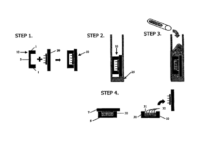
FIG 1 illustrates the four step fabrication method of the cell aggregation
device of the invention in general.
FIG 2 is a cross-section view illustrating one embodiment of the device
of the invention showing one cell suspension seeding chamber and six cell
aggregation recesses.
FIG 3 is a copy of a photographic reproduction showing the results of
controlled cellular aggregation using a device of the invention made as
described in Example 1. Figure 3A shows aggregation of normal human
fibroblast cells on a single large flat recess. Fig 3B shows aggregation in
flat-
bottomed 400 Firn diameter recesses. Figure 3C shows aggregation in
hemispherically-shaped 400 i_un diameter recesses. Figure 3D is a high
magnification of a single spherical aggregate of cells in one of the recesses
of
Figure 3 C. The scale bar = 300
FIG 4 is a copy of a photographic reproduction of the results of
Example 2 which shows a plan view of the array of aggregates of fibroblast
cells in 400 micron diameter aggregation recesses and a dose up of one of the
aggregates of fibroblast cells in a single recess.
Figure 5 illustrates the method of fabricating the encapsulated
aggregated live cells of the invention, as described in detail in Examples 6
and
7.
36
CA 02637663 2008-07-17
WO 2007/087402
PCT/US2007/002050
Figure 6 is a copy of a photographic reproduction of the microarray of
aggregated cells encapsulated in 1.8% alginate solution. After drying, the
microarray was peeled off the acrylamide cell aggregation device and
aggregates were observed with the features. Scale bar 300 rim.
Figures 7A and B are graphic illustrations of the results of the self-
assembly experiment described in Example 9A.
Figure 8 is a photographic reproduction of the results of the time-lapse
experiments using honeycomb-shaped recesses in the cell aggregation
devices, as described in detail in Example 9B.
Figure 9 is a histogram showing the results of the aggregation
experiments done using micro-molded agarose toroidal aggregation devices
seeded with untreated NHFs or NHFs treated with an aggregation modifying
agent, as described in Example 10.
Figures 10 and 11 are graphic representations of the results of the drug-
assisted aggregation experiments using the aggregation devices of the
invention having trough features, as described in Example 10.
Figure 12 is a photographic reproduction of some of the results from
the drug-assisted NI-IF cell aggregation experiment using honeycomb-
structured cell aggregation devices of the invention.
EXAMPLES
Example 1: Cell aggregation device fabrication and cell culture preparation
The fabrication method is illustrated for a general device of the
invention in Figure 1. Thirty millimeter Millicell CM cell culture inserts 10
(Millipore), which are essentially rigid plastic rings 1 having porous
polytetrafluoroethylene (PTFE) membranes 5 on their bottoms, were
employed as mold templates. The PTFE membrane, which is transparent
when wet, permits diffusion of culture media and visualization of cell
37
CA 02637663 2008-07-17
WO 2007/087402
PCT/US2007/002050
aggregates through an inverted microscope. AutoCAD 2005 software was
employed to design the shape and contours of various cell aggregation
devices and polyacrylamine substrates were made from the wax templates by
rapid prototype printing of sequential layers of wax to create the desired 3D
CAD designed topography. Mold topography was designed such that
gravitational force could funnel cells into the aggregation wells. The wax
template was designed to produce a flat square recess 1 mm in depth from the
top of the mold. The created software files were fed into a 3D Systems
ThermoJet() rapid prototyping machine for mold manufacture.
Once the molds were made, hydrogel casting was performed with a
multiple mini-vertical gel casting chamber Model #Gcc-210 from C.B.S.
Scientific. In the biological safety cabinet, six 30 mm Millicelle CM cell
culture inserts 10 were snapped onto six wax molds 20. The assembly was
placed in a vertical gel casting chamber 25 so that the composite was oriented
vertically between 2 glass plates (step 2 in Figure 1) and the bottom of the
gel
casting chamber. 100 ml of 15% pre-polymer solution was prepared by
mixing 37.5 ml of acrylamide/bisacrylamine (29:1 mix ratio, 40% solution;
Sigma-Aldrich A-7802) with 31.25 ml of Tris buffer, pH 6.8, and 31.25 ml
DMEM (1% penicillin/streptomycin) in two 50 ml conical centrifuge tubes.
The pre-polymer solution was degassed by exposure to vacuum for 30 sec.,
capped and transferred to a chemical fume hood. In the hood, 500 1 of
initiator, 10% APS (ammonium persulfate solution in deionized water; Sigma-
Aldrich A-9164) and 100 IA of TEMED (N,N,N,'-tetramethylethylenediame
99%; Sigma-Aldrich T-7024) catalyst was added to the 100 ml of pre-polymer
solution, mixed by inversion and transferred to the biological safety cabinet
for gelation.
In step 3, about 75 ml of the mixture was pipetted into vertical gel
casting chamber 25 to fill the spaces between molds 20 and inserts 10. The
38
CA 02637663 2008-07-17
WO 2007/087402
PCT/US2007/002050
remaining approximately 25 ml was left in a capped centrifuge tube as a tester
to confirm polymerization. Because the PTFE membranes, 5, were porous, the
solution flowed into the pores and polymerized, which tethered the hydrogel
to insert 10 at membrane 5. After 2 hours polymerization was complete and
the assembly was removed from casting chamber 25 by disassembly of the
chamber in the hood using aseptic technique. Excess polymerized hydrogel
was cut away from around inserts 10, wax molds 20 were removed and
inserts 10 now supporting the polyacrylamine hydrogel cell aggregation
device 30 were transferred to sterile 6-well tissue culture plates. Cell
aggregation device 30 can be seen in Figure 2 comprised of cell suspension
seeding chamber 31 and a plurality of cell aggregation recesses 32. The
volume of cell suspension seeding chamber 31 directly above aggregation
recesses 32 was about 300 1.
The foregoing is the preferred method for cells that are to be
15. aggregated and encapsulated in alginate. Otherwise, a simplified
procedure
is preferred in which the PTFE mold templates are not employed and the
devices of the invention are cast from a polydimethylsiloxane (PDMS), or
functionally equivalent polymer, instead of wax. For example, a PDMS mold
having the appropriate dimensions and structures is cast and molten agarose
solution (2-4% w/v in water) is added to the mold and allowed to cool. Once
cooled, the agarose can be removed from the mold, yielding a hydrogel that is
an exact negative replicate of the PDMS mold. Various sized molds may be
created to fit several standard sized tissue culture plates. To maintain
sterility, the protocol is performed in a biological safety cabinet using
autoclave-sterilized agarose solution and PDMS molds. This is the preferred
method of making the devices of the invention when the aggregated cells will
not be encapsulated in alginate.
39
CA 02637663 2008-07-17
WO 2007/087402
PCT/US2007/002050
Example 2: Cell seeding and aggregation of human diploid fibroblasts
3 ml of cell culture media (DMEM supplemented with 1%
penicillin/streptomycin as antibiotic, with and without fetal bovine serum)
was added to and the plates transferred to a cell culture incubator and
allowed to equilibrate overnight to rinse out any unreacted monomer,
neutralize pH and fill the pores of device 30 with the media. In a biological
safety cabinet, excess media was aspirated out of the 6-well tissue culture
plates. Normal human diploid fibroblast cells were trypsinized, counted and
resuspended to a cell density of 1.67 million cells per ml. 300 I (i.e.,
about
500,000 cells) were pipetted into each seeding chamber. Figure 2 illustrates
the cells 33, in various stages of the process of entering the aggregation
recesses 32, are shown bathed media 34, just after being pipetted into one of
the seeding chambers.
The hydrogel cell aggregation devices were transferred to the
= incubator for about 1.5 hours to allow cells to settle into the cell
aggregation
recesses and then transferred to a biological safety cabinet and to each well
of
the 6-well tissue culture plates was added 3 ml of additional media by slow
pipetting onto an inside wall of the well so as not to disturb any cells. The
devices were then returned to the incubator and allowed to stand. After 24
hours cell aggregates had formed and were retrieved by inverting the devices
within the wells of the 6-well plates (that still contained about 3 ml media)
for
5-10 minutes. The yield was between 80-90%. Mild centrifugation after
inversion (5 minutes, 25 C, at 500 rpm) increased the yield to 100%. Light
microscope images were taken during the 24 hour incubation period, at times
t= 0, 1, 2, 10 and 20. The results are shown in Figure 4, which is a copy of
the
photographic image taken from above (plan view) of the array of fibroblast
cell aggregates in 250 micron diameter aggregation recesses and a close up of
one fibroblast cell aggregate in a single recess. Cell aggregation could
dearly
CA 02637663 2008-07-17
WO 2007/087402
PCT/US2007/002050
be seen after 2 hours (data not shown) and the cells are completely aggregated
at 20 hours as shown in Figure 4.
Controlled aggregation within the device requires a cell-repellent
surface that is shaped to funnel cells together. This combination of surface
properties and geometry acts to increase intercellular interaction and cell
adhesion. In this example the effect of geometry on aggregation was
evaluated; cells were seeded onto cell-repellent polyacrylamide gels with
various topographical features. Devices containing cylindrical recesses with a
diameter of 400 }Am with either flat or hemisphere-shaped bottoms were
constructed. In addition, devices were constructed with a single large (17.6
mm X 17.6 mm X 1.8 mm) flat rectangular recess. These hydrogels were
seeded with normal human fibroblasts (2.5 X 10⁵ cells per device).
Photographic images taken 24 hours after seeding demonstrated that control
of the aggregation process is improved by geometries that utilize gravity to
funnel cells more closely together. As can be seen in Fig 3A, the single large
flat recess resulted in erratic and =controlled cell aggregation with
= irregularly shaped aggregates ranging in size from tens of microns to
several
millimeters. In the case of the 400 iirn diameter recesses with hemisphere-
shaped bottoms, cells formed a single spherical aggregate lying in the center
of each recess as can be seen in Figure 3C. In contrast, cells in the flat-
bottomed 400 1.= diameter recesses formed multiple irregularly-shaped cell
clusters scattered in each recess as can be seen in Figure 3B. A
single'spherical
aggregate formed in one of the recesses having the hemisphere-shaped
bottom is shown in Figure 3D.
Example 3: Aggregation of a mixed population of HUVEC and fibroblasts
Human umbilical cord vascular endothelial cells (HUVEC) were
seeded alone and simultaneously in co-culture with normal human diploid
41
CA 02637663 2008-07-17
WO 2007/087402
PCT/US2007/002050
fibroblasts, the latter of which were also seeded alone. To visualize the
different cell types, cell tracker Tm fluorescent living cell stain was used.
A
freshly trypsinized suspension of HUVECs was incubated in 5 1AM green
fluorescent cell tracker rm (in cell culture medium) and a fibroblast
suspension
was incubated in 5 tiM red fluorescent cell trackerTm for 45 minutes. Cell
suspensions were centrifuged, cell tracker Tm was aspirated off, and the cells
were re-suspellded in fresh media and incubated for 30 minutes.
Cell suspensions were then seeded into the seeding chambers of the
aggregation devices made in accordance with Example 1 above. 0.50 million
HUVECs alone (homogeneous population), 0.50 million fibroblasts alone
(homogeneous population) and 0.25 million HUVEC and 0.25 million
fibroblasts in co-culture (mixed population) were added to seeding chambers.
The three populations of cells were allowed to aggregate for 2 days and then
visualized using phase contrast as well as fluorescent microscopy. The
homogeneous populations of fibroblasts or HUVECs formed aggregates
within the aggregation recesses; and the mixed population of fibroblasts and
HUVECs formed heterotypic aggregates within the aggregation recesses. In
the mixed population, fibroblasts appeared to arrange themselves toward the
center of the aggregates while HUVECs localized on the periphery
surrounding the fibroblasts.
Example 4: Aggregation of murine mesothelial cells and macrophages
Murine malignant mesothelioma cells (40L) were seeded alone and
simultaneously in co-culture with normal primary peritoneal macrophages
(MO) isolated from an eGFP-transgenic mouse, the latter of which were also
seeded alone. In order to visualize the cells, cells suspended in CDMEM
culture medium were stained with the blue fluorescent dye 4,6-diamino-2-
42
CA 02637663 2008-07-17
WO 2007/087402
PCT/US2007/002050
phenylindole (DAPI ¨ Vector Laboratories) in accordance with the
manufacturer's protocol.
After staining, the three types of cell suspensions (homogeneous
populations of mesothelioma and macrophage cells, and the mixed
population of mesothelioma/macrophage cells) were seeded into the seeding
chambers of aggregation devices made in accordance with Example 1 above.
A total of 200 cells per aggregation recess were used; the mixed population in
a 3:1 'ratio of macrophage: mesothelioma cells. The three populations of cells
were incubated for 2 days and then visualized using fluorescent microscopy.
The homogeneous populations of mesothelioma cells formed spheroid
aggregates within the aggregation recesses. The homogeneous populations of
macrophages formed very loose aggregates within the aggregation recesses;
they were not expected to form tight aggregates because they do not form
intercellular junctions (in contrast to mesothelial or epithelial cells that
attach
to each other). However, while there was some variability among seeding
chambers, in co-cultures, the macrophages formed aggregates with the
neoplastic mesothelial cells in the aggregation recesses, similar to tumors in
vivo.
Example 5: Encapsulation of aggregated fibroblast cells
Aggregates of normal human diploid fibroblasts were made in accord
with Examples 1 and 2 above. The cell aggregation devices were typically
made the day before they would be used and were soaked in media for
approximately 24 hours to equilibrate them prior to using.
Briefly, aggregation wells of 400 and 600 1.tm diameter were printed
onto a wax mold using a three dimensional wax printer, and the gel was cast
onto this wax mold, creating a negative of the original mold as illustrated in
Figure 1. Each well was cylindrical is shape and terminated in a hemisphere
43
CA 02637663 2008-07-17
WO 2007/087402
PCT/US2007/002050
at one end. Cells were grown using conventional tissue culture technique.
Once cells were grown to confluence, they were passed and resuspended to a
cell density of 0.5 x 10⁶ cells per gel or per device. Cells were seeded into
the
cell aggregation device by slowly adding 150 IA (about 500,000 cells) to each
of the aggregation devices dropwise as illustrated in Figure 2. The cells were
allowed to sit and aggregates were typically observed 24 hours post-seeding.
After aggregation, media was changed every other day for up to five days
when aggregates were stable and mature. After five days, the cell aggregates
were encapsulated in situ in the cell aggregation device as square sheets as
described in Example 6.
Example 6: Preparation of microarrays of aggregates for encapsulation
Reference to Figure 5 may be made as the procedure detailed below is
illustrated therein. Because even cell culture alginate has inherent
impurities,
a 1.8% sodium alginate (Sigma) in lx Phosphate Buffered Saline (PBS)
solution first was mixed in a sterile field, then centrifuged at 4000rpm for
45
minutes to remove impurities. The supernatant was then filtered through a
0.42 in syringe filter to obtain sterile alginate solution. The cell
aggregation
device from Example 5 was placed in an empty container, excess media
bathing the aggregated cells was aspirated out as shown in step 1 of Figure 5,
and 300 I of alginate, the maximum volume the upper cell suspension
chamber in the cell aggregation device can hold, was slowly pipetted into the
chamber as shown in step 2. Next, in step 3, a 0.15M solution of CaCl2, was
added and crosslinking was allowed to occur for up to one hour in the
incubator. Because of the relatively low concentration of CaC12, the
aggregates of cells do not die during the crosslinking period.
In step 4, once the gels were crosslinked, leftover CaC12 was aspirated
out and the gels were allowed to dry at room temperature for between 2.5 to
44
CA 02637663 2008-07-17
WO 2007/087402
PCT/US2007/002050
3 hours. Because of the different dehydration rates of the alginate and the
slower acrylamide, the alginate containing the aggregated cells was readily
removed from the aggregation device. The alginate was peeled off the
acrylamide aggregation device in the form of a sheet 17.6 by 17.6 mm with
one aggregate per raised dome on one side of the microarray.
The alginate sheets were then visualized under an inverted light
microscope and a microarray of encapsulated aggregates of cells was seen
(Figure 6). MTT (3-(4,5-dimethylthiazol-2-y1)-2,5-diphenyltetrazolium
bromide) staining confirmed that all cells were alive and non-apoptotic.
Because MTT staining may mask a necrotic core in aggregates, a live/dead
stain (Molecular Probes) can be employed to discern the nature of the
aggregate core. The live/dead stain consists of two stains, calcein-AM and
ethidium homodimer (Eth-D). Calcein-AM is taken up by live cells where
cytoplasmic esterases cleave it and allow it to fluoresce green. Dead cells do
not take up calcein-AM, but instead, Eth-D is able to enter the dead cell's
nucleus, staining it red. Live/dead staining shows whether the core cells in
the aggregate are viable. For storage, sheets were immersed in 3mL culture
media (Dulbecco's Modified Eagle Media (DMEM, Invitrogen), 10% Fetal
Bovine Serum (FBS, Invitrogen) and 1% Penicillin-Streptomycin (Penn-Strep,
Invitrogen)), and media was changed every other day.
Example 7: Application of coating layer to encapsulated live cell aggregates
The microarray of aggregated live fibroblast cells from Example 6 was
then coated with a layer of poly-L-lysine to ensure complete encapsulation
and to assist in stabilizing the alginate. A 5% stock solution of poly-L-
lysine
(MW= 23,400, Sigma) in sterile water was mixed and then filtered through a
0.42 im syringe filter and then diluted to 0.1% by the addition of sterile
water. The microarray was washed with water two to three times to eliminate
CA 02637663 2008-07-17
WO 2007/087402
PCT/US2007/002050
salts from the storage media, the 0.1% PLL solution was then added and the
microarray was placed in an incubator and allowed to crosslink for fifteen to
twenty minutes, as illustrated in step 5 of Figure 5. Afterwards, the PLL-
water
solution was aspirated out, and the microarray was washed once again with
sterile water to eliminate excess PLL.
The general process as described in Examples 6 and 7 is illustrated in
Figure 5. In step 1, media was aspirated out of the cell suspension seeding
chamber of the aggregation device. In step 2, 300 1 of alginate was gently
pipetted into the chamber and onto the aggregated cells in their aggregation
= 10 recesses. Next, in step 3 the CaC12 solution was pipetted into the
device and
crosslinlcing was allowed to occur. In step 4, excess CaC12 was aspirated out,
the crosslinked alginate gel was allowed to air dry and then peeled off the
acrylamide aggregation device. In the next step, the sheet of encapsulated
cells were bathed in a solution of poly-L-lysine for an additional coating.
Example 8: Visualization studies
To ensure the encapsulation layer and the coating layer completely
encapsulate the cell aggregates, the alginate substrate and the PLL can be
dyed and visualized under confocal microscopy. Fluoresceinamine
(C2oHoNO3, Sigma) mixed in a concentration of 4.5mM is incubated with 1.8%
alginate solution, and stirred at room temperature. To removed unreacted
dye, the solution can be dialyzed against ion-free water at 4 C (26). PLL can
be labeled by protein coupling to fluorochromes such as Alexa 546. By
making the microarrays out of a solution of Fluoresceinamine-Alginate and
coating with Alexa 546-PLL, the microarrays could be visualized using
confocal microscopy, the results of which indicated that the PLL coating was
uniform and regular.
46
CA 02637663 2008-07-17
WO 2007/087402
PCT/US2007/002050
For characterization studies, a similar protocol was followed using a
0.1% solution of FITC-PLL instead of PLL (MW=23,400, Sigma), and the
microarrays made were kept wrapped in foil to prevent photobleaching of
labeled PLL. By staining the outer coating, epi-fluoresence microscopy can
confirm the location of cells relative to the outermost coating of the
microarray. Cells can also be stained (CellTrac.ker, Molecular Probes) in
order
to co-localize aggregates with respect to PLL.
To avoid subjecting the microarrays to the mechanical stress required
for SEM visualization, the samples can be visualized through an
environmental scanning electron microscope (ESEM) to confirm complete
encapsulation of the aggregates.
Example 9: Stable, Self-assembled, Non-spheroidal Aggregates of Cells
.Using differently shaped aggregation devices of the invention, we have
determined that different cells have different aggregation abilities and
characteristics, and different structures and forms into which they will
aggregate. For example, normal human fibroblasts (NHF) and rat
hepatocytes cells (H35 cells) exhibit significant differences when they
aggregate; in trough-shaped recesses, H35 cells readily form stable rod
structures while NHF cells form spheroids. Both form tori, but H35 tori are
more stable than NHF tori.
These differences can be employed
advantageously to construct stable, non-spheroidal structures of aggregated
cells for tissue engineering applications, for example, replacing tissue
engineering techniques such as tnicroinjection of contiguous spheroids within
a biocompatible matrix or electropatterning cells into desired arrays.
A. Some Form Rods, Some Form Spheres
Micro-molded, non-adhesive, cell aggregation devices, each having a
plurality of cell aggregation recesses in the shape of either troughs or tori
47
CA 02637663 2008-07-17
WO 2007/087402
PCT/US2007/002050
were made following the method described in Example 1. Agarose was
employed as the hydrogel material and the cell aggregation recess features
were as follows. Troughs were 400 m wide with bottoms rounded with 200
rn radii. There were 21 rows of troughs of increasing length per gel. Each
row had 11 troughs, two of which were 400 pm long, then one each of 600 pm
through 1800 pm increasing at 200 rn lengths, then two 2200 pm troughs.
The 2200 pm troughs were used for all experiments except those for Figure 9c,
which presents data from all trough lengths. Tori-shaped recesses were 800
.uirt deep, with circular track 400 Fim wide. The recess bottom was filleted
= with radius 200 pm. The peg diameter was 600 pm and that of the entire
feature was 1400 pm. There were 104 staggered tori per gel each separated by
250 pm.
NHF cells alone, H35 cells alone, and 50:50 mixtures of NHF and H35
cells were seeded into the trough aggregation devices and the tori aggregation
devices as described in Example 2 and allowed to aggregate. Briefly, NHF
cells derived from neonatal foreskins and H35 rat hepatoma cells were
expanded in DMEM supplemented with 10% fetal bovine serum (FBS) and 1
% penicillin/streptomycin solution. NHF cells were incubated in a 10% CO2
atmosphere and H35 cells were incubated in 5% CO2. The mixture
experiments were a 50:50 mix of both cell types and were maintained in a 5%
CO2 atmosphere. The cells were stained with fluorescent dyes (NHF cells
with CellTrac.ker Red CMTPX and H35 cells with CellTracker Green CMFDA
from Invitrogen) to allow their aggregation in the mixed populations to be
followed and their aggregated position visualized. Cells used in experiments
were between passage 3 and passage 9. Cell viability was determined using
the LIVE/DEAD Viability/Cytotoxicity Kit (Invitrogen) in accordance with the
manufacturer's instructions.
48
CA 02637663 2008-07-17
WO 2007/087402
PCT/US2007/002050
Excess medium was aspirated from the wells and 200 piL of a single cell
=
suspension was pipetted into the aggregation recess of each gel. Medium was
added to the wells after a 1.5 hour incubation period wherein cells settled
into
the aggregation recesses and commenced the self-assembly process. Medium
was exchanged every other day. The aggregation process was observed with
time-lapse microscopy using a Nikon TE2000-S microscope and a Hamamatsu
Orca-ER camera, outputting to Openlab v4Ø2 (Improvision). A z-stack of
images from the 4X objective with 10 ¨ 50 wri between focal planes was
collected at each time point using custom-designed Openlab 4Ø2
automations and images were analyzed using Volocity 3.1 (Improvision).
Bright-field and fluorescent images were obtained using an Olympus IX70
microscope equipped with an AxioCam MRc digital camera. For the time-
lapse experiments, medium was carefully added 20-30 minutes post seeding,
before the devices were transferred to the time lapse microscope. '
Measurements of morphological changes were performed with ImageJ data
analysis software. The length of a rod was the length of a line drawn from
end to end of the structure (long-axis length). The core circumference of tori
was measured as a continuous circumferential line located at the estimated
midpoint of the perpendicular width of the toroid.
Minutes post-seeding, cells settled into and coated the curved recess
bottoms with several layers of cells but did not fill the recesses. Self-
assembly
began immediately with cells coalescing and compacting to form smooth
. surfaced 3D structures. In trough-shaped recesses, cells self-assembled into
round-ended, cylindrical rods and shrank along their long-axes towards a
spheroid morphology. NHF cells and mixture cells aggregated first into rods
and then become spheroids in as few as 15 hours, but H35 cells maintained
the rod morphology and did not form spheroids. The NHF cells, the H35 cells
and the mixed cells all formed stable tori that conformed around the central
49
CA 02637663 2008-07-17
WO 2007/087402
PCT/US2007/002050
peg of the torus substrate. Despite identical seeding densities and similar
mean cell diameters (H35 = 17 21.4M, n=100; NHF = 20 3 M, n=20), there
were clear morphological differences in the tori. The NHF toroid was thinnest
while that of H35s was thickest. The thickness of the mixed cell population
was less regular with foci of higher cell density connected by thinner
regions.
While both NHF and mixed cells could be aggregated into stable tori with an
initial seeding density of 2x10⁶ cells/gel, H35 cells could be aggregated into
stable tori even with a seeding density as low as 1 x10⁶/gel (data not shown).
Once aggregated into a rod or tori shape, the cells could be easily removed
from the substrate. The removed aggregates maintained the final shape they
had been formed into during aggregation in their respective trough and
toroidal substrates.
To examine the kinetics of toroid and of rod formation, stability
and morphological changes over time were measured. NHF cells, H35 cells
and their 1:1 mix were seeded onto micro-molded gels with toroid or trough
features. The number of stable toroids with patent centers was measured over
time, n=728(H35), 511(NHF), 474(Mixed). At day 5, there were significantly
more intact H35 (88%) than NHF (30%) or mixed (60%) tori. Notably, stability
of mixed tori was intermediate between that of H35 and NHF. Core
circumferences of tori were maximal at time 0 when the cells coated the
bottom of the recesses. As self-assembly proceeded, the core circumference
decreased until the tori conformed to the peg. This process occurred at
strikingly different rates between H35 cells and both NHF cells and mixtures.
Steady state was achieved by day 2 in 1-135 cells, but in less than a day for
both
NHF cells and mixtures. These data show that stable tori have different core
circumferences depending on cell type with NI-IF cells the shortest, H35 cells
the longest and mixtures near midway between the two. It is interesting to
note that the rate of change in core circumference for mixtures and NHF cells
CA 02637663 2008-07-17
WO 2007/087402
PCT/US2007/002050
were similar, but mixtures formed more stable tori than NHF cells. Only H35
cells differed in their kinetics of rod and toroid changes with core
circumferences progressing at a slower rate than rod lengths (data not
shown).
In trough-shaped recesses, the length of the aggregated H35 rods
changed at a significantly slower rate than those of the aggregated NHF rods
and the aggregated mixed rods. NHF and mixed rods become spheroids
overnight, whereas H35 rods reached a final stability of 41% of their original
length by day 5. Data on rods formed in troughs of increasing length suggest
that this percentage remains constant, regardless of maximal length.
Interestingly, while tori and troughs seeded with NHF cells and mixed cells
progressed to their final aggregated morphology overnight, tori seeded H35
cells reached their final toroidal-shaped aggregated morphology in two days
and troughs seeded with H35 cells needed 5 days to reach their final rod-
shaped aggregated morphology. Mixed aggregates self-assembled with
kinetics similar to aggregated NHF cells alone.
Time-lapse microscopy was used to investigate the early kinetics of rod
and toroid self-assembly. Photos of rods in 1.6 mm troughs were taken at 10-
minute intervals and long axis lengths were measured and plotted as a
function of time. When NHF rods began to self-assemble into aggregates, a
necking or pinching in the middle and across the short axis was often
observed. The initial length of NHF rods remained nearly static for 20-30
minutes before rapidly shrinking by 40% over 50 minutes followed by a
transition to a slower period of further compaction (15%) over 1120 minutes.
Necking began within 10-20 minutes, but rapidly reduced by 50% over 30
minutes only to increase over 1150 minutes to a value greater than its
starting
condition. Final length and width were nearly identical, indicating spheroid
formation. Mixed rods also progressed to spheroids with kinetics similar to
51
CA 02637663 2008-07-17
WO 2007/087402
PCT/US2007/002050
NHF cells (mixture tau = 347 thins; NHF tau = 202 mins). In contrast, there
was no lag period for H35 rods and self-assembly proceeded with linear
kinetics at a much slower rate (tau = 1.21 x10⁸ mins).
To evaluate cell sorting within mixture tori, the H35 cells and the NHF
cells were differently labeled with fluorescent dyes as described above and
allowed to self-assemble for two days. Sorting occurred within 24 hours in
parallel with changes in core circumference and final cell type positions did
not change between day 1 and day 2. NHF cells formed a central ring
adjacent the toroid substrate; the H35 cells formed around and adjacent to the
NHF cells in the aggregated structure. While some mixing was apparent, the
NHF cells were clearly biased inwards towards the central peg, particularly in
thicker regions of the toroid. NI-IF cell morphologies were stretched and
more spindle-like, with less-distinct boundaries, most notably in areas of
high
stress. In contrast, H35 cells were more individual, rounded and spherical
regardless of their position within the toroid. While more evenly distributed
through the tori, they occupied the outermost regions away from the central
peg. After 2 days, these positions were consistent and slightly more apparent
than after the first day. By day 2, some of the mixed aggregates had moved
up and off the central pegs altogether, and patent centers were still
apparent.
In summary, in troughs up to 2.2 mm in length, H35 cells self-
assembled into aggregated rod-like structures and did not form spheroids as
would be predicted by many previous studies. NHF cells and the mixed
populations did aggregate into spheroids. The kinetics of self-assembly
revealed interesting differences as well. After a slight delay, NHF rod
lengths
decreased rapidly with exponential kinetics and then decreased more slowly
while approaching spheroid morphology by 1100 minutes. In contrast,
aggregated H35 rod lengths decreased with almost linear kinetics during the
first 24 hours then with slower kinetics over the proceeding four days.
52
CA 02637663 2008-07-17
WO 2007/087402
PCT/US2007/002050
Interestingly, the kinetics of the 1:1 mix of NHF cells and H35 cells was not
intermediate but was more similar to the kinetics of aggregated NHF cells
rather than aggregated H35 cells. The mix had the same slight delay followed
by an exponential decrease in rod length with a curve that was nearly
identical to that of aggregated NHF cells, except delayed by approximately 2
hours.
B.
Directed, Self-assembly and the Formation of Honeycomb Cell Aggregates
In this experiment, cell aggregation devices were formed from
agarose gels as described above, but agarose gels were micromolded with
honeycomb-shaped recesses instead of troughs or a tori. The honeycomb-
shaped recesses are essentially overlapping, tori-shaped recesses: There are
13
pegs (1 central, 6 concentric around the central peg and 6 outer pegs at 0 ,
60%
120% 180% 240% 300 and 360 ) within a network of recesses 4001IM wide and
with bottoms rounded with 20011m radii. Time lapse microscopy was again
used to monitor self-assembly of the NHF cells alone, the H35 cells alone and
50:50 mixtures of NI-IF cells and H35 cells as described above. The results
are
shown in Figure 8, at time 0 (top row), 10 hours post-seeding (middle row)
and 20 hours post-seeding (bottom row) with the aggregated NILF cells in
column A, the aggregated H35 cells in column B and the aggregated 1:1
mixture of NHF and H35 cells in column C. The aggregated NHF cells
quickly thinned, became taught and popped off the outer pegs of the
honeycomb shaped substrate. Once freed from the substrate, the honeycomb-
shaped aggregates of NHF cells contracted in a uniform manner but
maintained its shape. The aggregated H35 cells thinned more slowly,
maintaining contact with the outer pegs and staying within the molds. The
mixed cells exhibited an aggregation behavior intermediate between the
aggregated NFIF cells alone and the aggregated H35 cells alone. They
thinned more quickly than the aggregated H35 cells alone and only partially
53
CA 02637663 2008-07-17
WO 2007/087402
PCT/US2007/002050
popped off the pegs after 20 hours. Like the NHF aggregates, the
architectural integrity of the resulting free-floating aggregated NHF/H35
mixed honeycombs was preserved as the cells aggregated and the structures
contracted. Patency of the lumens was lost during this contraction process
but the general shape of the structures did not progress to spheroids.
To assay self-segregation in this complex, branching structure, labeled
H35 cells and NHF cells were seeded in the honeycomb mold and were
viewed one and two days later. As in labeled tori, sorting occurred within 24
hours, with NHF cells centrally located toward the pegs and H35 cells on the
periphery coating the entire structure. NHF cells had a spindle-like,
stretched
and smooth cell morphology (which relaxed to a rounder morphology when
freed from the substrate), while H35 cells were more rounded with visible cell
boundaries. Self-segregation varied with position in the honeycomb. Along
the outer edge of the 6 most outer pegs, the fibroblasts were located less
central and closer to the peg, as was the case for tor.
We also evaluated cell position and morphology of the mixture
honeycomb-shaped aggregates that freed themselves from the pegs. In these
relaxed structures, the patency of the lumens was maintained and the relative
cell positions remained unchanged, but there was a significant change in
fibroblast morphology. Instead of indistinct cell boundaries and elongated
spindle-like morphologies found in taught structures, NHF cells in relaxed
honeycombs were rounded and cell boundaries were easily discernible. This
was in contrast to H35 cells that remained round and distinct from neighbors
in both stretched and relaxed conformations.
The self-assembled honeycomb structures composed of the
aggregated cells that were created in this experiment demonstrate that tension
can be balanced and distributed throughout a complex self-assembled cellular
structure without the need for a scaffold. Honeycombs of NHF cells, H35 cells
54
CA 02637663 2008-07-17
WO 2007/087402
PCT/US2007/002050
and mixtures of NHF and H35 cells were each anchored by contact with the
outer edge of the 6 outer pegs. The structure had little if any contact with
the
inner 7 pegs. The time-lapse experiments clearly show that the entire
honeycomb structure is under tension and that the presence of NI-IF cells
significantly increases this tension. Honeycombs of NHF cells and
honeycombs of the NHF/H35 cell mixture spontaneously popped off the outer
pegs and the structures quickly contracted.
As freed honeycomb aggregated cells shrank, their lumens narrowed.
Shrinkage and narrowing was greatest for honeycombs of pure NHF cells,
least for pure H35 cells and the NHF/H35 cell mixture was intermediate.
Patency of the lumens was maintained for 2 days for NHF cells and as long as
5 days for H35 cells. Changes to the geometry of the mold such as increasing
the diameter of the agarose pegs may help to increase patency as well as
= control lumen size. Maintenance of lumen patency is another significant
difference between cell types in their self-assembly properties.
Tension also probably plays a role in the self-segregation of NI-IF cells
and H35 cells in complex shapes. In spheroids, different cell types self-
segregate and envelop one another due to differences in surface adhesion.
Foty, et al., Development 122: 1611-20 (1996). This was observed for NHF cells
and H35 cells in tori and honeycombs with NHF cells centrally located and
H35 cells coating the periphery. However, in tori, the ring of NHF cells was
not at the midpoint, but was located closer to the agarose peg. In
honeycombs, the band of NHF cells on the outer edges of the outer pegs, an
area of concentrated tension, was also closer to the agarose peg, but the band
of NHF cells in the interior of the honeycomb, where tension was balanced,
were more equally spaced.
That rod-shaped aggregates, tori-shaped aggregates and honeycomb-
shaped aggregates could be generated with H35 cells, a parenchymal cell
CA 02637663 2008-07-17
WO 2007/087402
PCT/US2007/002050
type, rather than NHF cells, a stromal cell type, is encouraging for
microscale
tissue engineering. Indeed, while we investigated a 1:1 ratio of H35 cells to
NHF cells, the physiologic ratio of hepatocytes to fibroblasts is closer to
0.5:1.
Bhatia et al., Biotechnology progress 14: 378-87 (1998). Together with our
findings that mixtures had intermediate stabilities, this suggests that the
constructs with more in vivo-like ratios and histologies may be more stable,
and thus better to employ in tissue engineering applications.
In sum, self-assembled rod-like aggregations of cells and honeycomb
aggregations of cells offer interesting new possibilities for the engineering
of
three-dimensional microtissues for in vitro and in vivo applications. Unlike a
spheroid whose ultimate size is severely limited by diffusion, the length of a
rod structure has no theoretical limit provided its radius remains within the
critical diffusion limit needed to maintain cell viability. Enmon et al.,
Biotechnology and bioengineering 72, 579-591 (2001); Griffin et al., Tissue
Eng 11:
257-66 (2005); Ambrosi et al., Journal of Mathematical Biology 48: 477-99
(2004).
= Such rod-like structures may have applications in bioreactors. The
honeycomb, which is essentially a combination of rods and tori in a structural
design, is well known as an efficient geometrical shape and may also have
applications in bioreactors as well as in tissue engineering. Three
dimensional
branching microtissues with open lumens more closely approximates the in
vivo environment and lumen structures offer the possibility of enothelialiling
self-assembled microtissues for transplantation.
Example 10: Drug Assisted Aggregation of Complex Microtissue Structures
The foregoing example demonstrated that the final aggregation
structure normally attained by the aggregated cells are cell-type dependent
and "preprogrammed", but that the pre-programmed structure may be
modified by changing the shape of the aggregation device employed to
56
CA 02637663 2008-07-17
WO 2007/087402
PCT/US2007/002050
aggregate the cells. This example goes further and demonstrates that the
dynamics of cellular self-aggregation can be controlled by the administration
of selected aggregation modifying agents during the aggregation process. In
other words, the rate and extent of aggregation is controllable by the
addition
of selected aggregation modifying agents to the cells before aggregation
begins. The experiment detailed below illustrates this principle using the
known cell contraction inhibitor Y-27632, although any other agent that has
the effect of modifying the dynamics of aggregation may be employed.
Micro-molded agarose toroidal aggregation devices (n=208) made as
described in Example 9 were seeded with untreated NHFs and NHFs treated
with 100p.M of the cell contraction inhibitor Y-27632 (Calbiochem (EMI)
Biosciences, Inc.), San Diego, CA) and the number of stable toroids with
patent centers was measured over time. By day 1, all untreated NHFs (n=104)
had progressed from toroids to spheroids while 90% of the treated NHFs
(n=104) still had patent centers. By day 2, only 5% of the treated NHFs still
had patent centers. This demonstrates that the native kinetics of cell
aggregation can be modified by treatment of the cells with an aggregation
. inhibitor. The data is shown in the Figure 9 histogram.
The effect was not limited to spheroidal cell aggregation structures.
Next, aggregation devices with trough features 2.2mrn in length were made as
described in previous examples and seeded with NHF cells, NHF cells in
serum-free medium, NHF cells treated with 50 M of Y-27632 and NHF cells
treated with 200 M of Y-27632 and the lengths of resulting rod shaped
aggregates were measured. The results are shown in Figure 10, in which
=
NHFs are represented by the solid triangles (A), NHFs in serum-free medium
are represented by the dear triangles (A), NHFs treated with 50 M of Y-27632
are represented by the inverted solid triangles (7) and NHFs treated with
200p./vI of Y-27632 are represented by the inverted clear triangles (V).
57
CA 02637663 2013-12-19
WO 2007/087402
PCT/IIS2007/002050
Increasing Y-27632 dramatically slowed length change kinetics in rods. H35
rods, which remain at 41% their trough length after 5 days, are included for
reference are represented by the solid circles (0).
= Gels with trough features 2.2mm in length were then seeded with
NHFs or with NHFs treated initially with 100h1v1 Y-27632 and imaged every
minutes. At 1.5hrs, the Y-27632 solution was removed and replaced with
normal medium. The results are shown in Figure 11 in which the untreated
NHFs are represented by the solid inverted triangle, the treated N1-1Fs are
represented by the clear inverted triangle and the arrow indicates the change
10 of media at 1.5hrs. The results demonstrate that upon removal of the
aggregation inhibiting drug, the NHF cells aggregated into rods, progressing
with similar kinetics to those of untreated NHFs.
Lastly, NHFs were seeded onto honeycomb-structured cell aggregation
devices made as described in Example 9B and maintained in Y-27632 solution.
While untreated NHFs contracted up and off of pegs within 1 day, treated
NI-IFs remained intact in the honeycomb-shaped recesses at day 5. An
example of one treated NHF honeycomb is shown in Figure 12. Viability
staining revealed that the majority of the cells were living at day 5.
These results demonstrate that the native kinetics of cell aggregation in
the cell aggregation devices of the invention may be modified by treatment of
the cells with an aggregation inhibitor. Accordingly, the kinetics of
aggregation can be controlled by treatment with aggregation modifying
agents to either speed up or slow down aggregation as needed by the
treatment regimen and the tissue repair and/or reconstruction protocol.
All of the compositions and methods disclosed herein can be
made and executed without undue experimentation in light of the disclosure.
Although the compositions and methods of the invention have been
58
CA 02637663 2013-12-19
WO 2007/087402
PCT/IIS2007/002050
= described in terms of preferred embodiments, it will be apparent to those
having ordinary skill in the art that variations may be made to the
compositions and methods. Fore example, certain agents and compositions that
are
chemically related by be substituted for the agents described herein if the
same or
similar results would be achieved.
In case of conflict, this specification including defmitions will control. In
addition the material methods and examples are illustrative only and not
intended to
be limiting.
59