Note: Descriptions are shown in the official language in which they were submitted.
CA 02637760 2008-08-28
METHOD AND APPARATUS FOR ENHANCING FLUX RATES
OF A FLUID IN A MICROPORATED BIOLOGICAL TISSUE
This application is a divisional application of co-pending application Serial
No.
2,329,167, filed March 5, 1999.
BACKGROUND OF THE INVENTION
Field of the Invention
The invention relates generally to the monitoring of analytes in the body and
the
transdermal delivery of drugs to the body. More particularly, the invention
relates to
enhancing the rate of flux of a substance collected from or delivered to a
biological
tissue through the poration of the skin or other biological membrane and the
application
io of a flux enhancer to the porated biological membrane.
Descrivtion of Related Art
The transfer of materials across biological membranes is necessary in the
practice of a variety of medical and other procedures. For example, to
minimize
complications resulting from diabetes, diabetics must periodically monitor and
control
their blood glucose levels. Typically, blood glucose monitoring is achieved by
taldng a
sample of blood or other body fluid, and measuring the glucose level present
in the
sample. Historically, the samples have been obtained by piercing the slcin
with a needle
or lancet. It is also frequently necessary to deliver a drug through the skin
or other
biological membrane. Most frequently, drugs are delivered transdermally by
injection
2o with a needled syringe. Such invasive sampling and drug delivery methods
entail a
number of disadvantages, most notably, discomfort and potential infection.
In an effort to address the inherent disadvantages of invasive sampling and
delivery methods, several minimally invasive and non-invasive sampling and
delivery
techniques have been developed. "Minimally invasive," as used herein, refers
to
techniques in which a biological membrane or tissue is invaded by forming
small holes
or micropores in the surface of a tissue or membrane, but do not substantially
damage
the underlying, non-surface portions of the tissue or membrane. As used
herein, "non
invasive" refers to techniques not requiring the entry of a needle, catheter,
or other
invasive medical instnmient into the body. It has previously been discovered
that blood
glucose levels can be determined from an analysis of interstitial fluid, the
clear fluid
occupying the spaces between cells in the body, samples of which can be
obtained
CA 02637760 2008-08-28
2
through the skin by previously known minimally invasive or non-invasive
sampling
techniques . Previously known minimally invasive or noninvasive methods of
sampling interstitial fluid, however, have not been fully successful for blood
glucose
monitoring purposes. One challenge facing minimally invasive or non-invasive
methods is the ability to acquire a large enough sample of interstitial fluid
in a short
time to enable accurate glucose measurement with low cost disposable assay
techniques.
The skin presents the largest, most readily accessible biological membrane
through which an analyte may be collected or a drug delivered. Mucosal and
buccal
to membranes present feasible, but less accessible, sites for collection and
delivery.
Unfortunately, the skin and, to a somewhat lesser extent, the mucosal and
buccal
membranes, are highly resistant to the transfer of materials therethrough. The
skin
generally comprises two main parts: the epidermis and the dermis. The
epidermis
forms the outer portion of the skin, and itself comprises several distinct
layers. The
outermost layer of the epidermis, the stratum corneum, is composed of
denucleated,
keratinized, clear, dead cells, and is typically between 10-30 m thick. The
stratum
comeum is chiefly responsible for the skin's barrier properties and,
therefore, is the
layer of skin forming the primary obstacle to the transdermal flux of analytes
out of the
body and of drugs or other foreign materials or organisms into the body.
There have been significant advancements made in the transdermal transport of
substances across a biological membrane by creating micropores in the
biological
membrane. See, for example, U.S. Patent No. 5,885,211 filed September 5, 1997,
entitled "Microporation of Human Skin for Drug Delivery and Monitoring
Applications".
Neverthdless, there is a need to improve upon these techniques and
particularly increase
the rate at which substances are transported through a biological membrane.
SUMMARY OF THE INVENTION
Briefly, one aspect of the present invention involves a method for enhancing
the
flux rate of a fluid through biological tissue. The method generally comprises
the
delivering an effective amount of a flux enhancer into the tissue through at
least one
micropore in the tissue. Depending on the specific application, the flux
enhancer is
CA 02637760 2008-08-28
3
delivered to the micropore through any of a number of mechanisms, exatnples of
which
are described below. The depth of poration and of application of the flux
enhancer can
also be adjusted to suit the desired application.
Another aspect of the present invention involves a method of harvesting an
i analyte from tissue beneath a biological membrane. The method preferably
includes
the steps of porating the biological membrane to form at least one micropore,
delivering
an effective amount of a flux enhancer to the tissue through the micropore,
and
collecting a quantity of the analyte through the micropore. Again, the
mechanism for
delivering the flux enhancer can vary to suit the application, as can the
depth of
io poration and application of the flux enhancer. The application of a motive
force, such
as suction, pressure, electric field, sonic energy, or concentration gradient,
can also be
employed to further enhance the rate of analyte harvesting.
Yet another embodiment of the present invention provides a method of
delivering a drug through a biological membrane. The method preferably
comprises
15 porating a site of the membrane to form at least one micropore, delivering
an effective
amount of a flux enhancer into the micropore, and introducing a drug through
the at
least one micropore. Again, the mechanism for delivering the flux enhancer can
vary to
suit the application, as can the depth of poration and application of the flux
enhancer.
The application of a motive force, such as iontophoresis, pressure, electric
field, sonic
20 energy, or concentration gradient can also be employed to further enhance
the rate of
drug delivery into the tissue.
Still a further aspect of the present invention provides involves a device for
facilitating the formation of micropores in a biological membrane and for
enhancing the
rate of flux of a fluid therethrough.
25 These and other features and advantages of the present invention will
become
apparent from the following description of the preferred embodiments, taken in
conjunction with the drawings.
BRIEF DESCRIPTION OF THE DRAWINGS
FIG. I is an enlarged cross-sectional view of a section of a biological
membrane
30 and underlying tissue porated according to one or more embodiments of the
present
invention.
CA 02637760 2008-08-28
4
FIG. 2 is an enlarged diagram showing the use of a probe for delivery of a
flux
enhancer according to one embodiment of the present invention.
FIG. 3 is an enlarged diagram showing the delivery of a flux enhancer from a
reservoir using a probe according to another embodiment of the present
invention.
FIG. 4 is an exploded view showing a device for facilitating the poration of a
biological membrane and the delivery of a flux enhancer according to the
present
invention.
FIG. 5 is an enlarged view showing the delivery of a flux enhancer from the
device shown in FIG. 4.
FIG. 6 is an exploded view showing another device for facilitating the
poration
of a biological membrane and the delivery of a flux enhancer according to the
present
invention.
FIGs. 7 is side view of a device suitable for microporating tissue and
delivering
a drug into the microporated tissue.
FIG. 8 is a bottom view of the device of FIG. 7.
FIG. 9 is a side view of another device suitable for microporating tissue and
delivering a drug into the microporated tissue.
DETAILED DESCRIPTION OF THE PREFERRED EMBODIMENTS
The present invention will now be described in detail, by reference to several
preferred embodiments. The embodiments described in detail herein are
presented by
way of example only, and are not intended to limit the scope of the invention
defined in
the claims, and equivalents thereof. Words and phrases used herein are
intended to
= have their ordinary meanings, as understood by a person of ordinary skill in
the art to
which this invention pertains, unless otherwise defined.
D i i ions
Unless the context clearly dictates otherwise, "a," "an," and "the" includes
both
singular and plural referents. Thus, for example, reference to delivery of "a
drug"
contemplates delivery of one or more drugs, reference to "a flux enhancer"
contemplates one or more flux enhancers, and reference to "an analyte"
contemplates
one or more analytes. Also, unless the context clearly dictates otherwise,
"in" means
CA 02637760 2008-08-28
"in" or "on." As used herein, "including," "includes," or the like, means
including,
without limitation.
As used herein, "organism" or "individual" or "subject" or "body" refers to
the
entire human, animal, or plant being acted upon by the methods described
herein.
5 As used herein, "biological tissue" or "tissue" means any component
comprising some portion of an organism, including but not limited to: cells;
intercellular substances surrounding cells; biological membranes; bone;
collagen;
fluids, including blood; epithelial tissue, including the skin; connective
tissue; blood
vessels; muscle tissue; nerve tissue; and the like.
As used herein, "biological membrane" or "membrane" means any tissue
material present within a living organism forming a barrier between distinct
tissues or
areas of an organism, or between tissue of an organism and the external
environment,
and includes without limitation: the skin; mucosal membranes; buccal
membranes; the
outer layers of a plant; and the walls of a cell or a blood vessel.
t5 As used herein, "skin" means the epidermis, which includes the stratum
corneum, and the denmis.
As used herein, "mucous membrane" or "mucosa" refers to the epithelial linings
of the mouth, nasopharynx, throat, respiratory tract, urogenital tract, anus,
eye, gut and
all other surfaces accessible via an endoscopie device such as the bladder,
colon, lung,
blood vessels, heart and the like.
As used herein, the "buccal membrane" includes the mucous membrane of the
mouth.
As used herein, "into" or "in" a biological membrane or layer thereof includes
penetration in or through only one or more layers (e.g., all or part of the
stratum
corneum or the entire outer layer of the skin or portion thereof).
As used herein, "through" a biological membrane or layer thereof means
through the entire depth of the biological membrane or layer thereof.
As used herein, "transdermal" or "percutaneous" or "transmembrane" or
"transmucosal" or "transbuccal" refers to passage of a substance into or
through the
subject biological mcmbrane or tissue, in any direction.
CA 02637760 2008-08-28
6
As used herein, "poration," "microporation," or any such similar term means
the
formation of a small hole or pore to a desired depth in or through a
biological
membrane or tissue. The microporation process referred to herein is
distinguished from
electroporation principally by the minimum dimensions of the micropores
formed.
s Micropores shall be no smaller than I micron across and at least 1 micron in
depth,
whereas the openings formed with electroporation are typically only a few
nanometers
in any dimension. Preferably the hole or micropore will be no larger than
about 1 mm
in diameter, and more preferably no larger than about 300 m in diameter, and
will
extend to a selected depth, as described hereinafter.
As used herein, "micropore" or "pore" means an opening as described above,
formed by the microporation method.
As used herein "ablation" means the controlled removal of material which may
include cells or other components comprising some portion of a biological
membrane
or tissue caused by any of the following: kinetic energy released when some or
all of
the vaporizable components of such material have been heated to the point that
vaporization occurs and the resulting rapid expansion of volume due to this
phase
change causes this material, and possibly some adjacent material, to be
removed from
the ablation site; thermal, mechanical, or sonic decomposition of some or all
of the
tissue at the poration site.
As used herein, "flux enhancer" means any material that increases the rate of
flow of a fluid through a biological tissue or membrane by any mechanism. The
fluid
can be, for example: a bioactive agent, drug, analyte, dye, stain,
microparticle,
microsphere, compound, or some other chemical formulation. As described in
greater
detail below, the subject fluid flow can be, for example, the flow of
interstitial fluid out
of a porated biological tissue or membrane, or can be the flow of a drug into
a porated
biological tissue or membrane. Representative examples of mechanisms by which
a
flux enhancer can increase the rate of flow of a fluid through a tissue
include, without
limitation: the reduction of the fluid's viscosity; the dilation of
intercellular pathways
within the tissue; the reduction of the barrier properties of the capillary
walls.
Materials that can be used as flux enhancers include, but are not liniited to,
ammonia related substances such as ammonia gas, ammonia heparin, and ammonia
CA 02637760 2008-08-28
7
bicarbonate; vasodilators such as histamine, Platelet Activating Factor (PAF),
bradykinin, nicotinic acid, and nitroglycerin; inflammatory mediators such as
autacoids
(histamine, bradykinin, eicanosoids such as prostaglandins, leukotrienes, and
thromboxane), cytokines, and interleukins; neurotransmitters such as substance
P,
acetyicholine, and neurokinin A; growth factors such as Platelet-Derived
Growth Factor
(PDGF), and Vascular Endothelial Growth Factor (VEGF); mast cell degranulators
such as substance P, and mastoparan; extracellular matrix adhesion inhibitors
such as
anti integrins, and disintegrins; enzymes such as hyaluronidase, trypsin, and
papain;
fungistatic compounds such as benzoic acid; compounds which release
neuropeptides
from nerve terminals such as capsaicin; keratolytic agents such as lactic
acid, glycolic
acid, and salicylic acid; blistering agents such as cantharidin;
anticoagulants such as
heparin and sodium fluoride; food oils such as mustard oil and peppermint oil;
anti-
pruritics such as camphor; diuretics such as ethacrynate sodium and
furosemide; and
capillary penneability enhancers (extravasants) such as VEGF, PAF,
leukotrienes,
t5 kinins (bradykinin & kallidin), histamine, and estrogen. One material which
has proven
effective as a flux enhancer is an ammonia-based solution sold by Tender
Corp., of
Littleton, NH, under the trademark AfterBite.
An "effective amount" of a flux enhancer is the quantity of material necessary
to
produce the desired increase of flow rate through the tissue.
As used herein, the term "bioactive agent," "drug," "pharmacologically active
agent," or "deliverable substance" or any other similar tenm means any
chemical or
biological material or compound suitable for delivery by the methods
previously known
in the art and/or by the methods taught in the present invention, that induces
a desired
effect, such as a biological or pharmacological effect, which may include but
is not
limited to (1) having a prophylactic effect on the organism and preventing an
undesired
biological effect such as preventing an infection, (2) alleviating a condition
caused by a
disease, for example, alleviating pain or inflammation caused as a result of
disease, (3)
either alleviating, reducing, or completely eliminating the disease from the
organism,
and/or (4) placing within the viable tissue layers of the organism of a
compound or
fotrnulation that can react, optionally in a reversible manner, to changes in
the
concentration of a particular analyte and in so doing cause a detectable shift
in this
CA 02637760 2008-08-28
8
compound or folmulation's measurable response to the application of energy to
this
area. This energy may be electromagnetic, mechanical, or acoustic.
An "effective amount" of a drug means a sufficient amount thereof to effect a
desired biological or pharmacological effect.
As used herein, "analyte" means any chemical or biological material or
compound in an organism, suitable for sampling from a biological tissue or
membrane
by the technology taught in this present invention, or by technology
previously known
in the art, the presence, concentration, or other characteristics of which are
sought to be
detennined. Glucose is a specific example of an analyte because it is a sugar
suitable
for passage through the skin, and individuals, for example those having
diabetes, might
want to know their blood glucose levels. Other examples of analytes include,
but are
not limited to, such compounds as sodium, potassium, bilirubin, urea, ammonia,
calcium, lead, iron, lithium, salicylates, antibodies, hormones, or an
exogenously
delivered substance and the like.
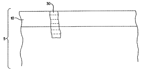
Referring first to FIG. 1, the present invention is directed to a method for
enhancing the rate of flux of a fluid collected from or delivered to a
biological tissue 5
comprising a biological membrane 10. At certain depths in the biological
membrane
and in the sub-membrane tissue, there are cells and capillaries, and
interstitial fluid
suffuses the spaces between the cells and capillaries. For example, in skin
there are
capillaries in the dermis.
The biological tissue 5 is porated with one or more micropores 30 (typically
several pores are formed at a site). The depth of a micropore can be
selectively varied,
according to the desired application. For example, FIG. 1 shows several
possible
depths of poration. The micropore 30 may extend into various depths of the
biological
membrane 10, or through the biological membrane 10 into sub-membrane tissue.
For
example, it may be desirable to porate to a sufficient depth into the
biological
membrane 5 to obtain more direct access to capillaries therein. An example is
to porate
into the dermis. The advantages of porating to selected depths is described in
more
detail hereinafter. Poration of the tissue 5, to form one or more micropores
30 of a
selected depth therein, can be carried out by methods including ablation or
micropuncture of the tissue 5 by a probe, hot wire or other heat source, an
optical
CA 02637760 2008-08-28
9
energy source, a sonic energy source, a microlancet, a high pressure fluid
jet, or by
some other energy source. Several exemplary embodiments of poration methods
and
devices for implementing the present invention are disclosed herein.
One such poration technique employs a heated probe, which is used to form one
or more micropores of a selected depth in a biological tissue or membrane. The
heated
probe is useful in the embodiments shown in FIGs. 2 and 3. A heated probe can
deliver
sufficient energy into or through the hydrated viable tissue layers beneath
the outer
layer of the biological membrane so that the poration process can continue
into the
tissue to a selected depth, penetrating through deeper layers including, e.g.,
in the case
of the skin, through the epidermis, the dermis, and into the subcutaneous
layers below if
desired. The principle concern of a system is designed to create a micropore
extending
some distance into or through the viable tissues beneath the stratum comeum,
mucosal
or buccal membranes is how to minimize both the damage to the adjacent tissue
and the
sensation to the subject during the poration process. Experimentally, it has
been shown
that a suitable heated probe is a solid, electrically or optically heated
element, with the
active heated probe tip physically defined to be no more than a few hundred
microns
across and protruding up to a few millimeters from the supporting base. A
single pulse,
or multiple pulses of current through the heated probe can deliver enough
thenmal
energy into or through the tissue to allow the ablation to penetrate as deep
as the
physical design allows. The support base may act as a component to limit the
extent of
the penetration into the tissue, essentially restricting the depth to which
the heated
probe can penetrate into a micropore to contact fresh, unporated tissue. If
the electrical
and thermal properties of said heated probe, when it is in contact with the
tissues, allow
the energy pulse to modulate the temperature of the heated probe rapidly
enough, this
type of deep tissue poration can be accompGshed with essentially no pain to
the subject.
Experiments have shown that if the required amount of thermal energy is
delivered to
the probe within less than roughly 20 milliseconds, than the procedure is
painless.
Conversely, if the energy pulse must be extended beyond roughly 20
milliseconds, the
sensation to the subject increases rapidly and non-linearly as the pulse width
is
extended.
CA 02637760 2008-08-28
An electrically heated probe design that supports this type of selected depth
poration can be built by bending a 50 to 150 micron diameter tungsten wire
into a sharp
kink, forming approximately an 180 degree bend with a minimal internal radius
near
the midpoint of the wire. This miniature 'V' shaped piece of wire can then be
mounted
5 such that the point of the 'V' extends some distance out from a support
piece which
has conductive electrodes deposited upon it. The distance to which the wire
extends out
from the support will define the maximum penetration distance into or through
the
tissue when the wire is heated. Each leg of the tungsten 'V' will be attached
to one of
the electrodes on the support carrier which in turn can be connected to the
current
to pulsing circuit. When the current is delivered to the wire in an
appropriately controlled
fashion, the wire will rapidly heat up to the desired temperature to effect
the thermal
ablation process, either in a single pulse or in multiple pulses of current.
By monitoring
the dynamic impedance of the probe and knowing the relationship between the
coefficient of resistance and the temperature of the tungsten element, closed
loop
control of the temperature of the heated element can easily be established.
Also, by
dynamically monitoring the impedance through the slcfn from the contact point
of the
probe (acting as an electrode) and a second electrode placed some distance
away from
the contact point of the probe, the depth of the pore can be controlled based
on the
different impedance properties of the tissue as a function of penetration
depth.
An optically heated probe design that supports this type of selected depth
poration can be built by taking an optical fiber and placing on one end a tip
comprised
of a solid cap or coating. A light source such as a laser diode is coupled
into the other
end of the fiber. The side of the tip closest to the fiber has a high enough
absorption
coefficient over the range of wavelengths, or selected wavelengths, emitted by
the light
source such that when the photons reach the end of the fiber and strike this
absorbing
material, some of them will be absorbed and subsequently cause the tip to heat
up. The
specific design of this tip, fiber, and source assembly may vary widely;
however, fibers
= with gross diameters of 50 to 1000 microns across commercially available and
sources
emitting up to thousands of watts of optical energy are similarly commercially
available. The tip fonming the actual heat-probe can be fabricated from t high-
melting
point material, such as tungsten, and attached to the fiber by machining it to
allow the
CA 02637760 2008-08-28
t~
insertion of the fiber into a cylindrical bore within the tip. If the distal
end of the tip
has been fabricated to limit the thermal diffusion away from this tip and back
up the
supporting cylinder attaching the tip to the fiber within the time frame of
the optical
pulse widths used, the photons incident upon this tip will elevate the
temperature
rapidly on both the fiber side and the contact side which is placed against
the tissue
surface. The positioning of the fiber/tip assembly onto the tissue surface can
be
accomplished with a simple mechanism designed to hold the tip against the
tissue
surface under some spring tension such that as the tissue beneath it is
ablated allowing
the tip to advaiice into the tissue. This allows the thermal ablation process
to continue
into or through the tissue as far as one desires. An additional feature of
this optically
heated probe design is that by monitoring the black body radiated energy from
the
heated tip that is collected by the fiber, a very simple closed loop control
of the tip
temperature can be effected. Also, as described earlier, by dynamically
monitoring the
impedance through the body from the contact point of the probe and a second
electrode
is placed some distance away from the contact point of the probe, the depth of
the pore
can be determined based on the different impedance properties of the tissue as
a
function of the probe penetration into the tissue.
Impedance can be used to determine the depth of a pore made by any technique
because it is well known that different tissue structures have different
impedance
characteristics. Accordingly, impedance can be used as an input to a control
system for
making pores of a selected depth. The impedance measured may be a complex
impedance measured with a device (such as a network analyzer) that applies a
signal
with selected frequency components between two or more electrodes (one of
which
preferably being the heated probe) on or in the tissue to highlight the
impedance
properties of the selected tissues.
Delivery of the flux enhancer can be accomplished by a variety of methods and
devices, several examples of which are more fully described herein. The
delivery of the
flux enhancer into the tissue can be carried out separately from the
microporation of the
biological membrane, or alternatively, microporation and delivery of the flux
enhancer
can be perfotmed substantially simultaneously. If separately carried out, the
flux
enhancer may be delivered before or. after microporation of the biological
membrane.
CA 02637760 2008-08-28
12
FIG. 2 shows the use of a probe or penetration device 40 for delivery of a
quantity of flux enhancer 42 into biological tissue 5. The probe 40 can be
provided
with a sharp tip or edge 44, capable of penetrating or piercing the biological
membrane
of the tissue 5, thereby allowing the probe 40 to serve as a poration means
for
5 forming a micropore 30 in the tissue 5. Altematively, the probe 40 can
comprise a
heated element for porating the biological membrane 10, such as the above-
described
heated probe comprising an electrically-heated probe or an optically-heated
probe. In
yet another alternate embodiment, the micropore 30 can be separately formed by
other
.poration means, the probe 40 serving solely to deliver the flux enhancer 42
to the
i0 micropore 30.
The flux enhancer 42 can be carried in a tube 46 within the probe 40, or can
be
carried on the outer surface 48 of the probe. Transfer means is provided for
transferring
or releasing at least a portion of the flux enhancer 42 carried by the probe
40 into the
tissue. For example, flux enhancer 42 can be injected into the tissue 5 from a
tube 46 in
t 5 the probe by a syringe or other pressurization means connectedto the
probe.
Alternatively, heating of the probe 40 can serve to release the flux enhancer
42, such as
by vaporization of a portion of the quantity of flux enhancer carried on the
probe 40.
Another embodiment is shown in FIG. 3. A carrier device 50 comprises a
reservoir 52 containing an effective amount of flux enhancer 42 for
positioning on or
adjacent to the surface of a biological membrane. The probe or penetration
device 40
(described above) is inserted into and through the reservoir 52 to release the
flux
enhancer 42 and to form the micropore 30. The sharp tip 44 penetrates the
carrier
device 50 and the biological membrane 10. Alternatively, the probe 40 is a
heated
probe and forms the micropore by thermal ablation.
Preferably, sufficient energy is applied to the flux enhancer 42 to vaporize
at
least a portion of the flux enhancer. The vaporizadon of the flux enhancer 42
provides
several advantages. For example, in its vapor state, a flux enhancer 42 such
as
ammonia more readily permeates into the tissue, thereby better enhancing the
flux rate
of fluids in the tissue S. Vaporization of the flux enhancer 42 also allows
the
pressurized release of the flux enhancer into the micropore 30, which further
enhances
the flux rate of fluids in the tissue. A variety of methods and energy sources
can be
CA 02637760 2008-08-28
13
used to vaporize the flux enhancer 42, including: kinetic energy transfer,
such as by
application of ultrasound to the reservoir 52 of flux enhancer 42;
electromagnetic
radiation, such as microwave heating; conduction; or convection. Several
examples are =
discussed in greater detail below.
For example, energy for the vaporization of the flux enhancer is provided by
conduction, through the introduction of a probe 40, heated by the mechanisms
above-
described. The introduction of the heated probe 40 through the reservoir 52 of
flux
enhancer 42 and into the tissue 5, in a single step, advantageously porates
the tissue 5
substantially simultaneously with the vaporization and delivery of the flux
enhancer 42
to the tissue 5. Registration of the reservoir 52 of flux enhancer 42 on the
tissue 5 over
the site where the micropore is to be fonned is maintained to ensure delivery
into the
micropore 30.
Turning to FIG. 4, a carrier device according to another embodiment is
generally shown at reference numeral 100. The carrier device 100 comprises a
substrate layer 102, an energy absorbing layer 104, an effective amount of
flux
enhancer 106, and a cover layer 108 transparent to the electromagnetic energy.
The
substrate layer 102 supports the various components of the device, and defines
an
aperture 110 therein which is aligned with the energy absorbing layer 104 and
the
transparent cover layer 108. The energy absorbing layer 104 is positioned on a
bottom
surface of the substrate layer 102 whereby it can be placed in contact with
the
biological membrane. A reservoir or chamber is defined in the space of the
aperture
110 between the energy absorbing layer 104 and the transparent cover layer
108. Thus,
the reservoir is sealed in a sandwich-like structure between the energy
absorbing layer
104 and the transparent cover layer 108. This reservoir contains the flux
enhancer 106.
A collecting element 112, such as an absorptive assay layer (well lrnown in
the art) is
optionally also included on another portion of the substrate layer 102. The
substrate
layer 102 may include an adhesive on a bottom portion thereof for attachment
to skin,
for example. Other attaclunent means, such as surgical tape, etc., are also
suitable.
A device for perfoming many functions in a inicroporationlharvesting/analysis
procedure is disclosed elsewhere.
CA 02637760 2008-08-28
14
The quantity of flux enhancer 106 can comprise a solid, gel, liquid, or vapor.
It
has been found that the use of a liquid flux enhancer reduces lateral heat
transfer in the
carrier device 100, thereby reducing any burning sensation observed by a
person.
Fusion means, such as a chemical binder or a carrier liquid or gel, can be
provided for
maintaining the quantity of flux enhancer 106 intact.
The substrate layer 102 is preferably made of bio-compatible material, such as
polycaprolactone or celluloseacetate which are commercially available.
The energy absorbing layer 104 is capable of absorbing energy received from an
external source, such as a laser or other source of focused optical energy,
converting
that energy to heat, and transferring that heat to a target portion of the
tissue to form at
least one micropore by ablation. For example, the energy absorbing layer 104
comprises a dye layer, which can be formed of any energy absorbing material
reactive
i5 with the external energy source, or of a non-absorbing substrate having an
absorbing
material applied thereon. A plastic film carrier treated with copper
phthalocyanine
(CPC) dye has been found to provide acceptable energy absorption from a source
of
light at wavelengths between 750-950 nm. Other materials are known to absorb
optical
energy at specific ranges of wavelengths, and can be used with energy sources
generating optical energy within those ranges. In this embodiment, it is
preferable that
the energy source not generate energy at wavelengths that are absorbed by the
target
biological tissue, so that the possibility of inadvertent injury to the tissue
is minimized.
The use of the carrier device 100 is described in conjunction with FIG. 5. The
carrier device 100 is positioned on the surface of a biological tissue to be
porated, such
as skin. A source of electromagnetic energy 120, such as optical energy, is
focused at
the transparent cover layer 108 on the carrier device 100. The focused optical
energy is
absorbed by the energy absorbing layer 104 thereby heating it. Heat generated
by
absorption of the focused optical energy is transferred to the flux enhancer
106 to heat
it ultimately to a temperature to vaporize at least a portion of it.
Substantially
simultaneously therewith, heat generated by absorption of the focused optical
energy is
transferred to the tissue 5 beneath the energy absorbing layer 104 to ablate
one or more
CA 02637760 2008-08-28
micropores 30 in the tissue. The heat generated by the absorption of the
focused optical
energy eventually destroys a portion of the energy absorbing layer 104,
melting or
burning an opening therethrough, allowing the release of at least a portion of
the flux
enhancer 106 (now vaporized) into the tissue 5 through the micropore 30.
5 The carrier device 100 initially comprises an intact energy absorbing layer,
which is normally impermeable to the flux enhancer 106, but which is made to
rupture
so as to release the flux enhancer 106 upon absorption of sufficient energy
from the
external energy source. By simultaneously ablating the tissue with optical
energy and
vaporizing the flux enhancer through one carrier device, there is inherently
provided
lo registration with the fonned micropores to ensure delivery of the vaporized
flux
enhancer into the micropores.
For collection applications, the carrier device 100 is placed over the porated
site
so that the collecting element 112 is in position to collect fluid, such as
interstitial fluid.
Additionally, suction may be applied directly over the site before, during or
after the
15 micropores are formed and the flux enhancer is released. Such suction
devices are well
known in the art.
For drug delivery applications, the carrier device 100 further comprises a
quantity of a drug 114 for delivery into the tissue. The drug can be in solid,
gel, liquid
or vapor form. The quantity of drug 114 can be contained in the reservoir of
the carrier
device 100 as described above, together with the flux enhancer 106. FIGs. 7-9
illustrate
devices which are suitable for delivery flux enhancer during microporation and
delivery
of a drug into microporated tissue.
Another embodiment of a carrier device is shown in FIG. 6. The carrier device
200 comprises a transparent substrate layer 202 and an energy absorbing layer
204 on a
bottom surface thereof. The energy absorbing layer 204 is treated with or
otherwise
incorporates a quantity of flux enhancer 206. The flux enhancer 206 may be in
granular
or powdered form and is applied to or incorporated in the energy absorbing
layer 204.
The energy absorbing layer 204 may comprise a structure similar to the
photosensitizing assembly described elsewhere.
For example, ammonia bi-carbonate crystals are suspended in
CA 02637760 2008-08-28
16
the energy absorbing layer 204 based on the fabrication techniques described
in the
aforementioned co-pending application. An adhesive may be provided on the
bottom
surface of the substrate layer 202 to attach the device to a site. The device
200 is used
in the same manner as device 100 shown in FIG. 5. As described above, the
energy
absorbing layer 204 absorbs energy from an external energy source, thereby
heating it.
Heat is transferred to the underlying tissue to ablate a portion of the tissue
and to form
one or more micropores. In addition, the heat in the energy absorbing layer
vaporizes
the flux enhancer incorporated therein, thereby releasing it into the
micropore(s). The
energy absorbing layer 204 could also be treated with a quantity of a drug
that would be
io suitably released by heat vaporization.
The delivery of flux enhancer into the target biological tissue through a
micropore increases the rate at which fluids will flow through the tissue.
Poration of
the tissue to a selected depth allows the delivery of flux enhancer to a
selected tissue or
a selected portion of a tissue. The present invention thereby provides
significantly
improved results, as compared to previously known collection and delivery
methods.
Turning to FIGs. 7 and 8, a device for containing a drug to be delivered to
microporated tissue and for delivery a flux enhancer, is shown generally at
reference
numeral 300. The device 300 is a reservoir patch containing a quantity of a
drug
mixture 310 in gaseous, liquid gel or solid form. The reservoir is defined and
contained
between a upper membrane 320 which is sealed to a lower membrane 330. On the
bottom surface of the lower membrane 330 a printed circuit 340 is disposed,
preferably
in a central region of the lower membrane 330. At selected points in the
printed circuit
340 a plurality of electrically heated probes 342 are connected and attached.
More
specifically, the electrically heated probes 342 are connected between two
sets of
electrically conductive contact pads 344 and 346. The electrically heated
probes 342
are similar to the heated wires described in the foregoing, and in the co-
pending
applications. Furthermore, around a periphery of the lower membrane 330,
adhesive
may be provided to hold the device 300 onto the tissue. The entire lower
membrane
300 may be sealed by pealable cover that keeps the device sterile and
protecting the
circuit 340 from exposure. Thus, the device 300 may be completely disposable.
A
quantity of flux enhancer may be applied to the surface of the electrically
heated probes
CA 02637760 2008-08-28
17
342 by coating the probes with a mixture containing one of the flux enhancers
described in the foregoing.
In operation, the device 300 is installed on the surface of the tissue or
biological
membrane. The adhesive cover is removed and the device is firmly attached.
Electrical
current is supplied to the circuit 340, energizing the electrically heated
probes 342. The
probes 342 will heat up and thermally ablate the tissue forming the micropores
therein.
In addition, the flux enhancer mixture on the surface of the electrically
heated probes
vaporizes and is taken up into the tissue through the micropores.
Substantially
simultaneous with the thermal creation of the micropores, the lower membrane
330 will
melt and multiple channels will be formed for delivering the drug mixture 310
into the
micropores created in the tissue.
Once the micropores have been fonmed, the electrically heated probes 342 can
be connected to a source of very mild electrical voltage to electroporate the
tissue. This
is further described in U. S. Patent 6,022,316, entitled
"Apparatus And Method For Electroporation Of Microporated Tissue For Enhancing
Flux Rates For Monitoring And Delivery Applications," issued on February 8,
2000.
See also International Application No. PCT/US97/24127, entitled
"Microporation of Biological Tissue For Delivery of Bio-Active Agents," filed
December 30, 1997. An AC
or DC current can be created across the micropores to induce an active ion-
pumping of
the drug into the tissue. This could be further enhanced by providing two
separate
reservoirs with complementary charged solutions in each to support a DC ion
flow
which would yield a net positive flux of the drug into the tissue.
Another technique to control the flux rate is to use the electrically heated
probes, or separate heated probes built into the device, to heat up the drug
mixture,
causing subsequent thermal expansion and an increase in the pressure within
the
reservoir. This heating process could be extended to the point where some
portion of
the drug mixture is vaporized. This would generate a.large increase in the
pressure
30f inside the reservoir; which would remain high until the vapor subsequently
condensed
and the pressure is returned to an initial passive state. Still another
technique is to
CA 02637760 2008-08-28
18
contact the electrically heated probes with the drug mixture and use them as
electrodes
by developing a potential between them as a means to electrolyze some elements
of the
solution to liberate a gas from the liquid or gel mixture, causing the desired
increase in
pressure.
~ Turning to FIG. 9, another device, shown generally at reference numeral 400
is
provided to delivery flux enhancer to microporated tissue and to delivery a
drug into the
tissue. The device 400 is a reservoir patch device that contains a reservoir
of a drug
mixture 410, similar to that described in conjunction with FIGs. 7 and 8. The
drug
mixture 410 is contained within a chamber defined by an upper membrane 420 and
a
to lower membrane 430. The lower membrane 430 is formed of a non-porous
plastic
material which has been treated with an energy absorbing compound similar to
that
described in conjunetion with FIG. 6. In addition, a quantity of flux enhancer
is
suspended within the energy absorbing compound of the lower membrane 430. The
upper membrane 420 is formed of optically transparent material to allow the
passage of
t i optical energy therethrough. The drug mixture 410 is likewise transparent
to optical
energy.
The operation of the device 400 is similar to that described in conjunction
with
FIG. 6. That is, the device is applied to the tissue and a beam of optical
energy, shown
at reference numeral 450 is focused onto device through the upper membrane 420
onto
20 the lower membrane 430. The lower membrane 430 responds to the optically
energy to
heat up and form micropores therein; and also melts at focused spots of the
optical
energy, thus permitting the drug mixture 410 to be released into the tissue
through the
micropores. The lower membrane 430 could be further treated with an adhesive
around
its entire surface, or its peripheral edges, or can be treated with a compound
designed to
23 reduce the thecmal impedance between the heated spots and the tissue
surface.
Alteraatively, the device 400 may comprise two chambers or reservoirs, one for
the
flux enhancer and one for the drug.
Preferably, the devices described herein are constructed such that the entire
drug
reservoir, poration elements, and optionally controlling circuitry and power
source can
30 be contained in a cone-time-use package approximately the size of a pocket
watch or
smaller. This platfortn is suitable for applications such as post-operative
delivery of
CA 02637760 2008-08-28
19
pain killers or other acute treatment regimens for which a controllable
transdennal
delivery system would be useful.
In drug delivery applications, the delivery of a flux enhancer to the
biological
tissue allows increased rates of drug introduction into target tissue or into
the
bloodstream. Thus, a given quantity of a drug can be delivered to a tissue or
into the
bloodstream in a shorter period of time, as compared with previously known
delivery
methods. Also, by the selective control of the depth of poration and of the
delivery of
flux enhancer, the present invention permits effective delivery of a drug to
deeper tissue
or tissue more remote from the site of application than could be achieved
using
io previous non-invasive or minimally invasive delivery techniques. For
example, the
present invention enables delivery of a drug to the capillary depth of a
tissue, thereby
providing improved uptake into the bloodstream before the drug is metabolized
by the
reaction of the body to a foreign substance. This is accomplished by porating
the
tissue to a selected depth whereby the flux enhancer can be delivered to the
walls of the
capillaries or to tissue adjacent the capillaries, and delivering the flux
enhancer and the
drug through the micropore. lontophoresis, sonic energy, mechanical pressure
and
manipulation or other motive forces can be used to further enhance the rate of
drug
delivery. Delivery of the flux enhancer and the drug in this manner results in
faster
uptake of the drug into the bloodstream. Thus, the rate of uptake of a drug by
the
subject organism can be controlled as desired by appropriate selection of the
depth of
poration and application of the flux enhancer and control of the other motive
forces
described herein.
The above-described embodiments are given as illustrative examples only, and
are not intended to be limiting or exhaustive. It will be readily apparent to
those of
ordinary skill in the art that many additions, deletions and modifications may
be made
without departing from the spirit and scope of the present invention, as
defined by the
claims below.