Note: Descriptions are shown in the official language in which they were submitted.
CA 02637835 2008-07-18
WO 2007/083929 PCT/KR2007/000300
MARKER AND METHOD FOR CANCER DIAGNOSIS
TECHNICAL FIELD
The present invention relates to a diagnostic cancer marker using variation in
the
gene expression of a granulocyte colony stimulating factor (G-CSF) and a
method
for preparing the same, and more specifically, relates to a method for
diagnosing
1 o cancer and/or assessing the state of cancer progression using an
oligonucleotide
having the 3'-terminal end of exon 2 region linked to the 5'-terminal end of
exon 4
region of a G-CSF gene as a diagnostic cancer marker.
BACKGROUND ART
Cancer diagnosis is generally achieved by (1) morphological analysis using
microscopes such as an optical microscope or electron microscope, (2)
immunohistochemical assays which detect proteins specifically expressed in
cancer
tissues (Iran, Biomed. J., 3:99, 1999; Lancet, 2:483, 1986), or (3) molecular
diagnosis which analyzes abnormal biomolecules found in cancer tissues, such
as
mutated genes. In comparison with the molecular diagnosis, the morphological
and immunohistochemical diagnosis requires much longer time and higher cost.
Since the molecular diagnosis has a relatively simple procedure and a short
time to
yield results, it has become a main subject in developing novel diagnostic
methods
for cancer. Recently, Health Digit Inc.developed a protein chip system for
diagnosing various cancers, and gained on approval for clinical tests from the
Chinese State Drug Administration (CSDA) for the first time in the world
(www.health-digit.com). However, the protein chip system does not use only one
1
CA 02637835 2008-07-18
WO 2007/083929 PCT/KR2007/000300
biomarker to diagnose all kinds of cancer, but uses 10 or more proteins as
biomarkers.
To effectively apply such diagnostic methods to cancer diagnosis, it is most
important to select and use cancer diagnostic markers capable of more
accurately
and easily detecting cancer incidence. Several genes (Steve, M. et al., J.
Clin.
Oncology, 20:3165-3175, 2002; Sridlhar, R. et al., J. Clin. Oncology,
20:1932-1941, 2002) and proteins (Goessl, et al., Urology, 58:335-338, 2001;
Zhou, et al., Breast Cancer Res. Treat., 66:217-224, 2001; Korea Pat.
Publication
1 o No. 2001-0061173) have been reported as diagnostic cancer markers, and
some of
them are being clinically used for diagnosis of cancer. Among conventional
cancer biomarkers, CEA, BFP, TPA and IAP, which have low organ specificity,
have low sensitivity, thus generating false positive data. Also, the
biomarkers
which have high organ specificity, such as AFP, PIVKA II, Esterase I , CA19-9,
CA50, Span-1 antigen, CA15-3 and BCA 225, are useful only for target organs.
Many researchers have attempted to find genes having diagnostic applications,
in
developing diagnostic cancer marker candidates showing different results
according
to pathological and physiological condition using microarray technology (Liu,
H.X.
et al., Nat. Genet., 27:55-58, 2001; Wilson, C.A. et al., Oncogene, 14:1-16,
1997;
Weissensteiner, T., Nucleic Acids Res., 26:687, 1998; Zolezzi, F. et al., Am.
J. Med.
Genet., 71:366-370, 1997; Mottes J.R. and Iverson, L.E., Neuron, 14:613-623,
1995; Crook, R. et al., Nat. Med., 4:452-455, 1998; Jiang, Z.H. and Wu, J.Y.,
Proc.
Soc. Exp. Biol. Med., 220:64-72, 1999).
However, since diagnostic cancer marker candidates found by the above
mentioned
methods are mostly composed of expressed sequence tag (EST), they are just
defined as a characteristic of data and thus it is difficult to select
reliable specific
candidates and to catch on the very genes from which they are originated.
3 o Specifically, the number of genes is known by human genome analysis and it
is also
2
CA 02637835 2008-07-18
WO 2007/083929 PCT/KR2007/000300
known that many isoforms or variants are expressed there from to have
biological
function and its complexity. Therefore, it has become another big subject for
the
future to find out that, in which gene and condition variants throughout the
whole
genome are expressed and what their functions are. These various variants can
be a
good basis to figure out the correlation between the formation of abnormal
variants
among them and possibility of causing cancer (Cartegni, L. et al., Nat. Rev.
Genet.,
3:285-298, 2002; Schweighoffer, F. et al., Pharmacogenomics, 1:187-197, 2000;
Blencowe, B.J., Treds Biochem. Sci., 25:106-110, 2000; Cooper, T.A. and
Mattox,
W., Am. J. Hum. Genet., 61:259-266, 1997).
The present inventors have also conducted studies for a long time to develop a
new
diagnostic cancer marker which can diagnose various kinds of cancers,
consequently, confirmed that deletion of exon 3 region was specifically shown
in
tumor cells or tumor tissues during transcription of G-CSF gene, thereby
filing an
application regarding a method for diagnosing cancer using G-CSF mRNA, cDNA
variants fragment or protein as a diagnostic cancer marker (WO 2003/027288
Al).
In microarray which uses G-CSF gene fragment as a diagnostic cancer marker of
the above application patent, any one or more fragments among exons 1, 2, 4
and 5
DNA fragments of G-CSF gene together with exon 3 DNA fragment of G-CSF gene
2 o are used as nucleic acid probes to detect G-CSF gene fragment having
deleted exon
3 region among biological samples. This inventive method for diagnosing
cancer,
by detecting deletion of exon 3 region of G-CSF gene expression is one of the
technologies which diagnose cancer using characteristics of gene variants, and
is
considered to be a useful diagnostic cancer marker candidate, since the
variants
appear in most cancer.
Meanwhile, most genes including G-CSF gene generally express many isoforms
and variants, so, probe fragments fixed on a microarray must have high
sensitivity
in detecting the deletion of exon 3 region of G-CSF gene. Also, the expression
of
3 o normal G-CSF or their fragments can exist together with that of mutated G-
CSF
3
CA 02637835 2008-07-18
WO 2007/083929 PCT/KR2007/000300
isoforms in tumor cells or tumor tissues, thus diagnosis for cancer only by
detecting
the presence of exon 3 region of G-CSF in its gene expression can lead to loss
of
credibility or low sensitivity and, moreover, it has a problem in assessing
the state
of cancer progression.
Accordingly, the present inventors have made extensive efforts to develop a
new
nucleic acid probe for detecting G-CSF gene fragment not having exon 3 region
which can satisfy the above requirement or solve the above problem, and as a
result,
found that it has remarkably increased high sensitivity in cancer diagnosis
compared
1 o with other probes, when an oligonucleotide containing a nucleic acid
sequence
having the 3'-terminal end of exon 2 region of G-CSF gene linked to the 5'-
terminal
end of exon 4 region of G-CSF gene is used as a diagnostic cancer marker, and
confirmed that the state of cancer progression can be accurately diagnosed by
using
an oligonucleotide containing nucleic acid sequence having 3'-terminal end of
exon
2 region of G-CSF gene linked to the 5'-terminal end of exon 4 region of G-CSF
gene together with an oligonucleotide having sequences of a part or the entire
region of exon 3 region of G-CSF gene as a diagnostic cancer marker, thereby
completing the present invention.
SUMMARY OF THE INVENTION
Therefore, the main object of the present invention is to provide an
oligonucleotide
for diagnosing cancer, essentially containing a nucleic acid sequence of a
splice
junction site having the 3'-terminal end of exon 2 region linked to the 5'-
terminal
end of exon 4 region of a granulocyte colony stimulating factor gene.
Another object of the present invention is to provide a diagnostic kit for
cancer
diagnosis containing the oligonucleotide and a method for diagnosing cancer
using
3 0 the oligonucleotide.
4
CA 02637835 2008-07-18
WO 2007/083929 PCT/KR2007/000300
To achieve the above objects, the present invention provides an
oligonucleotide for
a diagnostic cancer marker, essentially containing a nucleic acid sequence of
a
splice junction site having the 3'-terminal end of exon 2 region linked to the
5'-
terminal end of exon 4 region of a G-CSF gene.
Preferably, the oligonucleotide according to the present invention essentially
contains nucleic acid sequences of SEQ ID NOs: 1 or 2.
The present invention also provides a diagnostic kit for cancer diagnosis
containing
the oligonucleotide.
In the present invention, the diagnostic kit for cancer diagnosis is
preferably a
diagnostic kit for assessing the state of cancer progression which
additionally
contains an oligonucleotide essentially containing sequences of a part or the
entire
region of the exori 3 region of G-CSF gene.
The present invention also provides a method for diagnosing cancer, the method
comprising the steps of: (a) obtaining a G-CSF nucleic acid sample from a
mammal
2 o biological sample; (b) amplifying the obtained G-CSF nucleic acid sample;
and (c)
detecting oligonucleotide containing a nucleic acid sequence of a splice
junction site
having the 3'-terminal end of exon 2 region linked to the 5'-terminal end of
exon 4
region of a G-CSF gene, in the amplified sample.
In the inventive method, the step (c) preferably contains the step in which
simultaneously detects an oligonucleotide containing sequences of a part or
the
entire region of the exon 3 region together with an oligonucleotide containing
a
nucleic acid sequence of splice junction site having the 3'-terminal end of
exon 2
region linked to the 5'-terminal end of exon 4 region of a G-CSF gene, in the
3 o amplified sample.
5
CA 02637835 2008-07-18
WO 2007/083929 PCT/KR2007/000300
Other features and embodiments of the present invention will be more fully
apparent from the following detailed description and appended claims.
BRIEF DESCRIPTION OF DRAWINGS
FIG. 1 is a schematic diagram of a process of producing normal protein and
variants
from human G-CSF gene.
FIG. 2 shows a junction region of an exon 2 region and an exon 3 region which
can
be formed by two types (type A, type B) of exon 2 region of human G-CSF gene.
FIG. 3 shows positions to which primers used in PCR is attached and expected
PCR
products according to the positions.
FIG. 4 is a design of DNA chip which consists of probes of each region of
amplified
G-CSF gene (E2: a probe designed from exon 2 region of a G-CSF gene; E2E3a: a
probe designed from a junction site having the 3'-terminal end of exon 2
region
linked to the 5'-terminal end of exon 3 region of a type A G-CSF gene; E2E3b:
a
probe designed from a junction site having the 3'-terminal end of exon 2
region
linked to the 5'-terminal end of exon 3 region of a type B G-CSF gene; E3-1,
E3-3,
E3-4 and E3-6: probes designed from exon 3 region of a G-CSF gene; E2E4a: a
probe designed from a junction site having the 3'-terminal end of exon 2
region
linked to the 5'-terminal end of exon 4 region of a type A G-CSF gene; E2E4b:
a
probe designed from a junction site having the 3'-terminal end of exon 2
region
linked to the 5'-terminal end of exon 4 region of a type B G-CSF gene; P: a
probe
constructed to distinguish positions by fluorescent labels as a position
marker; N: a
negative control (spotting solution).
6
CA 02637835 2008-07-18
WO 2007/083929 PCT/KR2007/000300
FIG. 5 shows the results of hybridization using DNA chip of FIG. 4. Red
circles
show probes showing signals.
FIG. 6 shows the results of hybridization in DNA chip of FIG. 4 according to
types
of cells and tissues (A: normal human blood, B: lung cancer (A549), C: large
intestine cancer (SES-T), D: stomach cancer (1:AGS, 2: YCC-2, 3: HwangOO, E:
cervical cancer (1: C33A, 2: HeLa), F: breeding (HT1080), G: breast cancer
(MDA-
MB-231), H: pancreas cancer (Capan-2), I: liver cancer (SK-Hepl), J: malignant
melanoma (SK-Mel), K: leukemia (Jurket cDNA library), L: embryonic kidney
(293)). Red circles show signals of probes capable of distinguishing between
1. o cancer tissues and normal tissues.
FIG. 7 is a schematic view of a DNA chip which consists of probes designed to
detect a splice junction site of G-CSF gene (E2E4: a probe designed from a
junction
site having the 3'-terminal end of exon 2 region linked to the 5'-terminal end
of
exon 4 region of two types (type A, type B) of G-CSF gene; E2E3: a probe
designed
from a junction site having the 3'-terminal end of exon 2 region and the 5'-
terminal
end of exon 3 region of two types (type A, type B) of G-CSF gene; P: a probe
constructed to distinguish positions by fluorescent signals as a position
marker; N: a
negative control (spotting solution).
FIG. 8 shows the results of hybridization in DNA chip of FIG. 7 according to
types
of cells and tissues. Red circles show a probe which can specifically show
signals
in case of cancer.
FIG. 9 is a schematic view of a DNA chip on which probes, designed from each
exon region of G-CSF gene to examine diagnosis efficiency of each probe, are
fixed.
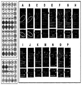
FIG. 10 is a schematic view showing positions of each probe in G-CSF gene.
Probes included in an ellipse among probes for G-CSF gene not having exon 3
are
7
CA 02637835 2008-07-18
WO 2007/083929 PCT/KR2007/000300
designed from the 3'-terminal end of exon 2 and the 5'-terminal end of exon 4
of
splicing variants.
FIG. 11 shows signal intensities of probes according to types of cells and
probe
candidates showing effectiveness, which can be deduced there from.
FIG. 12 shows the results of hybridization using DNA chip of FIG. 9. Red
circles
show a probe which can specifically show signals in only cancer.
FIG. 13 shows the results of hybridization using DNA chip of FIG. 9 according
to
types of cells and tissues. Images of hybridization reactions which is
obtained by
Scanarray 5000 (A: normal blood (WBC), B: 293 (embryonic cell line), C: SES-N
(normal large intestine), D: SES-T (large intestine cancer), E: Colo205 (colon
cancer cell line), F: DLD-1 (colon cancer cell line), G: Hwang00 (stomach
cancer
cell), H: YCC-3 (stomach cancer cell line), J: MDA-MB-231 (breast cancer cell
line), K: NCI-H460 (lung cancer cell line), L: Caki-2 (kidney cancer cell
line), M:
Capan-2 (pancreas cancer cell line), N: SK-Mel2(malignant melanoma), 0: HepG-2
(hepatocellular carcinoma), P: SK-Hepl (liver cancer cell line)). Red circles
show
a probe which can specifically show signals in case of cancer.
FIG. 14 shows DNA chips which are prepared by mixing each probe with spotting
solution. The part marked with a blue square is a region on which a probe
representing cancer is located.
FIG. 15 shows the results of hybridization of products obtained by amplifying
nucleotide sequence of human G-CSF gene derived from normal and tumor clinical
samples using primers of SEQ ID NO: 32 and SEQ ID NO: 33 according to
Example 8 and Example 9 with DNA chip of FIG. 14.
8
CA 02637835 2008-07-18
WO 2007/083929 PCT/KR2007/000300
DETAILED DESCRIPTION OF THE INVENTION, AND
PREFFERED EMBODIMENTS
The present invention relates to a method for diagnosing cancer and/or
assessing the
state of cancer progression, using an oligonucleotide which essentially
contains a
nucleic acid sequence of a splice junction site having 3'-terminal end of exon
2
region linked to the 5'-terminal end of exon 4 as G-CSF gene variants fragment
generated after post-transcription process by genetic analysis method
including a
microarray. In other words, the present invention relates to a method for
1 o diagnosing cancer and/or assessing the state of cancer progression using G-
CSF
gene variants obtained by deleting an exon 3 region in G-CSF gene to link an
exon
2 region and an exon 4 region, as a diagnostic cancer marker.
Exons 1-5 of G-CSF gene are normally linked during splicing process in normal
human body, however splicing occurs in a form of a variant not having exon 3
in
tumor cells or tumor-progressing cells to produce mRNA not having exon 3 (FIG.
1). Exon 2 region of a human G-CSF gene has two types (type A, type B),
therefore, a junction site of exon 2 region and exon 3 region also has two
types
(FIG. 2). Also, as a result of splicing of G-CSF gene, G-CSF mRNA having all
2 o exon 1-exon 5 is isolated from a normal cell, and mRNA not having exon 3
is
isolated from a tumor cell, and the above mentioned difference of mRNAs can be
confirmed by PCR using primers specific to G-CSF gene (FIG. 3).
Molecular biological methods which are used in identifying both genes
specifically
expressed (or suppressed) in tumor cells and genetic mutation are exemplified
by
PCR (Bottema, C.D., Mutat. Res., 233:93-102, 1993; Nelson, D.L., Curr. Opin.
Genet. Dev., 1:62-68, 1991; Pourzand, C. and Cerutti, P., Mutat. Res.,
288:113-121, 1993; Holland, P.M. et al., Proc. Natl. Acad. Sci. USA, 8:7276-
7280,
1991), Single-Stranded Conformation Polymorphism (SSCP, Glavac, D., Hum.
3 o Mutat., 19:384-394, 2002; Strippoli, P. et al., Int. J. Mol. Med., 8:567-
572, 2001),
9
CA 02637835 2008-07-18
WO 2007/083929 PCT/KR2007/000300
DNA Sequencing Analysis (Sanger, F. et al., Proc. Natl. Acad. Sci. USA,
74:6463-5467, 1997), Protein Truncation Test (Hardy, C.A., Methods Mol. Biol.,
187:87-108, 2002), automatic nucleotide sequence analysis (Boutin, P. et al.,
Hum.
Mutat., 15(2):201-203, 2000), study of loss of heterozygosity (Yang, Q. et
al., Clin.
Cancer Res., 8:2890-2893, 2002), study of microsatellite instability (Furlan,
D. et
al., J. Pathol., 197:603-609, 2002), gene analysis using MALDI-TOF (Leushner
J.,
Expert. Rev. Mol. Dign., 1:11- 18, 2001), gene analysis by hybridization
(Wetmur,
J.G., Critical Reviews in Biochem. Mol. Biol., 26:227-259, 1991), gene
analysis
using DNA chips (Goessl et al., Urology, 58:335-338, 2001; Zhou et al., Brest
Cancer Res. Treat., 66:217-224, 2001; Korea Pat. Publication No. 2001-
0061173),
analysis using protein chips (Pharmacogenomics, 1:385-393, 2000). Therefore
those skilled in the art will understand that they can easily detect the
existence of
splice junction site of specific variants according to the present invention,
generated
in post-transcriptional process of G-CSF by properly using well-known
molecular
biological methods including the above mentioned methods. The present
inventors
found that the most effective probes capable of detecting the existence of the
variants are only probe candidates capable of detecting the existence of
splice
junction site, thereby inventing a diagnostic method by which the existence
thereof
can be detected. However, among the above mentioned methods, the detection of
specific variants generated during post-transcriptional process of G-CSF
according
to the present invention is preferably and easily performed by using PCR,
hybridization reaction and DNA chip.
To perform cancer diagnosis according to the present invention, a G-CSF gene
or
variants thereof should first be obtained from tissue specimens or cells.
Since a
DNA sample for a specific gene is typically obtained from normal tissues or
cells at
a very small amount, the specific gene should be amplified by PCR and for such
amplification, primers suitable for such amplification should be designed. In
the
present invention, to amplify a part or an entire region of splice junction
site of an
3 0 exon 2 region and an exon 4 region, DNA nucleic acid fragments to be used
as
CA 02637835 2008-07-18
WO 2007/083929 PCT/KR2007/000300
primers in PCR for detecting the existence of the splice junction site is
required.
That is, the primers, as used herein, refer to oligonucleotides capable of
amplifying
a nucleotide sequence of G-CSF gene, comprising a part or an entire region of
the
splice junction site of an exon 2 region and an exon 4 region. Those skilled
in the
art will be able to easily design such primers. Those skilled in the art will
be able
to easily design such primers. Therefore, all primers capable of amplifying G-
CSF
gene variants comprising a part or an entire region of the splice junction
site, which
can be designed by those skilled in the art, are intended to fall within the
scope of
the present invention.
In accordance with an aspect of the present invention, there is provided a
gene
microarray or membrane to which a DNA fragment comprising a splice junction
site having the 3'-end of an exon 2 linked to the 5'-end of exon 4 of the G-
CSF
gene is immobilized, which is useful for diagnosis of cancer. The gene
microarray
includes DNA chips effective in detection of a gene by hybridization including
applying to a complementary oligonucleotide probe immobilized on the surface
of a
slide glass treated with a specific chemical reagent. Non-limiting examples of
the
membrane, which can be used instead of the slide glass in hybridization, may
include all membranes capable of immobilizing DNA fragments; and preferably,
nylon and nitrocellulose membranes.
Fixing the probes on the surface of a slide glass and a membrane can be easily
achieved by the conventional technique known in the art. In addition,
preparation
of targets, hybridization and stripping will be performed according to the
conventional techniques common in the art.
In another aspect of the present invention, there is included a composition
for
diagnosis of cancer, comprising a DNA fragment containing a splice junction
site
having the 3'-end of an exon 2 linked to the 5'-end of exon 4of G-CSF gene and
a
3 0 diagnostically acceptable conventional carrier. In a further aspect of the
present
11
CA 02637835 2008-07-18
WO 2007/083929 PCT/KR2007/000300
invention, there is included a diagnostic kit comprising a DNA fragment
containing
a splice junction site having the 3'-end of an exon 2 linked to the 5'-end of
exon 4
of the G-CSF gene and a DNA microarray using the DNA fragment.
Examples
Hereinafter, the present invention will be described in more detail by
specific
examples. However, the present invention is not limited to these examples, and
it
is obvious to those of ordinary skill in the field of the present invention
that
numerous variations or modifications could be made within the spirit and scope
of
the present invention.
Example 1: Preparation of sample from tissues (cells)
The normal cell lines and tumor cell lines used in Examples of the present
invention
are given in Table 1, below. The underlined samples have the same result as
those
of the normal cell lines in Table 1.
The tumor cell lines listed in Table 1 can be obtained from the cell
collection
centers listed in Table 1. The tumor cell line, obtained from the cancer
metastasis
research center at College of Medicine, Yonsei University, was prepared as
follows.
After ascitic fluid was aseptically obtained from advanced cancer patients,
supplemented with heparin in an amount of 10 units per ml to prevent clumping
of
cells and centrifuged at 400xg for 10 min. The precipitated cells obtained by
centrifuge were cultured in a 25cm2 culture flask. In case of containing a
large
number of erythrocytes, Ficoll-hypaque density gradient centrifugation at
800xg
was performed to separate mononuclear cells from erythrocytes, and the
obtained
mononuclear cell phase was incubated at 37 C under 5% COZ. After incubation
for 1 day (16-18 hours), the culture medium was centrifuged at 400xg for 10
min,
3 o and the precipitated cells were cultured in a new 25cm2 culture flask.
During
12
CA 02637835 2008-07-18
WO 2007/083929 PCT/KR2007/000300
culturing, cells were observed under a phase contrast microscope, and the
culture
medium was replaced twice or three times per week. When tumor cell colonies
were formed, the tumor cell clusters were obtained by treatment with trypsin-
EDTA
or by obtaining colony or by using scrapers, or the fluid containing tumor
cells was
centrifuged to remove normal cells. The resulting pure tumor cells were stored
at
frozen states according to their passages.
Human leucocyte cells can be obtained as follows. After 8mL of blood was
transferred into 50mL of Coming tube, 24mL of RBC lysis buffer was added and
1 o the mixture was left to stand at 4 C for 1 Omin,while stirring it
occasionally. After
centrifuging the mixture at 2,000rpm at 4 C for 12min and confirming
leucocytic
pellet, a supemant was removed. If RBC (red blood cell) was left, said process
was repeated. TRIZOL was added to finally obtain leucocytic pellet to separate
RNA.
Table 1
Cell types Cell collection centers
Culture Cancer A549 Lung cell line ATCC CCL-185
cell lines HCT116 ATCC CCL-247
Colon cancer cell
Co1o205 line ATCC CCL-222
DLD-1 ATCC CCL-221
HeLa Cervical cancer ATCC CCL-2
C33A cell line ATCC HTB-231
HT1080 Breeding cancer ATCC CCL-121
cell line
AGS ATCC CRL-1739
YCC-2 Stomach cancer Cancer metastasis research center, College
cell line of Medicine, Yonsei University
YCC-3 Cancer metastasis research center, College
of Medicine, Yonsei University
MDA-MB- Breast cancer cell
ATCC HTB-26
231 line
Caki-2 Kidney cancer Cancer metastasis research center, College
cell line of Medicine, Yonsei University
13
CA 02637835 2008-07-18
WO 2007/083929 PCT/KR2007/000300
Capan-2 Pancreas cancer Cancer metastasis research center, College
cell line of Medicine, Yonsei University
Hepatoma cell
HepG-2 ATCC HB-8065
line
Liver cancer cell
SK-Hep-1 line ATCC HTB-52
Malignant
SK-Mel2 melanoma cell ATCC HTB-68
line
Jurket
Cancer metastasis research center, College
cDNA Leukemia
of Medicine, Yonsei University
library
Brian cancer cell
U87-MG Korea cell line bank KCLB3004
line
Normal 293 Embryonic Cancer metastasis research center, College
kidney cell line of Medicine, Yonsei University
SES-T Intestine cancer Cancer metastasis research center, College
Cancer of Medicine, Yonsei University
Hwang00 Stomach cancer Cancer metastasis research center, College
Tissue of Medicine, Yonsei University
Human Cancer metastasis research center, College
Leucocyte
Normal Blood of Medicine, Yonsei University
SES-N intestine Cancer metastasis research center, College
of Medicine, Yonsei University
Example 2: Preparation of mRNA and cDNA from cell lines
Total RNA was isolated from each tumor cell line, normal cell line and normal
tissue using Trizol Reagent (Gibco-BRL, USA). lml of Trizol Reagent was added
to a tissue sample ground after quickly freezing using liquid nitrogen,
followed by
incubation at room temperature for 5min. 0.2m1 of chloroform was added to the
resulting tissue sample, vigorously vortexed for 15sec and incubated at room
temperature for 5 min. After centrifugation at 12,000xg at 4 C for 15min, the
resultant aqueous phase was transferred to a new tube. An equal volume of
isopropanol was added to the tube, and the tube was placed at 4 C for 10min.
After centrifugation at 12,000xg at 4 C for 10min, the supematant was
carefully
14
CA 02637835 2008-07-18
WO 2007/083929 PCT/KR2007/000300
discarded, and the pellet was washed with 70% ethanol, followed by
centrifugation
at 7,500xg at 4 C for 5min. After being dried, the RNA pellet was dissolved in
RNase-free water.
To synthesize cDNA from mRNA isolated from each cell line, and human-derived
tumor and normal cell line, RT-PCR was performed as follows. 2 ug of total RNA
was mixed with 1a of an oligo(dT)16-primer, and RNase-free water was added up
to a final volume of 11 ,cce. This mixture was heated at 90 C for 5min, and
placed
on ice, immediately after completion of the heating. After putting 40 of a
reaction buffer, 2a of 10mM dNTPs, 1,t.ce of RNase inhibitor and 2,cce of
reverse
transcriptase into another tube, 8.5 ,cd of the RNA mixture was added to the
pre-
mixture tube, followed by incubation at room temperature for 10min. The
reaction
mixture was incubated at 42 C for 90min, and then at 95 C for 15min.
Immediately after the incubation at 95 C, the mixture was placed on ice to
terminate
reaction, thus yielding a cDNA sample.
Example 3: Preparation of DNA chip 1 for examining effectiveness of probe for
cancer diagnosis
In order to investigate whether a DNA chip can be used as a tool for detection
of a
splice junction site of G-CSF mRNA or cDNA, various DNA fragment probes
capable of being immobilized on a glass plate was prepared as follows. On
probe
corresponding to a part of exon 2 of G-CSF, four non-overlapping probes
corresponding to exon 3, and one probe corresponding to a part of exon 4, were
designed to consist of 20 nucleotides each. Since two different G-CSF mRNAs
(human G-CSFa and human G-CSFb mRNAs) are generated by alternative splicing
in the exon 2 region (Tshuchiya, M. et al., EMBO J., 5:575-581, 1986), two
types
of probes comprising a region corresponding to exon 2 were prepared, based on
the
two different G-CSF mRNAs. Nucleic acid sequences thereof are shown in Table
2.
CA 02637835 2008-07-18
WO 2007/083929 PCT/KR2007/000300
Table 2
Probe name Nucleic acid sequences SEQ ID NO: Positions
E 2 CTG CAG CTG CTG CTG TGG CAC 3 Exon 2
E2E3a AGA AGC TGT GTG CCA C 4 Exon 2-3
E2E3b TGA GTG AGT GTG CCA C 5 Exon 2-3
E3-1 TGT GCC ACC TAC AAG CTG TG 6 Exon 3
E3-3 GAG CTG GTG ATG CTC GGA 7 Exon 3
E3-4 GGA CAC TCT CTG GGC ATC 8 Exon 3
E3-6 GGA CAC TCT CTG GGC ATC 9 Exon 3
E4 GCA GGC TGC TTG AGC CAA 10 Exon 4
E2E4a AGA AGC TGG CAG GCT G 11 Exon 2-4
E2E4b TGA GTG AGG CAG GCT G 12 Exon 2-4
To confer ability to be immobilized on a glass plate, when synthesizing all
DNA
fragment probes, a base having an amino group was inserted to the 3'-end of
the
probes using an aminolinker column (Cruachem, Glasgrow, Scotland), and slide
glass coated with aldehyde residues (CEL Associates, Inc., Huston Taxas, USA)
were used.
After being dissolved in 3 x SSC (0.45 M NaCl, 15 mM C6HSNaA, pH 7.0), the
DNA probes were immobilized on the slide glass by accumulating the DNA probes
using a microarrayer manufactured by the present inventors (Yoon et al., J.
Microbiol. Biotechnol., 10:21-26, 2000), and reacting for over lhr under about
55%
humidity, and then leaving the glass at room temperature for 6hrs (FIG. 4).
Herein,
the probes were arranged at intervals of 180 gm on the glass at an amout of
100 M,
thus producing a microarray. Immobilization of probes through reaction between
amine groups of probes and aldehyde groups on the glasses was estimated by
staining with SYBRO green II (Molecular Probes, Inc., Leiden, Netherlands).
16
CA 02637835 2008-07-18
WO 2007/083929 PCT/KR2007/000300
Example 4: Preparation of tar2et sample for detecting specific variants
Asymmetric PCR was carried out using mRNA or cDNA isolated from each cell
line of Example 2 as a template under the conditions of denaturation at 94 C
for
5min, 30 cycles of denaturation at 94 C for lmin, annealing at 50-56 C for
lmin
and extention at 72 C for 30sec, followed by final extention at 72 C for
5min. A
primer set used in Asymmetric PCR is as follows. A reverse primer was labeled
with FITC for detection.
Forward primer: 5'-ACC CCC CTG GGC CCT GCC-3' (SEQ ID NO: 13)
Reverse primer: FITC-5'CTG CTG CCA GAT GGT GGT-3' (SEQ ID NO:
14)
PCR products were separated on an agarose gel. From the result of
electrophoresis,
double strand DNA and single stranded DNA fragments were produced in each
PCR sample (FIG. 3). After amplifying G-CSF gene by asymmetric PCR, a
hybridization solution (6 x SSPE, 20 %(v/v) foramide) was added to 15 fd of
the
amplified product up to a final volume of 200 0. The mixture was applied on a
slide glass (a DNA chip 1 for cancer diagnosis, FIG. 4) having an immobilized
probe, and the glass was covered with a probe-clip pressseal incubation
chamber
(Sigma Co., St. Louis, MO.), followed by incubation in a shaking incubator at
30 C
for 6 hours to induce binding of the probe complemantary to the amplified
product.
Thereafter, the glass was washed over 5 min with 3 x SSPE (0.45 M NaCl, 15 mM
C6H5Na3O7, pH7.0), 2 x SSPE (0.3M NaC1, 10mM C6H5Na3O7, pH7.0), and then 1
x SSPE (0.15 M NaCI, 5 mM C6H5Na3O7, pH7.0).
Example 5: Test result of DNA chip 1 for cancer dia2nosis
After target products amplified by Asymmetric PCR was applied to the DNA chip
3 0 prepared in Example 3, they were scanned using Scanarray 5000 (GSI
Lumonics
17
CA 02637835 2008-07-18
WO 2007/083929 PCT/KR2007/000300
Inc., Bedford, MA., USA). To predict results regarding probe, in case of the
plasmid having no deletion of exon 3 in G-CSF gene, signals were detected by
applying on the DNA chip. In contrast, in case of the exon 3-deleted G-CSF-
containing plasmid, signals were detected by applying on the DNA chip, wherein
the plasmids have nucleotide sequences of SEQ ID NOs: 26 and 27.
As a result, as shown in FIG. 5, only E2E4a probe showed signals on a deletion
site.
This plasmid had type A exon 2 (FIG. 1). On the contrary, in the case of
plasmid
in which G-CSF gene was not deleted, E2E3a probe and probes in exon 3 region
1 o showed signals. If the sequences of no deletion and of deletion of exon3
in G-CSF
were mixed, mixed results of the two cases can be predicted. FIG. 6 shows the
hybridization results by Scanarray 5000 after target DNA according to each
cell was
applied to DNA chip of FIG. 4. As shown in FIG. 6, in case where probes
produced
from exon 2 and exon 4 junction region, which only specific variants can have,
cells
could be detected by each probe.
Example 6: Preparation of DNA chip 2 for examining effectiveness of probe for
cancer diagnosis and test results
2 o To examine effectiveness of E2E4 for cancer diagnosis, a new type DNA chip
2
was prepared (FIG. 7). To easily decode, the DNA chip 2 was designed to have
two types of exons (E2E4 of FIG. 7 contains both type A and type B, E2E3
contains
both type A and type B of E2E3a and E2E3b). Probes were immobilized by the
same immobilization method described in Example 3, and as a result of
hybridization of a target sample prepared in Example 4, as shown in FIG. 8, it
was
confirmed that probes constructed from the junction region of exon 2 and exon
4 is
the most powerful in developing a system which can easily diagnose cancer
using
produced DNA chip. To strengthen signal intensity, probes having nucleic acid
sequences in Table 3 below were applied on the basis of a nucleic acid
sequence of
3 o splice junction site.
18
CA 02637835 2008-07-18
WO 2007/083929 PCT/KR2007/000300
Table 3
Probe name Nucleic acid sequences SEQ ID NO: Positions
E2E4a GGA GAA GCT GGC AGG CTG CT 1 Exon 2-4
E2E4b GGT GAG TGA GGC AGG CTG CT 2 Exon 2-4
Example 7: Preparation of DNA chip 3 for examining effectiveness of probe for
cancer diagnosis
To examine whether probes constructed from exon 2 and exon 4 junction region
were the most powerful; DNA chip 3 was prepared by designing probes from each
nucleotide sequence in each region (FIG. 9). FIG. 10 shows the rough position
of
1. o each probe in G-CSF gene, and Table 4 shows nucleic acid sequence of each
probe.
Probes were fixed by the same immobilization method described in Example 3.
Table 4
Probe name Nucleic acid sequences SEQ ID NO: Positions
E2 1 GAG CTT CCT GCT CAA GTG CT 15 Exon 2
E2 2 AGA GCT TCC TGC TCA AGT GC 16 Exon 2
E2 3 GCA AGT GAG GAA GAT CCA GG 17 Exon 2
E2 4 CCA GAG CTT CCT GCT CAA GT 18 Exon 2
E2 5 CAA GTG AGG AAG ATC CAG GG 19 Exon 2
E2E4 CTGGTGAGTGGCAGGCTGCT 20 Exon 2-3-4
E2E4 1 AGA AGC TGG CAG GCT G 9 Exon 2-4
E2E4 2 TGA GTG AGG CAG GCT G 10 Exon 2-4
E2E4a GGA GAA GCT GGC AGG CTG CT 13 Exon 2-4
E2E4b GGT GAG TGA GGC AGG CTG CT 14 Exon 2-4
E2E3 1 AGA AGC TGT GTG CCA A 2 Exon 2-3
E2E3 2 TGA GTG AGT GTG CCA C 3 Exon 2-3
E3 3 GAG CTG GTG CTG CTC GGA 5 Exon 3
E3 4 GGA CAC TCT CTG GGC ATC 6 Exon 3
E3 6 GGA CAC TCT CTG GGC ATC 7 Exon 3
E4 1 CTT TTC CTC TAC CAG GGG CT 21 Exon 4
E4 2 CAT AGC GGC CTT TTC CTC TA 22 Exon 4
E4 3 TTT TCC TCT ACC AGG GGC TC 23 Exon 4
19
CA 02637835 2008-07-18
WO 2007/083929 PCT/KR2007/000300
E4 4 TAG CGG CCT TTT CCT CTA CC .24 Exon 4
E4 5 CGG CCT TTT CCT CTA CCA G 25 Exon 4
E4 GCA GGC TGC TTG AGCC CAA 8 Exon 4
Example 8: Test of DNA chip 3 for examinin effectiveness of probe for cancer
dinnosis
After target products amplified by Asymmetric PCR described in Example 4 was
applied to the DNA chip 3 prepared in Example 7 (FIG. 9), they were scanned
using
Scanarray 5000 (GSI Lumonics Inc., Bedford, MA., USA). In advance, the chip
signals were tested both in case of using the sequence having no deletion of
exon 3
and in case of using the sequence not having exon 3 in G-CSF gene by applying
on
the DNA chip.
FIG. 11 shows the results of each probe according to applied biological
samples.
Samples marked with green in the left side of Table show the results of
samples
classified as normal, samples marked with red in the middle show the results
of
samples classified as cancer. The right side represents the results on final
candidates of diagnostic cancer markers by analyzing all the results thereof.
The
degree of yellow in each column of Table shows the presence of a signal and
intensity thereof, and red colors in column of the right side of Table
represent strong
probe candidates having effectiveness, which can detect cancer.
As shown in FIG. 11, probes constructed from exon 2 and exon 4 junction region
are a powerful probe which can detect cancer among probe candidates which are
designed from each exon region. Herein, a probe for type A shows signals with
high intensity in most case of cancer and in case where signals are detected
on SEQ
ID NO: 4 and SEQ ID NO: 1 simultaneously, it could be interpreted that the
probe
has cancer-specific variants having exon 2 of type A. In the same line, in
case
where signals are detected on SEQ ID NO: 5 and SEQ ID NO: 2 simultaneously, it
CA 02637835 2008-07-18
WO 2007/083929 PCT/KR2007/000300
could be interpreted that the probe has cancer-specific variants having exon 2
of
type B (FIG. 1).
On the contrary, powerful candidates which can distinguish normal cells is a
probe
(SEQ ID NO: 8) from exon 3 region, however because signals are shown in almost
all samples among cancer samples to which this probe is applied,
distinguishing
between normal samples and cancer samples is impossible using the existence of
this probe. Also, because probes from different site of exon 3 region doesn't
show
signals in both normal sample and cancer sample due to their weak sensitivity,
1. o distinguishing between normal sample and cancer sample is impossible
using the
probes.
Target sample was amplified by Asymmetric PCR using a plasmid having exon 2
region of type A as a template as described in Example 4 and the target sample
was
applied to DNA chip 3 (FIG. 9). As a result, as shown in FIG. 12, E2E4a probe
only showed signals in a plasmid sample in which exon 3 of G-CSF gene was
deleted. Nucleotide sequences of plasmids having no deletion of G-CSF gene and
deletion of G-CSF gene are SEQ ID NO: 26 and SEQ ID NO: 27, respectively.
FIG. 13 shows the results of detection by Scanarray 5000 after target DNA
according to each sample was applied to DNA chip 3 of FIG. 11. As shown in
FIG. 13, it was confirmed that the existence of cancer could be detected by
existence of signals on the probes constructed from splice junction site of
exon 2
region and exon 4 region, which is the only basis of distinguishing cancer
cells by
each probe. Sites marked with red circles are diagnostic cancer markers whose
effectiveness was confirmed by the present inventors.
Example 9: Isolation of RNA from blood or tissues of normal individuals and
patients
21
CA 02637835 2008-07-18
WO 2007/083929 PCT/KR2007/000300
Total RNA from each cancer cell lines, normal blood and normal tissues was
isolated using TRIZOL REAGENT (GIBCO-BRL, USA). In case of blood, it was
isolated using TRIZOL LS REAGENT (GIBCO-BRL, USA). To prepare a
sample, blood and LS REAGENT are added in a ratio of 1:3. According to case,
blood sample was previously diluted in a ratio of 1:1, then REAGENT can be
added
in a ratio of 1:3. 0.75 mt of TRIZOL LS Reagent was added to 0.25 M of blood
sample (or diluted blood sample) and RNA can be extracted according to
protocol.
In case of tissues, 1mL of Trizol reagent was added to a tissue sample ground
after
quickly freezing using liquid nitrogen to isolate RNA according to protocol.
The resulting tissue sample added with 1mL of Trizol Reagent was incubated at
room temperature for 5min. The resulting tissue sample was supplemented with
0.2 mL of chloroform, vigorously mixed for 15sec, and incubated at room
temperature for 5min. After centrifugation at 12,000xg at 4 C for 15min, the
resultant aqueous phase was transferred to a new tube. An equal volume of
isopropanol was added to the tube, and the tube was placed at 4 C for 10min.
After centrifugation at 12,000xg at 4 C for 10min, the supernatant was
carefully
discarded, and the pellet was washed with 70% ethanol, followed by
centrifugation
at 7,500xg at 4 C for 15min. After being dried, the RNA pellet was dissolved
in
2 o RNase-free water.
Example 10: Amplification of G-CSF gene from RNA
To synthesize cDNA from mRNA isolated from each cell line, and human-derived
tumor and normal cell line and to amplity G-CSF gene, RT-PCR was performed as
follows. 1-2,ug of total RNA and 8a of ONE-STEP PCR premix (Intron Inc.,
Korea) were mixed with primers of SEQ ID NOs: 28 and 29 in Table 5, and RNase-
free water was added up to a final volum of 20 0. Then, G-CSF gene can be
directly amplified from RNA by carrying out an amplification reaction under
the
22
CA 02637835 2008-07-18
WO 2007/083929 PCT/KR2007/000300
condition described in Table 5. GAPDH was amplified using primers of SEQ ID
NO: 30 and SEQ ID NO: 31 and it was used as a control for RNA amplification.
Table 5
SEQ ID NO: Primer Name Nucleic acid sequences (5'-->3')
28 1 Exl-Fw AGA GCC CCA TGA AGC TGA T
29 2 ex5-Re GAC ACC TCC AGG AAG CTC TG
30 3 GAPDH F CAT CTT CCA GGA GCG AGA CC
31 4 GAPDH R TCC ACC ACC CTG TTG CTG TA
32 5 Full F ACC CCC CTG GGC CCT GCC
33 6 E4fullRe CTG CTG CCA GAT GGT GGT
Table 6
One Cycle
Reverse transcription reaction 45 C/30min
Non-activation of RTase 90 C /5min
3-Step-Cycling
Denaturation 94 C /20-60sec
Annealing 50 C /20-60sec
Extension 72 C / l min/kb
Number of cycles: 35
One Cycle
Final extension 72 C /5min
hG-CSF was amplified using 1-2 ,cce of first PCR product as template, which
was
amplified by ONE-STEP PCR method (Table 2), based on 50 0 of total reaction
1. o volume with primers of SEQ ID NO: 32 and SEQ ID NO: 33, wherein SEQ ID
NO:
33 was labeled with fluorescence (Cy5 or different kind of fluorescence).
Asymmetric PCR which has a big difference in addition ratio of forward primer
-(SEQ ID NO: 32) and reverse primer (SEQ ID NO: 33) from 1:5 to 1:10 was
secondarily performed to obtain final amplification products.
GAPDH can be also obtained by labeling reverse primer (SEQ ID NO: 31) with
fluorescence to perform an amplification reaction, as described the above.
23
CA 02637835 2008-07-18
WO 2007/083929 PCT/KR2007/000300
Example 11: Preparation of DNA chip for applyin2 to dia2nosis of patients and
hybridization results
DNA chip was prepared by mixing each probe (E2E4a and E2E4b) in 3X SSC
spotting solution at a concentration of 50 M (FIG. 14). The part marked with a
blue square in FIG. 14 is the region on which probes indicating cancer are
located.
PCR products from normal individuals and patients amplified using primers of
SEQ
1 o ID NO: 32 and SEQ ID NO: 33 were hybridized to the DNA chip according to
Example 8 and Example 9 (FIG. 15). To identify a control for the experiment,
GAPDH amplified using primers of SEQ ID NO: 30 and SEQ ID NO: 31 were also
hybridized. Applied patients were shown in each figure. In FIG. 15, purple
ellipticals show signals in probes indicating cancer.
As shown in FIG. 15, as a result of application of DNA chip for cancer
diagnosis
according to the present invention to diagnosis of patients, it was confirmed
that
probes for cancer diagnosis according to the present invention are excellent
as a
diagnostic cancer marker on the prepared DNA chip.
INDUSTRIAL APPLICABILITY
As described and proven above in detail, the present invention provides an
oligonucleotide essentially containing a nucleic acid sequence of a splice
junction
site having the 3'-terminal end of exon 2 region linked to the 5'-terminal end
of
exon 4 region of a G-CSF gene, a diagnostic kit for cancer diagnosis
containing the
oligonucleotide and a method for diagnosing cancer using the nucleic acid
molecule.
According to the present invention, cancer can be quickly and exactly
diagnosed
3 o using variation of a G-CSF gene.
24
CA 02637835 2008-07-18
WO 2007/083929 PCT/KR2007/000300
Although a specific embodiment of the present invention has been described in
detail, those skilled in the art will appreciate that this description is
merely a
preferred embodiment and is not construed to limit the scope of the present
invention. Thus, the substantial scope of the present invention will be
defined by
the accompanying claims and equivalents thereof.