Note: Descriptions are shown in the official language in which they were submitted.
CA 02638028 2008-07-22
WO 2007/089633 PCT/US2007/002252
ABLATION DEVICE WITH LOCKOUT FEATURE
CROSS REFERENCE TO RELATED APPLICATIONS
This application claims the benefit of U.S. Provisional Application having
Serial
No. 60/762,699, filed January 27, 2006, entitled "ABLATION DEVICE AND METHOD,"
which application is incorporated herein by reference in its entirety.
This application also incorporates by reference in their entirety the
following co-
pending U.S. Patent Applications: application having Serial No. , filed on the
same
day as the present application, entitled "ABLATION DEVICE AND SYSTEM FOR
GUIDING ABLATION DEVICE INTO BODY" and having Attorney Docket No.
MTI0050/US (P-24242.01); and, application having Serial No. , filed on the
same
day as the present application, entitled "METHODS OF USING ABLATION DEVICE
AND OF GUIDING ABLATION DEVICE INTO BODY" and having Attorney Docket No.
MT10053/US (P-24242.02).
FIELD OF THE INVENTION
The present invention relates generally to the treatment of tissue of a
patient with
ablative energy and, more particularly, to the an ablation device having a
flexible shaft
allowing for ease in surgical placement of the ablation device, and/or having
a lockout
feature that helps to prevent inadvertent application of ablative energy.
BACKGROUND OF THE INVENTION
Although the present invention contemplates devices, systems and methods
relating
to ablation of many types of tissue, in particular, the present application
will focus on
ablation devices and keys features thereof, systems of guiding or placing
ablation devices,
and methods of using ablation devices and of guiding ablation devices into a
body, for the
ablation of heart tissue or tissue near the heart. Also, the present invention
contemplates the
CA 02638028 2008-07-22
WO 2007/089633 PCT/US2007/002252
-2-
use of the described ablation devices, systems and methods to treat various
conditions,
however, the present application will focus particularly on treatment of heart
arrhythmias
(e.g., atrial fibrillation).
In a normal heart, contraction and relaxation of the heart muscle (myocardium)
takes place in an organized fashion as electrochemical signals
pass'sequentially through the
myocardium from the sinoatrial (SA) node located in the right atrium to the
atrialventricular
(AV) node and then along a well defined route which includes the His-Purkinje
system into
the left and right ventricles. Sometimes abnormal rhythms occur in the atrium
which are
referred to as atrial arrhythmia. Three of the most common arrhythmia are
ectopic atrial
tachycardia, atrial fibrillation, and atrial flutter. Arrhythmia can result in
significant patient
discomfort and even death because of a number of associated problems,
including the
following: (1) an irregular heart rate, which causes a patient discomfort and
anxiety; (2) loss
of synchronous atrioventricular contractions, which compromises cardiac
hemodynamics
resulting in varying levels of congestive heart failure; and (3) stasis of
blood flow, which
increases vulnerability to thromboembolism. It is sometiines difficult to
isolate a specific
pathological cause of the arrhythmia although it is believed that the
principal mechanism is
one or a multitude of stray circuits within the left and/or right atrium.
These circuits or stray
electrical signals are believed to interfere with the normal electrochemical
signals passing
from the SA node to the AV node and into the ventricles.
Treatment of arrhythmias may be accomplished by a variety of approaches,
including drugs, surgery, implantable pacemakers/defibrillators, and catheter
ablation.
While arrhythmic drugs may be the treatment of choice for many patients, these
drugs may
only'mask the symptoms and do not cure the underlying cause. Implantable
devices, on the
other hand, usually can correct an arrhythmia only after it occurs. Surgical
and catheter-
based treatments, by contrast, may actually cure the problem usually by
ablating the
abnormal arrhythmogenic tissue or abnormal pathway responsible for the
arrhythmia. The
CA 02638028 2008-07-22
WO 2007/089633 PCT/US2007/002252
-3-
catheter-based treatments rely on the application of various destructive
energy sources to the
target tissue including direct current energy sources to the target tissue
including direct
current electrical energy, radiofrequency electrical energy, microwave energy,
laser energy,
cryoenergy, ultrasound, and the like.
One surgical method of treating atrial fibrillation is the "Maze" procedure,
which
relies on a prescribed pattern of incisions to anatomically create a
convoluted path, or maze,
for electrical propagation within the left and right atria. The procedure
employs incisions in
the right and left atria which divide the atria into electrically isolated
portions which in turn
results in an orderly passage of a depolarization wave front from the SA node
to the AV
node, while preventing reentrant wave front propagation. The Maze procedure
has been
found very effective in curing arrhythmias. However, the procedure is
technically difficult.
The procedure also'requires open heart surgery, in which the breastbone is
divided and the
surgeon has direct access to the heart.
More recently, Maze-like procedures have been developed utilizing ablation
catheters that can form lesions on the endocardium to effectively create a
maze for electrical
conduction in a predetermined path. Typically, the lesions are formed by
ablating tissue
with an electrode carried by the catheter. Ablative energy, e.g., high
intensity focused
ultrasound (HIFU) energy, radiofrequency (RF) energy, microwave energy and/or
laser
energy, applied to the electrode, causes significant physiological effects in
the tissue
resulting from thermal and/or mechanical changes or effects. By controlling
the energy
level, the amount of heat generated in the tissue and the degree of tissue
damage or change
can also be controlled. Ablation uses lower levels of voltage that creates
sufficient heat to
cause a desired cell damage, but leaves the tissue structure intact so as to
effectively block
electrical pathways within the tissue. Irrigation of the electrode(s) during
the ablation
procedure with saline or other conductive fluid can decrease the interface
impedance, cool
the tissue, and allow for a greater lesion depth.
CA 02638028 2008-07-22
WO 2007/089633 PCT/US2007/002252
-4-
A treatment for atrial fibrillation, in particular, includes ablation around
the
pulmonary veins, which procedure is called pulmonary vein antrum isolation.
Almost all
the atrial fibrillation signals are believed to come from the four pulmonary
veins and move
to the atria. Ablation of the area of the atria that connects to the pulmonary
veins provides
circular scar tissue that blocks impulses firing within the pulmonary veins
from moving to
the atria, thereby disconnecting the pathway of abnormal rhythm and preventing
atrial
fibrillation.
Most previous ablation devices have been designed to access the heart via a
mid-
line sternotomy (i.e., an open surgical procedure). More recently, ablation of
cardiac tissue
can be carried out through a minimally invasive route, such as between the
ribs, through a
sub-xyphoid incision or via catheter that is introduced through a vein, and
into the heart.
Such minimally invasive procedures are generally performed off-pump, which
means the
heart is beating during the procedure. Such procedures gerierally require
several ports for
medical devices to enter the area of the heart and perform the procedures.
Ablation of a precise location within the heart requires precise placement of
an
ablation device within or near the heart. Precise positioning of the ablation
device is
especially difficult because of the physiology of the heart, particularly as
such recently
developed procedures generally occur off-pump. As discussed earlier, in some
cases,
dissection of tissue is necessary to guide or deliver specialized medical
devices to their
desired location in the body. In particular, with regard to pulmonary vein
antrum isolation,
tissue connecting each pair of pulmonary veins to pericardial reflections is
often dissected
allowing ablation device placement on and/or around the pulmonary veins.
In general, if prior art devices for dissection are used, and if guidance of a
specialized medical device to a location after the dissection is desired,
separate devices are
used for dissection and for placing the specialized medical device. Prior art
devices that
allow for both dissection and placement of another device, in particular with
regard to
CA 02638028 2008-07-22
WO 2007/089633 PCT/US2007/002252
-5-
ablation devices, require suturing a catheter at or near the end of the device
while the end of
the device is near the heart. Suturing near a beating heart involves risk of
negative
consequences.
Another challenge to placing ablation devices within or near the heart is that
the
anatomy of individual patients may differ, requiring different entry points or
ports to gain
access to the heart. Some current ablation devices include ablating elements
connected to
rigid elements that are difficult to position within a patient. Manipulation
of such rigid
elements is problematic and can lead to tissue damage. Also, if a location of
an orifice or
port does not allow access to a desired part of the heart using such a rigid
element, another
port inust be made in order to reach the desired part.
Ablation devices used for cardiac ablation may have integrated electrodes into
jaws
of a forceps-like device, which can clamp and ablate tissue between thejaws.
Generally the
controls for applying ablative energy through the electrodes are located
outside the body.
Often the controls are located on a generator or switch device that is remote
from the
handheld portion of the ablation device. Such separate controls may cause the
surgeon to
direct attention away from the patient. In addition, such separate controls
may be out of
reach of the surgeon, which means another person may need to manipulate the
controls.
These issues relating to the proximity of the controls to the surgeon can
result in erroneous
application of ablative energy at undesired locations in a patient or at
undesired times during
an ablation procedure. Additionally, with regard to some minimally invasive
procedures in
particular, such remote controls or switches may be required to be moved
around the
operating room as the surgeon moves around to access different parts of the
body, which is
not desired. Even if controls for activating the ablative energy source are
located on a
handle of the ablation device that is in the hands of the surgeon, during
manipulation and
placement of the device within a body, the ablative energy controls (e.g.,
trigger) can be
accidentally activated when not desired.
CA 02638028 2008-07-22
WO 2007/089633 PCT/US2007/002252
-6-
Therefore, there is a need for novel ablation devices, systems for guiding
ablation
devices into bodies and methods of both using ablation devices and of guiding
ablation
devices into bodies, which can improve ablation procedures. In'particular, the
ablation
procedures can be improved by decreasing the number of ports necessary to
properly access
areas of the heart. In addition, ablation procedures may be improved by
reducing or
eliminating undesired tissue damage such as that caused by using rigid
elements to deliver
ablating elements. Also, ablation procedures may be improved by avoiding
inadvertent
application of ablative energy at an undesired location in a body.. Further,
ablation
procedures may be improved by localizing controls to a handle portion that is
held by the
surgeon.
Some previous ablation devices are described in the following publications,
which
are herein incorporated by reference in their entireties: U.S. Patent
Application Publication
No. US 2006/0009759 Al (Christian et al.); U.S. Patent Application Publication
No. US
2006/0036236 A 1(Rothstein et al.); U.S. Patent Application Publication No. US
2006/0020263 Al (Rothstein et al.); and, U.S. Patent Application Publication
No. US
2006/0041254 A l(Francischelli et al.).
SUMMARY OF THE INVENTION
The present invention relates to ablation of tissue during surgical
procedures. The
present invention is of particular applicability for use during minimally
invasive surgical
procedures or endoscopic procedures, such as during ablation procedures on a
heart (e.g.,
pulmonary antrum isolation). The device includes a set of clamping jaws with
ablating
elements, which are connected to a handle assembly by a flexible neck, with
controls for
opening and closing the clamping jaws and applying ablative energy controlled
remotely in
the handle. The flexible neck in the device allows the clamping jaws, and
ablating elements,
to be easily maneuvered and placed in a desired location in a body. The device
also
preferably includes a lockout mechanism that prevents the ablative energy from
being
CA 02638028 2008-07-22
WO 2007/089633 PCT/US2007/002252
-7-
applied unless the clampingjaws, including the ablating elements, are in a
closed position.
Preferably, the ablative energy cannot be applied unless the user has
deactivated the lockout
mechanism. The present invention also preferably includes a system used to
guide the
ablation device to a location in a body where ablation is desired.
The present invention provides advantages over prior art devices and methods
for
ablating tissue. One advantage is that the flexible nature of the neck allows
the ablation
device to fit the anatomies of different patients. Another advantage is that
using an ablation
device with such a flexible neck can reduce the number of ports of entry into
a body that
need to be made to perform an ablation procedure, because more areas of the
heart may be
reached by the device using a single port. Yet another advantage of the
present invention is,
because the clamping jaws may be in a parallel configuration in a closed
position and
because the neck is flexible, the jaw end of the device may fit easily through
small ports
used in minimally invasive procedures. A further advantage of the present
invention is the
flexibility of the neck allows a surgeon to use a variety of approaches to an
ablation
procedure. An additional advantage is that the clamping jaws are a floating
jaw design,
which can function with a variety of tissue configurations or thicknesses. A
still further
advantage is that ablative energy may only be applied when the clamping jaws
are in a
closed position and the lockout mechanism is deactivated by the user, which
avoids
applying ablative energy to undesired tissue while maneuvering the device into
a body.
Further, the controls for the device are conveniently located on the handle,
which is being
held and controlled by the user. An advantage of the system of the present
invention is the
option for the ablation device to be able to be rapidly associated and
disassociated with a
guide wire system to assist in placement of the ablation device.
A first embodiment of the present invention is a device for ablating tissue at
a
desired location in a body, the device comprising: a pair ofjaws moveable
between a spaced
apart open position and a closed position, the pair ofjaws comprising at least
one ablating
CA 02638028 2008-07-22
WO 2007/089633 PCT/US2007/002252
-8-
element for ablating tissue located between the jaws; a handle comprising
controls for
remotely controlling the movement of the jaws and the at least one ablative
element,
wherein the controls for the at least one ablative element comprise a trigger
mechanism for
applying ablative energy to the at least one ablating element; a neck
connecting the jaws and
handle; and a lockout mechanism for preventing the trigger mechanism from
applying
ablative energy when the jaws are in the open position. The trigger mechanism
may be
positioned on the handle and moveable from a locked position to an unlocked
position and
in the locked position the trigger mechanism prevents ablative energy from
being applied.
The lockout mechanism may comprise a lockout flag and the trigger mechanism
comprises
a trigger, and wherein when the jaws are in the open position, the lockout
flag prevents the
trigger from being able to activate application of ablative energy. The device
may further
comprise a lever to move thejaws from the open position to the closed
position, the trigger
mechanism comprises a trigger, and the lockout mechanism comprises a movable
element
that is movable between a first position to prevent movement of the trigger
and a second
position permitting movement of the trigger and an operative connection and
the movable
element is operatively connected to the lever such that once the lever moves
the jaws to the
closed position the movable element is moved to the second position.
A second embodiment is a device for ablating tissue at a desired location in a
body,
the device comprising: a pair ofjaws moveable between a spaced apart open
position and a
closed position, the pair ofjaws comprising at least one ablating element for
ablating tissue
located between the jaws; a handle comprising controls for remotely
controlling the
movement of the jaws and the at least one ablative element, wherein the
controls for the at
least one ablative element comprise a trigger mechanism for applying ablative
energy to the
at least one ablating element and the controls for the movement of the jaws
comprise a lever
adapted to close the jaws as the lever is squeezed and to lock when the jaws
are in the closed
position; a neck connecting the jaws and handle; and a lockout mechanism for
preventing
CA 02638028 2008-07-22
WO 2007/089633 PCT/US2007/002252
-9-
the trigger mechanism from applying ablative energy when the jaws are in the
open
position. Before the lever locks, the lockout mechanism prevents the trigger
mechanism
from applying ablative energy. After the lever is locked and the jaws are in
the closed
position; the trigger mechanism may apply ablative energy. The lockout
mechanism may
comprise a lockout flag and the trigger mechanism may comprise a trigger, and
wherein
when the jaws are in the open position, the lockout flag may prevent the
trigger from being
able to activate application of ablative energy. The lockout flag may prevent
the trigger
from activating ablative energy by preventing pulling of the trigger. The
lockout flag may
be a visual and tactile indicator that the trigger may not apply ablative
energy. When the
jaws are in a closed position and locked, the lockout flag may recess into an
aperture in the
trigger and allows the trigger to activate application of ablative energy.
BRIEF DESCRIPTION OF THE DRAWINGS
The present invention will be further explained with reference to the appended
Figures, wherein like structure is referred to by like numerals throughout the
several
views, and wherein:
Fig. I is a plan view of an ablation system, in accordance with the present
invention,
showing an ablation device, first and second guide members, and a guide member
adapter;
Fig. 2 is a plan view of an embodiment of a portion of a jaw assembly portion
of an
ablation device, in accordance with the present invention;
Fig. 3 is a top view of an embodiment of ajaw assembly portion of an ablation
device, in accordance with the present invention, showing the jaw assembly in
an open
position and with a nose component and a spring sleeve retainer.component
shown in wire
frame or phantom;
Fig. 4 is the same jaw assembly as in Fig. 3 except showing the jaw assembly
in a
more closed position than Fig. 3, and with jaws parallel to each other;
CA 02638028 2008-07-22
WO 2007/089633 PCT/US2007/002252
-10-
Fig. 5 is a plan view of ajaw assembly portiori, a neck portion and power and
fluid
delivery conduits connected to the jaw assembly portion, in accordance with
the present
invention;
Fig. 6 is an exploded view of Fig. 5;
Fig. 7 is a close-up view of the jaw assembly portion of Fig. 6; .
Fig. 8 is a plan view of an embodiment of a portion of a handle portion of an
ablation device, in accordance with the present invention, shown with one of
two halves of a
handle casing removed to expose components inside the handle, and with a pull
wire
extending proximally into the handle;
Fig. 9 is a side view of an embodiment of a handle potion of an ablation
device, in
accordance with the present invention, shown with one of two halves of a
handle casing
removed to expose components inside the handle, and with a neck attached to
the handle;
Fig. 10 is a side view of a clutch assembly, in accordance with the present
invention;
Fig. 11 is a plan view of the clutch assembly of Fig. 10;
Fig. 12 is an exploded view of the clutch assembly of Figs. 10 and 11;
Fig. 13 is another exploded view of the clutch assembly of Figs. 10 and I 1
from a
different vantage point from Fig. 12;
Fig. 14 is a exploded view a lever portion (and attached components) of a
handle
assembly, in accordance with the present invention;
Fig. 15 is a plan view of an embodiment of a portion of a handle portion of an
ablation device, in accordance with the present invention, shown with one of
two halves of a
handle casing removed to expose components inside the handle, and with a neck
attached to
the handle; .
Fig. 16 is a plan view of some components of a lockout mechanism, in
accordance
with the present invention;
CA 02638028 2008-07-22
WO 2007/089633 PCT/US2007/002252
-I1-
Fig. 17 is another plan view of the same components of the lockout rnechanism
in
Fig. 16 from a different vantage point;
Fig. 18 is an exploded view of the components of the lockout mechanism of Fig.
17;
Fig. 19 is a cross-sectional view of a portion of the handle assembly showing
the
jaw activation lever in a locked position with the lockout feature
deactivated;
Fig. 20 is a cross-sectional view of the same portion of the handle assembly
as in.
Fig. 19, showingjaw activation lever released with the lockout feature
activated;
Fig. 21 is a plan view of an embodiment of a portion of a handle portion of an
ablation device, in accordance with the present invention, shown with one of
two halves of a
handle casing removed to expose components inside the handle, including power
wires and
fluid delivery conduits;
Fig. 22 is a side view of a cord assembly, in accordance with the present
invention;
Fig. 23 is a side view of an embodiment of a jaw assembly portion of an
ablation
device, in accordance with the present invention, showing curvature of a
portion of the jaw
assembly comprising clamping jaws;
Fig. 24 is a side view of an embodiment of a jaw assembly portion of an
ablation
device in accordance with the present invention showing curvature of a portion
of the jaw
assembly comprising clamping jaws;
Fig. 25 is a plan view of a posterior side of a heart showing two ablation
devices
closed around the two pairs of pulmonary veins as in an approach to pulmonary
antrum
isolation resulting in box lesions;
Fig. 26 is a plan view of a posterior side of a heart showing two ablation
devices
closed around the two pairs of pulmonary veins as in an approach to pulmonary
antrum
isolation resulting in encircling island lesions;
CA 02638028 2008-07-22
WO 2007/089633 PCT/US2007/002252
-12-
Fig. 27 is a schematic illustration of a pia]monary vein ostium (not shown in
relation
to a heart), including a right pair and a left pair of pulmonary veins, with
the view being
from the anterior side of a body;
Fig. 28 is a schematic illustration of the pulmonary vein ostium of Fig. 27
and
showing a step in a method of guiding and using an ablation device, in
accordance with the
present invention, in which a first guide member is inserted posterior to
upper right and left
pulmonary veins;
Fig. 29 is a similar view to Fig. 28, showing a subsequent step in the method
in
which a second guide member is inserted posterior to lower right and left
pulmonary veins;
Fig. 30 is a similar view to Fig. 29, showing a subsequent step in the method
in
which an ablation device, in accordance with the present invention, is shown
attached to the
first and second guide members;
Fig. 31 is a plan view of a portion of a shroud assembly on a distal end of a
clamping jaw of an ablation device, in accordance with the present invention,
shown
separated from an end portion of a guide member, in accordance with the
present invention;
Fig. 32 is a similar view to Fig. 31, showing a step in a method of inserting
the end
portion of the guide member being into an orifice of the shroud assembly;
Fig. 33 is a similar view to Fig. 32, showing a subsequent step in the method
in
which the end portion of the guide member is inserted into an orifice of the
shroud
assembly;
Fig. 34 is a plan view of a shroud assembly on a distal end of a clamping jaw
of an
ablation device and of an end portion of a guide member, in accordance with
the present
invention, showing a step in a method of inserting the guide member into the
shroud
assembly;
Fig. 35 is a similar view to Fig. 30, showing a subsequent step in the method
in
which the ablation device is pulled into place around the right pair of
pulmonary veins;
CA 02638028 2008-07-22
WO 2007/089633 PCT/US2007/002252
-13-
Fig. 36 is a similar view to Fig. 35, showing a subsequent step in the method
in
which the ablation device is in an open position after ablation and an
ablation lesion is
shown;
Fig. 37 is a similar view to Fig. 36, showing a subsequent step in the method
in
which the ablation device is withdrawn;
Fig. 38 is a top view of ajaw assembly and of an end portion of a guide
member, in
accordance with the present invention, showing the guide member connected to
one
clamping jaw of the jaw assembly, and an arrow indicating the direction the
guide member
be moved for removal from the clamping jaw, which is a step in a method for
removing the
guide member from the clamping jaw;
Fig. 39 is a similar view to Fig. 38, showing a subsequent step in the method
in
which the guide member is moved toward the interior of the clamping jaws in
order to
remove the guide member;
Fig. 40 is a similar view to Fig. 39, showing a subsequent step in the method
in
which the guide member is removed from the clamping jaw;
Fig. 41 is a similar view to Fig. 40, showing a subsequent step in the method
in
which the ablation device is removed from the guide members;
Fig. 42 is a similar view to Fig. 41, showing a subsequent step in the method
in
which the ablation device attached to the two guide members on the opposite
ends from a
prior step;
Fig. 43 is a similar view to Fig. 42, showing a subsequent step in the method
in
which the ablation device is pulled into place for ablation surrounding the
left pair of
pulmonary veins;
Fig. 44 is a similar view to Fig. 43, showing a subsequent step in the method
in
which the ablation device is in an open position after ablation and an
ablation lesion is
shown;
CA 02638028 2008-07-22
WO 2007/089633 PCT/US2007/002252
-14-
Fig. 45 is a similar view to Fig. 44, showing a subsequent step in the method
in
which the ablation device is withdrawn;
Fig. 46 is a similar view to Fig. 45, showing the resulting pulmonary ostium,
with
two ablation lesions, after the previous steps in the method;
Fig. 47 is a schematic illustration of a pulmonary vein ostium (not shown in
relation
to a heart), including a right pair and a left pair of pulmonary veins, with
the view being
from the anterior side of a body, showing a step in a method in which a
dissector/guide is
placed with a distal end surrounding the right pair of pulmonary veins;
Fig. 48 is a plan view of an end portion of a guide member being inserted into
a
guide member adapter, in accordance with the present invention, as indicated
by arrow;
Fig. 49 is a plan view of a guide member connected to a guide member adapter,
in
accordance with the present invention;
Fig. 50 is a similar view to Fig. 49, showing a subsequent step in the method
in
which a guide member with attached guide member adapter is shown attached to
the distal
end of the dissector/guide;
Fig. 51 is a similar view to Fig. 50, showing a subsequent step in the method
in
which the dissector/guide is withdrawn and pulls the guide member to surround
the right
pair of pulmonary veins;
Fig. 52 is a similar view to 51, showing a subsequent step in the method in
which
the dissector/guide is removed from the guide member;
Fig. 53 is a similar view to Fig. 52, showing a subsequent step in the method
in
which an ablation device, in accordance with the present invention, is
attached to the guide
member;
Fig. 54 is a similar view to Fig_ 53, showing a subsequent step in the method
in
which the ablation device is pulled into, place for ablation surrounding the
right pair of
pulmonary veins;
CA 02638028 2008-07-22
WO 2007/089633 PCT/US2007/002252
-15-
Fig. 55 is a similar view to Fig. 54, showing a subsequent step in the method
in
which the ablation device is in an open position after ablation and an
ablation lesion is
shown;
Fig. 56 is a similar view to Fig. 55, showing a subsequent step in the method
in
which the guide member and the ablation device are withdrawn;
Fig. 57 is a similar view to Fig. 56, showing a subsequent step in a method in
which
the dissector/guide is placed with the distal end surrounding the left pair of
pulmonary
veins;
Fig. 58 is a similar view to Fig. 57, showing a subsequent step in the method
in
which a guide member with attached guide member adapter is shown attached to
the distal
end of the dissector/guide;
Fig. 59 is a similar view to Fig. 58, showing a subsequent step in the method
in
which the dissector/guide is withdrawn and pulls the guide member to surround
the left pair
of pulmonary veins;
Fig. 60 is a similar view to Fig. 59, showing a subsequent step in the method
in
which the dissector/guide is removed from the guide member;
Fig. 61 is a similar view to Fig. 60, showing a subsequent step in the method
in
which an ablation device, in accordance with the present invention, is
attached to the guide
member;
Fig. 62 is a similar view to Fig. 61, showing a subsequent step in the method
in
which the ablation device is pulled into place for ablation surrounding the
left pair of
pulmonary veins;
Fig. 63 is a similar view to Fig. 62, showing a subsequent step in the method
in
which the ablation device is in an open position after ablation and an
ablation lesion is
shown; and
CA 02638028 2008-07-22
WO 2007/089633 PCT/US2007/002252
-16-
Fig. 64 is a similar view to Fig. 63, showing a subsequent step in the method
in
which the guide member and the ablation device are withdrawn.
DESCRIPTION OF THE PREFERRED EMBODIMENTS
In the following detailed description of the preferred embodiments, reference
is
made to the accompanying Figures which form a part hereof, and in which is
shown by way
of illustration specific embodiments in which the invention may be practiced.
It is to be
understood that other embodiments may be utilized and structural or logical
changes may be
made without departing from the scope of the present invention. The following
detailed
description, therefore, is not to be taken in a limiting sense, and the scope
of the present
invention is defined by the appended claims.
With reference to the accompanying Figures, wherein like components are
labeled
with like numerals throughout the several Figures, ablation devices, ablation
=systems, and
methods of use thereof are disclosed, taught and suggested by the multiple
embodiments for
the purpose of ablation of tissue in a subject body. It is understood that any
of the ablation
devices, systems and methods, in accordance with the present invention, have
applicability
for use in any part of a subject's body, including the human body or other
animals or
creatures, where ablation is useful. The present invention is described below
as developed
for the application of ablation of cardiac tissue, and in particular for
pulmonary vein antrum
isolation, in the t reatment of atrial fibrillation, as described above in the
Background
section. However, it is conterimplated that the ablation devices, systems and
methods may be
used for treating any condition for which ablation of tissue is useful.
A device contemplated by the present invention preferably includes basic
functionality for ablating tissue in a location in a body. Such a device
preferably includes a
manner of allowing clamping jaws, and included ablating elements, to be easily
maneuvered
and placed in a desired location in a body. In addition, such a device
preferably includes a
manner of preventing ablative energy from being applied unless the clamping
jaws,
CA 02638028 2008-07-22
WO 2007/089633 PCT/US2007/002252
-17-
including the ablating elements, are in a closed position and the user has
deactivated a
mechanism that deactivates an ablative energy source. Also, such a device
preferably
includes controls that are in close proximity to the user, and more preferably
on a handheld
portion of the device. Still further, such a device may be part of a system
for guiding the
device to a location in a body. Such a system preferably includes a manner of
attaching,
detaching and possibly reattaching at least one guide member to the ablation
device in order
to assist in guiding the ablation device to a desired location in a body.
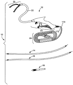
With reference initially to Fig. 1, an exemplary ablation system 10, including
an
exemplary ablation device 12, is illustrated. The ablation systeml0 also
comprise at least
one'guide member (shown with first and second guide members 14, 16) and,
optionally, a
guide member adapter 18. The ablation device 12 may be used alone or with one
or both of
the guide members 14, 16, which may attach or connect to the ablation device
12 and may
pull the ablation device 12 into a desired position where ablation may take
place. The guide
members 14, 16 may also preferably be able to be attached or connected to an
ablation
device, or other device, detached from the device and then reattached. The
guide member
adapter 18 shown may be used and attached to one of the guide members 14 or 16
in order
to allow a guide device (e.g., such as a device described in U.S. Patent
Applications having
Serial Numbers , , having titles "DEVICE AND SYSTEM FOR
SURGICAL DISSECTION AND/OR GUIDANCE OF OTHER MEDICAL DEVICES
INTO BODY" and "METHOD OF SURGICAL DISSECTION AND/OR GUIDANCE OF
OTHER MEDICAL DEVICES INTO BODY" and having Attorney Docket Nos.
MTI0049/US (P-22921.02), and MT10052/US (P-22921.03), all respectively, which
are co-
pending and filed the same day as the present application) to guide or place
the ablation
device 12 in order to perform an ablation procedure (e.g., as shown in Figs.
26, 47 - 66, and
described below).
CA 02638028 2008-07-22
WO 2007/089633 PCT/US2007/002252
-18-
The exemplary embodiment of the ablation device 12 shown in Fig. I generally
comprises; ajaw assembly 20, a flexible neck 22 connecting the jaw assembly 20
to a
handle assembly 24, and a cord assembly 26 attached to the handle assembly 24.
Each of
the general portions of the ablation device 12, and its components, will be
discussed in
detail below.
In order to ablate desired tissue, the tissue is retained or clamped using the
jaw
assembly 20 of the ablation device 12 prior to ablation. Fig. 2 illustrates a
plan view of a
portion of the jaw assembly 20. The portion of the jaw assembly 20 shown
includes a pair
of clamping jaws (right 28a and left 28b) that are primarily mirror images of
each other, and
when in a closed position allow the jaw assembly 20 to clamp tissue. Fig. 2
also includes a
shroud assembly 30 on the distal end of each jaw 28a, 28b, which provide a
means for
attaching and detaching a guide member to and from, respectively, the distal
end of each
jaw 28a, 28b (the details of the guide member will be discussed below).
Additionally, Fig. 2
includes a nose 32 in which the proximal ends of the jaws 28a, 28b, and other
components
used to open and close the jaws 28a, 28b, are housed and assembled. Also in
Fig. 2, ajaw
return spring sleeve 33 is shown positioned over a portion of the nose 32.
Fig. 2 also illustrates the jaws 28a, 28b as being preferably curved, other
shapes are
also possible. The purpose of the curvature illustrated in Fig. 2 is to allow
the jaws 28a, 28b
to.fit around certain anatomical features, such as blood vessels, and to clamp
tissue in a
desired location with respect to such anatomical features.
In order to clamp and release tissue, the jaws 28a, 28b of the jaw assembly 20
preferably move between an open position (as seen in Figs. 1-3) and a closed
position. So
as to move between the open and closed positions, the jaw assembly 20
preferably includes
components as depicted in Fig. 3. Fig. 3 illustrates a top view of a preferred
embodiment of
the jaw assembly 20, shown with the nose 32 and jaw return spring sleeve 33 in
wire frame
or phantom. Preferably, the components of the jaw assembly 20 are configured
so that as
CA 02638028 2008-07-22
WO 2007/089633 PCT/US2007/002252
-19-
the jaws 28a, 28b begin to close, the movement is pivotal or scissor-like, but
as the jaws
28a, 28b move closer to each.other, the jaws 28a, 28b ultimately close in a
parallel
configuration, as shown in the partially closed position of the jaws 28a, 28b
in Fig. 4. By
"scissor-like" it is meant that the orientation of the jaws is angular with
respect to each other
as if from a pivot point when the jaws 28a, 28b are in a generally open
position. As the jaws
28a, 28b begin to be closed and continue to move toward one another, they
pivot with
respect to one another much like a scissor moves until they reach a certain
point at which
they move parallel to one another. By having the jaws 28a, 28b ultimately come
together in
a generally parallel configuration, substantially all of the tissue-contacting
side surface of
the jaws 28a, 28b comes into contact with tissue at about the same time and
may exert a
more even force on the tissue along the length of the tissue-contacting side
surface of the
jaws 28a, 28b. Also, the jaws 28a, 28b are preferably able to float to a
limited degree with
respect to one another as they close or as they are closed together to
facilitate contact with
uneven tissue surfaces as will also be further described below.
A purpose of the jaws 28a, 28b being moveable and being able to both close
(i.e.,
approximate) and open is to clamp and release tissue to be ablated, as
discussed above.
However, another purpose of the approximating jaws 20 is to allow the jaw
assembly 20,
while in a substantially closed position, to be sized and shaped to be able to
pass through a
12 mm or other size of trocar port in a patient during minimally invasive
surgery.
The jaw assembly 20 is preferably configured such that the jaws 28a, 28b are
able to
compensate for a variation in tissue configurations or thicknesses. The design
of the jaw
assembly 20 is preferably configured so that the jaws 28a, 28b close in an
independently
floating fashion. In particular, the floating jaw assembly 20 permits tissue
of varying
thicknesses to be clamped in the jaws 28a, 28b with the jaws 28a, 28b coming
into contact
with tissue generally along their lengths. For example, thicker tissue can be
located closer
CA 02638028 2008-07-22
WO 2007/089633 PCT/US2007/002252
-20-
to the nose 32 than thinner tissue, and the jaws 28a, 28b will not be held
open by the thick
tissue, but will close and contact tissue along their lengths.
Controls for clamping and ablating tissue are located remotely from the jaws
28a,
28b and are preferably located in the handle assembly 24 that may preferably
be handheld.
Fig. 5 shows components that extend distally from the handle assembly 24,
through the neck
22 and to the jaw assembly 20 in order to control clamping and ablation in the
jaw assembly
20, as well as the components of the jaw assembly 20 and neck 22. In the
exemplary
embodiment shown in Fig. 5, components that extend to the jaw assembly 20 -
through the
neck 22 from the handle assembly 24, are two power source wires 34 (e.g.,
radiofrequency
(RF) wires) intertwined with two fluid delivery conduits 36 (e.g., saline
delivery tubes), and
a pull wire 35. Fig. 6 is an exploded view of all the components of the
portion of the
preferred ablation device 12 shown in Fig. 5. Fig. 7 is a close-up view of a
substantial
amount of the exploded jaw assembly 20 shown in Fig. 6. Referring to Figs. 5-
7, the
components of the preferred embodiment shown will be described below. However,
it
should be noted that the described embodiment is preferred and other
variations including
ablation devices powered and/or controlled in other ways as known or developed
that may
include some of the components discussed and/or additional components not
discussed are
also contemplated by the present invention.
Referring to Figs. 5-7, and beginning with the jaw assembly 10, the preferred
jaw
assembly 20 of the present invention includes two jaws 28a, 28b with each jaw
28a, 28b
including a housing 38a, 38b (respectively). The purpose of the housing 38a,
38b is to
house the components necessary to approximate the jaws 28a, 28b and to ablate
tissue
(which will be discussed below). The housings 38a, 38b are preferably made of
an
electrically insulating material, and include at least two channels, each that
run lengthwise,
with a first channel 40a on each jaw 28a, 28b facing each other so as to
contact tissue
between them and a second channel 40b in each housing 38a, 38b facing
oppositely. Jaw
CA 02638028 2008-07-22
WO 2007/089633 PCT/US2007/002252
-21 -
arms 42a, 42b are provided as to fit into the second channels 40b in the jaw
housings 38a,
38b. The jaw arms 42a, 42b are shown retained in the housings 38a, 38b by
electrically
insulated covers 44a, 44b that are held in place in the housings 38a, 38b. The
jaw arms 42a,
42b are controllably moveable and are operatively connected with the housings
38a, 38b and
attached to other jaw assembly 20 components in order to provide controlled
movement to
the jaws 28a, 28b. The jaw arms 42a, 42b include elongate portions 46a, 46b
that are
retained in the second channels 40b of the respective jaw housings 38a, 38b.
Also, as seen
in Fig. 7, the jaw arms 42a, 42b preferably include slots 48a, 48b that are
proximal to the
elongate portions 46a, 46b and that angle towards the interior of the jaw
assembly 20, or
tissue-contacting side of the jaws 28a, 28b, as the slots 48a, 48b extend
proximally. The jaw
arms 42a, 42b also each preferably include a pin 50a, 50b on the proximal end
of each
respective jaw arm 42a, 42b, with the pin 50a extending downward on the right
arm 42a and
the pin 50b extending upward on the left jaw ann 42b in the illustrated
orientation. The
slots 48a, 48b and pins 50a, 50b of the jaw arms 42a, 42b cooperate with other
components
in the jaw assembly 20, which will be discussed below, in order to open and
close the jaws 28a, 28b.
In order to ablate tissue, a fluid assisted elongate electrode assembly is
preferably
provided in the channel 40a in each housing 38a, 38b. The electrode assembly
preferably
comprises an elongate tubular electrode 52a, 52b that is retained in the
channel 40a and as
such are preferably provided within lumens of porous electrode supports 54a,
54b.
Preferably, the elongate tubular electrodes 52a, 52b include a series of fluid
ports (not seen
in Figs.) that are open from an internal fluid passage (not shown) and
oriented toward the
tissue-contacting side of each jaw 28a, 28b so that a conductive fluid may'be
dispensed from
the electrodes 52a, 52b through the series of fluid ports then migrate
laterally through the
pores of the porous electrode support 54a, 54b and around its circumference to
thoroughly
and uniformly wet the porous electrode support 54a, 54b along the right and
left jaws 28a,
CA 02638028 2008-07-22
WO 2007/089633 PCT/US2007/002252
-22-
28b. The conductive fluid (e.g., saline) is preferably provided to each of the
electrodes 52a,
52b through separate fluid delivery conduits 36a, 36b (only end portions of
the fluid
delivery conduits 36a, 36b are shown in Fig. 7).
The elongate tubular electrodes 52a, 52b are preferably formed of thin-walled,
malleable stainless steel tubing extending between a proximal open end 56a,
56b and a
distal, closed end 58a, 58b. The series of fluid ports are formed, e.g., laser
drilling, though
the sidewall of the tubing from a lumen inside and preferably extend in a
single line,
although the fluid ports could be formed in any selected array extending
around the
circumference of the sidewall of the tubing. The electrode supports 54a, 54b
preferably
comprise a porous polymer such as PorexTM plastic.
The elongate tubular electrodes 52a, 52b are flat electrodes that are
preferred
because the flat design allows for more energy to be applied to tlie surface
of tissue,to be
ablated. However, other types and shapes of electrodes or ablating elements
are also
contemplated by the present invention. Other possible ablating elements are
energy transfer
elements that transfer energy to target tissue. For example, energy may be
conductive
elements that may supply RF energy (as shown in Figs), HIFU energy, microwave
energy,
thermal energy, cryogenic energy or ultrasound energy to target tissue. Energy
transfer
elements may be, for example, laser elements for supplying laser light to
target tissue. Two
or more energy transfer elements or conductive elements may be arranged in a
bipolar
arrangement (as shown in Figs.) wherein at least one element is used as a
positive electrode
and at least one element is used as a negative electrode. One or more energy
transfer
elements or conductive elements of the ablation device 12 may be arranged in a
monopolar
arrangement wherein at least one element is used as one electrode and an
indifferent
electrode is placed elsewhere on the patient's body such as the back, thigh or
shoulder or
another site other than the ablation device 12 site.
CA 02638028 2008-07-22
WO 2007/089633 PCT/US2007/002252
-23-
Energy transfer elements or conductive elements may comprise one or more
conductive materials or blends including titanium, titanium alloys, TiNi
alloys, shape
memory alloys, super elastic alloys, aluminum oxide, platinum, platinum
alloys, stainless
steels, stainless steel alloys, MP35N, eigiloy, haynes 25, satellite,
pyrolytic carbon, silver
carbon, conductive metals, conductive polymers or plastics, and/or conductive
ceramics.
Energy transfer elements or conductive elements may not be conductive but may
serve as a
conduit to deliver a conductive material such as a conductive fluid. Energy
transfer or
conductive elements may be porous. For example, energy transfer elements or
conductive
elements may comprise porous polymers, metals, or ceramics. Energy transfer
elements or
conductive elements may be coated with non-stick coatings such as PTFE or
other types of
coatings as discussed herein. In particular, the energy transfer elements may
comprise one
or more coatings, e.g., hydrophilic coatings. Energy transfer elements or
conductive
elements may be flexible thereby allowing them to conform to the surface of
target tissue.
Energy transfer elements or conductive elements may be malleable thereby
allowing a
surgeon to shape them to conform to the surface of target tissue.
Energy transfer elements or conductive elements may comprise one or more metal
conductors such as windings inside a polymer or a conductive mesh material.
The energy
transfer elements or conductive elements may comprise tubes for delivery of
fluids. The
tubes may comprise holes or slots. A polymer tube may be placed inside a metal
tube to
control fluid delivery through energy transfer elements or conductive
elements. One or
more of the energy transfer elements or conductive elements may be used as one
or more
nerve stimulation electrodes and/or as one or more cardiac stimulation
electrodes.
Electrodes may be used for cardiac pacing, defibrillation, cardioversion,
sensing, stimulation
and/or mapping.
Energy transfer elements or conductive elements may comprise needles designed
to
penetrate tissues such as fat and muscle. For example, energy transfer
elements or
CA 02638028 2008-07-22
WO 2007/089633 PCT/US2007/002252
- 24 -
conductive elements may be designed to penetrate fat on the heart thereby
allowing the
energy transfer elements or conductive elements to reach cardiac tissue. The
needles may
allow fluids such as conductive fluids, chemicals such as ablation chemicals,
drugs,
biological agents and/or cells to pass through. The needles may allow a vacuum
or suction
to pass through.
In additional embodiments, the ablation device 12 of the present invention may
include means for tracking the position of the ablation device 12. The means
for tracking
the position of the ablation device 12 may include, for example, sensors and
imaging
devices. An example of a disclosure of such a tracking means is described in
U.S. Patent
Application Publication US 2006/0229594 Al (Francischelli et al.), and is
herein
incorporated by reference in its entirety.
Adhesive may be applied to maintain the elongate tubular electrodes 52a, 52b
and
porous electrode supports 54a, 54b in the channels 40a in the jaw housings
38a, 38b. The
adhesive used may not block migration of conductive fluid around the porous
electrode
supports 54a, 54b.
In order to supply energy or power to the elongate tubular electrodes 52a,
52b,
power source wires 34, in the preferred embodiment, extend distally from a
power source
(preferably separate from ablation device 12) through the neck 22 and are
soldered to the
elongate tubular electrodes 52a, 52b, for example, as shown in Fig. 7 (only
portions of wires
34 shown in Fig. 7), which is preferably at a location where the electrodes
52a, 52b are not
surrounded by electrode supports 54a, 54b.
Other methods of irrigating the electrodes or ablating elements, besides that
method
described above, are also contemplated by the present invention. The purpose
of irrigation
of the electrodes with saline or other conductive fluid is to help decrease
the interface
impedance, cool the tissue, and allow for a greater lesion depth. Irrigation
can also help
prevent tissue or fat from clogging the electrodes and help keep the
electrodes clean.
CA 02638028 2008-07-22
WO 2007/089633 PCT/US2007/002252
- 25 -
Figs. 6 and 7 show other components that cooperate with the jaw arms 42a, 42b
in
order to approximate the jaws 28a, 28b. The figures illustrate two halves 32a,
32b of the
nose 32. The two halves, as shown, preferably have the same shape and are made
to mate or
connect together as shown, and house components used for approximation. Two
identical
pins 60a, 60b are disposed, as shown in Fig. 7, between and attached to the
two halves 32a,
32b of the nose 32. The slot 48a on jaw arm 42a is slidably retained on pin
60a and slot 48b
is slidably retained on jaw arm 42b. The pins 50a, 50b on the jaw arms 42a,
42b are
moveably retained in triangular-shaped openings 62a, 62b on the top and bottom
of a clevis
64 that is moveably retained in the nose 32. The pins 50a, 50b on the jaw arms
42a, 42b are
also moveably retained in openings 51a, 51b in the nose halves 32a, 32b. The
clevis 64, at
its proximal end, is attached to the pull wire 35. From the clevis 64, the
pull wire 35
extends proximally through a distal neck retainer barb 66, which is attached
to the nose
halves 32a, 32b by extensions 68a, 68b on the distal neck retainer barb 66 as
being fitted
within apertures 70a, 70b on the nose halves 32a, 32b. The purpose of the
distal neck
retainer barb 66 is to attach the neck 22 to the nose 32 so that the pull wire
35 moves
relative to the neck 22 and nose 32 as they are operatively fixed together.
In order to close the jaws 28a, 28b while in an open position, the pull wire
35 is
pulled from the proximal portion of the device 12 (how this is performed is
discussed in
more detail below with regard to the handle portion 24), which results in the
clevis 64
moving proximally within a formed interior cavity of the nose 32. As the
clevis 64 is pulled
proximally, it exerts force on the jaw arms 42a, 42b, which are connected to
the clevis 64 by
the pins 50a, 50b. As the jaw arms 42a, 42b are pulled proximally for an
initial distance
within the nose 32, the slots 48a, 48b slide along the pins 60a, 60b in the
nose 32, which
moves the jaws 28a, 28b toward each othe'r in a scissor-like motion with the
pins 60a, 60b
located at an intermediate point within the slots 48a, 48b. At that point, the
jaws 28a, 28b
are preferably substantially parallel as controlled by the shape of the slots
48a, 48b and
CA 02638028 2008-07-22
WO 2007/089633 PCT/US2007/002252
-26-
interaction with the pins 60a, 60b. Once the jaws 28a, 28b are substantially
parallel (but not '
yet closed), further pulling proximally on the clevis 64 pulls the jaws 28a,
28b further
proximally as well. The pins 50a, 50b are extending through the slots 62a, 62b
in the clevis
64 are guided through the slots 51a, 51b in the nose halves 32a, 32b. The
shape of slots
51 a, 51b force the pins 50a, 50b and thus the jaws 28a, 28b to move toward
each other as
the pull wire 35 is further moved proximally relative to the neck 22 and nose
32. At the
same time, the width of slots 62a, 62b of the clevis 64 permit inward movement
of pins 50a,
50b. Also, pins 60a, 60b slide along slots 48a, 48b. The combination of
interactions
between pins 50a, 50b and 60a, 60b, and slots 48a, 48b and 51a, 51b results in
the jaws 28a,
28b moving toward each other in a substantially parallel position until the
jaws 28a, 28b are
in a substantially closed position (contacting each other)_ The slots 51 a,
51b also limit how
far the clevis 64 may move proximally in the nose 32. This arrangement of pins
and slots
also permits the jaws 28a, 28b to float to the degree permitted by the
interaction of the pins -
and slots so that the jaws 28a, 29b can adjust in orientation relative to one
another based
upon counter-pressure applied to the jaws surfaces from the engagement with
tissue.
The pull wire 35 extends from the handle 24 portion through the neck 22 and
into
the jaw assembly 20 through a lumen in the distal neck retainer barb 66. Fig.
6 shows that
the pull wire 35 is surrounded by an incompressible coil 72b, which is then
further
surrounded by a sleeve 72a. A preferred material for the sleeve 72a is
polyimide, although
other materials are also contemplated. The purpose of such a sleeve 72a is to
protect the
pull wire 35 as it is pulled through parts of the jaw assembly 20, and also as
the components
are bent and moved around in the flexible neck 22. Preferably, as shown in
Fig. 6, the
power source wires 34 and fluid delivery conduits 36 are also spirally wound
through the
neck 22 for strain relief.
In order to return the jaws 28a, 28b from a closed position to an open
position, the
jaw assembly 20 includes a jaw return spring 74 (see Fig. 6) (which happens
when no
. ' . .
CA 02638028 2008-07-22
WO 2007/089633 PCT/US2007/002252
-27-
tension is placed on the pull wire 35). Fig. 6 also shows that the jaw return
spring 74 is
preferably held in place surrounding the nose 32 at its proximal end by a
retaining ring 76
that provides bias between the end of the nose 32 and the clevis 64 to move
the clevis 64
distally. The spring 74 is provided in contact with the clevis 64 and exerts
force in a distal
direction on the clevis 64 in order to return the jaws arms 42a, 42b to an
open position.
Also shown in Fig. 6, is a jaw return spring sleeve 78 that covers the jaw
return spring 74 -
and retaining ring 76. Other biasing arrangements with other components and/or
configurations that would also return the jaws 28a, 28b to an open position
are also
contemplated by the present invention.
The pull wire 35 extends proximally in the device 12 from the jaw assembly 20,
through the neck 22 and into the handle 24. As the pull wire 35 enters the
handle 24, the
pull wire 35 is fed through a proximal neck retainer barb 80 (shown on Fig.
6), which
attaches the neck 22 to the handle assembly 24, and the pull wire 35 continues
-into the
handle assembly 24 and attaches at its distal end to a wire terminal 82 (also
shown on Fig.
6). The wire terminal 82 is held in place in the handle 24 using a set screw
84 (Fig. 6).
The pull wire 35 is preferably made of stainless steel, although other
suitable
materials may be used, with a solid wound coil surrounding the pull wire 35.
The preferred
configuration of the pull wire 35 and surrounding coil is an incompressible
coil. Other
suitable materials and/or designs that act as an incompressible coil are also
contemplated by
the present invention. A purpose of the incompressible coil configuration is
to maintain the
overall length of the pull wire 35 when the portion of the pull wire 35 that
extends through
the flexible neck 22 is flexed or twisted etc.
The jaw assembly 20 is functionally connected to the handle assembly 24 by the
neck 22. A purpose of the neck 22 is to provide a shaft or lumen through which
components
(e.g., power source wires 34, fluid delivery conduits 36 and pull wire 35) may
extend
between the jaw assembly 24 and the handle assembly 24. The length of the neck
22 then is
CA 02638028 2008-07-22
WO 2007/089633 PCT/US2007/002252
- 28 -
preferably related to the distance required in a procedure to allow the jaw
assembly 20 to be
at an desired anatomical location with the handle assembly 24 being outside
the body (i.e.,
ex vivo).
The neck 22, which attaches the jaw assembly 20 to the handle 24, is
preferably
flexible or "floppy" in nature. In one embodiment, the neck 22 may be flexible
or floppy
like a rope, for example. The flexible or "floppy" nature may thereby allow a
guide member
or device to be used to easily position the jaw assembly 20 of the ablation
device 12 into a
position to ablate tissue. The flexible nature of the neck 22 enables the
ablation device 12 to
be used with many different anatomies found in different patients. The neck 22
may be
capable of effectively transmitting torque.
Preferably, the neck 22 is made of extruded polyurethane with a 304 stainless
steel
braid. However, other suitable components or designs that provide the desired
flexibility of
the neck 22 are also contemplated by the present invention.
In order to control approximation of the jaws 28a, 28b and application of
ablative
energy, which both take place at or near the jaw assembly 20 of the ablation
device 12
preferably when the jaw assembly 20 is placed at a desired location in a body,
the controls
for approximation and ablation are preferably located ex vivo. Preferably, the
controls are
located in and/or on the handle assembly 24, which remains ex vivo during an
ablation
procedure. Preferably, the handle assembly 24 comprises a handle casing 86
having two
mating handle casing halves (one half of which is shown in Fig. 8 as 86a) for
housing the
other components and for providing a hand piece for the user of the device.
Also,
preferably, the handle assembly 24 may be held in the hand of a user.
As discussed previously, in order to cause the components of the jaw assembly
20
to close thejaws 28a, 28b, the pull wire 35 is pulled proximally using
controls in the handle
assembly 24. Referring to Figs. 8 and 9, in general, in order to pull the pull
wire 35
proximally, ajaw activation lever 122 is squeezed or moved toward the handle
casing 86a
CA 02638028 2008-07-22
WO 2007/089633 PCT/US2007/002252
-29-
(only one half shown) by the user, which results in coordinated and controlled
movement of
various linked components that work together to pull the pull wire 35
proximally. The
handle assembly 24 also includes components that enable the pull wire 35 to be
held in the
proximal position and that enable the movement of the components to be
reversed to allow
for release of the pull wire 35 and opening of the jaws 28a, 28b.
In the preferred embodiment shown in the figures, and in Figs. 8 and 9 in
particular,
the pull wire 35 extends from the neck 22 into the handle casing 86 through
the proximal
neck retainer barb 80, and is connected to the wire terminal 82. Preferably,
the wire
terminal 82 is held in place with the set screw 84. The ends of the wire
terminal 82 are
preferably attached to two rollers-88 that are retained in recesses (one
recess in the handle
housing half 86a, seen in Figs. 8, 15 as 89a) in both halves 86a, 86b (not
shown) of the
handle casing 86, which allow the rollers 88 to rotate and provide
predetennined paths for
the rollers 88. The wire terminal 82 is also placed through an aperture 90 in
a distal end of a
link arm 92, with the aperture 90 being sized and shaped to retain the wire
terminal 82.
In general, a basic purpose of the clutch assembly 94 is to translate the
motion of
the jaw activation lever 122, both toward and away from the handle casing 86,
into
generally proximal and distal, respectively, motion of the link arm 92. The
link arm 92, in
turn, moves the pull wire 35 proximally or distally, which closes or opens the
jaws 28a, 28b,
respectively.
The clutch assembly 94, as shown in Figs. 8-13, generally preferably includes
the
link arm 92 that is connected to the pull wire 35 and which is attached to
other components
of the clutch assembly 94 that pivot around an axle 110 and that are attached
to a cam 104
that may be rotated by movement of the jaw activation lever 122. The clutch
assembly also
preferably includes components that generally allow overdrive slip (i.e.,
components that
comprise an overdrive mechanism) so that, for example, once the jaws 28a, 28b
are closed
around tissue with a certain force, the jaw activation lever 122 may continue
to be squeezed
CA 02638028 2008-07-22
WO 2007/089633 PCT/US2007/002252
-30-
toward the handle casing 86 and the cam 104 rotated in order to, for example,
lock the lever
122 in place, without additional proximal pulling on the pull wire 35 nor
further
approximation of the jaws 28a, 28b. The clutch assembly 94 also preferably
includes a
tension adjuster mechanism by which to adjust the tension in the overdrive
slip to
accommodate different thicknesses of tissue to be ablated, for example.
More particularly, with regard to the components of the clutch assembly 94, in
order
to close the jaws 28a, 28b, the pull wire 35 is pulled proximally as the wire
terminal 82 is
pulled proximally in the recesses (one of which is 89a) by the link arm 92.
The purpose of
allowing the rollers 88 and attached wire terminal 82 to rotate in the
recesses (one of which
is 89a), while the link arm 92 of the clutch assembly 94 moves generally
proximally, is to
prevent bending the pull wire 35 in the handle assembly 24, which could in
turn cause
tension and fracture the pull wire 35 as it extends out through the neck 22
and into the jaw
assembly 20.
In particular, Figs 10-13 show that the clutch assembly 94 includes the link
arm 92
which is attached distally to the wire terminal 82 (as discussed above) and
proximally to a
clutch 96 using a pin 98 and a clip 100 (Fig. 13) with the pin 98 (Figs. 12,
13) extending
through an appropriately sized and shaped aperture 102 on the link arm 92 and
an aperture
(not shown) on the clutch 96, which are both are coaxially aligned. The
purpose of the
clutch 96 is to move the link arm 92, which in turn moves the pull wire 35.
The clutch 96 is
preferably also attached to a clutch (or torsion) spring 106 (shown in Figs. 8-
13).'
Preferably, a rotor 108 is attached to the clutch spring 106 opposite the
clutch 96, with the
rotor 108 including a screw 112 and anchor 114 to adjust the tension in the
clutch spring
106. The cam 104 is attached to the rotor 108. As shown in the figures, the
cam 104
includes a slot 124 into which the jaw activation lever 122 is moveably
retained. There is an
axle 110 running through apertures in the clutch 96, the cam 104 and the rotor
108, with the
axle 110 being held in place using another clip 100.
CA 02638028 2008-07-22
WO 2007/089633 PCT/US2007/002252
-31 -
The clutch spring 106 tension may be adjusted by tightening or loosening the
screw
112 and anchor 114. In particular, in the embodiment shown in the figures,
tightening the
screw 112 will wind the clutch spring 106 tighter.
Referring to Figs. 8, 9, and 14, the handle assembly 24 also comprises the jaw
activation (or closure) lever 122, which includes an extension portion 136
that is moveabiy
attached to the cam 104 of the clutch assembly 94. The exemplary attachment of
the
extension 136 of the lever 122 shown is made by fitting a slot 124 of the cam
104 around a
roller 126 in the extension 136 (Fig. 14), which is placed in a groove 128 in
the extension
136 of the lever 122 and held in place using a pin 130 placed through an
aperture 132 and
two apertures 134 in the extension 136, which are coaxially aligned. The
roller 126 of the
jaw activation lever 122 is then able to roll along the slot 124 in the cam
104, allowing the
two to move with respect to one another, in a predetermined path, while
staying moveably
connected. The lever 122 pivots about a point 121, where the lever 122
attaches to the
handle casing 86. The lever 122 is preferably ergonomically shaped to fit in
the hand of a
user.
In order to activate, or close the jaws 28a, 28b, the lever 122 is squeezed or
otherwise moved toward the handle casing 86. Moving the lever 122 in such a
way results
in the extension portion 136 of the lever 122 moving into the handle casing
86, which in
turn pivots the cam 104 counter-clockwise (as in Figs. 8, 9) which through the
components
of the clutch assembly 94 pivots the clutch 96 counter clockwise (as in Figs.
8, 9). As a
result, the clutch 96 pulls the link arm 92 generally proximally, and the wire
terminal 82
moves proximally as well along the path of the recesses (one is 89a) in the
handle casings
86a, 86b. Accordingly, the pull wire 35, attached to the wire terminal 82, is
pulled
proximally into the handle assembly 24, thereby closing the jaws 28a, 28b.
Fig. 15
illustrates the positions of the handle 24 components when the jaws 28a, 28b
are in a
substantially closed position.
CA 02638028 2008-07-22
WO 2007/089633 PCT/US2007/002252
-32-
With the jaws 28a, 28b in a closed position, the components of the handle
assembly
24 generally resemble Fig. 15. If further force is placed on the lever 122
(i.e., lever ] 22 is
lifted or squeezed farther toward the handle casing 86), the overdrive slip
described above
prevents further tension from being placed on the pull wire 35. However,
preferably, the
lever 122 is moved toward the handle casing 86 further in order to lock the
lever 122 in
place, which in turn locks the jaws 28a, 28b in a locked position. Once the
jaws 28a, 28b
are locked in the closed position, a lockout feature of the present invention
is deactivated,
allowing for ablative energy to be applied. Such a lockout feature will be
discussed in detail
below.
The jaw closure mechanism described above is one exemplary such mechanism. It
is also contemplated by the present invention that the jaws 28a, 28b may be
driven by either
a mechanical mechanism, e.g., a drive cable or wire in a compression jacket, a
hydraulic
mechanism, e.g., a piston powered by fluid pressure, and/or an electrical
mechanism, e.g., a
servo motor. Each of the jaw closure mechanisms described above would allow
neck 22 to
remain flexible or floppy when the jaws 28a, 28b were either in an open
position and/or a
closed position.
In the present invention, preferably the ablation device 12 includes a
mechanism for
preventing inadvertent application of ablative energy, which is referred to as
a lockout
mechanism or feature. In order to avoid inadvertent ablation, the lockout
mechanism is
preferably incorporated into the handle assembly 24. An example of such a
lockout
mechanism is included in the embodiments shown in Figs. 8, 9, and 15, and
functiorns by
preventing an ablative energy source from being activated unless the jaws 28a,
28b are
locked in a substantially closed position.
Before the jaws 28a, 28b are locked in a closed position, some components of
the
handle assembly 24, in the exemplary device 12, prevent ablative energy from
being
applied. In particular, the exemplary embodiment prevents ablative energy from
being
CA 02638028 2008-07-22
WO 2007/089633 PCT/US2007/002252
- 33 -
applied by preventing a trigger 140 on the device 12 from being pulled. The
mechanism for
preventing the trigger 140 from being pulled to apply ablative energy may be
referred to as a
lockout mechanism. In the lockout mechanism illustrated, there is preferably a
visual and/or
tactile lockout flag 142 on or near the trigger 140 that indicates when the
lockout
mechanism is engaged or activated. While the lockout mechanism is activated,
the lockout
flag 142 extends through an aperture in the trigger 140 and can be seen and
felt on the
trigger 140, and when deactivated the lockout flag is recessed in the aperture
in the trigger
.. 140..
Additional components of the exemplary lockout mechanism can be seen
separately
in Figs. 16-18. These components are parts of a power trigger subassembly 138
which
comprises the trigger 140 that is pivotally attached to the lockout flagl42 by
a pin 144. The
lockout flag 142 is attached via a slot 149 (Fig. 18) and a pin 148 that
connects to a lockout
slider 150. The lockout slider 150 is slidably retained in a lockout rail 152
with notches 143
on the sides of the slider 150 and channels 145 on the sides of the rail 152
in which the
notches 143 may slide and a spring 154 between the rail 152 and slider 150,
holding them
apart on the proximal end of the power trigger subassembly 138. Also, on the
proximal end
of the rail 152, there is an extension or tail 156, which may depress a power
switch to turn
on the ablative energy source_
The power trigger subassembly is incorporated into the remainder of the handle
assembly 24 as seen in Figs. 8, 9, and 15. The pin 144 that allows the trigger
140 to pivot
with respect to the lockout flag 142 is also connected to the two handle
casing halves (one
half of which is shown as 86a). Also, bosses 147 on the outer sides of the
rail 152 are
connected to the handle casing halves (one is 86a) such that the rail may
rotate or tilt with
respect to the handle casing (one half of which is 86a). The components,
therefore,'
generally allow the slider 160 to move proximally and distally within the rail
that is attached
to the handle casing., The slider 150 may pull the lockout flag 142 proximally
which
CA 02638028 2008-07-22
WO 2007/089633 PCT/US2007/002252
-34-
retracts the flag 142 into the trigger 140 and allows the trigger 140 to be
free to rotate about
pin 144. When the trigger 140 is free to rotate about pin 144, it may then be
pulled or
depressed such that the trigger 140 pushes up on the slider 150, which in turn
causes the rail
152 to pivot or rotate about the bosses 147 such that the extension or tail
156 on the rail 152
may press on the power switch 164 to activate ablative energy application. By
releasing the
trigger 140, a torsion spring 162 pushes down on the rail 152 which pivots or
rotates about
the bosses 147. This causes the slider 150 to push down on the trigger 140,
which will
rotate about pin 144, which in turn causes the extension or tail 156 on the
rail 152 to release
pressure on the power switch 164 with further deactivates ablative energy
application.
In order to move the slider 150 proximally to cause deactivation of the
lockout
mechanism, referring to Figs. 8 and 15, the extension 136 of the jaw
activation lever 122
moves through slot 151 in the slider 150 (as lever 122 is squeezed) until
proximal surfaces
137 of the extension 136 contact the proximal surfaces 153 in slot 151 in the
slider 150,
which moves the slider 150 proximally with respect to the rail 152 and in turn
pulls the
lockout flag 142 proximally so that the lockout flag 142 is recessed in the
trigger 140.
When the lockout flag 142 is recessed enough in order for the trigger 140 to
be
depressed, the lever 122 is also locked into the handle casing (one half of
which is 86a). In
the exemplary embodiment shown, a pawl 158 is attached to the handle casing
(one half of
which is 86a) and extends through slot 151 in the slider 150. The pawl 158
also has a
tension spring 160 attached proximally. The lever 122 may be locked in the
squeezed
position when as the extension 136 is moving into the handle casing (one half
of which is
86a) the pawl 158 catches on a projection 196 in the extension 136, which can
be seen in the
cross section of Fig. 19. With the pawl 158 caught on the projection 196, and
with the
lockout mechanism deactivated as described above, the trigger 140 may be
depressed, which
causes the rail 152 to pivot such that the extension 156 on the rail 152
depresses the power
CA 02638028 2008-07-22
WO 2007/089633 PCT/US2007/002252
- 35 -
switch 164. The power switch 164 is preferably connected to a power source
that is
preferably located remotely from the handle assembly 24.
In order to release the jaw activation lever 122, open the jaws 28a, 28b on
the jaw
assembly 20, and reactivate the lockout mechanism, a lever'release button 192
(Fig. 14)
disposed in the lever 122 is pressed, squeezed or otherwise moved proximally
into the lever
122. A cross-section of a portion of the handle portion 24 is shown in Fig. 20
showing what
happens after the lever release button 192 has pushed the pawl 158 proximally
so it is not
caught on the projection 196 on the lever 122. As a result, the pawl 158 is
pushed
proximally and releases the jaw activation lever 122. The jaw activation lever
122 then
moves away from the handle casing (one half of which is 86a) and the jaws 28a,
28b, are
allowed to open. Distal surfaces 135 of extension 136 maintain contact with
distal surfaces
157 of slider 150 and drive the slider 150 distally, which pushes the lockout
flag 142 and
causes it to pivot about the axis of pin 144. As a result, the lockout flag
142 extends
through the aperture 146 in the trigger 140. In the preferred embodiment
shown, the
presence of the lockout flag 146 in such a position indicates both visually,
as well as
tactilely, to a user that the lockout mechanism is engaged and that ablative
energy may not
be applied.
The lockout mechanism illustrated in the figures and described above is one
example of such a mechanism that prevents ablating energy from being
inadvertently
applied at an undesired location in a body. Other lockout mechanisms that
prevent such
inadvertent or accidental application of ablating energy at an undesired
location in a body
are also contemplated by the present invention. For example, it is
contemplated that a
lockout mechanism may be controlled through feedback from any number of
sensors on the
device, and in particular on the jaws of the device. Such sensors, could for
example, sense
whether or not they are clamped on desired tissue, which could in turn
deactivate the
lockout mechanism and allow ablative energy to be turned off and on. Any
suitable
CA 02638028 2008-07-22
WO 2007/089633 PCT/US2007/002252
-36-
feedback mechanisms are contemplated by the present invention for use in a
lockout
mechanism.
In order to supply power and fluid to the fluid assisted elongate electrode
assembly
that is preferably part of the ablation device 12, power source wires 34 and
fluid delivery
conduits 36 need to extend from a power source and a fluid source through the
handle
assembly 24, neck 22 and into the jaw assembly 20. There is discussion above
of the
preferred route for the power source wires 34 and fluid delivery conduits 36
through the
neck 22 and jaw assembly 20. In the handle assembly 24, a preferred route of
the power
source wires 34 and fluid delivery conduits 36 is illustrated in Fig. 21. As
shown in Fig. 21,
the handle halves (86a only shown) may be provided with a series of laterally
extending,
perpendicular intemal walls 168 that may include slots and/or recesses for
routing power
source wires 34 and fluid delivery conduits 36 and that extend through the
handle casing 86.
The power source wires 34 and fluid delivery conduits 36 are routed from the
proximal end
of the handle assembly 24 to the distal end, where they travel through
apertures in the
proximal neck retainer barb 80, where they may continue on to the neck 22 and
jaw
assembly 20.
The power source and fluid source are preferably located remotely from the
ablation
device 12. As seen in Fig. 1, a cord assembly 26 is attached to the handle
assembly 24
through which to provide the power and fluid. In the cord assembly 26, a power
source
supply cord 172 retains the power source wires 34, and a fluid source supply
cord 170
retains the fluid delivery conduits 36.
Fig. 22 shows the cord assembly 26 with a portion of the cords 170, 172
removed.
It provides a closer view of connectors for the fluid and power sources. A
fluid connector
174, such as that shown, preferably connects the fluid cord 170 to a fluid
source. The
female connector 174 is preferably a female luer, as shown. The fluid source
may be a
standard IV tubing system. In addition, the ablation device preferably
includes a
CA 02638028 2008-07-22
WO 2007/089633 PCT/US2007/002252
-37-
mechanism or device for controlling the amount of and the application of a
fluid, such that
the fluid is properly applied for fluid assisted ablation. A power connector
176 is also
shown in Fig. 22, and preferably connects the power cord 172 to a power
source. The cord
assembly 26, and all components, shown and described are exemplary, and other
suitable
alternatives for delivering power and fluid to the handle assembly 24 are also
contemplated
by the present invention.
The ablation device 12 may incorporate one or more switches to facilitate
regulation
of one or more components or features of ablation device 12 by the operator.
For example,
one or more switches may control the supply of irrigation fluid and/or
ablation energy to the
jaw assembly 20 of ablation device 12. The one or more switches may be, for
example, a
hand switch, a foot switch and/or a voice-activated switch comprising voice-
recognition
technologies. The one or more switches may be incorporated on and/or in handle
24 of
ablation device 12.
In the preferred embodiment shown in the figures, a power source switch 164
(e.g.,
RF switch) is included in the handle assembly 24 (Fig. 9). In order to supply
power to the
power source switch, and in order to activate or deactivate a remote power
source from the
power switch 164, there is preferably a set of wires 166 extending from the
switch 164, out
the proximal end of the handle assembly 24, and to a power source. Fig. 21
illustrates some
exemplary wires 166 leading from the power switch 164, which may extend
proximally
through the power cord 172 to the power source.
Although not illustrated in the figures, the ablation device 12 may include
one or
more sensors or sensing elements to monitor one or more components or
features. For
example, preferably, the ablation device 12 may have the capability to monitor
transmurality
of ablation lesions. An example of a preferred algorithm used to monitor
transmurality is
disclosed in co-pending Provisional Patent Application, having Serial Nurnber
60/832,242,
and is incorporated herein by reference in its entirety.
CA 02638028 2008-07-22
WO 2007/089633 PCT/US2007/002252
-38-
The ablation device 12 described above may, preferably, be part of a system 10
(Fig. 1) for guiding the ablation device 12 to a desired location in a body.
Other
components of such a system 10 may comprise the first and second guide members
14, 16
and the guide member adapter 18. Detail about the guide members 14, 16 and the
guide
member adapter 18 is provided below with regard to the discussion of methods
of using the
ablation device 12 and methods of guiding the ablation device 12 into a
location in a body.
As part of a system 10 for guiding the ablation device 12 to a desired
location in a
body, the ablation device may include different jaw assemblies 20 for
attachment to the
neck 22 of the device 12. In particular, different jaw assemblies 20 that may
be provided in
such a system 10 may have jaws 28a, 28b with different curvatures or shapes. A
purpose of
having such differentjaw assemblies is to accommodate different ablation
procedures at
different anatomical locations, as well 'as to accommodate the differing
anatomy of
individual patients. Figs. 23 and 24 show side views of two different
embodiments of the
jaw assembly 20 of the present invention. The two illustrative embodiments
show clamping
jaws 28a, 28b having different curvatures. As described above, a purpose of
different
curvatures may be to accommodate different ablation procedures. For example,
the
embodiment of the clamping jaws 28a, 28b (28b only shown) in Fig. 23 is
preferably suited
for a box lesions approach to pulmonary antrum isolation, as shown in Fig. 25
(on heart
174). Moreover, the embodiment of the clamping jaws 28a, 28b (28b only shown)
shown in
Fig. 24, which includes more curvature than those in Fig. 23, is preferably
suited for a
encircling island lesions approach to pulmonary antrum isolation, as shown in
Fig. 26. The
purpose of having clamping jaws 28a, 28b with more curvature for the
encircling island
lesions approach (Fig. 26) is to allow the clampingjaws 28a, 28b to be placed
on tissue
close to where the pulmonary veins end and the left atrium begins. Both the
box lesions and
encircling island lesions approaches will be described in more detail below
with regard to
the methods described below. In Figs. 25 and 26, although two sets of clamping
jaws 28a,
CA 02638028 2008-07-22
WO 2007/089633 PCT/US2007/002252
-39-
28b are shown clamped or closed around or near both sets of pulmonary veins,
this would
preferably not be done simultaneously while a heart is off-pump. Generally,
ablation of
only one set of pulmonary veins is performed at a time so that blood flow is
not occluded in
the other set of pulmonary veins.
The ablation device 12 and/or system 10 inay be used in ablation procedures in
various areas in a body where ablation of tissue is desired. In particular,
the ablation device
12 and sy'stem 10 is suitable for use in pulmonary antrum isolation. As
described above
there are different surgical approaches to pulmonary antrum isolation. With
reference to
Figs. 27-64, detail regarding use of the ablation device 12 and/or system 10,
in accordance
with the present invention, in both box lesions and encircling island lesions
approaches to
pulmonary antrum isolation is provided below. Figs. 27-30, 35-37, 41-47, 50-64
are
schematic illustrations, and may not be anatomically correct or drawn to
scale. The figures
are provided to help in understanding methods of the present invention.
Figs. 27-46 illustrate steps in a method ofusirig the ablation device 12
and/or
guiding the ablation device 12, with the surgical approach being a box lesions
approach.
Fig. 27 illustrates schematically a pulmonary ostium 176 with the view being
from the
posterior side of a body and heart. The pulmonary ostium 176 includes two sets
of
pulmonary veins, right 180 and left 178. Fig. 28 shows a step in a method of
guiding and
using an ablation device 12, in accordance with the present invention, in
which a first guide
member 14 is inserted posterior to the upper right and left pulmonary veins.
The guide
member 14 may be inserted through incisions in a minimally invasive procedure,
for
example. Four incisions or ports may be necessary in the procedure, allowing
access to the
pulmonary ostium 176 from four directions. The guide member, in this step and
any other
step described below, may be placed posterior to the upper right and left
pulmonary veins
using any known or future developed technique and/or device. Fig. 29 shows a
subsequent
step in the method in which a second guide member 16 is inserted posterior to
the lower
CA 02638028 2008-07-22
WO 2007/089633 PCT/US2007/002252
- 40 -
right and left pulmonary veins. Fig. 30 shows another subsequent step in which
an ablation
device 12 is attached at both distal ends of the jaws 28a, 28b to the first 14
and second 16
guide members.
The guide members 14, 16 or device may comprise a length of single or multi-
lumen tubing, for example. An active guide connection may be included which
has a
connector member or device 216, e.g., a ball-in socket fitting, a lure fitting
and/or a suture,
located at one or more ends of the guide members 14, 16, for connection
between the distal
end portion of an ablation device (shroud 30). The guide members 14, 16 may
include
reference markings, to provide, for eicample, depth or length references. The
guide member
14 or 16 may comprise two or more lengths of tubing, and the separate tubing
sections may
be color coded to facilitate differentiation between each other. In one
embodiment, the
guide member 14 or 16 may be used to safely pull the jaws 28a, 28b of the
ablation device
12 into place if the neck 22 of the ablation device 12 is loose or floppy,
e.g., the user cannot
actively push or poke the jaws 28a, 28b into tissue, thereby causing
undesirable tissue
damage. The guide member 14 or 16 may include one or more blunt ends. The
guide
member 14 or 16 may include a suture on its distal end. Fig. I includes the
guide members
14, 16 with sutures 15 on both ends. The suture(s) may be made of any suitable
suture
material. The purpose of the suture is to allow another instrument (e.g.,
forceps) to easily
grab the suture on the end of the guide member 14 or 16 and pull the guide
member 14 or 16
into a desired location. Also, the guide member 14 or 16 may include a wire
backbone and
an active guide connector.
In order to attach the first and second guide members 14, 16 to the distal
ends of the
jaws 28a, 28b of the ablation device 12, an attachment rrieans, such as that
illustrated in
Figs. 31-33 may be used. Fig. 31 shows the distal end of ajaw, which is called
a shroud 30.
The end of the guide member 14 includes a barb 15 that is shown in Fig. 32 as
being fit into
a socket 31 in the shroud. Preferably, the barb will fit into the socket 31
when the guide
CA 02638028 2008-07-22
WO 2007/089633 PCT/US2007/002252
-41-
member 14 or 16 is held at an angle and maneuvered into the socket. Fig. 33
shows the
final connected configuration. Fig. 34 shows another embodirnent of the shroud
30 in which
a covering 33 to the shroud 30 is provided. A purpose of the shroud 30 may be
to prevent
the barb 15 and other parts of the shroud 30 from catching on tissue as the
ablation device
12 and guide member 14 are pulled through a body.
Fig. 35 illustrates a subsequent step in the method, in which the ablation
device 12
is pulled into place with the jaws 20 surrounding the left set of pulmonary
veins 178. The
ablation device 12 is pulled into place by pulling the ends of the guide
members 14, 16,
which are opposite the ends to which the ablation device 12 is attached, out
through the
incisions or ports on the right side of the body. The next step, not
illustrated, is to clamp the
jaws 28a, 28b of the ablation device 12 on the surrounded tissue and activate
the ablative
energy to cause ablation. Fig. 36 illustrates a subsequent step in which the
jaws 28a, 28b are
opened and a lesion 182 can be seen that encircles the left pair of pulmonary
veins 178. The
next step, as in Fig. 37, is to withdraw the ablation device 12 back out of
the body. The
guide members 14 and 16 still attached. In order to remove the guide members
14, 16, the
method depicted in Figs. 38-40 may be used. As can be seen in Figs. 38-40, the
guide
member 14, may be moved toward the interior of the jaws 28a, 28b, as indicated
by arrow in
Fig. 38. The guide member 14 may be removed from the shroud 30 after the guide
member
14 is moved or tilted inward a certain degree. Fig. 41 illustrates the
subsequent step after
removal of the ablation device 12, in which the guide members 14, 16 are
returned to their
starting position, and the lesion 182 remains. The steps for ablating the left
two pulmonary
veins are then repeated on the left side of pulmonary ostium 176 (in Figs. 42-
45), and the
result is the pulmonary ostium, as seen in Fig. 46, with two overlapping
lesions 182, 184.
Fig. 49 illustrate steps in a method of using the ablation device 12 and
guiding the
ablation device 12 with a dissector/guide 186, with the surgical approach
being a encircling
island lesions approach. In a first step of the method, the dissector/guide
186 is inserted into
CA 02638028 2008-07-22
WO 2007/089633 PCT/US2007/002252
- 42 -
a port of incision for minimally invasive procedures (but may also be used for
open
procedures). An exemplary, preferred device is disclosed in co-pending U.S.
Patent
Application having Serial No. , filed on the same day as the present
application,
entitled "DEVICE AND SYSTEM FOR SURGICAL DISSECTION AND/OR GUIDANCE
OF OTHER MEDICAL DEVICES INTO BODY" and having Attorney Docket No.
MTI0049/US (P-22921.02), and is incorporated herein by reference in its
entirety. Fig. 47
shows the dissector/guide 186 articulated around the left pair of pulmonary
veins 178. As
described in the co-pending application described immediately above, a guide
wire may be
fed through the dissector/guide 186, and extended out another incision or port
from one used
for entry of the dissector/guide 186. The guide wire may then be attached to
the guide
member 14, and withdrawn back through the dissector/guide 186, which may pull
the guide
member 14 into contact with the distal end of the dissector/guide 186, as
shown in Fig. 50.
In the system 10 of the present invention, the guide member may have a guide
member
adapter 18 attached to an end of the guide member 14 in order to allow the
guide wire to be
attached. In Figs. 48-49, a guide wire adapter is shown, and with a guide
member 14
inserted into the guide member adapter 18, which allows the guide wire to be
attached to
guide member 14 (through adapter 18). The next step is shown in Fig. 51, in
which the
dissector/guide 186 is withdrawn through its port of entry and pulls the
attached guide
member into the desired location surrounding the pulmonary veins as shown. In
a
subsequent step, the dissector/guide 186 is removed from the guide member 14,
leaving the
guide member 14 in place (Fig. 52). A subsequent step, shown in Fig. 53
illustrates an
ablation device 12 being attached by one of its clampingjaws 28a to the guide
member 14..
The ablation device 12, next, is pulled into place around the pair of
pulmonary veins 178 by
pulling on the guide member 14. Thejaws 28a, 28b are closed and ablative
energy is
applied to form a lesion 188, which can be seen in Fig. 55 after the jaws 28a,
28b were
CA 02638028 2008-07-22
WO 2007/089633 PCT/US2007/002252
- 43 -
opened. After removal of the guide member 14 and ablation device 12, the
lesion remains
on the left side of the pulmonary ostium 176 (Fig. 56).
The steps for ablating the right two pulmonary veins are then repeated on the
right
side of pulmonary ostium 176 (in Figs. 57-63), and the result is the pulmonary
ostium, as
seen in Fig. 64, with two lesions 188, 190.
The ablation system 10 and its components are preferably made of biocompatible
materials such as stainless steel, biocompatible epoxy or biocompatible
plastic. Preferably,
a biocompatible material prompts little allergenic response from the patient's
body and is
resistant to corrosion from being placed within the patient's body.
Furthermore the
biocompatible material preferably does not cause any additional stress to the
patient's body,
for example, it does not scrape detrimentally against any element within the
surgical cavity.
It will be appreciated by those skilled in the art that while the invention
has been
described above in connection with particular embodiments and examples, the
invention is
not necessarily so limited, and that numerous other embodiments, examples,
uses,
modifications and departures from the embodiments, examples and uses are
intended to be
encompassed by the claims attached hereto. The entire disclosure of each
patent and
publication cited herein is incorporated by reference, as if each such patent
or publication
were individually incorporated by reference herein.