Note: Descriptions are shown in the official language in which they were submitted.
CA 02638902 2013-03-07
78349-15
1
HUMAN MONOCLONAL ANTIBODIES TO FUCOSYL-GMI AND METHODS FOR
USING ANTI-FUCOSYL-GM1 ANTIBODIES
Cross-Reference to Related Applications
This application claims priority of U.S. Provisional Application Serial No.
60/748,915,
filed on December 8, 2005.
Background
Fucosyl-GM1 is a sphingolipid monosialoganglioside composed of a ceramide
lipid
component, which anchors the molecule in the cell membrane, and a carbohydrate
component
tha is exposed at the cell surface. Carbohydrate antigens are the most
abundantly expressed
antigens on the cell surface of cancers (Feizi T. (1985) Nature n4:53-7). In
some tumor types,
such as small cell lung cancer (SCLC), initial responses to chemotherapy are
impressive, but
chemo-refractory relapses rapidly follows. Intervention with novel
immunotherapeutics may
succeed in overcoming drug resistant relapse (Johnson DH. (1995) Lung Cancer
12 Sunni 3:S71-
5). Several carbohydrate antigens, such as gangliosides GD3 and GD2, have been
shown to
function as effective targets for passive immtmotherapy with MAbs (hie RF and
Morton DL.
(1986) PNAS 83:8694-8698; Houghton AN et al. (1985) PNAS 82:1242-1246).
Ganglioside
antigens have also been demonstrated to be effective targets for active
inununotherapy with
vaccines in clinical trials (Krug LM etal. (2004) Clinical Cancer Research
10:6094-6100;
Dickler MN et al. (1999) Clinical Cancer Research 5:2773-2779; Livingston PO
et al. (1994).1
Clin Onco1.12:1036-44). Indeed, serum derived from SCLC patients who developed
antibody
titers to Fucosyl-GM1 following vaccination with KLH conjugated antigen,
demonstrated
specific binding to tumor cells and tumor specific complement dependant
cytotoxicity (CDC).
Anti-Fucosyl-GM1 titer associated toxicities were mild and transient and three
patients with
limited-stage SCLC were relapse-free at 18, 24, and 30 months (Krug et al.,
supra; Dickler etal.,
supra).
Fucosyl-GM I expression has been shown in a high percentage of SCLC cases and
unlike
other ganglioside antigens, Fucosyl-GM1 has little or no expression in normal
tissues (Nilsson et
CA 02638902 2008-06-05
WO 2007/067992
PCT/US2006/061817
al. (1984) Glycoconjugate J1:43-9; Krug etal., supra; Brezicka etal. (1989)
Cancer Res
49:1300-5; Zhangyi etal. (1997) Int J Cancer 73:42-49; Brezicka et al. (2000)
Lung Cancer
2829-36; Fredman etal. (1986) Biochim Biophys Acta 875: 316-23; Brezicka et
al. (1991)
APMIS 99:797-802; Nilsson etal. (1986) Cancer Res 46:1403-7). The presence of
Fucosyl-GM1
has been demonstrated in culture media from SCLC cell lines, in tumor extracts
and serum of
nude mouse xenografts and in the serum of SCLC patients with extensive-stage
disease
(Vangsted et al. (1991) Cancer Res 51:2879-84; Vangsted etal. (1994) Cancer
Detect Prey
18:221-9). These reports provide convincing evidence for Fucosyl-GM1 as a
highly specific
tumor antigen, which may be targeted by an irrununotherapeutic.
Accordingly, agents that recognize Fucosyl-GM1, and methods of using such
agents, are
desired.
Summary
The present disclosure provides isolated monoclonal antibodies, in particular
human
monoclonal antibodies, that bind to Fucosyl-GM1 and that exhibit numerous
desirable
properties. These properties include high affinity binding to Fucosyl-GM1 and
binding to the
human small cell lung cancer cell line DMS-79 (Human SCLC ATCC # CRL-2049).
Also
provided are methods for treating a variety of Fucosyl-GM1 mediated diseases
using the
antibodies and compositions of this disclosure.
In one aspect, this disclosure pertains to an isolated monoclonal antibody, or
an antigen-
binding portion thereof, wherein the antibody (a) specifically binds to
Fucosyl-GM1; and (b)
binds to the human small cell lung cancer cell line DMS-79 (Human SCLC ATCC #
CRL-2049).
Preferably the antibody is a human antibody, although in alternative
embodiments the
antibody can be a murine antibody, a chimeric antibody or humanized antibody.
In another embodiment, this disclosure provides an isolated monoclonal
antibody, or
antigen binding protion thereof, wherein the antibody cross-competes for
binding to Fucosyl-
GM1 with a reference antibody, wherein the reference antibody (a) specifically
binds to Fucosyl-
GM1; and (b) binds to the human small cell lung cancer cell line DMS-79 (Human
SCLC ATCC
# CRL-2049). In certain embodiments, the reference antibody comprises: (a) a
heavy chain
variable region comprising the amino acid sequence of SEQ ID NO:!; and (b) a
light chain
2
CA 02638902 2008-06-05
WO 2007/067992
PCT/US2006/061817
variable region comprising the amino acid sequence of SEQ ID NO:7. In further
embodiments,
the reference antibody comprises: (a) a heavy chain variable region comprising
the amino acid
sequence of SEQ ID NO:2; and (b) a light chain variable region comprising the
amino acid
sequence of SEQ ID NO:8. In other embodiments, the reference antibody
comprises: (a) a heavy
chain variable region comprising the amino acid sequence of SEQ ID NO:3; and
(b) a light chain
variable region comprising the amino acid sequence of SEQ ID NO:9. In still
further
embodiments, the reference antibody comprises: (a) a heavy chain variable
region comprising
the amino acid sequence of SEQ ID NO:4; and (b) a light chain variable region
comprising the
amino acid sequence of SEQ ID NO:! 0. In yet further embodiments, the
reference antibody
comprises: (a) a heavy chain variable region comprising the amino acid
sequence of SEQ ID
NO:5; and (b) a light chain variable region comprising the amino acid sequence
of SEQ ID
NO:11. In even further embodiments, the reference antibody comprises: (a) a
heavy chain
variable region comprising the amino acid sequence of SEQ ID NO:6; and (b) a
light chain
variable region comprising the amino acid sequence of SEQ ID NO:12.
In one aspect, this disclosure pertains to an isolated monoclonal antibody, or
an antigen-
binding portion thereof, comprising a heavy chain variable region that is the
product of or
derived from a human VH 3-48 gene, wherein the antibody specifically binds
Fucosyl-GMl.
This disclosure further provides an isolated monoclonal antibody, or an
antigen-binding portion
thereof, comprising a light chain variable region that is the product of or
derived from a human
VK L15 gene, wherein the antibody specifically binds Fucosyl-GM1 .
A preferred combination comprises:
(a) a heavy chain variable region CDR1 comprising SEQ ID NO:13;
(b) a heavy chain variable region CDR2 comprising SEQ ID NO:19;
(c) a heavy chain variable region CDR3 comprising SEQ ID NO:25;
(d) a light chain variable region CDR1 comprising SEQ ID NO:31;
(e) a light chain variable region CDR2 comprising SEQ ID NO:37; and
(f) a light chain variable region CDR3 comprising SEQ ID NO:43.
Another preferred combination comprises:
(a) a heavy chain variable region CDR1 comprising SEQ ID NO:14;
(b) a heavy chain variable region CDR2 comprising SEQ ID NO:20;
(c) a heavy chain variable region CDR3 comprising SEQ ID NO:26;
3
CA 02638902 2008-06-05
WO 2007/067992
PCT/US2006/061817
(d) a light chain variable region CDR1 comprising SEQ ID NO:32;
(e) a light chain variable region CDR2 comprising SEQ ID NO:38; and
(f) a light chain variable region CDR3 comprising SEQ ID NO:44.
Another preferred combination comprises:
(a) a heavy chain variable region CDR1 comprising SEQ ID NO:15;
(b) a heavy chain variable region CDR2 comprising SEQ ID NO:21;
(c) a heavy chain variable region CDR3 comprising SEQ ID NO:27;
(d) a light chain variable region CDR1 comprising SEQ ID NO:33;
(e) a light chain variable region CDR2 comprising SEQ ID NO:39; and
(f) a light chain variable region CDR3 comprising SEQ ID NO:45.
Another preferred combination comprises:
(a) a heavy chain variable region CDR1 comprising SEQ ID NO:16;
(b) a heavy chain variable region CDR2 comprising SEQ ID NO:22;
(c) a heavy chain variable region CDR3 comprising SEQ ID NO:28;
(d) a light chain variable region CDR1 comprising SEQ ID NO:34;
(e) a light chain variable region CDR2 comprising SEQ ID NO:40; and
(f) a light chain variable region CDR3 comprising SEQ ID NO:46.
Another preferred combination comprises:
(a) a heavy chain variable region CDR1 comprising SEQ ID NO:17;
(b) a heavy chain variable region CDR2 comprising SEQ ID NO:23;
(c) a heavy chain variable region CDR3 comprising SEQ ID NO:29;
(d) a light chain variable region CDR1 comprising SEQ ID NO:35;
(e) a light chain variable region CDR2 comprising SEQ ID NO :41; and
(f) a light chain variable region CDR3 comprising SEQ ID NO:47.
Another preferred combination comprises:
(a) a heavy chain variable region CDR1 comprising SEQ ID NO:18;
(b) a heavy chain variable region CDR2 comprising SEQ ID NO:24;
(c) a heavy chain variable region CDR3 comprising SEQ ID NO:30;
(d) a light chain variable region CDR1 comprising SEQ ID NO:36;
(e) a light chain variable region CDR2 comprising SEQ ID NO:42; and
(f) a light chain variable region CDR3 comprising SEQ ID NO:48.
4
CA 02638902 2008-06-05
WO 2007/067992
PCT/US2006/061817
Other preferred antibodies of this disclosure, or antigen binding portions
thereof comprise:
(a) a heavy chain variable region comprising the amino acid sequence of SEQ ID
NO:1;
and
(b) a light chain variable region comprising the amino acid sequence of SEQ ID
NO:7.
Another preferred combination comprises:
(a) a heavy chain variable region comprising the amino acid sequence of SEQ ID
NO:2;
and
(b) a light chain variable region comprising the amino acid sequence of SEQ ID
NO:8.
Another preferred combination comprises:
(a) a heavy chain variable region comprising the amino acid sequence of SEQ ID
NO:3;
and
(b) a light chain variable region comprising the amino acid sequence of SEQ ID
NO:9.
Another preferred combination comprises:
(a) a heavy chain variable region comprising the amino acid sequence of SEQ ID
NO:4;
and
(b) a light chain variable region comprising the amino acid sequence of SEQ ID
NO:10.
Another preferred combination comprises:
(a) a heavy chain variable region comprising the amino acid sequence of SEQ ID
NO:5;
and
(b) a light chain variable region comprising the amino acid sequence of SEQ ID
NO:11.
Another preferred combination comprises:
(a) a heavy chain variable region comprising the amino acid sequence of SEQ ID
NO:6;
and
(b) a light chain variable region comprising the amino acid sequence of SEQ ID
NO:12.
The antibodies of this disclosure can be, for example, full-length antibodies,
for example
of an IgG1 or IgG4 isotype. Alternatively, the antibodies can be antibody
fragments, such as Fab
or Fab'2 fragments, or single chain antibodies.
This disclosure also provides an immunoconjugate comprising an antibody of
this
disclosure, or antigen-binding portion thereof, linked to a therapeutic agent,
such as a cytotoxin
or a radioactive isotope. This disclosure also provides a bispecific molecule
comprising an
antibody, or antigen-binding portion thereof, of this disclosure, linked to a
second functional
5
CA 02638902 2008-06-05
WO 2007/067992
PCT/US2006/061817
moiety having a different binding specificity than said antibody, or antigen
binding portion
thereof.
Compositions comprising an antibody, or antigen-binding portion thereof, or
immunoconjugate or bispecific molecule of this disclosure and a
pharmaceutically acceptable
carrier are also provided.
Nucleic acid molecules encoding the antibodies, or antigen-binding portions
thereof, of
this disclosure are also encompassed by this disclosure, as well as expression
vectors comprising
such nucleic acids and host cells comprising such expression vectors.
Moreover, this disclosure
provides a transgenic mouse comprising human immunoglobulin heavy and light
chain
transgenes, wherein the mouse expresses an antibody of this disclosure, as
well as hybridomas
prepared from such a mouse, wherein the hybridoma produces the antibody of
this disclosure.
In yet another aspect, this disclosure provides a method of treating or
preventing a
disease characterized by growth of tumor cells expressing Fucosyl-GM1,
comprising
administering to the subject the antibody, or antigen-binding portion thereof,
of this disclosure in
an amount effective to treat or prevent the disease. The disease can be, for
example, cancer, e.g.,
lung cancer (including small cell lung cancer).
In a preferred embodiment, this disclosure provides a method of treating
cancer in vivo
using an anti-Fucosyl-GM1 antibody. The anti-Fucosyl-GM1 antibody may be a
murine,
chimeric, humanized or human antibody. Examples of other cancers that may be
treated using
the methods of this disclosure include lung cancer, including small cell lung
cancer and non-
small cell lung cancer, renal cancer (e.g., renal cell carcinoma),
glioblastoma, brain tumors,
chronic or acute leukemias including acute lymphocytic leukemia (ALL), adult T-
cell leukemia
(T-ALL), chronic myeloid leukemia, acute lymphoblastic leukemia, chronic
lymphocytic
leukemia, lymphomas (e.g., Hodgkin's and non-Hodgkin's lymphoma, lymphocytic
lymphoma,
primary CNS lymphoma, T-cell lymphoma, Burkitt's lymphoma, anaplastic large-
cell
lymphomas (ALCL), cutaneous T-cell lymphomas, nodular small cleaved-cell
lymphomas,
peripheral T-cell lymphomas, Lennert's lymphomas, immunoblastic lymphomas, T-
cell
leukemia/lymphomas (ATLL), entroblastic/centrocytic (cb/cc) follicular
lymphomas cancers,
diffuse large cell lymphomas of B lineage, angioirrununoblastic
lymphadenopathy (AILD)-like T
cell lymphoma and HIV associated body cavity based lymphomas), embryonal
carcinomas,
undifferentiated carcinomas of the rhino-pharynx (e.g., Schmincke's tumor),
Castleman's
6
CA 02638902 2013-11-08
78349-15
7
disease, Kaposi's Sarcoma, multiple myeloma, Waldenstrom's macroglobulinemia
and other
B-cell lymphomas, nasopharangeal carcinomas, bone cancer, skin cancer, cancer
of the head
or neck, cutaneous or intraocular malignant melanoma, uterine cancer, rectal
cancer, cancer of
the anal region, stomach cancer, testicular cancer, uterine cancer, carcinoma
of the fallopian
tubes, carcinoma of the endometrium, carcinoma of the cervix, carcinoma of the
vagina,
carcinoma of the vulva, cancer of the esophagus, cancer of the small
intestine, cancer of the
endocrine system, cancer of the thyroid gland, cancer of the parathyroid
gland, cancer of the
adrenal gland, sarcoma of soft tissue, cancer of the urethra, cancer of the
penis, solid tumors
of childhood, cancer of the bladder, cancer of the kidney or ureter, carcinoma
of the renal
pelvis, neoplasm of the central nervous system (CNS), tumor angiogenesis,
spinal axis tumor,
brain stem glioma, pituitary adenoma, epidermoid cancer, squamous cell cancer,
environmentally induced cancers including those induced by asbestos, e.g.,
mesothelioma and
combinations of said cancers.
In yet another aspect, this disclosure provides use of the antibody, or
antigen-
binding portion thereof, as described herein, for the preparation of a
medicament for treating a
cancer characterized by growth of tumor cells expressing Fucosyl-GM1.
In yet another aspect, this disclosure provides use of the antibody, or
antigen-
binding portion thereof, as described herein, for treating a cancer
characterized by growth of
tumor cells expressing Fucosyl-GMl.
Other features and advantages of the instant disclosure will be apparent from
the following detailed description and examples which should not be construed
as limiting.
Brief Description of the Drawings
Figure IA shows the nucleotide sequence (SEQ ID NO:49) and amino acid
sequence (SEQ ID NO:1) of the heavy chain variable region of the 5B1 human
monoclonal
antibody. The CDRI (SEQ ID NO:13), CDR2 (SEQ ID NO:19) and CDR3 (SEQ ID NO:25)
regions are delineated and the V, D and J germline derivations are indicated.
CA 02638902 2013-11-08
78349-15
7a
Figure 1B shows the nucleotide sequence (SEQ ID NO:55) and amino acid
sequence (SEQ ID NO:7) of the light chain variable region of the 5B1 human
monoclonal
antibody. The CDR I (SEQ ID NO:31), CDR2 (SEQ ID NO:37) and CDR3 (SEQ ID
NO:43)
regions are delineated and the V and J germline derivations are indicated.
Figure 2A shows the nucleotide sequence (SEQ ID NO:50) and amino acid
sequence (SEQ ID NO:2) of the heavy chain variable region of the 5Bla human
monoclonal
antibody. The CDR1 (SEQ ID NO:14), CDR2 (SEQ ID NO:20) and CDR3 (SEQ ID NO:26)
regions are delineated and the V, D and J germline derivations are indicated.
CA 02638902 2012-11-19
=
77448-122
, 8
Figure 2B shows the nucleotide sequence (SEQ NO:56) and amino acid sequence
(SEQ ID NO:8) of the light chain variable region of the 5Bla human monoclonal
antibody. The
CDR1 (SEQ ID NO:32), CDR2 (SEQ ID NO:38) and CDR3 (SEQ ID NO:44) regions are
delineated and the V and j germline derivations are indicated.
Figure 3A shows the nucleotide sequence (SEQ ID NO:51) and amino acid sequence
(SEQ ID NO:3) of the heavy chain variable region of the 7D4 human monoclonal
antibody. The
CDR1 (SEQ ID NO:15), CDR2 (SEQ ID NO:21) and CDR3 (SEQ ID NO:27) regions are
delineated and the V, D and J germlhie derivations are indicated.
Figure 3B shows the nucleotide sequence (SEQ ID NO:57) and amino acid sequence
(SEQ ID NO:9) of the light chain variable region of the 7D4-,human monoclonal
antibody. The
CDR1 (SEQ ID NO:33), CDR2 (SEQ NO:39) and CDR3 (SEQ ID NO:45) regions are
delineated and the V and J germline derivations are indicated.
Figure 4A shows the nucleotide sequence (SEQ ID NO:52) and amino acid sequence
(SEQ ID NO:4) of the heavy chain variable region of the 7E4 human monoclonal
antibody. The
CDR1 (SEQ ID NO:16), CDR2 (SEQ ID NO:22) and CDR3 (SEQ ID NO:28) regions are
delineated and the V, D and J germline derivations are indicated.
Figure 4B shows the nucleotide sequence (SEQ ID NO:58) and amino acid sequence
(SEQ ID NO:10) of the light chain variable region of the 7E4 human monoclonal
antibody. The
CDR1 (SEQ ID NO:34), CDR2 (SEQ ID NO:40) and CDR3 (SEQ ID NO:46) regions are
delineated and the V and J gennline derivations are indicated.
Figure 5A shows the nucleotide sequence (SEQ ID NO:53) and amino acid sequence
(SEQ ID NO:5) of the heavy chain variable region of the 13138 human monoclonal
antibody.
The CDR1 (SEQ ID NO:17), CDR2 (SEQ ID NO:23) and CDR3 (SEQ ID NO:29) regions
are
delineated and the V, D and J germline derivations are indicated.
Figure 5B shows the nucleotide sequence (SEQ NO:59) and amino acid sequence
(SEQ NO:11) of the light chain variable region of the 13138 human monoclonal
antibody.
The CDR1 (SEQ ID NO:35), CDR2 (SEQ ID NO:41) and CDR3 (SEQ ID NO:47) regions
are
delineated and the V and J gennline derivations are indicated.
Figure 6A shows the nucleotide sequence (SEQ NO:54) and amino acid sequence
(SEQ ID NO:63) of the heavy chain variable region of the 3C4 human monoclonal
antibody.
CA 0 2 6 3 8 9 0 2 2 0 12 ¨ 11 ¨ 1 9
77448-122
9
The CDR1 (SEQ ID NO:65), CDR2 (SEQ ID NO:66) and CDR3 (SEQ ID NO:67) regions
are
delineated and the V, D and J germline derivations are indicated.
Figure 6B shows the nucleotide sequence (SEQ ID NO:60) and amino acid sequence
(SEQ ID NO:64) of the light chain variable region of the 3C4 human monoclonal
antibody.
The CDR1 (SEQ 113 NO:68), CDR2 (SEQ ID NO:69) and CDR3 (SEQ ID NO:70) regions
are
delineated and the V and J germline derivations are indicated.
Figure 7 shows the alignment of the amino acid sequences of the heavy chain
variable
regions 5B1, 5Bla, 7D4, 7E4, 13B8 and 18D5 with the human germline V11 3-48
amino acid
sequence (SEQ ID NO:61).
Figure 8 shows the alignment of the amino acid sequences of the light chain
variable
regions of 5B1, 5B1a, 7D4, 7E4, 13E48 and 18D5 with the human germline VK L15
amino acid
sequence (SEQ ID NO:62).
Figures 9A-C show the results of ELISA experiments demonstrating that human
monoclonal antibodies against Fucosyl-GM1 specifically bind to Fucosyl-GM1.
Figures 10A-C show the results of whole-cell ELISA experiments demonstrating
that
human monoclonal antibodies against Fucosyl-GM1 specifically bind to cells
expressing
Fucosyl-GM1.
Figures 11A-C show the results of flow cytometry experiments demonstrating
that (A and
B) the human monoclonal antibodies against Fucosyl-GM1 bind the cell surface
of cell lines
DM579 and H-4-II-E expressing Fucosyl-GM1, and that (C) the DM579 cells were
found to
continue expressing Fucosyl-GM1 in vivo (1.e., after implantation in a mouse).
Figures 12A and B show the results of Hum-Zap internalization experiments
demonstrating that human monoclonal antibodies against Fucosyl-GM1 can
internalize into
Fucosyl-GM1+ cells.
Figures 13A and B show the results of a complement dependent cytotoxicity
(CDC) cell
proliferation assay demonstrating that human monoclonal anti-Fucosyl-GM1
antibodies kill cell
lines (A) DMS79 and (B) H-4-1I-E that express Fucosyl-GM1
Figures 14A and B show the results of an antibody dependent cell cytotoxicity
(ADCC)
= cell proliferation assay demonstrating that human monoclonal anti-Fucosyl-
GM1 antibodies kill
cell lines expressing Fucosyl-GM1 in the absence of CD16 blockade,
CA 02638902 2008-06-05
WO 2007/067992
PCT/US2006/061817
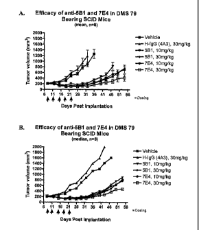
Figures 15A-C show the tumor volume over time in individual SCID mice that
were
implanted with DMS79 small cell lung cancer tumor cells (Fucosyl-GM1+). After
a tumor was
established, the mice were treated five times with one of the following
therapies: (A) PBS
(vehicle control); (B) human IgG1 (isotype control) at 30 mg/kg per mouse; (C)
anti-Fucosyl-
GM I monoclonal antibody 5B1 at 10 mg/kg per mouse; (D) anti-Fucosyl-GM1
monoclonal
antibody 5B1 at 30 mg/kg per mouse; (E) anti-Fucosyl-GM1 monoclonal antibody
7E4 at
mg/kg per mouse; or (F) anti-Fucosyl-GM1 monoclonal antibody 7E4 at 30 mg/kg
per mouse.
The tumor volume on the first day of treatment was about 200 mm3.
Figures 16A and B show the mean and median tumor volume, respectively, of the
mice
10 shown in Figure 15.
Figure 17 shows the mean group weight of the mice shown in Figure 15.
Detailed Description
In one aspect, the present disclosure relates generally to isolated monoclonal
antibodies,
particularly human monoclonal antibodies that bind specifically to Fucosyl-GM1
. In certain
embodiments, the antibodies of this disclosure exhibit one or more desirable
functional
properties, such as high affinity binding to Fucosyl-GM1 and/or the ability to
inhibit growth of
tumor cells in vitro or in vivo. In certain embodiments, the antibodies of
this disclosure are
derived from particular heavy and light chain germline sequences and/or
comprise particular
structural features such as CDR regions comprising particular amino acid
sequences. This
disclosure provides isolated antibodies, methods of making such antibodies,
immunoconjugates
and bispecific molecules comprising such antibodies and pharmaceutical
compositions
containing the antibodies, immunconjugates or bispecific molecules of this
disclosure. This
disclosure also relates to methods of using the antibodies, such as to treat
diseases such as
cancer.
In order that the present disclosure may be more readily understood, certain
terms are
first defined. Additional definitions are set forth throughout the detailed
description.
The term "immune response" refers to the action of, for example, lymphocytes,
antigen
presenting cells, phagocytic cells, granulocytes, and soluble macromolecules
produced by the
above cells or the liver (including antibodies, cytoldnes, and complement)
that results in
CA 02638902 2008-06-05
WO 2007/067992
PCT/US2006/061817
selective damage to, destruction of, or elimination from the human body of
invading pathogens,
cells or tissues infected with pathogens, cancerous cells, or, in cases of
auto immunity or
pathological inflammation, normal human cells or tissues.
A "signal transduction pathway" refers to the biochemical relationship between
a variety
of signal transduction molecules that play a role in the transmission of a
signal from one portion
of a cell to another portion of a cell. As used herein, the phrase "cell
surface receptor" includes,
for example, molecules and complexes of molecules capable of receiving a
signal and the
transmission of such a signal across the plasma membrane of a cell. An example
of a "cell
surface receptor" of the present disclosure is the Fucosyl-GM1 receptor.
The term "antibody" as referred to herein includes whole antibodies and any
antigen
binding fragment (i.e., "antigen-binding portion") or single chains thereof.
An "antibody" refers
to a glycoprotein comprising at least two heavy (H) chains and two light (L)
chains inter-
connected by disulfide bonds, or an antigen binding portion thereof. Each
heavy chain is
comprised of a heavy chain variable region (abbreviated herein as VH) and a
heavy chain
constant region. The heavy chain constant region is comprised of three
domains, CHI, CH2 and
CH3. Each light chain is comprised of a light chain variable region
(abbreviated herein as VI)
and a light chain constant region. The light chain constant region is
comprised of one domain,
CL. The VH and VL regions can be further subdivided into regions of
hypervariability, termed
complementarity determining regions (CDR), interspersed with regions that are
more conserved,
termed framework regions (FR). Each VH and VL is composed of three CDRs and
four FRs,
arranged from amino-terminus to carboxy-terminus in the following order: FR1,
CDR1, FR2,
CDR2, FR3, CDR3, FR4. The variable regions of the heavy and light chains
contain a binding
domain that interacts with an antigen. The constant regions of the antibodies
may mediate the
binding of the immunoglobulin to host tissues or factors, including various
cells of the immune
system (e.g., effector cells) and the first component (Clq) of the classical
complement system.
The term "antigen-binding portion" of an antibody (or simply "antibody
portion"), as
used herein, refers to one or more fragments of an antibody that retain the
ability to specifically
bind to an antigen (e.g., Fucosyl-GM1). It has been shown that the antigen-
binding function of
an antibody can be performed by fragments of a full-length antibody. Examples
of binding
fragments encompassed within the term "antigen-binding portion" of an antibody
include (i) a
Fab fragment, a monovalent fragment consisting of the VL, VH, CL and CHI
domains; (ii) a
11
CA 02638902 2008-06-05
WO 2007/067992
PCT/US2006/061817
F(ab')2 fragment, a bivalent fragment comprising two Fab fragments linked by a
disulfide bridge
at the hinge region; (iii) a Fd fragment consisting of the VH and Cm domains;
(iv) a Fv fragment
consisting of the VL and VH domains of a single arm of an antibody, (v) a dAb
fragment (Ward et
aL, (1989) Nature 341:544-546), which consists of a VH domain; and (vi) an
isolated
complementarity determining region (CDR). Furthermore, although the two
domains of the Fv
fragment, VL and VH, are coded for by separate genes, they can be joined,
using recombinant
methods, by a synthetic linker that enables them to be made as a single
protein chain in which
the VL and VH regions pair to form monovalent molecules (known as single chain
Fv (scFv); see
e.g., Bird et al. (1988) Science M:423-426; and Huston et al. (1988) Proc.
Natl. Acad. Sci. USA
85:5879-5883). Such single chain antibodies are also intended to be
encompassed within the
term "antigen-binding portion" of an antibody. These antibody fragments are
obtained using
conventional techniques known to those with skill in the art, and the
fragments are screened for
utility in the same manner as are intact antibodies.
An "isolated antibody", as used herein, is intended to refer to an antibody
that is
substantially free of other antibodies having different antigenic
specificities (e.g., an isolated
antibody that specifically binds Fucosyl-GM1 is substantially free of
antibodies that specifically
bind antigens other than Fucosyl-GM1). Moreover, an isolated antibody may be
substantially
free of other cellular material and/or chemicals.
The terms "monoclonal antibody" or "monoclonal antibody composition" as used
herein
refer to a preparation of antibody molecules of single molecular composition.
A monoclonal
antibody composition displays a single binding specificity and affinity for a
particular epitope.
The term "human antibody", as used herein, is intended to include antibodies
having
variable regions in which both the framework and CDR regions are derived from
human
germline immunoglobulin sequences. Furthermore, if the antibody contains a
constant region,
the constant region also is derived from human germline immunoglobulin
sequences. The
human antibodies of this disclosure may include amino acid residues not
encoded by human
germline immunoglobulin sequences (e.g., mutations introduced by random or
site-specific
mutagenesis in vitro or by somatic mutation in vivo). However, the term "human
antibody", as
used herein, is not intended to include antibodies in which CDR sequences
derived from the
12
CA 02638902 2008-06-05
WO 2007/067992
PCT/US2006/061817
germline of another mammalian species, such as a mouse, have been grafted onto
human
framework sequences.
The term "human monoclonal antibody" refers to antibodies displaying a single
binding
specificity which have variable regions in which both the framework and CDR
regions are
derived from human germline immunoglobulin sequences. In one embodiment, the
human
monoclonal antibodies are produced by a hybridoma which includes a B cell
obtained from a
transgenic nonhuman animal, e.g., a transgenic mouse, having a genome
comprising a human
heavy chain transgene and a light chain transgene fused to an immortalized
cell.
The term "recombinant human antibody", as used herein, includes all human
antibodies
that are prepared, expressed, created or isolated by recombinant means, such
as (a) antibodies
isolated from an animal (e.g., a mouse) that is transgenic or transchromosomal
for human
immunoglobulin genes or a hybridoma prepared therefrom (described further
below), (b)
antibodies isolated from a host cell transformed to express the human
antibody, e.g., from a
transfectoma, (c) antibodies isolated from a recombinant, combinatorial human
antibody library,
and (d) antibodies prepared, expressed, created or isolated by any other means
that involve
splicing of human immunoglobulin gene sequences to other DNA sequences. Such
recombinant
human antibodies have variable regions in which the framework and CDR regions
are derived
from human germline immunoglobulin sequences. In certain embodiments, however,
such
recombinant human antibodies can be subjected to in vitro mutagenesis (or,
when an animal
transgenic for human Ig sequences is used, in vivo somatic mutagenesis) and
thus the amino acid
sequences of the VH and VL regions of the recombinant antibodies are sequences
that, while
derived from and related to human germline VI{ and VL sequences, may not
naturally exist within
the human antibody germline repertoire in vivo.
As used herein, "isotype" refers to the antibody class (e.g., IgM or IgG1)
that is encoded
by the heavy chain constant region genes.
The phrases "an antibody recognizing an antigen" and "an antibody specific for
an
antigen" are used interchangeably herein with the term "an antibody which
binds specifically to
an antigen."
The term "human antibody derivatives" refers to any modified form of the human
antibody, e.g., a conjugate of the antibody and another agent or antibody.
13
CA 02638902 2008-06-05
WO 2007/067992
PCT/US2006/061817
The term "humanized antibody" is intended to refer to antibodies in which CDR
sequences derived from the germline of another mammalian species, such as a
mouse, have been
grafted onto human framework sequences. Additional framework region
modifications may be
made within the human framework sequences.
The term "chimeric antibody" is intended to refer to antibodies in which the
variable
region sequences are derived from one species and the constant region
sequences are derived
from another species, such as an antibody in which the variable region
sequences are derived
from a mouse antibody and the constant region sequences are derived from a
human antibody.
As used herein, an antibody that "specifically binds to Fucosyl-GM1" is
intended to refer
to an antibody that binds to Fucosyl-GM1 with a KD of 1 x 10⁴ M or less, more
preferably 5 x
104 M or less, more preferably 1 x 10⁴ M or less, more preferably 5 x 10'9 M
or less.
The term "Kassoc" or "Ka", as used herein, is intended to refer to the
association rate of a
particular antibody-antigen interaction, whereas the term "Kdis" or "Ka," as
used herein, is
intended to refer to the dissociation rate of a particular antibody-antigen
interaction. The term
"KD", as used herein, is intended to refer to the dissociation constant, which
is obtained from the
ratio of Ka to Ka (Le,. Ka/Ka) and is expressed as a molar concentration (M).
KD values for
antibodies can be determined using methods well established in the art. A
preferred method for
determining the KD of an antibody is by using surface plasmon resonance,
preferably using a
biosensor system such as a Biacoreil) system.
As used herein, the term "high affinity" for an IgG antibody refers to an
antibody having
a KD of 10-8M or less, more preferably 10-9 M or less and even more preferably
1040 M or less
for a target antigen. However, "high affinity" binding can vary for other
antibody isotypes. For
example, "high affinity" binding for an IgM isotype refers to an antibody
having a KD of 104 M
or less, more preferably 10-8 M or less, even more preferably 10-9 M or less.
As used herein, the term "subject" includes any human or nonhuman animal. The
term
"nonhuman animal" includes all vertebrates, e.g., mammals and non-mammals,
such as
nonhuman primates, sheep, dogs, cats, horses, cows, chickens, amphibians,
reptiles, etc.
Various aspects of this disclosure are described in further detail in the
following
subsections.
14
CA 02638902 2008-06-05
WO 2007/067992
PCT/US2006/061817
Anti-Fucosyl-GM1 Antibodies
The antibodies of this disclosure are characterized by particular functional
features or
properties of the antibodies. For example, the antibodies bind specifically to
Fucosyl-GM1,
preferably Fucosyl-GM1. Preferably, an antibody of this disclosure binds to
Fucosyl-GM1 with
high affinity, for example with a KD of 1 x i0 M or less. The anti-Fucosyl-GM1
antibodies of
this disclosure preferably exhibit one or more of the following
characteristics:
(a) specifically binds to Fucosyl-GM1; and
(b) binds to the human small cell lung cancer cell line DMS-79 (Human SCLC
ATCC #
CRL-2049).
Preferrably, the antibody binds to Fucosyl-GM1 with a KD of 5 x 10⁴ M or less,
binds to
Fucosyl-GM1 with a KD of lx 10.8M or less, binds to Fucosyl-GM1 with a KD of 5
x 10-9 M or
less, or binds to Fucosyl-GM1 with a KD of between 1 x 10⁴ M and 1 x 10⁻¹ M
or less.
Standard assays to evaluate the binding ability of the antibodies toward
Fucosyl-GM1 are known
in the art, including for example, ELISAs, Western blots and RIAs. The binding
kinetics (e.g.,
binding affinity) of the antibodies also can be assessed by standard assays
known in the art, such
as by ELISA, Scatchard and Biacore analysis.
Monoclonal Antibodies 5B1, 5Bla, 7D4, 7E4, 13B8 and 18D5
Preferred antibodies of this disclosure are the human monoclonal antibodies
5B1, 5Bla,
7D4, 7E4, 13B8 and 18D5, isolated and structurally characterized as described
in Examples 1
and 2. The VH amino acid sequences of 5B1, 5Bla, 7D4, 7E4, 13B8 and 18D5 are
shown in
SEQ ID NOs:1, 2, 3, 4, 5 and 6, respectively. The VL amino acid sequences of
5131, 5Bla, 7D4,
7E4, 13B8 and 18D5 are shown in SEQ ID NOs:7, 8, 9, 10, 11 and 12,
respectively.
Given that each of these antibodies can bind to Fucosyl-GM1, the VH and VL
sequences
can be "mixed and matched" to create other anti-Fucosyl-GM1 binding molecules
of this
disclosure. Fucosyl-GM1 binding of such "mixed and matched" antibodies can be
tested using
the binding assays described above and in the Examples (e.g., ELISAs).
Preferably, when VH
and VL chains are mixed and matched, a VH sequence from a particular VH/VL
pairing is replaced
with a structurally similar VH sequence. Likewise, preferably a VL sequence
from a particular
VH/VL pairing is replaced with a structurally similar VL sequence.
CA 02638902 2008-06-05
WO 2007/067992
PCT/US2006/061817
Accordingly, in one aspect, this disclosure provides an isolated monoclonal
antibody, or
antigen binding portion thereof comprising:
(a) a heavy chain variable region comprising an amino acid sequence selected
from the
group consisting of SEQ ID NOs:1, 2, 3,4, 5 and 6; and
(b) a light chain variable region comprising an amino acid sequence selected
from the
group consisting of SEQ ID NOs:7, 8, 9, 10, 11 and 12;
wherein the antibody specifically binds Fucosyl-GM1, preferably Fucosyl-GMl.
Preferred heavy and light chain combinations include:
(a) a heavy chain variable region comprising the amino acid sequence of
SEQ ID NO:1; and (b) a light chain variable region comprising the amino acid
sequence of SEQ
ID NO:7; or
(a) a heavy chain variable region comprising the amino acid sequence of
SEQ ID NO:2; and (b) a light chain variable region comprising the amino acid
sequence of SEQ
ID NO:8; or
(a) a heavy chain variable region comprising the amino acid sequence of
SEQ ID NO :3; and (b) a light chain variable region comprising the amino acid
sequence of SEQ
ID NO:9; or
(a) a heavy chain variable region comprising the amino acid sequence of
SEQ ID NO:4; and (b) a light chain variable region comprising the amino acid
sequence of SEQ
ID NO:10; or
(a) a heavy chain variable region comprising the amino acid sequence of
SEQ ID NO:5; and (b) a light chain variable region comprising the amino acid
sequence of SEQ
ID NO:11; or
(a) a heavy chain variable region comprising the amino acid sequence of
SEQ ID NO:6; and (b) a light chain variable region comprising the amino acid
sequence of SEQ
ID NO:12.
In another aspect, this disclosure provides antibodies that comprise the heavy
chain and
light chain CDR1s, CDR2s and CDR3s of 5B1, 5Bla, 7D4, 7E4, 13B8 and 18D5, or
combinations thereof. The amino acid sequences of the VH CDR1s of 5B1, 5Bla,
7D4, 7E4,
13138 and 18D5 are shown in SEQ ID NOs:13, 14, 15, 16, 17 and 18,
respectively. The amino
acid sequences of the VH CDR2s of 5B1, 5Bla, 7D4, 7E4, 13B8 and 18D5 are shown
in SEQ ID
16
CA 02638902 2008-06-05
WO 2007/067992
PCT/US2006/061817
NOs: 19, 20, 21, 22, 23 and 24, respectively. The amino acid sequences of the
VH CDR3s of
5B1, 5Bla, 7D4, 7E4, 13B8 and 18D5 are shown in SEQ ID NOs:25, 26, 27, 28, 29
and 30,
respectively. The amino acid sequences of the Vk CDR1s of 5B1, 5Bla, 7D4, 7E4,
13B8 and
18D5 are shown in SEQ ID NOs:31, 32, 33, 34, 35 and 36, respectively. The
amino acid
sequences of the Vk CDR2s of 5B1, 5Bla, 7D4, 7E4, 13B8 and 18D5 are shown in
SEQ ID
NOs:37, 38, 39, 40, 41 and 42, respectively. The amino acid sequences of the
Vk CDR3s of 5B1,
5Bla, 7D4, 7E4, 13B8 and 18D5 are shown in SEQ ID NOs:43, 44,45, 46,47 and 48,
respectively. The CDR regions are delineated using the Kabat system (Kabat, E.
A., et al. (1991)
Sequences of Proteins of Immunological Interest, Fifth Edition, U.S.
Department of Health and
Human Services, NIH Publication No. 91-3242).
Given that each of these antibodies can bind to Fucosyl-GM1 and that antigen-
binding
specificity is provided primarily by the CDR1, CDR2, and CDR3 regions, the VH
CDR1, CDR2,
and CDR3 sequences and Vk CDR1, CDR2, and CDR3 sequences can be "mixed and
matched"
(i.e., CDRs from different antibodies can be mixed and match, although each
antibody must
contain a VH CDR1, CDR2, and CDR3 and a Vk CDR1, CDR2, and CDR3) to create
other anti-
Fucosyl-GM1 binding molecules of this disclosure. Fucosyl-GM1 binding of such
"mixed and
matched" antibodies can be tested using the binding assays described above and
in the Examples
(e.g., ELISAs, Biacore analysis). Preferably, when VH CDR sequences are mixed
and matched,
the CDR1, CDR2 and/or CDR3 sequence from a particular VH sequence is replaced
with a
structurally similar CDR sequence(s). Likewise, when Vk CDR sequences are
mixed and
matched, the CDR1, CDR2 and/or CDR3 sequence from a particular Vk sequence
preferably is
replaced with a structurally similar CDR sequence(s). It will be readily
apparent to the ordinarily
skilled artisan that novel VH and VL sequences can be created by substituting
one or more VH
and/or VL CDR region sequences with structurally similar sequences from the
CDR sequences
disclosed herein for monoclonal antibodies antibodies 5B1, 5Bla, 7D4, 7E4,
13B8 and 18D5.
Accordingly, in another aspect, this disclosure provides an isolated
monoclonal antibody,
or antigen binding portion thereof comprising:
(a) a heavy chain variable region CDR1 comprising an amino acid sequence
selected
from the group consisting of SEQ ID NOs:13, 14, 15, 16,17 and 18;
(b) a heavy chain variable region CDR2 comprising an amino acid sequence
selected
from the group consisting of SEQ ID NOs:19, 20, 21, 22, 23 and 24;
17
CA 02638902 2008-06-05
WO 2007/067992
PCT/US2006/061817
(c) a heavy chain variable region CDR3 comprising an amino acid sequence
selected
from the group consisting of SEQ ID NOs:25, 26, 27, 28, 29 and 30;
(d) a light chain variable region CDR1 comprising an amino acid sequence
selected from
the group consisting of SEQ ID NOs:31, 32, 33, 34, 35 and 36;
(e) a light chain variable region CDR2 comprising an amino acid sequence
selected from
the group consisting of SEQ ID NOs:37, 38, 39, 40, 41 and 42; and
(f) a light chain variable region CDR3 comprising an amino acid sequence
selected from
the group consisting of SEQ ID NOs:43, 44,45, 46,47 and 48;
wherein the antibody specifically binds Fucosyl-GM1, preferably Fucosyl-GMl.
In a preferred embodiment, the antibody comprises:
(a) a heavy chain variable region CDR1 comprising SEQ ID NO:13;
(b) a heavy chain variable region CDR2 comprising SEQ ID NO:19;
(c) a heavy chain variable region CDR3 comprising SEQ ID NO:25;
(d) a light chain variable region CDR1 comprising SEQ ID NO:31;
(e) a light chain variable region CDR2 comprising SEQ ID NO:37; and
(f) a light chain variable region CDR3 comprising SEQ ID NO:43.
In another preferred embodiment, the antibody comprises:
(a) a heavy chain variable region CDR1 comprising SEQ ID NO:14;
(b) a heavy chain variable region CDR2 comprising SEQ ID NO:20;
(c) a heavy chain variable region CDR3 comprising SEQ ID NO:26;
(d) a light chain variable region CDR1 comprising SEQ ID NO:32;
(e) a light chain variable region CDR2 comprising SEQ ID NO:38; and
(f) a light chain variable region CDR3 comprising SEQ ID NO:44.
In another preferred embodiment, the antibody comprises:
(a) a heavy chain variable region CDR1 comprising SEQ ID NO:15;
(b) a heavy chain variable region CDR2 comprising SEQ ID NO:21;
(c) a heavy chain variable region CDR3 comprising SEQ ID NO:27;
(d) a light chain variable region CDR1 comprising SEQ ID NO:33;
(e) a light chain variable region CDR2 comprising SEQ ID NO:39; and
(f) a light chain variable region CDR3 comprising SEQ ID NO:45.
In another preferred embodiment, the antibody comprises:
18
CA 02638902 2008-06-05
WO 2007/067992
PCT/US2006/061817
(a) a heavy chain variable region CDR1 comprising SEQ ID NO:16;
(b) a heavy chain variable region CDR2 comprising SEQ ID NO:22;
(c) a heavy chain variable region CDR3 comprising SEQ ID NO:28;
(d) a light chain variable region CDR1 comprising SEQ ID NO:34;
(e) a light chain variable region CDR2 comprising SEQ ID NO:40; and
(f) a light chain variable region CDR3 comprising SEQ II) NO:46.
In another preferred embodiment, the antibody comprises:
(a) a heavy chain variable region CDR1 comprising SEQ ID NO:17;
(b) a heavy chain variable region CDR2 comprising SEQ ID NO:23;
(c) a heavy chain variable region CDR3 comprising SEQ ID NO:29;
(d) a light chain variable region CDR1 comprising SEQ ID NO:35;
(e) a light chain variable region CDR2 comprising SEQ ID NO:41; and
(f) a light chain variable region CDR3 comprising SEQ ID NO:47.
In another preferred embodiment, the antibody comprises:
(a) a heavy chain variable region CDR1 comprising SEQ ID NO:18;
(b) a heavy chain variable region CDR2 comprising SEQ ID NO:24;
(c) a heavy chain variable region CDR3 comprising SEQ ID NO:30;
(d) a light chain variable region CDR1 comprising SEQ ID NO:36;
(e) a light chain variable region CDR2 comprising SEQ ID NO:42; and
(1) a light chain variable region CDR3 comprising SEQ ID NO:48.
It is well known in the art that the CDR3 domain, independently from the CDR1
and/or
CDR2 domain(s), alone can determine the binding specificity of an antibody for
a cognate
antigen and that multiple antibodies can predictably be generated having the
same binding
specificity based on a common CDR3 sequence. See, for example, Klimka et al.,
British I of
Cancer 83(2):252-260 (2000) (describing the production of a humanized anti-
CD30 antibody
using only the heavy chain variable domain CDR3 of murine anti-CD30 antibody
Ki-4); Beiboer
et al., I Mol. Biol. 296:833-849 (2000) (describing recombinant epithelial
glycoprotein-2 (EGP-
2) antibodies using only the heavy chain CDR3 sequence of the parental murine
MOC-31 anti-
EGP-2 antibody); Rader et al., Proc. Natl. Acad. Sci. U.S.A. 95:8910-8915
(1998) (describing a
panel of humanized anti-integrin a133 antibodies using a heavy and light chain
variable CDR3
19
CA 02638902 2013-03-07
78349-15
domain of a murine anti-integrin avi33 antibody LM609 wherein each member
antibody
comprises a distinct sequence outside the CDR3 domain and capable of binding
the same epitope
as the parent muring antibody with affinities as high or higher than the
parent murine antibody);
Barbas et al, J Am. Chem. Soc. 116:2161-2162 (1994) (disclosing that the CDR3
domain
5 provides the most significant contribution to antigen binding); Barbas et
al., Proc. Natl. Acad.
Sci. U.S.A. 92:2529-2533 (1995) (describing the grafting of heavy chain CDR3
sequences of
three Fabs (SI-1, SI-40, and SI-32) against human placental DNA onto the heavy
chain of an
anti-tetanus toxoid Fab thereby replacing the existing heavy chain CDR3 and
demonstrating that
the CDR3 domain alone conferred binding specificity); and Ditzel et al ., J
Immunol. 157:739-
10 749 (1996) (describing grafting studies wherein transfer of only the
heavy chain CDR3 of a
parent polyspecific Fab LNA3 to a heavy chain of a monospecific IgG tetanus
toxoid-binding
Fab p313 antibody was sufficient to retain binding specificity of the parent
Fab).
Accordingly, within certain aspects, the present disclosure provides
monoclonal
15 antibodies comprising one or more heavy and/or light chain CDR3 domain
from a non-human
antibody, such as a mouse or rat antibody, wherein the monoclonal antibody is
capable of
specifically binding to Fucosyl-GM1 . Within some embodiments, such antibodies
comprising
one or more heavy and/or light chain CDR3 domain from a non-human antibody (a)
are capable
of competing for binding with; (b) retain the functional characteristics; (c)
bind to the same
20 epitope; and/or (d) have a similar binding affinity as the corresponding
parental non-human
antibody.
Within other aspects, the present disclosure provides monoclonal antibodies
comprising
one or more heavy and/or light chain CDR3 domain from a first human antibody,
such as, for
example, a human antibody obtained from a non-human animal, wherein the first
human
antibody is capable of specifically binding to Fucosyl-GM1 and wherein the
CDR3 domain from
the first human antibody replaces a CDR3 domain in a human antibody that is
lacking binding
specificity for Fucosyl-GMI to generate a second human antibody that is
capable of specifically
binding to Fucosyl-GM1. Within some embodiments, such inventive antibodies
comprising one
or more heavy and/or light chain CDR3 domain from the first human antibody (a)
are capable of
competing for binding with; (b) retain the functional characteristics; (c)
bind to the same epitope;
and/or (d) have a similar binding affinity as the corresponding parental first
human antibody.
CA 02638902 2008-06-05
WO 2007/067992
PCT/US2006/061817
Antibodies Having Particular Germline Sequences
In certain embodiments, an antibody of this disclosure comprises a heavy chain
variable
region from a particular gerrnline heavy chain immunoglobulin gene and/or a
light chain variable
region from a particular germline light chain immunoglobulin gene.
For example, in a preferred embodiment, this disclosure provides an isolated
monoclonal
antibody, or an antigen-binding portion thereof, comprising a heavy chain
variable region that is
the product of or derived from a human VH 3-48 gene, wherein the antibody
specifically binds
Fucosyl-GM1, preferably Fucosyl-GM1. In another preferred embodiment, this
disclosure
provides an isolated monoclonal antibody, or an antigen-binding portion
thereof, comprising a
light chain variable region that is the product of or derived from a human VK
L15 gene, wherein
the antibody specifically binds Fucosyl-GM1, preferably Fucosyl-GM1. In yet
another preferred
embodiment, this disclosure provides an isolated monoclonal antibody, or
antigen-binding
portion thereof, wherein the antibody:
(a) comprises a heavy chain variable region that is the product of or derived
from a
human VH 3-48 gene (which gene encodes the amino acid sequence set forth in
SEQ ID NO: 61);
(b) comprises a light chain variable region that is the product of or derived
from a human
VK L15 gene (which gene encodes the amino acid sequence set forth in SEQ ID
NO:62); and
(c) specifically binds to Fucosyl-GM1.
Examples of antibodies having VH and VK Of VH 3-48 and VK L15, respectively,
are 5B1,
5B1 a, 7D4, 7E4, 13B8 and 18D5.
As used herein, a human antibody comprises heavy or light chain variable
regions that is
"the product of' or "derived from" a particular germline sequence if the
variable regions of the
antibody are obtained from a system that uses human germline immunoglobulin
genes. Such
systems include immunizing a transgenic mouse carrying human immunoglobulin
genes with the
antigen of interest or screening a human immunoglobulin gene library displayed
on phage with
the antigen of interest. A human antibody that is "the product of' or "derived
from" a human
germline immunoglobulin sequence can be identified as such by comparing the
amino acid
sequence of the human antibody to the amino acid sequences of human germline
immunoglobulins and selecting the human germline immunoglobulin sequence that
is closest in
sequence e., greatest % identity) to the sequence of the human antibody. A
human antibody
that is "the product of' or "derived from" a particular human germline
imrnunoglobulin sequence
21
CA 02638902 2008-06-05
WO 2007/067992
PCT/US2006/061817
may contain amino acid differences as compared to the germline sequence, due
to, for example,
naturally-occurring somatic mutations or intentional introduction of site-
directed mutation.
However, a selected human antibody typically is at least 90% identical in
amino acids sequence
to an amino acid sequence encoded by a human germline immunoglobulin gene and
contains
amino acid residues that identify the human antibody as being human when
compared to the
germline immunoglobulin amino acid sequences of other species (e.g., murine
germline
sequences). In certain cases, a human antibody may be at least 95%, or even at
least 96%, 97%,
98%, or 99% identical in amino acid sequence to the amino acid sequence
encoded by the
germline immunoglobulin gene. Typically, a human antibody derived from a
particular human
germline sequence will display no more than 10 amino acid differences from the
amino acid
sequence encoded by the human germline immunoglobulin gene. In certain cases,
the human
antibody may display no more than 5, or even no more than 4, 3, 2, or 1 amino
acid difference
from the amino acid sequence encoded by the germline immunoglobulin gene.
Homologous Antibodies
In yet another embodiment, an antibody of this disclosure comprises heavy and
light
chain variable regions comprising amino acid sequences that are homologous to
the amino acid
sequences of the preferred antibodies described herein, and wherein the
antibodies retain the
desired functional properties of the anti-Fucosyl-GM1 antibodies of this
disclosure.
For example, this disclosure provides an isolated monoclonal antibody, or
antigen
binding portion thereof, comprising a heavy chain variable region and a light
chain variable
region, wherein:
(a) the heavy chain variable region comprises an amino acid sequence that is
at
least 80% homologous to an amino acid sequence selected from the group
consisting of SEQ ID
NOs:1, 2, 3, 4, 5 and 6;
(b) the light chain variable region comprises an amino acid sequence that is
at
least 80% homologous to an amino acid sequence selected from the group
consisting of SEQ ID
NOs:7, 8,9, 10, 11 and 12; and
the antibody exhibits one or more of the following properties:
(c) the antibody binds to Fucosyl-GM1 with a KD of 1 x 10⁻⁷ M or less;
22
CA 02638902 2013-03-07
78349-15
23
(d) the antibody binds to the human small cell lung cancer cell line DMS-79
(Human SCLC ATCC # CRL-2049).
In other embodiments, the Vti and/or VL amino acid sequences may be 85%, 90%,
95%,
96%, 97%, 98% or 99% homologous to the sequences set forth above. An antibody
having VIA
and VL regions having high (i.e., 80% or greater) homology to the Vll and -VL
regions of the
sequences set forth above, can be obtained by mutagenesis (e.g., site-directed
or PCR-mediated
mutagenesis) of nucleic acid molecules encoding SEQ ID NOs:49, 50, 51, 52, 53,
54, 55, 56, 57,
58, 59 and 60, followed by testing of the encoded altered antibody for
retained function (Le., the
functions set forth in (c) and (d) above) using the functional assays
described herein.
As used herein, the percent homology between two amino acid sequences is
equivalent to
the percent identity between the two sequences. The percent identity between
the two sequences
is a function of the number of identical positions shared by the sequences (L
e., % homology = #
of identical positions/total # of positions x 100), taking into account the
number of gaps, and the
length of each gap, which need to be introduced for optimal alignment of the
two sequences.
The comparison of sequences and determination of percent identity between two
sequences can
be accomplished using a mathematical algorithm, as described in the non-
limiting examples
below.
The percent identity between two amino acid sequences can be determined using
the
algorithm of E. Meyers and W. Miller (Comput. Appl. Biosci., 4:11-17 (1988))
which has been
incorporated into the ALIGN program (version 2.0), using a PAM120 weight
residue table, a gap
length penalty of 12 and a gap penalty of 4. In addition, the percent identity
between two ammo
acid sequences can be determined using the Needleman and Wunsch (J. MoL Biol.
48:444-453
(1970)) algorithm which has been incorporated into the GAP program in the GCG
software package (available
from Accehys, Inc. 10188 Telesis Court, Suite 100, San Diego, CA, 92121, USA),
using either a Blossum 62
matrix or a PAM250 matrix, and a gap weight of 16, 14, 12, 10, 8, 6, or 4 and
a length weight of 1, 2, 3, 4, 5, or 6.
Additionally or alternatively, the protein sequences of the present disclosure
can further
be used as a "query sequence" to perform a search against public databases to,
for example,
identify related sequences. Such searches can be performed using the )(BLAST
program
(version 2.0) of Altschul, eral. (1990) J
Biol. 215:403-10. BLAST protein searches can be
performed with the XBLAST program, score = 50, wordlength = 3 to obtain amino
acid
sequences homologous to the antibody molecules of this disclosure. To obtain
gapped
CA 02638902 2013-03-07
78349-15
24
alignments for comparison purposes, Gapped BLAST can be utilized as described
in Altschul et
al., (1997) Nucleic Acids Res, 25(17):3389-3402. When utilizing BLAST and
Gapped BLAST
programs, the default parameters of the respective programs (e.g., XBLAST and
NB LAST) can
be used.
Antibodies with Conservative Modifications
In certain embodiments, an antibody of this disclosure comprises a heavy chain
variable
region comprising CDR1, CDR2 and CDR3 sequences and a light chain variable
region
comprising CDR1, CDR2 and CDR3 sequences, wherein one or more of these CDR
sequences
comprise specified amino acid sequences based on the preferred antibodies
described herein
(e.g., 5B1, 5Bla, 7D4, 7E4, 13B8 or 18D5), or conservative modifications
thereof, and wherein
the antibodies retain the desired functional properties of the anti-Fucosyl-
GM1 antibodies of this
disclosure. Accordingly, this disclosure provides an isolated monoclonal
antibody, or antigen
binding portion thereof, comprising a heavy chain variable region comprising
CDR1, CDR2, and
CDR3 sequences and a light chain variable region comprising CDR1, CDR2, and
CDR3
sequences, wherein:
(a) the heavy chain variable region CDR3 sequence comprises an amino acid
sequence selected from the group consisting of amino acid sequences of SEQ ID
NOs:25, 26, 27,
28, 29 and 30, and conservative modifications thereof;
(b) the light chain variable region CDR3 sequence comprises an amino acid
sequence selected from the group consisting of amino acid sequence of SEQ ID
NOs:43, 44, 45,
46, 47 and 48, and conservative modifications thereof; and
the antibody exhibits one or more of the following properties:
(c) specifically binds to Fucosyl-GM I; and
(d) the antibody binds to the human small cell lung cancer cell line DMS-79
(Human SCLC ATCC # CRL-2049).
In a preferred embodiment, the heavy chain variable region CDR2 sequence
comprises an
amino acid sequence selected from the group consisting of amino acid sequences
of SEQ ID
NOs:19, 20, 21, 22, 23 and 24, and conservative modifications thereof; and the
light chain
variable region CDR2 sequence comprises an amino acid sequence selected from
the group
consisting of amino acid sequences of SEQ ID NOs:37, 38, 39, 40, 41 and 42,
and conservative
CA 02638902 2008-06-05
WO 2007/067992
PCT/US2006/061817
modifications thereof. In another preferred embodiment, the heavy chain
variable region CDR1
sequence comprises an amino acid sequence selected from the group consisting
of amino acid
sequences of SEQ ID NOs:13, 14, 15, 16, 17 and 18, and conservative
modifications thereof; and
the light chain variable region CDR1 sequence comprises an amino acid sequence
selected from
the group consisting of amino acid sequences of SEQ ID NOs:31, 32, 33, 34, 35
and 36, and
conservative modifications thereof.
As used herein, the term "conservative sequence modifications" is intended to
refer to
amino acid modifications that do not significantly affect or alter the binding
characteristics of the
antibody containing the amino acid sequence. Such conservative modifications
include amino
acid substitutions, additions and deletions. Modifications can be introduced
into an antibody of
this disclosure by standard techniques known in the art, such as site-directed
mutagenesis and
PCR-mediated mutagenesis. Conservative amino acid substitutions are ones in
which the amino
acid residue is replaced with an amino acid residue having a similar side
chain. Families of
amino acid residues having similar side chains have been defined in the art.
These families
include amino acids with basic side chains (e.g., lysine, arginine,
histidine), acidic side chains
(e.g., aspartic acid, glutamic acid), uncharged polar side chains (e.g.,
glycine, asparagine,
glutamine, serine, threonine, tyrosine, cysteine, tryptophan), nonpolar side
chains (e.g., alanine,
valine, leucine, isoleucine, proline, phenylalanine, methionine), beta-
branched side chains (e.g.,
threonine, valine, isoleucine) and aromatic side chains (e.g., tyrosine,
phenylalanine, tryptophan,
histidine). Thus, one or more amino acid residues within the CDR regions of an
antibody of this
disclosure can be replaced with other amino acid residues from the same side
chain family and
the altered antibody can be tested for retained function (i.e., the functions
set forth in (c) and (d)
above) using the functional assays described herein.
Antibodies that Bind to the Same Epitope as Anti-Fucosyl-GM1 Antibodies of
this disclosure
In another embodiment, this disclosure provides antibodies that bind to the
same epitope
on Fucosyl-GM1 as any of the Fucosyl-GM1 monoclonal antibodies of this
disclosure (i.e.,
antibodies that have the ability to cross-compete for binding to Fucosyl-GM1
with any of the
monoclonal antibodies of this disclosure). In preferred embodiments, the
reference antibody for
cross-competition studies can be the monoclonal antibody 5B1 (having VH and VL
sequences as
shown in SEQ ID NOs:1 and 7, respectively), or the monoclonal antibody 5Bla
(having VH and
CA 02638902 2008-06-05
WO 2007/067992
PCT/US2006/061817
VL sequences as shown in SEQ ID NOs:2 and 8, respectively), or the monoclonal
antibody 7D4
(having VH and VL sequences as shown in SEQ ID NOs:3 and 9, respectively), or
the
monoclonal antibody 7E4 (having VH and VL sequences as shown in SEQ ID NOs:4
and 10,
respectively), or the monoclonal antibody 13B8 (having VH and VL sequences as
shown in SEQ
ID NOs:5 and 11, respectively), or or the monoclonal antibody 18D5 (having VH
and VL
sequences as shown in SEQ ID NOs:6 and 12, respectively). Such cross-competing
antibodies
can be identified based on their ability to cross-compete with 5B1, 5Bla, 7D4,
7E4, 13B8 or
18D5 in standard Fucosyl-GM1 binding assays. For example, BIAcore analysis,
ELISA assays
or flow cytometry may be used to demonstrate cross-competition with the
antibodies of the
current disclosure. The ability of a test antibody to inhibit the binding of,
for example, 5B1,
5Bla, 7D4, 7E4, 13B8 or 18D5, to Fucosyl-GM1 demonstrates that the test
antibody can
compete with 5B1, 5Bla, 7D4, 7E4, 13B8 or 18D5 for binding to Fucosyl-GM1 and
thus binds
to the same epitope on Fucosyl-GM1 as 5B1, 5Bla, 7D4, 7E4, 13B8 or 18D5. In a
preferred
embodiment, the antibody that binds to the same epitope on Fucosyl-GM1 as 5B1,
5Bla, 7D4,
7E4, 13B8 or 18D5 is a human monoclonal antibody. Such human monoclonal
antibodies can
be prepared and isolated as described in the Examples.
Engineered and Modified Antibodies
An antibody of this disclosure further can be prepared using an antibody
having one or
more of the VH and/or VL sequences disclosed herein as starting material to
engineer a modified
antibody, which modified antibody may have altered properties from the
starting antibody. An
antibody can be engineered by modifying one or more residues within one or
both variable
regions (i. e. , VH and/or VI), for example within one or more CDR regions
and/or within one or
more framework regions. Additionally or alternatively, an antibody can be
engineered by
modifying residues within the constant region(s), for example to alter the
effector function(s) of
the antibody.
One type of variable region engineering that can be performed is CDR grafting.
Antibodies interact with target antigens predominantly through amino acid
residues that are
located in the six heavy and light chain complementarily determining regions
(CDRs). For this
reason, the amino acid sequences within CDRs are more diverse between
individual antibodies
than sequences outside of CDRs. Because CDR sequences are responsible for most
antibody-
26
CA 02638902 2013-03-07
78349-15
27
antigen interactions, it is possible to express recombinant antibodies that
mimic the properties of
specific naturally occurring antibodies by constructing expression vectors
that include CDR
sequences from the specific naturally occurring antibody grafted onto
framework sequences from
a different antibody with different properties (see, e.g., Riechmarui, L. et
al. (1998) Nature
332:323-327; Jones, P. etal. (1986) Nature 21:522-525; Queen, C. etal. (1989)
Proc. Natl.
Acad. See. U.S.A. 86:10029-10033; U.S. Patent No. 5,225,539 to Winter, and
U.S. Patent Nos.
5,530,101; 5,585,089; 5,693,762 and 6,180,370 to Queen et al.)
Accordingly, another embodiment of this disclosure pertains to an isolated
monoclonal
antibody, or antigen binding portion thereof, comprising a heavy chain
variable region
comprising CDRI, CDR2, and CDR3 sequences comprising an amino acid sequence
selected
from the group consisting of SEQ ID NOs:13, 14, 15, 16, 17 and 18, SEQ ID
NOs:19, 20, 21, 22,
23 and 24 and SEQ ID NOs:25, 26, 27, 28, 29 and 30, respectively, and a light
chain variable
region comprising CDRI, CDR2, and CDR3 sequences comprising an amino acid
sequence
selected from the group consisting of SEQ ID NOs:31, 32, 33, 34,35 and 36, SEQ
ID NOs:37,
38, 39, 40, 41 and 42 and SEQ ID NOs:43, 44, 45, 46,47 and 48, respectively.
Thus, such
antibodies contain the VH and V. CDR sequences of monoclonal antibodies 5B1,
5B I a, 7D4,
7E4, 13B8 or 18D5 yet may contain different framework sequences from these
antibodies.
Such framework sequences can be obtained from public DNA databases or
published
references that include germline antibody gene sequences. For example,
germline DNA
sequences for human heavy and light chain variable region genes can be found
in the "VBase"
human germline sequence database (available in Kabat, E. A., et al. (1991)
Sequences of Proteins
of Immunological Interest, Fifth Edition, U.S. Department of Health and Human
Services, NIH
Publication No. 91-3242; Tomlinson, I. M., etal. (1992) "The Repertoire of
Human Germline VH
Sequences Reveals about Fifty Groups of VH Segments with Different
Hypervariable Loops"
95 .1 Ma. Biol. 227:776-798; and Cox, J. P. L. et al. (1994) "A Directory
of Human Germ-line VH
Segments Reveals a Strong Bias in their Usage" Eur. J Immunol. 24:827-836). As
another
example, the germline DNA sequences for human heavy and light chain variable
region genes can
be found in the Genbank database. For example, the following heavy chain
germline sequences
found in the HCo7 HuMAb mouse are available in the accompanying Genbank
accession numbers:
1-69 (NG__0010109,
CA 02638902 2008-06-05
WO 2007/067992
PCT/US2006/061817
NT 024637 and BC070333), 3-33 (NG 0010109 and NT 024637) and 3-7 (NG 0010109
and
NT 024637). As another example, the following heavy chain germline sequences
found in the
HCo12 HuMAb mouse are available in the accompanying Genbank accession numbers:
1-69
(NG 0010109, NT_024637 and BC070333), 5-51 (NG_0010109 and NT 024637), 4-34
(NG 0010109 and NT 024637), 3-30.3 (?) and 3-23 (AJ406678).
Preferred framework sequences for use in the antibodies of this disclosure are
those that
are structurally similar to the framework sequences used by selected
antibodies of this disclosure,
e.g., similar to the Vll 3-48 framework sequences (SEQ ID NO:61) and/or the VK
L15
framework sequences (SEQ ID NO:62) used by preferred monoclonal antibodies of
this
disclosure. The VH CDR1, CDR2, and CDR3 sequences, and the VK CDR1, CDR2, and
CDR3
sequences, can be grafted onto framework regions that have the identical
sequence as that found
in the germline immunoglobulin gene from which the framework sequence derive,
or the CDR
sequences can be grafted onto framework regions that contain one or more
mutations as
compared to the germline sequences. For example, it has been found that in
certain instances it
is beneficial to mutate residues within the framework regions to maintain or
enhance the antigen
binding ability of the antibody (see e.g. ,U U.S. Patent Nos. 5,530,101;
5,585,089; 5,693,762 and
6,180,370 to Queen et al).
Another type of variable region modification is to mutate amino acid residues
within the
VH and/or VK CDR1, CDR2 and/or CDR3 regions to thereby improve one or more
binding
properties (e.g., affinity) of the antibody of interest. Site-directed
mutagenesis or PCR-mediated
mutagenesis can be performed to introduce the mutation(s) and the effect on
antibody binding, or
other functional property of interest, can be evaluated in in vitro or in vivo
assays as described
herein and provided in the Examples. Preferably conservative modifications (as
discussed
above) are introduced. The mutations may be amino acid substitutions,
additions or deletions,
but are preferably substitutions. Moreover, typically no more than one, two,
three, four or five
residues within a CDR region are altered.
Accordingly, in another embodiment, this disclosure provides isolated anti-
Fucosyl-GM1
monoclonal antibodies, or antigen binding portions thereof, comprising a heavy
chain variable
region comprising: (a) a VII CDR1 region comprising an amino acid sequence
selected from the
group consisting of SEQ ID NOs:13, 14, 15, 16, 17 and 18, or an amino acid
sequence having
one, two, three, four or five amino acid substitutions, deletions or additions
as compared to SEQ
28
CA 02638902 2008-06-05
WO 2007/067992
PCT/US2006/061817
ID NOs:13, 14, 15, 16, 17 and 18; (b) a VH CDR2 region comprising an amino
acid sequence
selected from the group consisting of SEQ ID NOs:19, 20, 21, 22, 23 and 24, or
an amino acid
sequence having one, two, three, four or five amino acid substitutions,
deletions or additions as
compared to SEQ ID NOs:19, 20, 21,22, 23 and 24; (c) a VH CDR3 region
comprising an amino
acid sequence selected from the group consisting of SEQ ID NOs:25, 26, 27, 28,
29 and 30, or an
amino acid sequence having one, two, three, four or five amino acid
substitutions, deletions or
additions as compared to SEQ ID NOs:25, 26, 27, 28, 29 and 30; (d) a VK CDR1
region
comprising an amino acid sequence selected from the group consisting of SEQ ID
NOs:31, 32,
33, 34, 35 and 36, or an amino acid sequence having one, two, three, four or
five amino acid
substitutions, deletions or additions as compared to SEQ ID NOs:31, 32, 33,
34, 35 and 36; (e) a
VK CDR2 region comprising an amino acid sequence selected from the group
consisting of SEQ
ID NOs:37, 38, 39, 40, 41 and 42, or an amino acid sequence having one, two,
three, four or five
amino acid substitutions, deletions or additions as compared to SEQ ID NOs:37,
38, 39, 40, 41
and 42; and (f) a VK CDR3 region comprising an amino acid sequence selected
from the group
consisting of SEQ ID NOs:43, 44, 45, 46, 47 and 48, or an amino acid sequence
having one, two,
three, four or five amino acid substitutions, deletions or additions as
compared to SEQ ID
NOs:43, 44, 45, 46, 47 and 48.
Engineered antibodies of this disclosure include those in which modifications
have been
made to framework residues within VH and/or VK, e.g. to improve the properties
of the antibody.
Typically such framework modifications are made to decrease the immtmogenicity
of the
antibody. For example, one approach is to "backmutate" one or more framework
residues to the
corresponding germline sequence. More specifically, an antibody that has
undergone somatic
mutation may contain framework residues that differ from the germline sequence
from which the
antibody is derived. Such residues can be identified by comparing the antibody
framework
sequences to the germline sequences from which the antibody is derived.
For example, for 7E4, amino acid residue #11 (within FR1) of VH is a serine
whereas this
residue in the corresponding VII 3-48 germline sequence is a leucine. To
return the framework
region sequences to their germline configuration, the somatic mutations can be
"backmutated" to
the germline sequence by, for example, site-directed mutagenesis or PCR-
mediated mutagenesis
(e.g., residue #11 of FR1 of the VH of 7E4 can be "backmutated" from serine to
leucine).
29
CA 02638902 2008-06-05
WO 2007/067992
PCT/US2006/061817
As another example, for 5B1, 5Bla, 7D4, 7E4, 13138 and 18D5, amino acid
residue #16
(within FR1) of VH is a glutamic acid whereas this residue in the
corresponding VH 3-48
germline sequence is a glycine. To return the framework region sequences to
their germline
configuration, for example, residue #16 of the VH of 5B1, 5Bla, 7D4, 7E4,
13138 and 18D5 can
be "backmutated" from glutamic acid to glycine. Such "backmutated" antibodies
are also
intended to be encompassed by this disclosure.
As another example, for 5B1, 5B 1 a, 7D4, 7E4, 13B8 and 18D5, amino acid
residue #23
(within FR1) of VH is a valine whereas this residue in the corresponding VH 3-
48 germline
sequence is an alanine. To return the framework region sequences to their
germline
configuration, for example, residue #23 of the VH of 5B1, 5Bla, 7D4, 7E4, 13B8
and 18D5 can
be "backmutated" from valine to alanine. Such "backmutated" antibodies are
also intended to be
encompassed by this disclosure.
As another example, for 7D4, amino acid residue #24 (within FR1) of VH is a
valine
whereas this residue in the corresponding VH 3-48 germline sequence is an
alanine. To return
the framework region sequences to their germline configuration, for example,
residue #24 of the
VH of 7D4 can be "backmutated" from valine to alanine. Such "backmutated"
antibodies are
also intended to be encompassed by this disclosure.
As another example, for 13B8, amino acid residue #29 (within FR1) of VH is a
leucine
whereas this residue in the corresponding VH 3-48 germline sequence is an
phenylalanine. To
return the framework region sequences to their germline configuration, for
example, residue #29
of the VH of 13B8 can be "backmutated" from leucine to phenylalanine. Such
"backmutated"
antibodies are also intended to be encompassed by this disclosure.
As another example, for 7D4, 13B8 and 18D5, amino acid residue #48 (within
FR2) of
VH is an isoleucine whereas this residue in the corresponding VH 3-48 germline
sequence is a
valine. To return the framework region sequences to their germline
configuration, for example,
residue #48 (residue #13vvithin FR2) of the VH of 7D4, 13B8 and 18D5 can be
"backmutated"
from isoleucine to valine. Such "backmutated" antibodies are also intended to
be encompassed
by this disclosure.
As another example, for 7D4 and 18D5, amino acid residue #84 (within FR3) of
VH is a
serine whereas this residue in the corresponding VH 3-48 germline sequence is
an asparagine. To
return the framework region sequences to their germline configuration, for
example, residue #84
CA 02638902 2008-06-05
WO 2007/067992
PCT/US2006/061817
(residue #18 within FR3) of the VH of 7D4 and 18D5 can be "backmutated" from
serine to
asparagine. Such "backmutated" antibodies are also intended to be encompassed
by this
disclosure.
Another type of framework modification involves mutating one or more residues
within
the framework region, or even within one or more CDR regions, to remove T cell
epitopes to
thereby reduce the potential immunogenicity of the antibody. This approach is
also referred to
as "deimmunization" and is described in futher detail in U.S. Patent
Publication No.
20030153043 by Carr et al.
In addition or alternative to modifications made within the framework or CDR
regions,
antibodies of this disclosure may be engineered to include modifications
within the Fc region,
typically to alter one or more functional properties of the antibody, such as
serum half-life,
complement fixation, Fc receptor binding, and/or antigen-dependent cellular
cytotoxicity.
Furthermore, an antibody of this disclosure may be chemically modified (e.g.,
one or more
chemical moieties can be attached to the antibody) or be modified to alter its
glycosylation, again
to alter one or more functional properties of the antibody. Each of these
embodiments is
described in further detail below. The numbering of residues in the Fc region
is that of the EU
index of Kabat.
In one embodiment, the hinge region of CT-I1 is modified such that the number
of cysteine
residues in the hinge region is altered, e.g., increased or decreased. This
approach is described
further in U.S. Patent No. 5,677,425 by Bodmer et al. The number of cysteine
residues in the
hinge region of CH1 is altered to, for example, facilitate assembly of the
light and heavy chains
or to increase or decrease the stability of the antibody.
In another embodiment, the Fc hinge region of an antibody is mutated to
decrease the
biological half life of the antibody. More specifically, one or more amino
acid mutations are
introduced into the CH2-CH3 domain interface region of the Fe-hinge fragment
such that the
antibody has impaired Staphylococcyl protein A (SpA) binding relative to
native Fe-hinge
domain SpA binding. This approach is described in further detail in U.S.
Patent No. 6,165,745
by Ward et al.
In another embodiment, the antibody is modified to increase its biological
half life.
Various approaches are possible. For example, one or more of the following
mutations can be
introduced: T252L, T254S, T256F, as described in U.S. Patent No. 6,277,375 to
Ward.
31
CA 02638902 2008-06-05
WO 2007/067992
PCT/US2006/061817
Alternatively, to increase the biological half life, the antibody can be
altered within the CH1 or
CL region to contain a salvage receptor binding epitope taken from two loops
of a CH2 domain
of an Fc region of an IgG, as described in U.S. Patent Nos. 5,869,046 and
6,121,022 by Presta et
In yet other embodiments, the Fc region is altered by replacing at least one
amino acid
residue with a different amino acid residue to alter the effector function(s)
of the antibody. For
example, one or more amino acids selected from amino acid residues 234, 235,
236, 237, 297,
318, 320 and 322 can be replaced with a different amino acid residue such that
the antibody has
an altered affinity for an effector ligand but retains the antigen-binding
ability of the parent
antibody. The effector ligand to which affinity is altered can be, for
example, an Fc receptor or
the Cl component of complement. This approach is described in further detail
in U.S. Patent
Nos. 5,624,821 and 5,648,260, both by Winter et al.
In another example, one or more amino acids selected from amino acid residues
329, 331
and 322 can be replaced with a different amino acid residue such that the
antibody has altered
Clq binding and/or reduced or abolished complement dependent cytotoxicity
(CDC). This
approach is described in further detail in U.S. Patent Nos. 6,194,551 by
Idusogie et al.
In another example, one or more amino acid residues within amino acid
positions 231
and 239 are altered to thereby alter the ability of the antibody to fix
complement. This approach
is described further in PCT Publication WO 94/29351 by Bodmer et al.
In yet another example, the Fc region is modified to increase the ability of
the antibody to
mediate antibody dependent cellular cytotoxicity (ADCC) and/or to increase the
affinity of the
antibody for an Fey receptor by modifying one or more amino acids at the
following positions:
238, 239, 248, 249, 252, 254, 255, 256, 258, 265, 267, 268, 269, 270, 272,
276, 278, 280, 283,
285, 286, 289, 290, 292, 293, 294, 295, 296, 298, 301, 303, 305, 307, 309,
312, 315, 320, 322,
324, 326, 327, 329, 330, 331, 333, 334, 335, 337, 338, 340, 360, 373, 376,
378, 382, 388, 389,
398, 414, 416, 419, 430, 434, 435, 437, 438 or 439. This approach is described
further in PCT
Publication WO 00/42072 by Presta. Moreover, the binding sites on human IgG1
for FcyR1,
FcyRII, FcyRIII and FcRn have been mapped and variants with improved binding
have been
described (see Shields, R.L. et al. (2001)J. Biol. Chem. 276:6591-6604).
Specific mutations at
positions 256, 290, 298, 333, 334 and 339 were shown to improve binding to
FcyRIII.
32
CA 02638902 2008-06-05
WO 2007/067992
PCT/US2006/061817
Additionally, the following combination mutants were shown to improve FcyRIII
binding:
T256A/S298A, S298A/E333A, S298A/K224A and S298A/E333A/K334A.
In still another embodiment, the glycosylation of an antibody is modified. For
example,
an aglycoslated antibody can be made (i.e., the antibody lacks glycosylation).
Glycosylation can
be altered to, for example, increase the affinity of the antibody for antigen.
Such carbohydrate
modifications can be accomplished by, for example, altering one or more sites
of glycosylation
within the antibody sequence. For example, one or more amino acid
substitutions can be made
that result in elimination of one or more variable region framework
glycosylation sites to thereby
eliminate glycosylation at that site. Such aglycosylation may increase the
affinity of the
antibody for antigen. Such an approach is described in further detail in U.S.
Patent Nos.
5,714,350 and 6,350,861 by Co et al.
Additionally or alternatively, an antibody can be made that has an altered
type of
glycosylation, such as a hypofucosylated antibody having reduced amounts of
fucosyl residues
or an antibody having increased bisecting GlcNac structures. Such altered
glycosylation patterns
have been demonstrated to increase the ADCC ability of antibodies. Such
carbohydrate
modifications can be accomplished by, for example, expressing the antibody in
a host cell with
altered glycosylation machinery. Cells with altered glycosylation machinery
have been
described in the art and can be used as host cells in which to express
recombinant antibodies of
this disclosure to thereby produce an antibody with altered glycosylation. For
example, the cell
lines Ms704, Ms705, and Ms709 lack the fucosyltransferase gene, FUT8 (alpha
(1,6)
fucosyltransferase), such that antibodies expressed in the Ms704, Ms705, and
Ms709 cell lines
lack fucose on their carbohydrates. The Ms704, Ms705, and Ms709 FUT8'" cell
lines were
created by the targeted disruption of the FUT8 gene in CHO/DG44 cells using
two replacement
vectors (see U.S. Patent Publication No. 20040110704 by Yamane et al. and
Yamane-Ohnuki et
al. (2004) Biotechnol Bioeng 87:614-22). As another example, EP 1,176,195 by
Hanai et al.
describes a cell line with a functionally disrupted FUT8 gene, which encodes a
fucosyl
transferase, such that antibodies expressed in such a cell line exhibit
hypofucosylation by
reducing or eliminating the alpha 1,6 bond-related enzyme. Hanai et al. also
describe cell lines
which have a low enzyme activity for adding fucose to the N-acetylglucosamine
that binds to the
Fe region of the antibody or does not have the enzyme activity, for example
the rat myeloma cell
33
CA 02638902 2008-06-05
WO 2007/067992
PCT/US2006/061817
line YB2/0 (ATCC CRL 1662). PCT Publication WO 03/035835 by Presta describes a
variant
CHO cell line, Led l 3 cells, with reduced ability to attach fucose to
Asn(297)-linked
carbohydrates, also resulting in hypofucosylation of antibodies expressed in
that host cell (see
also Shields, R.L. et al. (2002) J. Biol. Chem. 277:26733-26740). PCT
Publication WO
99/54342 by Umana et al. describes cell lines engineered to express
glycoprotein-modifying
glycosyl transferases (e.g., beta(1,4)-N-acetylglucosaminyltransferase III
(GnTIII)) such that
antibodies expressed in the engineered cell lines exhibit increased bisecting
GlcNac structures
which results in increased ADCC activity of the antibodies (see also Umana et
al. (1999) Nat.
Biotech. 17:176-180). Alternatively, the facose residues of the antibody may
be cleaved off
using a fucosidase enzyme. For example, the fucosidase alpha-L-fucosidase
removes fucosyl
residues from antibodies (Tarentino, A.L. et al. (1975) Biochem. M:5516-23).
Another modification of the antibodies herein that is contemplated by this
disclosure is
pegylation. An antibody can be pegylated to, for example, increase the
biological (e.g., serum)
half life of the antibody. To pegylate an antibody, the antibody, or fragment
thereof, typically is
reacted with polyethylene glycol (PEG), such as a reactive ester or aldehyde
derivative of PEG,
under conditions in which one or more PEG groups become attached to the
antibody or antibody
fragment. Preferably, the pegylation is carried out via an acylation reaction
or an alkylation
reaction with a reactive PEG molecule (or an analogous reactive water-soluble
polymer). As
used herein, the term "polyethylene glycol" is intended to encompass any of
the forms of PEG
that have been used to derivatize other proteins, such as mono (CI-CIO) alkoxy-
or aryloxy-
polyethylene glycol or polyethylene glycol-maleimide. In certain embodiments,
the antibody to
be pegylated is an aglycosylated antibody. Methods for pegylating proteins are
known in the art
and can be applied to the antibodies of this disclosure. See for example, EP 0
154 316 by
Nishimura et al. and EP 0 401 384 by Ishikawa etal.
Methods of Engineering Antibodies
As discussed above, the anti-Fucosyl-GM1 antibodies having VII and VK
sequences
disclosed herein can be used to create new anti-Fucosyl-GMI antibodies by
modifying the VH
and/or VK sequences, or the constant region(s) attached thereto. Thus, in
another aspect of this
disclosure, the structural features of an anti-Fucosyl-GM1 antibody of this
disclosure, e.g. 5B1,
5Bla, 7D4, 7E4, 13B8 or 18D5, are used to create structurally related anti-
Fucosyl-GM1
34
CA 02638902 2008-06-05
WO 2007/067992
PCT/US2006/061817
antibodies that retain at least one functional property of the antibodies of
this disclosure, such as
binding to Fucosyl-GMl. For example, one or more CDR regions of 5B1, 5Bla,
7D4, 7E4,
I3B8 or 18D5, or mutations thereof, can be combined recombinandy with known
framework
regions and/or other CDRs to create additional, recombinantly-engineered, anti-
Fucosyl-GM1
antibodies of this disclosure, as discussed above. Other types of
modifications include those
described in the previous section. The starting material for the engineering
method is one or
more of the VH and/or VK sequences provided herein, or one or more CDR regions
thereof. To
create the engineered antibody, it is not necessary to actually prepare (L e.,
express as a protein)
an antibody having one or more of the VH and/or VK sequences provided herein,
or one or more
CDR regions thereof. Rather, the information contained in the sequence(s) is
used as the starting
material to create a "second generation" sequence(s) derived from the original
sequence(s) and
then the "second generation" sequence(s) is prepared and expressed as a
protein.
Accordingly, in another embodiment, this disclosure provides a method for
preparing an
anti-Fucosyl-GM1 antibody comprising:
(a) providing: (i) a heavy chain variable region antibody sequence comprising
a
CDR1 sequence selected from the group consisting of SEQ ID NOs:13, 14, 15, 16,
17 and 18, a
CDR2 sequence selected from the group consisting of SEQ ID NOs:19, 20, 21,22,
23 and 24,
and/or a CDR3 sequence selected from the group consisting of SEQ ID NOs:25,
26,27, 28,29
and 30; and/or (ii) a light chain variable region antibody sequence comprising
a CDR1 sequence
selected from the group consisting of SEQ ID NOs:31, 32, 33, 34, 35 and 36, a
CDR2 sequence
selected from the group consisting of SEQ ID NOs:37, 38, 39, 40, 41 and 42,
and/or a CDR3
sequence selected from the group consisting of SEQ ID NOs:43, 44, 45, 46, 47
and 48;
(b) altering at least one amino acid residue within the heavy chain variable
region
antibody sequence and/or the light chain variable region antibody sequence to
create at least one
altered antibody sequence; and
(c) expressing the altered antibody sequence as a protein.
Standard molecular biology techniques can be used to prepare and express the
altered
antibody sequence.
Preferably, the antibody encoded by the altered antibody sequence(s) is one
that retains
one, some or all of the functional properties of the anti-Fucosyl-GM1
antibodies described
herein, which functional properties include, but are not limited to:
CA 02638902 2008-06-05
WO 2007/067992
PCT/US2006/061817
(a) the antibody binds to Fucosyl-GM1 with a KD of 1 x 10⁴ M or less;
(b) binds to the human small cell lung cancer cell line DMS-79 (Human SCLC
ATCC # CRL-2049).
The functional properties of the altered antibodies can be assessed using
standard assays
available in the art and/or described herein, such as those set forth in the
Examples (e.g., flow
cytometry, binding assays).
In certain embodiments of the methods of engineering antibodies of this
disclosure,
mutations can be introduced randomly or selectively along all or part of an
anti-Fucosyl-GM1
antibody coding sequence and the resulting modified anti-Fucosyl-GM1
antibodies can be
screened for binding activity and/or other functional properties as described
herein. Mutational
methods have been described in the art. For example, PCT Publication WO
02/092780 by Short
describes methods for creating and screening antibody mutations using
saturation mutagenesis,
synthetic ligation assembly, or a combination thereof. Alternatively, PCT
Publication WO
03/074679 by Lazar et al. describes methods of using computational screening
methods to
optimize physiochemical properties of antibodies.
Nucleic Acid Molecules Encoding Antibodies of this disclosure
Another aspect of this disclosure pertains to nucleic acid molecules that
encode the
antibodies of this disclosure. The nucleic acids may be present in whole
cells, in a cell lysate, or
in a partially purified or substantially pure form. A nucleic acid is
"isolated" or "rendered
substantially pure" when purified away from other cellular components or other
contaminants,
e.g., other cellular nucleic acids or proteins, by standard techniques,
including alkaline/SDS
treatment, CsC1 banding, column chromatography, agarose gel electrophoresis
and others well
known in the art. See, F. Ausubel, et al., ed. (1987) Current Protocols in
Molecular Biology,
Greene Publishing and Wiley Interscience, New York. A nucleic acid of this
disclosure can be,
for example, DNA or RNA and may or may not contain intronic sequences. In a
preferred
embodiment, the nucleic acid is a cDNA molecule.
Nucleic acids of this disclosure can be obtained using standard molecular
biology
techniques. For antibodies expressed by hybridomas (e.g., hybridomas prepared
from transgenic
mice carrying human immunoglobulin genes as described further below), cDNAs
encoding the
36
CA 0 2 6 3 8 9 0 2 2 0 12 ¨ 11 ¨ 1 9
77448-122 =
37
light and heavy chains of the antibody made by the hybridoma can be obtained
by standard PCR
amplification or cDNA cloning techniques. For antibodies obtained from an
immunoglobulin
gene library (e.g., using phage display techniques), nucleic acid encoding the
antibody can be
recovered from the library.
Preferred nucleic acids molecules of this disclosure are those encoding the VH
and VL
sequences of the 5B1, 5Bla, 71344, 7P4, 13B8 or 3C4 monoclonal antibodies. DNA
sequences
encoding the VH sequences of 5B1, 5B la, 7D4, 7E4, 13B8 and 18D5 are shown in
SEQ ID
NOs: 49,50, 51, 52, 53 and 54, respectively. DNA sequences encoding the VL
sequences of
5B1, 5B la, 7D4, 7E4, 13B8 and 3C4 are shown in SEQ NOs:55, 56, 57, 58, 59 and
60,
respectively.
Once DNA fragments encoding VH and VL segments are obtained, these DNA
fragments can be further manipulated by standard recombinant DNA techniques,
for example to
convert the variable region genes to full-length antibody chain genes, to Fab
fragment genes or to
a scFv gene. In these manipulations, it VL- or VH-encoding DNA fragment is
operatively linked
to another DNA fragment encoding another protein, such as an antibody constant
region or a
flexible linker. The term "operatively linked", as used in this context, is
intended to mean that the
two DNA fragments are joined such that the amino acid sequences encoded by the
two DNA
fragments remain in-frame.
The isolated DNA encoding the VH region can be converted to a full-length
heavy chain
gene by operatively linking the VH-encoding DNA to another DNA molecule
encoding heavy
chain constant regions (CU!, CH2 and CH3). The sequences of human heavy chain
constant
region genes are known in the art (see e.g., Kabat, E. A., el al. (1991)
Sequences of Proteins of
Immunological Interest, Fifth Edition, U.S. Department of Health and Human
Services, NIH
Publication No. 91-3242) and DNA fragments encompassing these regions can be
obtained by
standard PCR amplification. The heavy chain constant region can be an IgGl,
IgG2, IgG3, Ig04,
IgA, IgE, IgM or IgD constant region, but most preferably is an IgG1 or Ig04
constant region.
For a Fab fragment heavy chain gene, the VH-encoding DNA can be operatively
linked to
another DNA molecule encoding only the heavy chain CHI constant region.
The isolated DNA encoding the VL region can be converted to a full-length
light chain
gene (as well as a Fab light chain gene) by operatively linking the VL-
encoding DNA to another
DNA molecule encoding the light chain constant region, CL. The sequences of
human light =
CA 02638902 2008-06-05
WO 2007/067992
PCT/US2006/061817
chain constant region genes are known in the art (see e.g., Kabat, E. A., et
al. (1991) Sequences
of Proteins of Immunological Interest, Fifth Edition, U.S. Department of
Health and Human
Services, NIH Publication No. 91-3242) and DNA fragments encompassing these
regions can be
obtained by standard PCR amplification. The light chain constant region can be
a kappa or
lambda constant region, but most preferably is a kappa constant region.
To create a scFv gene, the VH- and VL-encoding DNA fragments are operatively
linked
to another fragment encoding a flexible linker, e.g., encoding the amino acid
sequence (G1y4 -
Ser)3, such that the VH and VL sequences can be expressed as a contiguous
single-chain protein,
with the VL and VH regions joined by the flexible linker (see e.g., Bird etal.
(1988) Science
242:423-426; Huston et al. (1988) Proc. Natl. Acad. Sci. USA 85:5879-5883;
McCafferty etal.,
(1990) Nature 348:552-554).
Production of Monoclonal Antibodies of this disclosure
Monoclonal antibodies (mAbs) of the present disclosure can be produced by a
variety of
techniques, including conventional monoclonal antibody methodology e.g., the
standard somatic
cell hybridization technique of Kohler and Milstein (1975) Nature 256: 495.
Although somatic
cell hybridization procedures are preferred, in principle, other techniques
for producing
monoclonal antibody can be employed e.g., viral or oncogenic transformation of
B lymphocytes.
The preferred animal system for preparing hybridomas is the murine system.
Hybridoma
production in the mouse is a very well-established procedure. Immunization
protocols and
techniques for isolation of immunized splenocytes for fusion are known in the
art. Fusion
partners (e.g., murine myeloma cells) and fusion procedures are also known.
Chimeric or humanized antibodies of the present disclosure can be prepared
based on the
sequence of a murine monoclonal antibody prepared as described above. DNA
encoding the
heavy and light chain immunoglobulins can be obtained from the murine
hybridoma of interest
and engineered to contain non-murine (e.g., human) immunoglobulin sequences
using standard
molecular biology techniques. For example, to create a chimeric antibody, the
murine variable
regions can be linked to human constant regions using methods known in the art
(see e.g., U.S.
Patent No. 4,816,567 to Cabilly etal.). To create a humanized antibody, the
murine CDR
regions can be inserted into a human framework using methods known in the art
(see e.g., U.S.
38
CA 02638902 2013-03-07
78349-15
39
Patent No. 5,225,539 to Winter, and U.S. Patent Nos. 5,530,101; 5,585,089;
5,693,762 and
6,180,370 to Queen et aL).
In a preferred embodiment, the antibodies of this disclosure are human
monoclonal
antibodies. Such human monoclonal antibodies directed against Fucosyl-GM1 can
be generated
using transgenic or transchromosomic mice carrying parts of the human immune
system rather
than the mouse system. These transgenic and transchromosomic mice include mice
referred to
herein as HuMAb mice and KM mice, respectively, and are collectively referred
to herein as
"human Ig mice."
The HuMAb mouse (Medarex, Inc.) contains human immunoglobulin gene miniloci
that encode unrearranged human heavy (IA and y) and K light chain
immunoglobulin sequences,
together with targeted mutations that inactivate the endogenous tt and ic
chain loci (see e.g.,
Lonberg, et al. (1994) Nature 368(6474): 856-859). Accordingly, the mice
exhibit reduced
expression of mouse IgM or K, and in response to immunization, the introduced
human heavy
and light chain transgenes undergo class switching and somatic mutation to
generate high
affinity human IgGK monoclonal (Lonberg, N. et al. (1994), supra; reviewed in
Lonberg, N.
(1994) Handbook of Experimental Pharmacology 113:49-101; Lonberg, N. and
Huszar, D.
(1995) Intern. Rev. Immunol. 13: 65-93, and Harding, F. and Lonberg, N. (1995)
Ann. N.Y. Acad
Sci. 764:536-546). The preparation and use of HuMab mice, and the genomic
modifications
carried by such mice, is further described in Taylor, L. etal. (1992) Nucleic
Acids Research
20:6287-6295; Chen, J. et al. (1993) International Immunology 5: 647-656;
Tuaillon et al.
(1993) Proc. Natl. Acad. Sci. USA 90:3720-3724; Choi etal. (1993) Nature
Genetics 4:117-123;
Chen, J. etal. (1993) EMBO J. 12: 821-830; Tuaillon et aL (1994)J. ImmunoL
152:2912-2920;
Taylor, L. et aL (1994) International Immunology 6: 579-591; and Fishwild, D.
et al. (1996)
Nature Biotechnology 14: 845-851.
See further, U.S. Patent Nos. 5,545,806; 5,569,825;
5,625,126; 5,633,425; 5,789,650; 5,877,397; 5,661,016; 5,814,318; 5,874,299;
and 5,770,429; all
to Lonberg and Kay; U.S. Patent No. 5,545,807 to Surani et PCT Publication
Nos. WO
92/03918, WO 93/12227, WO 94/25585, WO 97/13852, WO 98/24884 and WO 99/45962,
all to
Lonberg and Kay; and PCT Publication No. WO 01/14424 to Korman etal.
CA 02638902 2008-06-05
WO 2007/067992
PCT/US2006/061817
In another embodiment, human antibodies of this disclosure can be raised using
a mouse
that carries human immunoglobulin sequences on transgenes and transchomosomes,
such as a
mouse that carries a human heavy chain transgene and a human light chain
transchromosome.
Such mice, referred to herein as "KM miceTm", are described in detail in PCT
Publication WO
02/43478 to Ishida et al.
Still further, alternative transgenic animal systems expressing human
immunoglobulin
genes are available in the art and can be used to raise anti-Fucosyl-GM1
antibodies of this
disclosure. For example, an alternative transgenic system referred to as the
Xenomouse
(Abgenix, Inc.) can be used; such mice are described in, for example, U.S.
Patent Nos.
5,939,598; 6,075,181; 6,114,598; 6, 150,584 and 6,162,963 to Kucherlapati
etal.
Moreover, alternative transchromosomic animal systems expressing human
immunoglobulin genes are available in the art and can be used to raise anti-
Fucosyl-GM1
antibodies of this disclosure. For example, mice carrying both a human heavy
chain
transchromosome and a human light chain tranchromosome, referred to as "IC
mice" can be
used; such mice are described in Tomizuka et al. (2000) Proc. Natl. Acad. Sci.
USA 97:722-727.
Furthermore, cows carrying human heavy and light chain transchromosomes have
been
described in the art (Kuroiwa et al. (2002) Nature Biotechnology 20:889-894)
and can be used to
raise anti-Fucosyl-GM1 antibodies of this disclosure.
Human monoclonal antibodies of this disclosure can also be prepared using
phage display
methods for screening libraries of human immunoglobulin genes. Such phage
display methods
for isolating human antibodies are established in the art. See for example:
U.S. Patent Nos.
5,223,409; 5,403,484; and 5,571,698 to Ladner et cd.; U.S. Patent Nos.
5,427,908 and 5,580,717
to Dower etal.; U.S. Patent Nos. 5,969,108 and 6,172,197 to McCafferty et al.;
and U.S. Patent
Nos. 5,885,793; 6,521,404; 6,544,731; 6,555,313; 6,582,915 and 6,593,081 to
Griffiths etal.
Human monoclonal antibodies of this disclosure can also be prepared using SCID
mice
into which human immune cells have been reconstituted such that a human
antibody response
can be generated upon immunization. Such mice are described in, for example,
U.S. Patent Nos.
5,476,996 and 5,698,767 to Wilson et al.
Immunization of Human Ig Mice
CA 02638902 2008-06-05
WO 2007/067992
PCT/US2006/061817
When human Ig mice are used to raise human antibodies of this disclosure, such
mice can
be immunized with a purified or enriched preparation of Fucosyl-GM1 antigen
and/or
recombinant Fucosyl-GM1, or an Fucosyl-GM1 fusion protein, as described by
Lonberg, N. et
al. (1994) Nature 368 (6474): 856-859; Fishwild, D. et al. (1996) Nature
Biotechnology 14: 845-
851; and PCT Publication WO 98/24884 and WO 01/14424. Preferably, the mice
will be 6-16
weeks of age upon the first infusion. For example, a purified or recombinant
preparation (5-50
ps) of Fucosyl-GM1 antigen can be used to immunize the human Ig mice
intraperitoneally.
Detailed procedures to generate fully human monoclonal antibodies to Fucosyl-
GM1 are
described in Example 1 below. Cumulative experience with various antigens has
shown that the
transgenic mice respond when initially immunized intraperitoneally (IP) with
antigen in
complete Freund's adjuvant, followed by every other week IP immunizations (up
to a total of 6)
with antigen in incomplete Freund's adjuvant. However, adjuvants other than
Freund's are also
found to be effective. In addition, whole cells in the absence of adjuvant are
found to be highly
immunogenic. The immune response can be monitored over the course of the
immunization
protocol with plasma samples being obtained by retroorbital bleeds. The plasma
can be screened
by ELISA (as described below), and mice with sufficient titers of anti-Fucosyl-
GM1 human
immunoglobulin can be used for fusions. Mice can be boosted intravenously with
antigen 3 days
before sacrifice and removal of the spleen. It is expected that 2-3 fusions
for each immunization
may need to be performed. Between 6 and 24 mice are typically immunized for
each antigen.
Usually both HCo7 and HCo12 strains are used. In addition, both HCo7 and HCol2
transgene
can be bred together into a single mouse having two different human heavy
chain transgenes
(HC07/HC012). Alternatively or additionally, the KM mouse Tm strain can be
used, as described
in Example 1.
Generation of Hybridomas Producing Human Monoclonal Antibodies of this
disclosure
To generate hybridomas producing human monoclonal antibodies of this
disclosure,
splenocytes and/or lymph node cells from immunized mice can be isolated and
fused to an
appropriate immortalized cell line, such as a mouse myeloma cell line. The
resulting hybridomas
can be screened for the production of antigen-specific antibodies. For
example, single cell
suspensions of splenic lymphocytes from immunized mice can be fused to one-
sixth the number
of P3X63-Ag8.653 nonsecreting mouse myeloma cells (ATCC, CRL 1580) with 50%
PEG.
41
CA 02638902 2008-06-05
WO 2007/067992
PCT/US2006/061817
Alternatively, the single cell suspensions of splenic lymphocytes from
immunized mice can be
fused using an electric field based electrofusion method, using a Cyto Pulse
large chamber cell
fusion electroporator (Cyto Pulse Sciences, Inc., Glen Burnie, MD). Cells are
plated at
approximately 2 x 10⁵ in flat bottom microtiter plate, followed by a one week
incubation in
DMEM high glucose medium with L-glutamine and sodium pyruvate (Mediatech,
Inc., Herndon,
VA) and further containing 20% fetal Bovine Serum (Hyclone, Logan, UT), 18%
P388DI
conditional media, 5% Origen Hybridoma cloning factor (BioVeris, Gaithersburg,
VA), 4 mM L-
glutamine, 5mM HEPES, 0.055 mM13-mercaptoethanol, 50 units/ml penicillin, 50
mg/ml
streptomycin and 1X Hypoxanthine-aminopterin-thymidine (HAT) media (Sigma; the
HAT is
added 24 hours after the fusion). After one week, cells cultured in medium in
which HAT was
used was replaced with HT. Individual wells can then be screened by ELISA for
human
monoclonal IgM and IgG antibodies. Once extensive hybridoma growth occurs,
medium can be
observed usually after 10-14 days. The antibody secreting hybridomas can be
replated, screened
again, and if still positive for human IgG, the monoclonal antibodies can be
subcloned at least
twice by limiting dilution. The stable subclones can then be cultured in vitro
to generate small
amounts of antibody in tissue culture medium for characterization.
To purify human monoclonal antibodies, selected hybridomas can be grown in two-
liter
spinner-flasks for monoclonal antibody purification. Supernatants can be
filtered and
concentrated before affinity chromatography with protein A-sepharose
(Pharmacia, Piscataway,
N.J.). Eluted IgG can be checked by gel electrophoresis and high performance
liquid
chromatography to ensure purity. The buffer solution can be exchanged into
PBS, and the
concentration can be determined by 0D280 using 1.43 extinction coefficient.
The monoclonal
antibodies can be aliquoted and stored at -80 C.
Generation of Transfectomas Producing Monoclonal Antibodies of this disclosure
Antibodies of this disclosure also can be produced in a host cell transfectoma
using, for
example, a combination of recombinant DNA techniques and gene transfection
methods as is
well known in the art (e.g., Morrison, S. (1985) Science 229:1202).
For example, to express the antibodies, or antibody fragments thereof, DNAs
encoding
partial or full-length light and heavy chains, can be obtained by standard
molecular biology
42
CA 02638902 2008-06-05
WO 2007/067992
PCT/US2006/061817
techniques (e.g., PCR amplification or cDNA cloning using a hybridoma that
expresses the
antibody of interest) and the DNAs can be inserted into expression vectors
such that the genes
are operatively linked to transcriptional and translational control sequences.
In this context, the
term "operatively linked" is intended to mean that an antibody gene is ligated
into a vector such
that transcriptional and translational control sequences within the vector
serve their intended
function of regulating the transcription and translation of the antibody gene.
The expression
vector and expression control sequences are chosen to be compatible with the
expression host
cell used. The antibody light chain gene and the antibody heavy chain gene can
be inserted into
separate vector or, more typically, both genes are inserted into the same
expression vector. The
antibody genes are inserted into the expression vector by standard methods
(e.g., ligation of
complementary restriction sites on the antibody gene fragment and vector, or
blunt end ligation if
no restriction sites are present). The light and heavy chain variable regions
of the antibodies
described herein can be used to create full-length antibody genes of any
antibody isotype by
inserting them into expression vectors already encoding heavy chain constant
and light chain
constant regions of the desired isotype such that the VH segment is
operatively linked to the CH
segment(s) within the vector and the VK segment is operatively linked to the
CL segment within
the vector. Additionally or alternatively, the recombinant expression vector
can encode a signal
peptide that facilitates secretion of the antibody chain from a host cell. The
antibody chain gene
can be cloned into the vector such that the signal peptide is linked in-frame
to the amino
terminus of the antibody chain gene. The signal peptide can be an
immunoglobulin signal
peptide or a heterologous signal peptide (i.e., a signal peptide from a non-
immunoglobulin
protein).
In addition to the antibody chain genes, the recombinant expression vectors of
this
disclosure carry regulatory sequences that control the expression of the
antibody chain genes in a
host cell. The term "regulatory sequence" is intended to include promoters,
enhancers and other
expression control elements (e.g., polyadenylation signals) that control the
transcription or
translation of the antibody chain genes. Such regulatory sequences are
described, for example,
in Goeddel (Gene Expression Technology. Methods in Enzymology 185, Academic
Press, San
Diego, CA (1990)). It will be appreciated by those skilled in the art that the
design of the
expression vector, including the selection of regulatory sequences, may depend
on such factors
as the choice of the host cell to be transformed, the level of expression of
protein desired, etc.
43
CA 02638902 2008-06-05
WO 2007/067992
PCT/US2006/061817
Preferred regulatory sequences for mammalian host cell expression include
viral elements that
direct high levels of protein expression in mammalian cells, such as promoters
and/or enhancers
derived from cytomegalovirus (CMV), Simian Virus 40 (SV40), adenovirus, (e.g.,
the
adenovirus major late promoter (AdMLP) and polyoma. Alternatively, nonviral
regulatory
sequences may be used, such as the ubiquitin promoter or 13-globin promoter.
Still further,
regulatory elements composed of sequences from different sources, such as the
SRcc promoter
system, which contains sequences from the SV40 early promoter and the long
terminal repeat of
human T cell leukemia virus type 1 (Takebe, Y. et al. (1988) Mol. Cell. Biol.
8:466-472).
In addition to the antibody chain genes and regulatory sequences, the
recombinant
expression vectors of this disclosure may carry additional sequences, such as
sequences that
regulate replication of the vector in host cells (e.g., origins of
replication) and selectable marker
genes. The selectable marker gene facilitates selection of host cells into
which the vector has
been introduced (see, e.g., U.S. Pat. Nos. 4,399,216, 4,634,665 and 5,179,017,
all by Axel et al.).
For example, typically the selectable marker gene confers resistance to drugs,
such as G418,
hygromycin or methotrexate, on a host cell into which the vector has been
introduced. Preferred
selectable marker genes include the dihydrofolate reductase (DHFR) gene (for
use in dhfr- host
cells with methotrexate selection/amplification) and the neo gene (for G418
selection).
For expression of the light and heavy chains, the expression vector(s)
encoding the heavy
and light chains is transfected into a host cell by standard techniques. The
various forms of the
term "transfection" are intended to encompass a wide variety of techniques
commonly used for
the introduction of exogenous DNA into a prokaryotic or eukaryotic host cell,
e.g.,
electroporation, calcium-phosphate precipitation, DEAE-dextran transfection
and the like.
Although it is theoretically possible to express the antibodies of this
disclosure in either
prokaryotic or eukaryotic host cells, expression of antibodies in eukaryotic
cells, and most
preferably mammalian host cells, is the most preferred because such eukaryotic
cells, and in
particular mammalian cells, are more likely than prokaryotic cells to assemble
and secrete a
properly folded and immunologically active antibody. Prokaryotic expression of
antibody genes
has been reported to be ineffective for production of high yields of active
antibody (Boss, M. A.
and Wood, C. R. (1985) Immunology Today 6:12-13).
Preferred mammalian host cells for expressing the recombinant antibodies of
this
44
CA 02638902 2008-06-05
WO 2007/067992
PCT/US2006/061817
disclosure include Chinese Hamster Ovary (CT-JO cells) (including dhfr- CHO
cells, described in
Urlaub and Chasin, (1980) Proc. Natl. Acad. Sci. USA 77:4216-4220, used with a
DHFR
selectable marker, e.g., as described in R. J. Kaufman and P. A. Sharp (1982)
Mol. Biol.
159:601-621), NSO myeloma cells, COS cells and SP2 cells. In particular, for
use with NSO
myeloma cells, another preferred expression system is the GS gene expression
system disclosed
in WO 87/04462, WO 89/01036 and EP 338,841. When recombinant expression
vectors
encoding antibody genes are introduced into mammalian host cells, the
antibodies are produced
by culturing the host cells for a period of time sufficient to allow for
expression of the antibody
in the host cells or, more preferably, secretion of the antibody into the
culture medium in which
the host cells are grown. Antibodies can be recovered from the culture medium
using standard
protein purification methods.
Characterization of Antibody Binding to Antigen
Antibodies of this disclosure can be tested for binding to Fucosyl-GM1 by, for
example,
standard ELISA. Briefly, microtiter plates are coated with purified Fucosyl-
GM1 at 0.25 tig/m1
in PBS, and then blocked with 5% bovine serum albumin in PBS. Dilutions of
antibody (e.g.,
dilutions of plasma from Fucosyl-GM1-immunized mice) are added to each well
and incubated
for 1-2 hours at 37 C. The plates are washed with PBS/Tween and then incubated
with
secondary reagent (e.g., for human antibodies, a goat-anti-human IgG Fc-
specific polyclonal
reagent) conjugated to alkaline phosphatase for 1 hour at 37 C. After washing,
the plates are
developed with pNPP substrate (1 mg/ml), and analyzed at OD of 405-650.
Preferably, mice
which develop the highest titers will be used for fusions.
An ELISA assay as described above can also be used to screen for hybridomas
that show
positive reactivity with Fucosyl-GM1 immunogen. Hybridomas that bind with high
avidity to
Fucosyl-GM1 are subcloned and further characterized. One clone from each
hybridoma, which
retains the reactivity of the parent cells (by ELISA), can be chosen for
making a 5-10 vial cell
bank stored at -140 C, and for antibody purification.
To purify anti-Fucosyl-GM1 antibodies, selected hybridomas can be grown in two-
liter
spinner-flasks for monoclonal antibody purification. Supernatants can be
filtered and
concentrated before affinity chromatography with protein A-sepharose
(Pharmacia, Piscataway,
CA 02638902 2008-06-05
WO 2007/067992
PCT/US2006/061817
NJ). Eluted IgG can be checked by gel electrophoresis and high performance
liquid
chromatography to ensure purity. The buffer solution can be exchanged into
PBS, and the
concentration can be determined by 0D280 using 1.43 extinction coefficient.
The monoclonal
antibodies can be aliquoted and stored at -80 C.
To determine if the selected anti-Fucosyl-GM1 monoclonal antibodies bind to
unique
epitopes, each antibody can be biotinylated using commercially available
reagents (Pierce,
Rockford, IL). Competition studies using unlabeled monoclonal antibodies and
biotinylated
monoclonal antibodies can be performed using Fucosyl-GM1 coated-ELISA plates
as described
above. Biotinylated mAb binding can be detected with a strep-avidin-alkaline
phosphatase
probe.
To determine the isotype of purified antibodies, isotype ELISAs can be
performed using
reagents specific for antibodies of a particular isotype. For example, to
determine the isotype of
a human monoclonal antibody, wells of microtiter plates can be coated with 1
pg/m1 of anti-
human immunoglobulin overnight at 4 C. After blocking with 1% BSA, the plates
are reacted
with 1 pg /ml or less of test monoclonal antibodies or purified isotype
controls, at ambient
temperature for one to two hours. The wells can then be reacted with either
human IgG1 or
human IgM-specific alkaline phosphatase-conjugated probes. Plates are
developed and analyzed
as described above.
Anti-Fucosyl-GM1 human IgGs can be further tested for reactivity with Fucosyl-
GM1
antigen by Western blotting. Briefly, Fucosyl-GM1 can be prepared and
subjected to sodium
dodecyl sulfate polyacrylamide gel electrophoresis. After electrophoresis, the
separated antigens
are transferred to nitrocellulose membranes, blocked with 10% fetal calf
serum, and probed with
the monoclonal antibodies to be tested. Human IgG binding can be detected
using anti-human
IgG alkaline phosphatase and developed with BCIP/NBT substrate tablets (Sigma
Chem. Co., St.
Louis, Mo.).
Immunoconjugates
In another aspect, the present disclosure features an anti-Fucosyl-GM1
antibody, or a
fragment thereof, conjugated to a therapeutic moiety, such as a cytotoxin, a
drug (e.g., an
inu-nunosuppressant) or a radiotoxin. Such conjugates are referred to herein
as
46
CA 02638902 2008-06-05
WO 2007/067992
PCT/US2006/061817
"immunoconjugates". Immunoconjugates that include one or more cytotoxins are
referred to as
"immunotoxins." A cytotoxin or cytotoxic agent includes any agent that is
detrimental to (e.g.,
kills) cells. Examples include taxol, cytochalasin B, gramicidin D, ethidium
bromide, emetine,
mitomycin, etoposide, tenoposide, vincristine, vinblastine, colchicine,
doxorubicin,
daunorubicin, dihydroxy anthracin dione, mitoxantrone, mithramycin,
actinomycin D, 1-
dehydrotestosterone, glucocorticoids, procaine, tetracaine, lidocaine,
propranolol, and puromycin
and analogs or homologs thereof. Therapeutic agents also include, for example,
antimetabolites
(e.g., methotrexate, 6-mercaptoputine, 6-thioguanine, cytarabine, 5-
fluorouracil decarbazine),
alkylating agents (e.g., mechlorethamine, thioepa chlorambucil, melphalan,
earn-Justine (BSNU)
and lomustine (CCNU), cyclothosphamide, busulfan, dibromomarmitol,
streptozotocin,
mitomycin C, and cis-dichlorodiamine platinum (II) (DDP) cisplatin),
anthracyclines (e.g.,
daunorubicin (formerly daunomycin) and doxorubicin), antibiotics (e.g.,
dactinomycin (formerly
actinomycin), bleomycin, mithramycin, and anthramycin (AMC)), and anti-mitotic
agents (e.g.,
vincristine and vinblastine).
Other preferred examples of therapeutic cytotoxins that can be conjugated to
an antibody
of this disclosure include duocarmycins, calicheamicins, maytansines and
auristatins, and
derivatives thereof. An example of a calicheamicin antibody conjugate is
commercially
available (MylotargTm; Wyeth-Ayerst). Examples of therapeutic cytotoxins may
be found, for
example, in US Patent Nos: 6548530 and 6281354 and US Patent application Nos:
US
2003/0064984, US 2003/0073852 and US 2003/0050331.
Cytotoxins can be conjugated to antibodies of this disclosure using linker
technology
available in the art. Examples of linker types that have been used to
conjugate a cytotoxin to an
antibody include, but are not limited to, hydrazones, thioethers, esters,
disulfides and peptide-
containing linkers. A linker can be chosen that is, for example, susceptible
to cleavage by low
pH within the lysosomal compartment or susceptible to cleavage by proteases,
such as proteases
preferentially expressed in tumor tissue such as cathepsins (e.g., cathepsins
B, C, D).
For further discussion of types of cytotoxins, linkers and methods for
conjugating
therapeutic agents to antibodies, see also Saito, G. et al. (2003) Adv. Drug
Deliv. Rev. 55:199-
215; Trail, P.A. et al. (2003) Cancer Immunol. Imrnunother. 52:328-337; Payne,
G. (2003)
Cancer Cell 3:207-212; Allen, T.M. (2002) Nat. Rev. Cancer 2:750-763; Pastan,
I. and
47
CA 02638902 2008-06-05
WO 2007/067992
PCT/US2006/061817
Kreitman, R. J. (2002) Curr. Opin. Investig. Drugs 3:1089-1091; Senter, P.D.
and Springer, C.J.
(2001) Adv. Drug Deily. Rev. 53:247-264.
Antibodies of the present disclosure also can be conjugated to a radioactive
isotope to
generate cytotoxic radiopharrnaceuticals, also referred to as
radioimmunoconjugates. Examples
of radioactive isotopes that can be conjugated to antibodies for use
diagnostically or
therapeutically include, but are not limited to, iodine131, indiumm ,
yttrium90 and lutetium177.
Method for preparing radioimmunconjugates are established in the art. Examples
of
radioimmunoconjugates are commercially available, including ZevalinTM (IDEC
Pharmaceuticals) and BexxarTM (Corixa Pharmaceuticals), and similar methods
can be used to
prepare radioimmunoconjugates using the antibodies of this disclosure.
The antibody conjugates of this disclosure can be used to modify a given
biological
response, and the drug moiety is not to be construed as limited to classical
chemical therapeutic
agents. For example, the drug moiety may be a protein or polypeptide
possessing a desired
biological activity. Such proteins may include, for example, an enzymatically
active toxin, or
active fragment thereof, such as abrin, ricin A, pseudomonas exotoxin, or
diphtheria toxin; a
protein such as tumor necrosis factor or interferon-y; or, biological response
modifiers such as,
for example, lymphokines, interleulcin-1 ("IL-1"), interleukin-2 ("IL-2"),
interleukin-6 ("IL-6"),
granulocyte macrophage colony stimulating factor ("GM-CSF"), granulocyte
colony stimulating
factor ("G-CSF"), or other growth factors.
Techniques for conjugating such therapeutic moiety to antibodies are well
known, see,
e.g., Amon etal., "Monoclonal Antibodies For Immunotargeting Of Drugs In
Cancer Therapy",
in Monoclonal Antibodies And Cancer Therapy, Reisfeld et al. (eds.), pp. 243-
56 (Alan R. Liss,
Inc. 1985); Hellstrom et al.," Antibodies For Drug Delivery", in Controlled
Drug Delivery (2nd
Ed.), Robinson et al. (eds.), pp. 623-53 (Marcel Dekker, Inc. 1987); Thorpe,
"Antibody Carriers
Of Cytotoxic Agents In Cancer Therapy: A Review", in Monoclonal Antibodies
'84: Biological
And Clinical Applications, Pinchera etal. (eds.), pp. 475-506 (1985);
"Analysis, Results, And
Future Prospective Of The Therapeutic Use Of Radiolabeled Antibody In Cancer
Therapy", in
Monoclonal Antibodies For Cancer Detection And Therapy, Baldwin et al. (eds.),
pp. 303-16
(Academic Press 1985), and Thorpe et al., "The Preparation And Cytotoxic
Properties Of
Antibody-Toxin Conjugates", hnmunol. Rev., 62:119-58 (1982).
48
CA 02638902 2008-06-05
WO 2007/067992
PCT/US2006/061817
Bispecific Molecules
In another aspect, the present disclosure features bispecific molecules
comprising an anti-
Fucosyl-GM1 antibody, or a fragment thereof, of this disclosure. An antibody
of this disclosure,
or antigen-binding portions thereof, can be derivatized or linked to another
functional molecule,
e.g., another peptide or protein (e.g., another antibody or ligand for a
receptor) to generate a
bispecific molecule that binds to at least two different binding sites or
target molecules. The
antibody of this disclosure may in fact be derivatized or linkd to more than
one other functional
molecule to generate multispecific molecules that bind to more than two
different binding sites
and/or target molecules; such multispecific molecules are also intended to be
encompassed by
the term "bispecific molecule" as used herein. To create a bispecific molecule
of this disclosure,
an antibody of this disclosure can be functionally linked (e.g., by chemical
coupling, genetic
fusion, noncovalent association or otherwise) to one or more other binding
molecules, such as
another antibody, antibody fragment, peptide or binding mimetic, such that a
bispecific molecule
results.
Accordingly, the present disclosure includes bispecific molecules comprising
at least one
first binding specificity for Fucosyl-GM1 and a second binding specificity for
a second target
epitope. In a particular embodiment of this disclosure, the second target
epitope is an Fc
receptor, e.g., human FcyRI (CD64) or a human Fca receptor (CD89). Therefore,
this disclosure
includes bispecific molecules capable of binding both to FcyR or FcccR
expressing effector cells
(e.g., monocytes, macrophages or polymorphonuclear cells (PNINs)), and to
target cells
expressing Fucosyl-GM1. These bispecific molecules target Fucosyl-GM1
expressing cells to
effector cell and trigger Fc receptor-mediated effector cell activities, such
as phagocytosis of an
Fucosyl-GM1 expressing cells, antibody dependent cell-mediated cytotoxicity
(ADCC), cytokine
release, or generation of superoxide anion.
In an embodiment of this disclosure in which the bispecific molecule is
multispecific, the
molecule can further include a third binding specificity, in addition to an
anti-Fc binding
specificity and an anti-Fucosyl-GM1 binding specificity. In one embodiment,
the third binding
specificity is an anti-enhancement factor (EF) portion, e.g., a molecule which
binds to a surface
protein involved in cytotoxic activity and thereby increases the immune
response against the
49
CA 02638902 2013-03-07
78349-15
target cell. The "anti-enhancement factor portion" can be an antibody,
functional antibody
fragment or a ligand that binds to a given molecule, e.g., an antigen or a
receptor, and thereby
results in an enhancement of the effect of the binding determinants for the Fc
receptor or target
cell antigen. The "anti-enhancement factor portion" can bind an Fc receptor or
a target cell
5 antigen. Alternatively, the anti-enhancement factor portion can bind to
an entity that is different
from the entity to which the first and second binding specificities bind. For
example, the anti-
enhancement factor portion can bind a cytotoxic T-cell (e.g. via CD2, CD3,
CD8, CD28, CD4,
CD40, ICAM-1 or other immune cell that results in an increased immune response
against the
target cell).
10 In one embodiment, the bispecific molecules of this disclosure comprise
as a binding
specificity at least one antibody, or an antibody fragment thereof, including,
e.g., an Fab, Fab',
F(a11)2, Fv, or a single chain Fv. The antibody may also be a light chain or
heavy chain dimer,
or any minimal fragment thereof such as a Fv or a single chain construct as
described in Ladner
et al. U.S. Patent No. 4,946,778.
15 In one embodiment, the binding specificity for an Fey receptor is
provided by a
monoclonal antibody, the binding of which is not blocked by human
immunoglobulin G (IgG).
As used herein, the term "IgG receptor" refers to any of the eight y-chain
genes located on
chromosome 1. These genes encode a total of twelve transmembrane or soluble
receptor
isoforrns which are grouped into three Fey receptor classes: FcyRI (CD64),
FcyRH(CD32), and
20 FcyRIII (CD16). In one preferred embodiment, the Fey receptor a human
high affinity FcyRI.
The human FeyRI is a 72 IcDa molecule, which shows high affmity for monomeric
IgG (108- 109
M-').
The production and characterization of certain preferred anti-Fey monoclonal
antibodies
are described by Fanger et al. in PCT Publication WO 88/00052 and in U.S.
Patent No.
25 4,954,617. These antibodies
bind to an epitope of FcyRI, FcyRII or FcyRIII at a site which is distinct
from the Fcy binding site
of the receptor and, thus, their binding is not blocked substantially by
physiological levels of
IgG. Specific anti-FeyRI antibodies useful in this disclosure are mAb 22, mAb
32, mAb 44,
mAb 62 and mAb 197. The hybridoma producing mAb 32 is available from the
American Type
30 Culture Collection, ATCC Accession No. HB9469. In other embodiments, the
anti-Fcy receptor
CA 02638902 2008-06-05
WO 2007/067992
PCT/US2006/061817
antibody is a humanized form of monoclonal antibody 22 (H22). The production
and
characterization of the H22 antibody is described in Graziano, R.F. et al.
(1995) .]: Immunol 155
(10): 4996-5002 and PCT Publication WO 94/10332. The H22 antibody producing
cell line was
deposited at the American Type Culture Collection under the designation
HA022CL1 and has
the accession no. CRL 11177.
In still other preferred embodiments, the binding specificity for an Fc
receptor is provided
by an antibody that binds to a human IgA receptor, e.g., an Fe-alpha receptor
(FcaRI (CD89)),
the binding of which is preferably not blocked by human immunoglobulin A
(IgA). The term
"IgA receptor" is intended to include the gene product of one a-gene (Fecal)
located on
chromosome 19. This gene is known to encode several alternatively spliced
transmembrane
isoforms of 55 to 110 kDa. FcaRI (CD89) is constitutively expressed on
monocytes/macrophages, eosinophilic and neutrophilic granulocytes, but not on
non-effector cell
populations. FcaRI has medium affinity (.-:: 5 X 10⁷M-1) for both IgAl and
IgA2, which is
increased upon exposure to cytoldnes such as G-CSF or GM-CSF (Morton, H.C. et
al. (1996)
Critical Reviews in Immunology 16:423-440). Four FcaRI-specific monoclonal
antibodies,
identified as A3, A59, A62 and A77, which bind FcaRI outside the IgA ligand
binding domain,
have been described (Monteiro, R.C. et al. (1992).1 Immunol. 148:1764).
FcaRI and FcyRI are preferred trigger receptors for use in the bispecific
molecules of this
disclosure because they are (1) expressed primarily on immune effector cells,
e.g., monocytes,
PMNs, macrophages and dendritic cells; (2) expressed at high levels (e.g.,
5,000-100,000 per
cell); (3) mediators of cytotoxic activities (e.g., ADCC, phagocytosis); (4)
mediate enhanced
antigen presentation of antigens, including self-antigens, targeted to them.
While human monoclonal antibodies are preferred, other antibodies which can be
employed in the bispecific molecules of this disclosure are murine, chimeric
and humanized
monoclonal antibodies.
The bispecific molecules of the present disclosure can be prepared by
conjugating the
constituent binding specificities, e.g., the anti-FcR and anti-Fucosyl-GM1
binding specificities,
using methods known in the art. For example, each binding specificity of the
bispecific
molecule can be generated separately and then conjugated to one another. When
the binding
specificities are proteins or peptides, a variety of coupling or cross-linking
agents can be used for
51
CA 02638902 2008-06-05
WO 2007/067992
PCT/US2006/061817
covalent conjugation. Examples of cross-linking agents include protein A,
carbodiimide, N-
succinimidyl-S-acetyl-thioacetate (SATA), 5,5'-dithiobis(2-nitrobenzoic acid)
(DTNB), o-
phenylenedimaleimide (oPDM), N-succinimidy1-3-(2-pyridyldithio)propionate
(SPDP), and
sulfosuccinimidyl 4-(N-maleimidomethyl) cyclohaxane-l-carboxylate (sulfo-SMCC)
(see e.g.,
Karpovsky et al. (1984)J. Exp. Med. 160:1686; Liu, MA et al. (1985) Proc.
Nall. Acad. Sci.
USA 82:8648). Other methods include those described in Paulus (1985) Behring
Ins. Mitt. No.
78, 118-132; Brennan et al. (1985) Science 229:81-83), and Glennie et al.
(1987)J. Immunol.
139: 2367-2375). Preferred conjugating agents are SATA and sulfo-SMCC, both
available from
Pierce Chemical Co. (Rockford, IL).
When the binding specificities are antibodies, they can be conjugated via
sulfhydryl
bonding of the C-terminus hinge regions of the two heavy chains. In a
particularly preferred
embodiment, the hinge region is modified to contain an odd number of
sulfhydryl residues,
preferably one, prior to conjugation.
Alternatively, both binding specificities can be encoded in the same vector
and expressed
and assembled in the same host cell. This method is particularly useful where
the bispecific
molecule is a mAb x mAb, mAb x Fab, Fab x F(ab)2 or ligand x Fab fusion
protein. A
bispecific molecule of this disclosure can be a single chain molecule
comprising one single chain
antibody and a binding determinant, or a single chain bispecific molecule
comprising two
binding determinants. Bispecific molecules may comprise at least two single
chain molecules.
Methods for preparing bispecific molecules are described for example in U.S.
Patent Number
5,260,203; U.S. Patent Number 5,455,030; U.S. Patent Number 4,881,175; U.S.
Patent Number
5,132,405; U.S. Patent Number 5,091,513; U.S. Patent Number 5,476,786; U.S.
Patent Number
5,013,653; U.S. Patent Number 5,258,498; and U.S. Patent Number 5,482,858.
Binding of the bispecific molecules to their specific targets can be confirmed
by, for
example, enzyme-linked immunosorbent assay (ELISA), radioimmunoassay (RIA),
FACS
analysis, bioassay (e.g., growth inhibition), or Western Blot assay. Each of
these assays
generally detects the presence of protein-antibody complexes of particular
interest by employing
a labeled reagent (e.g., an antibody) specific for the complex of interest.
For example, the FcR-
antibody complexes can be detected using e.g., an enzyme-linked antibody or
antibody fragment
which recognizes and specifically binds to the antibody-FcR complexes.
Alternatively, the
52
CA 02638902 2013-03-07
78349-15
53
complexes can be detected using any of a variety of other immunoassays. For
example, the
antibody can be radioactively labeled and used in a radioimmunoassay (RIA)
(see, for example,
Weintraub, B., Principles of Radioimmunoassays, Seventh Training Course on
Radioligand
Assay Techniques, The Endocrine Society, March, 1986).
The radioactive isotope can be detected by such means as the use of a y
counter or a
scintillation counter or by autoradiography.
Pharmaceutical Compositions
In another aspect, the present disclosure provides a composition, e.g., a
pharmaceutical
composition, containing one or a combination of monoclonal antibodies, or
antigen-binding
portion(s) thereof, of the present disclosure, formulated together with a
pharmaceutically
acceptable carrier. Such compositions may include one or a combination of
(e.g., two or more
different) antibodies, or immunoconjugates or bispecific molecules of this
disclosure. For
example, a pharmaceutical composition of this disclosure can comprise a
combination of
antibodies (or immunoconjugates or bispecifics) that bind to different
epitopes on the target
antigen or that have complementary activities.
Pharmaceutical compositions of this disclosure also can be administered in
combination
therapy, i.e., combined with other agents. For example, the combination
therapy can include an
anti-Fucosyl-GM1 antibody of the present disclosure combined with at least one
other anti-
inflammatory or immunosuppressant agent. Examples of therapeutic agents that
can be used in
combination therapy are described in greater detail below in the section on
uses of the antibodies
of this disclosure.
As used herein, "pharmaceutically acceptable carrier" includes any and all
solvents,
dispersion media, coatings, antibacterial and antifungal agents, isotonic and
absorption delaying
agents, and the like that are physiologically compatible. Preferably, the
carrier is suitable for
intravenous, intramuscular, subcutaneous, parenteral, spinal or epidermal
administration (e.g., by
injection or infusion). Depending on the route of administration, the active
compound, i.e.,
antibody, immunoconjuage, or bispecific molecule, may be coated in a material
to protect the
compound from the action of acids and other natural conditions that may
inactivate the
compound.
CA 02638902 2008-06-05
WO 2007/067992
PCT/US2006/061817
The pharmaceutical compounds of this disclosure may include one or more
pharmaceutically acceptable salts. A "pharmaceutically acceptable salt" refers
to a salt that
retains the desired biological activity of the parent compound and does not
impart any undesired
toxicological effects (see e.g., Berge, S.M., etal. (1977) J. Pharm. Sci. 66:1-
19). Examples of
such salts include acid addition salts and base addition salts. Acid addition
salts include those
derived from nontoxic inorganic acids, such as hydrochloric, nitric,
phosphoric, sulfuric,
hydrobromic, hydroiodic, phosphorous and the like, as well as from nontoxic
organic acids such
as aliphatic mono- and dicarboxylic acids, phenyl-substituted alkanoic acids,
hydroxy alkanoic
acids, aromatic acids, aliphatic and aromatic sulfonic acids and the like.
Base addition salts
include those derived from alkaline earth metals, such as sodium, potassium,
magnesium,
calcium and the like, as well as from nontoxic organic amines, such as
dibenzylethylenediamine, N-methylglucamine, chloroprocaine, choline,
diethanolamine,
ethylenediamine, procaine and the like.
A pharmaceutical composition of this disclosure also may include a
pharmaceutically
acceptable anti-oxidant. Examples of pharmaceutically acceptable antioxidants
include: (1)
water soluble antioxidants, such as ascorbic acid, cysteine hydrochloride,
sodium bisulfate,
sodium metabisulfite, sodium sulfite and the like; (2) oil-soluble
antioxidants, such as ascorbyl
palmitate, butylated hydroxyanisole (BHA), butylated hydroxytoluene (BHT),
lecithin, propyl
gallate, alpha-tocopherol, and the like; and (3) metal chelating agents, such
as citric acid,
ethylenediamine tetraacetic acid (EDTA), sorbitol, tartaric acid, phosphoric
acid, and the like.
Examples of suitable aqueous and nonaqueous carriers that may be employed in
the
pharmaceutical compositions of this disclosure include water, ethanol, polyols
(such as glycerol,
propylene glycol, polyethylene glycol, and the like), and suitable mixtures
thereof, vegetable
oils, such as olive oil, and injectable organic esters, such as ethyl oleate.
Proper fluidity can be
maintained, for example, by the use of coating materials, such as lecithin, by
the maintenance of
the required particle size in the case of dispersions, and by the use of
surfactants.
These compositions may also contain adjuvants such as preservatives, wetting
agents,
emulsifying agents and dispersing agents. Prevention of presence of
microorganisms may be
ensured both by sterilization procedures, supra, and by the inclusion of
various antibacterial and
antifungal agents, for example, paraben, chlorobutanol, phenol sorbic acid,
and the like. It may
also be desirable to include isotonic agents, such as sugars, sodium chloride,
and the like into the
54
CA 02638902 2008-06-05
WO 2007/067992
PCT/US2006/061817
compositions. In addition, prolonged absorption of the injectable
pharmaceutical form may be
brought about by the inclusion of agents which delay absorption such as
aluminum monostearate
and gelatin.
Pharmaceutically acceptable carriers include sterile aqueous solutions or
dispersions and
sterile powders for the extemporaneous preparation of sterile injectable
solutions or dispersion.
The use of such media and agents for pharmaceutically active substances is
known in the art.
Except insofar as any conventional media or agent is incompatible with the
active compound,
use thereof in the pharmaceutical compositions of this disclosure is
contemplated.
Supplementary active compounds can also be incorporated into the compositions.
Therapeutic compositions typically must be sterile and stable under the
conditions of
manufacture and storage. The composition can be formulated as a solution,
microemulsion,
liposome, or other ordered structure suitable to high drug concentration. The
carrier can be a
solvent or dispersion medium containing, for example, water, ethanol, polyol
(for example,
glycerol, propylene glycol, and liquid polyethylene glycol, and the like), and
suitable mixtures
thereof. The proper fluidity can be maintained, for example, by the use of a
coating such as
lecithin, by the maintenance of the required particle size in the case of
dispersion and by the use
of surfactants. In many cases, it will be preferable to include isotonic
agents, for example, sugars,
polyalcohols such as mannitol, sorbitol, or sodium chloride in the
composition. Prolonged
absorption of the injectable compositions can be brought about by including in
the composition
an agent that delays absorption, for example, monostearate salts and gelatin.
Sterile injectable solutions can be prepared by incorporating the active
compound in the
required amount in an appropriate solvent with one or a combination of
ingredients enumerated
above, as required, followed by sterilization microfiltration. Generally,
dispersions are prepared
by incorporating the active compound into a sterile vehicle that contains a
basic dispersion
medium and the required other ingredients from those enumerated above. In the
case of sterile
powders for the preparation of sterile injectable solutions, the preferred
methods of preparation
are vacuum drying and freeze-drying (lyophilization) that yield a powder of
the active ingredient
plus any additional desired ingredient from a previously sterile-filtered
solution thereof.
The amount of active ingredient which can be combined with a carrier material
to
produce a single dosage form will vary depending upon the subject being
treated, and the
particular mode of administration. The amount of active ingredient which can
be combined with
CA 02638902 2008-06-05
WO 2007/067992
PCT/US2006/061817
a carrier material to produce a single dosage form will generally be that
amount of the
composition which produces a therapeutic effect. Generally, out of one hundred
per cent, this
amount will range from about 0.01 per cent to about ninety-nine percent of
active ingredient,
preferably from about 0.1 per cent to about 70 per cent, most preferably from
about 1 per cent to
about 30 per cent of active ingredient in combination with a pharmaceutically
acceptable carrier.
Dosage regimens are adjusted to provide the optimum desired response (e.g., a
therapeutic response). For example, a single bolus may be administered,
several divided doses
may be administered over time or the dose may be proportionally reduced or
increased as
indicated by the exigencies of the therapeutic situation. It is especially
advantageous to
formulate parenteral compositions in dosage unit form for ease of
administration and uniformity
of dosage. Dosage unit form as used herein refers to physically discrete units
suited as unitary
dosages for the subjects to be treated; each unit contains a predetermined
quantity of active
compound calculated to produce the desired therapeutic effect in association
with the required
pharmaceutical carrier. The specification for the dosage unit forms of this
disclosure are dictated
by and directly dependent on (a) the unique characteristics of the active
compound and the
particular therapeutic effect to be achieved, and (b) the limitations inherent
in the art of
compounding such an active compound for the treatment of sensitivity in
individuals.
For administration of the antibody, the dosage ranges from about 0.0001 to 100
mg/kg,
and more usually 0.01 to 5 mg/kg, of the host body weight. For example dosages
can be 0.3
mg/kg body weight, 1 mg/kg body weight, 3 mg/kg body weight, 5 mg/kg body
weight or 10
mg/kg body weight or within the range of 1-10 mg/kg. An exemplary treatment
regime entails
administration once per week, once every two weeks, once every three weeks,
once every four
weeks, once a month, once every 3 months or once every three to 6 months.
Preferred dosage
regimens for an anti-Fucosyl-GM1 antibody of this disclosure include 1 mg/kg
body weight or 3
mg/kg body weight via intravenous administration, with the antibody being
given using one of
the following dosing schedules: (i) every four weeks for six dosages, then
every three months;
(ii) every three weeks; (iii) 3 mg/kg body weight once followed by 1 mg/kg
body weight every
three weeks.
In some methods, two or more monoclonal antibodies with different binding
specificities
are administered simultaneously, in which case the dosage of each antibody
administered falls
within the ranges indicated. Antibody is usually administered on multiple
occasions. Intervals
56
CA 02638902 2008-06-05
WO 2007/067992
PCT/US2006/061817
between single dosages can be, for example, weekly, monthly, every three
monthgs or yearly.
Intervals can also be irregular as indicated by measuring blood levels of
antibody to the target
antigen in the patient. In some methods, dosage is adjusted to achieve a
plasma antibody
concentration of about 1-1000 .ug /ml and in some methods about 25-300 ji.g
/ml.
Alternatively, antibody can be administered as a sustained release
formulation, in which
case less frequent administration is required. Dosage and frequency vary
depending on the half-
life of the antibody in the patient. In general, human antibodies show the
longest half life,
followed by humanized antibodies, chimeric antibodies, and nonhuman
antibodies. The dosage
and frequency of administration can vary depending on whether the treatment is
prophylactic or
therapeutic. In prophylactic applications, a relatively low dosage is
administered at relatively
infrequent intervals over a long period of time. Some patients continue to
receive treatment for
the rest of their lives. In therapeutic applications, a relatively high dosage
at relatively short
intervals is sometimes required until progression of the disease is reduced or
terminated, and
preferably until the patient shows partial or complete amelioration of
symptoms of disease.
Thereafter, the patient can be administered a prophylactic regime.
Actual dosage levels of the active ingredients in the pharmaceutical
compositions of the
present disclosure may be varied so as to obtain an amount of the active
ingredient which is
effective to achieve the desired therapeutic response for a particular
patient, composition, and
mode of administration, without being toxic to the patient. The selected
dosage level will
depend upon a variety of pharmacokinetic factors including the activity of the
particular
compositions of the present disclosure employed, or the ester, salt or amide
thereof, the route of
administration, the time of administration, the rate of excretion of the
particular compound being
employed, the duration of the treatment, other drugs, compounds and/or
materials used in
combination with the particular compositions employed, the age, sex, weight,
condition, general
health and prior medical history of the patient being treated, and like
factors well known in the
medical arts.
A "therapeutically effective dosage" of an anti-Fucosyl-GM1 antibody of this
disclosure
preferably results in a decrease in severity of disease symptoms, an increase
in frequency and
duration of disease symptom-free periods, or a prevention of impairment or
disability due to the
disease affliction. For example, for the treatment of tumors, a
"therapeutically effective
57
CA 02638902 2008-06-05
WO 2007/067992
PCT/US2006/061817
dosage" preferably inhibits cell growth or tumor growth by at least about 20%,
more preferably
by at least about 40%, even more preferably by at least about 60%, and still
more preferably by
at least about 80% relative to untreated subjects. The ability of a compound
to inhibit tumor
growth can be evaluated in an animal model system predictive of efficacy in
human tumors.
Alternatively, this property of a composition can be evaluated by examining
the ability of the
compound to inhibit, such inhibition in vitro by assays known to the skilled
practitioner. A
therapeutically effective amount of a therapeutic compound can decrease tumor
size, or
otherwise ameliorate symptoms in a subject. One of ordinary skill in the art
would be able to
determine such amounts based on such factors as the subject's size, the
severity of the subject's
symptoms, and the particular composition or route of administration selected.
A composition of the present disclosure can be administered via one or more
routes of
administration using one or more of a variety of methods known in the art. As
will be
appreciated by the skilled artisan, the route and/or mode of administration
will vary depending
upon the desired results. Preferred routes of administration for antibodies of
this disclosure
include intravenous, intramuscular, intradermal, intraperitoneal,
subcutaneous, spinal or other
parenteral routes of administration, for example by injection or infusion. The
phrase "parenteral
administration" as used herein means modes of administration other than
enteral and topical
administration, usually by injection, and includes, without limitation,
intravenous, intramuscular,
intraarterial, intrathecal, intracapsular, intraorbital, intracardiac,
intradermal, intraperitoneal,
transtracheal, subcutaneous, subcuticular, intraarticular, subcapsular,
subarachnoid, intraspinal,
epidural and intrasternal injection and infusion.
Alternatively, an antibody of this disclosure can be administered via a non-
parenteral
route, such as a topical, epidermal or mucosal route of administration, for
example, intranasally,
orally, vaginally, rectally, sublingually or topically.
The active compounds can be prepared with carriers that will protect the
compound
against rapid release, such as a controlled release formulation, including
implants, transdermal
patches, and microencapsulated delivery systems. Biodegradable, biocompatible
polymers can
be used, such as ethylene vinyl acetate, polyanhydrides, polyglycolic acid,
collagen,
polyorthoesters, and polylactic acid. Many methods for the preparation of such
formulations are
patented or generally known to those skilled in the art. See, e.g., Sustained
and Controlled
Release Drug Delivery Systems, J.R. Robinson, ed., Marcel Dekker, Inc., New
York, 1978.
58
CA 02638902 2013-03-07
78349-15
59
Therapeutic compositions can be administered with medical devices known in the
art.
For example, in a preferred embodiment, a therapeutic composition of this
disclosure can be
administered with a needleless hypodermic injection device, such as the
devices disclosed in
U.S. Patent Nos. 5,399,163; 5,383,851; 5,312,335; 5,064,413; 4,941,880;
4,790,824; or
4,596,556. Examples of well-known implants and modules useful in the present
disclosure
include: U.S. Patent No. 4,487,603, which discloses an implantable micro-
infusion pump for
dispensing medication at a controlled rate; U.S. Patent No. 4,486,194, which
discloses a
therapeutic device for administering medicants through the skin; U.S. Patent
No. 4,447,233,
which discloses a medication infusion pump for delivering medication at a
precise infusion rate;
U.S. Patent No. 4,447,224, which discloses a variable flow implantable
infusion apparatus for
continuous drug delivery; U.S. Patent No. 4,439,196, which discloses an
osmotic drug delivery
system having multi-chamber compartments; and U.S. Patent No. 4,475,196, which
discloses an
osmotic drug delivery system. Many other
such implants, delivery systems, and modules are known to those skilled in the
art.
In certain embodiments, the human monoclonal antibodies of this disclosure can
be
formulated to ensure proper distribution in vivo. For example, the blood-brain
barrier (BBB)
excludes many highly hydrophilic compounds. To ensure that the therapeutic
compounds of this
disclosure cross the BBB (if desired), they can be formulated, for example, in
liposomes. For
methods of manufacturing liposomes, see, e.g., U.S. Patents 4,522,811;
5,374,548; and
5,399,331. The liposomes may comprise one or more moieties which are
selectively transported
into specific cells or organs, thus enhance targeted drug delivery (see, e.g.,
V.V. Ranade (1989)
.1 Clin. Pharmaeol. 29:685). Exemplary targeting moieties include folate or
biotin (see, e.g.,
U.S. Patent 5,416,016 to Low et al.); mannosides (Umezawa etal., (1988)
Biochem. Biophys.
Res. Commun. 153:1038); antibodies (P.G. Bloeman et al. (1995) FEBS Lett.
357:140; M. Owais
etal. (1995) Antimicrob. Agents Chemother. 39:180); surfactant protein A
receptor (Briscoe et
al. (1995)Am. I Physiol. 1233:134); p120 (Schreier et al. (1994) I Biol.
('hem. 269:9090); see
=
also K. Keinanen; M.L. Laulckanen (1994) FEBS Lett. 346:123; J.J. Killion;
I.J. Fidler (1994)
Immunomethods 4:273.
Uses and Methods of this disclosure
CA 02638902 2008-06-05
WO 2007/067992
PCT/US2006/061817
The antibodies, antibody compositions and methods of the present disclosure
have
numerous in vitro and in vivo diagnostic and therapeutic utilities involving
the diagnosis and
treatment of Fucosyl-GM1 mediated disorders. In a preferred embodiment, the
antibodies of the
present disclosure are human antibodies. For example, these molecules can be
administered to
cells in culture, in vitro or ex vivo, or to human subjects, e.g., in vivo, to
treat, prevent and to
diagnose a variety of disorders. As used herein, the term "subject" is
intended to include human
and non-human animals. Non-human animals includes all vertebrates, e.g.,
mammals and non-
mammals, such as non-human primates, sheep, dogs, cats, cows, horses,
chickens, amphibians,
and reptiles. Preferred subjects include human patients having disorders
mediated by Fucosyl-
GM1 activity or having disorders coincident with a Fucosyl-GM1 mediated
activity. The
methods are particularly suitable for treating human patients having a
disorder associated with
aberrant Fucosyl-GM1 expression or increased Fucosyl-GM1 presence. When
antibodies to
Fucosyl-GM1 are administered together with another agent, the two can be
administered in either
order or simultaneously.
Given the specific binding of the antibodies of this disclosure for Fucosyl-
GM1, the
antibodies of this disclosure can be used to specifically detect Fucosyl-GM1
expression on the
surface of cells and, moreover, can be used to purify Fucosyl-GM1 via
immunoaffinity
purification.
This disclosure further provides methods for detecting the presence of Fucosyl-
GM1
antigen in a sample, or measuring the amount of Fucosyl-GM1 antigen,
comprising contacting
the sample, and a control sample, with a human monoclonal antibody, or an
antigen binding
portion thereof, which specifically binds to Fucosyl-GM1, under conditions
that allow for
formation of a complex between the antibody or portion thereof and Fucosyl-
GM1. The
formation of a complex is then detected, wherein a difference complex
formation between the
sample compared to the control sample is indicative the presence of Fucosyl-
GM1 antigen in the
sample.
Fucosyl-GM1 is expressed in small cell lung cancer, but not detected in normal
lung or
other tissues (Nilsson et al. (1984) Glycoconjugate J1:43-9; Krug et al.
(2004) Clin Cancer Res
10:6094-100). An anti-Fucosyl-GM1 antibody may be used alone to inhibit the
growth of
cancerous tumors. Alternatively, an anti-Fucosyl-GM1 antibody may be used in
conjunction
CA 02638902 2008-06-05
WO 2007/067992
PCT/US2006/061817
with other immunogenic agents, standard cancer treatments or other antibodies,
as described
below.
Anti-fucosyl GM1 monoclonal antibodies have been demonstrated to mediate
potent
CDC directed against fucosyl GM1 positive cell lines (Livingston PO et al.
(1994)J Clin
Onco1.12:1036-44; Brezicka etal. (2000) Cancer Immunol Immunother 49:235-42).
Further, it
has been reported that the complement activation, induced by monoclonal
antibodies specific to
fucosyl-GM1, in combination with cytostatic drugs result in synergistic
cytotoxic effects on
fucosyl-GM1 expressing cells lines (Brezicka and Einbeigi (2001) Tumour Biol
22:97-103).
Finally, anti-fucosyl-GM1 monoclonal antibodies have also been reported to
inhibit the
engraftment of fucosyl-GM1 expressing tumor cells in nude mice (Brezicka et
al. (1991) Int J
Cancer 49:911-8). These data support the development of a fully human
monoclonal antibody to
fucosyl-GM1 as an immunotherapeutic for the treatment of SCLC either alone or
in combination
with chemotherapeutic agents.
Preferred cancers whose growth may be inhibited using the antibodies of this
disclosure
include cancers typically responsive to immunotherapy. Non-limiting examples
of preferred
cancers for treatment include lung cancer (including small cell lung cancer
and non-small cell
lung cancer). Examples of other cancers that may be treated using the methods
of this
disclosure include colon cancer (including small intestine cancer), lung
cancer, breast cancer,
pancreatic cancer, melanoma (e.g., metastatic malignant melanoma), acute
myeloid leukemia,
kidney cancer, bladder cancer, ovarian cancer and prostate cancer, renal
cancer (e.g., renal cell
carcinoma), glioblastoma, brain tumors, chronic or acute leukemias including
acute lymphocytic
leukemia (ALL), adult T-cell leukemia (T-ALL), chronic myeloid leukemia, acute
lymphoblastic
leukemia, chronic lymphocytic leukemia, lymphomas (e.g., Hodgkin's and non-
Hodgkin's
lymphoma, lymphocytic lymphoma, primary CNS lymphoma, T-cell lymphoma,
Burkitt's
lymphoma, anaplastic large-cell lymphomas (ALCL), cutaneous T-cell lymphomas,
nodular
small cleaved-cell lymphomas, peripheral T-cell lymphomas, Lennert's
lymphomas,
immunoblastic lymphomas, T-cell leukemia/lymphomas (ATLL),
entroblastic/centrocytic (cb/cc)
follicular lymphomas cancers, diffuse large cell lymphomas of B lineage,
angioimmunoblastic
lymphadenopathy (AILD)-like T cell lymphoma and HIV associated body cavity
based
lymphomas), embryonal carcinomas, undifferentiated carcinomas of the rhino-
pharynx (e.g.,
Schmincke's tumor), Castleman's disease, Kaposi's Sarcoma, multiple myeloma,
Waldenstrom's
61
CA 02638902 2008-06-05
WO 2007/067992
PCT/US2006/061817
macroglobulinemia and other B-cell lymphomas, nasopharangeal carcinomas, bone
cancer, skin
cancer, cancer of the head or neck, cutaneous or intraocular malignant
melanoma, uterine cancer,
rectal cancer, cancer of the anal region, stomach cancer, testicular cancer,
uterine cancer,
carcinoma of the fallopian tubes, carcinoma of the endometrium, carcinoma of
the cervix,
carcinoma of the vagina, carcinoma of the vulva, cancer of the esophagus,
cancer of the small
intestine, cancer of the endocrine system, cancer of the thyroid gland, cancer
of the parathyroid
gland, cancer of the adrenal gland, sarcoma of soft tissue, cancer of the
urethra, cancer of the
penis, solid tumors of childhood, cancer of the bladder, cancer of the kidney
or ureter, carcinoma
of the renal pelvis, neoplasm of the central nervous system (CNS), tumor
angiogenesis, spinal
axis tumor, brain stem glioma, pituitary adenoma, epidermoid cancer, squarnous
cell cancer,
environmentally induced cancers including those induced by asbestos, e.g.,
mesothelioma and
combinations of said cancers.
Furthermore, given the expression of Fucosyl-GM1 on various tumor cells, the
human
antibodies, antibody compositions and methods of the present disclosure can be
used to treat a
subject with a tumorigenic disorder, e.g., a disorder characterized by the
presence of tumor cells
expressing Fucosyl-GM1 including, for example, lung cancer (including small
cell lung cancer
and non-small cell lung cancer), colon cancer (including small intestine
cancer), melanoma (e.g.,
metastatic malignant melanoma), acute myeloid leukemia, lung cancer, breast
cancer, bladder
cancer, pancreatic cancer, ovarian cancer and prostate cancer. Examples of
other subjects with a
tumorigenic disorder include subjects having renal cancer (e.g., renal cell
carcinoma),
glioblastoma, brain tumors, chronic or acute leukemias including acute
lymphocytic leukemia
(ALL), adult T-cell leukemia (T-ALL), chronic myeloid leukemia, acute
lymphoblastic
leukemia, chronic lymphocytic leukemia, lymphomas (e.g., Hodgkin's and non-
Hodgkin's
lymphoma, lymphocytic lymphoma, primary CNS lymphoma, T-cell lymphoma,
Burkitt's
lymphoma, anaplastic large-cell lymphomas (ALCL), cutaneous T-cell lymphomas,
nodular
small cleaved-cell lymphomas, peripheral T-cell lymphomas, Lennert's
lymphomas,
immunoblastic lymphomas, T-cell leukemia/lymphomas (ATLL),
entroblastic/centrocytic (cb/cc)
follicular lymphomas cancers, diffuse large cell lymphomas of B lineage,
angioimmunoblastic
lymphadenopathy (AILD)-like T cell lymphoma and HIV associated body cavity
based
lymphomas), embryonal carcinomas, undifferentiated carcinomas of the rhino-
pharynx (e.g.,
Schmincke's tumor), Castleman's disease, Kaposi's Sarcoma, multiple myeloma,
Waldenstrom's
62
CA 02638902 2008-06-05
WO 2007/067992
PCT/US2006/061817
macroglobulinemia and other B-cell lymphomas, nasopharangeal carcinomas, bone
cancer, skin
cancer, cancer of the head or neck, cutaneous or intraocular malignant
melanoma, uterine cancer,
rectal cancer, cancer of the anal region, stomach cancer, testicular cancer,
uterine cancer,
carcinoma of the fallopian tubes, carcinoma of the endometrium, carcinoma of
the cervix,
carcinoma of the vagina, carcinoma of the vulva, cancer of the esophagus,
cancer of the small
intestine, cancer of the endocrine system, cancer of the thyroid gland, cancer
of the parathyroid
gland, cancer of the adrenal gland, sarcoma of soft tissue, cancer of the
urethra, cancer of the
penis, solid tumors of childhood, cancer of the bladder, cancer of the kidney
or ureter, carcinoma
of the renal pelvis, neoplasm of the central nervous system (CNS), tumor
angiogenesis, spinal
axis tumor, brain stem glioma, pituitary adenoma, epidermoid cancer, squamous
cell cancer,
environmentally induced cancers including those induced by asbestos, e.g.,
mesothelioma and
combinations of said cancers.
Accordingly, in one embodiment, this disclosure provides a method of
inhibiting growth
of tumor cells in a subject, comprising administering to the subject a
therapeutically effective
amount of an anti-Fucosyl-GM1 antibody or antigen-binding portion thereof.
Preferably, the
antibody is a human anti-Fucosyl-GM1 antibody (such as any of the human anti-
Fucosyl-GM1
antibodies described herein). Additionally or alternatively, the antibody may
be a chimeric or
humanized anti-Fucosyl-GM1 antibody.
In one embodiment, the antibodies (e.g., human monoclonal antibodies,
multispecific and
bispecific molecules and compositions) of this disclosure can be used to
detect levels of Fucosyl-
GM1 or levels of cells which contain Fucosyl-GM1 on their membrane surface,
which levels can
then be linked to certain disease symptoms. Alternatively, the antibodies can
be used to inhibit
or block Fucosyl-GM1 function which, in turn, can be linked to the prevention
or amelioration of
certain disease symptoms, thereby implicating Fucosyl-GM1 as a mediator of the
disease. This
can be achieved by contacting an experimental sample and a control sample with
the anti-
Fucosyl-GM1 antibody under conditions that allow for the formation of a
complex between the
antibody and Fucosyl-GM1. Any complexes formed between the antibody and
Fucosyl-GM1
are detected and compared in the experimental sample and the control.
In another embodiment, the antibodies (e.g., human antibodies, multispecific
and
bispecific molecules and compositions) of this disclosure can be initially
tested for binding
63
CA 02638902 2008-06-05
WO 2007/067992
PCT/US2006/061817
activity associated with therapeutic or diagnostic use in vitro. For example,
compositions of this
disclosure can be tested using the flow cytometric assays described in the
Examples below.
The antibodies (e.g., human antibodies, multispecific and bispecific
molecules,
immunoconjugates and compositions) of this disclosure have additional utility
in therapy and
diagnosis of Fucosyl-GM1-related diseases. For example, the human monoclonal
antibodies, the
multispecific or bispecific molecules and the imrnunoconjugates can be used to
elicit in vivo or
in vitro one or more of the following biological activities: to inhibit the
growth of and/or kill a
cell expressing Fucosyl-GM1; to mediate phagocytosis or ADCC of a cell
expressing Fucosyl-
GM1 in the presence of human effector cells; or to block Fucosyl-GM1 ligand
binding to
Fucosyl-GMl.
In a particular embodiment, the antibodies (e.g., human antibodies,
multispecific and
bispecific molecules and compositions) are used in vivo to treat, prevent or
diagnose a variety of
Fucosyl-GM1-related diseases. Examples of Fucosyl-GM1-related diseases
include, among
others, lung cancer (including small cell lung cancer and non-small cell lung
cancer).
Suitable routes of administering the antibody compositions (e.g., human
monoclonal
antibodies, multispecific and bispecific molecules and immunoconjugates ) of
this disclosure in
vivo and in vitro are well known in the art and can be selected by those of
ordinary skill. For
example, the antibody compositions can be administered by injection (e.g.,
intravenous or
subcutaneous). Suitable dosages of the molecules used will depend on the age
and weight of the
subject and the concentration and/or formulation of the antibody composition.
As previously described, human anti-Fucosyl-GM1 antibodies of this disclosure
can be
co-administered with one or other more therapeutic agents, e.g., a cytotoxic
agent, a radiotoxic
agent or an immunosuppressive agent. The antibody can be linked to the agent
(as an
immunocomplex) or can be administered separate from the agent. In the latter
case (separate
administration), the antibody can be administered before, after or
concurrently with the agent or
can be co-administered with other known therapies, e.g., an anti-cancer
therapy, e.g., radiation.
Such therapeutic agents include, among others, anti-neoplastic agents such as
doxorubicin
(adriamycin), cisplatin bleomycin sulfate, carmustine, chlorambucil and
cyclophosphamide
hydroxyurea which, by themselves, are only effective at levels which are toxic
or subtoxic to a
patient. Cisplatin is intravenously administered as a 100 mg/ dose once every
four weeks and
adriamycin is intravenously administered as a 60-75 mg/ml dose once every 21
days. Co-
64
CA 02638902 2013-03-07
78349-15
administration of the human anti-Fucosyl-GM1 antibodies or antigen binding
fragments thereof,
of the present disclosure with chemotherapeutic agents provides two anti-
cancer agents which
operate via different mechanisms which yield a cytotoxic effect to human tumor
cells. Such co-
administration can solve problems due to development of resistance to drugs or
a change in the
5 antigenicity of the tumor cells which would render them unreactive with
the antibody.
In one embodiment, immunoconjugates of this disclosure can be used to target
compounds (e.g., therapeutic agents, labels, cytotoxins, radiotoxoins
immunosuppressants, etc.)
to cells which have Fucosyl-GM1 cell surface receptors by linking such
compounds to the
antibody. For example, an anti-Fucosyl-GM1 antibody can be conjugated to any
of the toxin
10 compounds described in US Patent Nos. 6,281,354 and 6,548,530, US patent
publication Nos.
20030050331, 20030064984, 20030073852 and 20040087497 or published in WO
03/022806.
Thus, this disclosure also provides
methods for localizing ex vivo or in vivo cells expressing Fucosyl-GM1 (e.g.,
with a detectable
label, such as a radioisotope, a fluorescent compound, an enzyme or an enzyme
co-factor).
15 Alternatively, the immunoconjugates can be used to kill cells which have
Fucosyl-GM1 cell
surface receptors by targeting cytotoxins or radiotoxins to Fucosyl-GIvIl
Target-specific effector cells, e.g., effector cells linked to compositions
(e.g., human
antibodies, multispecific and bispecific molecules) of this disclosure can
also be used as
therapeutic agents. Effector cells for targeting can be human leukocytes such
as macrophages,
20 neutrophils or monocytes. Other cells include eosinophils, natural
killer cells and other IgG- or
IgA-receptor bearing cells. If desired, effector cells can be obtained from
the subject to be
treated. The target-specific effector cells can be administered as a
suspension of cells in a
physiologically acceptable solution. The number of cells administered can be
in the order of 108-
109 but will vary depending on the therapeutic purpose. In general, the amount
will be sufficient
25 to obtain localization at the target cell, e.g., a tumor cell expressing
Fucosyl-GM1 and to effect
cell killing by, e.g., phagocytosis. Routes of administration can also vary.
Therapy with target-specific effector cells can be performed in conjunction
with other
techniques for removal of targeted cells. For example, anti-tumor therapy
using the
compositions (e.g., human antibodies, multispecific and bispecific molecules)
of this disclosure
30 and/or effector cells armed with these compositions can be used in
conjunction with
chemotherapy. Additionally, combination immunotherapy may be used to direct
two distinct
CA 02638902 2008-06-05
WO 2007/067992
PCT/US2006/061817
cytotoxic effector populations toward tumor cell rejection. For example, anti-
Fucosyl-GM1
antibodies linked to anti-Fc-gamma RI or anti-CD3 may be used in conjunction
with IgG- or
IgA-receptor specific binding agents.
Bispecific and multispecific molecules of this disclosure can also be used to
modulate
FcyR or FcyR. levels on effector cells, such as by capping and elimination of
receptors on the cell
surface. Mixtures of anti-Fe receptors can also be used for this purpose.
The compositions (e.g., human antibodies, multispecific and bispecific
molecules and
immunoconjugates) of this disclosure which have complement binding sites, such
as portions
from IgGl, -2 or -3 or IgM which bind complement, can also be used in the
presence of
complement. In one embodiment, ex vivo treatment of a population of cells
comprising target
cells with a binding agent of this disclosure and appropriate effector cells
can be supplemented
by the addition of complement or serum containing complement. Phagocytosis of
target cells
coated with a binding agent of this disclosure can be improved by binding of
complement
proteins. In another embodiment target cells coated with the compositions
(e.g., human
antibodies, multispecific and bispecific molecules) of this disclosure can
also be lysed by
complement. In yet another embodiment, the compositions of this disclosure do
not activate
complement.
The compositions (e.g., human antibodies, multispecific and bispecific
molecules and
immunoconjugates) of this disclosure can also be administered together with
complement.
Accordingly, within the scope of this disclosure are compositions comprising
human antibodies,
multispecific or bispecific molecules and serum or complement. These
compositions are
advantageous in that the complement is located in close proximity to the human
antibodies,
multispecific or bispecific molecules. Alternatively, the human antibodies,
multispecific or
bispecific molecules of this disclosure and the complement or serum can be
administered
separately.
Accordingly, patients treated with antibody compositions of this disclosure
can be
additionally administered (prior to, simultaneously with or following
administration of a human
antibody of this disclosure) with another therapeutic agent, such as a
cytotoxic or radiotoxic
agent, which enhances or augments the therapeutic effect of the human
antibodies.
66
CA 02638902 2013-03-07
78349-15
67
In other embodiments, the subject can be additionally treated with an agent
that
modulates, e.g., enhances or inhibits, the expression or activity of Fey or
Fey receptors by, for
example, treating the subject with a cytolcine. Preferred cytokines for
administration during
treatment with the multispecific molecule include of granulocyte colony-
stimulating factor (G-
CS F), granulocyte- macrophage colony-stimulating factor (GM-CSF), interferon-
y (IFN-y) and
tumor necrosis factor (TNF).
The compositions (e.g., human antibodies, multispecific and bispecific
molecules) of this
disclosure can also be used to target cells expressing FcyR or Fucosyl-GMI,
for example for
labeling such cells. For such use, the binding agent can be linked to a
molecule that can be
detected. Thus, this disclosure provides methods for localizing ex vivo or in
vitro cells
expressing Fe receptors, such as FcyR or Fucosyl-GM1. The detectable label can
be, e.g., a
radioisotope, a fluorescent compound, an enzyme or an enzyme co-factor.
Also within the scope of the present disclosure are kits comprising the
antibody
compositions of this disclosure (e.g., human antibodies, bispecific or
multispecific molecules, or
immunoconjugates) and instructions for use. The kit can further contain one
more more
additional reagents, such as an immunosuppressive reagent, a cytotoxic agent
or a radiotoxic
agent or one or more additional human antibodies of this disclosure (e.g., a
human antibody
having a complementary activity which binds to an epitope in the Fucosyl-GM1
antigen distinct
from the first human antibody). Kits typically include a label indicating the
intended use of the
contents of the kit. The term label includes any writing, or recorded material
supplied on or with
the kit, or which otherwise accompanies the kit.
The present disclosure is further illustrated by the following examples which
should not
be construed as further limiting.
CA 02638902 2008-06-05
WO 2007/067992
PCT/US2006/061817
Examples
Example 1: Generation of human monoclonal antibodies against Fucosyl-GM1
Antigen
Immunization protocols utilized as antigen Salmonella minnesota adsorbed
fucosyl-GM1
(Northwest Biotherapeutics, Inc.).
Trans genic HuMab and KM miceTm
Fully human monoclonal antibodies to Fucosyl-GM1 were prepared using the HCo7,
HCo12, HCo7+HCo12 strains of HuMab transgenic mice and the KM strain of
transgenic
transchromosomic mice, each of which express human antibody genes. In each of
these mouse
strains, the endogenous mouse kappa light chain gene has been homozygously
disrupted as
described in Chen et al. (1993) EMBO J. 12:811-820 and the endogenous mouse
heavy chain
gene has been homozygously disrupted as described in Example 1 of PCT
Publication WO
01/09187. Each of these mouse strains carries a human kappa light chain
transgene, KCo5, as
described in Fishwild et al. (1996) Nature Biotechnology 14:845-851. The HCo7
strain carries
the 1-ICo7 human heavy chain transgene as described in U.S. Patent Nos. 5,
770, 429; 5,545,806;
5,625,825; and 5,545,807. The HCo12 strain carries the HCo12 human heavy chain
transgene as
described in Example 2 of WO 01/09187 or example 2 WO 01/14424. The HCo7+HCo12
strain
carries both the 1-ICo7 and the HCo12 heavy chain transgenes. The KM strain
contains the SC20
transchromosome as described in PCT Publication WO 02/43478. All of these
strains are
referred to herein as HuMAb mice.
HuMab and KM Immunizations:
To generate fully human monoclonal antibodies to Fucosyl-GM1, HuMab mice and
KM
miceTm were immunized with Fucosyl-GM1 adsorbed by lyophilization onto the
surface of acid
treated Salmonella Minnesota (Northwest Biotherapuetics, Inc.). General
immunization schemes
for HuMab mice are described in Lonberg, N. et al (1994) Nature 368(6474): 856-
859; Fishwild,
D. et al. (1996) Nature Biotechnology 14: 845-851 and PCT Publication WO
98/24884. The
mice were 6-16 weeks of age upon the first infusion of antigen.
68
CA 02638902 2008-06-05
WO 2007/067992
PCT/US2006/061817
Transgenic mice were immunized intraperitonealy (IP) or subcutaneously (Sc)
with
antigen in complete Freund's adjuvant twice, followed by 2-4 weeks IP
immunization (up to a
total of 8 immunizations) with the antigen in incomplete Freund's adjuvant.
The immune
response was monitored by ELISA (described below) of sera obtained from retro-
orbital bleeds.
Mice with sufficient titers of anti-fucosyl-GM1 human inununogolobulin were
used for fusions.
Mice were boosted intravenously with antigen 3 and 2 days before sacrifice and
removal of the
spleen.
Selection of HuMab or KM MiceTM Producing Anti-Fucosyl-GM1 Antibodies:
To select HuMab or KM miceTM producing antibodies that bound Fucosyl-GM1, sera
from immunized mice were tested by ELISA as described by Fishwild, D. et al.
(1996). Briefly,
natural fucosyl-GM1 purified from either fucosyl transferase transfectant cell
lines (Northwest
Biotherapeutics, Inc.) or from bovine brain (Matreya, Inc) was solubilized in
methanol at 1
mg/ml and passively adsorbed onto polypropylene microtiter plates, 50 ul/well,
by air-drying at
room temperature for 1-2 hrs. Similarly, plates coated with related control
antigens such as GM1
were prepared as a counter-screen for cross reactive antibodies. Assay plates
were then blocked
with 250 pd/well of 1% ovalbumin in PBS for 1 hr at room temperature.
Dilutions of plasma
from fucosyl-GM1 immunized mice were added to each well and incubated for 1-2
hours at
ambient temperature. The plates were washed with PBS and then incubated for 1
hour at room
temperature with either a goat-anti-human IgG Fc or with goat-anti-human IgM
Fe polyclonal
antibodies each conjugated to horseradish peroxidase (HRP). After washing, the
plates were
developed with TMB substrate and analyzed by spectrophotometer at OD 450nm.
Mice that developed the highest titers of anti-fucosyl-GM1 antibodies were
used for
fusions. Fusions were performed as described below and hybridoma supernatants
were tested for
anti-fucosyl-GM1 activity by ELISA.
Generation of Hybridomas Producing Human Monoclonal Antibodies to Fucosyl-GM1:
Splenocytes were isolated from the HuMab mice and KM-miceTm and were fused to
a
mouse myeloma cell line either using PEG based upon standard protocols or
electric field based
electrofusion using a Cyto Pulse large chamber cell fusion electroporator
(Cyto Pulse Sciences,
Inc., Glen Burnie, MD). The resulting hybridomas were then screened for the
production of
antigen-specific antibodies.
69
CA 02638902 2008-06-05
WO 2007/067992
PCT/US2006/061817
Single cell suspensions of splenic lymphocytes from immunized mice were fused
to one-
fourth the number of P3X63-Ag8.653 non-secreting mouse myeloma cells (ATCC,
CRL 1580)
or SP2/0 non-secreting mouse myeloma cells (ATCC, CRL 1581) using 50% PEG
(Sigma).
Cells were plated at a density of about lx10 5/well in flat bottom microtiter
plates and incubated
approximately 2 weeks in selective medium containing 10% fetal bovine serum,
10% P388D1
(ATCC, CRL TIB-63) conditioned medium, 3-5% origen (IGEN) in DMEM (Mediatech,
CRL
10013, with high glucose, L-glutamine and sodium pyruvate) plus 5 mM HEPES,
0.055 mM 2-
mercaptoethanol, 50 mg/m1 gentamicin and lx HAT (Sigma, CRL P-7185). After 1-2
weeks,
cells were cultured in medium in which the HAT was replaced with HT.
Individual wells were
then screened by ELISA (described above) for human anti-fucosyl-GM1 IgG and
IgM
antibodies.
The hybridomas with specific binding activity in the ELISA assay screen were
further
tested by FACS (described below) for specific binding to fucosyl-GM1 adsorbed
to mammalian
cells. Hybridomas exhibiting the highest specific binding by ELISA and FACS
were sub-
cloned at least twice by limiting dilution. The resulting stable sub-clones
were then cultured in
vitro to generate small amounts of monoclonal antibody in tissue culture
medium. The ELISA
screen was repeated to confirm the activity of the sub-clones. The sub-clones
with highest
activity in the ELISA were scaled up to produce sufficient conditioned medium
(typically 1L) for
purification of monoclonal anti-fucosyl-GM1 for further characterization.
Hybridoma clones 5B1, 5Bla, 7D4, 7E4, 13B8, 13B8a and 18D5 were selected for
further analysis.
Example 2: Structural characterization of human monoclonal antibodies 5B1,
5Bla, 7D4,
7E4, 13B8 and 18D5
The cDNA sequences encoding the heavy and light chain variable regions of the
5B1,
5Bla, 7D4, 7E4, 13B8 and 18D5 monoclonal antibodies were obtained from the
5B1, 5Bla,
7D4, 7E4, 13B8 and 18D5 hybridomas, respectively, using standard PCR
techniques and were
sequenced using standard DNA sequencing techniques.
The nucleotide and amino acid sequences of the heavy chain variable region of
5B1 are
shown in Figure lA and in SEQ ID NO:49 and 1, respectively.
CA 02638902 2008-06-05
WO 2007/067992
PCT/US2006/061817
The nucleotide and amino acid sequences of the light chain variable region of
5B1 are
shown in Figure 1B and in SEQ ID NO:55 and 7, respectively.
Comparison of the 5B1 heavy chain immunoglobulin sequence to the known human
germline immunoglobulin heavy chain sequences demonstrated that the 581 heavy
chain utilizes
a VH segment from human germline VH 3-48, a D segment from the human germline
1-1, and a
JH segment from human germline JH 6b. The alignment of the 5B1 VH sequence to
the
germline VH 3-48 sequence is shown in Figure 7. Further analysis of the 5B1 VH
sequence
using the Kabat system of CDR region determination led to the delineation of
the heavy chain
CDR1, CDR2 and CD3 regions as shown in Figures lA and 7, and in SEQ ID NOs:13,
19 and
25, respectively.
Comparison of the 5B1 light chain immunoglobulin sequence to the known human
germline immunoglobulin light chain sequences demonstrated that the 5B1 light
chain utilizes a
VL segment from human germline VK L15 and a JK segment from human germline JK
4. The
alignment of the 5B1 VL sequence to the germline VK L15 sequence is shown in
Figure 8.
Further analysis of the 5B1 VL sequence using the Kabat system of CDR region
determination
led to the delineation of the light chain CDR1, CDR2 and CD3 regions as shown
in Figures 1B
and 8, and in SEQ ID NOs:31, 37 and 43, respectively.
The nucleotide and amino acid sequences of the heavy chain variable region of
5Bla are
shown in Figure 2A and in SEQ ID NO:50 and 2, respectively.
The nucleotide and amino acid sequences of the light chain variable region of
5Bla are
shown in Figure 2B and in SEQ ID NO:56 and 8, respectively.
Comparison of the 5Bla heavy chain immunoglobulin sequence to the known human
germline immunoglobulin heavy chain sequences demonstrated that the 5Bla heavy
chain
utilizes a VH segment from human germline VH 3-48, a D segment from the human
germline 1-
1, and a JH segment from human germline JH 6b. The alignment of the 5Bla VH
sequence to
the germline VH 3-48 sequence is shown in Figure 7. Further analysis of the
5Bla VH
sequence using the Kabat system of CDR region determination led to the
delineation of the
heavy chain CDR1, CDR2 and CD3 regions as shown in Figures 2A and 7, and in
SEQ ID
NOs:14, 20 and 26, respectively.
Comparison of the 5Bla light chain immunoglobulin sequence to the known human
germline immunoglobulin light chain sequences demonstrated that the 5B la
light chain utilizes a
71
CA 02638902 2008-06-05
WO 2007/067992
PCT/US2006/061817
VL segment from human germline VK L15 and a JK segment from human germline JK
4. The
alignment of the 5Bla VL sequence to the germline VK L15 sequence is shown in
Figure 8.
Further analysis of the 5B1a VL sequence using the Kabat system of CDR region
determination
led to the delineation of the light chain CDR1, CDR2 and CD3 regions as shown
in Figures 2B
and 8, and in SEQ ID NOs:32, 38 and 44, respectively.
The nucleotide and amino acid sequences of the heavy chain variable region of
7D4 are
shown in Figure 3A and in SEQ ID NO:51 and 3, respectively.
The nucleotide and amino acid sequences of the light chain variable region of
7D4 are
shown in Figure 3B and in SEQ ID NO:57 and 9, respectively.
Comparison of the 7D4 heavy chain immunoglobulin sequence to the known human
germline immunoglobulin heavy chain sequences demonstrated that the 7D4 heavy
chain utilizes
a VH segment from human germline VH 3-48, a D segment from the human germline
1-1, and a
JH segment from human germline JH 6b. The alignment of the 7D4 VH sequence to
the
germline VH 3-48 sequence is shown in Figure 7. Further analysis of the 7D4 VH
sequence
using the Kabat system of CDR region determination led to the delineation of
the heavy chain
CDR1, CDR2 and CD3 regions as shown in Figures 3A and 7, and in SEQ ID NOs:15,
21 and
27, respectively.
Comparison of the 7D4 light chain immunoglobulin sequence to the known human
germline immunoglobulin light chain sequences demonstrated that the 7D4 light
chain utilizes a
VL segment from human germline VK L15 and a JK segment from human germline JK
4. The
alignment of the 7D4 VL sequence to the germline VK L15 sequence is shown in
Figure 8.
Further analysis of the 7D4 VL sequence using the Kabat system of CDR region
determination
led to the delineation of the light chain CDR1, CDR2 and CD3 regions as shown
in Figures 3B
and 8, and in SEQ ID NOs:33, 39 and 45, respectively.
The nucleotide and amino acid sequences of the heavy chain variable region of
7E4 are
shown in Figure 4A and in SEQ ID NO:52 and 4, respectively.
The nucleotide and amino acid sequences of the light chain variable region of
7E4 are
shown in Figure 4B and in SEQ ID NO:58 and 10, respectively.
Comparison of the 7E4 heavy chain immunoglobulin sequence to the known human
germline immunoglobulin heavy chain sequences demonstrated that the 7E4 heavy
chain utilizes
a VH segment from human germline VH 3-48, a D segment from the human germline
1-1, and a
72
CA 02638902 2008-06-05
WO 2007/067992
PCT/US2006/061817
JH segment from human germline JH 6b. The alignment of the 7E4 VH sequence to
the
germline VH 3-48 sequence is shown in Figure 7. Further analysis of the 7E4 VH
sequence
using the Kabat system of CDR region determination led to the delineation of
the heavy chain
CDR1, CDR2 and CD3 regions as shown in Figures 4A and 7, and in SEQ ID NOs:16,
22 and
28, respectively.
Comparison of the 7E4 light chain immunoglobulin sequence to the known human
germline immunoglobulin light chain sequences demonstrated that the 7E4 light
chain utilizes a
VL segment from human germline VK L15 and a JK segment from human germline JK
4. The
alignment of the 7E4 VL sequence to the germline VK L15 sequence is shown in
Figure 8.
Further analysis of the 7E4 VL sequence using the Kabat system of CDR region
determination
led to the delineation of the light chain CDR1, CDR2 and CD3 regions as shown
in Figures 4B
and 8, and in SEQ ID NOs:34, 40 and 46, respectively.
The nucleotide and amino acid sequences of the heavy chain variable region of
13B8 are
shown in Figure 5A and in SEQ ID NO:53 and 5, respectively.
The nucleotide and amino acid sequences of the light chain variable region of
13B8 are
shown in Figure 5B and in SEQ ID NO:59 and 11, respectively.
Comparison of the 13B8 heavy chain immunoglobulin sequence to the known human
germline immunoglobulin heavy chain sequences demonstrated that the 13B8 heavy
chain
utilizes a VH segment from human germline VH 3-48, a D segment from the human
germline 1-
1, and a JH segment from human germline JH 6b. The alignment of the 13B8 VH
sequence to
the germline VH 3-48 sequence is shown in Figure 7. Further analysis of the
13B8 VH sequence
using the Kabat system of CDR region determination led to the delineation of
the heavy chain
CDR1, CDR2 and CD3 regions as shown in Figures 5A and 7, and in SEQ ID NOs:11,
17 and
23, respectively.
Comparison of the 13B8 light chain immunoglobulin sequence to the known human
germline immunoglobulin light chain sequences demonstrated that the 13B8 light
chain utilizes a
VL segment from human germline VK L15 and a JK segment from human germline JK
4. The
alignment of the 13B8 VL sequence to the germline VK L15 sequence is shown in
Figure 8.
Further analysis of the 13B8 VL sequence using the Kabat system of CDR region
determination
led to the delineation of the light chain CDR1, CDR2 and CD3 regions as shown
in Figures 5B
and 8, and in SEQ ID NOs:35, 41 and 47, respectively.
73
CA 0 2 6 3 8 9 0 2 2 0 1 2 ¨ 1 1 ¨ 1 9
77448-122
74
The nucleotide and amino acid sequences of the heavy chain variable region of
3C4 are
shown in Figure 6A and in SEQ ID NO:54 and 63, respectively.
The nucleotide and amino acid sequences of the light chain variable region of
3C4 are
shown in Figure 6B and in SEQ ID NO:60 and 64, respectively.
Comparison of the 18D5 heavy chain immunoglobulin sequence to the known human
gemdine immunoglobulin heavy chain sequences demonstrated that the 18D5 heavy
chain
utilizes a VII segment from human germline 'VH 3-48, a D segment from human
germline 1-1,
. and a JH segment from human germline JH 6b. The alignment of the 18D5 VII
sequence to the
germline VII 3-48 sequence is shown in Figure 7. Further analysis of the 18D5
VII sequence
using the Kabat system of CDR region determination, led to the delineation of
the heavy chain
CDR1, CDR2 and CD3 regions as shown in Figures 7, and in SEQ ID NOs:18, 24 and
30, respectively.
Comparison of the 18D5 light chain immunoglobulin sequence to the known human
germline immunoglobulin light chain sequences demonstrated that the 18D5 light
chain utilizes a
VL segment from human germline VK L15 and a .TK segment from human germline JK
4. The
alignment of the 18D5 VL sequence to the germline VK L15 sequence is shown in
Figure 8.
Further analysis of the 18D5 VL sequence using the Kabat system of CDR region
determination
led to the delineation of the light chain CDR1, CDR2 and CD3 regions as shown
in Figures
8, and in SEQ ID NOs:36, 42 and 48, respectively.
Examtge 3. Preparation of cells doped with Pacosyl-GM1
Cells were prepared that contained fucosyl-GM1 embedded in the plasma membrane
for
use in binding assays. Purified fucosyl-GM1 was solubilized in a 1:1 mixture
of
chloroform:methanol in a glass tube and evaporated to dryness under nitrogen.
PBS without Ca
or Mg was added to the dry fucosyl-OM1 such that the concentration of antigen
was 200 g/rnl;
an emulsion was created by vortexing for 5 minutes followed by sonication for
5 minutes. A
suspension of target cells, typically HEK 293 (human kidney, ATCC #CRL-1573)
or Daudi
(human Burkitt's lymphoma, ATCC # CCL-213) was prepared in PBS at 2 x 10⁶
cells/ml. Equal
volumes of fucosyl-GM1 emulsion and cell suspension were mixed to a final
concentration of
=
100 lag antigen/106 cells/ml. Cells plus antigen were incubated to allow
fucosyl-GM1
CA 02638902 2008-06-05
WO 2007/067992
PCT/US2006/061817
intercalation into the membrane for 15 minutes at 37 C, followed by 30 minutes
at room
temperature with occasional mixing throughout. Cells were centrifuged at 1000
rpm for 10
minutes. The supemate was discarded and the "doped"cells were resuspended at a
density of 4 x
106 cells/ml in FACS buffer (PBS without Ca or Mg + 1% human serum +2% FBS +
2mM
EDTA). Similarly, control cells were prepared "doped" with a related antigen
such as GM1 or
with no antigen.
Example 4. Characterization of binding specificity and binding kinetics of
anti-Fucosyl-GM1 human monoclonal antibodies
Binding specificity by ELISA
Purified, monoclonal, human anti-Fucosyl-GM1 antibodies were tested for
specific
binding to purified fucosyl-GM1 by ELISA, by both recombinant antigen and
doped cells. All
procedures were performed on ice. Conditioned medium from ELISA positive
hybridoma
cultures were mixed with antigen or cells in "V" bottom plates and were
incubated 1 hour. Cells
were washed and bound antibody was detected with an HRP conjugated mouse anti-
human IgG
Fe secondary antibody. After washing, the plates were developed with a
colorimetric substrate,
pelleted by centrifugation and supemate was transferred to a flat-bottom
microtiter assay plate
for analysis by spectrophotometer. The results are shown in Figures 9 (antigen
ELISA) and 10
(whole cell ELISA). The anti-fucosyl-GM1 antibodies were shown to bind
specifically to
fucosyl-GMl.
Binding specificity by flow cvtometry
Naturally expressing fucosy-GM1 positive cells lines such as H-4-II-E (Rat
hepatoma
ATCC# CRL-1548) or DMS-79 (Human SCLC ATCC # CRL-2049) were used to determine
the
specificity of Fucosyl-GM1 human monoclonal antibodies by flow cytomety. All
staining
procedures were performed on ice. Binding of an anti-Fucosyl-GM1 human
monoclonal
antibody was assessed by incubating the transfected cells with the anti-
Fucosyl-GM1 human
monoclonal antibody at a concentration of 10 pg/ml. The cells were washed and
binding was
detected with a FITC-labeled anti-human IgG Ab. Flow cytometric analyses were
performed
using a FACScan flow cytometry (Becton Dickinson, San Jose, CA). The results
are depicted in
CA 02638902 2008-06-05
WO 2007/067992
PCT/US2006/061817
Figure 11. The anti-Fucosyl-GM1 human monoclonal antibody bound to the H-4-II-
E and DMS-
79 cell lines. This data demonstrates the specificity of anti-Fucosyl-GM1
human monoclonal
antibodies for Fucosyl-GMl.
Example 5. Internalization of anti-Fucosyl-GM1 monoclonal antibody
Anti-Fucosyl-GM1 HuMAbs were tested for the ability to internalize into
Fucosyl-GM1-
expressing cell lines using a Hum-Zap internalization assay. The Hum-Zap assay
tests for
internalization of a primary human antibody through binding of a secondary
antibody with
affinity for human IgG conjugated to the toxin saporin.
Cell lines expressing fucosyl-GM1 (H-4-II-E or DMS 79) were suspended in
culture
medium and dispensed into mierotiter cell culture plates at 7500 cells/well.
Serial dilutions of
test antibodies and isotype controls are prepared in culture medium and mixed
with a 2:1 molar
excess of saporin conjugated secondary antibody (Hum-ZapTM, Advanced Targeting
Systems,
San Diego, CA, IT-22-25) for 1 hour on ice. Antibody plus saporin conjugate
mixtures were
then mixed with cells and incubated at 37 C, 48-72 hours. Cell viability was
measured with the
addition of MTS (Promega) O.D. is read after a further 4 hour incubation, with
absorbance
inversely proportional to internalization. The results are shown in Figures
12. This data
demonstrates that the human anti-fucosyl GM1 antibodies can internalize into
cell lines
expressing fueosyl-GMl.
Example 6. Complement dependent cytotoxicity effect of anti-Fucosyl GM!
antibodies
Target cells, either "doped" or naturally expressing cell lines were suspended
to 106/mL
in CDC buffer (RPMI1640). Human complement (HC) was diluted 1:3 in cell line
growth
medium. Serial dilutions of test antibodies and isotype controls were
prepared. Cells,
complement and antibodies were mixed in equal volumes in a microtiter assay
plate and
incubated at 37 C for 2 hours. Alamar blue was added to each well and the
plates were
incubated for an additional 21 hrs at 37 C. The plates were read on a
fluorescent plate reader
using a 530 urn absorption/590 nm emission profile, with cell viability being
proportional to
fluorescence units. The results are shown in Figure 13. Human and mouse
control antibodies
76
CA 02638902 2008-06-05
WO 2007/067992
PCT/US2006/061817
exhibit robust, dose dependant CDC activity on both DMS 79 and H-4-II-E cells.
Isotype
control antibodies show no significant cytotoxicity. CDC activity on fucosyl-
GM1 negative cell
lines, such as Ramos and ARH77 is negligible
Example 7. Assessment of ADCC activity of anti-Fucosyl GM1 antibodies
In this example, anti-fucosyl GM1 monoclonal antibodies were tested for the
ability to
kill fucosyl GM1 expressing cell lines in the presence of effector cells via
antibody dependent
cellular cytotoxicity (ADCC) in a fluorescence cytotoxicity assay.
ADCC activity is measured using the Delfia System (Perkin-Elmer). Briefly,
effector
cells are cultured from human peripheral blood mononuclear cells (PBMC) by
overnight
stimulation with 200 u/ml IL-2 and are resuspended to 2 x 10⁷/ml. Target cells
are diluted to
106/m1 and are loaded with a fluorescence enhancing ligand (BATDA) by
incubating 20 minutes
and are diluted to 2 x 10⁷ cells/ml. Effector and target cells are combined at
a 100:1 ratio in a
microtiter assay plate and mixed with a serial dilution of test antibody and
isotype control.
Plates are incubated 1 hour at 37 C, centrifuged to pellet cells and 20 ul of
supemate is removed
and mixed with Eu solution (Perkin-Elmer). Fluorescence resulting from
released ligand
combined with Eu is measured on a Fusion plate reader (Perkin-Elmer) and is
proportional to
cell lysis. Assay wells with effector cells in the absence of antibody and
with detergent control
for background lysis and complete lysis respectively allowing antibody
specific lysis to be
calculated. The results are shown in Figure 14. This data demonstrates that
the anti-fucosyl
GM1 antibodies are cytotoxic to cells expressing fucosyl-GM1 on the cell
surface.
Example 8. Immunohistochemistry of lung carcinoma
The ability of the anti-fucosyl-GM1 HuMAb 7E4 to recognize fueosyl-GM1 by IHC
was
examined using clinical biopsies of small cell lung carcinoma (SCLC) and non-
small cell lung
carcinoma (NSCLC).
For immunohistochemistry, 5mm frozen sections (Ardais Inc, USA) prepared from
clinical biopsies of small cell lung carcinoma (SCLC) and non-small cell lung
carcinoma
(NSCLC). After drying for 30 minutes, sections were fixed with methanol (at
room temperature
77
CA 02638902 2008-06-05
WO 2007/067992
PCT/US2006/061817
for 10 minutes) and air-dried for 5 minutes. Slides were rinsed in PBS and
then pre-incubated
with 10% normal goat serum in PBS for 20 mm and subsequently incubated with 10
mg/ml
biotinylated 7E4 in PBS with 10% normal goat serum for 30 min at room
temperature. Slides
were washed three times with PBS and then incubated for 30 min with
streptavidin-FITC (
DAKO) at room temperature. Slides were washed again with PBS and incubated
with Goat anti-
FITC HRP conjugate (DAKO) for 30 minutes at room temperature. Slides were
washed again 3x
with PBS. Diaminobenzidine (Sigma) was used as an HRP substrate, resulting in
brown staining
of tissues positive for 7E4 binding. After washing with distilled water,
slides were counter-
stained with hematoxyllin for 1 min to show tissue structure. Subsequently,
slides were washed
for 10 seconds in running distilled water and mounted in glycergel (DAKO).
Clinical biopsy
itnmunohistochemical staining displayed positive staining in both the SCLC and
NSCLC
sections. Only malignant cells were positive in each case, adjacent normal
lung tissue was not
stained. Overall prevalence in lung carcinoma was 5/13 samples tested (2/6
SCLC and 3/7
NSCLC). MC was negative for 7E4 binding to normal lung tissue.
Example 9. Production of nonfucosylated HuMAbs
Antibodies with reduced amounts of fucosyl residues have been demonstrated to
increase
the ADCC ability of the antibody. In this example, an anti-Fucosyl GM1 HuMAb
is produced
that is lacking in fucosyl residues.
The CHO cell line Ms704-PF, which lacks the fucosyltransferase gene, FUT 8
(Biowa,
Inc., Princeton, NJ) is electroporated with a vector which expresses the heavy
and light chains of
an anti-Fucosyl GM1 HuMAb. Drug-resistant clones are selected by growth in Ex-
Cell 325-PF
CHO media (TRH Biosciences, Lenexa, KS) with 6 mM L-glutamine and 500 ug/m1
G418
(Invitrogen, Carlsbad, CA). Clones are screened for IgG expression by standard
ELISA assay.
Example 10. In vivo Efficacy of anti-Fucosyl-GM1 human monoclonal antibodies
DMS79 small cell lung cancer cells (Fucosyl-GM1+) were subcutaneously
implanted in
in male SCID mice (5 x 10⁶ cells/mouse) for a time sufficient (about 8 days)
to permit the
formation of tumors. On day 8 post-implantation, tumor measurements were taken
and mice
78
CA 02638902 2008-06-05
WO 2007/067992
PCT/US2006/061817
were randomized based on mean tumor volume (about 200 mm3) into six groups of
eight mice
each for subsequent antibody therapy. On days 8 11, 15, 18, and 22 post-
implantation, mice
were injected intraperitoneally (i.p.) as the following groups: (A) PBS
(vehicle control); (B)
human IgG1 (isotype control) at 30 mg/kg per mouse; (C) anti-Fucosyl-GM1
monoclonal
antibody 5B1 at 10 mg/kg per mouse; (D) anti-Fucosyl-GM1 monoclonal antibody
5B1 at
30 mg/kg per mouse; (E) anti-Fucosyl-GM1 monoclonal antibody 7E4 at 10 mg/kg
per mouse;
or (F) anti-Fucosyl-GM1 monoclonal antibody 7E4 at 30 mg/kg per mouse. The
monoclonal
antibody compositions used in these experiments had low levels of endotoxin
and did not
significantly aggregate. Using an electronic precision caliper, tumors were
measured three
dimensionally (height x width x length) and tumor volume was calculated. Tumor
measurements
were taken twice a week for the duration of the experiment (61 days). Mice
were euthanized
when the tumors reached a designated tumor end-point (a particular tumor
volume such as 1500
mm3 and/or when the mice showed sign of discomfort or greater than about 15%
weight loss).
To examine whether the anti-Fucosyl-GM1 monoclonal antibodies delayed tumor
growth, the day tumor reached a volume of 1000 mm3 was monitored. Both of the
7E4 and 5B1
anti-Fucosyl-GM1 monoclonal antibodies significantly delayed tumor growth
compared to
vehicle and isotype controls (see Table 1). The antibody efficacy appears to
be dose-dependant
with the 30 mg/kg treatment groups showing a greater response in each case.
Moreover, tumor
volumes in mice treated with the 7E4 antibody at 30 mg/kg never reached 1000
mm3 and had a
mean tumor volume of 600 mm3 at the termination of the study on day 61.
Table 1.
Day Tumor Volume at
Treatment 1000 mm
PBS (vehicle control) 34
h-IgG1 (isotype control) 32
Anti-5B1 (10 mg/kg) 56
Anti-5B1 (30 mg/kg) 60
Anti-7E4 (10 mg/kg) 57
Anti-7E4 (30 mg/kg) >61
79
CA 02638902 2008-06-05
WO 2007/067992
PCT/US2006/061817
To examine whether Fucosyl-GM1 continued to be expressed by the DMS79 cells in
vivo, binding of monoclonal antibodies 7E4 and 5B1 to DMS79 cells was analyzed
by FACS
prior to implantation and after recovery of DM579 cells from untreated tumors.
The antibodies
bound pre- and post-implantation of DMS79 cells demonstrated that Fucosyl-GM1
expression
levels were maintained in vivo (Figure 11C).
Figure 15A shows that all control mice (Groups A and B) except one reached the
tumor
end-point well before day 61. Figure 15B shows that the group treated with 10
mg/kg of anti¨
Fucosyl-GMI 5B1 antibody (Group C) had two mice that reached the tumor
end¨point and six
mice with tumors having a volume ranging from about 600 mm3 to 1000 mm3, while
none of the
eight mice in the group treated with 30 mg/kg of anti¨Fucosyl-GM1 5B1 antibody
(Group D)
reached the tumor end-point by day 61 (having a volume of 1000 mm3 or less).
Figure 15C
shows that none of the eight mice in the group treated with 10 mg/kg anti-
Fucosyl-GM1
monoclonal antibody 7E4 (Group E) reached the tumor end-point by day 61
(having a volume of
about 1200 mm3 or less, and one mouse being tumor free). Figure 15C also shows
that none of
the eight mice in the group treated with 30 mg/kg anti-Fucosyl-GM1 monoclonal
antibody 7E4
(Group F) reached the tumor end-point by day 61 (having a volume of about 800
mm3 or less,
and two mice being tumor free). Figure 16 shows the (A) mean and (B) median
tumor volumes
measured at day 61. The antibody efficacy appears to be dose-dependant with
the 30 mg/kg
treatment groups showing a greater response in each case as compared to the
controls.
Table 2.
Tumor Growth Inhibition (TGI) Tumor Free, Day
40
Group
(Day 32) %
(#/total)
Mean Vol.* % MedianVol.* %
PBS 897 882
h-IgG1 1189 1066
Anti-581 (10mg/kg) 227 81 201 81
Anti-5B1 (30mg/kg) 174 85 172 84
Anti-7E4 (10mg/kg) 177 85 175 84 12.5 %
(1/8)
Anti-7E4 (30mg/kg) 116 90 141 87 25 % (2/8)
CA 02638902 2008-06-05
WO 2007/067992
PCT/US2006/061817
This study indicates that, in a murine tumor model, treatment with anti-
Fucosyl-GM1
antibody functions in a dose dependent manner and has a significantly greater
effect on tumor
growth than the vehicle and isotype controls. The anti-Fucosyl-GM1 antibody
treatment also did
not cause the mice to lose weight or result in any other significant side-
effects, indicating that
these antibodies are safe and well tolerated (Figure 17). Indeed, the anti-
Fucosyl-GM1
antibodies showed a percent tumor growth inhibition (TGI %) ranging from 81 to
90 % on day
32 (Table 2). In addition, treatment with antibody 7E4 at a 30 mg/kg dose
resulted in 25% of the
mice being tumor free.
The present disclosure is not to be limited in scope by the specific
embodiments
described herein. Indeed, various modifications of this disclosure, in
addition to those described
herein, will become apparent to those skilled in the art from the foregoing
description and the
accompanying figures. Such modifications are intended to fall within the scope
of the appended
claims. This disclosure is, therefore, to be limited only by the terms of the
appended claims
along with the full scope of equivalents to which the claims are entitled.
81
CA 0 2 63 8 90 2 2 0 12 ¨ 1 1 ¨ 1 9
77448-122
82
SEGUENCE LISTING
SEQ ID NO: SEQUENCE SEQ ID NO: SEQUENCE
_
1 VH a.a. 5B1 32 VK CDRI a.a. 5Bla
_
2 VH a.a. 5Bla 33 VK CDR1 a.a. 7D4
.
3 VH a.a. 7D4 34 VK CDR1 a.a. 7E4
_
4 VH a.a. 7E4 35 VK CDR1 a.a. 13B8
. 5 VH a.a. 13B8 36 , VK CDR1 a.a.
18D5 _
6 VH a.a. 18D5 37 VK CDR2 a.a. 5B1 _
_
7 'VK a.a. 5B1 38 VK CDR2 a.a. 5Bla
_
8 VK a.a. 5Bla 39 VK CDR2 a.a. 7D4
.
9 VK a.a. 7D4 40 'VIC CDR2 a.a.
7E4
VK ca. 7E4 41 VK CDR2 a.a. 13138
11 VK a.a. 13B8 42 VK CDR2 a.a. 18D5
12 VK a.a. 18D5 43 VK CDR3 a.a. 5B1
13 VH CDR1 a.a. 5B1 44 VK CDR3 a.a. 5Bla
_
14 VH CDR1 a.a. 5Bla 45
VK CDR3 a.a. 7D4 _
VII CDR1 a.a. 7D4 46 VK CDR3 a.a. 7E4
16 VII CDR1 a.a. 7E4 47 VK CDR3 a.a. 1388
17 VII CDR1 a.a. 1388 48 'VK CDR3 a.a.
18D5
18 VII CDR1 a.a. 18D5 49 VII n.t. 5B1
19 VII CDR2 a.a. 5B1 50 VII n.t. 5Bla
VII CDR2 a.a. 5Bla 51 VII n.t 7D4
21 VII CDR2 a.a. 7D4 52 VII n.t. 7E4
22 VII CDR2 a.a. 7E4 53 VH n.t. 13B8
_
23 VH CDR2 a.a. 1388 54 VH n.t. 3C4
r
1
24 VII CDR2 a.a. 18D5 55 VK a.7. 5B1
VH CDR3 a.a. 5B1 56 VK n.t 5Bla .
. _
26 VH CDR3 a.a. 5Bla 57 VK n.t 7D4
27 VII CDR3 a.a. 7D4 58 'VK n.t. 7E4
28 VH CDR3 a.a. 7E4 59 VK n.t 13B8
'
29 VII CDR3 a.a. 13B8 60 VK n.t. 3C4
VII CDR3 a.a. 18D5 61 VH 3-48 germline a.a. .
31 VK CDR1 a.a. 5B1 62 VK L15 gernaline
a.a.
CA 02638902 2012-11-19
82a
SEQUENCE LISTING IN ELECTRONIC FORM
In accordance with Section 111(1) of the Patent Rules, this
description contains a sequence listing in electronic form in ASCII
text format (file: 77448-122 Seq 30-10-12 v3.txt).
A copy of the sequence listing in electronic form is available from
the Canadian Intellectual Property Office.
The sequences in the sequence listing in electronic form are
reproduced in the following table.
SEQUENCE TABLE
<110> Medarex Inc.
Vistica, Cynthia A
Brams, Peter
Holmes, Eric H
Witte, Alison
Cardarelli, Josephine M
<120> Human monoclonal antibodies to fucosyl-GM1 and methods for using
anti-fucosyl-GM1 antibodies
<130> 077375.0699
<140> PCT/U506/61817
<141> 2006-12-08
<150> 60/748,915
<151> 2005-12-08
<160> 77
<170> PatentIn version 3.4
<210> 1
<211> 122
<212> PRT
<213> Homo sapiens
<400> 1
Glu Val Gln Leu Val Glu Ser Gly Gly Gly Leu Val Gln Pro Gly Glu
1 5 10 15
Ser Leu Arg Leu Ser Cys Val Ala Ser Gly Phe Thr Phe Ser Arg Tyr
20 25 30
Lys Met Asn Trp Val Arg Gln Ala Pro Gly Lys Gly Leu Glu Trp Val
35 40 45
Ser Tyr Ile Ser Arg Ser Gly Arg Asp Ile Tyr Tyr Ala Asp Ser Val
50 55 60
Lys Gly Arg Phe Thr Ile Ser Arg Asp Asn Ala Lys Asn Ser Lou Tyr
65 70 75 80
CA 02638902 2012-11-19
82b
Leu Gin Met Asn Ser Leu Arg Asp Glu Asp Thr Ala Val Tyr Tyr Cys
85 90 95
Ala Gly Thr Val Thr Thr Tyr Tyr Tyr Tyr Phe Gly Met Asp Val Trp
100 105 110
Gly His Gly Thr Thr Val Thr Val Ser Ser
115 120
<210> 2
<211> 121
<212> PRT
<213> Homo sapiens
<400> 2
Glu Val Gin Leu Val Glu Ser Gly Gly Gly Leu Val Gin Pro Gly Glu
1 5 10 15
Ser Leu Arg Leu Ser Cys Val Ala Ser Gly Phe Thr Phe Ser Arg Tyr
20 25 30
Lys Met Asn Trp Val Arg Gin Ala Pro Gly Lys Gly Leu Glu Trp Val
35 40 45
Ser Tyr Ile Ser Arg Ser Gly Arg Asp Ile Tyr Tyr Ala Asp Ser Val
50 55 60
Lys Gly Arg Phe Thr Ile Ser Arg Asp Asn Ala Lys Asn Ser Leu Tyr
65 70 75 80
Leu Gln Met Asn Ser Leu Arg Asp Glu Asp Thr Ala Val Tyr Tyr Cys
85 90 95
Ala Gly Thr Val Thr Thr Tyr Tyr Tyr Phe Gly Met Asp Val Trp Gly
100 105 110
His Gly Thr Thr Val Thr Val Ser Ser
115 120
<210> 3
<211> 122
<212> PRT
<213> Homo sapiens
<400> 3
Glu Val Gin Leu Val Glu Ser Gly Gly Gly Leu Val Gin Pro Gly Glu
1 5 10 15
Ser Leu Arg Leu Ser Cys Val Val Ser Gly Phe Thr Phe Ser Arg Tyr
20 25 30
Lys Met Asn Trp Val Arg Gin Ala Pro Gly Lys Gly Leu Glu Trp Ile
35 40 45
Ser Tyr Ile Ser Arg Ser Gly Arg Asp Ile Tyr Tyr Ala Asp Ser Val
50 55 60
Lys Gly Arg Phe Thr Ile Ser Arg Asp Asn Ala Lys Asn Ser Leu Tyr
65 70 75 80
Leu Gin Met Ser Ser Leu Arg Asp Glu Asp Thr Ala Val Tyr Tyr Cys
85 90 95
Ala Gly Thr Val Thr Thr Tyr Tyr Tyr Tyr Phe Gly Met Asp Val Trp
100 105 110
Gly Leu Gly Thr Thr Val Thr Val Ser Ser
115 120
CA 02638902 2012-11-19
82c
<210> 4
<211> 122
<212> PRT
<213> Homo sapiens
<400> 4
Glu Val Gin Leu Val Glu Ser Gly Gly Gly Ser Val Gin Pro Gly Glu
1 5 10 15
Ser Leu Arg Leu Ser Cys Val Ala Ser Gly Phe Thr Phe Ser Arg Tyr
20 25 30
Lys Met Asn Trp Val Arg Gin Ala Pro Gly Lys Gly Leu Glu Trp Val
35 40 45
Ser Tyr Ile Ser Arg Ser Gly Arg Asp Ile Tyr Tyr Ala Asp Ser Val
50 55 60
Lys Gly Arg Phe Thr Ile Ser Arg Asp Asn Ala Lys Asn Ser Leu Tyr
65 70 75 80
Leu Gin Met Asn Ser Leu Arg Asp Glu Asp Thr Ala Val Tyr Tyr Cys
85 90 95
Ala Gly Thr Val Thr Thr Tyr Tyr Tyr Asp Phe Gly Met Asp Val Trp
100 105 110
Gly Gin Gly Thr Thr Val Thr Val Ser Ser
115 120
<210> 5
<211> 122
<212> PRT
<213> Homo sapiens
<400> 5
Glu Val Gin Leu Val Glu Ser Gly Gly Gly Leu Val Gin Pro Gly Glu
1 5 10 15
Ser Leu Arg Leu Ser Cys Val Ala Ser Gly Phe Thr Leu Ser Arg Tyr
20 25 30
Lys Met Asn Trp Val Arg Gin Ala Pro Gly Lys Gly Leu Glu Trp Ile
35 40 45
Ser Tyr Ile Ser Arg Ser Gly Arg Asp Ile Tyr Tyr Ala Asp Ser Val
50 55 60
Lys Gly Arg Phe Thr Ile Ser Arg Asp Asn Ala Lys Asn Ser Leu Tyr
65 70 75 80
Leu Gin Met Asn Ser Leu Arg Asp Glu Asp Ser Ala Val Tyr Tyr Cys
85 90 95
Ala Gly Thr Val Thr Thr Tyr Tyr Tyr Tyr Phe Gly Met Asp Val Trp
100 105 110
Gly Gin Gly Thr Thr Val Thr Val Ser Ser
115 120
<210> 6
<211> 122
<212> PRT
<213> Homo sapiens
<400> 6
Glu Val Gin Leu Val Glu Ser Gly Gly Gly Leu Val Gin Pro Gly Glu
1 5 10 15
CA 02638902 2012-11-19
82d
Ser Leu Arg Leu Ser Cys Val Ala Ser Gly Phe Thr Phe Ser Arg Tyr
20 25 30
Lys Met Asn Trp Val Arg Gln Ala Pro Gly Lys Gly Leu Glu Trp Ile
35 40 45
Ser Tyr Ile Ser Arg Ser Gly Arg Asp Ile Tyr Tyr Ala Asp Ser Val
50 55 60
Lys Gly Arg Phe Thr Ile Ser Arg Asp Asn Ala Lys Asn Ser Leu Tyr
65 70 75 80
Leu Gln Met Ser Ser Leu Arg Asp Glu Asp Thr Ala Val Tyr Tyr Cys
85 90 95
Ala Gly Thr Val Thr Thr Tyr Tyr Tyr Tyr Phe Gly Met Asp Val Trp
100 105 110
Gly Leu Gly Thr Thr Val Thr Val Ser Ser
115 120
<210> 7
<211> 107
<212> PRT
<213> Homo sapiens
<400> 7
Asp Ile Gln Met Thr Gin Ser Pro Ser Ser Leu Ser Ala Ser Val Gly
1 5 10 15
Asp Arg Val Thr Ile Thr Cys Arg Ala Ser Gin Gly Ile Ser Ser Trp
20. 25 30
Leu Ala Trp Tyr Gln Gln Lys Pro Glu Lys Ala Pro Lys Ser Leu Ile
35 40 45
Tyr Ala Ala Ser Ser Leu Gln Ser Gly Val Pro Ser Arg Phe Ser Gly
50 55 60
Ser Gly Ser Gly Thr Asp Phe Thr Leu Thr Ile Ser Ser Leu Gln Pro
65 70 75 80
Glu Asp Phe Ala Thr Tyr Tyr Cys Gln Gln Tyr Asn Ser Tyr Pro Pro
85 90 95
Thr Phe Gly Gly Gly Thr Lys Val Glu Ile Lys
100 105
<210> 8
<211> 107
<212> PRT
<213> Homo sapiens
<400> 8
Asp Ile Gln Met Thr Gln Ser Pro Ser Ser Leu Ser Ala Ser Val Gly
1 5 10 15
Asp Arg Val Thr Ile Thr Cys Arg Ala Ser Gln Gly Ile Ser Ser Trp
20 25 30
Leu Ala Trp Tyr Gln Gin Lys Pro Glu Lys Ala Pro Lys Ser Leu Ile
35 40 45
Tyr Ala Ala Ser Ser Leu Gln Ser Gly Val Pro Ser Arg Phe Ser Gly
50 55 60
Ser Gly Ser Gly Thr Asp Phe Thr Leu Thr Ile Ser Ser Leu Gln Pro
65 70 75 80
CA 02638902 2012-11-19
82e
Glu Asp Phe Ala Thr Tyr Tyr Cys Gin Gin Tyr Asn Ser Tyr Pro Pro
85 90 95
Thr Phe Gly Gly Gly Thr Lys Val Glu Ile Lys
100 105
<210> 9
<211> 107
<212> PRT
<213> Homo sapiens
<400> 9
Asp Ile Gin Met Thr Gin Ser Pro Ser Ser Leu Ser Ala Ser Val Gly
1 5 10 15
Asp Arg Val Thr Ile Thr Cys Arg Ala Ser Gin Gly Ile Ser Ser Trp
20 25 30
Leu Ala Trp Tyr Gin Gin Lys Pro Glu Lys Ala Pro Lys Ser Leu Ile
35 40 45
Tyr Ala Ala Ser Ser Leu Gin Ser Gly Val Pro Ser Arg Phe Ser Gly
50 55 60
Ser Gly Ser Gly Thr Asp Phe Thr Leu Thr Ile Ser Cys Leu Gin Pro
65 70 75 80
Glu Asp Phe Ala Thr Tyr Tyr Cys Gin Gin Tyr Asn Ser Tyr Pro Pro
85 90 95
Thr Phe Gly Gly Gly Thr Lys Val Glu Ile Lys
100 105
<210> 10
<211> 107
<212> PRT
<213> Homo sapiens
<400> 10
Asp Ile Gin Met Thr Gin Ser Pro Ser Ser Leu Ser Ala Ser Val Gly
1 5 10 15
Asp Arg Val Thr Ile Thr Cys Arg Ala Ser Gin Gly Ile Ser Ser Trp
20 25 30
Leu Ala Trp Tyr Gin Gin Lys Pro Glu Lys Ala Pro Lys Ser Leu Ile
35 40 45
Tyr Ala Ala Ser Ser Leu Gin Ser Gly Val Pro Ser Arg Phe Ser Gly
50 55 60
Ser Gly Ser Gly Thr Asp Phe Thr Leu Thr Ile Ser Ser Leu Gin Pro
65 70 75 80
Glu Asp Phe Ala Thr Tyr Tyr Cys Gin Gin Tyr Asn Ser Tyr Pro Pro
85 90 95
Thr Phe Gly Gly Gly Thr Lys Val Glu Ile Lys
100 105
<210> 11
<211> 107
<212> PRT
<213> Homo sapiens
CA 02638902 2012-11-19
82f
<400> 11
Asp Ile Gin Met Thr Gin Ser Pro Ser Ser Leu Ser Ala Ser Val Gly
1 5 10 15
Asp Arg Val Thr Ile Thr Cys Arg Ala Ser Gin Gly Ile Ser Ser Trp
20 25 30
Leu Ala Trp Tyr Gin Gin Lys Pro Glu Lys Ala Pro Lys Ser Leu Ile
35 40 45
Tyr Ala Ala Ser Ser Leu Gin Ser Gly Val Pro Ser Arg Phe Ser Gly
50 55 60
Ser Gly Ser Gly Thr Asp Phe Thr Leu Thr Ile Ser Ser Leu Gin Pro
65 70 75 80
Glu Asp Phe Ala Thr Tyr Tyr Cys Gin Gin Tyr Asn Ser Tyr Pro Pro
85 90 95
Thr Phe Gly Gly Gly Thr Lys Val Glu Ile Lys
100 105
<210> 12
<211> 107
<212> PRT
<213> Homo sapiens
<400> 12
Asp Ile Gin Met Thr Gin Ser Pro Ser Ser Leu Ser Ala Ser Val Gly
1 5 10 15
Asp Arg Val Thr Ile Thr Cys Arg Ala Ser Gin Gly Ile Ser Ser Trp
20 25 30
Leu Ala Trp Tyr Gin Gin Lys Pro Glu Lys Ala Pro Lys Ser Leu Ile
35 40 45
Tyr Ala Ala Ser Ser Leu Gin Ser Gly Val Pro Ser Arg Phe Ser Gly
50 55 60
Ser Gly Ser Gly Thr Asp Phe Thr Leu Thr Ile Ser Cys Leu Gin Pro
65 70 75 80
Glu Asp Phe Ala Thr Tyr Tyr Cys Gin Gin Tyr Asn Ser Tyr Pro Pro
85 90 95
Thr Phe Gly Gly Gly Thr Lys Val Glu Ile Lys
100 105
<210> 13
<211> 5
<212> PRT
<213> Homo sapiens
<400> 13
Arg Tyr Lys Met Asn
1 5
<210> 14
<211> 5
<212> PRT
<213> Homo sapiens
CA 02638902 2012-11-19
82g
<400> 14
Arg Tyr Lys Met Asn
1 5
<210> 15
<211> 5
<212> PRT
<213> Homo sapiens
<400> 15
Arg Tyr Lys Met Asn
1 5
<210> 16
<211> 5
<212> PRT
<213> Homo sapiens
<400> 16
Arg Tyr Lys Met Asn
1 5
<210> 17
<211> 5
<212> PRT
<213> Homo sapiens
<400> 17
Arg Tyr Lys Met Asn
1 5
<210> 18
<211> 5
<212> PRT
<213> Homo sapiens
<400> 18
Arg Tyr Lys Met Asn
1 5
<210> 19
<211> 17
<212> PRT
<213> Homo sapiens
<400> 19
Tyr Ile Ser Arg Ser Gly Arg Asp Ile Tyr Tyr Ala Asp Ser Val Lys
1 5 10 15
Gly
CA 02638902 2012-11-19
82h
<210> 20
<211> 17
<212> PRT
<213> Homo sapiens
<400> 20
Tyr Ile Ser Arg Ser Gly Arg Asp Ile Tyr Tyr Ala Asp Ser Val Lys
1 5 10 15
Gly
<210> 21
<211> 17
<212> PRT
<213> Homo sapiens
<400> 21
Tyr Ile Ser Arg Ser Gly Arg Asp Ile Tyr Tyr Ala Asp Ser Val Lys
1 5 10 15
Gly
<210> 22
<211> 17
<212> PRT
<213> Homo sapiens
<400> 22
Tyr Ile Ser Arg Ser Gly Arg Asp Ile Tyr Tyr Ala Asp Ser Val Lys
1 5 10 15
Gly
<210> 23
<211> 17
<212> PRT
<213> Homo sapiens
<400> 23
Tyr Ile Ser Arg Ser Gly Arg Asp Ile Tyr Tyr Ala Asp Ser Val Lys
1 5 10 15
Gly
<210> 24
<211> 17
<212> PRT
<213> Homo sapiens
<400> 24
Tyr Ile Ser Arg Ser Gly Arg Asp Ile Tyr Tyr Ala Asp Ser Val Lys
1 5 10 15
Gly
CA 02638902 2012-11-19
82i
<210> 25
<211> 13
<212> PRT
<213> Homo sapiens
<400> 25
Thr Val Thr Thr Tyr Tyr Tyr Tyr Phe Gly Met Asp Val
1 5 10
<210> 26
<211> 12
<212> PRT
<213> Homo sapiens
<400> 26
Thr Val Thr Thr Tyr Tyr Tyr Phe Gly Met Asp Val
1 5 10
<210> 27
<211> 13
<212> PRT
<213> Homo sapiens
<400> 27
Thr Val Thr Thr Tyr Tyr Tyr Tyr Phe Gly Met Asp Val
1 5 10
<210> 28
<211> 13
<212> PRT
<213> Homo sapiens
<400> 28
Thr Val Thr Thr Tyr Tyr Tyr Asp Phe Gly Met Asp Val
1 5 10
<210> 29
<211> 13
<212> PRT
<213> Homo sapiens
<400> 29
Thr Val Thr Thr Tyr Tyr Tyr Tyr Phe Gly Met Asp Val
1 5 10
<210> 30
<211> 13
<212> PRT
<213> Homo sapiens
CA 02638902 2012-11-19
82j
<400> 30
Thr Val Thr Thr Tyr Tyr Tyr Tyr Phe Gly Met Asp Val
1 5 10
<210> 31
<211> 11
<212> PRT
<213> Homo sapiens
<400> 31
Arg Ala Ser Gin Gly Ile Ser Ser Trp Leu Ala
1 5 10
<210> 32
<211> 11
<212> PRT
<213> Homo sapiens
<400> 32
Arg Ala Ser Gin Gly Ile Ser Ser Trp Leu Ala
1 5 10
<210> 33
<211> 11
<212> PRT
<213> Homo sapiens
<400> 33
Arg Ala Ser Gin Gly Ile Ser Ser Trp Leu Ala
1 5 10
<210> 34
<211> 11
<212> PRT
<213> Homo sapiens
<400> 34
Arg Ala Ser Gin Gly Ile Ser Ser Trp Leu Ala
1 5 10
<210> 35
<211> 11
<212> PRT
<213> Homo sapiens
<400> 35
Arg Ala Ser Gin Gly Ile Ser Ser Trp Leu Ala
1 5 10
CA 02638902 2012-11-19
82k
<210> 36
<211> 11
<212> PRT
<213> Homo sapiens
<400> 36
Arg Ala Ser Gin Gly Ile Ser Ser Trp Leu Ala
1 5 10
<210> 37-
<211> 7
<212> PRT
<213> Homo sapiens
<400> 37
Ala Ala Ser Ser Leu Gin Ser
1 5
<210> 38
<211> 7
<212> PRT
<213> Homo sapiens
<400> 38
Ala Ala Ser Ser Leu Gin Ser
1 5
<210> 39
<211> 7
<212> PRT
<213> Homo sapiens
<400> 39
Ala Ala Ser Ser Leu Gin Ser
1 5
<210> 40
<211> 7
<212> PRT
<213> Homo sapiens
<400> 40
Ala Ala Ser Ser Leu Gln Ser
1 5
<210> 41
<211> 7
<212> PRT
<213> Homo sapiens
CA 02638902 2012-11-19
821
<400> 41
Ala Ala Ser Ser Leu Gin Ser
1 5
<210> 42
<211> 7
<212> PRT
<213> Homo sapiens
<400> 42
Ala Ala Ser Ser Leu Gin Ser
1 5
<210> 43
<211> 9
<212> PRT
<213> Homo sapiens
<400> 43
Gin Gin Tyr Asn Ser Tyr Pro Pro Thr
1 5
<210> 44
<211> 9
<212> PRT
<213> Homo sapiens
<400> 44
Gin Gin Tyr Asn Ser Tyr Pro Pro Thr
1 5
<210> 45
<211> 9
<212> PRT
<213> Homo sapiens
<400> 45
Gin Gin Tyr Asn Ser Tyr Pro Pro Thr
1 5
<210> 46
<211> 9
<212> PRT
<213> Homo sapiens
<400> 46
Gin Gin Tyr Asn Ser Tyr Pro Pro Thr
1 5
CA 02638902 2012-11-19
82m
<210> 47
<211> 9
<212> PRT
<213> Homo sapiens
<400> 47
Gin Gin Tyr Asn Ser Tyr Pro Pro Thr
1 5
<210> 48
<211> 9
<212> PRT
<213> Homo sapiens
<400> 48
Gin Gin Tyr Asn Ser Tyr Pro Pro Thr
1 5
<210> 49
<211> 366
<212> DNA
<213> Homo sapiens
<400> 49
gaggtgcagc tggtggagtc tgggggaggc ttggtacagc ctggggagtc cctgagactc 60
tcctgtgtag cctctggatt caccttcagt aggtataaga tgaactgggt ccgccaggct 120
ccagggaagg gactggaatg gatttcatac attagtcgta gtggtcgtga catatactac 180
gcagactctg tgaagggccg attcaccatc tccagagaca atgccaagaa ctcactgtat 240
ctgcaaatga gcagcctgag agacgaggac acggctgtgt attactgtgc gggaactgtc 300
acgacatatt attattactt cggtatggac gtctggggcc tagggaccac ggtcaccgtc 360
tcctca 366
<210> 50
<211> 366
<212> DNA
<213> Homo sapiens
<400> 50
gaggtgcagc tggtggagtc tgggggaggc ttggtgcagc ctggggagtc cctgagactc 60
tcctgtgtag cctctggatt tactttcagt agatataaga tgaactgggt tcgccaggct 120
ccagggaagg gactggaatg ggtttcatac atcagtcgta gtggccgtga catttactac 180
gcagactctg tgaagggccg attcaccatc tccagagata atgccaagaa ctcactgtat 240
ctgcaaatga acagcctgag agacgaggac acggctgtat attactgtgc gggaactgta 300
acgacatact actactactt cggtatggac gtctggggcc acgggaccac ggtcaccgtc 360
tcctca 366
<210> 51
<211> 363
<212> DNA
<213> Homo sapiens
CA 02638902 2012-11-19
32n
<400> 51
gaggtgcagc tggtggagtc tgggggaggc ttggtgcagc ctggggagtc cctgagactc 60
tcctgtgtag cctctggatt tactttcagt agatataaga tgaactgggt tcgccaggct 120
ccagggaagg gactggaatg ggtttcatac atcagtcgta gtggccgtga catttactac 180
gcagactctg tgaagggccg attcaccatc tccagagata atgccaagaa ctcactgtat 240
ctgcaaatga acagcctgag agacgaggac acggctgtat attactgtgc gggaactgta 300
acgacatact actacttcgg tatggacgtc tggggccacg ggaccacggt caccgtctcc 360
tca 363
<210> 52
<211> 366
<212> DNA
<213> Homo sapiens
<400> 52
gaagtgcagc tggtggagtc tgggggaggc tcggtacagc ctggggagtc cctgagactc 60
tcctgtgtag cctctggatt caccttcagt aggtacaaga tgaactgggt ccgccaggct 120
ccagggaagg gactggaatg ggtttcatac attagtcgta gtggtcgtga catatactac 180
gcagactctg tgaagggccg attcaccatc tccagagaca atgccaagaa ctcactgtat 240
ctgcaaatga acagcctgag agacgaggac acggctgtgt attactgtgc gggaactgta 300
acgacatact actacgactt cggtatggac gtctggggcc aagggaccac ggtcaccgtc 360
tcctca 366
<210> 53
<211> 366
<212> DNA
<213> Homo sapiens
<400> 53
gaggtgcagc tggtggagtc tgggggaggc ttggtacagc ctggggagtc cctgagactc 60
tcgtgtgtag cctctggatt caccctcagt aggtataaga tgaactgggt ccgccaggct 120
ccagggaagg gactggaatg gatttcatac atcagtcgta gtggtcgtga catatactac 180
gcagactctg tgaagggccg attcaccatc tccagagaca atgccaagaa ctcactgtat 240
ctgcaaatga acagcctgcg agacgaggac tcggctgtgt attactgtgc gggaactgta 300
acgacatact actactactt cggtatggac gtctggggcc aagggaccac ggtcaccgtc 360
tcctca 366
<210> 54
<211> 366
<212> DNA
<213> Homo sapiens
<400> 54
gaggtgcagc tggtggagtc tgggggaggc ttggtacagc ctggggagtc cctgagactc 60
tcgtgtgtag cctctggatt caccctcagt aggtataaga tgaactgggt ccgccaggct 120
ccagggaagg gactggaatg gatttcatac atcagtcgta gtggtcgtga catatactac 180
gcagactctg tgaagggccg attcaccatc tccagagaca atgccaagaa ctcactgtat 240
ctgcaaatga acagcctgcg agacgaggac tcggctgtgt attactgtgc gggaactgta 300
acgacatact actactactt cggtatggac gtctggggcc aagggaccac ggtcaccgtc 360
tcctca 366
CA 02638902 2012-11-19
82o
<210> 55
<211> 321
<212> DNA
<213> Homo sapiens
<400> 55
gacatccaga tgacccagtc tccatcctca ctgtctgcat ctgtaggaga cagagtcacc 60
atcacttgtc gggcgagtca gggtattagc agctggttag cctggtatca gcagaaacca 120
gagaaagccc ctaagtccct gatctatgct gcatccagtt tgcaaagtgg ggtcccatca 180
aggttcagcg gcagtggatc tgggacagat ttcactctca ccatcagctg cctgcagcct 240
gaagattttg cgacttatta ctgccaacag tataatagtt accctcccac tttcggcgga 300
gggaccaagg tggagatcaa a 321
<210> 56
<211> 321
<212> DNA
<213> Homo sapiens
<400> 56
gacatccaga tgacccagtc tccatcctca ctgtctgcat ctgtaggaga cagagtcacc 60
atcacttgtc gggcgagtca gggtattagc agctggttag cctggtatca gcagaaacca 120
gagaaagccc ctaagtccct gatctatgct gcatccagtt tgcaaagtgg ggtcccatca 180
aggttcagcg gcagtggatc tgggacagat ttcactctca ccatcagcag cctgcagcct 240
gaagattttg caacttatta ctgccaacag tataatagtt accctcccac tttcggcgga 300
gggaccaagg tggagatcaa a 321
<210> 57
<211> 321
<212> DNA
<213> Homo sapiens
<400> 57
gacatccaga tgacccagtc tccatcctca ctgtctgcat ctgtaggaga cagagtcacc 60
atcacttgtc gggcgagtca gggtattagc agctggttag cctggtatca gcagaaacca 120
gagaaagccc ctaagtccct gatctatgct gcatccagtt tgcaaagtgg ggtcccatca 180
aggttcagcg gcagtggatc tgggacagat ttcactctca ccatcagcag cctgcagcct 240
gaagattttg caacttatta ctgccaacag tataatagtt accctcccac tttcggcgga 300
gggaccaagg tggagatcaa a 321
<210> 58
<211> 321
<212> DNA
<213> Homo sapiens
<400> 58
gacatccaga tgacccagtc tccatcctca ctgtctgcat ctgtaggaga cagagtcacc 60
atcacttgtc gggcgagtca gggtattagc agctggttag cctggtatca gcagaaacca 120
gagaaagccc ctaagtccct gatctatgct gcatccagtt tgcaaagtgg ggtcccatca 180
aggttcagcg gcagtggatc tgggacagat ttcactctca ccatcagctg cctgcagcct 240
gaagattttg cgacttatta ctgccaacag tataatagtt accctcccac tttcggcgga 300
gggaccaagg tggagatcaa a 321
CA 02638902 2012-11-19
82p
<210> 59
<211> 321
<212> DNA
<213> Homo sapiens
<400> 59
gacatccaga tgacccagtc tccatcctca ctgtctgcat ctgtaggaga cagagtcacc 60
atcacttgtc gggcgagtca gggtattagc agctggttag cctggtatca gcagaaacca 120
gagaaagccc ctaagtccct gatctatgct gcatccagtt tgcaaagtgg ggtcccatca 180
aggttcagcg gcagtggatc tgggacagat ttcactctca ccatcagcag cctgcagcct 240
gaagattttg caacttatta ctgccaacag tataatagtt accctcccac tttcggcgga 300
gggaccaagg tggagatcaa a 321
<210> 60
<211> 321
<212> DNA
<213> Homo sapiens
<400> 60
gacatccaga tgacccagtc tccatcctca ctgtctgcat ctgtaggaga cagagtcacc 60
atcacttgtc gggcgagtca gggtattagc agctggttag cctggtatca gcagaaacca 120
gagaaagccc ctaagtccct gatctatgct gcatccagtt tgcaaagtgg ggtcccatca 180
aggttcagcg gcagtggatc tgggacagat ttcactctca ccatcagcag cctgcagcct 240
gaagattttg caacttatta ctgccaacag tataatagtt accctcccac tttcggcgga 300
gggaccaagg tggagatcaa a 321
<210> 61
<211> 98
<212> PRT
<213> Homo sapiens
<400> 61
Glu Val Gin Leu Val Glu Ser Gly Gly Gly Leu Val Gin Pro Gly Gly
1 5 10 15
Ser Leu Arg Leu Ser Cys Ala Ala Ser Gly Phe Thr Phe Ser Ser Tyr
20 25 30
Ser Met Asn Trp Val Arg Gin Ala Pro Gly Lys Gly Leu Glu Trp Val
35 40 45
Ser Tyr Ile Ser Ser Ser Ser Ser Thr Ile Tyr Tyr Ala Asp Ser Val
50 55 60
Lys Gly Arg Phe Thr Ile Ser Arg Asp Asn Ala Lys Asn Ser Leu Tyr
65 70 75 80
Leu Gin Met Asn Ser Leu Arg Asp Glu Asp Thr Ala Val Tyr Tyr Cys
85 90 95
Ala Arg
<210> 62
<211> 95
<212> PRT
<213> Homo sapiens
CA 02638902 2012-11-19
82q
<400> 62
Asp Ile Gin Met Thr Gin Ser Pro Ser Ser Leu Ser Ala Ser Val Gly
1 5 10 15
Asp Arg Val Thr Ile Thr Cys Arg Ala Ser Gin Gly Ile Ser Ser Trp
20 25 30
Leu Ala Trp Tyr Gin Gin Lys Pro Glu Lys Ala Pro Lys Ser Leu Ile
35 40 45
Tyr Ala Ala Ser Ser Leu Gin Ser Gly Val Pro Ser Arg Phe Ser Gly
50 55 60
Ser Gly Ser Gly Thr Asp Phe Thr Leu Thr Ile Ser Ser Leu Gin Pro
65 70 75 80
Glu Asp Phe Ala Thr Tyr Tyr Cys Gin Gin Tyr Asn Ser Tyr Pro
85 90 95
<210> 63
<211> 122
<212> PRT
<213> Homo sapiens
<400> 63
Glu Val Gln Leu Val Glu Ser Gly Gly Gly Leu Val Gin Pro Gly Glu
1 5 10 15
Ser Leu Arg Leu Ser Cys Val Ala Ser Gly Phe Thr Phe Ser Arg Tyr
20 25 30
Lys Met Asn Trp Val Arg Gin Ala Pro Gly Lys Gly Leu Glu Trp Ile
35 40 45
Ser Tyr Ile Ser Arg Ser Gly Arg Asp Ile Tyr Tyr Ala Asp Ser Val
50 55 60
Lys Gly Arg Phe Thr Ile Ser Arg Asp Asn Ala Lys Asn Ser Leu Tyr
65 70 75 80
Leu Gin Met Ser Ser Leu Arg Asp Glu Asp Thr Ala Val Tyr Tyr Cys
85 90 95
Ala Gly Thr Val Thr Thr Tyr Tyr Tyr Tyr Phe Gly Met Asp Val Trp
100 105 110
Gly Leu Gly Thr Thr Val Thr Val Ser Ser
115 120
<210> 64
<211> 107
<212> PRT
<213> Homo sapiens
<400> 64
Asp Ile Gin Met Thr Gin Ser Pro Ser Ser Leu Ser Ala Ser Val Gly
1 5 10 15
Asp Arg Val Thr Ile Thr Cys Arg Ala Ser Gin Gly Ile Ser Ser Trp
20 25 30
Leu Ala Trp Tyr Gin Gin Lys Pro Glu Lys Ala Pro Lys Ser Leu Ile
35 40 45
Tyr Ala Ala Ser Ser Leu Gin Ser Gly Val Pro Ser Arg Phe Ser Gly
50 55 60
Ser Gly Ser Gly Thr Asp Phe Thr Leu Thr Ile Ser Cys Leu Gin Pro
65 70 75 80
CA 02638902 2012-11-19
82r
Glu Asp Phe Ala Thr Tyr Tyr Cys Gln Gln Tyr Asn Ser Tyr Pro Pro
85 90 95
Thr Phe Gly Gly Gly Thr Lys Val Glu Ile Lys
100 105
<210> 65
<211> 5
<212> PRT
<213> Homo sapiens
<400> 65
Arg Tyr Lys Met Asn
1 5
<210> 66
<211> 17
<212> PRT
<213> Homo sapiens
<400> 66
Tyr Ile Ser Arg Ser Gly Arg Asp Ile Tyr Tyr Ala Asp Ser Val Lys
1 5 10 15
Gly
<210> 67
<211> 13
<212> PRT
<213> Homo sapiens
<400> 67
Thr Val Thr Thr Tyr Tyr Tyr Tyr Phe Gly Met Asp Val
1 5 10
<210> 68
<211> 11
<212> PRT
<213> Homo sapiens
<400> 68
Arg Ala Ser Gln Gly Ile Ser Ser Trp Leu Ala
1 5 10
<210> 69
<211> 7
<212> PRT
<213> Homo sapiens
<400> 69
Ala Ala Ser Ser Leu Gln Ser
1 5
CA 02638902 2012-11-19
82s
<210> 70
<211> 9
<212> PRT
<213> Homo sapiens
<400> 70
Gin Gin Tyr Asn Ser Tyr Pro Pro Thr
1 5
<210> 71
<211> 25
<212> PRT
<213> Homo sapiens
<400> 71
Gly Thr Val Thr Thr Tyr Tyr Tyr Tyr Phe Gly Met Asp Val Trp Gly
1 5 10 15
Gin Gly Thr Thr Val Thr Val Ser Ser
20 25
<210> 72
<211> 25
<212> PRT
<213> Homo sapiens
<400> 72
Gly Thr Val Thr Thr Tyr Tyr Tyr Tyr Phe Gly Met Asp Val Trp Gly
1 5 10 15
Leu Gly Thr Thr Val Thr Val Ser Ser
20 25
<210> 73
<211> 25
<212> PRT
<213> Homo sapiens
<400> 73
Gly Thr Val Thr Thr Tyr Tyr Tyr Tyr Phe Gly Met Asp Val Trp Gly
1 5 10 15
His Gly Thr Thr Val Thr Val Ser Ser
20 25
<210> 74
<211> 24
<212> PRT
<213> Homo sapiens
<400> 74
Gly Thr Val Thr Thr Tyr Tyr Tyr Phe Gly Met Asp Val Trp Gly His
1 5 10 15
Gly Thr Thr Val Thr Val Set Ser
CA 02638902 2012-11-19
82t
<210> 75
<211> 25
<212> PRT
<213> Homo sapiens
<400> 75
Gly Thr Val Thr Thr Tyr Tyr Tyr Tyr Phe Gly Met Asp Val Trp Gly
1 5 10 15
Leu Gly Thr Thr Val Thr Val Ser Ser
20 25
<210> 76
<211> 25
<212> PRT
<213> Homo sapiens
<400> 76
Gly Thr Val Thr Thr Tyr Tyr Tyr Asp Phe Gly Met Asp Val Trp Gly
1 5 10 15
Gin Gly Thr Thr Val Thr Val Ser Ser
20 25
<210> 77
<211> 12
<212> PRT
<213> Homo sapiens
<400> 77
Pro Thr Phe Gly Gly Gly Thr Lys Val Glu Ile Lys
1 5 10