Note: Descriptions are shown in the official language in which they were submitted.
CA 02639862 2008-09-29
USE OF A TRANSLUMINAL ENDOSCOPE AND INSTRUMENTS
Field Of The Invention
[0001] The present invention relates to the use of a transluminal
endoscope and instruments.
Background Of The Invention
[0002] The traditional method of abdominal surgery involves creating an
incision in a patient large enough so that the surgeon can work with and
handle
directly the patient's organs and tissues. Unfortunately, this traditional
method
carries with it a relatively high risk of infection due to the exceptional
amount of
exposure to which the patient's internal organs are subjected during the
surgery.
Other significant drawbacks associated with traditional methods of abdominal
surgery are the length of recovery time required for a patient and the
significant
pain suffered because of the size of the incision.
[0003] These negative effects of surgical treatment were significantly
mitigated by the introduction of endoscopic surgery. Endoscopic surgery
generally involves making one or more relatively small incisions in a
patient's
abdomen and then inserting one or more small surgical tools. The surgical
tools
are generally mounted on one end of a long, thin element having on the other
end a handle and a means for actuating or manipulating the surgical tool. The
endoscopic surgical tools are also often outfitted with optical and light-
delivery
channels so that the surgeon can view the area of the surgery.
CA 02639862 2008-09-29
-2-
[0004] While the advent of endoscopic surgical techniques significantly
reduced the drawbacks of traditional surgical techniques, endoscopic surgery
still
involves a relatively high risk of infection, a relatively long recovery
period, and
significant pain for the patient. Recently, these negative effects have been
even
further reduced by the introduction of transiuminal endoscopic surgery.
[0005] In transgastric surgery, which is a type of transiuminal surgery
which utilizes a patient's gastric tract, an endoscopic tool is inserted into
the
patient's mouth and fed to the patient's stomach. The wall of the patient's
stomach can then be punctured so that the tool can access other parts of the
patient's abdomen. An incision in the wall of the stomach is preferable to
external incisions because there are no nerve endings in the stomach.
Transluminal endoscopic surgery reduces patient pain and recovery time as well
as the risk of infection. In other types of transluminal surgery, the
endoscopic
tool is inserted into a patient's rectum, colon, or vagina. All or nearly all
locations
in a patient's abdominal cavity can be accessed via at least one of these body
cavities.
(00061 Methods of transluminal surgery traditionally require the use of a
complicated endoscopic tool. The endoscopic tool that is inserted into the
patient
for transluminal surgery generally includes one or more surgical tools, an
optical
channel, one or more light channels, and/or one or more channels for
evacuation
or insufflation. The tools often have other unique features. First, they
preferably
are designed such that insertion into the patient's body is easy and causes
the
patient a minimum of trauma. Second, the tool preferably provides a means for
multiple surgical tools to be used to exert force or perform functions in
multiple
directions at the surgical site. This is more difficult in transluminal
surgery
because there is only one possible angle of approach since the tools are
preferably inserted in the same place, for example, the patient's mouth. In
CA 02639862 2008-09-29
-3-
conventional endoscopic surgery on the other hand, tools can be inserted at
multiple locations so that the surgeon has an advantageous `working triangle.'
The working triangle allows the surgeon to exert force in multiple directions
and
therefore better perform surgical tasks. In transluminal surgery it is more
difficult
to create this working triangle since the tools are inserted parallel to one
another.
[0007] There are various examples in the prior art of endoscopic tools
which are intended for or could be used in transluminal surgery and which
attempt to address the concerns described above. For example, U.S. Patent No.
6,066,090 to Yoon, U.S. Patent No. 6,352,503 to Matsui et al., and U.S. Patent
No. 7,029,435 to Nakao all disclose endoscopic surgical apparatuses which can
be used in transluminal surgical techniques.
[ooos] A significant drawback which all of these endoscopic surgical
systems have in common is that they are complicated to deploy. This
disadvantage is particularly important in methods of transluminal surgery
because the system must be capable of quickly and easily switching between a
state in which the system is easily moved through a patient's body cavities
and a
state in which the surgical tools are `triangulated.' During a typical
transiuminal
procedure, the system may be switched between these two states at least three
times. The capabilities of the endoscopic tool or tools employed by the
surgeon
are vital to the ease, efficiency, and ultimately the success of any
transiuminal
surgical procedure.
[ooo9] Therefore, what is needed is a means to use an endoscope that
minimizes the strain on a surgical patient, minimizes the patient's recovery
time,
reduces the risk to the patient of infection, and is effective for a wide
variety of
surgical procedures.
CA 02639862 2008-09-29
-4-
Summary Of The Invention
[00010] It is an object of the present invention to provide a use of a
transiuminal endoscope and instruments which minimizes the strain on a
surgical
patient.
[00011] It is a further object of the present invention to provide a use of a
transiuminal endoscope and instruments which minimizes the patient's recovery
time.
[00012] It is yet a further object of the present invention to provide a use
of a transiuminal endoscope and instruments which reduces the risk to the
patient of infection.
[00013] It is still a further object of the present invention to provide a use
of a transluminal endoscope and instruments which is effective for a wide
variety
of surgical procedures.
[00014] These and other objects are accomplished by one embodiment of
the present invention which provides a use of a transiuminal endoscopic
apparatus, comprising: providing an endoscopic apparatus including a tubular
member having a plurality of channels along its longitudinal axis, a handle
located on a proximal end of the tubular member, two or more arms pivotably
connected to a distal end of the tubular member by hinges, wherein said arms
have guiding channels passing therethrough adapted to receive endoscopic
tools; passing a first endoscopic tool through one of said guiding channels,
wherein said first endoscopic tool is able to create an incision sized to
accommodate the tubular member; pivoting the arms about the hinges into an
open position using a mechanism on the handle such that the guiding channels
CA 02639862 2008-09-29
-5-
passing through the arms create a desired degree of triangulation for
endoscopic
tools disposed in the guiding channels; operating at least one triangulated
endoscopic tool; and pivoting the arms about the hinges into a closed position
using a mechanism on the handle.
[00015] In some embodiments, the step of creating an incision in a body
cavity wall further comprises the steps of pivoting the arms about the hinges
into
an open position using a mechanism on the handle such that the guiding
channels passing through the arms create a desired degree of triangulation for
endoscopic tools disposed in the guiding channels; using at least one
endoscopic
tool to create the incision; and pivoting the arms about the hinges into a
closed
position using a mechanism on the handle.
[00016] In some embodiments, the step of creating an incision in a body
cavity wall further comprises the steps of advancing an endoscopic tool from
the
tubular member through an opening between the arms, wherein the arms include
a ramp for deflecting the endoscopic tool; using the endoscopic tool to create
the
incision; and withdrawing the endoscopic tool into the tubular member.
[00017] In some embodiments, the step of providing an endoscopic
apparatus further comprises that the arms are interchangeable with arms of
different configurations and also includes the step of selecting two arms
having a
desired configuration from a group of interchangeable arms of different
configurations. In some embodiments, the step of providing an endoscopic
apparatus further comprises that one of the channels is an optical channel for
the
transmission of images and at least one other of the channels is an
illumination
channel for the transmission of light and when the arms are in the closed
position
an opening is defined for viewing of an area proximal to the distal end via
the
optical channel and illumination of an area proximal to the distal end via the
at
CA 02639862 2008-09-29
-6-
least one illumination channel; and the step of advancing the distal end of
the
tubular member to a desired surgical site further comprises the step of using
the
optical channel to view an area proximal to the distal end of the tubular
member.
[oools] In some embodiments, the step of passing a first endoscopic tool
through one of said guiding channels further comprises the steps of pivoting
the
arms about the hinges into an open position using a mechanism on the handle
such that the guiding channels passing through the arms create a desired
degree
of triangulation for the first endoscopic tool disposed in the guiding
channel;
operating the first endoscopic tool; and pivoting the arms about the hinges
into a
closed position using a mechanism on the handle.
[00019] In some embodiments, the step of passing a first endoscopic tool
through one of said guiding channels further comprises: advancing the first
endoscopic tool from the tubular member through an opening between the arms,
wherein the arms include a ramp for deflecting the endoscopic tool; operating
the
first endoscopic tool; and withdrawing the endoscopic surgical tool into the
tubular member.
[00020] In some embodiments, the use further comprises pivoting the
arms about the hinges into an open position using a mechanism on the handle so
as to provide a wider view of an area proximal to the distal end via an
optical
channel disposed in the tubular member.
[00021] In some embodiments, the use further comprises insufflating a
cavity in the region adjacent to the distal end of the tubular member using an
insufflation channel disposed in the tubular member; and monitoring the
pressure
in the cavity in the region adjacent to the distal end of the tubular member
via an
insufflation channel disposed in the tubular member.
CA 02639862 2008-09-29
-7-
[000221 In some embodiments, the use further comprises introducing a
device for delivery of fluid or gaseous matter through a channel in the
tubular
member for the delivery of fluid or gaseous matter to a region adjacent to the
distal end of the tubular member.
[000231 In some embodiments, the use further comprises providing an
endoscopic retrieving bag for the removal of material from a region adjacent
to
the distal end of the tubular member.
[000241 In some embodiments, the operating at least one triangulated
endoscopic tool comprises attaching a clip in a region adjacent to the distal
end
of the tubular member.
[00025] In some embodiments, the operating at least one triangulated
endoscopic tool comprises forming a stitch in a region adjacent to the distal
end
of the tubular member.
[000261 In some embodiments, the operating at least one triangulated
endoscopic tool comprises cauterizing a region adjacent to the distal end of
the
tubular member.
[00027] In some embodiments, the operating at least one triangulated
endoscopic tool comprises employing a laser in a region adjacent to the distal
end of the tubular member.
[000281 In some embodiments, the providing an endoscopic apparatus
further comprises that the distal end of the tubular member articulates.
CA 02639862 2008-09-29
-8-
[00029] In some embodiments, the use further comprises articulating the
distal end of the tubular member into a desired position relative to a region
adjacent to the distal end of the tubular member.
[00030] In some embodiments, pivoting the arms about the hinges into an
open position a first and a second time further comprises displacing material
using the arms.
[00031] In some embodiments, providing an endoscopic apparatus further
comprises that the arms are interchangeable with arms of different
configurations; and further comprises selecting two arms having a desired
configuration from a group of interchangeable arms of different
configurations.
[00032] In some embodiments, selecting two arms having a desired
configuration includes selecting arms adapted to grasp material.
[00033] In some embodiments, selecting two arms having a desired
configuration includes selecting arms adapted to cut material.
[00034] In some embodiments, selecting two arms having a desired
configuration includes selecting arms which form an obturator shape when the
arms are in a closed position.
[00035] In some embodiments, providing an endoscopic apparatus further
comprises that one of said channels is an optical channel for the transmission
of
images and at least one other of said channels is an illumination channel for
the
transmission of light and wherein when the arms are in a closed position an
opening is defined for viewing of an area proximal to the distal end via said
optical channel and illumination of an area proximal to the distal end via
said at
CA 02639862 2008-09-29
-9-
least one illumination channel; and the use further comprises using the
optical
channel to view an area proximal to the distal end of the tubular member.
[00036] In some embodiments, the use further comprises using the arms
to hold material removed from a region adjacent to the distal end of the
tubular
member.
Brief Description Of The Drawings
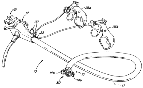
[00037] FIG. 1 is a perspective view of an endoscopic apparatus for use
according to one embodiment of the present invention.
[00038] FIG. 2 is a perspective view of the distal end of the endoscopic
apparatus of FIG. 1 showing the arms in an open position and surgical tools
protruding from the working channels therein.
[00039] FIG. 3 is a perspective view of the distal end of the endoscopic
apparatus of FIG. 1, with arms in a closed position.
[0004o] FIG. 4 is an end view of the distal end of the endoscopic
apparatus of FIG. 1, with arms in a closed position.
[00041] FIG. 5 is a perspective view of the handle on the proximal end of
the endoscopic apparatus of FIG. 1.
[00042] FIG. 6 is a second perspective view of the handle on the proximal
end of the endoscopic apparatus of FIG. 1.
CA 02639862 2008-09-29
-10-
[00043] FIG. 7 is a schematic view of the endoscopic apparatus of FIG. 1
being inserted into the esophagus of a patient.
[oo044] FIG. 8 is a schematic view of the distal end of the endoscopic
apparatus of FIG. 1 in the stomach of a patient.
[00045] FIG. 9 is a schematic view of the endoscopic apparatus of FIG. 1
being used in a gallbladder dissection according to one embodiment of the
present invention.
Detailed Description Of The Drawings
[00046] The use of a transiuminal endoscope and instruments according
to an embodiment of the present invention utilizes an endoscopic apparatus
such
as apparatus 10 shown in Figure 1. The endoscopic apparatus 10 includes
tubular member 11 and handle 12 which is located on a proximal end 31 of
tubular member 11. At the distal end 30 of tubular member 11 is a head portion
13 of the apparatus 10, having two pivotable arms 14a and 14b fixed thereon.
The arms 14a and 14b are attached by hinges to the head portion 13 of the
apparatus 10. Two surgical tools 25a and 25b are also shown in Figure 1. The
surgical tools 25a and 25b are shown inserted into the endoscopic apparatus 10
at proximal terminals 22 of working channels running along the longitudinal
axis
of the tubular member 11. The surgical tools utilized with apparatus 10, such
as
tools 25a and 25b, generally have tools and articulating portions on their
distal
ends which are controlled using a handle portion on a proximal end.
[00047] The term "tubular member" as used throughout this application
refers to many possible configurations. In one embodiment, the tubular member
11 has a shaft at its proximal end that is attached to the handle 12 and is
CA 02639862 2008-09-29
-11-
substantially inflexible. Attached to the shaft portion is a series of
articulating
vertebrae, the articulation of which is controlled by the surgeon using
control
mechanisms on the handle. In that embodiment, the head portion 13 is either
the
last vertebra of the series of vertebrae or a special member attached to the
last
vertebra. In another embodiment, the tubular member 11 could be a single
element, constructed out of a flexible material designed to have a selected
degree of plasticity and elasticity. In that embodiment, the head portion 13
may
or may not be a separate element distinct from the tubular member 11, but
merely the most distal portion of the tubular member 11.
[00048] Figure 2 shows a close-up view of the head portion 13 of the
endoscopic apparatus 10. Arms 14a and 14b are shown in an open position.
The arms 14a and 14b include the guiding channels 16a and 16b, which align
with working channels passing through tubular member 11 (not visible in Figure
2). Guiding channels 16a and 16b receive and guide surgical tools 25a and 25b
which are shown protruding from the guiding channels 16a and 16b. The angle
of arms 14a and 14b determines the angle that surgical tools 25a and 25b
approach a surgical site. The surgeon may select an angle for the arms 14a and
14b such that the surgical tools 25a and 25b emerge parallel to each other, at
an
angle less than parallel, or at an angle more than parallel. The configuration
of
arms 14a and 14b will affect the degree to which the arms can be opened. In
the
embodiment shown in Figure 2, arms 14a and 14b have tissue displacing
members 26a and 26b formed on their outer surface for manipulating and
displacing tissue. In many embodiments, arms 14a and 14b do not include
displacing members 26a and 26b but such arms are still advantageously used to
temporarily displace and manipulate tissue. Further, in some embodiments the
head portion 13 of an endoscopic apparatus 10, including the arms 14a and 14b,
is electrically isolated so as to enable electrosurgical procedures.
CA 02639862 2008-09-29
-12-
[00049] Figure 2 also shows the distal terminals of channels 15a-d,
wherein channel 15a is an optical channel, channel 15b is a third working
channel, channels labeled 15c are illumination channels, and channel 15d is a
fluid or air channel. In general, illumination channels 15c provide light to
the
surgical site so that the surgeon may view the site via the optical channel
15a.
Fluid or air channel 15d may be used to deliver air, water, pharmaceutical
fluids,
or the like to the surgical site. Fluid or air channel 15d is used in some
embodiments to provide insufflation in the vicinity of the distal end 30 of
the
apparatus 10. In some embodiments, working channel 15b is used for
insufflation. Fluid or air channel 15d may also be used as a means for sensing
the ambient pressure at the surgical site. Alternatively, pressure-sensing may
be
accomplished at other points on the head portion 13. The third working channel
15b may be advantageously employed as a means for evacuating fluids from the
surgical site. In some embodiments, small particles of solid matter may also
be
evacuated by channel 15b.
[0005o] The third working channel 15b does not pass through the guiding
channels 16a and 16b in arms 14a and 14b. This gives the surgeon the ability
to
easily exert force in directions parallel to the axis of the tubular member
11.
Thus, the surgeon is provided with the ability to exert force in many
directions at
the surgical site: forward or backward along the axis of the tubular member 11
or
at various angles according to the angles of arms 14a and 14b.
[00051] Figure 3 shows head portion 13 of the endoscopic apparatus 10
with arms 14a and 14b in a closed position. A hinge 24 is shown, which
pivotably connects the arm 14b to the head portion 13. Arm 14a is connected to
tubular member 11 in the same fashion, however this connection is not shown in
Figure 3. In the closed position, arms 14a and 14b provide a ramp for a
surgical
tool or instrument passing through working channel 15b in some embodiments.
CA 02639862 2008-09-29
-13-
This ramp is formed by the shape of the arms 14a and 14b or by protruding
members formed on the inner surface of the arms 14a and 14b. This ramp
brings the tool or instrument directly into the field of view of optical
channel 15a.
[00052] Figure 4 shows an alternative view of the head portion 13 of the
endoscopic apparatus 10 with arms 14a and 14b in a closed position. This view
shows one advantageous configuration of the apparatus 10, in which when the
arms 14a and 14b are in a closed position, they define an opening 17. The
opening 17 allows for utilization of the optical channel 15a, the fluid
delivery
channel 15d, and at least one of the illumination channels 15c in this
embodiment even when the arms 14a and 14b are in a closed position. This
allows a surgeon to more safely and effectively employ the endoscopic
apparatus
10, for example, during insertion of the apparatus into the body of a patient.
Also, as stated above, the arms 14a and 14b provide a ramp for a surgical tool
emerging from working channel 15b.
[00053] Methods according to the present invention require the
performance of a variety of surgical tasks. In order to maximize the
efficiency of
these methods, the apparatus 10 is advantageously employed in methods
according to the present invention using arms of various configurations. The
various configurations of arms are interchangeable in the endoscopic apparatus
and one set of arms can be easily substituted for another set. Because of the
wide variety of surgical procedures which employ the method according to the
present invention, arms having different configurations are desirable for
optimal
performance of the system. The optimal arm configuration depends, for
example, on such things as the organ on which surgery is to be performed, the
type of surgery to be performed, or the condition of the patient.
CA 02639862 2008-09-29
-14-
[00054] For example, in some embodiments the arms 14a and 14b are
constructed out of transparent material so that the optical channel 15a and
the
illumination channels 15c may be utilized even when the arms are in a closed
position. In such a design, the opening 17 shown in Figure 4 may not be
necessary and the arms 14a and 14b could completely cover the head portion 13
of the endoscopic apparatus 10. This configuration further eases insertion of
the
system into a patient. As a second example, in some embodiments the outer
surface of the arms provides a means for tissue manipulation at the surgical
site.
The outer surface could have members formed thereon for displacing tissue. In
such a case, the arms are used to move tissue aside or obtain the desired
degree of stretching of tissue. In a further example, the arms may also grasp
tissue or organs to stabilize or remove them from the surgical site. Finally,
some
arm configurations may include a blade for snipping or cutting tissue. Certain
arm configurations will perform these tasks better than other configurations.
Thus, it is highly desirable to have the ability to interchange the arms
located on
the head portion 13 or even the entire head portion itself.
[oo055] Most arm configurations that are advantageously employed in the
present invention have a shape such that when the arms are in a closed
position,
they act as an obturator or blunt-tip trocar. This obturator or blunt-tip
trocar
shape allows for easier insertion into the body because arms of this shape
will
harmlessly and temporarily displace tissue during insertion.
[00056] Figures 5 and 6 show a close-up view of the handle 12 according
to one embodiment of the invention. The handle 12 is attached at a proximal
end
of tubular member 11. The proximal terminals 22 of working channels 16a, 15b,
and 16b as well as the proximal terminals 21 of the fluid or air channel 15d
are
shown. Camera controls 19 for controlling optical components utilizing the
optical channel 15a are shown in Figure 5. In some embodiments, the camera
CA 02639862 2008-09-29
-15-
controls 19 control the degree of focus and zoom of the camera so that the
surgeon is ensured a clear view of the surgical site. In some embodiments of
the
present invention, the system is advantageously adapted to permit video
recording of the surgery for later analysis or educational purposes. Figures 5
and 6 also show dial controls 40 and 41 which control the articulation and
positioning of the distal end of the apparatus 10.
[00057] Figure 6 also shows control switch 20 for controlling the position
of the arms on the distal end. The control switch 20 may be of the sliding
type as
shown, a rotatable knob type, or any other appropriate design. In some
embodiments, this switch advantageously has a locking mechanism so that the
arms can be locked in a position selected by the surgeon.
[00058] The following is an exemplary use of the endoscopic apparatus in
the performance of a gallbladder dissection according to the present
invention.
Figures 7-9 show illustrations at key points in the method. First, an
endoscopic
apparatus is provided and appropriate pivotable arms are selected. The
configuration of the arms will depend on the particular surgery, and could
include
arms adapted to grasp, cut, and/or displace tissue. Then, as shown in Figure
7,
the endoscopic apparatus 10 is inserted into a patient's mouth 50 and advanced
down the patient's esophagus 51. During this step, the arms are in a closed
position to minimize strain and trauma on the patient. The endoscopic
apparatus
is advanced into the stomach 52. The apparatus is guided during insertion with
a
high degree of accuracy even when the arms are in the closed position using
the
optical and illumination channels. This is possible either because of the
advantageous opening 17 present between the pivotable arms 14 or because the
arms 14 are constructed out of a transparent material.
CA 02639862 2008-09-29
-16-
[000591 Once the distal end of the apparatus 10 reaches the stomach, the
anterior gastric wall 53 is identified. It is intended that the apparatus 10
will be
moved through an incision made at the appropriate location in this part of the
stomach. To make the necessary incision 54, the arms 14a and 14b of the
apparatus 10 are pivoted about their hinges into an open position and a
deflectable hook or cutting needle 55 is advanced through one of the working
channels of the apparatus 10. The hook or cutting needle 55 is then used to
create a gastrotomy 54 sized to accommodate the endoscopic apparatus 10.
Once this is done, the arms 14a and 14b are pivoted about their hinges to a
closed position, creating the obturator or blunt-tip trocar shape, and the
distal end
of the apparatus is advanced through the gastrotomy and into the peritoneal
cavity.
[000601 In some embodiments, the incision 54 is created by advancing a
tool 55 in the working channel 15b and making the incision 54 with the arms
14a
and 14b in a closed position. The tool 55 will, in some embodiments, be
deflected by a ramp formed by the arms 14a and 14b when they are in a closed
position. The tool 55 will be brought into the surgeon's field of view and the
proper incision 54 may be made.
[00061] Once inside the peritoneal cavity, the arms are pivoted about
their hinges into an open position if a wide view via the optical channel is
desired.
The distal end is then articulated in a`retroflex' maneuver in order to access
the
right upper abdominal quadrant and to identify the gallbladder 56. During this
process, insufflation is often desired and is accomplished using a channel of
the
endoscopic apparatus (such as the fluid delivery channel 15d). The intra-
peritoneal pressure is also monitored using a channel of the endoscopic
apparatus. In some embodiments, the pivoting of the arms also serves to
CA 02639862 2008-09-29
-17-
displace tissue to a selected degree to achieve a desired amount of stretching
or
to create more room for the performance of surgical tasks.
[00062] The gallbladder 56 is then manipulated using surgical tools 25a
and 25b introduced through the working channels and the guiding channels 16a
and 16b of the arms. A third surgical tool 58, such as an irrigation-
aspiration
device, may be introduced through the third working channel 15b. Triangulation
of the surgical tools is achieved as a result of the angle of the arms in
their open
position.
[00063] The gallbladder 56 and its pedicle are dissected using a blunt
coagulation tip on one of the surgical tools. The cystic duct is isolated,
clipped,
and divided with articulated scissors. After completing the dissection of the
gallbladder from the liver, the gallbladder is placed in an endoscopic
retrieving
bag 57 which was advanced to the surgical site parallel to the endoscopic
apparatus 10. In some embodiments, the endoscopic retrieving bag 57 is
advanced down the working channel 15b and then expanded as it exits the
apparatus 10. Once the gallbladder is in the bag 57, in such embodiment, it
may
be held between and within the arms 14a and 14b in a closed position while the
apparatus 10 is withdrawn from the patient. Many other surgical tasks are
possible using surgical tools introduced through the working channels. Such
surgical tools include cauterizing tools, lasers, clippers, cutters, and the
like.
[00064] The surgical tools are withdrawn from the surgical site and back
through the guiding channels 16a and 16b so that the arms may be pivoted into
a
closed position. The apparatus 10 and the retrieving bag 57 are withdrawn from
the peritoneal cavity into the stomach. Once the apparatus 10 is again in the
stomach 52, the arms are pivoted to an open position which again creates
triangulation of the surgical tools which are necessary to close the
gastrotomy
CA 02639862 2008-09-29
-18-
54. The gastrotomy 54 is closed by such means as surgical clips, suture,
endoloop, or the like. In some embodiments, the configuration of the arms
includes a suturing system incorporated into the arms themselves.
[00065] In order to remove the retrieving bag containing the gallbladder
through the patient's mouth, the apparatus 10 must first be completely
removed.
After closing the gastrotomy 54, the arms of the apparatus 10 are pivoted into
a
closed position and the apparatus 10 is completely withdrawn from the body of
the patient. Finally, the retrieving bag 57 is withdrawn via the patient's
mouth.
[00066] In addition to gallbladder dissection procedures such as that just
described, the uses of the present invention are applied to a wide variety of
other
surgical procedures. These procedures include, but are not limited to,
appendectomy, splenectomy, mucosectomy, cholecystectomy, liver resection,
small bowel enteroscopy, small bowel resection, tubal ligation,
gastrointestinal
fistulas, peritoneoscopy, fundoplication, gastroplasty, gastro-entero-
anastomosis,
adrenalectomy, common bile duct exploration, ileo-cecal resection, ileoplasty,
and endoluminoplasty.
[00067] Some embodiments of the use according to the present invention
are performed under robotic or electronic control. This allows for highly
precise
and effective remote surgery.
[00068] The simplicity of the use of the present invention is well illustrated
by an analogy to traditional laparoscopic methods. In a traditional
laparoscopic
method, a trocar having a tip and sheathed in a cannula is inserted into the
patient's abdomen. In order to proceed with the surgery, the tip must be
removed so that surgical tools will have access to the surgical site. The use
of
the present invention is much simpler, as the blunt-tip trocar shaped arms are
CA 02639862 2008-09-29
-19-
quickly and easily pivoted into and out of position. Thus, it is not necessary
to
completely withdraw a trocar tip or the surgical instruments every time the
instrument reaches a surgical site or must be advanced further within the
patient's body.
[00069] Thus, the use of the transluminal endoscope and instruments of
the present invention are substantial improvements over the prior art. The
present invention simplifies transluminal surgery and thus improves the safety
and effectiveness of transluminal surgical procedures. The risk of infection,
the
recovery time, and the pain associated with surgery are all reduced.
[00070] Although the invention has been described with reference to a
particular arrangement of parts, features, steps, and the like, these are not
intended to exhaust all possible arrangements of features or steps, and indeed
many other modifications and variations will be ascertainable to those of
skill in
the art.