Note: Descriptions are shown in the official language in which they were submitted.
CA 02639889 2008-06-05
WO 2007/076347 PCT/US2006/062288
CHARGE-INTEGRATING RETINAL PROSTHESIS AND METHOD
FIELD OF THE INVENTION
[0001] The present invention relates generally to retinal prostheses and, in
particular, to a retinal prosthesis configured to integrate charge resulting
from
photoconduction caused by incident light.
BACKGROUND
[0002] Many human retinal diseases cause vision loss by partial to
complete destruction of the vascular layers of the eye. that include. the
choroid
and choriocapillaris, both of which nourish the outer anatomical retina and a
portion of the inner anatomical retina of the eye.
[0003] Many other retinal diseases cause vision loss due to partial to
complete degeneration of one or both of the two anatomical retinal layers
directly, due to inherent abnormalities of these layers. The components of the
retinal layers include Bruch's membrane and retinal pigment epithelium which
comprise the "outer anatomical retinal layee', and the photoreceptor, outer
nuclear, outer plexiform, inner nuclear, inner plexiform, amacrine cell, .
ganglion cell and nerve fiber layers which comprise the "inner anatomical
retinal layer", also known as the "neuroretina". The outer portion of the
neuroretina is comprised of the photoreceptor and bipolar cell layers and is
also known as the "outer retina" which is to be distinguished from the "outer
anatomical retinal laye-" as defined above. Loss of function of the outer
retina
is commonly the result of dysfunction of the outer anatomical retinal layer
that
provides nourishment to the outer retina and/or direct defects of the outer
retina itself. The final common result is dysfunction of the outer retina that
contains the light sensing cells, the photoreceptors. These "outer retina"
diseases include age-related macula degeneration, retinitis pigmentosa,
choroidal disease, long-term retinal detachment, diabetic retinopathies,
Stargardt's disease, choroideremia, Best's disease, and rupture of the
choroid. The inner portion of the neuroretina, however, often remains
-1 -
CA 02639889 2008-06-05
WO 2007/076347 PCT/US2006/062288
functionally and anatomically quite intact and may be activated by the
appropriate stimuli.
[0004] There are currently numerous efforts underway to develop
prosthetic devices that may be used to replace some degree of visual function
to patients with the diseases described above. Many of the approaches are
premised on the hypothesis that acute electrical stimulation using an array of
stimulation electrodes may be used to form patterned vision. Typically, these
approaches rely on relatively complex systems in which a video camera or
similar device is used to capture images for subsequent processing and
eneoding. The encoded information thereafter controls electrical stimulation
provided via an array of electrodes implanted proximate to retinal tissues.
Typically, the electrode array is implanted epiretinally (on the ganglion cell
layer side of the neuroretina) or subretinally (between the outer retina and
the
outer anatomical retinal layer, as defined above) and is connected through a
wired or wireless connection to the appropriate control circuitry. In some
instances, the wired connection must traverse the sclera, the tough outer
coating of the eye often referred to as the white portion of the eye.
Regardless, an advantage of providing such a connection between the control
circuitry and the stimulating array is the ability to control the level of
electrical
stimulation delivered to neural tissues.
[0005] However, in addition to being relatively complex, systems of the
type described above fail to take advantage of the eye's natural movements
and its ability to focus images on the retina. They instead produce
stimulation
in a pattern not necessarily having any relationship to the eye's spatial
orientation.
[0006] One approach that does take advantage of the eye's natural
movements and focusing ability is the ASR device developed by Optobionics
Corporation. Comprising an array of several thousand electrode-tipped,
independent photodiodes, the ASR device is implanted in the subretinal
space of the eye. The photodiodes are designed to essentially mimic the
function of missing or non-functioning photoreceptors by converting incident
light to electrical stimulation that may be further processed by the remaining
-2-
CA 02639889 2008-06-05
WO 2007/076347 PCT/US2006/062288
retinal cell layers. Because of its simple design, the ASR device offers
several advantages over other, more complex retinal prosthesis systems.
While it is believed that the ASR device can be configured to generate
sufficient electrical stimulation to reach stimulation thresholds, thereby
inducing neuronal responses, efficacy of the device could be enhanced and
perhaps better controlled through the provision of additional power.
[0007] Therefore, it=would be advantageous to provide a retinal prosthesis
that combines the simplicity and ability to exploit natural eye movements of
the ASR device with the power control capability of other, more complex
retinal prosthesis systems.
SUMMARY
[0008] The present invention comprises a retinal prosthesis that provides
power control capabilities through the temporal integration or accumulation of
electrical charge. In particular, a retinal prosthesis in accordance with the
present invention comprises at least one stimulating component, each
stimulating component in turn comprising a photojunction element (e.g., a
photodiode) in electrical communication with an electrode. A pulse
generation circuit in electrical communication with the photojunction element
provides a reverse-bias signal to the photojunction element and, from time to
time, provides a pulsatile forward-bias signal to the photojunction element.
In
a presently preferred embodiment, an array of photojunction elements is
provided, which array is electrically coupled to the pulse generation circuit
via
a common electrode. An at least partially-implanted energy source is used to
provide power to the pulse generation circuit. In various embodiments of the
present invention, the implanted portion of the energy source as well as the
pulse generation circuit may be implanted in an extra-ocular (i.e., within the
body but outside the eye) and/or intra-ocular (i.e., within the eye)
configuration.
[0009] During application of the reverse-bias signal, through the process of
photoconduction, light incident upon the photojunction element causes
electrical charge to be accumulated over time in a manner akin to the
-3-
CA 02639889 2008-06-05
WO 2007/076347 PCT/US2006/062288
accumulation of charge in a single pixel element of an electronic camera
during a single exposure. Upon subsequent application of the pulsatile
forward-bias signal, electrical continuity through the photojunction element
results, and the accumulated electrical charge is injected via the electrode
into retinal tissues adjacent the electrode, thereby stimulating the retina.
By
appropriately selecting the parameters of the reverse-bias signal, a
sufficient
amount of charge may be accumulated (and subsequently released by the
pulsatile forward-bias signal) to ensure that stimulation thresholds are met.'
In
this manner, control over stimulus currents may be improved while still
retaining advantageous use of the eye's natural focusing and imaging
capabilities, thereby increasing the probability of successful clinical
outcomes.
BRIEF DESCRIPTION OF THE DRAWINGS
[00101 FIG. 1 is a schematic illustration of a retinal prosthesis in
accordance with the present invention.
[00111 FIG. 2 is a partial cutaway view and schematic illustration of an
embodiment of a retinal prosthesis in accordance with the present invention,
and further illustrating alternative embodiments of various aspects in
accordance with the present invention.
[0012] FIG. 3 is an illustration of waveforms in accordance with prior art
techniques.
[0013] FIG. 4 is a flowchart illustrating operation of a retinal prosthesis
system in accordance with the present invention.
[0014] FIG. 5 is an illustration of waveforms further describing operation of
a retinal prosthesis system in accordance with the present invention.
DETAILED DESCRIPTION OF THE PRESENTLY PREFERRED
EMBODIMENTS
[0015] Referring now to FIG. 1, a schematic illustration of a retinal
prosthesis 100 in accordance with the present invention is provided. In
particular, the retinal prosthesis comprises at least one stimulating
component
102 (only one shown) itself comprising a photojunction element 104 in
electrical communication with an electrode 106 via a first terminal 108 of the
-4-
CA 02639889 2008-06-05
WO 2007/076347 PCT/US2006/062288
photojunction element 104. In a presently preferred embodiment, an array of
stimulating components 102 is provided. The photojunction element 104 is in
further electrical communication with a pulse generation circuit (or pulse
generator) 112 via a second terminal 110 and a conductive element 111: As
described in greater detail below, the conductive element 111 may comprise
one or more conductors, such as flexible, electrically-independent wires or
conductive traces, configured for entirely intra-ocular implantation or for
trans-
scleral routing outside the eye. The pulse generation circuit 112 is
electrically
coupled to a return electrode 114 and to an energy source 116.
[001 fi] In practice, the- photojunction element 104 may comprise any
device or combination of devices capable of exhibiting distinct modes of (i)
photoconduction and (ii) rectification (i.e., one-way conduction). For
example,
a combination of a photoconductive cell such as a cadmium sulfide cell or a
cadmium selenide cell in parallel with a silicon diode would suffice, assuming
adequate biocompatible encapsulation. In a preferred embodiment; the
required combination of photoconduction and rectification is attained using a
photodiode, as known in the art. Such photodiodes operate in a
photoconductive mode when reverse-biased, and exhibit appreciable
conductance in the opposite direction only when forward-biased. When no
bias conditions are externally imposed on a photodiode, it operates in a
photovoltaic mode, although such photovoltaic operation is not required for
the present invention. An example of a retinal prosthesis incorporating the
use of photodiodes is the ASR'o device by Optobionics Corporation, which
may be used to implement the present invention as described in greater detail
below. The ASR device comprises thousands of electrode-tipped
photodiodes formed in an appropriately doped silicon substrate using well
known semiconductor processing techniques.
[0017] A number of well known materials suitable for delivering charge to
biological tissues may be used to provide the electrode 106. Titanium,
platinum, iridium and oxides thereof are but a few examples of suitable
electrode materials, although iridium oxide is preferred. The electrode 106 is
also preferably configured for implantation proximate to retinal tissues. In
-5-
CA 02639889 2008-06-05
WO 2007/076347 PCT/US2006/062288
practice, this implies that the electrodes have dimensions on the order of a
few microns to tens of microns while still retaining the ability to provide
adequate reversible charge injection to reach stimulation thresholds. The
terminais 108, 110 comprise electrically conductive paths; techniques for
creating such paths are readily known to those having ordinary skill in the
art.
[0018] The pulse generation circuit 112 generates a reverse-bias signal
and, from time to time, a pulsatile forward-bias signal which signals are used
to control operation of the photojunction element. In a presently preferred
embodiment, where the photojunction element comprises a photodiode, the
reverse-bias signal may comprise a positive voltage (when applied to the
cathode terminal of the photodiode) and the pulsatile forward-bias signal may
comprise a relatively brief negative voltage (also applied to the cathode
terminal). When continuously applied in succession, the reverse-bias and
pulsatile forward-bias signals in essence form an asymmetric voltage
waveform, such as the one illustrated in, and further described with reference
to, FIG. 5. As also described in further detail below, the reverse-bias signal
causes the photojunction 104 and electrode 106 to effectively integrate
electrical current resulting from the photoconduction induced by incident
light
upon the photojunction 104. The subsequent forward-bias pulse establishes
electrical continuity and thereby allows the accumulated charge to be
released via the electrode into the surrounding environment, i.e., retinal
tissues. Those having ordinary skill in the art will recognize that a variety
of
well know circuits may be used for the pulse generation circuit 112, such as
appropriately configured multivibrator or oscillator circuits fabricated using
discrete components or employing integrated circuits such as the well-known
555 timer IC, where the pulse generation circuit 112 is configured to reside
outside the body. In a preferred embodiment, the pulse generation circuit 112
is configured for implantation in the body and is therefore preferably
fabricated as a standalone integrated circuit or within the same substrate as
the stimulating component 102 such that it may be suitably encapsulated for
implantation.
-6-
CA 02639889 2008-06-05
WO 2007/076347 PCT/US2006/062288
[0019] The pulse generation circuit 112 may also comprise one or more
control inputs 113 that provide the ability to change the configuration of the
reverse-bias and pulsatile forward-bias signals. For example, in certain
circumstances, it may be desirable to change the duration of the forward-bias
pulses, or to change their amplitude. Techniques for controlling such
parameters are well known in the art. Additionally, in order to provide
electrical continuity through- the entire electrical circuit established by
the
pulse generation circuit 1-12, stimulating component 102 and the biological
tissue, a remote return electrode 114 is electrically coupled to the pulse
generation circuit 112.
[0020] The energy source 116 provides power to the pulse generation
circuit 112. As described in greater detail with reference to FIG. 2, the
energy
source may reside entirely outside the body with only wired connections
providing the power signal to the pulse generator 112. However, in a
preferred embodiment, at least a portion of the energy source is implanted
within the body, either extra-ocularly or intra-ocularly.
[0021] Referring now to FIG. 2, a partial cutaway view and schematic
illustration of an embodiment of a retinal prosthesis in accordance with the
present invention, and further illustrating alternative embodiments of various
aspects in accordance with the present invention, is provided. A photodiode
array 202 comprising a plurality of photodiodes 204 formed in a
semiconductor substrate 206 (preferably silicon) is provided. As shown, the
anode of each photodiode 204 is in electrical communication with a uniquely
corresponding electrode 208; as described above, each pairing of photodiode
204 and uniquely corresponding electrode 208 constitutes a stimulating
component 102. The electrodes 208 are preferably fashioned out of iridium
oxide and are fabricated on a surface of the substrate 206 intended to' face
incident light 220 and, optionally, any additional light 230 beyond normal,
ambient light 220. The cathode of each photodiode 204 is in electrical
communication with a common electrode 210, which is preferably fabricated
from the same material as the stimulating electrodes 208. Finally, an
insulating layer 212 is provided which electrically isolates the common
-7-
CA 02639889 2008-06-05
WO 2007/076347 PCT/US2006/062288
electrode 210 from the environment surrounding the retinal prosthesis, i.e.,
the conductive ocular environment. Additionally, the insulating layer should
be biocompatible and biodurable. Thus, for example, the insulating layer 212
may be fabricated using non-conductive polymers such as parylene, or other
materials such as diamond-like carbon. Note that the photodiode array 202
and its constituent elements in FIG. 2 are not shown to scale; the dimensions
shown are for illustrative purposes only. Furthermore, additional
biocompatibility/biodurability coatings (not shown) may be disposed on those
surfaces (other than the stimulating electrodes 208) that would otherwise
come into contact with the -biological environment, as known in the art.
[0022] FIG. 2 also schematically illustrates alternative embodiments for the
configuration of the pulse generator 240, 240' and the energy source. For
example, as illustrated on the lower left-hand portion of FIG. 2, the energy
source is embodied by an inductive coupling system comprising a primary coil
250 that uses electromagnetic signals 254 (typically in the kilohertz to
megahertz frequency range) to transfer power via a secondary coil 252 and
rectifying circuitry (not shown for ease of illustration). As shown in this
example, the primary coil 250 may be configured for external positioning as in
the case of a coil mounted anteriorly or temporally on a pair of glasses or
goggles. The secondary coil 252 and associated rectifying circuitry are
implanted within the body but outside the eye, i.e., extra-ocularly. Such an
arrangement of primary and second coils 250, 252 is further described in
published patent application WO 03/061537. Likewise, the pulse generator
240 and return electrode 242 are also implanted extra-ocularly. In this case,
only the photodiode array 202 and a portion of a trans-scieral conductive
element 256 are implanted intra-ocularly. Obviously, in this example, like the
photodiode array 202, the other implanted components 240, 252, 256 would
need to be encapsulated in an appropriate biocompatible/biodurable coating.
Furthermore, although the pulse generator 240, return electrode 242 and
secondary coil 252 (and associated circuitry) have been described as
configured for extra-ocular placement, it is possible that some or all of
these
components could be configured for intra-ocular placement as known in the
-8-
CA 02639889 2008-06-05
WO 2007/076347 PCT/US2006/062288
art. For example, as taught by Humayun in published patent application WO
99/45870, the secondary coil and associated rectifying circuitry can be
implanted within the eye entirely.
[0023] Yet another configuration for an alternative energy source is
illustrated in the lower right-hand portion of FIG. 2. In this example, the
energy source is embodied by a combination of a light source 260 and a
photovoltaic element 262. The light source 260 may comprise any source of
light in the relevant portion of the spectrum, preferably in the non-visible
range, e.g., an infrared light-emitting diode or an infrared laser. Light 264
is
transmitted to the implanted photovoltaic element -262, =such as a photodiode,
which converts the transmitted light 264 into electrical power that is
thereafter
provided to the pulse generator 240'. As shown in FIG. 2, the photovoltaic
element 262, the pulse generator 240', the return electrode 242' and the
conductive element 266 are all implanted intra-ocularly. As in the previous
example, however, this is not a necessity as some or all of those components
may be implanted extra-ocularly. For example, U.S. Patent No. 6,427,087
issued to Chow illustrates the use of a photovoltaic element implanted within
the anterior chamber or lens capsule of an eye. = However, such a
photovoltaic element may also be positioned extra-ocularly, for example, on
the surface of the sclera but underneath the conjunctiva. In that case, the
pulse generator 240' might also be positioned somewhere within the eye's
orbit (the bony cavity within which the eye resides) with only a trans-scleral
conductive element 266, in addition to the photodiode array 202, positioned
within the eye.
[0024] One other alternative embodiment is further illustrated in FIG. 2. In
particular, rather than using a remote return electrode 242, 242', a return
electrode 270 may be incorporated into the photodiode array 202, as
illustrated by the dotted lines forming a grid pattern around the stimulating
electrodes 208. The electrodes 208 are electrically isolated from the return
electrode grid 270 by buffer regions 272 surrounding each electrode 208. In
practice, such an arrangement results in a more tightly confined distribution
of
electrical currents surrounding each stimulating electrode 208, which may
-9-
CA 02639889 2008-06-05
WO 2007/076347 PCT/US2006/062288
provide greater resolution in the pattern of stimulation provided by the
retinal
prosthesis. Although the electrode grid 270 is illustrated as a continuous,
common electrode, it is also possible, though less preferred, to provide more
than one return electrode 270 up to and including a uniquely corresponding
return electrode for each electrode 208. Regardless, if the return electrode
270 is disposed in such close proximity to the stimulating electrodes 208, one
or more additional conductive elements 274 would need to be provided to
complete the electrical circuit with the pulse generator 240'.
[0025] Referring now to FIGS. 3-5, operation of the present invention is
further described and compared- with prior art techniques: In F1G. 3,
operation of a retinal prosthesis proposed by Palanker et al. in their paper
"Design of a high-resolution optoelectronic retinal prosthesis", J. Neural
Eng.
2 (2005) S105-S120 (hereinafter, "Palanker") is described. The scheme set
forth in Palanker is premised on the concept of providing pulsed excitation
both optically and electrically to a photodiode array of the type described
above such that reverse-biasing of the photodiodes occurs in synchrony with
the pulsed optical signals. Thus, as illustrated by the voltage waveform, V, a
forward-bias signal 302 is provided to the photodiodes and, from time to time,
a pulsatile reverse-bias signal 304 is also provided. During the reverse-bias
pulses 304, the photodiodes operate in a photoconduction mode as noted
above. Controlled optical pulses, whose intensity is illustrated by the
waveform labeled N, are provided in synchrony with the pulsatile reverse-bias
signals 304. Thus, the resulting current conducted by the photodiode (upon
which the optical pulses 306, 307 are incident) will be directly proportional
to
the intensity of the incident optical pulse. This is illustrated in FIG. 3
where a
first optical pulse 306 having a first amplitude, A, is provided resulting in
a
cathodic current 308 of amplitude A' and a subsequent anodic current 309 of
amplitude A". In contrast, second optical pulse 307 having a second
amplitude, B, which is less than the first amplitude A, results in a cathodic
current 310 having amplitude B' less than amplitude A' and a subsequent
anodic current 311 having amplitude B" less than amplitude A". Note that the
areas beneath the cathodic current waveforms 308, 310 and their respective
-10-
CA 02639889 2008-06-05
WO 2007/076347 PCT/US2006/062288
anodic current waveforms 309, 311 are substantially identical, thereby
beneficially maintaining zero net charge injection over time. In summary,
Palanker describes a system in which quantities of injected charge are
determined by amplitudes of incident light pulses.
[0026] Referring now to FIG. 4, operation of the present invention is more
fully described. Initially, the parameters for the reverse-bias signal 502 and
the pulsatile forward-bias signal 504 are determined at step 402. In
particular, the amplitudes for the signals 502, 504 are determined, as well as
the timing of the pulsatile forward-bias signal 504. In a preferred
embodiment, the voltages used must take into consideration 'the so-called
"water window" which defines electrochemical potential limits within which the
oxidation and reduction of water may be avoided. Exceeding these limits
typically causes irreversible damage to the stimulating electrodes and may
also cause harmful pH changes in the surrounding tissue. As known in the
art, the anodic potential of the stimulating electrode should not exceed 0.8
V,
and the cathodic potential should not exceed -0.6 V, each with respect to a
AgiAgCl reference. Additionally, the amplitude of individual pulses 504, 506
may be varied, as shown.
[0027] In a preferred embodiment, the pulsatile forward-bias signals 504
occur in a periodic manner at a fixed frequency and for specific durations.
For example, it is expected that pulse repetition frequencies from 1 Hz to 50
Hz may be employed with pulse durations of not less than 0.1 milliseconds
and not more than 100 milliseconds. Although the pulsatile forward-bias
signals 504 are preferably provided at a fixed frequency, this is not a
requirement and they may be provided in an aperiodic manner or at varying
frequencies.
[0028] Referring again to FIG. 4, an optional step 404 of.providing
additional light, beyond ambient light, is shown. As described above, the
Palanker method varies the amount of charge delivered by any individual
photodiode by varying the amplitude of the light pulses delivered to that
photodiode. In contrast, the present invention controls the amount of charge
to be delivered by accumulating charge over time, which charge may be
-11 -
CA 02639889 2008-06-05
WO 2007/076347 PCT/US2006/062288
provided using nothing more than ambient light. However, it is recognized
that in certain low light situations, it may be desirable to deliver
additional light
thereby enabling sufficient charge accumulation. Beneficially, such additional
illumination, which is preferably in the infrared portion of the spectrum and
delivered using glasses or goggles as described above, may be continuous
rather than pulsed.
[0029] At step 406, the reverse-bias signal 502 is applied to the at least
one photojunction element causing the stimulating element to operate in a
photoconduction mode and thereby integrate current over time. The process
of accurnulating charge results from the manner in which electrodes
exchange charge with biological tissue. As known in the art, stimulating
electrodes of the type employed in the present invention establish a
capacitive interface with the surrounding aqueous environment by the
formation of a so-called "electrical double layer" and, in the case of iridium
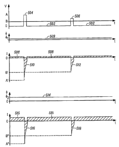
oxide electrodes, also through reversible valence transitions within the
electrode material. The present invention exploits this capacitive property
when the reverse-biased photojunction operates in a photoconductive mode
whereby incident light generates a photocurrent that accumulates on the
capacitance of the electrode and induces a concomitant change in the
potential of the electrode. This is illustrated in FIG. 5 where, during
application of the reverse-bias signal 502, an anodic charge 508 is built up
over time. Note that the amplitude of the reverse-bias signal 502 affects the
maximum level of charge accumulation; higher amplitudes lead to greater
maximum charge levels and lower amplitudes lead to lesser maximum charge
levels.
[0030] The process of accumulating charge continues until the charge
capacity of the electrode is met or until a pulsatile forward-bias pulse 504
is
applied, as shown at step 408. For example, as shown in FIG. 5, an anodic
charge 508 is accumulated for the duration of each integration period. At step
410, the pulsatile forward-bias signal 504 is applied to the photojunction,
establishing electrical continuity through the photojunction. This continuity
results in a change in potential at the electrode causing the previously
-12-
CA 02639889 2008-06-05
WO 2007/076347 PCT/US2006/062288
accumulated charge to be injected into the tissue as illustrated by the
cathodic waveform 510, 512. The amplitude of the cathodic waveform 510 is
determined by the amount of current accumulated during the integration
period, which in turn depends on both the lighting conditions and the duration
of the integration period. Additionally, the amplitude of the cathodic
waveform
510 is controlled by the amplitude of the pulsatile forward-bias signal 504.
This is illustrated in FIG. 5 where a first forward-bias pulse 504 of
amplitude A
results in a cathodic curren# having amplitude A'. In contrast, a subsequent
forward-bias pulse 506 of amplitude B (less than A) results in a corresponding
cathodic current having amplitude -B' (less than A'). An illumination
intensity
cp2 shown in waveform 514, which is greater than the illumination intensity
cQ1
shown in waveform 509, results in a greater accumulation of charge 515 and
a greater cathodic current with amplitude A" in pulse 516 larger than A' in
510. Note that the amplitude of the cathodic waveform 510, 512, does not
depend significantly upon the amplitude of the incident light present during
the forward-bias pulse, as in the Palanker method, but rather on the total
amount of charge accumulated during the reverse-bias period, and on the
amplitude of the forward-bias pulse 504. Thus, if, for a particular
photojunction, an insufficient quantity of light is incident upon the
photojunction in between applications of forward-bias pulses (i.e., during
application of the reverse-bias signal) to establish accumulation of a
sufficient
amount of charge to reach the retinal stimulation threshold, the subsequent
cathodic current will be insufficient to create a perceptual response.
Conversely, a perceptual response will result if there is a sufficient
quantity of
light during the integration period to generate sufficient charge to reach the
stimulation threshold. The charge capacity of the electrode should be large
enough such that the full charge capacity of the electrode should be met only
in relatively bright light conditions, and even then, only after substantially
all of
the integration period has passed. Stated another way, the electrode charge
capacity shouldn't be so low, or the integration period so long, that even
modest amounts of light will cause the capacity limit to be reached in a small
fraction of the integration period. The electrode capacity should be large
-13-
CA 02639889 2008-06-05
WO 2007/076347 PCT/US2006/062288
enough to allow for intermediate levels of light to accumulate charges to
varying degrees above the stimulation threshold without reaching the charge
capacity limit.
[0031] In a relatively simple implementation of the present invention, at the
conclusion of the pulsatile forward-bias signal 504, 506, the reverse-bias
signal 502 is again applied to the photojunction and the process described
above is repeated, as illustrated by the dotted line exiting step 410.
Referring
again to FIG. 4, however, it is possible to ascertain, at step 412, whether
ambient lighting conditions (specifically excluding any additional
illumination
applied, for example; as -part of step 404) have changed to a sufficient
degree
to merit modifying any additional illumination being provided, or not, as the
case may be. Changes to the overall ambient light conditions can be
detected using additional photodetectors as known in the art, or by monitoring
the current delivered by the pulse generator 240. The basis for using the
charge delivered by the pulse generator as a measure of the ambient lighting
conditions is that the total amount of charge supplied by the pulse generator
during each forward-bias pulse will be substantially identical to the total
amount of charge accumulated by all of the elements in the array 202 during
the integration period, and therefore substantially proportional to the
average
illumination intensity on the array. If additional photodetectors are used,
they
may be deployed in such a manner that they are not influenced by any
additional illumination provided. For example, the additional photodetectors
may reside external to the body on the glasses or goggles used to support the
additional illumination light source. The signal derived from photodetectors
or
from monitoring the pulse generator output may be used to test the ambient
change condition set forth in step 412. For example, a decrease in the
ambient light intensity may be used to cause an increase in the additional
external illumination provided at step 414. Likewise, a sufficient increase in
ambient illumination (for example, when a person walks out of a relatively
dark indoor environment to a bright outdoor environment) may be used to
cause the additional external illumination to be decreased or even terminated
at step 414.
-14-
CA 02639889 2008-06-05
WO 2007/076347 PCT/US2006/062288
[0032] Additionally, changes to the ambient lighting may necessitate
changes to the timing and amplitude parameters of the reverse-bias and
pulsatile forward-bias signals, as described above at step 402. For example,
when ambient light levels decrease, it may be desirable to decrease the
frequency of the forward-bias pulses thereby allowing longer integration
times. In brighter ambient conditions, it may be advantageous to decrease
the amplitude of the forward-bias pulses thereby decreasing the amount of
injected charge. In this manner, operation of the present invention may be
refined to provide even greater control of a retinal prosthesis.
[0033] - The present invention provides a technique for providing greater
control over the quantities of electrical charge that may be delivered by a
retinal prosthesis comprising photojunction elements, while still retaining
the
ability of such prostheses to exploit the natural movements and focusing
ability of the eye. This is achieved through the application of reverse-bias
signals that allow the photojunctions to operate in an integration mode
whereby charges are accumulated over time. Application of subsequent
forward-bias pulses cause the accumulated charge to be injected into the
retinal tissue. By accumulating charge over a relatively long integration
period and delivering the accumulated charge in a comparatively short
interval, the present invention achieves stimulus currents that are
substantially greater than the photocurrents generated by the incident light,
and control over the stimulus currents is achieved through adjustment of the
timing and amplitude of the forward- and reverse-bias pulses.
[0034] Although particular embodiments have been disclosed herein in
detail, this has been done for purposes of illustration only and is not
intended
to be limiting with respect to the scope of the appended claims that follow.
In
particular, it is contemplated by the inventors that various substitutions,
alterations, and modifications may be made to the invention without departing
from the spirit and scope of the invention as defined by the claims. Other
aspects, advantages, and modifications are considered to be within the scope
of the following claims.
-15-