Note: Descriptions are shown in the official language in which they were submitted.
CA 02640349 2008-10-03
SURGICAL PORTAL WITH FOAM
AND FABRIC COMPOSITE SEAL ASSEMBLY
BACKGROUND
1. Field of the Disclosure
The present disclosure relates to surgical devices and, more particularly,
relates to a surgical portal apparatus incorporating a seal assembly adapted
for use during
a minimally invasive, e.g., a laparoscopic, surgical procedure.
2. Description of the Related Art
Minimally invasive surgical procedures including both endoscopic and
laparoscopic procedures permit surgery to be performed on organs, tissues and
vessels far
removed from an opening within the tissue. Laparoscopic and endoscopic
procedures
generally require that any instrumentation inserted into the body be sealed,
e.g.,
provisions may be made to ensure that gases do not enter or exit the body
through the
incision as, for example, in surgical procedures in which the surgical region
is insufflated.
These procedures typically employ surgical instruments which are introduced
into the
body through a cannula. The cannula has a seal assembly associated therewith.
The seal
assembly is intended to form a substantially fluid tight seal about the
instrument to
preserve the integrity of the established pneumoperitoneum.
1
CA 02640349 2008-10-03
SUMMARY
The present disclosure is directed to further improvements in seal
assemblies for use with portal apparatii during a surgical procedure. In
accordance with
one embodiment, a surgical portal assembly includes a portal adapted to
provide access to
underlying tissue and having a longitudinal opening extending along a
longitudinal axis
of the portal and a seal. The seal includes internal surfaces having a passage
for
reception and passage of a surgical object in substantial sealed relation
therewith and
defines a seal axis. The seal includes a foam segment comprising a foam
material and a
fabric segment comprising a fabric material and being mounted relative to the
foam
segment. The fabric segment may be disposed adjacent one of the proximal and
distal
surfaces of the foam segment. The fabric segment may include a fabric layer
which is in
juxtaposed relation with the one of the proximal and distal surfaces of the
foam segment.
The fabric layer may include slots therethrough to facilitate passage of the
surgical object
through the seal. The slots may be arranged to extend radially outwardly
relative to the
seal axis.
The seal may have first and second fabric layers mounted in juxtaposed
relation with respective proximal and distal surfaces of the foam segment.
Alternatively,
the seal may further include an elastomeric segment comprising an elastomeric
material
having less elasticity than the foam material of the foam segment. The
elastomeric
segment is mounted in juxtaposed relation to the fabric layer. The seal may
include, from
proximal to distal, the elastomeric segment, the fabric layer and the foam
segment.
2
CA 02640349 2008-10-03
A second layer of fabric may be mounted adjacent the distal surface of the
foam segment
and a third layer of fabric may be mounted adjacent a proximal surface of the
elastomeric
segment.
The seal may include an outer seal area and an inner seal area. The inner
seal area generally tapers in a distal direction to define a general funnel
configuration or a
general sloped configuration which is adapted to facilitate insertion of the
surgical object
and minimize potential of inversion of the inner seal area upon withdrawal of
the surgical
object.
The portal may include a portal housing and a portal sleeve extending
from the portal housing. In this embodiment, the seal may include a
substantially annular
retention member which is received within a corresponding annular recess of
the portal
housing to assist in mounting the seal within the portal housing.
BRIEF DESCRIPTION OF THE DRAWINGS
Preferred embodiments of the present disclosure will be better appreciated
by reference to the drawings wherein:
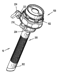
FIG. 1 is a perspective views of a portal system in the form of a cannula
assembly and a seal assembly in accordance with the principles of the present
disclosure;
FIG. 2 is a side plan view of the portal system of FIG. 1;
FIG. 3 is a side cross-sectional view of the portal system;
FIG. 4 is an enlarged view of the area of detail depicted in FIG. 3;
FIG. 5 is a perspective view illustrating the seal assembly detached from
the cannula assembly;
3
CA 02640349 2008-10-03
FIG. 6 is a perspective view with parts separated of the seal assembly
illustrating the seal housing, the object seal and the locking ring;
FIGS. 7A, 7B and 7C are respective plan, perspective and side-cross-
sectional views of the object seal;
FIG. 7D is a perspective view with parts illustrating the components of
the object seal;
FIGS. 8A, 8B and 8C are respective front, side and rear plan views of the
locking ring;
FIGS. 9A-9E are views illustrating a sequence of assembling the seal
components of the seal assembly; and
FIG. 10 is a flow chart illustrating a method for performing a surgical task
with the portal system of FIG. 1.
DETAILED DESCRIPTION
The portal system of the present disclosure incorporates a seal assembly
which, either alone or in combination with a seal internal to a cannula
assembly, provides
a substantial seal between a body cavity of a patient and the outside
atmosphere before,
during and after insertion of an object through the cannula assembly.
Moreover, the seal
assembly is capable of accommodating objects of varying diameters, e.g.,
instruments
from about 4.5 mm to about 15 mm, by providing a gas tight seal with each
instrument
when inserted. The flexibility of the seal assembly greatly facilitates
endoscopic surgery
where a variety of instruments having differing diameters are often needed
during a
single surgical procedure.
4
CA 02640349 2008-10-03
The seal assembly contemplates the introduction and manipulation of
various types of instrumentation adapted for insertion through a trocar and/or
caruiula
assembly while maintaining a fluid tight interface about the instrumentation
to prevent
gas and/or fluid leakage from the established pneumoperitoneum so as to
preserve the
atmospheric integrity of a surgical procedure. Specifically, the seal assembly
accommodates angular manipulation of the surgical instrument relative to the
seal axis.
This feature of the seal assembly desirably minimizes the entry and exit of
gases and/or
fluids to/from the body cavity. Examples of instrumentation include clip
appliers,
graspers, dissectors, retractors, staplers, laser probes, photographic
devices, endoscopes
and laparoscopes, tubes, and the like. Such instruments will be collectively
referred to
herein as "instruments or instrumentation".
The seal assembly may be a component of a portal system adapted to
provide access to an underlying site. The seal assembly may be readily
incorporated into
a portal, such as a conventional trocar device or cannula, and provides the
device with
sealing capability about an inserted instrument.
The seal assembly may also be adapted to receive and form a seal about a
physician's arm or hand during a hand-assisted laparoscopic procedure. In this
application, the seal assembly is a component of an access member which is
introduced
within the body to provide access to underlying tissue in, e.g., the abdominal
cavity.
Referring now to the drawings, in which like reference numerals identify
identical or substantially similar parts throughout the several views, FIGS. 1-
2 illustrate a
portal system 10 of the present disclosure incorporating seal assembly 100
mounted to an
access device such as cannula assembly 200. Cannula assembly 200 may be any
portal
CA 02640349 2008-10-03
member suitable for the intended purpose of accessing a body cavity and
typically
defines a passageway permitting introduction of instruments therethrough.
Cannula
assembly 200 is particularly adapted for use in laparoscopic surgery where the
peritoneal
cavity is insufflated with a suitable gas, e.g., C02, to raise the cavity wall
from the
internal organs therein. Cannula assembly 200 is typically used with an
obturator
assembly (not shown) which may be blunt, a non-bladed, or a sharp pointed
instrument
positionable within the passageway of the cannula assembly 200. The obturator
assembly
is utilized to penetrate the abdominal wall or introduce the cannula assembly
200 through
the abdominal wall, and then subsequently is removed from the cannula assembly
200 to
permit introduction of the surgical instrumentation utilized to perform the
procedure
through the passageway.
With respect to FIGS. 3-4, in conjunction with FIGS. 1-2, cannula
assembly 200 includes cannula sleeve 202 and cannula housing 204 mounted to a
proximal end of the cannula sleeve 202. Cannula sleeve 202 defines a
longitudinal axis
"t" extending along the length of the cannula sleeve 202 and has proximal (or
leading)
and distal (or trailing) ends 206, 208. Mounted adjacent proximal end 206 of
cannula
sleeve 202 is sleeve flange 210. Cannula sleeve 202 further defines an
internal
longitudinal passage 212 dimensioned to permit passage of surgical
instrumentation.
Cannula sleeve 202 may be formed of any suitable medical grade material, such
as
stainless steel or other rigid materials, including polymeric materials, such
as
polycarbonate, or the like. Cannula sleeve 202 may be transparent or opaque.
The
diameter of cannula sleeve 202 may vary, but, typically ranges from about 3.0
to about 18
mm. Cannula sleeve 202 may or may not include means for facilitating retention
of the
6
CA 02640349 2008-10-03
cannula sleeve 202 within tissue. Such means may include a plurality of
locking
elements or ribs such as, e.g., the locking arrangement disclosed in commonly
assigned
U.S. Patent Application Serial No. 11/170,824 to Smith filed June 30, 2005,
the entire
contents of the `824 disclosure being hereby incorporated by reference herein.
Cannula housing 204 may be connected to sleeve flange 210 of cannula
sleeve 202 by conventional means including a bayonet coupling, a threaded
connection,
snap fit, ultrasonic welding or any other means envisioned by one skilled in
the art
including, e.g., adhesive means. Cannula assembly 200 may also incorporate 0-
ring seal
214 disposed between sleeve flange 210 and cannula housing 204 to assist in
sealing the
interior passages of cannula assembly 200. Cannula housing 204 may be a single
monolithically formed unit or composed of several components connected to each
other
through any of the aforementioned connection means. Cannula housing 204
further
includes diametrically opposed housing grips 216 dimensioned and arranged for
gripping
engagement by the fingers of the clinician. Additionally or altematively,
suture anchors
or filaments may extend from cannula housing 210, e.g., from housing grips
216, for
attachment to the epidermis of the patient.
With reference to FIGS. 1-5, cannula housing 204 further includes valve
218. Valve 218 may be a zero-closure valve such as a duck-bill valve having a
slit 220
which is adapted to close in the absence of a surgical object and/or in
response to
insufflation gases of the pressurized cavity. In the alternative, valve 218
may be a gel
seal, balloon valve, or a flapper valve. Cannula housing 204 may further
include port 222
to which stop cock valve 224 is attached. Port 222 permits the introduction of
7
CA 02640349 2008-10-03
insufflation gases though cannula sleeve 202 via opening of stop cock valve
224 to assist
in maintaining the integrity of the pneumoperitoneum. Stop cock valve 224 may
be any
conventional valve. As best depicted in FIG. 5, cannula housing 204 further
includes at
least one, e.g., three, peripheral grooves 226. Grooves 226 extend in an axial
direction
and are preferably equidistally spaced about the periphery of the cannula
housing 204.
Grooves 226 assist in releasably mounting seal assembly 100 to cannula
assembly 200.
Referring now to FIGS. 5-6, in conjunction with FIGS. 3-4, seal assembly
100 will be discussed in detail. Seal assembly 100 includes seal housing,
generally
identified as reference numeral 102, composite object seal 104 which is
disposed within
the seal housing 102 and internal locking element 106. Seal housing 102 houses
the
sealing components of the assembly and defines the outer valve or seal body of
the seal
assembly 100. Seal housing 102 defines central seal housing axis "b" which is
preferably
parallel to the axis "t" of cannula sleeve 202 and, more specifically,
coincident with the
axis "t" of the cannula sleeve 102 when seal assembly 100 is mounted to
cannula
assembly 200. Seal housing 102 may be integrally or monolithically formed as a
single
unit or may incorporate multiple components which, when assembled together,
form the
seal housing 102. In the illustrated embodiment, seal housing 102 is a single
unit formed
of a suitable polymeric material. Suitable polymeric materials include
polycarbonates
polystyrenes, ABS or any other material contemplated by one skilled in the
art.
Seal housing 102 includes proximal end wall 108 defining central aperture
110 and internal annular collar 112 depending from the end wall 108 and
coaxially
arranged about seal housing axis "b". Central aperture 110 and annular collar
112
receive the surgical object and collectively define an internal dimension or
diameter
8
CA 02640349 2008-10-03
adapted to permit passage of relatively large sized instruments. Annular
collar 112 also
may limit the degree of lateral or offset movement of the surgical object,
e.g., surgical
instrument, relative to seal axis "b" by, defining an outer limit of movement
of the
instrument. Seal housing 102 further defines internal peripheral recess or
channel 114
(FIG. 4) approximately adjacent the longitudinal midpoint of the seal housing
102 for
receiving a component of object seal 104 as will be discussed.
Seal housing 102 further includes mounting collar 116 adjacent its distal
end. Mounting collar 116 may be selectively releasably connectable to cannula
housing
204 to cooperatively releasably couple seal assembly 100 to cannula assembly
200.
Various means for releasably securing or connecting mounting collar to cannula
housing
are envisioned including a bayonet coupling, snap-fit, frictional fit, tongue
and groove
arrangement, threaded arrangement, cam-lock mechanisms or the like. One
methodology
contemplated will be discussed in greater detail hereinbelow. Alternatively,
seal housing
102 may be permanently secured to cannula housing 204. Mounting collar 116 may
have
an irregular exterior surface to facilitate engagement by the clinician. In
one
embodiment, mounting collar includes an arrangement of spaced recesses 118 to
assist in
gripping of seal assembly 100.
With particular reference to FIGS. 7A-7D, in view of FIGS. 4 and 6,
composite object seal 104 will be discussed in detail. Object seal 104
includes from,
proximal to distal, retention member 120, first fabric segment 122,
elastomeric segment
124, second fabric segment 126, foam segment 128 and third fabric segment 130.
Retention member 120 is general annular or ring-like in shape and may be
fabricated
from a suitable biocompatible relatively rigid material such as polypropylene,
nylon,
9
CA 02640349 2008-10-03
ABS, polycarbonate, stainless steel, titanium or any other suitable material.
Retention
member 120 assists in mounting object seal 104 within seal housing 102 by its
reception
within peripheral channel 114 of the seal housing 102 (FIG. 4). First fabric
segment 122,
second fabric segment 126 and third fabric segment 130 may each be in the form
of disc-
shaped layers. Fabric segments 122, 126, 130 generally enhance the structural
integrity
of object seal 104 by providing a support lattice or structure to encapsulate
and support
foam segment 128. Fabric segment 122,130 also may prevent foam segment 128 of
object seal 104 from contact with the instrument during insertion and possibly
withdrawal
of the instrument through object seal 104. Fabric segments 122, 126, 130 also
enhance
seal durability and may reduce object or instrument insertion forces. A
suitable fabric
material for each of fabric segments 122, 126, 130 includes a SPANDEXTM
material
containing, e.g., 20% LYCRATM which is commercially available from Milliken of
South
Carolina. The fabric may comprise a woven, knitted, braided, or non-woven
material of
polymeric materials. Other fabric materials are also envisioned. For example,
a synthetic
material such as nylon, Kevlar (Trademark of E.I. DuPont de Nemours and
Company) or
any other material that will expand and compress about an instrument inserted
therethrough is envisioned. In addition, the fabric may be coated, e.g., on
its interior with
urethane, silicon or other flexible lubricious materials to facilitate passage
of an
instrument or other object, through the seal.
Each fabric segment 122, 126, 130 may define an aperture, opening or
passage to permit passage of the surgical object. Single or multiple
intersecting slits
within any one or more of fabric segments 122, 126, 130 are also envisioned.
For
example, second fabric segment 126 may define at least one, possibly, a
plurality of slits
CA 02640349 2008-10-03
132 extending outwardly from seal axis "b". In one embodiment, slits 132 are
substantially linear and extend radially outwardly relative to seal axis "b".
Other
arrangements are envisioned including non-linear slits, serpenditious slits,
intersecting
slits. Slits 132 may be equidistally and radial spaced about seal axis. Slits
132 may
assist in reducing insertion and withdrawal forces needed to advance the
object into the
surgical site by reducing radial constriction of the inner areas of fabric
segment 130 about
the object.
With continued reference to FIGS. 7A-7D, elastomeric segment 124 in
fabricated from a suitable elastomeric or thermoplastic polymer which may
exhibit less
elasticity than the elasticity of the material of foam segment 128.
Elastomeric segment
124 provides protection to foam segment 128 by minimizing the potential of
puncture
with the surgical object, e.g., the instrument during insertion. Elastomeric
segment 128
may be fabricated from a polyisoprene; however, other elastomers are also
envisioned
including neoprene, silicone, butyl, nitrile and Buna-N.
Foam segment 128 is fabricated from a foam material (closed cell or open
cell) such as, in one embodiment, a thermoplastic material comprising a
foaming agent.
Foam segment 128 may be the primary sealing component about the surgical
object. In
one embodiment, foam segment 128 is fabricated from a polyisoprene impregnated
or
injected with a foaming agent including, e.g., CELOGEN", EXPANDEX", and
OPEXTM chemical foaming agents. Foam segment 128 has sufficient elasticity to
bend
and deform about the inserted instrument while conforming about the outer
dimensioning
of the object, e.g., instrument, thereby establishing a fluid tight seal about
the object.
Foam segment 128 is sufficiently compliant to absorb off axis motion of the
instrument.
11
CA 02640349 2008-10-03
Moreover, the compliant characteristics of foam segment 128 may substantially
rriinimize
the formation of a gap around the instrument during off-set manipulation of
the
instrument. The presence of a gap would otherwise permit the undesired release
of gases
from the underlying pneumoperitioneum.
In the assembled condition of object seal 104, best depicted in FIG. 7C,
the object seal 104 defines a generally tapered or funneled profile whereby
the inner area
of the object seal 104 slopes at an oblique angle with respect to the seal
axis "b". The
funneled characteristic may assist in guiding the instrument toward central
aperture 134
during initial introduction of the instrument. The funneled characteristic
also may
substantially minimize the potential of inversion of object seal 104 during
withdrawal of
the instrument. More particular, object seal 104 defines proximal seal surface
104p
adjacent fabric segment 122 and distal seal surface 104p adjacent fabric
segment 130
defining an arcuate, parabolic or hyperbolic profile. In one embodiment,
proximal seal
sui-face 104p may define a surface portion having a radius of curvature "m"
=ranging from
about .540 inches to about .620 inches, and distal seal surface 104d may
define a surface
portion having a radius of curvature "k" ranging from about .430 inches to
about .500
inches. Other dimensioning and radii of curvatures are also envisioned. The
axial length
"j" of object seal 104 ranges from about .47 inches to about .53 inches
(excluding
retention member 120). This overall dimensioning effected through the sloped
arrangement provides an exaggerated funneled profile to object seal 104,
which,
facilitates directing or funneling of the surgical object through object seal
104 and, also,
minimizes the potential of inversion of the object seal 104 during withdrawal
of the
object.
12
CA 02640349 2008-10-03
Object seal 104 may be manufactured via conventional means. In one
method, fabric segments 122, 126, 130, elastomeric segment 124 and foam
segment 128
may be compression molded while the elastomeric material of the elastomeric
segment
124 and/or the foam material of the foam segment 128 is subjected to heat to
at least
partially embed the fabric material of fabric segments 122, 126, 130 into the
elastomeric
and/or foam segments 124, 128. Alternatively, the components of object seal
104 may be
attached with adhesives, cements, or the like. The methodologies for attaching
fabric
segments 122, 126, 130 to one or both of elastomeric and foam segments 124,
128 as
disclosed in certain embodiments of the U.S. Patent. No, 6,702,787 to Racenet
also may
be utilized. The entire disclosure of U.S. Patent No. 6,702,787 to Racenet is
hereby
incorporated by reference herein. Another method for fabricating seal 104 is
disclosed in
(H-US-00875), the entire contents of which are hereby incorporated by
reference herein.
Once object seal 104 is assembled or manufactured, central seal aperture 134
may be
punched through the composite materials with a die punch or made via a molding
process
which provides the seal aperture 134. Alternatively, the respective components
of object
seal 104 may be provided with cooperative aperture or slits and then assembled
via any
of the aforementioned methodologies.
In the alternative, object seal 104 may be a substantially flat or planar
septum seal having an aperture, slit or the like. Object seal 104 also may
comprise a gel
composition or very-soft thermoplastic elastomer in addition to, or in lieu
of, foam
segment 128. Gels and soft thermoplastic materials contemplated for use are
known ~.
under the trade names VERSAFLEXTM, FLEXPLAST", DYANFLEXTM and
13
CA 02640349 2008-10-03
MONPRENETM. Other suitable materials include soft silicone and polyurethane
composites. These materials may be adapted to be sufficiently pliable.
Object seal 104 may incorporate a lubricant or a therapeutic or
pharmacological agent. Suitable lubricants include a coating of
hydrocyclosiloxane
prepared by plasma polymerization process. Such a coating is disclosed in U.S.
Pat. No.
5,463,010 to Hu et al., the entire contents of which is hereby incorporated by
reference.
Examples of therapeutic or pharmacological agents include antimicrobials,
antibacterials,
hemostatic, moisture-providing agents, such as saline, healing agents,
lubricious agents,
analgesics, antiseptics, growth factors, and/or anti-inflammatory agents.
Referring now to FIGS. 8A-8C, in conjunction with FIGS. 4 and 6,
locking ring 106 of seal assembly 100 will be discussed. Locking ring 106
serves a dual
function of securing object seal 104 within seal housing 102 and providing an
intemal
seal within the passageway of the assembled seal and cannula assemblies 100,
200.
Locking ring 106 may be fabricated from a material exhibiting some resiliency
including
any elastomeric material hereinabove described. Locking ring 106 includes
outer
segment 136 and internal gasket segment 138. Outer segment 136 defines a
plurality
(e.g., three) peripheral recesses 140 equidistally spaced about the periphery.
Recesses
140 assist in mounting locking ring 106 within seal housing 102. Internal
gasket segment
138 of locking ring 106 may contact distal seal surface 104d of object seal
104 in the
assembled condition of seal assembly 100. Outer segment 136 and internal
gasket
segment 138 may be monolithically formed or may consist of separate components
received to each other by conventional means. Intemal gasket segment 138 may
be more
elastic than outer segment 136. Outer segment 136 may incorporate first and
second 0-
1 4
CA 02640349 2008-10-03
ring seals 142 within annular recesses of the outer segment to assist in
sealing the internal
opening of seal housing 102.
The assembly of seal assembly 100 will now be discussed. With reference
to FIGS. 9A-9F, in conjunction with FIG. 4, object seal 104 is aligned with
the internal
area of seal housing 102 and advanced within the seal housing 102 whereby
retention
member 120 is received within channel 114 of seal housing 102 (see also FIG.
4).
Locking ring 106 is thereafter positioned whereby peripheral recesses 140 of
the locking
ring 106 are aligned with mounting tabs 142 extending radially inwardly from
the interior
wall of seal housing 102. In particular, as best depicted in FIGS. 9D-9E,
mounting tabs
142 are disposed within the interior of seal housing 102. At least two, e.g.,
three,
mounting tabs 142 and associated peripheral recesses 140 of locking ring 106
are
envisioned. Locking ring 106 is advanced into seal housing 102 with mounting
tabs 142
being received within peripheral recesses 140 of the locking ring 106.
Thereafter,
locking ring 106 is rotated relative to seal housing 102 to cause mounting
tabs 142 of seal
housing 102 to ride along peripheral surfaces on the underside of the locking
ring 106 to
be out of alignment with respective peripheral recesses 140. In this
arrangement as
depicted in FIG. 9E, mounting tabs 140 are secured against the underside of
locking ring
106. It is envisioned that mounting tabs 140 may initially ride along inclined
or cam
surfaces 144 provided on the underside of locking ring 106 and adjacent
recesses 140 to
draw the locking ring 106 against object seal 104. It is further envisioned
that the
underside of locking ring 106 may have locking recesses 146 formed therein to
assist in
securing locking tabs 106 relative to seal housing 102. FIG. 9D illustrates
the assembled
seal assembly 100.
CA 02640349 2008-10-03
Seal assembly 100 may be associated with, or joined to, cannula assembly
200 in a variety of ways. In a preferred embodiment, seal housing 102 of seal
assembly
100 and cannula housing 204 of cannula assembly 200 are adapted to detachably
engage
each other, e.g., through a bayonet lock, threaded attachment, latching
attachment, or like
mechanical means. In one preferred arrangement, seal housing 102 includes at
least two,
preferably, three housing locking detents 148 as best depicted in FIGS. 9D-9E.
Locking
detents 148 are aligned with and subsequently received within, axial recesses
of cannula
housing 204 (FIG. 5) during mounting of seal assembly 100 to cannula assembly
200. In
one embodiment, seal housing 102 and cannula housing 104 are rotated relative
to each
other to position housing locking detents beneath on annular ledge 228 defined
within the
cannula housing 104 to receive the components. Alternatively, seal housing 102
and
cannula housing 104 may be releasably secured to each other via a friction fit
or the like.
Seal assembly may be mounted to cannula assembly 100 before, during, or after,
application of cannula assembly 200 within the operative site.
FIG. 10 is a flow chart 500 illustrating the use of seal assembly 100 and
cannula assembly 200 in connection with the performance of a surgical task
during a
laparoscopic procedure. The peritoneal cavity is insufflated to establish the
pneumoperitonum (Step 502). Seal assembly 100 is mounted to cannula assembly
200
(step 504) as discussed hereinabove. The assembled portal system 10 is
introduced into
an insufflated abdominal cavity typically utilizing a sharp or non-blade
trocar obturator to
access the cavity (step 506) and the obturator is removed. An instrument may
be
advanced through portal system 10 (step 508) by inserting the instrument into
seal
assembly 100 through object seal 104 whereby the portions defining aperture
134 of the
16
CA 02640349 2008-10-03
object seal 104 stretch to accommodate the instrument in substantial sealed
relation
therewith. The instrument is distally passed through valve 218 and into the
body cavity.
The desired surgical task is performed with the instrument (510). During
manipulation of
the instrument, at least the foam material of foam segment 128 conforms about
the
instrument to prevent formation of any gaps on opposed sides of the
instrument. Fabric
segments 122, 126, 130, and elastomeric segment 124 assist in supporting and
preserving
the integrity of foam segment 128 during insertion, manipulation and
withdrawal of the
surgical instrument.
It will be understood that various modifications and changes in form and
detail may be made to the embodiments of the present disclosure without
departing from
the spirit and scope of the invention. Therefore, the above description should
not be
construed as limiting the invention but merely as exemplifications of
preferred
embodiments thereof. Those skilled in the art will envision other
modifications within
the scope and spirit of the present invention as defined by the claims
appended hereto.
Having thus described the invention with the details and particularity
required by the
patent laws, what is claimed and desired protected is set forth in the
appended claims.
17