Note: Descriptions are shown in the official language in which they were submitted.
CA 02640759 2008-07-30
WO 2007/054553 PCT/EP2006/068318
Implant
The invention relates to an implant in accordance with the preamble of
claim 1.
Components of joint implants are usually fastened to resection surfaces of
bones. Due to naturally given kinematics of the joints, the connection
points at which the components of the implants are fastened to the bone
are frequently stressed parallel to the connection plane and must also
partly be able to absorb tension stresses or tiling stresses which attempt
to separate the implant component from the bone.
Such stress forms occur, for example, in the tibial component of a knee
joint.
It is known from the prior art, for example, to introduce central shafts into
the medullary space of long bones or to provide the component with
spigots which take up the fixing of the component on the bone with
respect to transverse stress. With glenoid components of shoulder joint
prostheses, it is known to screw them or to fix them in the bone with
individual spigots.
DE 198 03 183 describes a tibial component as well as a femoral
component of a total knee joint prosthesis which have conical bores at
their sides facing the bone and provided for the implantation in whicli
fastening pins can be arranged for the fastening of the bone in the
implant. In accordance with the teaching communicated there, the pins
are positioned such as is most suitable for the respectively present bone
CA 02640759 2008-07-30
2
structure. A further going specification of this teaching is not
communicated by DE 198 03 183, nor does it communicate any teaching
for the skilled person which can be realized directly as to which
positioning of the pins is suitable for which bone structure.
EP 577 529 sets forth an implant which has pin-shaped anchorage
elements arranged in scattered form for fastening to the bone.
EP 013 864 communicates the teaching of anchoring an implant in the
bone by means of a few anchorage spigots having a wave-like outer profile.
DE 1 164 019 sets forth a cap for the replacement of the joint surface of a
femoral head which is to be anchored by means of three pins. The majority
of pins serves for the security against rotation of the cap-shaped implant.
The document emphasizes the importance for the teaching communicated
there of the anchorage of the pins in the cortical bone tissue of the lateral
femur and/or of Adam's arch.
The invention starts from the fact of providing an implant of the initially
named kind with pin-shaped anchorage elements which can be introduced
into the bone material and displace bone tissue there. A plurality of
anchorage pins are in particular used which are in particular arranged as
a pin field or in a plurality of pin fields. The implant therefore in
particular
finds stability not by the fixing by means of individual fastening elements,
but rather by the cooperation of the totality of pins which are arranged so-
to-say as a bed of nails. The force required for the anchorage of the
implant is thus substantially distributed over the total resection surface.
Particularly with poor and in particular osteoporotic bone tissue, a local
CA 02640759 2008-07-30
3
strong stress on the bone tissue is also thereby avoided and the individual
regions of the bone in which the introduction of force takes place are
substantially distributed over the total resection surface and in particular
over the spongious region of the resection surface, whereby the bone
receives stimulation to bone growth in the total region of the resection
surface. A positive effect is in particular achieved on the bone quality in
the total region of the anchorage due to the growth stimulation distributed
over a large area.
The arrangement of the pins and/or the geometry of the pins is/are
selected to be different in different regions of the fastening side of the
implant. The arrangement of the pins is in this connection to be
understood as the position of the pins relative to one another on the
fastening side, that is, for example, the spacing of the pins from one
another and the number of the pins per unit of area of the fastening side
of the implant. The geometry of the pins is here to be understood, in the
widest sense, as both the length and the cross-sectional surface, the
shape of the cross-section and the design of the pins in their longitudinal
extent. The geometry of the pins is preferably selected such that they can
be introduced as such into the bone without any predrilling of the bone.
On the penetration into the bone tissue, the pins displace bone tissue and
are held thereby. The total bone tissue consists of bony tissue and a
medullary portion. For reasons of simplicity, the medullary portion of' the
bone will here, and in the following, also be subsumed under the bone
tissue even though it is per se not entirely accurate to call the medullary
portion of the bone tissue. The bony tissue includes cortical bone tissue
and spongious bone tissue. The components of the bone combined here as
bony tissue have a structure, unlike the medulla, and are therefore able to
CA 02640759 2008-07-30
4
transmit a force. Both the strength of the anchorage of the anchorage pins
in the bone and the force required for the introduction of the anchorage
pins into the bone substantially depend on how much bony tissue, that is
cortical bone tissue and, at the resection surface of a bone, in particular
spongious bone tissue, is displaced by an anchorage pin. In the implant
described here, the length of the anchorage pins and/or the cross-
sectional area of the anchorage pins per unit of area of the fastening side
is therefore selected differently in dependence on the density of the bone
opposite which a surface segment of the implant is provided, that is in
dependence on the proportion of the bony tissue in the total tissue. For
example, in regions of a relatively lower proportion of bony tissue in the
total tissue, the anchorage pins are selected to be longer and/or
anchorage pins having a larger cross-sectional surface are selected and/or
anchorage pins of a different cross-sectional shape are selected and/or
more pins are arranged per unit of area of the fastening side. Seen in total,
the volume of the anchorage pints per unit of area of the fastening side of
the implant is therefore selected to be the larger, the smaller the
proportion of the bony tissue in the total bone tissue is. The geometry
and/or the arrangement of the pins is accordingly selected corresponding
to the proportion of bony tissue in the total tissue of the bone material
opposite which the region of the fastening side is provided such that in
total less tissue, that is bony tissue and medulla, is displaced in regions of
a relatively high proportion of bony tissue than in regions of a relatively
lower proportion of bony tissue. In an embodiment of an implant, the
geometry and/or the arrangement of the anchorage pins is selected such
that the volume of the anchorage pins per unit of area of the fastening
side behaves substantially inversely proportionally to the proportion of
bony tissue in the total tissue of the bone opposite which the implant is
CA 02640759 2008-07-30
provided. Substantially the same amount of bony tissue per unit of area of
the fastening side of the implant is thereby displaced at each point in the
bone opposite which the implant is provided. The introduction of force of
the anchorage pins is thus distributed evenly over the force transmitting
5 structured bone components such that again in total more bony tissue is
used for the force transmission in regions of a lower proportion of bony
tissue. In regions in which proportionally less bony tissue is present, in
which in other words the bone substance is less resistant, the
introduction of force is accordingly distributed over a larger volume and
the local load on the tissue is thus reduced.
In an embodiment of the implant, the anchorage pins are arranged such
that they only, or substantially only, penetrate into the spongiosa on
implantation and thus receive a primary fixation by displacement of the
trabeculae, whereas an ongrowth of bone tissue at the pins takes place
after a certain time. In this embodiment, no pin-like anchorage elements,
or only comparatively short anchorage elements, i.e. at most a few mm
long, for example up to 2 to 3 or 5 mm, are provided in the region of the
cortical bone.
In an embodiment of an implant of the described kind, the geometry
and/or the arrangement of the anchorage pins is/are also selected in
dependence on the orientation of the trabeculae in addition to the
dependence on the proportion of the bony tissue in the total bone tissue in
the region of the implant which is provided for arrangement on a
spongious region of the resection area. In a further embodiment, the
anchorage pins are only arranged in regions of the implant which are
provided on a spongious region of the resection area, whereas no pins, or
CA 02640759 2008-07-30
6
at most very short pins, actually tips, are arranged in the region provided
for arrangement on the cortex. In this respect, an embodiment of the
implant is characterized in that the length of the anchorage pins and/or
the number of the anchorage pins per unit of area of the fastening side of
the implant and/or the cross-sectional surface of the individual anchorage
elements increase from the rim of the fastening side of the implant toward
the center of the surfaces.
Pins can, for example, be considered as anchorage pins which have a
constant cross-section over their total longitudinal extent, in particular
cylindrical pins, pins which reduce in their cross-sectional areas towards
the tip, in particular conical pins, as well as pins whose longitudinal
extents have regions of constant cross-section as well as regions of
variable cross-section. Pins which have a converging cross-section have a
half angle in this region which amounts to a maximum of 5 , 4 , 3 or 2 .
The angle is, for example, small enough to ensure an at least
approximately self-locking seat of the anchorage pin in the bone material.
The geometry of the cross-section of the anchorage pins is, for example,
circular, but can easily also be a polygon, in particular triangular or
rectangular, or can have a cruciform shape or a star shape, or can also be
a hollow section, with this design easily being able to differ in the
anchorage pins which are arranged in different regions of the fastenirig
side of an implant or in different anchorage pins. The size of the cross-
section of the anchorage pins can then be given by a diameter of a circle
circumscribed at the pin cross-section at the base of the pin and is, for
example, in the range of 0.5 millimeters to 3 millimeters.
CA 02640759 2008-07-30
7
An embodiment of the implant is characterized in that the anchorage pins
vary with respect to their length and/or with respect to the number of pins
per unit of area of the fastening side and/or with respect to their cross-
sectional area.
In another embodiment, at least two anchorage pins are connected to one
another at the ends of the anchorage pins adjacent to the implant by a
wall-like structure which extends, on the one hand, from one anchorage
pin to the other anchorage pin and, on the other hand, extends by a
vertical extent from the fastening side to the remote end of the anchorage
pins, with the vertical extent being smaller than the length of the
anchorage pins and with the vertical extent of the wall-like structure
amounting to between 1 mm and 4 mm in a more specific embodiment.
This arrangement inter alia improves the security of the pins as required
against kinking on the implantation, which is important for the reason
that the pins are preferably pressed into the bone without predrilling.
Furthermore, an implant is characterized in that the fastening is effected
by the totality of the pins which are arranged in the manner of a bed of
nails. In this connection, in an embodiment, the spacings (s) between the
longitudinal axes of the anchorage pins amount to at least 1 mm, in
particular 1.5 mm and furthermore at least 2 mm and no more than 10
mm, in particular no more than 5 mm and furthermore no more than 3
mm. In this connection, the surface density of the anchorage pins
amounts, for example, at a minimum to around 1 pin/cm2 and at a
maximum to 30/cm2, for example at least 3/cm2. Furthermore, in an
exemplary embodiment of the implant, the anchorage pins are arranged at
CA 02640759 2008-07-30
8
least regionally on an equidistant grid, in particular on a base grid of
equilateral triangles.
In an embodiment, at least one anchorage pin has a section diverging
toward the fastening side of the implant, with the half angle (a) of the
divergence amounting to a maximum of 5 , 4 , 3 or 2 . Such an acute
cone on the one hand facilitates the introduction of the anchorage pins
into the bone, on the one hand, and ensures a good and reliable stability
of the pins, on the other hand.
For example, an implant is characterized in that the cross-sections of the
anchorage pins have circumscribed circles whose diameters at the base of
the pins, i.e. at the end facing the fastening side of the implant, amount to
a least 0.5 mm, and in particular to at least 1 mm. Furthermore, these
diameters at the base of the pins amount in an exemplary embodiment to
at most 3 mm, in particular at most 2 mm and furthermore in particular
at most 1 mm. The length of a longest anchorage pin of the anchorage
pins, for example, amounts to a minimum of 8 mm, in particular a
minimum of 15 mm or a minimum of 20 mm and in an embodiment to a
minimum of 25 mm. In a further embodiment, the length of the anchorage
pins generally amounts to a minimum of 2 mm, in particular to a
minimum of 3 mm or to a minimum of 5 mm and in an embodiment to a
minimum of 10 mm. The maximum length of the anchorage pins in an
embodiment of the implant is a maximum of 50 mm, in particular a
maximum of 35 mm and in a special embodiment a maximum of 25 mm.
In an embodiment, an implant has at least one anchorage pin having a
length of at least 15 mm and in particular of at least 25 mm.
CA 02640759 2008-07-30
9
The anchorage pins furthermore in particular have length/diameter ratios
of at least 3, with at least some anchorage pins having length/diameter
ratios of at least 8 and in particular of at least 10 or 12 in a further
development.
The longitudinal axes of the anchorage pins are in particular arranged
substantially parallel to one another and furthermore substantially
perpendicular to a resection surface associated with the implant.
Generally, an implant of the kind proposed here is in particular especially
advantageous when it is provided for the implantation on a bone region
with comparatively poor bone quality which distributes the force
transmission over a large area and an adaptation of the density of the
force flow to the local bone quality takes place.
Further criteria for the design of the geometry of the anchorage pins and
their arrangement on the fastening side of the implant result from the
dependent claims and the embodiments.
In a further development of the implant, at least one guide element and/or
centering element is arranged on the fastening side and in particular
extends further away from the fastening side than the longest anchorage
pin. On the placement of the implant, the guide element and/or centering
element is introduced into a predrilled opening of the bone and thus
defines the position of the implant on the bone before the actual
anchorage of the implant by means of the anchorage pins. In another
further development of the implant, the implant has two such guide
elements and/or centering elements. They then define the position and the
CA 02640759 2008-07-30
orientation of the implant on the bone before the pins penetrate into the
bone and fix the implant.
An embodiment of the implant described above is a proximal tibial
5 implant, in particular a tibial plateau, such as is used for knee joint
prostheses.
A further embodiment is a distal femoral component of a knee joint
prosthesis. It can be a monocondylar or a bicondylar femoral component.
Another embodiment is a proximal femoral component for a hip joint in
particular a cap for arrangement on the femoral head such as is used for
so-called "resurfacing". In an embodiment, the proximal femoral
component has a metallic articulation surface which, for example, has a
shape error of less than 10 pm or even less than 2}un. Such a proximal
femoral component is, for example, suitable for use with a metal-to metal
slide pairing in a hip joint prosthesis.
The described implant is naturally also suitable as an acetabulum
component of a hip joint prosthesis.
Equally, the implant can be used as a component of a shoulder joint
prosthesis which is fastened to the scapula or to the humerus by means of
the anchorage pins.
Another embodiment relates to an intervertebral implant which is
anchored in the vertebral bodies by means of the anchorage pins.
CA 02640759 2008-07-30
11
This list cannot and does not want to be exclusive.
An embodiment of the implant is characterized in that comparatively
densely adjacent anchorage pins are provided substantially over the total
surface provided opposite the resection surface of a bone and in particular
the spongious region of the resection surface and their spacing from one
another amounts to a few mm, for example to 3 mm or to 5 mm.
The anchorage pins ensure a good primary fixation of the implant in the
bone due to static friction of the displaced bone volume at the plurality of
pins. The primary fixation achieved in this manner is the larger, the
steeper the sides of the pins are. If, however, the pins have a pronounced
conical shape which is enlarged toward the base of the pins at a
comparatively large divergence angle, the stability is lower and the implant
can be removed more easily for any required following interventions. The
pins can equally be made with different surface properties to influence the
secondary fixation by the ongrowth of bone. Generally, the secondary
anchorage will be more pronounced with a rough surface than with
comparatively smooth surfaces. A coating with hydroxilapatite or the like
is also possible. The pins are, for example, made from titanium.
The features of implants of the kind described here and described above
and in the claims can naturally also be combined among one another.
An implant of the kind described above is suitable for the implantation
using bone cement and also for cement-free implantation.
CA 02640759 2008-07-30
12
The implant described above will be described in more detail in the
following with reference to embodiments shown in the drawing. There are
shown in detail
Figure 1 a schematic representation of a tibial implant of the described
kind as well as a plan view of a resection surface of a tibia,
with the implant being provided for arrangement thereon;
Figure 2 a further embodiment of the implant from Figure 1;
Figure 3 a side view of a femoral component of a knee joint prosthesis
in the described and claimed manner;
Figure 4 a schematic representation of a femoral head prosthesis as
well as of a femur prepared for implantation;
Figure 5 various exemplary geometries of anchorage pins;
Figure 6 an example for the arrangement of pins on a grid;
Figure 7 various exemplary pin cross-sections;
Figure 8 an embodiment of an arrangement of anchorage pins;
Figure 9 a further embodiment of a tibial implant as well as a plan view
of a prepared tibia;
Figure 10 a further example of a tibial implant;
CA 02640759 2008-07-30
13
Figure 11 a tibial implant from Figure 10 in the implanted state;
Figure 12 a shoulder implant in which the humerus component and
also the glenoid component are anchored in the described
manner;
Figure 13 a first view of the glenoid component of Figure 12;
Figure 14 a second view of the glenoid component of Figure 12; and
Figure 15 the humerus component of Figure 12.
The embodiments serve for the better understanding of the invention and
should not be used for a restriction of the invention described in the
claims.
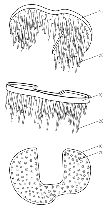
In Figure 1, a tibial plateau 10 is shown in a very simplified representation
and is made in the proposed manner. Figure la shows a plan view of' the
fastening side with which the implant is provided for contact with the
resection surface of the bone. Anchorage pins 20 are arranged on the
fastening side of the tibial plateau 10. They are arranged on a square grid
in this Figure, but this is in no way compulsory; any other desired
arrangement patterns appropriate for the application are also possible.
Figure lb shows a view along the line marked by B-B in Figure la. Figure
1 c shows a plan view of the resection surface of a tibia 1. The cortex 3 at
the rim of the resection surface substantially comprises solid bone tissue
and is only slightly deformable and is comparatively brittle. In the region
CA 02640759 2008-07-30
14
of the spongiosa 2, the bone tissue is, as already described above,
composed of bony tissue and medulla and has a greater deformability. As
indicated by the differently dense hatching, the proportion of bony tissue
in the total tissue is different in different regions of the resection
surface.
In the example shown here, the proportion of the bony tissue reduces from
the cortex toward the center, whereby the strength also reduces and the
deformability of the bone increases. It can be recognized with respect: to
Figure lb that the length of the anchorage pins is adapted to this
circumstance and is larger in the regions provided for arrangement in
regions of the bone with a low proportion of bony tissue than in regions of
a high proportion of bony tissue. In this example, all the anchorage pins
are shown as cylindrical pins having a constant cross-sectional surface.
The pins can naturally also be conical in total or regionally or can, for
example, converge acutely at the front end remote from the implant. It is
equally not compulsory that all the pins have the same cross-sectional
shape or longitudinal contour.
Figure 2 shows an alternative embodiment of such a tibial plateau. The
anchorage pins 20 on the tibial plateau 10 are arranged in a variable
density over the surface of the fastening side of the implant. As can be
recognized from Figure 2b, the anchorage pins 20 are almost of equal
length and converge acutely. An equivalent effect as is adopted in Figure 1
due to the longer anchorage pins is achieved by the reduction of the
spacing between the anchorage pins in the regions of the implant which
are provided for arrangement on regions of a resection surface with a.
lower proportion of bony material. Anchorage pins of different lengths and
other and/or different shapes can naturally also be used in the
embodiment shown in Figure 2.
CA 02640759 2008-07-30
A condyle implant 30 is shown schematically in Figure 3 which has
anchorage pins 20 of different lengths for anchorage at the resection
surface of a bone.
5
The use of an implant of the kind described here at the femoral
component of a hip joint prosthesis is shown in Figure 4. A femoral cap is
shown in Figures 4a and 4b as is used for the so-called resurfacing of a
femoral head. The "resurfacing" is characterized in that as little bone as
10 possible is removed and substantially only the cartilage of the natural
articulation surface is replaced by an artificial articulation surface. The
articulation surface of the femoral cap 40 preferably consists of metal, in
particular of a steel having a high carbon proportion, which is, however,
not material to the invention here. The femur 41 is shown schematically in
15 Figure 4c with the femoral neck 42, the femoral head 43 and the resection
surface 44 at the femoral head. The proportion of the bony tissue in the
total bone tissue is also different over this resection surface. For example,
cortex is located at the outer rim of the resection surface into which
anchorage pins can only be introduced with difficulty and with the risk
also being present of causing a splintering of the cortex with non-
predrilled holes for the introduction of the pins. In contrast, spongious
material having a large medulla proportion is located at the center of the
resection surface. The anchorage pins 20 of the femoral cap 40 are
therefore selected differently in different regions adapted to the bone
density and are longer at the center than at the rim, whereas at the far
rim, in regions which come to lie on the cortex, no anchorage pins are
arranged. The fastening of an implant described here by means of a
collective of pins adapted to the local bone structure and bone strength
CA 02640759 2008-07-30
16
can naturally also be used in the fastening of the associated acetabulum
component.
A plurality of possible embodiments of anchorage pins 20 are shown
schematically in Figure 5. They extend with a length I starting from the
implant 50. Some of the exemplary anchorage pins 20 have divergent
regions seen from the tip toward the implant. The half angle of the
divergence is marked by a. This angle is for example 2 , 3 , 4 or 5 . Small
angles, corresponding to very acute pins, facilitate the introduction which
in particular takes place into a non-predrilled bone and improve the
security of the seat of the pins in the bone. Pins with larger divergence
angles, in contrast, have a greater security against kinking on the
introduction and facilitate the explantation in any required revisions.
Figure 6 illustrates further geometrical connections of the arrangement of
anchorage pins on the fastening side of an implant. In this example, the
anchorage pins 20 are arranged on a grid of equilateral triangles. The
spacings between the central longitudinal axes of two adjacent anchorage
pins are marked by s. The density of the anchorage pins is the number of
pins within a surface element dA and is specified, for example, as the
number of pins with respect to a unit of area of, for example, 1 cmz.
Figure 7 illustrates different cross-sectional shapes of anchorage pins.
Furthermore, in some of these exemplary anchorage pins, the
circumscribed circle 25 is shown with the diameter d.
Figure 8 illustrates a further embodiment in which wall-like structures 23
whose height h is smaller than the length I of the anchorage pins 20 are
CA 02640759 2008-07-30
17
arranged between anchorage pins 20, with the height within such a wall-
like structure also being able to be variable, for example such that the
height of the wall-like structure is larger adjacent to the anchorage pins
than at the center between two anchorage pins; wall-like structures of
constant height are naturally also possible or those in which the height is
larger at the center between two anchorage pins than adjacent to the
anchorage pins. The vertical extent of the wall-like structure amounts, for
example, to between one millimeter and four millimeters and should be
limited such that not too may blood vessels are cut by these wall-like
structures within the bone.
Figure 9 shows a modification of the tibial plateau of Figure 1. On the
fastening side, the tibial plateau 10 has, in addition to the anchorage pins
20, guide elements 21 which project further from the fastening side than
the longest anchorage pin. Two positioning bores 4 are introduced into the
resection surface of the tibia shown in Figure 9c and their spacing
corresponds to the spacing of the guide elements 21. On the placement of
the tibial plateau onto the resection surface, the guide elements 21 first
penetrate into the bores 4 of the tibia and position the tibial plateau there
with respect to position and direction before the implant is fixed in the
bone by pressing or hammering the anchorage pins into the bone tissue.
The guide elements 21 have a conical tip region in addition to the
cylindrical guide region for the easier introduction into the bores 4. For
the best possible function, the cylindrical guide region of the guide
elements is longer than the longest anchorage pin such that the position
and direction of the implant are already securely fixed before the
penetration of the anchorage pins.
CA 02640759 2008-07-30
18
Figure 10 shows an exemplary tibial plateau 10 in which the arrangement
and geometry of the anchorage pins 20 of the complex bone density
distribution is adapted to a real resection surface of a tibia. Figure 1:1
shows this tibial plateau on a tibia 1 in the implanted state.
A shoulder prosthesis is shown in Figure 12 in which the components are
designed in the manner described and claimed. A cap 60, which is made,
for example, from a metallic material and has an articulation surface 61 is
anchored in the humerus 9 by means of anchorage pins 20 of different
length. The application of such a cap as a joint surface replacement is
likewise actually a "resurfacing" in the manner explained in connection
with Figure 4. A glenoid component 70 having an articulation surface 71
is likewise anchored in the scapula 8 by means of a plurality of anchorage
pins 20 arranged in a pin field. The surgeon frequently finds very poor
bone quality at these points which makes a secure primary fixation of the
implants more difficult. The distribution of the anchorage over the
plurality of pins which are shaped and arranged such that the force is
distributed over a bone volume which is the larger, the smaller the local
density and stability of the bone is, has a very advantageous effect. Due to
the high porosity of the bone which is often found at the implantation
sites, the implantation by pressing in or hammering in the implants is
facilitated, whereas the distribution of the holding forces over a number of
pins facilitates the primary fixation. A large surface of the pins 20 is
available for a secondary fixation by ongrowth on the bone. The strength
of the hold and the intensity of the connection to the bone can be
influenced in a manner known per se by the geometry of the pins as
described above and their surface properties, in particular their
roughness. The glenoid component is shown enlarged in Figures 13 and
CA 02640759 2008-07-30
19
14. In this example, it comprises, in addition to the anchorage pins 20, an
articulation surface 71 which is made, for example, of polyethylene, in
particular of a highly cross-linked and/or a high-molecular polyethylene,
as well as a multilayer wire mesh which is made, for example, of titanium
and is known, for example, under the name "Sulmesh". This is very
suitable for the anchorage of the polyethylene articulation surface and
likewise promotes an ongrowth of the bone. Figure 15 shows the humerus
component which has the shape of a hollow sphere 60 here and in whose
interior the anchorage pins 20 are arranged. The pins are longer at the
center of the anchorage surface than at its rim both in the glenoid
component 70 and in the humerus component 60.
The embodiments described above only represent some selected examples
from the possibilities which are available to the skilled person. Further
embodiments of the invention easily become clear to the skilled person in
light of these embodiments.
The implantation of the implants described here takes place in that the
corresponding bone is cut. The anchorage pins are pressed into the bone
structure of the arising resection surface without corresponding holes
having been predrilled. On the use of implants having centering spigots
and/or guide spigots, such as is shown in Figure 9, bores are introduced
for the reception of the spigots and the bone; no bores are introduced for
the reception of the spigots and the bone. The friction of the pins in the
bone tissue and the large number of pins, as well as their fitting - whether
due to the arrangement or to the geometry - to the local bone density
ensure a good primary anchoring of the implant in the bone so that the
CA 02640759 2008-07-30
implantation can take place free of cement; it is naturally also possible
additionally to fix the implant on the resection surface with bone cement.
When an implant is provided, as shown in Figure 9, with guide elements
5 and/or centering elements, corresponding guide bores and/or centering
bores have to be introduced into the resection surface, in particular by
means of a suitable gauge. They are dimensioned, for example, such that
they can receive the guide elements and/or centering elements with as
little clearance as possible. On the implantation, the guide elements
10 and/or centering elements are then first introduced into the associated
bores of the resection surface, whereby the position and/or direction of
the implant on the resection surface are defined and the implant is then
pressed in or hammered in using the anchorage pins for which no holes
were predrilled.
CA 02640759 2008-07-30
21
Reference numeral list
1 bone
2 spongiosa
3 cortex
4 bore
8 scapula
9 humerus
implant, tibial plateau
10 20 anchorage pin
21 guide spigot and/or centering spigot
23 areal connection structure, wall-like structure
25 circumscribed circle of an anchorage pin
30 distal femoral component
40 femoral cap
41 femur
42 femoral neck
43 femoral head
44 resection surface
50 implant
60 humerus implant
61 articulation surface of the humerus implant
70 glenoid implant
71 articulation surface of the glenoid implant
74 wire mesh structure
d thickness of an anchorage pin, diameter of the circumscribed circle
of an anchorage pin
CA 02640759 2008-07-30
22
dA surface element
I length of an anchorage pin
H height of the wall-like structure
S spacing of two anchorage pins
a angle