Note: Descriptions are shown in the official language in which they were submitted.
CA 02640972 2011-06-30
AUTOLOGOUS BONE HARVEST DURING
OSTEOTOMY AND BONE DRILLING PROCEDURES
BACKGROUND OF THE INVENTION
Field of the Invention
The present invention has to do with apparatus and methods for performing
osteotomies and drilling holes in bones. More specifically, the invention
relates to
apparatus and methods for harvesting bone from the operating site during the
osteotomy or
bone drilling procedure so that it can be used to augment the bone fusion
process.
The Related Art
Osteotomies are routinely performed for surgical access or to divide (and
reposition) a bone for the correction of a skeletal deformity. Holes may be
drilled in bones
for various reasons to accommodate screws, pins and various other implantable
devices
and materials or to take a bone sample for analysis.
One of the more common examples of an osteotomy for surgical access is a
craniotomy. In this procedure, the surgeon removes a significant portion of
the patient's
skull (termed a craniotomy flap, a cranial flap, a skull flap or bone flap)
for access to the
brain. The removed section of the skull is set aside in a sterile field and at
the end of
surgery, it is returned to its original position and affixed to the native
skull, typically with
plates and screws. The intent of the surgeon is to restore the patient's skull
to its original
contour and to provide physical protection for the brain. The ideal outcome
would be
complete fusion of the craniotomy flap to the native skull, leaving no long
term bony
deficit or weakness. In addition, many surgeons would prefer there to be
minimal foreign
bodies remaining and no imaging artifacts postoperatively. Unfortunately this
is difficult
to accomplish with the current surgical techniques.
The surgical instrument used to cut the craniotomy (a craniotome) utilizes a
rotating cutter approximately 2mm in diameter. The bone that is removed by
this
instrument is lost during surgery and as a result, when the cranial flap is
returned to its
original position, there is a gap around the entire periphery which
corresponds to the
diameter of the cutter. This gap creates a number of problems. The most
obvious
1
CA 02640972 2008-07-30
WO 2007/133240 PCT/US2006/033463
deficiency is that bone-to-bone contact, essential for achieving bony fusion,
is
impossible around the periphery of the cranial flap. This continuous gap (or
kerf)
creates a surgical "dead space" which is never desirable, it also allows soft
tissue (the
scalp and dura) to intrude into this space and inhibit bony healing. The step-
off
between the skull and cranial flap also may result in a cosmetic deformity for
the
patient. To combat these problems, surgeons use one or more strategies which
have
their own shortcomings. For example, the surgeon may choose to bias the
cranial flap
toward one side of the craniotomy. This produces bone-to-bone contact in a
local
area but increases the gap elsewhere around the periphery.
The surgeon may also elect to fill the gap between the skull and skull flap
with
a material which will encourage bony fusion. These fill materials can be
autologous,
allograft, or artificial. Autologous bone grafts are harvested directly from
the patient
and are the "gold standard," since they are inherently biocompatible,
osteoconductive,
osteoinductive, and osteogenic. Harvesting autologous bone is currently
carried out
by taking bone from a part of the patient's body other than the'surgical site.
This
results in additional surgical time and the additional (surgical) harvest has
its own
attendant risk of complications such as donor site pain and morbidity.
Allografts,
derived from donor (cadaver) tissues, are only osteoconductive, and they
involve
considerable cost, pose the risk of disease transmission and are objectionable
to
certain religious groups. Artificial materials such as alloplastic bone cement
are
another alternative. These bone cements are almost always used in conjunction
with
plates and screws. The drawbacks to this approach include substantial
additional
cost, risk of infection and no certainty that the bone cement will ever
remodel into
actual bone.
While this problem is illustrated with a craniotomy example, it occurs
whenever
an osteotomy is created strictly for surgical access and the bones must be
returned to
their original positions in order to prevent a postoperative deformity or a
functional
problem. In the skull alone, this problem exists in skull base surgery,
craniofacial
tumor surgery and mandibular osteotomies for oncologic resection. At the
conclusion
of all these procedures, the surgical goal is to restore the original bony
anatomy. This
precludes achieving bone-to-bone contact of the severed ends since they must
remain
separated by the width of the blade (or cutter) used for the osteotomy.
Perforations (or holes) are routinely created in bones for surgical access and
other reasons. These perforations may be performed for biopsy purposes, to
create
2
CA 02640972 2008-07-30
WO 2007/133240 PCT/US2006/033463
access for minimally invasive surgery or as the prelude to an osteotomy. An
example
of the latter is the burr hole that is initially created in the skull which
allows the
craniotome to be inserted for completion of the craniotomy. In these cases, it
is
desirable to close the perforation, preferably in a manner which restores the
bone to
its original condition. Additionally, holes are routinely drilled into bone as
a step in
preparation for orthopaedic screw or pin insertion. Most of these cases would
also
benefit from the availability of autologous bone graft.
When osteotomies are used to divide a bone so that it may be repositioned to
correct a surgical deformity, a different problem exists. In many cases, bone
graft
material is needed to fill the gaps created as the bones are repositioned and
severed
bony ends move relative to each other. This is obviously the case where a gap
is
intentionally created, such as an osteotomy to elevate a collapsed tibial
plateau. It
also may occur when the intent of the osteotomy is to decrease the bone
volume. In
these surgeries it is not uncommon for the contours of the bony ends to be
slightly
mismatched and in these cases the surgeon may elect to augment the fusion with
additional bone graft material. As previously discussed, allograft bone,
autogenous
bone or alloplastic materials may all be used in such situations, each with
their related
problems.
In all these procedures where an osteotomy (or perforation) is necessary, a
common problem exists: bone is removed by the osteotomy or drilling instrument
and
at the conclusion of surgery, additional bone is required to complete the
reconstruction.
The current surgical practice is to manually irrigate the bone as it is cut
and
also to manually suction off the resulting solids and liquids into the
operating room's,
non-sterile vacuum system. These activities are performed concurrently by
other
operating room personnel while the surgeon operates the osteotomy instrument.
Some of the shortcomings of these practices are detailed in the following text
which is
excerpted from the USC Neurosurgery website.
(http://uscneurosurgery.com/infonet/ecrani/instruments.htm).
Irrigation
With even optimal illumination and magnification and organization of his
field, the surgeon is still incapacitated by obscuring blood, cloudy
irrigation fluid, or other debris. Efficient intracranial surgery requires
3
CA 02640972 2008-07-30
WO 2007/133240 PCT/US2006/033463
keeping the operative field clear of physical and visual obstacles by
diligent irrigation, attentive aspiration, and meticulous hemostasis.
Irrigation and aspiration are complimentary aspects of surgical field
maintenance. The irrigating-aspirating assistant must concentrate on
following the movements of the surgeon's hands visually and with
irrigant and suction. Areas of surgical interest are most safely
addressed at the time of maximal cleanliness; immediately after they
have been washed clean and aspirated dry.
Irrigant should be squirted onto the field under enough pressure to
displace blood, but if the bulb is squeezed too hard and fluid issues
under too much pressure, fluid from the bulb will be reflected back
against the stream because it cannot dissipate fast enough, with the
consequence that a splashing of mixed blood-irrigant fluid ends up in the
surgeon's face and widely scattered across the field. Better control of
the stream from the irrigation fluid bulb is achieved by manipulating it
with the dominant hand.
The primarily aqueous solution used for surgical irrigation not only
dilutes the blood but pushes it ahead of the irrigant stream. This
washing force is greatest at the tip of a irrigation bulb where the irrigant
fluid pressure is maximal.
Suction
Blood accumulates with irrigation fluid in dependent portions of the field
as it escapes and is washed from lacerated vessels. The bloody fluid
then interferes with the working of the electrocautery devices used to
stop further bleeding from the openings in the vessels. To this is added
the problem of blood's opacity, so that even in small quantities as even a
thin layer, it obscures the surgical field.
Suction is a maintenance activity, keeping the operative field clear of
debris, blood, or smoke that can obstruct visualization. Whenever
possible the suction attachment should be held in the non-dominant
hand.
Surgical field suction instrumentation attaches to the same suction
canisters which provide suction for anesthesia. Distally non-sterile,
proximally sterile tubing connects the suction device to the distal end of
4
CA 02640972 2008-07-30
WO 2007/133240 PCT/US2006/033463
the metal suction handle and tip. The proximal end of the metal sucker
connects to the suction tubing.
The importance and difficulty of performing simultaneous irrigation and
suction
in concert with the surgeon's movements are detailed above. Later in the text
they
discuss the importance of irrigation when cutting the bone:
Bone is perforated and/or cut in the course of any intracranial trauma
surgery. Irrigation accomplishes two purposes in the setting of drilling
bone. First, it cools down the bone. This is important in terms of the
mechanics of bone cutting. The bits cut more effectively through cooler
bone and in the absence of bone dust that can clog its rotations.
These comments are directed toward neurosurgical craniotomies but the same
principles apply to all osteotomies and perforations. Proper irrigation not
only
improves the efficiency of the cutting instrument, it also prevents thermal
necrosis of
the bone which can later retard the healing process. This principle takes on
even
greater importance when one intends to collect the bone particles generated
during
the cutting process and reuse them in surgery. Irrigation has traditionally
been
conducted using a liquid. But according to the present invention we can
irrigate with a
liquid or compressed gas source or a combination of liquid and a compressed
gas
source. The compressed gas can be chilled if required and also can be
intermixed
with a fluid (e.g., saline).
Up until now, a reliable and essentially free source of autogenous bone has
been overlooked by the surgical community. Manufacturers of surgical cutting
instruments have incorporated irrigation on some instruments but none have
ever
proposed taking the concept one step further - collecting the bone particulate
in a
sterile fashion for later use in the bony reconstructive phase of the surgery.
We have now developed apparatus and methods for sterilely collecting and
containing the particulate bone created during osteotomy and bone drilling
procedures. The apparatus and methods also enable more controlled irrigation
of the
bone as it is cut or drilled and a reduction in the amount of patient bone
that is
scattered or aerosolized during surgery.
The terms particulate bone, bone particulate and bone particles are used
interchangeably in this patent and all are intended to have the same meaning.
5
CA 02640972 2012-04-24
SUMMARY OF THE INVENTION
A collection module is provided on the cutting end, also referred to herein as
the
distal end, of a bone cutting tool to prevent the scatter and loss of
particulate bone created
at the operating site during an osteotomy or bone drilling procedure. The
collection module
suctions off the bone particulate as well as irrigant, blood and other body
fluids and
reduces contamination of the surgical field from the cutting operation. The
module can be
partially or completely disposable.
The collection module contains a suction port which evacuates the particulate
bone from the cutting operation. A sterile containment module is provided
downstream for
io collecting the particulate bone and separating it from irrigant and body
fluids suctioned
off from the surgical field.
An irrigation system is incorporated in some cutting tools and when it is not,
it can
be incorporated in the collection module to provide a reliable and effective
source of
irrigation to the cutting area. The irrigant prevents thermal necrosis,
prevents the formation
of bone dust, improves cutting efficiency and improves visibility within the
surgical field.
As previously disclosed, the irrigation system in our invention can disperse
fluids, gasses
or a combination of the two.
The sterile bone particles which are harvested according to the invention are
used
to augment the reconstructive portion of the surgery. The particulate bone can
be used
"as is" or mixed with any number of readily available additives such as, but
not limited
to:
a. Patient's blood;
b. Patient's platelet rich plasma (PRP);
c. Bone morphogenic proteins;
d. Other bone growth factors; and
e. Antibiotics.
In one embodiment, a collection module is provided for collecting particulate
bone
from a bone for use with a bone cutting or drilling tool having a distal end
with a bone
cutting or drilling element and a foot plate each having a diameter and
extending distally
from the bone cutting or drilling tool, comprising:
a housing having:
an inner wall having distal and proximal ends;
6A
CA 02640972 2012-04-24
an outer wall coaxially disposed around the inner wall and spaced therefrom
and having a distal end extending distally from the distal end of the inner
wall; and
an end wall affixed to and capping the distal end of the outer wall,
the inner wall being sized for sealing and removable engagement of the
proximal end of the apparatus with the distal end of the bone cutting or
drilling tool,
the end wall having a slot therethrough sufficiently larger than the diameter
of
the bone cutting or drilling element and the foot plate to permit the bone
cutting or drilling
element and the foot plate to extend therethrough and to allow suctioning
through the slot of
bone particulate from an operating site;
a suction channel disposed between the inner wall and the outer wall an in
suctioning communication with the slot; and
a flexible portion disposed around the housing giving proximal and distal
ends, the
proximal end being affixed to the proximal end of the outer wall and the
distal end extending
distally from the end wall, the distal end of the flexible portion comprising
an open cylindrical
tube,
the flexible portion of the collection module allowing the open distal end of
the
cylindrical tube to follow irregularities in the bone surface when the
collection module is in
sealing engagement with the distal end of the bone cutting or drilling tool.
In another embodiment, an apparatus is provided for cutting or drilling bone
and
collecting particulate bone from the bone comprising:
a bone cutting or drilling tool having a distal end with a bone cutting or
drilling
element and a foot plate, each having a diameter and extending distally from
the bone
cutting or drilling tool; and
a collection module having:
a housing having:
an inner wall having distal and proximal ends;
an outer wall coaxially disposed around the inner wall and spaced
therefrom and having a distal end extending distally from the distal end of
the inner
wall; and
an end wall affixed to and capping the distal end of the outer wall,
the inner wall being sized for sealing and removable engagement of the
proximal end of the apparatus with the distal end of the bone cutting or
drilling tool,
6B
CA 02640972 2012-04-24
the end wall having a slot therethrough sufficiently larger than the diameter
of
the bone cutting or drilling element and the foot plate to permit the bone
cutting or drilling
element and the foot plate to extend therethrough and to allow suctioning
through the slot
of bone particulate from an operating site;
a suction channel disposed between the inner wall and the outer wall an in
suctioning communication with the slot; and
a flexible portion disposed around the housing giving proximal and distal
ends, the
proximal end being affixed to the proximal end of the outer wall and the
distal end extending
distally from the end wall, the distal end of the flexible portion comprising
an open cylindrical
1o tube,
the flexible portion of the collection module allowing the open distal end of
the
cylindrical tube to follow irregularities in the bone surface when the
collection module is in
sealing engagement with the distal end of the bone cutting or drilling tool.
BRIEF DESCRIPTION OF THE DRAWINGS
The drawing figures are provided for purposes of illustrating the elements of
the
invention and are not intended to be drawn to scale.
FIG. 1 is an expanded perspective view of a bone cutting tool (a craniotome)
of the
invention which has been provided with integral irrigation and suction
systems. A
collection module of the invention is illustrated to the left before
attachment to the tool. The
craniotome is attached to a handpiece which in turn is attached to a pneumatic
line or an
electric power source.
6C
CA 02640972 2008-07-30
WO 2007/133240 PCT/US2006/033463
FIG. 2 is a perspective view of the craniotome of FIG. I with the collection
module and the pneumatic line attached.
FIG. 3 is an elevation view of the craniotome of FIG. 2.
FIG. 3A is a view of the left end of FIG. 3.
FIG. 4 is a section view of FIG. 3A taken at section line 4-4 of FIG. 3A and
illustrating a portion of the suction system.
FIG. 4A is a section of FIG. 3A taken at section line 3-3 of FIG. 3A and
illustrating a portion of the irrigation system.
FIG. 5 is an elevation view of a collection module of the invention.
FIG. 6 is a distal end view of the collection module of FIG. 5.
FIG. 7 is a section view of the collection module of FIGS. 5 and 6.
FIG. 8 is an expanded elevation view of a standard prior art craniotome and a
collection module of the invention. This embodiment of a collection module is
for use
with standard craniotomes and is illustrated to the left before attachment to
the tool.
FIG. 9 is an elevation view of the craniotome of FIG. 8 with the collection
module and the pneumatic line attached.
FIG. 9A is a view of the left end of FIG. 9.
FIG. 10 is a section view of FIG. 9A taken at section line 10-10 of FIG. 9.
FIG. 11 is an elevation view of the collection module of FIGS. 8-10.
FIG. 12 is a distal end view of FIG. 11.
FIG. 13 is a section view of the collection module of FIGS. 11 and 12 taken at
section line 13-13 of FIG. 12.
FIG. 14 is an illustration of an apparatus of the invention in operation
during a
cranial osteotomy.
FIG. 15 is a perspective view of a drill guide of the invention which can
suction
and collect bone particulate during a bone drilling procedure.
FIG. 16 is a bottom view of FIG. 15.
FIGS. 17 and 18 are partial section views of FIG. 16. FIG. 17 is taken at
section line 17-17 of FIG. 16 and FIG. 18 is taken at section line 18-18 of
FIG. 16.
FIG. 19 is a perspective view of the guide of FIG. 15 illustrating the
relationship
of the guide to a drill and a bone plate.
FIG. 20 is a partial section of FIG. 19 taken at section line 20-20 of FIG.
19.
FIG. 21 is a perspective view of another embodiment of a bone particulate
collection system for use with a drill.
7
CA 02640972 2008-07-30
WO 2007/133240 PCT/US2006/033463
FIG. 22 is a distal end view of FIG. 21.
FIG. 23 is a section view of FIG. 22 taken at section line 23-23 of FIG. 22.
FIG. 24 is an elevation view of a transparent embodiment of the FIG. 21
collection module affixed to a drill.
FIG. 25 is an enlarged section view of a portion of FIG. 24.
FIG. 26 illustrates a sterile containment module of the invention.
DESCRIPTION OF THE PREFERRED EMBODIMENT
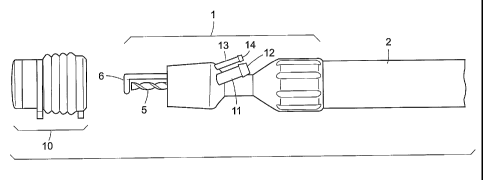
FIG. I is an expanded perspective view of a bone cutting tool of the invention
having integral irrigation and suction systems. The tool is a craniotome which
is used
to cut an opening in the skull for brain surgery. The craniotome 1 is attached
to a
handpiece 2 which in turn is attached to a pneumatic line 3 (see Figs. 2-4) or
an
electric power source. Cutting burr 5 is actuated by a foot switch (not shown)
and the
foot plate 6 is used to guide the tool along the inside of the skull in order
to prevent
penetration of the dura. A suction tube 11 is provided with a barbed fitting
12 and an
irrigation tube 13 has a barbed fitting 14. Collection module 10 is
illustrated before it
is attached to craniotome 1.
FIG. 2 is a perspective view of FIG. 1 with the collection module 10 of the
invention attached to the craniotome. A flexible bellows 15 is shown in this
embodiment with a shield 16 and an elastomeric seal 17 at the distal end. The
shield
16 normally will be comprised of a relatively stiff, clear plastic tube.
FIG. 3 is an elevation view of FIG. 2 and FIG. 4 is a section view of FIG. 3.
FIG. 4 illustrates suction tube 11 which has an open mouth 23 at its distal
end
around cutting burr 5. FIG. 4A is a different section view of FIG. 3 which
illustrates
irrigation tube 13 of the irrigation system.
FIG. 5 illustrates the collection module 10 in an elevation.view and FIG. 6
illustrates the distal end of the collection module 10. FIG. 7 is a section
view of the
collection module 10.
FIG. 8 illustrates in expanded elevation another embodiment of the invention.
Collection module 110 is made for use with a standard prior art craniotome
101. FIG.
9 is an elevation view of craniotome 101 with collection module 110 affixed
thereto.
The collection module 110 comprises a suction tube 111 having a barbed fitting
112,
an irrigation tube 113 having a barbed fitting 114, a flexible bellows 115 and
a clear
tubular shield 116. An optional indicator tab 119 is also illustrated. The
craniotome
has a foot plate 106 and a cutting burr 105.
8
CA 02640972 2008-07-30
WO 2007/133240 PCT/US2006/033463
FIG. 10 is a section view of FIG. 9 illustrating the relationship of the
elements of
collection module 110 to the craniotome 101. In particular, the suction tube
111
connects to a suction channel 121 and the irrigation tube 113 connects to an
irrigation
channel 123.
FIG. 11 is an elevation view of the collection module 110 by itself. The
collection module 110 provides the irrigation and suction capability needed to
carry
out the objectives of the invention when a standard craniotome having no
irrigation or
suction capability is employed. (Some commercially available craniotomes have
irrigation capability in which case the embodiment of FIG. 11 described herein
can be
made with suction capability but without irrigation capability as will be
apparent to
those skilled in the art.) This embodiment does not employ a seal of the type
illustrated as element 17 in FIGS. 1-7. In FIG. 12, the distal end of the
module is
illustrated with an opening 118 for a cutting burr and foot plate. An
irrigation port 133
is also provided. Referring to the section view FIG. 13, the irrigation port
133 and the
irrigation channel 123 are illustrated as well as the suction channel 121 and
a suction
port 131.
FIG. 14 illustrates the operation of the distal (cutting) end of the
embodiment of
the invention illustrated in FIGS. 1-7. The craniotome 1 has a cutting burr 5
(and burr
shaft 5a) and an integral foot plate 6. Unlike current instruments, however,
the
improved craniotome of the invention has many advantageous features. In this
embodiment, the craniotome also incorporates internal passages for suction and
irrigation. Each of these terminates proximally in a barbed fitting. The
collection
module 10 comprises an elastomeric bellows 15, a clear tubular shield 16 and
an
elastomeric seal 17. The collection module can constitute a preassembled,
sterile,
disposable item, although other configurations are certainly possible.
The collection module 10 is adapted to the distal end of the craniotome 1 (as
shown in FIGS. 2-4). Module 10 mates with the outer diameter of the craniotome
1
and is sealingly engaged therewith. The two are aligned in the correct
orientation to
set the slot 18 in the seal 17 in-line with the footplate 6. Optional
indicator tabs 19 (in
the direction that the instrument will cut, arrow 20) can be used to
facilitate correct
orientation. The bellows 15 is constructed from an elastomer, allowing it to
flex so that
the distal portion of the collection module 10 can follow the irregularities
of the skull 30
without excessive resistance. On the other end of the bellows is an internal
lip seal 22
which prevents debris from being forced into the radial space between the
craniotome
9
CA 02640972 2008-07-30
WO 2007/133240 PCT/US2006/033463
1 and the bellows 15. It should be noted that the cutting burr, or the drill
bit or saw
blade in other tools, may or may not extend beyond the distal end of the
module when
the tool is not in use. This is because the collection module is sufficiently
flexible to
allow such burr, bit or blade to extend beyond the distal end of the module
when the
tool is in use.
The shield 16 is a relatively stiff, clear tubular section that forms the
radial wall
of the collection module 10. Attached to the distal end of the shield 16 is
the
elastomeric seal 17. Ideally this would be a relatively clear material as well
to aid in
visualizing the cut. The seal 17 has an optionally, outwardly domed flexible
end with a
slot 18 to better contain and suction the bone particulate. The domed shape
limits the
contact area with the bone to reduce resistance. As the surgeon operates the
craniotome, he applies both sideways force to cut as well as upward force to
keep the
tip of the footplate 6 in contact with the underside of the skull. This allows
the
footplate to ride between the dura 4 (the outer covering of,the brain 104) and
the inner
table of the skull 30. Ahead of the cutting burr 5 is solid skull 30 and
trailing the
cutting burr is the kerf 31. The rotation of the cutting burr 5 and its
helical flutes help
to draw much of the bone particulate 32 upwards into a collection, chamber 24
of the.
collection module. A funnel shaped depression or mouth 23 at the junction of
the
suction tube 11 and the distal face of the craniotome guides these bone
fragments,
into the suction tube 11 and draws in by vacuum additional bone particles,
irrigant.and
bodily fluids. The suction tube 11 is connected to a sterile vacuum tube 40. A
barbed
fitting 12 is provided for this connection. The sterile vacuum tube 40 is
connected
downstream to a containment module 60 as will be discussed later. (See FIG.
26.)
Suction is applied to tube 40 and the result is that all material aspirated
into the
collection module 10 (bone fragments, irrigant, blood, tissue, etc.) is
evacuated in the
direction of arrow 41. The irrigation system is not illustrated because it
is.behind the
suction system in this drawing. But the irrigation system is illustrated and
discussed
above in connection with FIGS. 1, 2 and 4A. Irrigant supply can be,most easily
provided from a pressurized IV bag of saline or from a hand syringe,
peristaltic pump,
sterile compressed gas source, or other common means. When the irrigant is a
combination of gas and liquid an additional channel can be provided in either
the
craniotome of the invention (see FIGS. 1-4 and 14) or the collection module,
for the
purpose of introducing a second irrigation means. This additional channel
could
CA 02640972 2008-07-30
WO 2007/133240 PCT/US2006/033463
communicate with the liquid channel to serve as a mixing device as will be
apparent to
those having skill in the art based on the disclosures herein.
FIG. 15 is a perspective view of a drill guide of the invention which can
suction
and collect bone particulate in a sterile environment during a bone drilling
procedure.
The guide 201 comprises a handle 202 and a collection module 210. Sterile
vacuum
tube 241 connects to suction tube 211 and irrigant supply tube 243 connects to
irrigation tube 213 during operation of the guide. Opening 218 accommodates a
drill
bit 205 (see FIGS. 19 and 20) and irrigation and suctioning take place
generally
through the same opening. A bottom view of guide 201 is illustrated in FIG.
16.
FIGS. 17 and 18 are section views of collection module 210 taken through line
A - A and line B - B, respectively, of FIG. 16. The FIG. 17 section
illustrates a barbed
fitting 212 at the end of suction tube 211 and the connection of tube 211 with
suction
chamber 221. Irrigation channel 223 and irrigation ports 233 are illustrated.
The FIG.
18 section illustrates another part of suction chamber 221. The FIG. 18
section also
illustrates the barbed fitting 214 at the end of irrigation tube 213 and the
connection of
tube 213 with irrigation channel 223.
A perspective view illustrating the relationship of the guide with a drill
203, drill
bit 205 and a bone plate 206 is illustrated in FIG. 19. FIG. 20 is a partial
section of
FIG. 19 illustrating the relationship of drill bit 205 to the suction chamber
221, irrigation
channel 223 and irrigation ports 233. During drilling, bone particulate is
carried
upward by the drill bit 205 and by suction. Suction vacuum tube 241 is
connected to
suction tube 211 and the particulate bone is carried by vacuum to asterile
containment module 60 (see FIG. 26). The operating area is irrigated by
irrigant~
exiting irrigation ports 233.
FIG. 21 is a perspective view of another embodiment of a bone particulate
collection system for use with a drill. Collection module 310 is comprised of
an outer
telescoping section 301 and an inner telescoping section 302. A spring 304 is
biased
between section 301 and distal end section 303. When drilling, inner
telescoping
section 302 telescopes into outer telescoping section 301 and when the
drilling is
complete spring 304 returns section 302 to its original position (as
illustrated). Sterile
vacuum tube 341 and irrigant supply tube 343 are also illustrated.
FIG. 22 is a distal end view of the collection module 310 also illustrating
opening 318 which accommodates a drill bit 305 (see FIGS. 24 and 25) and
irrigation
and suctioning take place through the same opening.
11
CA 02640972 2008-07-30
WO 2007/133240 PCT/US2006/033463
FIG. 23 is a section view of collection module 310 illustrating a collection
chamber 321 and irrigation duct 323 in relation to opening 318.
FIG. 24 is an elevation view of a transparent embodiment of collection module
310 affixed to drill 303 having a drill bit 305. An enlarged section view of a
portion of
FIG. 24 is provided in FIG. 25. Arrow 320 illustrates the direction of the
telescoping
movement of section 302 into section 301 when the drill bit is drilled into a
bone.
Spring 304 causes section 302 to return to the position illustrated when
drilling is
completed. Sterile vacuum tube 341 is in suctioning communication with suction
chamber 321 and irrigant supply tube 343 is in irrigating communication with
irrigation
duct 323. The suctioning and irrigating operations function in the same manner
as the
other embodiments of the invention discussed above.
FIGS. 1-25 depict just a few possible configurations of a cutting or drilling
and
collection apparatus of the invention which would be consistent with the
method of the
invention. The principles of the invention can easily be adapted to other
osteotomy
instruments (e.g. an oscillating saw, a rotary saw or a reciprocating saw) to
achieve
the same results.
According to the method of the invention, a surgeon can simultaneously cut or
drill bone and irrigate and suction with essentially no additional effort.
Eliminated is
the splatter of the irrigant and cutting debris and also the need for an
assistant to
precisely coordinate with the movements of the surgeon as he or she irrigates
and
suctions. These benefits however, are secondary to the main purpose of the
apparatus and method of the invention, namely, the ability to collect the
sterile bone
particulate generated by the osteotomy or drilling process for use in the
reconstructive
portion of the procedure.
FIG. 26 illustrates an embodiment of a sterile containment module 60 for the
separation of the bone particles 32 from liquids 33, the liquids comprising
irrigant and
body fluids. Unlike traditional hospital suction systems, this is a sterile
system so that
the bone particles collected can be reused in the reconstructive portion of
surgery.
The aspirate from the containment chamber is conveyed though the sterile
vacuum tube 40 to the containment module 60. The aspirate consists of bone
particles, irrigant, small amounts of tissue, blood and other body fluids. The
containment module comprises three sterile parts: the canister 61, the
collection cup
62 and the cover 63. Of course, other embodiments are certainly possible and
would
be apparent to those skilled in the art based upon the disclosures herein. It
is
12
CA 02640972 2008-07-30
WO 2007/133240 PCT/US2006/033463
envisioned that all three items would be provided as a sterile unit for single
use. All
could be produced (molded) from a clear polymer for visualizing the contents.
The
suction tube 40 connects to a fitting 64 molded into the cover. A second
fitting 65 is
then connected to the hospital suction system in a sterile fashion through
tube 66.
The suction travels in the direction of the arrows 67. When the aspirate
enters the
canister 61, a deflector 68 forces the flow downward and gravity then
separates the
contents (solid and liquid) from the air flow. The solids and liquids fall
into the cup 62
and settle to the bottom where perforations 69 allow the liquid to drain into
the bottom
of the canister 61. Optionally the cup may be fitted with a filter to better
trap the
smaller bone particles. At the conclusion of the osteotomy or drilling
procedure, the
bone particles in the cup can be left to drain until needed, at which point
the cover 63
is removed and the cup 62 is extracted with its sterile contents. As mentioned
previously, the bone particles can then be used "as is" or mixed with other
biological
additives for use in the reconstructive portion of the procedure.
In today's operating room environment, the contents of the canister 61
described above are simply suctioned into the non-sterile hospital system and
discarded. A valuable and much-needed commodity, (autologous) bone graft,_ is
simply wasted and later replaced with autograft harvested from a second site,
allograft
or with alloplastic materials.
13