Note: Descriptions are shown in the official language in which they were submitted.
CA 02641310 2011-01-05
WO 2007/092453 PCT/US2007/003164
IGF-IR ANTAGONISTS AS ADJUVANTS FOR
TREATMENT OF PROSTATE CANCER
FEDERAL FUNDING
[0001] The present invention was made in part with United States Government
support under Grant No. CA85859 from the National Institutes of Health. and
Grant No.
W81XWH-04-1-0912 from the Department of Defense. Accordingly, the United
States
Government has certain rights in this invention.
FIELD OF THE INVENTION
[0003] The present invention relates to a method of treating prostate cancer
with
androgen deprivation therapy and an insulin-like growth factor receptor (IGF-
1R) antagonist.
The method inhibits or delays transition of androgen dependent cancer to
androgen
independent cancer and significantly decreases risk of recurrence and improves
treatment
outcome.
BACKGROUND OF THE INVENTION
[0004] Prostate cancer is the most common nonskin cancer and second most
common
cause of cancer mortality in US men. Most prostate cancer is initially
androgen dependent
(AD). Prostate cancer cells initially require androgen for continued
proliferation. Response
to ablation of testosterone through androgen deprivation therapy (ADT), either
surgically
(orchiectomy) or medically (GnRH agonistsnr estrogens), leads to rapid
induction of
apoptosis of sensitive prostate cancer cells. The positive resrionse rate is
about 86% based on
decrease in prostate specific antigen (PSA) and stabilization or decrease in
tumor volume.
The cell death that occurs generally takes place within the first few days to
a week.
However, the positive response is followed by a period of growth arrest in
which remaining
cells tend not to die. After 18-36 months following hormone ablation, growth
recurs in 90%
of cases. Invariably, surviving cancer cells become androgen independent or
unresponsive,
and androgen-independent (Al) tumor growth follows. Since ADT is initially
very effective,
CA 02641310 2008-08-01
WO 2007/092453 PCT/US2007/003164
a therapy that could take advantage of the benefits of ADT and extend or
enhance its effects
would be of great benefit.
[0005] Androgen independence appears to arise by a variety of mechanisms.
Mutations in the androgen receptor gene are rare at diagnosis, but increase
after exposure to
the anti-androgen flutamide. However, these mutations do not occur in the
majority of
patients and do not explain most cases of hormone-refractory disease. High
levels of bc1-2
are seen with greater frequency in advanced disease as compared to localized
disease. Thus,
the ability to induce apoptosis diminishes as the disease progresses. The
proliferation of cells
harboring mutations of the tumor suppressor gene p53, the loss of TGF-/3
receptors, and the
expression of peptide growth factors likely play a role in the development of
a hormone-
refractory state. However, these processes do not explain the rapidity and
frequency of
development.
[0006] The insulin-like growth factor receptor (IGF-lR) is a ubiquitous
transmembrane tyrosine kinase receptor that is essential for normal fetal and
post-natal
growth and development. IGF-IR can stimulate cell proliferation, cell
differentiation,
changes in cell size, and protect cells from apoptosis. It has also been
considered to be quasi-
obligatory for cell transformation (reviewed in Adams et al., Cell. Mel. Life
Sci 57:1050-93
(2000); Baserga, Oncogene 19:5574-81 (2000)). IGF-IR is located on the cell
surface of
most cell types and serves as the signaling molecule for growth factors IGF-I
and IGF-II
(collectively termed henceforth IGFs). IGF-IR also binds insulin, albeit at
three orders of
magnitude lower affinity than it binds to IGFs. IGF-IR is a pre-formed hetero-
tetramer
containing two alpha and two beta chains covalently linked by disulfide bonds.
The receptor
subunits are synthesized as part of a single polypeptide chain of 180kd, which
is then
proteolytically processed into alpha (130kd) and beta (95kd) subunits. The
entire alpha chain
is extracellular and contains the site for ligand binding. The beta chain
possesses the
transmembrane domain, the tyrosine kinase domain, and a C-terminal extension
that is
necessary for cell differentiation and transformation, but is dispensable for
mitogen signaling
and protection from apoptosis.
[0007] IGF-IR is highly similar to the insulin receptor (IR),
particularly within the
beta chain sequence (70% homology). Because of this homology, recent studies
have
demonstrated that these receptors can form hybrids containing one IR dimer and
one IGF-IR
dimer (Pandini et al., Clin. Canc. Res. 5:1935-19 (1999)). The formation of
hybrids occurs in
both normal and transformed cells and the hybrid content is dependent upon the
2
CA 02641310 2008-08-01
WO 2007/092453 PCT/US2007/003164
concentration of the two homodimer receptors (IR. and IGF-IR) within the cell.
In one study
of 39 breast cancer specimens, although both IR and IGF-IR were over-expressed
in all tumor
samples, hybrid receptor content consistently exceeded the levels of both homo-
receptors by
approximately 3-fold (Pandini et al., Clin. Canc. Res. 5:1935-44 (1999)).
Although hybrid
receptors are composed of IR. and IGF-IR. pairs, the hybrids bind selectively
to IGFs, with
affinity similar to that of IGF-IR, and only weakly bind insulin (Siddle and
Soos, The IGF
System. Humana Press. pp. 199-225. 1999). These hybrids therefore can bind
IGFs and
transduce signals in both normal and transformed cells.
[0008] Endocrine expression of IGF-I is regulated primarily by growth hormone
and
produced in the liver, but recent evidence suggests that many other tissue
types are also
capable of expressing IGF-I. This ligand is therefore subjected to endocrine
and paracrine
regulation, as well as autocrine in the case of many types of tumor cells (Yu,
H. and Rohan,
3., J. Natl. Cancer Inst. 92:1472-89 (2000)).
[0009] The androgen receptor (AR) consists of 3 functional and structural
domains:
an N-terminal (modulatory) domain; a DNA binding domain (Interpro Accession
No.
EPR001628) that mediates specific binding to target DNA sequences (ligand-
responsive
elements); and a hormone binding domain. The N-terminal domain (NTD) is unique
to the
androgen receptors and spans approximately the first 530 residues; the highly-
conserved
DNA-binding domain is smaller (around 65 residues) and occupies the central
portion of the
protein; and the hormone ligand binding domain (LBD) lies at the receptor C-
terminus. In
the absence of ligand, steroid hormone receptors are thought to be weakly
associated with
nuclear components; hormone binding greatly increases receptor affinity. The
interaction
among androgen receptor (AR), androgen, and prostate cancer is complex.
Distribution of
AR between .the nucleus and cytoplasm is affected by androgen and androgen
withdrawal.
For example, AR immunoreactivity is observed only in the nuclei of LuCaP 35
cells grown in
intact male mice, but strong immunoreactivity is observed in the cytoplasm and
nuclei of
LuCaP 35 grown in intact male mice and subsequently castrated.
SUMMARY OF THE INVENTION
[0010] This invention relates to treatment of androgen dependent tumors such
as
prostate cancer. Prostate tumors are typically stimulated by androgens such as
testosterone,
and exhibit androgen dependent (AD) growth. Therefore, treatment of prostate
cancer
typically involves therapy that deprives prostate cancer cells of androgen.
However, a large
3
CA 02641310 2008-08-01
WO 2007/092453 PCT/US2007/003164
proportion of prostate cancers eventually transition to androgen independence
(Al). It has
been discovered that administration of an IGF-IR antagonist in combination
with androgen
deprivation therapy (ADT) inhibits or prevents transition of AD tumors to AT
tumors.
[0011] Accordingly, the invention provides a method of treatment of an
androgen
dependent cancer by administering androgen deprivation therapy and an IGF-IR
antagonist.
In an embodiment of the invention, the androgen dependent cancer is prostate
cancer.
[0012] According to the invention, the IGF-]R antagonist can be an
extracellular
antagonist or an intracellular antagonist and more than one antagonist may be
employed.
More generally, the invention relates to inhibition of the IFG-IR signal
transduction and to
modulation of component of the pathway so as to inhibit transition of tumor
cells from AD to
AT. Extracellular antagonists include, but are not limited to proteins or
other biological
molecules that bind to IGF-IR or its ligand (IGF). In certain embodiments of
the invention,
the extracellular antagonist inhibits binding of IGF-IR to IGF. In one
embodiment, the
binding protein is an antibody, such as, for example, ]MC-Al2. In another
embodiment, the
binding protein is a soluble ligand binding fragment of IGF-IR. Intracellular
antagonists can be biological molecules, but are usually small molecules. In
an embodiment
of the invention, the IGF-IR antagonist is a small molecule selected from
AG1024, NVP-
AEW541, and BMS-554417.
[0013] The effectiveness of various antagonists to inhibit IGF-IR signal
transduction
can be observed, for example, by assaying the state of IGF-ER. signal
transduction pathway
components. In one embodiment, inhibition of IGF-IR is observed in the reduced
phosphorylation of Akt. In another embodiment, inhibition of IGF-IR. signaling
is observed
in the reduced expression of survivin or tubulin13-peptide (TUBB).
[0014] An IGF-IR. antagonist of the invention is used with any form of ADT. In
an
embodiment of the invention, ADT comprises orchiectomy. In another embodiment
of the
invention, ADT comprises administration of a luteinizing hormone-releasing
hormone
analog. In another embodiment, ADT comprises administration of an
antiandrogen. In yet
another embodiment, an adrenal androgen inhibitor is administered. According
to the
invention, two or more methods of ADT can be combined.
[0015] The invention further provides for inhibition of signaling through Akt.
Accordingly, the invention includes administration of modulators of signal
transduction
proteins that activate Akt. In one embodiment, such a modulator is an
antagonist of EGFR.
4
CA 02641310 2008-08-01
WO 2007/092453 PCT/US2007/003164
[0016] According to the invention, an IGF-IR antagonist is administered as an
adjuvant for ADT. In one embodiment, ADT and administration of an IGF-IR
antagonist are
initiated at about the same time. In another embodiment, ADT is initiated
first, and an IGF-
IR antagonist is administered before the androgen-independent cancer becomes
androgen-
independent. The invention further provides for use of anti-neoplastic agents
with ADT and
IGF-1R. antagonist administration. In an embodiment of the invention, an IGF-
1R antagonist
and an ADT agent are used together as a neoadjuvant for surgical or radiation
treatment of
prostate cancer.
[0017] The invention also provides compositions comprising an IGF-IR
antagonist
and an ADT agent in a dosage form.
BRIEF DESCRIPTION OF THE FIGURES
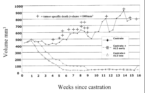
[0018] Figure 1 depicts a study in which LuCap35 subcutaneous xenografts in
SC]D
mice were observed. All mice were castrated when the average tumor size
reached 400 mm3.
The control group of mice received castration alone. In two other groups, LVIC-
Al2 was
administered three times per week starting one or two weeks after castration.
[0019] Figure 2 depicts levels of PSA in the castrated control mice and in
castrated
mice treated with INIC-Al2 starting one (early) or two (late) weeks after
castration.
[0020] Figure 3 depicts the distribution of androgen receptor (AR) in response
to
stimulation of IGF-IR with IGF and/or antagonism of IGF-IR with IMC-Al2.
Levels of
cytoplasm and nuclear AR were assessed by Western Blots.
[0021] Figure 4 depicts the effect of an IGF-IR. antagonist (IMC-Al2) on the
distribution of androgen receptor (AR) in androgen dependent xenograft tumors
of LuCaP 35
cells in intact mice (left column) and androgen independent xenograft tumors
of LuCaP 35V
cells in castrated mice (right column).
[0022] Figure 5 depicts the correlation between AR score and tumor volume. R-=
0.66, p <0.01. Castrate only values are in the open circles and Castrate + Al2
early and late
values are in the closed circles. Values are the mean value for 100 nuclei
graded per tumor.
[0023] Figure 6 depicts gene expression changes between two time periods for
subcutaneous Al2-treated tumors. Out of 3170 unique genes on the array with
sufficient data
to test, there were 21 up-regulated (including many androgen-regulated,
denoted by "*") and
41 down-regulated with q-value in the late time period when tumors began to
recur
compared to the early time period.
CA 02641310 2008-08-01
WO 2007/092453 PCT/US2007/003164
[0024] Figure 7A depicts the correlation between survivin copy number score
and
tumor volume (r= 0.66, p --Ø01). Figure 7B depicts the correlation between
tubulin beta
peptide 3 copy number score and tumor volume (r = 0.59, p Ø01). Castrate
only values are
in the open circles and Castrate + Al2 early and late values are in the closed
circles, Each
value is the mean of three PCR runs.
DETAILED DESCRIPTION OF THE INVENTION
[0025] It has been discovered that inhibitors of IGF-IR are useful in
therapies for
treatment of prostate cancer. In particular, administration of an IGF-lR
antagonist in
combination with androgen deprivation therapy (ADT) results in improved
treatment
outcome relative to ADT alone.
[0026] It has been observed that androgens up-regulate insulin-like growth
factor-I
receptor expression and may sensitize prostate cancer to the effects of IGF-I.
Similarly, the
transition to androgen independence that is observed in prostate cancer cells
can result from
adaptations of the cell that increase androgen receptor signaling such as
increased levels of
AR that make the cell sensitive to low levels of circulating androgen or AR
mutations
allowing activation by nonandrogen steroids. Indeed, evidence demonstrates
that IGF-I
signaling can actually mediate AR translocation to the nucleus of tumor cells
and lead to up-
regulation of AR-dependent genes. In this fashion, it is proposed that IGF-I
can promote the
conversion of androgen-dependent prostate cancer to androgen-independent,
following
hormone ablation therapy, by promoting AR signaling in the absence of
circulating levels of
androgen. Recent data from men and from human prostate xenografts has also
shown that
current methods of androgen ablation fail to decrease prostatic androgens to
levels that no
longer result in activation of the androgen receptor. The prostate may
actually be able to
synthesize DHT from several precursor steroids and possibly acetate.
[0027] It therefore follows that inhibition of IGF-I signaling concomitant
with
hormone ablation therapy may prevent or prolong the time until conversion of
prostate cancer
to androgen-independent disease, significantly delaying the onset of
recurrence. Antagonists
of IGF-IR may therefore be an effective adjuvant therapy to androgen
deprivation strategies
to treat newly diagnosed and locally advanced or metastatic hormone-dependent
prostate
cancer.
[0028] The use of IGF-IR. antagonists with androgen withdrawal also has the
potential
to block IGF mediated recovery from apoptosis. Mechanisms by which IGF-IR can
abrogate
6
CA 02641310 2008-08-01
WO 2007/092453 PCT/US2007/003164
apoptosis include inhibition of ras-raf-map kinase, PI3 kinase including mTOR
and forkhead
signaling, and 14-3-3. Another mechanism by which IGF-IR inhibition can
prolong the
effects of androgen withdrawal is by maintaining the tumor in cell cycle
arrest following
initial apoptosis.
[0029] Previous studies have demonstrated that IGF-IR antagonists can have a
positive effect when used to treat xenografts of both androgen dependent and
androgen
independent prostate cancers. Growth of the xenografts, while slowed, was not
arrested or
reversed. It has now been discovered that antagonists of IGF-IR are
particularly useful for
treatment of prostate cancer when administered with androgen deprivation
therapy (ADT).
Typically, prostate tumors transition to androgen independence, and become
insensitive to
ADT. As has been previously observed, such androgen insensitive tumors tend
not to show
strong responses to IGF-IR antagonists. However, as demonstrated herein, the
time for
progression of prostate tumors from AD to Al is significantly prolonged by a
therapy that
combines ADT with administration of an IGF-IR antagonist. During that extended
period,
the tumors diminish in size, and PSA levels are reduced. The combined therapy
reduces the
high risk of recurrence that is seen with ADT alone, and reduces the risk that
metastatic
cancer will develop. Treatment with an IGF-IR antagonists is also advantageous
for
treatment of advanced prostate cancer in which metastases potentially are
present or have
been diagnosed.
[0030] In models incorporating prostate cancer cells,.AR translocation from
cytoplasm to nucleus is observed to be induced not only by androgen
stimulation, but also,
though to a lesser extent, by IGF-IR stimulation. Even in the presence of
androgen, AR
translocation in the presence of androgen and IGF is reduced by an IGF-IR.
antagonist.
[0031] In the prostate, following castration, low levels of androgens are
still
detectable. It is also reported that expression of IGF-IR, which signals
through Akt, first
decreases in response to castration, but then increases, and further that
growth factor
stimulation of Akt enhances AR signaling to low levels of androgen.
[0032] As demonstrated herein, treatment with an IGF-IR antagonist
significantly
delays regrowth of tumors in castrated mice. Further, there is a good
correlation between
decreased nuclear AR and decreased tumor volume. This suggests that inhibition
of IGF-IR
signaling plays a considerable role in inhibiting AR driven tumor progression.
In the
experiments described herein, IGF-IR signaling is inhibited using an antibody
designated
Al2, that binds to IGF-IR. Previous experiments with Al2 and similar
antibodies show that
7
CA 02641310 2008-08-01
WO 2007/092453 PCT/US2007/003164
there is decreased phosphorylation (i.e., activation) of a various signal
transduction
molecules, including ERK and MAPK, and particularly Akt.. The effect of
inhibition of IGF-
IR has been observed in a variety of tumor cell types, including the M12
prostate tumor line
(Wu, J.D. et al., 2005, Clin. Cancer Res. 11:3065-74) and MCF7 breast cancer
cells
(Burtrum, D. et al., 2003, Cancer Res. 63:8912-21). Thus, it should be
appreciated that the
same or similar adjuvant activity observed herein for an IGF-IR. antagonist
would be
observed for agents that exert the same or similar effect on Akt activation.
[0033] Treatment with an IGF-IR antagonist is observed to result in inhibition
of AR
trartslocation to the nucleus. The inhibition can be observed histochemically
or by
fluorescence microscopy, as well as in reduced expression levels of AR induced
genes. Two
genes associated with resistance to castration, survivin and tubulini6-peptide
are regulated by
IGF-IR through Akt activation. Expression of the genes is suppressed in
castrated mice
treated with an IGF-IR antagonist as compared to castration alone. Similar
inhibitory effects
on AR translocation and Akt activated gene expression would be observed in
response to an
Akt specific inhibitor or an antagonist of another signal transduction pathway
involving Akt
to a significant degree.
[0034] A variety of IGF-IR antagonists can be used according to the invention.
The
IGF-IR antagonists can be extracellular antagonists or intracellular
antagonists. The
extracellular and intracellular IGF-IR antagonists can be biological
molecules, small
molecules, or any other substance that inhibits activation of IGF-IR, for
example by
interaction with the extracellular binding region of the receptor (i.e.,
extracellular antagonist),
by inhibiting phosphorylation of the intracellular tyrosine kinase domain of
IGF-IR, or by
inhibiting interaction with of activation of any other cellular component
involved in the IGF-
IR signaling pathway, thereby ultimately inhibiting gene activation or
cellular proliferation.
[0035] In an embodiment of the present invention, an extracellular IGF-IR.
antagonist
interacts with the extracellular ligand binding region of the receptor through
sufficient
physical or chemical interaction between the antagonist and the extracellular
binding region
of the receptor, such that binding of IGF-IR and its ligand (IGF) is blocked
and tyrosine
kinase activity of the receptor is inhibited. One of skill in the art would
appreciate that
examples of such chemical interactions, which include association or bonding,
are known in
the art and include covalent bonding, ionic bonding, hydrogen bonding, and the
like between
the antagonist and the extracellular binding region. In an embodiment of the
invention, the
extracellular IGF-IR antagonist is a biological molecule. Biological molecules
include, but
8
CA 02641310 2008-08-01
WO 2007/092453 PCT/US2007/003164
are not limited to, antibodies or antibody fragments that bind to IGF-IR.. In
another
embodiment, the IGF-IR antagonist can be a small molecule that blocks ligand
binding to
IGF-IR. In another embodiment, the extracellular antagonist is a substance
that sequesters or
degrades IGF-IR. ligands. One example is a soluble extracellular fragment of
IGF-IR that
binds to IGF. Another example of such a substance is an IGF binding protein
(IGFBP) that
can bind to IGF such as to limit IGF receptor activation, such as, for
example, IGFBP-1,
IGFBP-2, and IGFBP-3. In another embodiment of the invention, a small molecule
inhibitor
binds to the ligand binding domain of IGF-IR and blocks binding and receptor
activation by
an IGF-IR ligand.
[0036] Although not wishing to be bound by theory, it is thought that the
extracellular
IGF-IR antagonist inhibits all signal transduction cascades initiated by the
conformation
changes in the extracellular region of the IGF-DR. following IGF-IR.
activation. This
inhibition includes surface IGF-112. as well as those IGF-IR that have been
internalized within
a cell. For example, it is thought that activated receptor tyrosine ldnases
(RTKs) can be
internalized via a clathrin-coated pit into an endosome, while still
maintaining their signaling
activity. Following internalization, such receptors are either recycled back
to the cell surface
or degraded in the endosome or lysosome.
[0037] Another way to inhibit IGF-IR mediated signal transduction is by down-
regulation IGF-IR expression. In an embodiment of the invention, an IGF-IR
antagonist
binds to the receptor and promotes receptor internalization and degradation.
In another =
embodiment, an IGF-IR antagonist reduces expression of the receptor.
[0038] Biological molecules, in the context of the present invention, include
all amino
acids, nucleotides, lipids and polymers of monosaccharides that generally have
a molecular
weight greater than 650 D. Thus, biological molecules include, for example,
oligopeptides,
polypeptides, peptides, and proteins, oligonucleotides and polynucleotides
such as, for
example, DNA and RNA, and oligosaccharides and polysaccharides. Biological
molecules
further include derivatives of' any of the molecules described above. For
example, derivatives
of biological molecules include lipids and glycosylation derivatives or
oligopeptides,
polypeptides, peptides, and proteins. Derivatives of biological molecules
further include lipid
derivatives of oligosaccharides and polysaccharides, e.g. lipopolysaccharides.
Most
typically, biological molecules are antibodies or functional derivatives
thereof.
[0039] Small molecules include organic compounds, such as heterocycles,
peptides,
saccharides, steroids, and the like, organometallic compounds, salts of
organic compounds
9
CA 02641310 2008-08-01
WO 2007/092453 PCT/US2007/003164
and organometallic compounds, and inorganic compounds. Atoms in a small
molecule are
linked together via covalent and ionic bonds; the former is typical for small
organic
compounds such as small molecule tyrosine kinase inhibitors and the latter is
typical of small
inorganic compounds. The arrangement of atoms in a small organic molecule may
represent
a chain, e.g. a carbon-carbon chain or carbon-heteroatom chain or may
represent ,a ring
containing carbon atoms, e.g. benzene or a polycyclic system, or a combination
of carbon and
heteroatoms, i.e., heterocycles such as a pyrimidine or quinazoline. Although
small
molecules can have any molecular weight they generally include molecules that
would
otherwise be considered biological molecules, except their molecular weight is
not greater
than 650 D. Small molecules include both compounds found in nature, such as
hormones,
neurotransmitters, nucleotides, amino acids, sugars, lipids, and their
derivatives as well as
compounds made synthetically, either by traditional organic synthesis, bio-
mediated
synthesis, or a combination thereof. See e.g. Ganesan, Drug Discov. Today
7(1): 47-55 (Jan.
2002); Lou, Drug Discov. Today, 6(24): 1288-1294 (Dec. 2001). The compounds
may be
modified to enhance efficacy, stability, pharmaceutical compatibility, and the
like.
{0040] The intracellular IGF-IR. antagonists can be biological molecules, such
as
mutant receptor subunits, intracellular binding proteins (e.g.,
intracellularly expressed
fragments of antibodies) and the like. In a preferred embodiment, the
intracellular
antagonists are small molecules. The small molecule inhibitors include but are
not limited to
small molecules that modify or block the ATP binding domain, substrate binding
regions, or
kinase domain of IGF-IR. The small molecule inhibitors also include substances
that are
inhibitors of other components of the IGF-IR. signal transduction pathway,
including, but not
limited to, ras-mitogen activated protein kinase (MAPK) pathway, and the
phospatidylinosito1-3 kinase (PI3K)-Akt pathway.
[0041] To identify antagonists, small molecule libraries can be screened for
inhibitory
activity using high-throughput biochemical, enzymatic, or cell based assays.
The assays can
be formulated to detect the ability of a test compound to inhibit binding of
IGF-IR to IGF-IR
ligands or substrate IRS-1 or to inhibit the formation of functional receptors
from IGF-IR
dimers. The intracellular IGF-IR antagonist may inhibit the tyrosine kinase
activity of IGF-
IR by binding to or inhibiting activation of the intracellular region bearing
a kinase domain or
by binding to or inhibiting activation of any intracellular protein involved
in the signaling
pathway of IGF-]R. Small molecule antagonists of IGF-IR include, for example,
the insulin-
like growth factor-I receptor selective kinase inhibitors NVP-AEW541 (Garcia-
Echeverriaõ.
=
CA 02641310 2008-08-01
WO 2007/092453 PCT/US2007/003164
C. et al., 2004, Cancer Cell 5:231-9) and NVP-ADW742 (Mitsiades, C. et al.,
2004, Cancer
Cell 5:221-30), INSM-18 (Insmed Incorporated), which selectively inhibits IGF-
IR and
HERZ, and the tyrosine kinase inhibitor tryphostins AG1024 and AG1034
(Parrizas, M. et al.,
1997, Endocrinology 138:1427-33) which inhibit phosphorylation by blocking
substrate
binding and have a significantly lower IC50 for inhibition of IFG-IR
phosphorylation than for
IR phosphorylation. The cyclolignan derivative picropodophyllin (PPP) is
another IGF-IR
antagonist that inhibits IGF-IR phosphorylation without interfering with IR
activity (Gimita,
A. et al., 2004, Cancer Res. 64:236-42). Other small molecule IGF-IR
antagonists include
the benzimidazol derivatives BMS-536924 (Wittman, M. et al., 2005, J. Med.
Chem.
48:5639-43) and BMS-554417 (Haluska P. et al., 2006, CancerRes. 66:362-71),
which inhibit
IGF-IR and IR almost equipotently. For compounds that inhibit receptors in
addition to IGF-
IR, it should be noted that IC50 values measured in vitro in direct binding
assays may not
reflect IC50 values measured ex vivo or in vivo (i.e., in intact cells or
organisms). For
example, where it is desired to avoid inhibition of IR, a compound that
inhibits IR. in vitro
may not significantly affect the activity of the receptor when used in vivo at
a concentration
that effectively inhibits IGF-IR.
[0042] Antisense oligodeoxynucleotides, antisense RNAs and small inhibitory
RNAs
(siRNA) provide for targeted degradation of mRNA, thus preventing the
translation of
proteins. Accordingly, expression of receptor tyrosine kinases and other
proteins critical for
IGF signaling can be inhibited. The ability of antisense oligonucleotides to
suppress gene
expression was discovered more than 25 yr ago (Zamecnik and Stephenson, 1978,
Proc. NatL
Acad. Sci. USA_ 75:280-84). Antisense oligonucleotides base pair with mRNA and
pre-
mRNAs and can potentially interfere with several steps of RNA processing and
message
translation, including splicing, polyadenylation, export, stability, and
protein translation
(Sazani and Kole, 2003, J. Clin. Invest. 112:481-86). However, the two most
powerful and
widely used antisense strategies are the degradation of mRNA or pre-mRNA via
RNaseH and
the alteration of splicing via targeting aberrant splice junctions. RNaseH
recognizes
DNA/RNA heteroduplexes and cleaves the RNA approximately midway between the 5'
and 3'
ends of the DNA oligonucleotide. Inhibition of IGF-IR by antisense
oligonucleotides is
exemplified in Wraight, Nat. BiotechnoL 18:521-6.
[0043] Innate RNA-mediated mechanisms can regulate mRNA stability, message
translation, and chromatin organization (Mello and Conte, 2004, Nature.
431:338-42).
11
CA 02641310 2008-08-01
WO 2007/092453 PCT/US2007/003164
Furthermore, exogenously introduced long double-stranded RNA (dsRNA) is an
effective
tool for gene silencing in a variety of lower organisms. However, in mammals,
long dsRNAs
elicit highly toxic responses that are related to the effects of viral
infection and interferon
production (Williams, 1997, Biochem. Soc. Trans. 25:509-13). To avoid this,
Elbashir and
colleagues (Elbashir, et al., 2001, Nature. 411:494-98) initiated the use of
siRNAs composed
of 19-mer duplexes with 5' phosphates and 2 base 3' overhangs on each strand,
which
selectively degrade targeted mRNAs upon introduction into cells.
[0044] The action of interfering dsRNA in mammals usually involves two
enzymatic
steps. First, Dicer, an RNase 111¨type enzyme, cleaves dsRNA to 21-23-mer
siRNA
segments. Then, RNA-induced silencing complex (RISC) unwinds the RNA duplex,
pairs one
strand with a complementary region in a cognate mRNA, and initiates cleavage
at a site 10
nucleotides upstream of the 5' end of the siRNA strand (Hannon, 2002, Nature.
418:244-51).
Short, chemically synthesized siRNAs in the 19-22 mer range do not require the
Dicer step
and can enter the RISC machinery directly. It should be noted that either
strand of an RNA
duplex can potentially be loaded onto the RISC complex, but the composition of
the
oligonucleotide can affect the choice of strands. Thus, to attain selective
degradation of a
particular mRNA target, the duplex should favor loading of the antisense
strand component
by having relatively weak base pairing at its 5' end (Khvorova, 2003, Cell
115:209-16).
Exogenous siRNAs can be provided as synthesized oligonucleotides or expressed
from
plasmid or viral vectors (Paddison and Harmon, 2003, Curr. Opin. MoL Ther.
5:217-24). In
the latter case, precursor molecules are usually expressed as short hairpin
RNAs (shRNAs)
containing loops of 4-8 nucleotides and stems of 19-30 nucleotides; these are
then cleaved
by Dicer to form functional siRNAs.
[0045] Anti-IGF-]R antibodies to be used according to the present invention
exhibit
one or more of following properties:
[0046] 1) The antibodies bind to the external domain of IGF-IR and
inhibit binding of
IGF-1 or IGF-II to IGF-IR. Inhibition can be determined, for example, by a
direct binding
assay using purified or membrane bound receptor. In this embodiment, the
antibodies of the
present invention, or fragments thereof, preferably bind IGF-IR at least as
strongly as the
natural ligands of IGF-IR (IGF-I and IGF-II).
[0047] 2) The antibodies neutralize IGF-IR. Binding of a ligand, e.g.,
IGF-I or IGF-
II, to an external, extracellular domain of IGF-IR stimulates
autophosphorylation of the beta
subunit and phosphorylation of TEG-lR substrates, including MAPK, Akt, and IRS-
1.
12
CA 02641310 2008-08-01
WO 2007/092453 PCT/US2007/003164
[0048] Neutralization of IGF-IR includes inhibition, diminution, inactivation
and/or
disruption of one or more of these activities normally associated with signal
transduction.
Neutralization can be determined in vivo, ex vivo, or in vitro using, for
example, tissues,
cultured cell, or purified cellular components. Neutralization includes
inhibition of IGF-IR /
IR heterodimers as well as IGF-IR homodimers. Thus, neutralizing IGFAR has
various
effects, including inhibition, diminution, inactivation and/or disruption of
growth
(proliferation and differentiation), angiogenesis (blood vessel recruitment,
invasion, and
metastasis), and cell motility and metastasis (cell adhesion and
invasiveness).
[0049] One measure of IGF-IR neutralization is inhibition of the tyrosine
kinase
activity of the receptor. Tyrosine kinase inhibition can be determined using
well-known
methods; for example, by measuring the autophosphorylation level of
recombinant kinase
receptor, and/or phosphorylation of natural or synthetic substrates. Thus,
phosphorylation
assays are useful in determining neutralizing antibodies in the context of the
present
invention. Phosphorylation can be detected, for example, using an antibody
specific for
phosphotyrosine in an ELISA assay or on a western blot. Some assays for
tyrosine kinase
activity are described in Panek et al., 1997, J. Pharrnacol. Exp. Thera. 283:
1433-44 and
Batley et al., 1998, Life ScL 62:143-50. Antibodies of the invention cause a
decrease in
tyrosine phosphorylation of IGF-IR of at least about 75%, preferably at least
about 85%, and
more preferably at least about 90% in cells that respond to ligand.
[0050] Another measure of IGF-1R. neutralization is inhibition of
phosphorylation of
downstream substrates of IGF-IR. Accordingly, the level of phosphorylation of
MAPK, Akt,
or IRS-1 can be measured. The decrease in substrate phosphorylation is at
least about 50%,
preferably at least about 65%, more preferably at least about 80%.
[0051] In addition, methods for detection of protein expression can be
utilized to
determine IGF-112. neutralization, wherein the proteins being measured are
regulated by IGF-
IR tyrosine kinase activity. An example of such a protein that is associated
with cancer
progression and drug resistance is survivin, which is a member of the
inhibitor of apoptosis
(LAP) family. While survivin regulation is complex and mediated by more than
one pathway,
regulation mediated by Akt and increased by IGF-1 has been demonstrated. See,
e.g., Zhang
et al., 2005, Oncogene, 24:2474-82. Methods for analyzing gene expression
include
inununohistochemistry (THC) for detection of protein expression, fluorescence
in situ
hybridization (FISH) for detection of gene amplification, competitive
radioligand binding
13
CA 02641310 2008-08-01
WO 2007/092453 PCT/US2007/003164
assays, solid matrix blotting techniques, such as Northern and Southern blots,
reverse
transcriptase polymerase chain reaction (RT-PCR) and ELISA. See, e.g., Grandis
et al.,
1996, Cancer, 78:1284-92; Shimizu et al., 1994, Japan J. Cancer Res., 85:567-
71; Sauter et
al., 1996, Am. J. Path., 148:1047-53; Collins, 1995, Glia 15:289-96; Radinsky
et al., 1995,
Clin. Cancer Res. 1:19-31; Petrides et al., 1990, Cancer Res. 50:3934-39;
Hoffmann et al.,
1997, Anticancer Res. 17:4419-26; Wikstrand et al., 1995, Cancer Res. 55:3140-
48.
[0052] Ex vivo assays can also be utilized to determine IGF-IR neutralization.
For
example, receptor tyrosine kinase inhibition can be observed by mitogenic
assays using cell
lines stimulated with receptor ligand in the presence and absence of
inhibitor. The MCF7
breast cancer line (American Type Culture Collection (ATCC), Rockville, MD) is
such a cell
line that expresses IGF-IR and is stimulated by IGF-I or IGF-II. Another
method involves
testing for inhibition of growth of IGF-]R -expressing tumor cells or cells
transfected to
express IGF-IR.. Inhibition can also be observed using tumor models, for
example, human
tumor cells injected into a mouse.
[0053] The antibodies of the present invention are not limited by any
particular
mechanism of IGF-IR neutralization. The anti-IGF-IR antibodies of the present
invention
can bind externally to the IGF-IR. cell surface receptor, block binding of
ligand (e.g., IGF-I or
IGF-II) and subsequent signal transduction Mediated via the receptor-
associated tyrosine
kinase, and prevent phosphorylation of the IGF-IR and other downstream
proteins in the
signal transduction cascade.
[0054] 3) The antibodies down modulate IGF-ER. The amount of IGF-IR present on
the surface of a cell depends on receptor protein production, internalization,
and degradation.
The amount of IGF-IR. present on the surface of a cell can be measured
indirectly, by
detecting internalization of the receptor or a molecule bound to the receptor.
For example,
receptor internalization can be measured by contacting cells that express IGF-
IR with a
labeled antibody. Membrane-bound antibody is then stripped, collected and
counted.
Internalized antibody is determined by lysing the cells and detecting label in
the lysates.
[0055] Another way is to directly measure the amount of the receptor present
on the
cell following treatment with an anti-IGF-IR. antibody or other substance, for
example, by
fluorescence-activated cell-sorting analysis of cells stained for surface
expression of IGF-IR.
Stained cells are incubated at 37 C and fluorescence intensity measured over
time. As a
14 =
=
CA 02641310 2008-08-01
WO 2007/092453 PCT/US2007/003164
control, part of the stained population can be incubated at 4 C (conditions
under which
receptor internalization is halted).
[0056] Cell surface IGF-IR can be detected and measured using a different
antibody
that is specific for IGF-IR and that does not block or compete with binding of
the antibody
being tested. (Burtrum, et al., 2003, Cancer Res. 63:8912-21) Treatment of an
IGF-IR
expressing cell with an antibody of the invention results in reduction of cell
surface IGF-IR.
In a preferred embodiment, the reduction is at least about 70%, more
preferably at least about
80%, and even more preferably at least about 90% in response to treatment with
an antibody
of the invention. A significant decrease can be observed in as little as four
hours.
[0057] Another measure of down-modulation is reduction of the total receptor
protein
present in a cell, and reflects degradation of internal receptors.
Accordingly, treatment of
cells (particularly cancer cells) with antibodies of the invention results in
a reduction in total
cellular IGF-IR.. In a preferred embodiment, the reduction is at least about
70%;more
preferably at least about 80%, and even more preferably at least about 90%.
[0058] For treatment of human subjects, the antibodies are preferably human
antibodies, but can also be humanized or chimeric antibodies. One preferred
human antibody
that binds to IGF-IR. is Al2 (See, W02005016970). Another preferred human
antibody is
2F8 (See, W02005016970). Useful antibodies further include anti-IGF-IR
antibodies that
compete with IIVIC-Al2 or INIC-2F8 for binding to IGF-IR, as well as
antibodies that bind to
other epitopes (i.e., antibodies that bind to other epitopes and exhibit
properties as previously
described such as ligand blocking, receptor internalization, etc., but do not
compete with
IMC-Al2 or IMIC-2F8). Other nonlimiting examples of neutralizing anti-IGF-IR
antibodies
useful according to the invention are described by Wang et al. (WO
2003/1000008; US
2004/0018191) and Singh et al. (WO 2003/106621; US 2003/0235582). The
nucleotide and
amino acid sequences of several antibodies mentioned herein are indexed in
Table 1.
Table 1. SEQ ID NOS for Antibody Variable Domains and CDRs
(nucleotide / amino acid)
Antibody VII CDRH1 CDRH2 CDR113 VL CDRL1 CDRL2 CDRL3
Name
Al2 1/2 13/14 15/16 17/18 9/10 25/26 27/28 29/30
2F8 1/2 13/14 15/16 17/18 5/6 19/20 21/22 23/24
11F8 37/38 31/32 33/34 35/36 45/46 39/40 41/42
43/44
C225 47/48 49/50
=
CA 02641310 2008-08-01
WO 2007/092453 PCT/US2007/003164
[0059] Antibodies that can be used according to the invention include complete
immunoglobulins, antigen binding fragments of immunoglobulins, as well as
antigen binding
proteins that comprise antigen binding domains of immunoglobulins. Antigen
binding
fragments of immunoglobulins include, for example, Fab, Fab', and F(ab')2.
Other antibody
formats have been developed which retain binding specificity, but have other
characteristics
that may be desirable, including for example, bispecificity, multivalence
(more than two
binding sites), compact size (e.g., binding domains alone).
[0060] Single chain antibodies comprise two variable domains lack some or all
of the
constant domains of the whole antibodies from which they are derived.
Therefore, they can
overcome some of the problems associated with the use of whole antibodies. For
example,
single-chain antibodies tend to be free of certain undesired interactions
between heavy-chain
constant regions and other biological molecules. Additionally, single-chain
antibodies are
considerably smaller than whole antibodies and can have greater permeability
than whole
antibodies, allowing single-chain antibodies to localize and bind to target
antigen-binding
sites more efficiently. Furthermore, the relatively small size of single-chain
antibodies makes
them less likely to provoke an unwanted immune response in a recipient than
whole
antibodies.
[0061] Multiple single chain antibodies, each single chain having one VH and
one VL
domain covalently linked by a first peptide linker, can be covalently linked
by at least one or
more peptide linker to form a multivalent single chain antibodies, which can
be monospecific
or multispecific. Each chain of a multivalent single chain antibody includes a
variable light
chain fragment and a variable heavy chain fragment, and is linked by a peptide
linker to at
least one other chain. The peptide linker is composed of at least fifteen
amino acid residues.
The maximum number of amino acid residues is about one hundred.
[0062] Two single chain antibodies can be combined to form a diabody, also
known
as a bivalent dimer. Diabodies have two chains and two binding sites, and can
be
monospecific or bispecific. Each chain of the diabody includes a VH domain
connected to a
VL domain. The domains are connected with linkers that are short enough to
prevent pairing
between domains on the same chain, thus driving the pairing between
complementary
domains on different chains to recreate the two antigen-binding sites.
Similarly, three single
chain antibodies can be combined to form a triabody, also known as a trivalent
trimer.
Triabodies are constructed with the amino acid terminus of a VL or VH domain
directly fused
to the carboxyl terminus of a VL or VH domain (Le., without any linker
sequence). Triabodies
16
CA 02641310 2008-08-01
WO 2007/092453 PCT/US2007/003164
can be monospecific, bispecific or trispecific. Bispecific antibodies that are
bivalent for each
antigen binding site have also been developed. For example, Zhu (WO 01/90192)
describes
an antibody with four binding sites that otherwise has the structure of, and
retains the effector
functions of, a naturally occurring antibody. Zhu (WO 2006/020258) discloses a
bispecific
antibody that incorporates two diabodies and Ig constant regions.
[0063] Thus, antibodies of the invention and fragments thereof include, but
are not
limited to, naturally occurring antibodies, bivalent fragments such as
(Fab')2, monovalent
fragments such as Fab, single chain antibodies, single chain Fv (scFv), single
domain
antibodies, multivalent single chain antibodies, diabodies, triabodies, and
the like that bind
specifically with antigens.
[0064] IGF-M. antogonists are exemplified herein by IMC-Al2, a human
monoclonal
antibody that binds to the extracellular domain of IGF and blocks binding of
IGF. Properties
of IMC-Al2 and a similar human antibody are provided in International
Publication WO
2005/016970.
[0065] Effects of IGF-IR antagonists of the invention on androgen dependent
prostate
cancer cells include one or more of the following. 1) IGF can mediate AR
activation or
translocation in the absence of androgen. IGF-IR antagonists of the invention
block IGF
mediated translocation. 2) IGF-IR antagonists mediate enhance cell killing or
inhibition of
tumor cell proliferation. 3) AR mediated androgen receptor activated gene
expression is
reduced. Genes demonstrating AR mediated expression include, for example, PSA
and
TMPRSS2 (a transmembrane serine protease).
[0066] According to the invention, an IGF-IR antagonist is administered to a
subject
having prostate cancer in coincidence with androgen deprivation therapy (ADT;
also call
hormonal therapy). The goal of ADT is to lower levels of the male hormones
(androgens,
such as testosterone) in the body. Androgens, produced mainly in the
testicles, can actually
stimulate prostate cancer cells to grow. Lowering androgen levels can usually
make prostate
cancers shrink or grow more slowly.
[0067] ADT is used in several situations: as first-line (initial) therapy
for patients
unable to have surgery or radiation or that can't be cured by these treatments
because the
cancer has already spread beyond the prostate gland; after initial treatment,
such as surgery or
radiation therapy, if the cancer remains or comes back; as an addition
(adjuvant) to radiation
therapy as initial treatment in certain groups of men at high risk for cancer
recurrence; and
before surgery or radiation (neoadjuvant therapy), in an attempt to shrink the
cancer and
17
CA 02641310 2008-08-01
WO 2007/092453 PCT/US2007/003164
make the other treatment more effective. According to the invention, an IGF-ER
antagonist is
administered in conjunction with ADT in any situation where ADT would
otherwise be
employed. The IGF-lR antagonist is an adjuvant that enhances and/or prolongs
the effect of
ADT.
[0068] There are several methods used for ADT. Orchiectomy involves removal of
the testicles, where more than 90% of the androgens, mostly testosterone, are
produced. With
this source removed, most prostate cancers shrink. Although permanent and
resulting in a
variety of undesirable side effects generally related to changing levels of
hormones in the
body, orchiectomy is probably the least expensive and simplest way to reduce
androgen
production and can be done as a simple outpatient procedure.
[0069] Luteinizing hormone-releasing hormone (LHRH) analogs (also called LHRH
agonists) lower testosterone levels as effectively as orchiectomy by
decreasing the androgens,
mainly testosterone, produced by the testicles. LHRH analogs are injected or
placed as small
implants under the skin and are given either monthly or every 3, 4, 6, or 12
months.
Examples of LHRH analogs include leuprolide, goserelin, and triptorelin.
Possible side
effects of LHRH analogs are similar to those of orchiectomy, and are largely
due to changes
in hormone levels.
[0070] Antian.drogens block the body's ability to use any androgens. Even
after
orchiectomy or during treatment with LHRH analogs, a small amount of androgens
is still
produced by the adrenal glands. Drugs of this type include flutamide,
bicalutamide, and
nilutamide. These drags are usually taken daily as pills.
[0071] Antiandrogen treatment is often combined with orchiectomy or LHRH
analogs. This combination is called combined androgen blockade (CAB). Further,
an
antiandrogen may be added if treatment with orchiectomy or an LHRH analog is
no longer
working by itself. Several recent studies have compared the effectiveness of
antiandrogens
alone with that of LHRH agonists. Most found no difference in survival rates,
but a few
found antiandrogens to be slightly less effective.
[0072) Side effects of antiandrogens in patients already treated by
orchiectomy or
with LHRH agonists are usually not serious. Diarrhea is the major side effect,
although
nausea, liver problems, and tiredness can also occur. The major difference
from LHRH
agonists is that antiandrogens have fewer sexual side effects and allow
maintenance of libido
and potency if used alone.
18
=
CA 02641310 2008-08-01
WO 2007/092453 PCT/US2007/003164
[0073] Adrenal androgen inhibitors can be administered because the low level
of
androgens produced by the adrenal glands may be sufficient to provide
continued stimulation.
Following androgen ablation, a subset of prostate cancer cells can become
hypersensitive to
androgens and the adrenal gland is the source of 5 to 10% of peripheral
testosterone. The two
most commonly used agents to inhibit adrenal androgen production are amino
glutethimide
and ketoconazole.
[0074] Other examples of androgen-suppressing drugs include diethylstilbestrol
(DES), megesterol acetate, cyproterone acetate, and prednisone Estrogens were
once the main
alternative to orchiectomy for men with advanced prostate cancer, but because
of their
possible side effects, which include blood clots and breast enlargement,
estrogens have been
largely replaced by LHRH analogs and antiandrogens.
[0075] According to the invention, a course of treatment with an IGF-IR
antagonist is
administered starting before, at the time of, or after initiation of ADT. The
course of
administration of an IGF-IR antagonist should coincide with ADT, but the
coincidence need
not be complete. For example, the IGF-IR antagonist can be administered any
time during
remission resulting from androgen withdrawal. In an embodiment of the
invention, the IGF-
IR antagonist is administered within 24 months of androgen withdrawal for
treatment of a
primary or metastatic tumors. In another embodiment, the IGF-IR. antagonist is
administered
within 18 months of androgen withdrawal. In an embodiment of the invention,
the IGF-IR
antagonist is administered during or near the end of the cell death period
observed upon ADT
treatment, and will still prevent or delay the subsequent outgrowth of Al
cells. In an
embodiment of the invention, administration of the IGF-IR. antagonist is
initiated within two
weeks of androgen withdrawal. In another embodiment, administration is begun
within one
week of androgen withdrawal.
[0076] IGF-IR antagonists of the invention can be administered with
antagonists that
neutralize other receptors involved in tumor growth. Of particular interest
are receptors
involved in a signal transduction pathway includes Akt. For example, signal
transduction
through EGFR or HER2 (erbB2) is thought to involve Act activation.
Accordingly, IGF-IR
antagonists of the invention may be combined with intracellular or
extracellular antagonists
of EGFR or HER2.
[0077] Antagonists of EGFR or HER2 include antigen-binding proteins that bind
to
the extracellular domain of EGFR or HER2 and block binding of one or more
ligands and/or
neutralize ligand-induced activation. The antagonists also include antibodies
or other binding
19
CA 02641310 2011-01-05
, .
WO 2007/092453
PCT/US2007/003164
proteins that bind to a ligand of EGFR and inhibits binding of EGFR to the
ligand. Ligands
for EGFR include, for example, EGF, TGF-a, amphiregulin, heparin-binding EGF
(HB-EGF)
and betacellulin. EGF and TGF-a are thought to be the main endogenous ligands
that result
in EGFR-mediated stimulation, although TGF-a has been shown to be more potent
in
promoting angiogenesis. EGFR antagonists also include substances that inhibit
EGFR
dimerization with other EGFR receptor subunits (t e., EGFR homodimers) or
heterodimerization with other growth factor receptors (e.g., HER2). EGFR
antagonists
further include biological molecules and small molecules, such as synthetic
kinase inhibitors
that act directly on the cytoplasmic domain of EGFR to inhibit EGFR-mediated
signal
transduction. ErbituxID (cetuximab; C225) is an example of an EGFR antagonist
antibody
that binds to EGFR and blocks ligand binding. Erbitux6) is a chimeric IgG1
antibody having
murine variable domains of M225 (See, e.g., WO 96/40210) and human constant
domains. A
human anti-EGFR antibody designated 11F8 is disclosed by Zhu (WO 2005/090407).
Other
anti-EGFR antibodies include EMD 72000 (matuzumab), VectibixTm (panitumtunab;
ABX-
*
EGF), TheraC1M(nimotuzumab), and Hu-Max-EGFR (zalutumumab). An example of a
small molecule EGFR antagonist is IRESSATM (ZD1939), which is a quinozaline
derivative
that functions as an ATP-mimetic to inhibit EGFR. SeeU U.S. Patent No.
5,616,582 (Zeneca
Limited). Another example of a small molecule EGFR antagonist is TARCEVATm
(OSI-
774), which is a 4-(substitutedphenylamino)quinozaline derivative [6,7-Bis(2-
methoxy-
ethoxy)-quinazolin-4-y1]- (3-ethynyl-phenyl)amine hydrochloride] EGFR
inhibitor. See WO
96/30347 (Pfizer Inc.); Moyer et al., Cancer Res., 57: 4838-48 (1997); Pollack
et al., .1
Pharmacol., 291: 739-48 (1999). TARCEVA1N may function by inhibiting
phosphorylation
of EGFR and its downstream PI3/Alct and MAP (mitogen activated protein) kinase
signal
transduction pathways resulting in p27-mediated cell-cycle arrest. See Hidalgo
et al.,
Abstract 281 presented at the 37th Annual Meeting of ASCO, San Francisco, CA,
12-15 May
2001.
[0078] While the antagonists can be administered separately, in certain
instances, it
can be desirable to combine the functions of two antagonists into a single
molecule, such as a
bispecific antibody or a dual inhibitor. Bispecific antibodies can be
engineered to combine
IGF-112 specificity with specificity for a different RTK or other cell surface
molecule.
Combinations of specificity with EGFR specificity of HER2
specificity are of
particular interest. An example of a bispecific antibody that binds to IGF-IR
and EGFR is
provided by Zhu (WO 2006/020258). Similarly, small molecules that inhibit IGF-
1R and a
* Trade-mark
CA 02641310 2008-08-01
WO 2007/092453 PCT/US2007/003164
second cellular component are available, or can be screened for. For example
as mentioned
above, INSM-18 (Insmed/University of California San Franscisco) inhibits IGF-
IR and
HER2/neu.
[0079] Another aspect of the present invention relates to pharmaceutical
compositions
containing the antagonists of the present invention or a pharmaceutically
acceptable salt,
hydrate or pro-drug thereof, in combination with a pharmaceutically acceptable
carrier. Such
compositions may be separate compositions of the IGF-IR antagonist and the ADT
agent or a
single composition containing both.
[0080] The compositions of the present invention may be in solid or liquid
form, in
solution or in suspension. Routes of administration include, for example,
oral, parenteral
(intravenous, intraperitoneal, subcutaneous, or intramuscular), topical,
transderrnal and by
inhalation.
[0081] For oral administration, the IGF-IR antagonist may be administered, for
example, in liquid form with an inert diluent or assimilable carrier, or
incorporated into a
solid dosage form. Examples of oral liquid and solid dosage forms include, for
example,
solutions, suspensions, syrups, emulsions, tablets, lozenges, capsules
(including soft gelatin
capsules), and the like. Oral dosage forms may be formulated as sustained
release products
using, for example, a coating to delay disintegration or to control diffusion
of the active
compound. Where necessary, the compositions may also include a solubilizing
agent.
[0082] Examples of injectable dosage forms include sterile injectable liquids,
including, for example, solutions, emulsions and suspensions. Injectable
dosage forms
further include solids such as sterile powders that are reconstituted,
dissolved or suspended in
a liquid prior to injection. Sterile injectable solutions are prepared by
incorporating the EGF-
IR antagonist and/or the ADT agent in the required amount in the appropriate
solvent with
various of the other ingredients enumerated above, as required, followed by
filtered
sterilization. Carriers typically include, for example, sterile water, saline,
injectable organic
esters, peanut oil, vegetable oil, and the like. Buffering agents,
preservatives, and the like can
be included in the administerable forms. Sterile formulations can be prepared
by heating,
irradiation, microfiltration, and/or by addition of various antibacterial and
antifungal agents,
such as, for example, parabens, chlorobutanol, phenol, sorbic acid,
thimerosal, and the like.
[0083] For topical administration, IGF-IR antagonists and the ADT agents of
the
present invention can be administered separately or together, for example, in
the form of gels,
creams, or ointments, or paints. Typical carriers for such application include
hydrophobic or
21
CA 02641310 2008-08-01
WO 2007/092453 PCT/US2007/003164
hydrophilic bases, oleaginous or alcoholic liquids, and dry powders. IGF-IR
antagonists and
ADT agents may also be incorporated in a gel or matrix base for application in
a patch,
optionally providing for controlled release of compound through a transdermal
barrier. IGF-
IR antagonists and ADT agents can also be formulated by known methods for
rectal
administration.
[0084] For administration by inhalation, IGF-IR antagonists and ADT agents of
the
present invention may be dissolved or suspended in, or adsorbed onto, a
suitable carrier for
use in a nebulizer, aerosol, or dry powder inhaler.
[0085] Suitable dosages can be determined by a physician or qualified medical
professional, and depend on factors such as the nature of the illness being
treated, the route of
administration, the duration of treatment, and the condition of the patient.
The IGF-IR
antagonists and ADT agents may be administered as frequently as necessary in
order to
obtain the desired therapeutic effect. Frequency of administration will
depend, for example,
on the nature of the dosage form used. One of skill in the art would
understand that dosages
and frequency of treatment depend on the tolerance of the individual patient
and on the
pharmacological and pharmacolcinetic properties of blocking or inhibitory
agent used.
Ideally, one wishes to achieve saturable pharmacokinetics for the agent used.
A loading dose
for an anti-IGF-IR. antibody can range, for example, from about 10 to about
1000 mg/m2,
preferably from about 200 to about 400 mg/m2. This can be followed by several
additional
daily or weekly dosages ranging, for example, from about 200 to about 400
mg/m2. An
exemplary dosage of an IGF-IR. antibody is 400 mg/m2 loading and 250 mg/m2
weekly
infusion. (For conversions between mg/kg and mg/m2 for humans and other
mammals, see
Freireich, E.J. et al., 1966, Cancer Chemother. Rep. 50:219-44.) The patient
is monitored for
side effects and the treatment is stopped when such side effects are severe.
Effective dosages
of the ADT agents are well known in the art.
[0086] One of skill in the art would also know how to monitor the progress of
the
treatment in order to determine an effective dose. For prostate cancer, one
such way is to
monitor PSA levels. Another is to monitor prostatic acid phosphatase (PAP).
Other ways to
monitor prostate cancers include ultrasound, computed tomography (CT),
magnetic
resonance imaging (MRI), and the like. Tissue samples can also be examined for
expression
and cellular distribution of AR, as well as expression of survivin and/or
TUBB.
[0087] In certain embodiments of the invention, treatments combining
administration
of IGF-IR antagonists with ADT can employ with one or more anti-neoplastic
agents. For
22
CA 02641310 2011-01-05
WO 2007/092453 PCT/U52007/003164
example, as noted above, ADT is often employed as a neoadjuvant for radiation
treatment of
prostate tumors. When the anti-neoplastic agent is radiation, the source of
the radiation can
be either external (external beam radiation therapy ¨ EBRT) or internal
(brachytherapy ¨ BT)
to the patient being treated.
[0088] The anti-neoplastic agent can be an alkylating agent or an anti-
metabolite.
Examples of alkylating agents include, but are not limited to, cisplatin,
cyclophosphamide,
melphalan, and dacarbazine. Examples of anti-metabolites include, but not
limited to,
doxorubicin, daunorubicin, and paclitaxel, gemcitabine.
[0089] Useful anti-neoplastic agents also include mitotic inibitors, such as
taxanes
docetaxel and paclitaxil. Topoisomerase inhibitors are another class of anti-
neoplastic agents
that can be used in combination with antibodies of the invention. These
include inhibitors of
topoisomerase I or topoisomerase II. Topoisomerase I inhibitors include
irinotecan (CPT-
11), aminocamptothecin, camptothecin, DX-8951f, topotecan. Topoisomerase II
inhibitors
include etoposide (VP-16), and teniposide (VM-26). Other substances are
currently being
evaluated with respect to topoisomerase inhibitory activity and effectiveness
as anti-
neoplastic agents. In a preferred embodiment, the topoisomerase inhibitor is
irinotecan
(CPT-11).
[0090] Throughout this application, various publications, reference texts,
textbooks,
technical manuals, patents, and patent applications are referred to.
[0091] It is to be understood and expected that variations in the principles
of
invention herein disclosed may be made by one skilled in the art and it is
intended that such
modifications are to be included within the scope of the present invention.
[0092] The following examples further illustrate the invention, but should not
be
construed to limit the scope of the invention in any way. Detailed
descriptions of
conventional methods, such as those employed in the construction of vectors
and plasmids,
and expression of antibodies and antibody fragments can be obtained from
numerous
publications, including Sambrook, Jet al., (1989) Molecular Cloning: A
Laboratory Manual,
2" ed., Cold Spring Harbor Laboratory Press; Coligan, J. et al. (1994) Current
Protocols in
Immunology, Wiley & Sons, Incorporated; Enna, S.J. et al. (1991) Current
Protocols in
23
CA 02641310 2011-01-05
WO 2007/092453 PCT/1JS2007/003164
Pharmacology, Wiley & Sons, Bonifacino, J.S. et al. (1999) Current Protocols
in Cell
Biology, Wiley & Sons.
EXAMPLES
[0093] Antagonism of IGF-IR inhibits tumor regrowth following ADT.
[0094] A preclinical model was developed to test the efficacy of inhibition of
IGF-IR
signaling using a human monoclonal IGF-IR antibody (IMC-Al2) with castration
on
recurrence of prostate cancer following castration. For the study, a xenograft
of LuCaP 35,
an androgen responsive human prostate cancer cell line, was implanted
subcutaneously into
the flank of male SCID mice. LuCaP 35 can transition to an androgen-
independent state and
can be used to evaluate molecular changes associated with this process. At
first, PSA levels
drop and tumor volume decreases, but after a period of 60-120 days, regrowth
of tumors is
observed. LuCaP 35 has metastatic potential and results in mixed bone lesions.
LuCaP 35
grown in intact male mice is androgen sensitive and responds to androgen
withdrawal in the
manner that is usually seen in patients.
[0095] LuCaP 35 cells were implanted subcutaneously into the flank of male SOD
mice. When the tumors reached a volume of ca. 400mm3, the mice were castrated
and
divided into three groups of 20 animals each. Group 1 controls received
castration alone,
Group 2 received castration and IMC-Al2 intraperitoneally three times a week
for 14 days
starting 7 days after castration and Group 3 received IMC-Al2 for14 days
beginning 14 days
after castration. After 14 days of IMC-Al2 no further therapy was
administered. The timing
of Al2 administration for 2 weeks beginning either 1 or 2 weeks after
castration was based
on published data with the LuCaP 35 cell line indicating that maximum
castration-induced
apoptosis occurs within four days of castration (Corey, E. et al., 2003,
Prostate 99:392-401).
Since inhibition of IGF-IR signaling can cause cell cycle arrest and prevent
cells from
undergoing apoptosis, it was decided to start administration of Al2 when
apoptosis was
"complete" following castration (Corey et al., 2003; Tennant, M. et aL, 2003,
Prostate,
56:115-22).
[0096] Blood samples were collected from orbital sinus weekly. The serum was
separated and PSA levels were determined using the IMx Total PSA Assay (Abbott
Laboratories, Abbott Park, IL). Tumors were measured twice weekly and ttunor
volume was
estimated by the formula: volume = LX W2/2. Mice were sacrificed if tumors
reached
1000=3 or when animal weight loss exceeded 20% of initial body weight. BrdU
was
24
* Trade-mark
CA 02641310 2008-08-01
WO 2007/092453 PCT/US2007/003164
injected i.p. into the mice 1 h before animals were sacrificed in order to
determine in vivo
tumor cell proliferation rate.
[0097] Upon castration, tumor growth was initially halted in all mice. (Fig.
1) In
mice treated with IMC-Al2, tumor volume decreased over the course of the study
and there
were no tumor specific deaths. In the untreated cohort, an increase in average
tumor volume
was evident by week 5, with tumor specific deaths (sacrificed) beginning in
the fourth week
and continuing through the study. Note that the plot of average tumor volume
is artificially
depressed for mice that did not receive IN4C-Al 2 as each death removed a
large tumor from
the averaged tumor set.
[0098] PSA levels were monitored in the LuCaP 35 xenograft mice. All mice
responded initially to hormone ablation and a similar drop in PSA levels was
observed in the
first week following castration (Fig. 2). In mice treated by castration alone,
after the initial
drop, PSA levels then increased over the course of the study starting at about
the second
week. In contrast, PSA levels in castrated mice that were treated with IMC-Al2
did not rise,
but remained near baseline.
[0099] This study demonstrates that blocking IGF-IR signaling and expression
after
castration with IGF-IR antibody, IMC-Al2, results in a significantly greater
decrease in
tumor volume than castration alone, p< 0.001, and significantly prolongs the
time to Al
tumor regrowth as determined by tumor volume and an increase in PSA, p< 0.001.
[0100] In control animals treated by castration alone, tumor growth stopped
for about
four weeks, but increased thereafter. Among animals treated by castration
alone more than
half were sacrificed due to tumor growth by 9 weeks following castration and
most animals
had been sacrificed by the end of 16 weeks. In contrast, all animals which
received IMC-Al2
were alive after 16 weeks.
[0101] The in vivo results presented demonstrate the effectiveness of
inhibition of
IGF-IR signal transduction. Notably, the IGF-IR antagonist was administered
over the course
of 14 days, and then halted. In a separate study in which Al2 was administered
in a similar
manner, some tumor regrowth was observed late in the study following
administration of
Al2. Two of 40 Group 2 and 3 animals had to be sacrificed because of tumor
volume by the
end of the study. Maintenance doses of an IGF-IR antagonist would prolong the
time to
tumor regrowth indefinitely.
[0102] To investigate whether there was a relationship between reduction in
tumor
volume in Al2 treated tumors and AR translocation, AR immunohistochemistry was
CA 02641310 2008-08-01
WO 2007/092453 PCT/US2007/003164
performed on tumors from each of the three groups, as shown in Fig. 5. A
nuclear AR
staining score was assigned to 100 nuclei from each tumor. Nuclei were scored
blindly by
two individuals and the mean of the two scores was counted as the score for
that tissue.
There is a significant positive correlation between tumor volume and nuclear
AR intensity, r
= 0.66, p
[0103] Antagonism of IGF-IR inhibits AR translocation.
[0104] .The effect of an stimulation and antagonism of IGF-IR on androgen
receptor
localization was assessed. LuCaP 35 cells were cultured with or without IGF-1
stimulation,
in the presence of absence of IMC-Al2. (Fig. 3) Cytoplasmic and nuclear
extracts were
prepared from treated cells and assessed by PAGE. The level of ERK was used to
equalize
loading of lanes. In cells stimulated with IGF-1, Th4C-Al2 caused a reduction
in the
proportion of androgen receptor observed in the nucleus.
[0105] Androgen receptor translocation was also assessed by
immunohistochemistry.
(Fig. 4).. LuCaP 35 (AD) xenograft tumors were grown in intact male and LuCaP
35V (Al)
xenograft tumors were grown in castrated mice. Test mice were treated with IMC-
Al2.
Serial sections of the tumors were prepared and stained with an AR specific
antibody. in
intact control mice, AR in androgen dependent LuCaP 35 tissue was localized
predominantly
in the nucleus. In tissue from test animals treated with IMC-Al2, AR staining
was observed
in the cytoplasm. In castrated control mice, AR in androgen independent LuCaP
35v cells
was distributed between nucleus and cytoplasm. In tissue from test animals
treated with
INIC-Al2, AR staining was predominantly in the cytoplasm.
[0106] In a similar experiment, the localization of AR was studied by
fluorescence
microscopy in tissue culture. Treatment with 108M DHT resulted in a
significant
redistribution of AR from cytoplasm to nucleus. Treatment with IGF-1 alone
resulted in a
partial redistribution of AR to the nucleus, and IMC-Al2 completely reversed
that effect.
[0107] Antagonism of IGF-IR inhibits AR dependent gene expression.
[0108] Survivin, which is an inhibitor of apoptosis, is strongly
expressed in several
human prostate cancer cell lines. In cell lines with intact androgen
receptors, androgen
stimulation with DHT increases survivin expression. Survivin expression is
also observed to
be mediated by AKT as IGF induced AKT signaling increases survivin expression
even in
AR-negative cell lines. A gene chip experiment to detect differential
expression of survivin
indicates that survivin expression is reduced upon treatment with IMC-Al2.
26
CA 02641310 2011-01-05
WO 2007/092453 PCT/US2007/003164
[0109] Custom cDNA microarrays were constructed as previously described [ref]
using clones derived from the Prostate Expression Data Base (PEDB), a sequence
repository
of human prostate expressed sequence tag (EST) data available to the. public.
(Nelson, P.S. et
al., 2002, Nucl. Acids Res. 30:218-20). Methods of labeling with Cy3 and Cy5
fluorescent
dyes, hybridization to the microarray slides, and array processing were as
described (Tusher,
V. et al., 2001, Proc. Natl. Acad. ScE U.S.A. 98:5116-21).
[0110] Three tumors were pooled in each experimental group. To provide a
reference
standard RNA for use on cDNA microarrays, equal amounts of total RNA were
isolated and
pooled from LNCaP, DU145, PC3, and CWR22rV1 cell lines (American Type Culture
Collection, Manassas, VA) growing at log phase in dye-free RPMI-1640 medium
supplemented with 10% fetal bovine serum (FBS; Life Technologies, Rockville,
MD). Total
MA was isolated from the pooled tumors and cell lines using Trizol
(hivitrogen, SanDiego,
CA). mRNA was amplified one round using the Ambion MessageAmpni II
Amplification
Kit (Ambion Inc, Austin, TX), and sample quality and quantity were assessed by
agarose gel
electrophoresis and absorbance at A260. Hybridization probes were labeled and
quality
control of the array experiments was performed as described previously
(Tusher, V. et al.,
2001). Differences in gene expression associated with treatment groups were
determined
using the SAM procedure (Chu, G., Narasimhan, B., Tibshirani, R. & Tusher, V.,
2002,
Significance analysis of microarrays (sarn) software, Stanford University)
with a false
discovery rate (FDR) of < 10% considered significant(37). Similarities between
samples
were assessed by unsupervised, hierarchical clustering of genes and samples
using Cluster 3.0
software (de Hoon et al., 2004, Bioinformatics 20:1453-4) and viewed by
TreeView (Page,
R.D., 1996, Comput. Appl. Biosci. 12:357-8).
[0111] Survivin and TUBB were also assayed by PCR using primers and methods
previously described (Wu, I. et al., 2006, Clin. Cancer Res. 12:6153-60). A
standard PCR
fragment of the target cDNA was purified. A series of dilutions of the
standards from lOngail
to 10-3 pg,4t1 were used for real-time RT-PCR to generate the standard curves.
One tig of
total RNA from each group of pooled tumor was used for first-strand cDNA
synthesis using
Superscript First Strand Synthesis System (Invitrogen). Real-time RT-PCR was
performed in
20 p1 of reaction mixture consisted of 1 Al of first strand of cDNA, specific
primers sets, and
Lightcycler FastStart DNA Master Plus SYBR Green using a Roche Lightcycler
following
the manufacturer's protocol (Roche, Nutley, NJ). RT-PCR products were
subjected to
27
* Trade-mark
CA 02641310 2008-08-01
WO 2007/092453 PCT/US2007/003164
melting curve analysis on Lightcycler software v3.5. The amplicon sizes were
confirmed by
agarose gel electrophoresis. Each sample was assayed in duplicate.
[0112] Castration combined with an IGF-IR antagonist is associated with a
decrease
in AR gene expression until recurrence of tumor. RNA samples from tumors
harvested in
each group at the time frames noted in Table 2 were analyzed on cDNA
microarrays. No
genes were found to be significantly altered between the time periods for
group 1 (castration
alone) when tested by two sample t-test in SAM (q-value 100 %) In addition,
unsupervised,
hierarchical clustering of known androgen-regulated genes did not segregate
the two time
periods. This may not be surprising since many of the animals in this group
had PSA
recurrence and increased nuclear AR scores compared to Groups 2 and 3 by day
40. In
contrast, there were significant gene expression changes between the two time
periods of
Al2-treated tumors. Out of 3170 unique genes on the array with sufficient data
to test, there
were 21 up-regulated (including many androgen-regulated) and 41 down-regulated
with
0% q-value in the late time period when tumors began to recur compared to the
early time
period (Fig. 6) Furthermore, unsupervised, hierarchical clustering of known
androgen-
regulated genes clearly differentiated the Al2-treated, two time periods into
two separate
clusters. These data indicate that nuclear AR expression is associated with AR
transcriptional
activity and prostate cancer progression through AR activation.
Table 2. cDNA Arrays at Each Time Point
Days Post Castration
20-60 70-150
Group 1 (castration) 3 3
Group 2 (castration + Al 2 early) 2 2
Group 3 (castration + Al2 late) 1 1
[0113] Expression of survivin and g Tubulin is significantly decreased by an
IGF-IR
antagonist. The microarray studies determined that survivin expression was
decreased in the
tumors treated with Al 2 antibody. As depicted in Fig. 7A, Qt-RT PCR on RNA
extracted
from tumors demonstrates a significant positive correlation between survivin
copy number
and tumor volume, r= 0.66, p s'0.01. A second gene recently implicated in IGF-
IR induced
tumor formation is 13-tubulin, TUBB (O'Connor, R., 2003, Horm. Metab. Res.
35:771-7;
Geller, J. et al., 1984, J. Urol. 132:693-700). TUBB was shown to be decreased
in the
28
CA 02641310 2011-01-05
WO 2007/092453
PCT/US2007/003164
microarrays and as shown in Fig. 7B, was shown in tumor specimens to correlate
positively
with tumor volume, r = 0.59, p 50.01, and to be significantly decreased in
groups 2 and 3
compared to group 1. A third gene that was not different over time on the
microarrays in
group I but was decreased in the two early time periods in the group 2 and 3
animals was
PSA. The change in PSA expression was confirmed by a similar pattern in the
serum PSA
levels.
[0114] Proliferation and Apoptosis
[0115] Apoptosis was determined by terminal deoxynucleotidyl transfe-rase-
mediated
nick end labeling (TUNEL) assay and propidium (PI) staining using the Apop-
Direct kit (BD
BioScience) as previously described (Wu, J.D. et al., 2005, Clin. Cancer Res.,
11:3065-74).
Briefly, 1x10⁶ cells from the single-cell suspension were fixed with 10%
neutral buffer
formalin (NBF) followed by 70% ethanol alcohol at ¨20 C for 30 min. After
several washes,
cells were penneablized with 0.1% Triton X-100 and incubated with FITC-
conjugated dUTP
and terminal deoxynucleotidyl transferase enzyme -(TdT) at 37 C for 1 h,
followed by an
incubation with PI/RNase buffer (100 Ag/m1 of P1,50 AghnlRNase ) at room
temperature for
60 min. Samples were analyzed by flow cytometry using a BD FACscan*. Data were
analyzed with CeliQuestPRO*software. Apoptosis was also determined using by
TUNEL
assay on formalin fixed tissue using the Apop-Tag kit (Millipore Co, MA)
following
manufacturer's recommendations. Apoptotic cells were determined per 300 cells
per tissue
slide.
[0116] As shown in Table 3, proliferation was significantly greater in the
Group 1
tumors compared to Group 2 and 3, p In contrast, apoptosis as determined by
TUNEL
staining was higher in the Group 1 compared to Groups 2 and 3, Table 3.
Table 3. Apoptosis and BRDU Uptake
** p <0.001 compared to castrate group.
Treatment Group Apoptosis (TUNEL) +/- SEM BRDU +/- SEM
Castrate 6.58 +/- 1.41 27.74 +/- 1.93
Castrate + Al2 early 1.29 +/- 0.49 ** 17.78 +/- 2.74 **
Castrate +Al2 late 1.16 +/-0.37 ** 12.36 +/- 1.75 **
29
* Trade-mark