Note: Descriptions are shown in the official language in which they were submitted.
CA 02641608 2008-08-05
WO 2007/092052 PCT/US2006/033974
SYSTEM, METHOD AND DEVICE FOR AIDING IN THE DIAGNOSIS OF
RESPIRATORY DYSFUNCTION
FIELD OF THE INVENTION
[0001] The present invention relates to the field of medical devices and more
particularly
to the field of cardiovascular and pulmonary medicine.
BACKGROUND OF THE INVENTION
[0002] A pulmonary embolism occurs when an embolus become lodged in lung
arteries,
thus blocking blood flow to lung tissue. An embolus is usually a blood clot,
known as a
thrombus, but may also comprise fat, amniotic fluid, bone marrow, tumor
fragments, or
even air bubbles that block a blood vessel. Unless treated promptly, a
pulmonary
embolism maybe fatal.
[0003] A pulmonary embolism may be difficult to detect because signs and
symptoms
may vary depending on the severity of the occurrence. For instance, a
pulmonary
embolism may be confused with a heart attack, pneumonia, hyperventilation,
congestive
heart failure or a panic attack. In other cases, there may be no symptoms at
all.
[0004] A physician will sometimes first eliminate the occurrence of other lung
diseases
before determining that the symptoms, if any, are caused by a pulmonary
embolism.
Traditional diagnostic methods of testing involve blood tests, chest X-rays,
and
electrocardiograms. These methods may typically be more effective in ruling
out other
possible problems than for actually diagnosing a pulmonary embolism. For
example, a
chest x-ray may reveal subtle changes in the blood vessel patterns after an
embolism
and signs of pulmonary infarction. However, chest x-rays may show normal lungs
even
when an embolism is present. Similarly, an electrocardiogram may show
abnormalities
that are mainly useful in establishing the possibility of a pulmonary
embolism.
[0005] As a pulmonary embolism alters the ability of the lungs to oxygenate
the blood
and to remove carbon dioxide from the blood, one method of diagnosing the
condition
involves taking a specimen of arterial blood and measuring the partial
pressure of
oxygen and carbon dioxide in the arterial blood (i.e., an arterial blood gas
analysis).
Although a pulmonary embolism often causes abnormalities in these
measurements, an
individual finding or combination of findings from the arterial blood gas
analysis does not
CA 02641608 2008-08-05
WO 2007/092052 PCT/US2006/033974
necessarily provide a reliable way to exclude or a specific way of diagnosing
a
pulmonary embolism. For instance, some patients with a documented pulmonary
embolism have normal oxygen and carbon dioxide contents of the arterial blood.
Accordingly, the arterial blood analysis may not reliably include or exclude
the diagnosis
of a pulmonary embolism.
[0006] The blood D-dimer assay is another diagnostic method that has become
available for commercial use. A D-dimer protein fragment is typically formed
when fibrin
is cleaved by plasmin and therefore produced naturally whenever clots form in
the body.
However, many studies have shown a D-dimer assay may produce a high degree of
false positives.
[0007] In an attempt to increase the accuracy of diagnostic procedures for
pulmonary
embolisms, physicians have recently turned to methods that can produce an
image of a
potentially afflicted lung. One such method is a nuclear perfusion study that
involves the
injection of a small amount of radioactive particles into a vein. The
radioactive particles
then travel to the lungs where they highlight the perfusion of blood in the
iung based
upon whether they can penetrate a given area of the lung. However, one
possible
drawback with this method is that an abnormal scan does not necessarily mean
that a
pulmonary embolism is present.
[0008] Pulmonary angiograms are another means of diagnosing a pulmonary
embolism.
During a pulmonary angiogram, a catheter is threaded into the pulmonary artery
so that
iodine dye can be injected into the bloodstream. The dye flows into the
regions of the
lung and is imaged using x-ray technology, which may indicate a pulmonary
embolism
as a blockage of flow in an artery. Pulmonary angiograms may be useful in
diagnosing
pulmonary embolisms but often presents health risks and can be expensive.
Spiral
volumetric computed tomography is another diagnostic tool that has recently
been
proposed as a possibly less invasive test for detecting a pulmonary embolism.
This
procedure's reported sensitivity has varied widely, however, it may only be
useful for
diagnosing an embolism in the central pulmonary arteries, as it may be
relatively
insensitive to clots in more remote regions of the lungs.
[0009] The above-discussed pulmonary vascular imaging tests have several
disadvantages in common. Many of the tests require ionizing radiation and
invasiveness
2
CA 02641608 2008-08-05
WO 2007/092052 PCT/US2006/033974
of, at a minimum, an intravenous catheter. Some tests also typically involve
costs of
more than $1,000 for the patient, take more than two hours to perform, and
require
special expertise such as a trained technician to perform the tests and
acquire the
images and a board-certified radiologist to interpret the images. Notably,
many of the
tests may not be completely safe for patients who are pregnant. As a result of
these
shortcomings, many of the imaging procedures currently used are not available
in many
outpatient clinic settings.
SUMMARY OF THE INVENTION
[0010] A system and method for aiding in the diagnosis of a respiratory
dysfunction is
described. More particularly, a system and method for aiding in the diagnosis
of one or
more pulmonary embolisms is described.
BRIEF DESCRIPTION OF THE DRAWINGS
[0011] Figure 1 is a schematic diagram of a medical device for aiding in the
diagnosis of
respiratory dysfunction in accordance with the present invention.
[0012] Figure 2 is a schematic block diagram of a system for aiding in the
diagnosis of
respiratory dysfunction in accordance with the present invention.
[0013] Figure 3 is a front view of a flow restrictor usable in the device and
system of the
present invention.
[0014] Figure 4a is a perspective view of a light restrictor usable in the
device and
system of the present invention according to one embodiment.
[0015) Figure 4b is a side view of a light restrictor usable in the device and
system of the
present invention according to one embodiment.
[0016] Figure 4c is a front view of a light restrictor usable in the device
and system of the
present invention according to one embodiment.
[0017] Figure 5a is a perspective view of a light restrictor usable in the
device and
system of the present invention according to another embodiment.
3
CA 02641608 2008-08-05
WO 2007/092052 PCT/US2006/033974
[0018] Figure 5b is a side view of a light restrictor usable in the device and
system of the
present invention according to another embodiment.
[0019] Figure 5c is a front view of a light restrictor usable in the device
and system of the
present invention according to another embodiment.
[0020] Figure 6 is an exploded isometric view of the device of the present
invention
including additional components.
[0021] Figure 7 is a cross sectional view of the device of the present
invention shown in
Figure 6.
[0022] Figure 8 is an inlet view of the device of the present invention shown
in Figure 6.
[0023] Figure 9 is an outlet view of the device of the present invention shown
in Figure
6.
[0024] Figure 10 is a top view of the device of the present invention shown in
Figure 6.
[0025] Figure 11 is a bottom view of the device of the present invention shown
in Figure
6.
[0026] Figure 12 is a top view of a disposable assembly in accordance with the
present
invention.
[0027] Figure 13 is a side cross-sectional view of the disposable assembly
shown in
Figure 12.
[0028] Figure 14 is a bottom view of the disposable assembiy shown in Figure
13.
[0029] Figure 15 is a cross-sectional view of the disposable assembly of the
present
invention in use with an electronic device of the present invention.
[0030] Figure 16 is a side cross-sectional view of a medical device for aiding
in the
diagnosis of a respiratory dysfunction in accordance with the present
invention.
4
CA 02641608 2008-08-05
WO 2007/092052 PCT/US2006/033974
[0031] Figure 17 is a side cross-sectional view of a disposable assembly of
usable in
conjunction with an electronic device in accordance with the present
invention.
[0032] Figure 18 is an isometric exploded view of a medical device for aiding
the
diagnosis of a respiratory dysfunction in accordance with the present
invention.
[0033] Figure 19 is a side cross-sectional view of the device of the present
invention
shown in Figure 18.
[0034] Figure 20 is an inlet view of the device of the present invention shown
in Figure
18.
[0035] Figure 21 is an outlet view of the device of the present invention
shown in Figure
18.
[0036] Figure 22 is a top view of the device of the present invention shown in
Figure 18.
[0037] Figure 23 is a bottom view of the device of the present invention shown
in Figure
18.
[0038] Figure 24 is a flowchart depicting a method for determining the
sufficiency of
respiration in accordance with the present invention.
[0039] Figure 25 is a flowchart depicting a method for calibrating a gas
sensor in
accordance with the present invention.
[0040] Figure 26 is a method for aiding in the diagnosis of a respiratory
dysfunction in
accordance with the present invention.
CA 02641608 2008-08-05
WO 2007/092052 PCT/US2006/033974
DETAILED DESCRIPTION OF THE PREFERRED EMBODIMENTS
[0041] The present invention includes a comprehensive solution to the
aforementioned
problems associated with the state of the art. In particular, the device,
system and
associated methods of the present invention include mechanical and associated
electronic means for ensuring the proper calibration and operation of the
internal sensing
components. Moreover, the system of the present invention is easily integrated
with
additional external sensors for further improving the data selection and
diagnostic
capabilities of the device. These and further benefits and advantages of the
present
invention are discussed in detail with reference to the Figures.
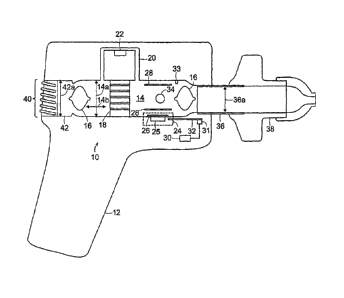
[0042] Figure 1 is a schematic diagram of a medical device 10 for aiding in
the diagnosis
of respiratory dysfunction, more particuiarly in the diagnosis of a pulmonary
embolism.
As shown herein, the device 10 of the present invention may be encompassed
within a
housing 12 that forms a unitary combination of the numerous elements and
subsystems
of the present invention readily adaptable for use in a diagnostic situation.
In the
preferred embodiments, the device 10 of the present invention is a handheld
unit, as
shown in Figure 1.
[0043] The device 10 generally includes an airway 14 that defines a diameter
or cross-
sectional dimension 14a substantially perpendicular to a longitudinal axis
14b. In
preferred embodiments, the airway 14 is cylindrical in nature, with the
longitudinal axis
14b being substantially the same as the flow of air while in use. The airway
14 is
bounded at an inlet by an inlet adapter 36 and at an outlet at an outlet
adapter 42. The
inlet adapter 36 is also substantially cylindrical in nature, defining a
diameter or cross-
sectional dimension 36a that is substantially perpendicular to the
longitudinal axis 14b.
In preferred embodiments, the inlet diameter 36a is less than the airway
diameter 14a,
for reasons discussed in greater detail below. The outlet adapter 42 is also
substantially
cylindrical in nature, defining a diameter or cross-sectional dimension 42a
that is
substantially perpendicular to the longitudinal axis 14b. As in the prior
instance, the
outlet diameter 42a is preferably less than the airway diameter 14a.
6
CA 02641608 2008-08-05
WO 2007/092052 PCT/US2006/033974
[0044] The inlet adapter 36 is adapted for receiving a disposable mouthpiece
38 through
which a patient may breath during use. Similarly, the outlet adapter 42 is in
communication with an outlet port 40 that may be integral with the housing 12.
In use, a
patient breathes air in and out through the disposable mouthpiece 38, which
causes the
passage of inhaled air as well as exhaled air through the airway 14. As
described more
fully herein, the device 10 utilizes a plurality of sensors to analyze the
content of the
exhaled air in order to aid in the diagnosis of a respiratory dysfunction.
[0045] A pair of light restrictors 16 is disposed within the airway 14
proximal to the inlet
adapter 36 and the outlet adapter 42. The light restrictors 16 shown herein
generally
define a symmetrical body having opposing convex surfaces that are oriented
along
longitudinal axis 14a of the airway 14. In preferred embodiments, the light
restrictors 16
have a diameter that is less than that of the airway diameter 14a but greater
than both
the inlet adapter 36a and the outlet adapter 42a, respectively. In this
manner, it is
possible for air to flow through the airway 14 around the light restrictors
16, but light itself
is unable to pass through the airway 14, thus protecting the internal sensors
from
interference or degradation, as discussed more fully below. Additionally,
given the
symmetrical convex shape of the light restrictors 16, a turbulent airflow that
is drawn
through the airway 14 becomes substantially laminar prior to its engagement
with the
plurality of sensors.
[0046] The device 10 further includes a flow restrictor 18 that is disposed in
the airway
14 between the pair of light restrictors 16. The flow restrictor 18 is adapted
for directing
a portion of air into a flow bypass channel 20 that is in communication with a
flow sensor
22. As shown in further detail below, the flow restrictor 18 forms a network
of passages
that cooperate to sufficiently occlude the airflow thereby directing air into
the bypass
channel and to the flow sensor 22. The flow restrictor 18 is preferably
configured for
insertion into the airway 14, and is thus preferably cylindrical in shape
having a diameter
substantially equal to that of the airflow diameter 14a.
[0047] The device 10 further includes an oxygen sensor 24 having an
emitter/sensor 25
and a lens 26. The preferred oxygen sensor 24 is a combination of a light
emitting diode
(LED) and a photodetector that is adapted for measuring the reflectivity of
the LED light
off of a selected surface. In most preferred embodiments, the LED emits light
in or
around the blue wavelengths that is directed by the lens 26 onto a coated
surface (not
7
CA 02641608 2008-08-05
WO 2007/092052 PCT/US2006/033974
shown) that is reactive to oxygen. As the level of oxygen in the airflow
varies, the
fluorescence of the coated surface also varies and the photodetector measures
this
variance. Known relationships between the reflective intensity of the coated
surface and
the measured photodetector values are utilized to compute the amount of oxygen
in the
airflow.
[0048] The device 10 further includes a carbon dioxide sensor 34 that is
disposed
adjacent to the oxygen sensor 24 in the airway 14. The carbon dioxide sensor
34 is
preferably a non-dispersive infrared sensor (NDIR), of the type known in the
art.
[0049] According to the present invention, the oxygen sensor 24 and the carbon
dioxide
sensor 34 are arranged so as to minimize the potential for error in the
computation of the
oxygen to carbon dioxide ratio of the airflow. More particularly, the oxygen
sensor 24
and the carbon dioxide sensor 34 are arranged so as to be mutually orthogonal
with the
longitudinal axis 14b. Or, as both sensors are preferably optical sensors,
they are
preferably arranged such that a first ray emanating from the oxygen sensor 24
and a
second ray emanating from the carbon dioxide 34 sensor and the longitudinal
axis 14b
are mutually orthogonal. This orientation provides a number of benefits,
including
synchronized data collection over the same volume of air as it passes through
the airway
14. Serial disposition of these sensors, as practiced in the state of the art,
does not
allow each sensor to operate independently upon the same volume of air at the
same
time, thus leaving open the possibility that changes in air temperature, flow
direction,
pressure or gaseous concentration will adversely affect the measured values of
oxygen
and carbon dioxide. The present invention solves this problem through the
aforementioned orthogonal orientation of the oxygen sensor 24 and the carbon
dioxide
sensor 34.
[0050] A pair of substantially planar air deflectors 28 are disposed within
the airway 14
to control the flow of air through the airway 14 as well as to prohibit any
signal or optical
interference between the oxygen sensor 24 and the carbon dioxide sensor 34. In
preferred embodiments, the air deflector 28 disposed nearest the oxygen sensor
24 has
a coated surface nearest the oxygen sensor 24, wherein the coated surface is
optically
sensitive to the presence of oxygen in the airflow. Alternatively, the coated
surface can
be placed on a disposable member (not shown) that can be removed from the
device 10
and replaced without affecting the functionality of the oxygen sensor 24.
8
CA 02641608 2008-08-05
WO 2007/092052 PCT/US2006/033974
[0051] The device 10 of the present invention further contains temperature
control
means 30 including at least a first thermometer 31 and a heating element 32,
wherein
the latter two elements preferably cooperate to maintain the temperature of
the airway
14 at a predetermined level. A second thermometer 33 is also preferably
disposed
within the airway 14 for measuring an air temperature as it passes there
through. More
specifically, variations in the temperature and relative humidity between
inhaled air and
exhaled air may cause unintended errors in the measurement of the oxygen to
carbon
dioxide ratios as measured by the present invention. Thus, while the first
thermometer
31 and the heating element 32 cooperate to maintain a predetermined
temperature on
the airway surface 14, the second thermometer 32 is configured for measuring
the
temperature of the air passing through the heated airway 14.
[0052] The temperature control means 30 of the present invention is adapted
for
maintaining the temperature of the airway 14 at a range between thirty-three
and forty-
three degrees Celsius. More preferably, the temperature control means 30 of
the
present invention is adapted for maintaining the temperature of the airway 14
at
approximateiy thirty-eight degrees Celsius.
[0053] The temperature control feature of the present invention provides a
number of
benefits, including removing any excess humidity from the exhaled air, warming
the
inhaled air so as to decrease the temperature gradient over the respiration
cycle of a
user, and increasing the sensitivity of the oxygen sensor 24 and the carbon
dioxide
sensor 34 by normalizing the relative humidity and temperature gradient over
the
respiration cycle.
[0054] The interaction of the various components of the present invention is
also
apparent in the block diagram of a system 100 according to the present
invention shown
in Figure 2. The system 100 includes the airway 14, the light restrictors 16,
and the flow
restrictor 18 for managing the entry, exit and flow of the user's breadth as
described
above. Additionally, the system 100 includes the temperature control means 30,
the
oxygen sensor 24, the carbon dioxide sensor 34, the flow sensor 22 and the
second
thermometer 33, all of which are coupled through various means known in the
art to a
controller 50. The system 100 further includes a pulse meter 42 that is
coupled to the
controller 50 and is further adapted for communication with the user's body in
order to
9
CA 02641608 2008-08-05
WO 2007/092052 PCT/US2006/033974
determine the user's heart rate.
[0055] Each of the measuring components of the system input their respective
data in
real time to the controller 50, which is adapted for receiving such
information and
computing a ratio of oxygen to carbon dioxide in the user's breath, which in
turn may be
indicative of a pulmonary embolism. The controller thus receives data
indicative of the
total volume of air expelled by the patient, the oxygen content of the exhaled
air, the
carbon dioxide content of the exhaled air, the temperature of the exhaled air
and the
heart rate of the user. The data is processed by the controller according to
the
methodology described herein, and the results are transmitted to a display 60
coupled to
the controller 50. The entire system 100 is adapted for use in a compact and
mobile
arrangement that is usable in a hospital environment. For example, a cart can
be readily
configured to include the controiler 50 and display 60, the former of which
can be
adapted for interface with the device 10 and pulse meter 42 of the present
invention in
order to compile the system 100 described herein.
[0056] As noted above, another novel aspect of the present invention is the
flow
restrictor 18 that is disposed in the airway 14 for directing air into the
bypass channel 22,
shown in the front view of Figure 3. As shown herein, the flow restrictor 18
generally
defines an annular edge portion 180 that is disposable within the airway 14.
It should be
understood that the flow restrictor 18 is annular in shape in order to fit
within a cylindrical
airway 14. In embodiments in which the airway 14 is non-cylindrical, the flow
restrictor
18 will have a matching profile so as to prevent the flow of air between the
edge portion
180 and the airway 14.
[0057] The flow restrictor 18 generally defines an interior space within the
edge portion
180 that is divided into multiple portions by a plurality of substantially
horizontal fins and
substantially vertical fins. Preferably, the horizontal and vertical fins are
oriented so as
to be substantially mutually orthogonal with the longitudinal axis 14b, shown
in Figure 1.
In the embodiment depicted in Figure 3, a first pair of horizontal fins 182
are disposed
opposite each other with a plurality of horizontal fins 188 disposed there
between.
Vertical fins 184, 186 bisect the horizontal fins 182, 188 as shown in order
to define a
plurality of openings 190 having substantially the same frontal area through
which air
may pass.
CA 02641608 2008-08-05
WO 2007/092052 PCT/US2006/033974
[0058] Although the shown combination of horizontal fins 182, 188 and vertical
fins 184,
186 defines a plurality of openings 190 having a particular geometry, it
should be
understood by those skiiied in the art that the size and shape of the
plurality of openings
190 can be readily altered provided that there is a marked consistency across
the
surface of the flow restrictor 18. In operation, the flow restrictor 18
permits the flow of
some air through the plurality of openings 190, while simuitaneously causing a
sufficient
buildup in air pressure to divert the air into the bypass channel 20 shown in
Figure 1.
Thus, for optimal performance the flow restrictor 18 should cause a uniform
diversion of
air without causing large deviations in the air pressure orthogonal to the
longitudinal axis
14b so as to provide a consistent and steady flow of air into the bypass
channel 20 to
the flow sensor 22.
[0059] The light restrictor 16 of the present invention is shown in two
alternate
embodiments in Figures 4a, 4b, 4c and 5a, 5b and 5c. In the first embodiment
shown,
the light restrictor 16 is generally defined by a body portion for
substantially prohibiting
the passage of light into the airway 14. Figure 4a is a perspective view of
the light
restrictor 16 illustrating the contours of a leading edge 16a. As seen in the
side view of
Figure 4b, the leading edge 16a and a trailing edge 16b are substantially
symmetrical
about the center 16c of the light restrictor 16.
[0060] As previously noted, the light restrictor 16 is adapted for use in the
device 10 of
the present invention, preferably being disposed at either end of the airway
14. In order
to permit the passage of air while limiting or eliminating the passage of
light into the
airway 14, the diameter of the light restrictor 16 about its center 16c is
preferably less
than that of the airway 14 but greater than that of the respective inlet
adapter 36 or outlet
adapter 42.
[0061] The contours of the leading edge 16a and the trailing edge 16b are
selected in
order to maximize the efficient flow of air about the light restrictor 16
while minimizing
any associated pressure drop along the traiiing edge 16b. The aerodynamics of
the light
restrictor 16 have the added benefit of creating a laminar flow of the air as
it passes
through the airway 14, thereby increasing the consistency and dependability of
the
sensor measurements. The act of respiration may create large pockets of low
pressure,
and the typical airflow through any closed space maybe significantiy
turbulent. However,
due to the specific design and shape of the light restrictor 16 described
herein, a
11
CA 02641608 2008-08-05
WO 2007/092052 PCT/US2006/033974
significant amount of that turbulence is eliminated in the process of
restricting light entry
into the airway 14.
[0062] The front view of Figure 4c illustrates the substantially circular
profile of the light
restrictor 16. As discussed above with reference to the flow restrictor 18, it
is
conceivable that the airway will not have a cylindrical shape, and thus the
cross-
sectional profile of the light restrictor 16 may vary accordingly.
Nevertheless, the
functional aspects of the light restrictor 16 are the same in any embodiment
or geometry.
Namely, the light restrictor 16 of the present invention accomplishes two
goals. First, the
light restrictor 16 must substantially or entirely occlude any ambient light
from irradiating
the oxygen sensor 24 and its associated components. Secondly, the light
restrictor 16
significantly reduces the turbulent flow of a user's breath through the airway
14 by
adopting contours that will induce a laminar flow of air, The first goal is
accomplished by
appropriately sizing the diameter of the light restrictor 16 relative to that
of the airway 14
and the inlet adapter 36 and outiet adapter 42. The second goal is
accomplished as
described above, by introducing a substantially symmetrical leading edge 16a
and
trailing edge 16 about a center 16c.
[0063] Figure 5a is a perspective view of a light restrictor 17 usable in the
device and
system of the present invention according to another embodiment. The light
restrictor 17
depicted herein is generally conical in shape, consisting of a series of
members
arranged in order of decreasing size and terminating at a cap 17a. As shown in
Figure
5b, there are spaces between each successive member in the light restrictor
17, thus
permitting significant airflow there through. However, as shown in Figure 5c,
the
members are arranged radially about the cap 17a such that no light can be
transmitted
directly through the light restrictor 17, i.e. each successive member
obstructs the
passage of light through the larger adjacent member, and the cap 17a prevents
light
from passing directly along the longitudinal axis 14b.
[0064] As depicted in Figures 5a, 5b and 5c, this plurality of members of the
light
restrictor 17 is shown as a series of rings or annuli having a largest
diameter 17b that is
preferably coextensive with or less than the airway diameter 14a.
Nevertheless, the light
restrictor 17 described herein can take innumerable forms depending upon the
cross-
sectional profile of the airway 14. For an airway 14 that is cylindrical in
nature, the most
efficient and aerodynamic form for the light restrictor 17 would be a series
of rings, as
12
CA 02641608 2008-08-05
WO 2007/092052 PCT/US2006/033974
shown and described above. However, in the instances in which the airway 14
cross-
section is square, elliptical or some other geometry, the specific shapes of
the members
can be varied accordingly to permit the passage of air with minimal pressure
drop while
prohibiting the entrance of light into the airway 14.
[0065] As described thus far, the present invention is a device and system
arranged
from various discreet components, including sensors, controlling means and the
associated flow and light restrictors. However, the present invention includes
numerous
additional components that are preferred for its operation in the manner
described
herein. For example, the device and system of the present invention, in more
preferred
embodiments, include various integrated computation and electronic elements
for
efficiently receiving, analyzing and transmitting the data to the controller
means 50.
[0066] Another embodiment of the present invention is described herein with
reference
to Figures 6 through 11. Figure 6 is an exploded isometric view of the device
and
system of the present invention including additional components useful in its
preferred
method of operation, and Figure 7 is a cross-sectional view of the same.
Figures 8 and
9 are inlet and outlet views of the device 10 of the present invention,
respectively.
Figures 10 and 11 are top and bottom views of the device described herein,
respectively.
[0067] To the extent that identical reference numerals are used herein, they
should be
understood to refer to similar elements as previously described. As in the
prior
embodiment described above, the device 10 of the present invention generally
includes
an airway 14 that is defined in part by a first body portion 140 and a second
body portion
142. The airway 14 contains, at or near the junction of the first body portion
and the
second body portion 142, a flow restrictor 18 of the type generally described
herein.
[0068] The first body portion 140 receives at least one air deflector 28
disposed within
the airway 14. A heating element 32, preferably integrated into the thermal
control
means described above with reference to Figures 1 and 2, is disposed within
the first
body portion 140 for maintaining the latter at a predetermined temperature. A
light
restrictor cradle 160 and a light restrictor 16 are also disposed within the
airway 14
defined by the first body portion 140. The light restrictor cradle 160 serves
multiple
functions, including maintaining the orientation and placement of the light
restrictor 16,
as well as varying the diameter within the airway 14 such that light may not
pass there
13
CA 02641608 2008-08-05
WO 2007/092052 PCT/US2006/033974
through. Finally, an inlet adapter 36 is disposed within the airway 14 defined
by the first
body portion 140, to which a mouthpiece or other breathing apparatus may be
attached
during use.
[0069] The first body portion 140 shown herein also contains, is coupled to,
or receives
a number of sensors and subsystems noted above. In particular, the first body
portion
140 includes receiving ports for receiving the carbon dioxide sensor 34, the
flow sensor
22, the oxygen sensor 24, and the temperature control means 30, including the
second
thermometer 33 disposabie within the airway 14, as shown in Figure 1.
Additionally, the
first body portion 140 includes openings or tunnels that are formative of the
flow bypass
channels 20, described in detail above.
[0070] Each of the aforementioned sensors is coupled to or integrated with its
associated electronic components, including for each sensor the necessary
circuitry and
processing means for converting raw signals sensed by the sensors into
eiectronic
signals that are adapted for processing by the controller 50 of the system 100
described
above. Each of the sensors within the device 10 are thus readily connectable
to the
controller 50, shown in Figure 2, through wired or wireless communications
means
known to those skilled in the art.
[0071] For example, as shown in Figure 7 the oxygen sensor 24 and its
components, the
emitter/sensor 25 and the lens 26, are shown integrated into an oxygen sensor
printed
circuit board (PCB) 240 that is attachable to the first body portion 140 as
shown.
Similariy, the flow sensor 22 and its associated structure is shown integrated
into a flow
sensor PCB that is attachable to the first body portion 140, preferably on a
side opposite
to that of the oxygen sensor PCB 240. The temperature control means 30 and its
associated components, including the second thermometer 33, are shown
integrated
into a temperature control PCB 320, which again is preferably disposed on a
side of the
first body portion 140 opposite to that of the oxygen sensor PCB 240.
[0072] The carbon dioxide sensor 34 is preferably dual-sided in nature, having
both an
emitter side and a detector side. Thus, the carbon dioxide sensor 34 consists
of a
carbon dioxide sensor emitter PCB 340, including a carbon dioxide sensor
emitter 35,
and a carbon dioxide detector PCB 344, including a carbon dioxide detector
(not shown).
In order to prevent interference between the oxygen sensor 24 and the carbon
dioxide
14
CA 02641608 2008-08-05
WO 2007/092052 PCT/US2006/033974
sensor 34, it is preferable to dispose the respective carbon dioxide PCBs 340,
344 on
opposing sides of the first body portion 140 that are adjacent to the oxygen
sensor PCB
240. In preferred embodiments, the device further includes at least one carbon
dioxide
sensor adaptor 342 for affixing the carbon dioxide PCBs 340, 344 that serve
the further
purpose of protecting the optical components of the carbon dioxide sensor 34.
[0073] The second body portion 142 also defines a portion of the airway 14.
The
second body portion 142 also is adapted to receive a light restrictor cradle
160 and a
light restrictor 16, both of which are buttressed on a distal end by an outlet
adapter 42.
As previously noted, the light restrictor cradle 160 serves multipie
functions, including
maintaining the orientation and placement of the light restrictor 16, as well
as varying the
diameter within the airway 14 such that light may not pass there through.
[0074] In another aspect of the present invention, a disposable assembly is
provided
that integrates the necessary airflow and light restriction features while
maintaining the
functional aspects associated with the device and system outlined above.
Figures 12,
13 and 14 are top, cross-sectional, and bottom views of the assembly 400 in
accordance
with a preferred embodiment.
[0075] Referring to Figures 12, 13 and 14 simultaneously, it is shown that the
assembly
400 includes an airway 404 that is coupled to a mouthpiece 402. In some
embodiments,
the airway 400 and the mouthpiece may be selectively coupled, such that each
component can be separately sterilized or disposed of following use.
Alternatively, the
assembly 400 can be readily designed as an integrated whole that is disposable
after
each and every use.
[0076] As seen best in Figure 13, the airway 404 defines an interior volume
that contains
a number of elements described above. The mouthpiece 402 defines an inner
surface
406 defining a first volume 410 across a first diameter. Entrance into the
interior of the
airway 14 is partially obstructed by a light restrictor 412 that is bounded on
either side by
a pair of ridges 408. The opposing light restrictor 412 is also bounded by a
pair of ridges
408 disposed along the interior of the airway 404. The respective ridges 408
function to
prevent the passage of iight into the interior of the airway 404 while
simultaneously
cooperating with the light restrictor 412 to permit the regular and laminar
flow of air there
through. As in prior embodiments, the ridges 408 may be designed as light
restrictor
CA 02641608 2008-08-05
WO 2007/092052 PCT/US2006/033974
cradles or changes in the shape of the interior of the airway 404. Likewise,
it should be
understood that different types of light restrictors, such as those described
herein, might
also be used in the assembly 400 of the present invention.
[0077] A flow restrictor 424 is disposed within the airway 404 between the
light
restrictors 412. As in prior preferred embodiments, the airway 404 is also
populated by
a pair of air deflectors 414. The air deflectors 414 are arranged about a
first sensor
window 420, as shown in Figure 13. In preferred embodiments, the sensor window
is
alignable with an optical sensor, such as a carbon dioxide sensor, which
requires the
further presence of the air deflectors 414 to avoid any interference with
other optical
sensors operating in the airway 404.
[0078] The airway 414 further defines a second sensor window 418 that is
disposed
opposite one of the air deflectors 414. In preferred embodiments, the second
sensor
window 418 is alignable with a second optical sensor, such as for example an
oxygen
sensor. In such an embodiment, it is further possible to dispose an oxygen-
sensitive
surface 416 on the near surface of the opposing air deflector 416. As
previously noted,
in the operation of both an oxygen and a carbon dioxide sensor, it is
preferable for the
air deflectors 414 to be aligned in such a manner so as to prevent any optical
interference between the sensors.
[0079] The airway 414 further defines a first sensor port 422 disposed along
the bottom
of the airway 414 as shown in Figures 13 and 14. The first sensor port 422 is
preferably
adapted for a thermometer or other means for measuring the temperature of air
that is
passing through the airway 414. A set of second ports 426 visible in Figures
12 and 13,
are disposed about the flow restrictor 424 for permitting the selective
passage of air out
of the airway 404. In preferred embodiments, the second ports 426 are
alignable with a
flow sensor of the type described herein in which air is directed through a
bypass
channel.
[0080] In its more preferred embodiments, the assembly 400 of the present
invention is
utilized in conjunction with an electronic device 500 as shown in Figure 15.
The
assembly 400, including each of the features described above, is shown
inserted into a
receptacle formed within an electronic device 500. The electronic device 500
includes a
bypass channel 502 that is in fluid communication with a flow sensor 504. In
preferred
16
CA 02641608 2008-08-05
WO 2007/092052 PCT/US2006/033974
embodiments, the bypass channel 502 is readily and automatically aligned with
the
second ports 426 of the airway 404 such that air passing through the airway
404 is
diverted into the bypass channel 502 by the flow restrictor 424. It is further
preferred
that the junction between the second ports 426 and the bypass channel 502 is
substantially airtight so as to cause minimal disruption in the airflow
through the airway
404.
[0081] The electronic device 500 shown herein further includes an oxygen
sensor 506
including an emitter/detector 508 and a lens 510 that are disposed within the
electronic
device 500 adjacent to the second sensor window 418. In preferred embodiments,
the
oxygen sensor 506 is oriented within the eiectronic device 500 such that
placement of
the assembly 400 therein automatically aligns the oxygen sensor 506 with the
oxygen
sensitive surface 416 located within the airway 404.
[0082] The eiectronic device 500 also includes a carbon dioxide sensor 520
that is
disposed therein such that insertion of the assembly 400 causes the carbon
dioxide
sensor 520 to be automatically aligned with the first sensor window 420. Both
the first
sensor window 420 and the second sensor window 418 are preferably composed of
a
material that is optically transparent across the spectra used by the
respective sensors.
Moreover, it is preferable for the first sensor window 420 and the second
sensor window
418 to be airtight so as to substantially minimize any disruption in the
airfiow through the
airway 404.
[0083] The first port 422 is in fluid communication with a thermometer 512 for
measuring
a temperature of the air passing through the airway 404. The first port 422 is
preferably
sealed against the thermometer 512 in an airtight fashion so as to
substantially eliminate
any turbulence in the airflow through the airway 404.
[0084] A temperature control system 530 including a temperature controller
514, a
heating element 516 and a second thermometer 518 is preferably disposed within
the
electronic device. In operation, the temperature control system 530 serves to
maintain
the electronic device 500 at a specified temperature. Thermal induction will
further
ensure that the airway 404, when properly inserted within the electronic
device 500, will
also be maintained at or about the specified temperature. In particular, it is
desirable to
maintain the airway 404 at a temperature of between thirty-three and forty-
three degrees
17
CA 02641608 2008-08-05
WO 2007/092052 PCT/US2006/033974
Celsius, although it is even more preferable to maintain a temperature of
approximately
thirty-eight degrees Celsius.
[0085] The benefits of temperature control within the present invention are
described
above including, without limitation, warming the inhaled air so as to decrease
the
temperature gradient over the respiration cycle of a user, and increasing the
sensitivity
of the oxygen sensor 506 and the carbon dioxide sensor 520 by normalizing the
relative
humidity and temperature gradient over the respiration cycle.
[0086] Another aspect of the present invention is shown in Figures 16 and 17
in which
the oxygen sensor is located within a second bypass channel. As shown herein,
a
device 700 is shown for aiding in the determination of a pulmonary
dysfunction. The
device 700 generally includes a body portion 702 defining an airway 704 there
through.
The airway 704 is bounded on one end by an inlet adapter 732 and on another
end by
an outlet adapter 734. A flow restrictor 724 of the type described above is
disposed
within the airway 704. In operation, the flow restrictor 724 diverts a
selected portion of
each inhaled and exhaled breath into a first bypass channel 706 and a second
bypass
channel 710.
[0087] A carbon dioxide sensor 726 of the type described above is disposed
adjacent to
the airway 704. Preferably, the carbon dioxide sensor 726 is optical in
nature. A
thermometer 730 is disposed in the airway 704 for measuring the temperature of
the
inhaled and exhaled breaths passing there through. As in prior embodiments,
the device
700 also includes a temperature control means 720 including a heating element
721, a
controller 722 and a second thermometer 723. As noted with respect to the
prior
embodiments, the temperature control means 720 is adapted for maintaining the
device
700 in general and the airway in particular 704 at a specified minimum
temperature. In
particular, the temperature control means 720 serves to maintain the airway
704 at a
temperature of between thirty-three and forty-three degrees Celsius, aithough
it is even
more preferable to maintain a temperature of approximately thirty-eight
degrees Celsius.
[0088] The benefits of temperature control within the present invention are
described
above, including removing any excess humidity from the exhaled air, warming
the
inhaled air so as to decrease the temperature gradient over the respiration
cycle of a
user, and increasing the sensitivity of the oxygen sensor 712 and the carbon
dioxide
sensor 726 by normalizing the relative humidity and temperature gradient over
the
18
CA 02641608 2008-08-05
WO 2007/092052 PCT/US2006/033974
respiration cycle.
[0089] The first bypass channel 706 is adapted for diverting a portion of the
airflow
towards a flow sensor 708 of the type described in detail above. The second
bypass
channel 710 is adapted for diverting a portion of the airflow towards an
oxygen sensor
712 of the type described above. In preferred embodiments, the oxygen sensor
712
includes an emitter 716, a detector 718 and an oxygen sensitive surface 714
disposed
across the second bypass channel 710 such that light emitted from the emitter
716 is
reflected from the oxygen sensitive surface 714 to the detector 718.
[0090] Unlike in prior embodiments in which the oxygen sensitive surface 714
was
protected from ambient light through the use of mechanical light restrictors,
the current
embodiment of the present invention maintains the lifetime of the oxygen
sensitive
surface 714 by disposing it within the second bypass channel 710. The benefits
of doing
so are apparent to those skilled in the art, including the ease of engineering
and desiging
the present embodiment without the use of light restrictors or labyrinths.
Moreover, as
the oxygen sensor itself 712 is disposed along the second bypass channel 710,
the flow
of air there through is readily measured and controlled according to the
mechanical
properties of the flow restrictor 724 and the operation of the flow sensor
708.
Additionally, by eliminating the need for light restrictors, the overall
volume of the airway
704 is lessened, therefore requiring less volume per breath in order to
properly and
consistently operate the sensors of the present invention. As noted in the
current
specification, it is a feature of the present invention that greater control
and
measurement precision over the relevant variables (flow, temperature, oxygen,
carbon
dioxide, and pulse rate) is instrumental in assuring accurate and predictive
diagnosis of
a respiratory dysfunction.
[0091] In yet another embodiment of the present invention, a disposable
assembly 800
is shown in Figure 17 that encompasses features of the aforementioned
disposable
assembly, but adapted for fitting into an electronic device having an oxygen
sensor
disposed in an ancillary bypass channel. As such, the assembly 800 includes a
mouthpiece 802 that is selectively coupled to or integrated with an airway 804
defining a
volume through which air may freely flow. A flow restrictor 808 is disposed
within the
airway 804 for directing a specified portion of the airflow into a pair of
bypass channels,
such as those described above with reference to Figure 17.
19
CA 02641608 2008-08-05
WO 2007/092052 PCT/US2006/033974
[0092] A window 806 is also provided in the airway 804 that is readily and
automatically
alignable with a carbon dioxide sensor (not shown) disposed within the
receiving device.
Similarly, a first port 814 is provided for permitting access to the airway
804 by a
thermometer (not shown) for measuring the air temperature of the airflow. The
numerous benefits of air temperature measurement are described herein. A
second set
of ports 810 and a third set of ports 812 are disposed on opposing surfaces of
the airway
804 in the manner shown in Figure 17. In preferred embodiments, the second
ports 810
and third ports 812 are readily aligned with a first and second bypass
channel, each
providing airflow to one of an oxygen sensor or a flow sensor, as described
above.
[0093] It is another feature of the present invention that each of the first
port 814, second
ports 810, and third ports 812 are coverable by a filtration media 816 having
microbial
properties. In such a manner, a user can readily discard the assembly 800 of
the
present invention after each use without risk of directly exposing any
electronic devices
or sensors to a patient's breath.
[0094] A similar embodiment of the present invention is shown in Figures 18
through 23.
Figure 18 is an isometric exploded view of a device 900 for aiding in the
diagnosis of a
respiratory dysfunction, and Figure 19 is a cross-sectional view of the same.
Figures 20
and 21 are an inlet and an outlet view of the device 900, respectively, and
Figures 22
and 23 are a top and a bottom view of the same. The device 900 of the present
invention, like that in the previous embodimerit, employs a pair of bypass
channels for
the oxygen and flow sensors, thus eliminating the need for any mechanical
light
restrictors or labyrinths.
[0095] The device 900 generally includes a body portion 902 defining an airway
914 and
a passage 912 configured for a carbon dioxide sensor emitter 932. The body
portion
902 is preferably designed to receive an inlet adapter 908 at an inlet end and
an outlet
adapter 906 at an outlet end. As in previous embodiments, a flow restrictor
904 is
disposed in the airway 914 for directing a selected portion of the airflow
into the
respective bypass channels. A flow sensor PCB 920, including a flow sensor
919, a first
bypass channel 921 and the requisite eiectronics and circuitry, is attachable
to the
bottom of the body portion 902. As in previous embodiments, the first bypass
channel
921 cooperates with the flow restrictor 904 to divert a portion of the airflow
to the flow
CA 02641608 2008-08-05
WO 2007/092052 PCT/US2006/033974
sensor 919, the resulting signal being adapted for transmission and processing
by a
controller 50 as part of the system 100 of the present invention.
[0096] The oxygen sensor PCB 944, including the oxygen sensor 945 and any
associated electronics and circuitry, is disposable on the top of the body
portion 902.
Optionally, a shield 946 may be disposed above the oxygen sensor PCB 944 for
protecting the circuitry and sensing components of the oxygen sensor 945 and
its
associated PCB. Although not shown in this embodiment, it should be understood
that
the preferred oxygen sensor 945 is similar to those previously described,
including an
emitter/detector pair in optical communication with an oxygen sensitive
surface. The
oxygen sensor PCB 944 is preferably disposable on or near a second bypass
channel
942, which as shown herein is formed by a pair of top portions 940 that are
conjoined
over the body portion 902 as shown in Figure 18. A pair of projections 948 are
shown
inserting into the airway 914, in communication with and forming portions of
the second
bypass channel 942. The projections 948 serve the dual purpose of filtering
impurities
and moisture from the air stream, as well as regulating the volume of air that
is
admissible into the second bypass channel for sensing by the oxygen sensor
945. As
should be understood by those skilled in the art, the particular formation of
the second
bypass channel 942 utilizing the projections 948 shown herein is merely
exemplary, and
other methods and designs are equally well suited for achieving the foliowing
benefits.
[0097] As noted before, positioning of the oxygen sensor 945 in communication
with the
second bypass channel 942 provides a number of benefits, most notably
simplifying the
design and implementation of the present invention by obviating the need for
mechanical
light restrictors. Moreover, by securing the oxygen sensitive surface on the
interior of a
dark structure, the life and performance of the oxygen sensor 945 wiil not
suffer due to
the influence of ambient light. Also, by eliminating light restrictors, the
overall volume of
the airway 914 is decreased, therefore reducing the volume per breath needed
in order
to properly and consistently operate the sensors of the present invention. As
noted
throughout the current specification, it is a feature of the present invention
that greater
control and measurement precision over the relevant variables (flow,
temperature,
oxygen, carbon dioxide, and pulse rate) is instrumental in assuring accurate
and
predictive diagnosis of a respiratory dysfunction. Accordingly, by minimizing
the voiume
necessary to induce performance of the sensors, the present invention results
in more
accurate and consistent measurements of the necessary parameters.
21
CA 02641608 2008-08-05
WO 2007/092052 PCT/US2006/033974
[0098] As in prior embodiments, the device 900 includes a carbon dioxide
sensor that is
disposable on either side of the body portion 902 in the form of a carbon
dioxide sensor
emitter PCB 924, including a carbon dioxide sensor emitter 932, and a carbon
dioxide
sensor detector PCB 922, including a carbon dioxide sensor detector 936. A
series of
couplers 930 connect the carbon dioxide sensor emitter 932 to its respective
PCB, and a
series of couplers 936 connect the carbon dioxide sensor detector 934 to its
respective
PCB. As in the previous embodiments, the carbon dioxide PCB's include all of
the
necessary electronics and circuitry for converting an optical signal into an
electrical
signal suitable for transmission to a controller 50 part of the system 100
described
previously.
[0099] A thermometer 950 is aiso shown in fluid communication with the airway
914 for
measuring a temperature of the air passing there through. Although not shown,
the
embodiment of the device 900 described herein is also adapted for utilizing a
temperature control means of the type previously described.
[0100] Turning to Figure 24, a method for determining the sufficiency of
respiration is
provided according to the present invention. As noted with respect to prior
aspects of
the present invention, one of the many important factors in accurately
measuring the
oxygen content of a patient's exhaled air is the volume of expired air that is
provided to
the various sensors described above. A patient that does not have fully
functional
alveoli may not exhale sufficiently to properly and consistently measure the
content of
his or her exhaled breathe, as the gas-exchanging process within the patient's
lungs
may be degraded. Accordingly, the present invention inciudes a method for
determining
whether any exhaled breath passed through the systems and devices described
above
is sufficient for diagnostic purposes.
[0101] Step S100 of the method includes measuring a volume of exhaled air. As
noted
above, the flow sensor of the present invention, operating in concert with the
temperature control features of the present invention, is adapted for
providing such a
measurement.
[0102] In step S102, the method recites determining a dead space volume in
response
to the volume of exhaled air. As is known in the art, the dead space volume
refers to the
22
CA 02641608 2008-08-05
WO 2007/092052 PCT/US2006/033974
portion of any tidal breath where gas exchange does not take place. There are
numerous methodologies known in the art for determining the dead space volume,
including for example the Fletcher-Fowler method. The Fletcher-Fowler method
includes measuring a carbon dioxide concentration across an exhalation period,
resulting in a curve representing the exhaled volume as a function of carbon
dioxide
concentration. Integration of the curve about an equilibrium point results in
the
calculation of a dead space volume, which may or may not be indicative of a
respiratory
dysfunction such as pulmonary embolism.
[0103] In step S106, the method recites inputting a constant multiplier,
preferably
between 1.5 and 1.9, and even more preferably between 1.6 and 1.8. In more
preferred
embodiments of the present invention, the constant multiplier is approximately
1.7. In
step S108, the non-gas exchanging volume is multiplied by the constant
multiplier,
resulting in a product that is determinative of respiratory sufficiency. For
example, if the
resulting product is greater than a predetermined value, then the exhalation
was
insufficient for diagnostic purposes. On the contrary, if the resulting
product is less than
the predetermined value, then the exhalation was sufficient for diagnostic
purposes and
the system and device of the present invention will have a sufficient volume
of air for
accurate and consistent measurements.
[0104] In step S110, the method recites the step of aitering the user as to
the respiratory
sufficiency. In preferred embodiments, this alert may be practiced through
automated
means such as a visual or aural signal emanated by the system or device of the
present
invention. For example, the display described above with reference to the
system of the
present invention may be adapted for signaling to a user the resulting product
in a visual
form, such as a green light for a sufficient breath and a red or yellow light
for an
insufficient breath. Many similar methods and modes of altering the user can
be readily
devised by those skilled in the art. Following the alert, the method includes
step S112
that recites providing feedback to a patient based upon respiratory
sufficiency.
Preferably, this step includes coaching a patient to breath more deeply or
exhale more
completely in the case of an insufficient respiration. As in the previous step
of the
method, step S112 can be readily automated and performed by the system and
device
of the present invention described above.
[0105] Figure 25 is a flowchart depicting a method for calibrating a gas
sensor in
23
CA 02641608 2008-08-05
WO 2007/092052 PCT/US2006/033974
accordance with the present invention. As noted with respect to the system and
device
of the present invention, the optimal performance of the sensors depends
heavily on the
environment in which the measurements are taken. Those skilled in the art will
recognize that Boyle's law, PV = nRT, wherein the pressure, volume, molecular
concentration and temperature of the airflow through the system and device of
the
present invention are all interrelated. As such, proper measurement of any
concentration of a gas preferably incorporates a simultaneous measurement of
the
temperature, as indicated with the incorporation of the temperature control
systems and
thermometers within the systems and devices described above. For any given
volume
and pressure of air, therefore, one can find an inverse relationship between
the
temperature T and the concentration n of a gas.
[0106] In step S200, the method recites the step of heating a predetermined
volume of
air to a first temperature, TI. In preferred embodiments, the heating of the
predetermined volume of air is accomplished by the temperature control means
and
associated controls within the device and system of the present invention.
Step S202
recites measuring the temperature of the volume of air at TI, and step S204
recites
measuring the concentration of a selected gas, such as oxygen, within the
volume of air
at TI. Accordingly, for a predetermined temperature Tl, the method provides
for a
measurement of the temperature and concentration of a selected gas, most
preferably
using the oxygen sensor of the system and device to measure an oxygen
concentration
within the volume of air.
[0107] In step S206, the volume of air is heated to a second temperature, T2.
In step
S208, the method recites measuring the temperature of the volume of air at T2.
Step
S210 recites measuring the concentration of the selected gas, preferably
oxygen, at T2.
As such, for the second temperature T2, the method provides for the
measurement of
the temperature and concentration of the selected gas within the same volume
of air.
[0108] As evident from Boyle's law, any increase in temperature should not
affect the
molecular concentration of the selected gas provided that the volume of the
sample is
heid constant. One would reasonably expect the pressure to increase, however,
with an
increase in temperature. Given the foregoing, the method includes step S212,
which is
calculating a variance between the concentration of the selected gas as
measured at
temperatures T, and T2.
24
CA 02641608 2008-08-05
WO 2007/092052 PCT/US2006/033974
[0109] To the extent that there is a variation in the measured concentration,
it must be
indicative of a calibration issue with the sensor, as there can be no change
in the
concentration of a gas within a closed volume. Thus the method recites in step
S214
correlating the concentration of the selected gas to a temperature in response
to any
calculated variance. As such, a user will be informed of the temperature
dependence of
the measurements provided by the sensor. For example, the performance of the
optical
oxygen sensor described herein is affected by temperature changes, and thus
the
methodology set forth above provides a user with the necessary relationship
between
the output signal of the sensor and the temperature as measured within the
device and
system.
[0110] Thus in step S216, the method provides for calibrating a sensor adapted
to sense
a concentration of the selected gas in response to its dependence on a
temperature.
The preferred method for calibration includes processing both temperature and
sensor
data as it is fed into a central controller, such as that described above with
reference to
the system of the present invention. Knowing the functional relationship
between the
temperature output and the gas sensor output permits a user to program or
otherwise
control the system to bias or vary the output of the gas sensor to provide a
temperature-
dependent, and thus more accurate, measurement of the concentration of the gas
within
a volume of air. Although oxygen is generally the selected gas, it should be
understood
that the method described herein is equally applicable to carbon dioxide or
any other
gaseous concentrations to be measured by the device and system of the present
invention.
[0111] Moreover, it is also preferable to measure the pressure of the ambient
air as well
as the humidity of the ambient air, as these values may directly or indirectly
affect the
ability of the gas sensor to properly measure the concentration of the
selected gas.
Suitable devices for measuring the pressure and humidity of the ambient air
are known
in the art, and preferably signals indicative of the aforementioned conditions
can be
readily integrated into the system and methodology of the present invention.
[0112] Another method practicable according to the system and device of the
present
invention is shown in the flowchart of Figure 26, which is a method for
recommending
further diagnostics in response to a variation in a user's heartbeat between
an exhalation
CA 02641608 2008-08-05
WO 2007/092052 PCT/US2006/033974
and an inhalation. In step S300, the method designates a first period as an
inhalation
period, and in step S302, the method designates a second period as an
exhalation
period. As described above, the system of the present invention includes a
pulse meter
or pulse-oximeter for measuring at least an individual's heart rate during the
testing
interval. As such, in step S304, the method recites measuring an individual's
heart rate
during the inhalation period; and step S306 includes measuring the
individual's heart
rate during the exhalation period.
[0113] In step S308, the method recites determining a first variation of the
individual's
heart rate during the inhalation and exhalation periods. It is anticipated
that there will be
some variation in heart rate, as this is the normal result from a healthy
individual. As
such, in step S310, the method recites inputting a nominal variation of a
heart rate
during the inhalation and exhalation periods that is representative of the
variation for a
healthy subject. In step S312, the method includes comparing the first
variation to the
nominal variation to determine an actual variation, i.e. wherein the presence
of an actual
variation is indicative of an abnormality in the individual's cardio-pulmonary
system. In
response to an actual variation that exceeds a predetermined value, for
example outside
the margin of error of the nominal and first variations, step S314 recites the
step of
recommending that further diagnostics be done on the individual. Said further
diagnostics may include any and all of the testing of gaseous concentrations
of exhaled
breath as well as any other test or measurements that may aid in the diagnosis
of a
respiratory dysfunction such as pulmonary embolism.
[0114] The present invention has been described herein with reference to its
most
preferred and exemplary embodiments with reference to the noted figures.
However, it
should be apparent to those skilled in the art that numerous deviations from
the
described embodiments can be readily devised without departing from the scope
of the
present invention as defined in the following claims.
26