Note: Descriptions are shown in the official language in which they were submitted.
CA 02642280 2011-06-30
METAL-ENHANCED CIIEMILUMINESCENCE (MEC)
STATEMENT OF GOVERNMENT RIGHTS
[001] This work was supported by the NIH G1\4070925 and the National Center
for Research
Resources, RR008119, and the United States Government may have right to this
invention.
CROSS REFERENCE TO RELATED APPLICATIONS
[002] The present application claims priority to U. S Provisional Patent
Application No.
60/773,037.
BACKGROUND OF THE INVENTION
Field of the Invention
[003] The present invention relates to bioassays, and more particularly, to
the use of
metallized surfaces to enhance intensity of chemilumirtescence species or
reactions in
chemiluminescence assays thereby increasing sensitivity and delectability of
same.
Background of the Related Art
[004] The use of light-producing chemical reactions for quantitative detection
in
biotechnology is increasing [1-7], especially with regard to
cherniluminescence based ligand-
binding assays [1-7]. The attractiveness of chemiluminescence as an analytical
tool lies
primarily in the simplicity of detection [8]; the fact that most samples have
no unwanted
background luminescence, as is typically observed in fluoreScence-based assays
[9]; and the
fact that no optical filters are required to separate the excitation
wavelengths and scatter [8], as
is also required for fluorescence-based detection 191.
[005] However, chemiluminescent based detection is currently limited by the
availability of
chemilUminescent probes, which is not a factor governing fluorescence based
detection [9].
Both fluorescence and chemiIuminescence based -technologies do however suffer
from an
inhereot need for increased sensitivity/detection limits [8, 5]. For
fluorescence, this is governed
by the quantum yield of the tagging fluorophore, the level of unwanted
background
fluorescence and the photostability of the fluorophore [9], where as for
chemiluminescence,
detection is limited by the quantum efficiency of the cherniluminescence
reaction or probe, arid
1
CA 02642280 2008-08-12
WO 2007/095527
PCT/US2007/062041
the time before depletion of the reactants [8]. For both detection systems, an
increased
luminescence yield would clearly benefit overall detectability and therefore
for bioassays, the
sensitivity towards a particular analyte.
[006] Recent developments have provided neW technology to enhance fluorescence
and that
can increase the 'system quantum yield [10-13], the photostability of the
fluorophore [10-13]
and by using spatially localized excitation can readily remove unwanted
background
fluorescence [14]. Specifically, techniques such as Metal-Enhanced
Fluorescence (MEF) [10-
20] also called Radiative Decay Engineering [21] and Surface Enhanced
fluorescence (SEF)
[22], have used nanosecond decay time fluorophores in close proximity to a
variety of different
sized [15] and shape [16,17] noble metal nanostructures to overcome the
shortcomings of
fluorescence technique. However, to date ne one has found any comparable
systems to
overcome the shortcomings of using chemiluminescent based reaction detection
methods.
SUMMARY OF THE INVENTION
[007] The present invention relates to surface plastrion-coupled
chemiluminescence (SPCC),
where the luminescence from chemically induced electronic excited states
couple to surface
plasmons in metallized particles or surfaces. Importantly, these plasmonic
emissions emitted
from a metallic particle or surface are generated without an external
excitation source but
instead from chemically induced electronically excited states.
[008] in one aspect, the present invention relates to bioassay systems
comprising metallic
surfaces for the enhancement of effects of chenalluminescence based reactions
positioned hear
the metallic surfaces, wherein metallic surface plasmons are excited by a
chemically induced
electronically excited state of a chemiluminescent species and radiation
emitted therefrom
providing an enhanced signal.
[009] In another aspect, the present invention relates to a bioassay for
measuring
concentration of receptor-ligand binding in a test sample, the method
comprising:
(a) preparing metallic structures immobilized on a surface wherein the
metallic
structures have positioned thereon a receptor molecule having affinity for a
ligand of interest;
(b) contacting the receptor molecule with the test sample suspected of
comprising
the ligand of interest, wherein the ligand of interest will bind to the
receptor
molecule to form a receptor-ligand complex;
2
CA 02642280 2008-08-12
WO 2007/095527
PCT/US2007/062041
(0) contacting the receptor-ligand complex with a detector molecule having
affinity for the ligand to form a receptor-ligand-detector complex, wherein
the
detector molecule comprises a chemiluminescent label;
(d) exposing the chemiluminescent label to a trigger solution that will
chemically
react with the cherniluninescent label metal complex to induce a chemically
electronically excited state; and
(e) measuring the intensity of radiation emitted from exited metallic surface
plasmons.
[0010] Preferably, the metallic surfaces take the form of metallic islands,
nanostructures,
colloids, porous matrix, metallic particles impregnated with a glass or
polymeric surface and/or
a continuous metallic surface. The metallic element may include any form that
exhibits surface
plasrnons such as noble metals including silver, gold, platinum and copper,
and more preferably
the metallic material is silver or gold,
[0011] In yet another aspect, the present invention relates to a method of
metal-enhanced
chemiluminescence sensing, comprising:
(a) applying a metallic material to a surface or within such surface used
in a detection system;
(b) introducing a solution containing at least one biomolecule for
disposing near the metallic surface, wherein The biomolecule
comprises a chemiluminescent label;
(c) triggering the chemiluminescent label to induce a chemically
electronically
excited state thereby generating metallic surface plasmons; and
(d) measuring the chemiluminescence signal.
[0012] In a still further aspect, the present invention provides a method for
detecting a targeted
pathogen in a sample, the method comprising:
providing a system !comprising:
i) a metallic surface, wherein the metallic surface has attached thereto
an immobilized capture nucleic acid sequence probe complementary to
a known nucleic acid sequence of the target pathogen; and
ii) a free capture nucleic acid sequence probe complementary to the
known nucleic acid sequence of the target pathogen, wherein the free
capture nucleic acid sequence probe has attached thereto a
3
CA 02642280 2008-08-12
WO 2007/095527 PCT/US2007/062041
cherniluminescent label;
b) contacting the sample with the immobilized capture nucleic acid
sequence probe, wherein the nucleic acid sequence of the target pathogen binds
to the immobilized capture nucleic acid sequence probe;
c) contacting the bound nucleic acid sequence of the target pathogen with
the free capture nucleic acid sequence probe for binding therewith;
d) introducing a trigger component to chemically react with the
chemiluminescent label thereby creating a chemically induce electronically
excited state that induces excited metallic surface plasmons; and
e) measuring the chemilumineseence signal intensity, wherein the signal
is enhanced relative to system that does not include metallic sutfaces.
[0013] The surface plasmon-coupled chemiluminescence signal may include
unpolarized, p-
polarized arid/or s-polarized signals.
[0014] In another aspect, the present invention relates to a system for
measuring
chernilurninescence, the system comprising:
a) a metallized surface positioned on a surface substrate;
b) a cormector molecule attached to the metallized surface for binding or
capture of a desired molecule in a testing sample;
0) a detector molecule having an affinity for the desired
molecule,
wherein the detector molecule comprises a chemiluminescence label;
d) a triggering component that chemically reacts with the
chemiluminescence label to generate a chemically induced electronically exited
state; and
ej a measuring device to measure surface plasmon coupled
emissions.
[0015] Yet another aspect of the present invention relates to an assay ldt,
wherein the assay kit
comprises
a) a substrate surface comprising a metallized surface;
b) a connector component for attachment to the metallized surface having
an affinity for a target component to be determined;
e) a detector molecule having an affinity for the target component,
wherein the detector molecule comprises a ehemilumineseerice label;
d) a triggering component that chemically reacts with the
chemilumineseenee label to generate a chemically induced electronically
exited state.
4
CA 02642280 2011-06-30
[0016] A still further aspect of the present invention relates to the use of
low power microwave
energy directed at the detection system comprising at least metallic particles
for heating of the
metallic and/or chemical components therein to enhance the detection system
and increase the
speed of chemical reactions therein.
[0017] Thus, another aspect of the present invention relates to a method for
increasing and
enhancing chemiluminescence signals, the method comprising;
(a) applying a metallic material to a surface or within such surface used
in a detection system;
(b) introducing a solution containing at least one biomolecule for
disposing near the metallic surface, wherein the biomolecule
comprises a chemiluminescent label;
(c) triggering the chemiluminescent label to induce a chemically
electronically
excited state thereby generating metallic surface plasmons;
(d) irradiating the system with microwave energy; and
(e) measuring the chemiluminescence signal.
[0017a] In a particular embodiment there is provided a method for measuring
concentration of
receptor-ligand binding complex in a test sample, the method comprising:
providing a surface
substrate comprising metallic particles positioned on the surface of the
substrate, wherein the
metallic particles are connected to a receptor molecule having binding
affinity for a ligand of
interest, wherein the metallic particles comprise metallic islands, metallic
nanostructures, or
metallic colloids; contacting the receptor molecule with the test sample
suspected of comprising
the ligand of interest, wherein the ligand of interest will bind to the
receptor molecule to form a
receptor-ligand complex; contacting the receptor-ligand complex with a
detector molecule having
binding affinity for the ligand to form a receptor-ligand-detector complex,
wherein the detector
molecule comprises a chemiluminescent label and wherein the chemiluminescent
label is
positioned 5 nm to 200 nm from the metallic particles; triggering the
chemiluminescent label to
induce a chemically electronically excited state to produce chemiluminescence
that couples with
metallic surface plasmons for excitement thereof; and measuring the intensity
of radiation emitted
from excited metallic surface plasmons and chemiluminescence signal.
[0018] Other features and advantages of the invention will be apparent from
the following
detailed description, drawings and claims.
CA 02642280 2011-06-30
BRIEF DESCRIPTION OF THE FIGURES
[0019] Figure 1 shows Metal-Enhanced Chetnilunainescence (MEC) on a silvered
surface,
Top, and photographs showing the enhanced luminescence, Bottom.
[0020] Figure 2 shows the metal-enhanced chemiluminescence on a silvered
surface as a
function of time, Top, and -the intensity of luminescence in terms of seconds,
Bottom.
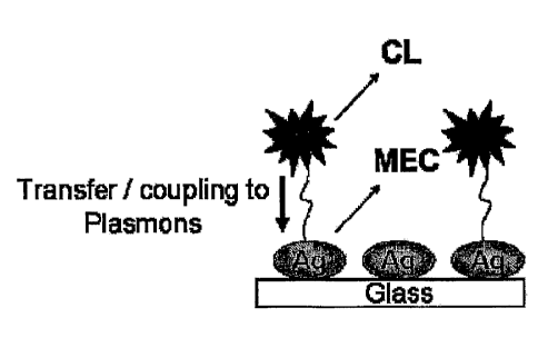
[0021] Figure 3 shows the proposed model for Metal-Enhanced Chemiluntinescence
(MEC).
The chemically induced electronically excited luminophore (C) transfers energy
to silver
plasmons (a resonance coupling interaction), which themselves radiate the
photophysical
properties of the excited species. CL ¨ Chemiluminescence, MEC ¨ Metal-
Enhanced
Chernituminescence, Ag ¨ Silver.
[0022] Figure 4 shows the experimental sample set-up Wherein the
chemiluminescence species
is placed between two glass slides comprising silver islands deposited
thereon.
5a
CA 02642280 2008-08-12
WO 2007/095527
PCT/US2007/062041
[0023] Figure 5 shows the chemiluminescence intensity measured on both SiFs
and glass as a
function of time (Top) and the data normalized (Top-insert). Normalized
chemihnninescence
intensity on both SiFs and a continuous silver film (Bottom). Photograph of
the emission from
both the continuous silver film and the SiFs (Bottom ¨ hisert). Ag ¨ Silver.
SiFs -Silver
Island Film.
[0024] Figure 6 shows the Experimental geometry used for measuring and/or
detecting surface
plasmon-coupled chemiluminescence (SPCC), Top, view from the top; bottom, side
view.
[0025] Figure 7 shows surface plasmon-coupled chemiluminescence from 20-nm-
thick
aluminum films. Top right, enlarged directional SPCC; top left, free-space
chemilmninescence
and SPCC; bottom, emission spectra of both the free-space chemiluminescence
and SPCC.
[0026] Figure 8 shows surface plasmon-coupled chemiluminescence from 45-nm-
thick silver
films. Top right, enlarged directional SPCC; top left, free-space
chemiluminescence and SPCC;
bottom, emission spectra of both the free-space chemiluminescence and SPCC.
[0027] Figure 9 shows surface plasmon-coupled chemiluminescence from 42-pm-
thick gold
films. Top right, enlarged directional SPCC; top left, free-space
chemilurninescence and SPCC;
bottom, emission spectra of both the free-space chemiluminescence and SPCC.
[0028] Figure 10 shows photographs of the coupled emission at various
polarizations for gold,
silver, and aluminum films, top to bottom, respectively, taken at their
respective SPCC peak
angles. See location of camera in Figures 7-9 (top right).
[0029] Figure 11 shows surface plasmon-coupled chemilurninescence (SPCC) and
free-space
chemilurninescence from a small sample chamber, top left, and the enlarged
coupled region,
top right. Bottom, emission spectra of both free-space chemiluminescence and
SPCC from the
small chamber.
[0030] Figure 12 shows that chemiluminescence intensity decays from aluminum
films for
both free space and coupled (top) and normalized to the same initial intensity
(bottom).
[0031] Figure 13 shows that chemiluminescence intensity decays from silver
films for both
free space and coupled (top) and normalized to the same initial intensity
(bottom).
6
CA 02642280 2008-08-12
WO 2007/095527
PCT/US2007/062041
[0032] Figure' 14 shows a setup for PIRP-acridan chemiluminescence assay on
both glass and
silvered slides.
[0033] Figure 15 shows 3D plots of acridan assay emission as a function of
time from glass
slides without (top) and with low-power microwave exposure/pulses (middle).
(Bottom)
Photographs showing the acridan emission both before (a) and after a low-power
microwave
pulse (b). Mw, microwave pulse. The concentration of BSA-biotin was 1,56 plVI.
[0034] Figure 16 shows 3D plots of the acridan assay cherniluminescence
emission as a
function of time from silvered glass slides (Ag) without (top) and with low-
power microwave
exposure/pulses (middle). (Bottom) Photographs showing the acridan emission
both before (a)
and after a low-power microwave pulse (b). Mw, microwave pulse. The
concentration of BSA-
biotin was 1.56 pM.
[0035] Figure 17 shows 3D plots of the acridan assay chemiluminescence
emission from both
glass (top) and silvered substrates (bottom). (Right) Emission spectra are the
average of 400 1-s
time points. In both cases, BSA-biotin was not irrmiobilized to the surfaces,
which were
exposed to microwave pulses at 100- and 200-s time points. The final
concentration of IIRP-
streptavidin in the assay was ¨10 Pg/mL.
[0036] Figure 18= shows the acridan chemilurninescence emission intensity as a
function of
time for different concentrations of surface-bound BSA-biotin. a, 156 pM BSA-
biotin; b, 15.6
pM BSA-biotin; c, 1.56 pM BSA-biotin; d, 156 fIVI BSA-biotin; and e, no BSA-
biotin.
[0037] Figure 19 shows photon flux integrated over 500 s of the assay shown in
Figure 18, for
different concentrations of BSA-biotin from both glass and silvered surfaces
(Ag). Baselines
correspond to integrated photon flux over 500 s for glass and silvered
surfaces (Ag) incubated
with 1% BSA solution and streptavidin HRP.
[0038] Figure 20 shows the procedure for the MT-1VfEC immunoassay (Mw, low-
power
microwave heating).
DETAILED DESCRIPTION OF THE INVENTION
[0039] Surface plasrnons are collective oscillations of free electrons at
metallic surfaces.
When a metallic article or surface is exposed to an electromagnetic wave, the
electrons in the
metal (plasmons) oscillate at the same frequency as the incident wave.
Subsequently, the
7
CA 02642280 2008-08-12
WO 2007/095527
PCT/US2007/062041
oscillating electrons radiate electromagnetic radiation with the same
frequency as the
oscillating electrons. It is this re-radiation of light at the same incident
wavelength that is often
referred to as plasmon emission. It the present invention chemically induced
electronic excited
states (cherniluminescence species) couple to surface plasmons to produce
emission intensities
greater than from about 5 to 1000-fold, as compared to a control sample
containing no metallic
surface. This approach is of significance for optically amplifying
ohemiluminescence based
clinical assays, potentially increasing analyteibiospecies delectability.
[0040] The term "biomolecule" means any molecule occurring in nature or a
derivative of such
a molecule. The biomolecule can be in active or inactive form. "Active form"
means the
biomolecule is in a form that can perform a biological function. "Inactive
form" means the
biomolecule must be processed either naturally or synthetically before the
biomolecule can
perform a biological function. Exemplary biomolecules include nucleic acids,
aromatic carbon
ring structures, NADH, FAD, amino acids, carbohydrates, steroids, flavins,
proteins, DNA,
RNA, oligonucleotides, peptide, nucleic acids, fatty acids, myoglobin, sugar
groups such as
glucose etc., vitamins, cofactors, porkies, pyrirxridines, formycin, lipids,
phytochrome,
phytoffuor, peptides, lipids, antibodies and phycobiliproptein.
[0041] The term "receptor-ligand" as used herein means any naturally Occurring
or unnaturally
occurring binding couple wherein the components have affinity for each other.
For example,
the binding couple may include an antibody/antigen complex, Viral coat
ligandfprotein cell
receptor or any combination of probe and binding partner, The term "receptor"
refers to a
chemical group, molecule, biological agent, naturally occurring or synthetic
that has an affinity
for a specific chemical group, molecule, virus, probe or any biological agent
target in a sample.
The choice of a receptor-ligand for use in the present invention will be
determined by nature of
the disease, condition, or infection to be assayed.
[0042] Embodiments of the present invention are applicable to
cherniluminescence labels or
moieties which participate in light-producing reactions in the presence of a
triggering agent or
cofactor. In the present application, for purposes of example and without
limitation, a preferred
embodiment will be discussed in terms of chemiluminescence labels and
triggering agent. The
label affixed to the detector molecule will be referred to as the "label" or
"label agent", For
purposes herein, "triggering agent or cofactor" is broadly used to describe
any chemical
species, other than the chemiluminescenee labels which participates in a
reaction and which
produces a detectable response. Chethiluminescence labels and triggering
agents produce a
light response.
8
CA 02642280 2008-08-12
WO 2007/095527
PCT/US2007/062041
[0043] Examples of suitable chemiluminescence labels include but without
limitation,
peroxidase, bacterial luciferase, firefly luciferase, functionalized iron-
porphyrin derivatives,
lurninal, isoluminol, acridinium esters, sulfonamide and others. A recent
chemiluminescent
label includes xanthine oxidase with hypoxanthine as substrate. The triggering
agent contains
perborate, an Fe-EDTA complex and luminol. Choice of the particular
chemiluminescence
labels depends upon several factors which include the cost of preparing
'labeled members, the
method to be used for covalent coupling to the detector molecule, and the size
of the detector
molecules and/or chemiluminescence label.
Comspondingly, the choice of
chemilurnineseence triggering agent will depend upon the particular
chemiluminescence label
being used.
[0044] Chemilumineseent reactions have been intensely studied and are well
documented in
the literature [39]. For example, peroxidase is well suited for attachment to
the detector
molecule for use as a chemiluminescence. The triggering agent effective for
inducing light
emission in the first reaction would then comprise hydrogen peroxide and
luminol. Other
triggering agents which could also be used to induce a light response in the
presence of
peroxidasc include isobutyraldettyde and oxygen.
[0045] Procedures for labeling detector molecules, such as antibodies or
antigens with
peroxidase are known in the art. For example, to prepare peroxidase-labeled
antibodies or
antigens, peroxidase and antigens or antibodies are each reacted with N-
succinimidyl 3-(2-
pyridyldithio) proprionate (hereinafter SPDP) separately. SPDP-labeled
peroxidase, or SPDP-
labeled antigen or antibody is then reacted with dithiothreitol to produce
thiol-labeled
peroxidase, or thiol-labeled antigen or antibody. The thipl derivative is then
allowed to couple
with the SPDP-labeled antigen or antibody, or SPDP-labeled peroxidase.
[0046] Techniques for attaching antibodies or antigens to solid substrates are
also well known
in the art. For example, antibodies may be coupled covalently using
glutaraldehyde to. a, silane
derivative of borosilicate glass.
[0047] Although chemiluminescence detection has been successfully implemented,
the
sensitivity and specificity of these reactions require further improvements to
facilitate early
diagnosis of the prevalence of disease. In addition, most protein detection
methodologies, most
notably western blotting, are still not reliable methods for accurate
quantification of low protein
concentrations without investing in high-sensitivity detection schemes.
Protein detection
methodologies are also limited by antigen-antibody recognition steps that are
generally
kinetically very slow and require long incubation times; e.g., western blots
require processing
9
CA 02642280 2008-08-12
WO 2007/095527
PCT/US2007/062041
times in excess of 4 h. Thus, both the rapidity and sensitivity of small-
molecule assays are still
critical issues to be addressed to improve assay detection.
[0048] Thus, in one embodiment, the application of low level microwave heating
of the
sample may be used to speed up any biOlogicalibiochemical kinetics within the
system.
Notably, low level microwaves do not destroy or denature proteins, DNA, or
RNA, but instead
heat the sample sufficiently to provide for accelerated kinetics such as
binding or hybridization.
In addition, themicrowaves are not scattered by the low density silver metal,
which is contrary
to most metal objects, such as that recognized by placing a spoon in a
microwave oven.
[0049] Microwaves (about 0.3 to about 300 GHz) lie between the infrared and
radio frequency
electromagnetic radiations, It is widely thought that microwaves accelerate
chemical and
biochemical reactions by the heating effect, where the heating essentially
follows the principle
of microwave dielectric loss. Polar molecules absorb microwave radiation
through dipole
rotations and hence are heated, where as non-polar molecules do not absorb due
to lower
dielectric constants are thus not heated. The polar molecules align themselves
with the external
applied field. In the conventional microwave oven cavity employed in this
work, the radiation
frequency (2450 MHz) Changes sign 2.45 x 10⁹ times per second. Heating occurs
due to the
tortional effect as the polar molecules rotate back and forth, continually
realigning with the
changing field, the molecular rotations being slower than the changing
electric field. The
dielectric constant, the ability of a molecule to be polarized by an electric
field, indicates the
capacity of the medium to be microwave heated. Thus, solvents such as water,
methanol and
dimethyl formamide are easily heated, where as microwaves are effectively
transparent to
hexane, toluene and diethylether.
[0050] For metals, the attenuation of microwave radiation arises from the
creation of currents
resulting from charge carriers being displaced by the electric field. These:
conductance
electrons are extremely mobile and unlike water molecules can be completely
polarized in 10-
18 s. In microwave cavity used in the present invention, the time required for
the applied
electric field to be reversed is far longer than this, in fact many orders of
magnitude. If the
metal particles are large, or form continuous strips, then large potential
differences can result,
which can produce dramatic discharges if they are large enough to break down
the electric
resistance of the medium separating the large metal particles.
[0051] Interestingly, and most appropriate for the new assay platform
described herein, small
metal particles do not generate sufficiently large potential differences for
this "arcing"
phenomenon to occur. However, as: discuss hereinbelow, the charge carriers
which are
CA 02642280 2008-08-12
WO 2007/095527
PCT/US2007/062041
displaced by the electric field are subject to resistance in the medium in
which they travel due
to collisions with the lattice phonons_ This leads to Ohmic heating of the
metal nanoparticles in
addition to the heating of any surface polar molecules. Intuitively, this
leads to localized
heating around the metallic nanostructures in addition to the solvent, thereby
rapidly
accelerating assay kinetics.
[0052] In the present invention, microwave radiation may be provided by an
electromagnetic
source having a frequency in a range between 0.3 and 10 GHz and a power level
in a range
between about 10 mvvatts and 400 watts, preferably from 30 mwatts to about 200
watts, and
more preferably from about 100 watts to 150 watts. Any source, known to one
skilled in the art
may be used, such as a laser having the capacity to emit energy in the
'microwave range, The
microwave radiation may be emitted continuously or intermittently, as desired,
to maintain the
metallic particles at a predetermined temperature such that it is capable of
increasing the speed
of chemical reactions not only in the assay system but also the
chemiluminescence species.
[0053] In the alternative, microwave energy can be supplied through a hollow
wave guide for
conveying microwave energy from a suitable magnetron. The microwave energy is
preferably
adjusted to cause an increase of heat within the metallic material without
causing damage to
any biological materials in the assay system.
[0054] In one embodiment the present invention provides far a metallic surface
and a
biomolecule capable of chemiluminescing, wherein the metallic surface and the
biomolecule
are separated by at least one film spacer layer. The thickness of said film
may be chosen so as
to enhance the chemiluminescence of the biomolecule by positioning the
biomolecule an
optimal distance from the metallic surface. The film spacer layer may be one
or multiple layers
of a polymer film, a layer farmed from a fatty acid or a layer formed from an
oxide_ In a
preferable embodiment,, the film spacer layers and the metallic surface are
chemically inert and.
do not bind to the biomolecules to be detected or to intermediates that are
bound to the
compounds to be detected, for example covalently bound. The layer formed from
a fatty acid
may be formed by a Langmuir-Blodgett technique. The film spacer layer may be a
spin coated
polymer film. The oxide layer may be formed from a deposition technique, such
as vapor
deposition.
[0055] Further, the metallic surface may be in the form of a porous three
dimensional matrix.
The three dimensional matrix may be a nano-porous three dimensional matrix.
The metallic
surface may include metal colloid particles and/or metal-silica composite
particles. The
metallic surface may comprise agglomerated metal particles and/or binary
linked particles or
11
CA 02642280 2008-08-12
WO 2007/095527
PCT/US2007/062041
metal particles in a polymer matrix. The three dimensional matrix may be
formed from
controlled pore glasses or using matrices assembled from the aggregation of
silver-silica
composites themselves. The matrices may be metallic nanoporous matrix, through
which
species will flow and be both detected and counted more efficiently. The
ability to
quantitatively count single flowing molecules under practical conditions may
have many
implications for medical diagnostics, the detection of biohazard organisms and
new and quieker
methods for DNA sequencing.
[0056] The emission enhancement may be observed at distances according to the
type of
chemiluminescence species to be detected and the type of metal. For example,
emission
enhancement may be observed when a chemilurninescence species is positioned
about 5 run to
about 200 mu to metal surfaces. Preferable distances are about 5 nm to about
30 nrn, and more
preferably, 5 ran to about 20 nm to metal surfaces. At this scale, there are
few phenomena that
provide opportunities for new levels of sensing, manipulation, and contra In
addition, devices
at this scale may lead to dramatically enhanced performance, sensitivity, and
reliability with
dramatically decreased size, weight, and therefore cost.
[0057] Different effects are expected for mirrors, sub-wavelength or semi-
transparent metal
surfaces, silver island films or metal colloids. More dramatic effects are
typically observed for
islands and colloids as compared to continuous metallic surfaces. The silver
islands have the
remarkable effect of increasing the emission intensity at least 5-fold while
decreasing the
lifetime 100-fold.
[0058] Light from the cherniluminescence reaction generated by the random
depopulation of a
chemically induced electronic state of a lurninophore and/or the plasmon
coupled emissions
from the metallic components can be detected using an optical detector,
positioned above
and/or below reaction sites. Various optical detectors, such as photodiode,
charge-coupled
device (CCD), photorriultiplier tube (PMT), or photon counting detector, have
different degree
of sensitivity. PMT and photon counting detectors can achieve an electronic
amplification
factor as high as 106-108. Conventional PMTs require a 'l kV power source, but
new
miniaturized detector requires. only a 5 V. Most of the chemihuninescence
emission
wavelengths are in the visible region. A narrow-band optical filter may be
used to ensure
detecting luminescence wavelengths. The system may include a microactuator,
detector,
microprocessor, electronics, a display, and translation stage. The output of
the detector may be
interfaced to an analog to digital converter and a microprocessor to calculate
analyte
concentration.
= 12
CA 02642280 2008-08-12
WO 2007/095527
PCT/US2007/062041
[0059] It is known that the extinction properties (C5) of metal particles can
be expressed as
both a combination of both absorption (CA) and scattering (Cs) factors, when
the particles are
spherical and have sizes comparable to the incident wavelength of light, i.e.
in the Mie
limit[26].
(1)
= CA +Cs = inl(a) ¨Icel2
67T
[0060] where k1 = 2'an1 Xo is the wavevector of the incident light in medium 1
and a is the
polarizability of a sphere with radius r, rri is the refractive index and Xo
the incident wavelength.
The term I ct I 2 is square of the modulus of a.
ce = 47r3 (a,¨ 61)1(e, + 2e1)
(2)
[0061] where si and cm are the dielectric and the complex dielectric constants
of the metal
respectively. The first term in equation 1 represents the cross section due to
absorption, CA,
and the second term, the cross section due to scattering, Cs. Current
interpretation of metal-
enhanced fluorescence [23] is one underpinned by the scattering component of
the Metal
extinction, Le. the ability of fluorophore-coupled plasmons to radiate
(plasmon scatter) [11].
Intuitively, larger particles have wavelength distinctive scattering spectra
(Cs) as compared to
their absorption spectra (CA) [26], facilitating plasmon coupled emission from
the larger
nanoparticles.
[0062] Surprisingly, the present invention shows that chemically induced
electronic excited
states (chemiluminescence species) also couple to surface plasmons, producing
emission
intensities from about 5 to about 1000 fold, as compared to a control sample
containing no
surface silver nanostructures. Thus, the present invention further shows that
surface plasmons
can be directly excited by chemically induced electronically excited
luminophores.
[0063] The present invention provides enhanced emissions Using metallized
nanostructures,
islands of elliptical, spherical, triangular or rod-like forms. In exemplary
cases, the elliptical
islands have aspect ratios of 3/2, and the spherical colloids have diameters
of 20-60 rim.
However, the invention is not limited to any particular geometry. Using known
coating
techniques, the placement of metallic islands could be controlled precisely,
as close as 50 nm
apart.
[0064] Metal Wand particles may be prepared in Clean beakers by reduction of
metal ions
using various reducing agents [10-13 and 27]. For example, sodium hydroxide is
added to a
13
CA 02642280 2008-08-12
WO 2007/095527
PCT/US2007/062041
rapidly stirred silver nitrate solution forming a brown precipitate. Ammonium
hydroxide is
added to re-dissolve the precipitate. The solution is cooled and dried quartz
slides are added to
the beaker, followed by glucose. After stirring for 2 minutes, the mixture is
warmed to 30 C.
After 10-15 minutes, the mixture turns yellow-green and becomes cloudy. A thin
film of silver
particles has formed on the slides as can be seen from their brown green
color. The slides are
rinsed with pure water prior to use.
[0065] Alternative procedures for preparing metal particles are also available
[2$-32]. Silver
is primarily used because of the familiar color from the longer s-urface
plasmon absorption of
[0066] Colloids can be prepared as suspensions by citrate reduction metals.
Preferred metals
are silver and gold. The size of the colloids and their homogeneity can be
determined by the
extensive publications on the optical properties of metal particles available
and the effects of
interface chemistry on the optical property of colloids [33].
[0067] Silver island films can be formed by a chemical reduction of a silver
salt on the quartz
surface and that are relatively simple to fabricate. However, this approach
does not provide a,
control of particle size, or distance of the chemiluminescent species from the
metallic surface.
[0068] Metal particles can be bound to a surface by placing functional
chemical groups such
as cyanide (ON), amine (N112) or thiol (SH), on a glass or polymer substrate.
Metal colloids are
lcriown to spontaneously bind to such surfaces with high affinity [34-35].
[0069] Positioning of the biomolecule or metal particle at a desired distance
can be achieved
by using a film. The film may be a polymer film, a Langmuir-Blodgett film or
an oxide film.
Proper distances may be achieved by using Langmuir-Blodgett films with fatty
acid spacers.
The fatty acids may be from natural sources, including concentrated cuts or
fractionations, or
synthetic alkyl carboxylic acids. Examples of the fatty acids include, but not
limited to,
caprylic (C8), eapric (C10), lauric (Cu), myristic (014), palmitic (C16),
stearic (C18), oleic (CH),
linoleic (C18), linolenic (CIO, ricinoleic (C18) arachidie (C20), gadolic
(C20), behenic (022) and
erucic (C22). The fatty acids with even numbered carbon chain lengths are
given as illustrative
though the odd numbered fatty acids can also be used.
[0070] Also, metal-chemiluminescenne species distances may be achieved by
using polymer
films. Examples of the polymer include, but not limited to, polyvinyl alcohol
(PVA).
Absorbance measurements and ellipsometry may be used to determine polymer film
thickness.
14
CA 02642280 2008-08-12
WO 2007/095527 PCT/US2007/062041
One type of polymer films is spin coated polymer films. The technology of spin
coated
polymer spacei films readily allows films to be coated onto a variety of
surfaces, with varied
thickness from >0.1 um. The coating can be performed on a spin eoater, which
allows uniform
surface thickness by varying polymer concentration (viscosity) and. spin
speed. For example,
Model P6700 spin coater (Specialty Coating Systems Inc.) allows uniform
surface thickness by
varying polymer concentration (viscosity) and spin speed.
[0071] Metallic colloids (or various other non-spherical shapes/particles) may
also be
incorporated into organic polymers, covalently or non-covalently, to form
polymeric matrices,
wherein the distance from diffusing species affords an increase in radiative
decay rate and thus,
an increase in quantum yield. Such polymeric matrices are ideal for
sensing/flowing sensing
applications of low concentration species.
[0072] Any chemiluminescent species may be used in the present invention that
provides for a
chemical reaction which produces the excited state responsible for the
observed emission
including, but not limited to the following excitation mechanisms:
R. + R!* R ¨R. + hv (single bond formation (radical-radical reaction))
sk* + =Re' --> RR + hv (double bond formation (radical-radical reaction))
R02 =R= + 02 ¨,2141t. + hv
1:t+ + R + hv (electron capture)
[0073] This embodiment of the present invention may have vast applications in
clinical
medicine, environmental monitoring applications, homeland security such as
rapid detection of
low concentration species, industrial processes, pharmaceutical industries
such as monitoring
species, and sensors for use in reduced atmospheres such as biohazard clean
rooms and
environments using space light.
[0074] Examples
[0075] 1. Radiating Plasrnons Generated from Chemically Induced
Electronic
Excited States
[0076] 1.2 Materials
[0077] Silver nitrate (99.9%), sodium hydroxide (99.996%), ammonium hydroxide
(30%),
trisodium citrate, 1)-glucose and premium quality APS-coated glass slides
(75x25 mm) were
CA 02642280 2008-08-12
WO 2007/095527 PCT/US2007/062041
obtained from Sigma-Aldrich (St. Loius, MO). The blue-glow Chemiluminescence
sticks used
were the "Color Bright" light sticks; obtained from Omniglow (West
Springfield, MA).
[0078] 1.3 Chemiluminescence
[00791 The chemiluminescent materials used in this study were obtained from
commercial
light glow sticks. These glow sticks contain the necessary reacting chemicals
encapsulated
within a plastic tube. The plastic tube contains a phenyl oxalate ester and a
fluorescent probe,
where the choice of dye simply determines the color of the luminescence [9].
For the examples
set forth herein, this choice is arbitrary as long as the luminophore emits in
the visible spectral
region, consistent with previous reports [10-13]. Inside the plastic tube lies
a glass capsule
containing the activating agent (hydrogen peroxide). Activation of the
chemicals is
accomplished with a bend, snap, and. a vigorous shake of the plastic tube
which breaks the glass
capsule containing the peroxide and mixes the chemicals to begin the
chemiluminescence
reaction. The hydrogen peroxide oxidizes the phenyl oxalate ester to a
peroxyacid ester and
phenol. The unstable peroxyacid ester decomposes to a peroxy compound and
phenol, the
process chemically inducing an electronic excited state.
[0080] 1.4 Formation of Silver Island Films (SiFs) on APS-coated
Glass
Substrates
[0081] The silver island films were made according to previously published
procedures
employing the ,chemical reduction of silver nitrate on glass microscope slides
using sodium
hydroxide, ammonium hydroxide and glucose [10-13].
[0082] 1.5 Chemiluminescence from SiFs and glass
[0083] The cherniluminescence experiments were performed using a blue emission
glow stick.
After chemiluminescence initiation, approximately 70 ul of the glow stick
fluid was placed
between two APS-coated microscope glass slides, clamped together. The glass
slides contained
silver island films on one end and were bare glass on the other end. The bare
end of the glass
served as the control sample by which to compare the benefits of using the
metal-enhanced
chemiluminescence phenomenon. Subsequently, the enhancement ratio, the
intensity from
silver! intensity from glass, Could be determined.
[0084] Chemiluminescence measurements
16
CA 02642280 2008-08-12
WO 2007/095527 PCT/US2007/062041
[0085] Chemiluminescence spectra were Collected using an Ocean Optics
spectrometer, model
SD 2000 (Dunedin, FL), connected to an Ocean Optics 1000 um diameter fiber
with an NA of
0.22 (Dunedin, FL). The fiber was positioned vertically on top of the slides
containing the
luminescening material. Spectra were collected with an integration time
ranging from between
4 and 10 seconds. The integration time was kept constant between the control
and silver island
film sample measurements.
[0086] 1.7 Results
[00871 Figure 1 top shows the luminescence emission spectra from between the
silvered glass
and glass plates. The emission from the silvered portion of the slide was
spatially averaged to
be about 4-5 times greater than the glass control side of the sample. In
addition, the volume
between both the sandwiched glass and silver slides was identical. Figure 1 ¨
bottom shows the
photographs of the slides, both before and after the addition of the
chemiluminescent material.
Approximately 70 pL of fluid was enough to form a thin coating across both
portions of the
slide, held by capillary action as the slides were sandwiched as shown in
Figure 4. The
enhanced chernilumineseence is clearly visible on the silvered portion as
shown in Figure 1
(bottom). Interestingly, the digital camera was not able to capture the blue
emission from the
thin fluid layer of the glass region of the slide, the intensity quite weak as
also shown in Figure
1 ¨ top.
[0088] Several control experiments were performed to determine the loss of
chemiluminescent
intensity, due to the depletion of the reactants, Figure 2. After a period of
60 minutes, most of
the emission from the silvered plates had gone, Figure 2- Top. Interestingly,
the luminescence
emission intensity changed very little in several tens of seconds, Figure: 2¨
bottom, which was
the time needed to measure both the intensity on silver and glass shown in
Figure 1, making the
comparison between both silver and glass a valid one. Finally, while not shown
here, the rate
of loss of luminescence was measured from both the silvered and glass portions
of the slide.
For both, the rate of chemiluminescence was almost identical, suggesting that
no chemical
interaction between the chemiluminescent reagents and silver occurred, the
enhanced
luminescence signals observed due to interactions with surface plasmons as
discussed below
[23].
[0089] ;Several detailed control experiments were undertaken to ascertain
whether silver could
catalyze the chemiluminescence reaction and account for the enhanced optical
signatures
observed, as compared to an interpretation in terms of a chemiluminescence-
based radiating
plasmon model. Figure 5 ¨ top shows the luminescence intensity as a function
of time. Clearly
17
CA 02642280 2008-08-12
WO 2007/095527 PCT/US2007/062041
the enhanced luminescence from the SiFs is visible, with the initial intensity
on SilverP.,- 3100
a.u. (at t = 0) as. compared to < 1511 on glass. Subsequently the rates of
loss of luminescence
were compared after the curves were normalized, Figure 5 ¨ top insert. The
rate of loss of
luminescence, which is due to the depletion of solution reactants and
therefore depletion over
time of excited states, Vvas found to follow first order decay kinetics and
could simply be
modeled to an exponential function of the form:
[0090] Luminescence Intensity, I= C + B-kt (3)
where C is the intensity at time t = oc, B is a pre-exponential factor and k
the rate of
luminescence depletion, units S-1: From Figure 5 ¨ Top insert, the rate of
depletion on silver
was found to be 1.7 times faster than on glass, 0.034 vs 0.019 s71
respectively. Two
explanations could initially describe this observation: Firstly, silver
catalysis of the
chemiluminescence reaction, or secondly, the high rate of transfer / coupling
of the
chemiluminescence to surface plasmons, rapidly reducing the excited state
lifetime of the
chemiluminescence species.
[0091] To eliminate silver based catalysis of the chemiluminescence reaction
as an explanation
for the enhanced signals, the luminescence rates were measured on both SiFs
and a continuous
silver strip. Interestingly, the rate of loss of luminescence was still found
to be greater on the
SiFs as compared to the continuous silver strip, Figure 5 ¨ bottom. In
addition, the emission
intensity was very low indeed from the continuous strip of silver, Figure 5 ¨
bottom insert.
Given that the continuous strip is indeed darker and that the rate is slower
than on SiFs, then
silver based catalysis can be eliminated as a possible explanation of the
observation of
increased signal intensities on the SiFs. Subsequently, these observations
suggest that
chemically induced electronic excited states (chemiluminescence species) can
readily
induce/couple to surface plasmons, facilitating metal-enhanced
chemiluminescence.
[0092] 1.8 con
[0093] With the chemiluminescence species shown here, it is theorized that
excited
chemiluminescence species couple to surface plasmons, which is turn radiate
the photophysical
properties of the chemically excited state, as shown in Figure 3.
Interestingly, the
chernilurninescent system described herein, wherein there is no external
excitation source for
direct illumination and no direct mode of excitation of the surface plasmons
suggests that the
surface plasmons are indeed excited from a chemically induced electronically
excited state of a
luthinophore. It is believed that this is the first observation of the
chemically induced
18
CA 02642280 2008-08-12
WO 2007/095527
PCT/US2007/062041
electronic excitation of surface plasmons.
[0094] 2, Directional and Polarized Emission of the Luminescence
[0095] The experimental geometry used for the surface plasmon-coupled
chemilurninescence
(SPCC) studies is shown in Figure 6.
[0096] 2.1 Materials and Methods
[0097] Premium quality ABS-coated glass slides (75 x 25 nun), silver wire
(99.99+% purity),
aluminum evaporation slugs (99.999% purity), and silicon monoxide pieces
(99.99% purity)
were obtained from Sigma-Aldrich (St. Loius, MO). Gold evaporation slugs
(99.999% purity)
were obtained from Research and PVD Material Corporation (Wayne, NJ).
CoverWell
imaging chamber gaskets with adhesive (20-mm diameter, 1-mm deep) were
obtained from
Molecular Probes (Eugene, OR). The smaller imaging chambers were built in-
house using
electrical black tape, double sticky tape, and microscope coverslips. Several
standard
ehemiluminescence kits from Ornnioglow (West Springfield, MA) and Night Magic
(Union
City, OH) were used as the source of chemiluminescence.
[0098] 2.2 Chemiluminescent Dyes
[0099] The chemilumineseent materials used in this study were obtained from
commercially
available kits and previously described in Example 1.
[00100] 2.3 Formation of Continuous Thin Films of Metal on APS-Coated
Glass
Substrates
[00101]Twerity nanometers of aluminum, 45 nut of silver, and 40 nan of gold
were deposited
on separate ABS-coated glass slides using an Edwards Auto 306 Vacuum
Evaporation chamber
(West Sussex, U.K.) under ultrahigh vacuum (<3 x 10⁻⁶ Torr). In each ca, the
metal
deposition step was followed by the deposition of 5 nm of silica via
evaporation without
breaking vacuum. This step served to protect the metal surface from attack by
the various
chemical species present in the chemiluminescence assay.
[00102] 2A Surface Plasmon-Coupled Chemilurninescence (SPCC) of Dyes on
Continuous Metal Films
19
CA 02642280 2008-08-12
WO 2007/095527 PCT/US2007/062041
[00103] The surface plasmon-coupled chemiluminescence (SPCC) experiments were
performed
using several different colors of the chemiluminescent dyes ranging from blue
to red. They
were carried out by first bending the plastic tube of the chemilurninescence
kit and shaking it
vigorously. This allowed the reaction mixtures to mix and begin to luminesce.
The tubes were
then cut with a scissor, and the reacting fluid was poured into a glass vial.
Approximately 150
FIL of the reacting fluid was then placed in an imaging chamber gasket with
adhesive (20-mm
diameter, 1-mm deep). This gasket was then pressed against an (APS-coated)
continuous
metal-coated and silica-capped microscope glass slide until they were stuck
together creating a
chamber containing the chemilurninescent dyes on the surface of the metal-
pealed glass slide.
For smaller samples, approximately 50 14L of the reacting fluid was placed in
an imaging
chamber built in-house attached to an (APS-coated) continuous metal-coated and
silica-capped
microscope glass slide.
[00104] 25 Surface Plasmon-Coupled Chemiluminescence (SPCC)
Measurements
[00105] The metal-coated slides containing the chemiluminescent dyes were
attached to a
hemicylindrical prism made with BK7 glass (n = 1.52), and the refractive index
was matched
using spectrophotometric grade: glycerol (n. = 1.475) between the back of the
glass slide
(uncoated side) and the prism. This unit was then placed on a precise 360*
rotatory stage
which was built in-house. The rotatory stage allowed the collection of light
at all angles around
the sample chamber. An Ocean Optics low OH 1000 Pm diameter optical fiber with
NA of
0.22 (Dunedin, FL) used for collecting the ehemiluminescence signals was
mounted on a holder
that was screwed onto the base of the rotatory stage. A pictorial
representation of the top and
side view of the setup is presented in Figure 6. Surface plasmon-coupled
chemiluminescence
(SPCC) spectra were collected using a model SD 2000 Ocean Optics spectrometer
(Dunedin,
FL) connected to the above-mentioned optical fiber. The spectra were collected
with an
integration time between 05 and 2 s (depending on the intensity of the various
SPCC signals).
Both unpolarized and p- and s-polarized signal information was collected for
the SPCC signal
(from 0 to 180 Q with respect to the front of the prism) and for the free-
space signal (from 180
to 3600 with respect to the front of the prism). A separate time-dependent
decay study was
performed on each chemiluminescent dye to study the comparative time-dependent
decay
profile of the SPCC signal and the free-space signal.
[00106] 2.6 Results
[00107] Figure 7 (top left) shows the surface plasmon-coupled
chernilurninescence (SPCC) and
the free-space emission from the blue chemiluminescent dye on a 20-nm aluminum
layer. It
CA 02642280 2008-08-12
WO 2007/095527
PCT/US2007/062041
can be seen that the free-space emission is of much higher magnitude than. the
SPCC signal.
This is because the sample 'chamber is 1-mm thick and only the luminophores
within
approximately 250 nm of the surface of silver are known to excite surface
plasmons [36, 24].
Hence, the majority of the luminophores the chamber do not eonple to plasmons
and so
radiate their energy in the form of free-space emission. Subsequently there
was an attempt to
use very thin films of liquid to alleviate this effect. However, the
hydrophobic nature of the
surface globulated the chemiluminescence liquid, preventing films <250 nm
thick to be
produced.
1.00108]Figure 7 (top right) is an enlarged figure showing the highly
directional and
predominantly p-polarized SPCC emission only,. suggesting that the observed
signal is due to
surface plasmons. This is in stark contrast to the free-space emission which
does not show any
polarization or directional preference. However, the signal at the SPCC peak
angle is not
entirely p-polarized. The camera located at the SPCC peak angle of the figure
depicts the
approximate angular position where photographs of the coupled emission at
various
polarizations were taken. These photographs are shown in Figure 10. Figure 7
(bottom) is the
normalized SPCC and free-space emission spectra showing a high degree of
overlap between
the spectra. This suggests the plasmen-coupled chemiluminescence has not
undergone any
changes in its spectral properties because of the interaction between the
luminescent species
and the metal surface.
[00109]Figure 8 (top left) shows the surface plasmon-coupled chemiluminescence
(SPCC) and
the free-space emission from the green cheiniluminescent dye on a 45-nm silver
layer. Similar
to the case of the blue dye on aluminum, it can also be seen here that the
free-space emission is
of greater magnitude than the SPCC signal. Figure 8 (top right) is an enlarged
figure showing
the highly directional and predominantly p-polarized SPCC emission only,
suggesting that the
observed signal is due to surface plastrons. This again is in stark contrast
to the free-space
emission which does not show any polarization or directional preference.
Figure 8 (bottom) is
the normalized SPCC and freerspace. emission spectra showing a high degree of
overlap
between the spectra, suggesting no additional interaction between the
luminescent species and
the metal surface.
[00110]Figure 9 (top left) shows the surface plasrnon-coupled
chemiluminescence (SPCC) and
the free-space emission from the red chenniluminescent dye on a 42-nm gold
layer. Figure 9
(top right) is an enlarged figure showing the highly directional and
predominantly p-polarized
SPCC emission only, suggesting that the observed signal is due to surface
plasmons. The
SPCC again is in stark contrast to the free-space emission which does not show
any
21
CA 02642280 2008-08-12
WO 2007/095527
PCT/US2007/062041
polarization or directional preference. Figure 9 (bottom) is the normalized
SPCC and free-
space emission spectra showing a high degree of overlap between the spectra,
suggesting no
other interaction between the luminescent species and the metal surface.
[00111]Figure 10 shows photographs of the coupled emission (from the prism
side) at the
respective SPCC peak angle from the various dyes at both s- and p-
polarizations as well as with
no polarization. The approximate angular location of the camera used obtaining
these
photographs is marked in Figures 7-9 (top right). This figure clearly shows
that the emission at
the SPCC peak angle is predominantly p-polarized for all three dyes (on all
three metals) thus
suggesting that surface plasmons are responsible for the SPCC signal. It can
be seen that the p-
polarized signal intensity at the SPCC peak angle is lciwer in magnitude than
the unpolarized
signal. This occurs because the entire SPCC signal consists of both p- and to
a lesser degree s-
polarized light, and also because the sheet polarizers used. in the experiment
have only 30-40%
peak transmission efficiency for both polarizations.
[00112]Initially, the broadness of the SPCC peak angles for all three dyes
which varied
between 20 and 25 degrees. Hence, to investigate whether the broadness of the
SPCC peak
angle is a function of the surface area of the sanaple, the experiments were
repeated on silver
using the green chemilumineseent dye with a sample chamber that had
approximately half the
surface area when compared to the samples made with commercially available
imaging
chambers that had been used thus far. Figure 11 (top left) shows the surface
plasrnon-coupled
cherniluminescence (SPCC) and the free-space eMi8SiOn from the green
cherniluminescent dye
on a 45-nm silver layer for the small imaging chambers. Figure 11 (top right)
is an enlarged
figure showing the highly directional and predominantly p-polarized SPCC
emission only.
Here, the broadness of the SPCC peak angle is approximately 20 degrees. It is
clear from this
figure that the broadness of the SPCC peak angle is not significantly affected
by the surface
area of the sample. An interesting observation in Figure 11 (top right) is the
decay in the SPCC
signal in the region between 90 and 180 degrees when compared to that in the 0-
90 degrees.
This is because the data was collected sequentially from 0 through 360
degrees. As a result, for
the small chamber with a lower volume of reactants, by the time the data in
the region between
90 and 180 degrees was collected, a signal reduction is observed because of
the depletion of
reactants (depletion of excited states) overtime. The broad angle distribution
shown in Figures
7-9 and ills attributed to the wave guide effect, given that our solution of
chemilumineseence
oceupied a sample chamber of 1-mm thickness. Figure 11 (bottom) is the
normalized SPCC
and free-space emission spectra showing a high degree of overlap between the
spectra,
suggesting no additional interaction between the luminescent species and the
metal surface in
the smaller imaging chambers built in-house.
22
CA 02642280 2008-08-12
WO 2007/095527 PCT/US2007/062041
[00113] The next round of experiments was performed to determine the rate of
decay of
luminescence for the blue and green chemilurrrinescent dyes as a function of
time for both the
five-space emission and the SPCC emission (with p-polarizers so that only
plasmon-coupled
emission was measured). By decay rate, it is meant the decrease in intensity
because of
depletion of reagents. The results of these experiments for the blue dye on
aluminum and green
dye on silver are shown in Figures 12 and 13, respectively. Figure 12 (top)
shows the decay of
free-space and SPCC emission as p. function of time for the blue dye on
alumintmi, with Figure
12 (bottom) image showing both the decay intensities normalized to their
respective values at t
= 0. The rate of loss of luminescence, which is due to the depletion of
solution reactants and
therefore a depletion over time of excited states, was found to follow first-
order decay kinetics
as shown herein above in formula (3).
[00114] The rate of depletion of the SPCC signal for the blue dye on aluminum
was found to be
only minimally greater than the free-space emission, 0.0003 versus 0.0002
respectively.
Since both the SPCC signal and the free-space emission signal decay are highly
dependent on
the rate of depletion of the same reactants (depletion of states)
in the sample chamber
over time, it is not surprising that the measured decay rates for both the
signals as shown in
Figure 12 are almost identical. T-Towever, this finding does indicate that
there are no localized
catalytic effects of the aluminum on the chemilurninescence reaction, as this
would be expected
to manifest in a larger difference in the SPCC luminescence decay rate (from
the free-space
decay rate) than is currently observed.
[00115]Figure 13 (top) shows the decay of free-space and SPCC emission as a
function of time
for the green chemiluminescent dye on silver, and Figure 13 (bottom) shows
both the decay
intensities normalized to their respective values at t = 0. The rate of
depletion of the SPCC
signal for the green dye on silver was found to be only minimally smaller than
the free-space
emission, 0.0005 versus 0.0006 s, respectively. It is again not surprising
that the measured
decay rates for both the signals as shown in Figure 12 are almost identical,
since both the SPCC
signal and the free-space emission signal decay are highly dependent on the
rate of depletion of
the same reactants in the sample chamber over time, This finding again
indicates no localized
catalytic or chemical effects of the silver on the chemiluminescence reaction
studied.
[00116] 2.7 Conclusions
[00117] The results of this study lead us to conclude that chemically induced
electronic excited
states of laminophores can excite surface plasmons on thin films of continuous
metal,
23
CA 02642280 2011-06-30
producing highly polarized and directional emission. This phenomenon is not
restricted to the
commercially available kits that were used in this study but rather can be
extended to the
myriad of chemiluminescent reactions .employed in biotechnology today to
increase signal
collection efficiency and hence the sensitivity of such essays. The -typical
thickness of the
functional surface of such assays are compatible with an approximately 250-nm
coupling
region, potentially alleviating unwanted background signals caused by
spontaneous reaction of
reagents or unwanted enzymatic activity and therefore increasing assay
sensitivity.
[00118] Another interesting observation is that SPCC occurs with gold films.
Since
luminophores within approximately 250 um of the surface of metal arc known to
exeite surface
olasmons, which is longer than the distances required for nonradiative
quenching of
luminescence, the potential of using gold as the metal surface becomes an
advantage. This is
because gold is chemically more stable than silver and the surface chemistry
of gold is well-
known and characterized [37]. Also, since gold films are widely used in
surface plasmon
resonance (SPR), this provides a robust technology base for the mass
production of suitable
gold films.
[901.191 3. Microwave Triggered Metal Enhanced Chemiluminescence
[00120] 3.1 Materials
[00121]Bovine-biotina.midocaproyI-labeled albumin (biotinlyated BSA); HRP-
labeled avidin,
silver nitrate (99.9%), sodium hydroxide (99.996%), ammonium hydroxide (30%),
trisodium
citrate, D-glucose, and premium qeality APS-coated glass slides (75 x 25 rnm)
were obtained
from Sigma-Aldrich. CoverWellTM imaging chamber gaskets with adhesive (20-mm
diameter, 1
mm deep) were obtained from Molecular Probes (Eugene, OR.). Steptavidin-FIRP
predauted
solution was obtained from Chemicon International Inc. Chemiluminescence
materials were
purchased from Amersham Biosciencea (ECL Plus Western blotting detection kit,
RPN2132).
ECL Phis utilizes a new technology, developed by Lumigen Inc., based on the
enzymatic
generation of at acridinium ester, which produces intense light emission at-
430 nm.
[00122] 3.2 Formation of Silver Island Films on.APS-Coated Glass
Substrates
[001231In a typical SiF preparation, a SOlution of silver nitrate (0.5 g in 60
niL of deionized
water) in a clean 100-mL glass beaker, equipped with a TeflonTm-coated stir
bar, is prepared and
placed on a Corning stirring/hot plate, While stirring at the quickest speed,
8 drops (-200 PL)
of freshly prepared 5% (w/v) sodium hydroxide solution are added. T his
results in the
24
CA 02642280 2008-08-12
WO 2007/095527 PCT/US2007/062041
formation of dark brown precipitates of silver particles. Approximately 2 mL
of ammonium
hydroxide is then added, drop by drop, to redissolve the precipitates. The
clear solntion is
cooled to 5 C by placing the beaker in an ice. bath, followed by soaking the
APS-coated glass
slides in the solution. While keeping the slides at 5 C, a fresh solution of D-
glucose (0.72 g in
15 niL of water) is added. Subsequently, the temperature of the mixture is
then warmed to
30 C. s the color of the mixture turns from yellow-green to yellow-brown, and
the color of the
slides become green, the slides are removed from the mixture, washed with
water, and
sonicated for 1 min at room temperature. SiF-deposited slides were then rinsed
with deionized
water several times and dried under a stream of nitrogen gas. Prior to assay
fabrication and
subsequent chemiluminescent experiments, imaging chamber gaskets with adhesive
(20-mm
diameter, 1 mm deep) were pressed against the silver-coated and silica-capped
microscope
glass slides until they were stuck together, creating a chamber.
[00124] 3.3 Preparation of the Model Protein Assay (Biotin-Avidin) on
Silver
Island Films and on Glass
[00125] The model assay used in the present experiment is based on the well-
known
interactions of biotin and avidin. Biotin groups are introduced to the glass
and silvered surfaces
through biotinylated BSA, which readily forms a monolayer on the surfaces of
glass and SiFs.
Biotinylated BSA is bound to SiFs and the glass by incubating 20 filL of
biotinylated BSA
solutions with different cencentrations in the imaging for ¨1 h. Chambers were
washed with
water to remove the unbound material. Imaging chambers were then incubated
with 20 isL of
1% aqueous BSA (w/v) for 1 h to minimize nonspecific binding of HRP-
streptavidin to
surfaces. Chambers were agath washed with water to remove the BSA blocking
solution.
Stock solutions of HRP-streptavidin were diluted 1:10 to a final concentration
of 100 ttg/tnL.
Twenty microliters of the HRP-streptavidin solution was subsequently added
into the
biotinylated BSA-coated glass and SiF-coated imaging chambers and typically
microwaved. for
20 s in the microwave cavity (0.7 ft3, GE compact microwave model JES735BF,
max power
700 W): The power setting was set to 2, which corresponded to 140 W over the
entire cavity.
In all the experiments performed with low-power microwaves, there was no
evidence of surface
drying. Following incubation, imaging chambers were again washed with water to
remove
unbound HRP-streptavidin material prior to the chemiluminescence experiments.
[00126] 3.4 Chemiluminescence Reagents
[00127]The ECL Western blotting detection kit contained two reagents that
yield a bright
chemiluminescent emission at 430 xim upon mixing. Solution A contained the
substrate
CA 02642280 2011-06-30
solution (peroxide formulation), and solution B contained the solution of the
luminescent
compound, acridan in dioxane and ethanol. HRP and hydrogen peroxide solution
(solution A)
catalyze the oxidation of the aeridan substrate (solution B), As a result,
acridinium ester
intermediates are formed and further react with peroxide to generate light
emission with a
maximum wavelength centered around 430 run.
[00128] .Chemiluminescence from Reagents on SiFs and Glass Surfaces:
[00129] The chemilurninescence experiments were performed with and without
microwave
heating inside the microwave cavity. During microwave heating, 30-s pulses
were applied at
three 100-s intervals. The pulses were applied at 30% power, which
corresponded to 210 W
over the entire cavity. In order to obtain the same initial chemilurninescence
emission for all
measurements, all cherniluminescent assays were undertaken by combining 40 FAL
of solution A
with 2.0 PL of solution B and immediately adding the entire solution to the
imaging chamber.
[00130]Data collection commenced immediately following addition of reagents
and terminated
when the photon count returned to baseline. Since the rate of photon emission
is directly
proportional to enzyme concentration, the photon flux was summed for a _fixed
time interval for
the points shown in Figure 16 to determine the relationship between protein
concentration and
signal intensity, cf. Figures 19 and 20.
[00131] 3.6 Chemiluminescence Detection
[00132] Chemiluminescence spectra were collected using an Ocean OpticsTM
spectrometer, model
SD 2000 (Dunedin, FL), connected to an Ocean Optics 1000,mm-diameter fiber
with an NA of
0,22. The fiber was positioned vertically on top of the slides containing the
chernilurninescent
reagents inside the microwave cavity. Chemiluminescent spectra and time-
dependent emission
intensities were collected with an integration time of 1000 ms for ¨500 s
unless otherwise
noted. The integration time was kept constant between the control and silver
island film
sample measurements. The real-color photographs were taken with an Olympus
Digital
camera (C-740, 3.2 Mega Pixel, 10x Optical Zoom) without the need for optical
filters.
[00133] 37 Results
[00134] To demonstrate protein detection with microwave triggered metal
enhanced
chemiluminescence (MT-MEC) on silver island films (SiFs), commercially
available
chemiluminescent reagents (acridan and peroxide) from Amersbam Biosciences was
used. The
26
CA 02642280 2008-08-12
WO 2007/095527
PCT/US2007/062041
model protein assay was constructed with biotinylated BSA surface-modified
substrates (SiFs
or glass), horseradish peroxidase-streptavidin (HRP-avidin) and
chemiluminescent reagents, as
demonstrated in Figure 14,
[00135]Biotinylated BSA was incubated on silvered or glass substrates for ¨1
h. A 1% aqueous
BSA solution was subsequently added to minimize nonspeeific binding of HRP-
streptavidin to
the surfaces. HRP-streptavidin was then added to the surfaces with bound
biotinylated BSA,
The strong binding affinity of streptavidin for biotin served as the basis for
the quantitative
determination of the BSA-biotin species on the glass and silvered surfaces. As
a result,
chemiluminescent reaction rates for these experiments are proportional to the
quantity of bound
biotinylated BSA HRP-streptavidin complexes, [38] where the dynamic range of
protein
concentration is proportional to the total luminescent photon flux for a
defined time interval.
[00136]Following surface modification of glass and silver surfaces, a
comparison was made
between traditional chemiluminescence reaction yields with microwave (Mw)
"trigger" reaction
yields. With the addition of the chemiluminescent mixtures to the
functionalited surfaces, the
emission data was collected for the MT-MEC assays within the microwave cavity
using a fiber
optic that is connected to a spectrofluorometer and a computer (not shown).
The microwave
cavity power was ¨140 W. Detection was accomplished through a fiber delivered
through a
small opening on the top of the microwave cavity. Imaging chambers were
placed_ in the
microwave, and wells of interest were alignedd, with the tip of the fiber to
optimize collection
efficiencies.
[00137] Figure 15 top shows the first 500 s of collection tithe for the
chemiluminescence
emission from the glasS surfaces. Figure 15, bottom, shows the
chemiluminescence emission
from the glass substrate under the same initial conditions, but the sample is
subjected to 30-s
microwave pulses at ¨100-s intervals. These results clearly show the "on-
demand" nature of
microwave-triggered chemiluminescence reactions. The most striking feature of
Figure 15 is
the enhancement of the photon flux upon. the application of discrete microwave
pulses. In
essence, these results demonstrate the feasibility of increasing 'reaction
rates of
chemiluminescent reactions and dramatically improving photon flux for finite
time intervals.
As a result, chemiluminescent reactions that typically generate limited light
emission over
extended periods of time can be subsequently accelerated with the addition of
low-power
microwave pulses.
[00138] Figure 16 demonstrates significant enhancement with microwave pulses
from silver
island films. Figure 16, top, shows metal-enhanced chemiluminescence. As
compared to the
27
CA 02642280 2008-08-12
WO 2007/095527
PCT/US2007/062041
results of Figure 15 top, it is evident that there is a pronounced increase in
photon flux front
the metal surfaces; cf. Figure 15, top, a 3-fold enhancement in signal is
observed from the
silvered surfaces shown in Figure 16. These results are further demonstrated
with the insets in
Figures 15 and 16 that show the real-color photographs of the chemilumineseent
reagents
(before and after Mw exposure) on glass and the SiF surfaces. When subjected
to low-power
microwaves as shown in Figure 16, bottom, chemilumine,scence from the silver
island films is
even further enhanced for the microwave pulse time intervals. It is theorized
that the high
photon flux evident upon delivery of microwave pulses to the metal surface is
attributed to
localized heating of the metal surfaces. The local temperature increase not
only accelerates the
rate of the chemilurninescerice reactions, but the proximity to the silver
allows for metal-
enhanced chemilmninescence. Thus, a reaction that traditionally is followed
over extended
periods of time can be "triggered" in short discrete time intervals with low-
power microwaves.
[00139] The microwave heating of the whole sample (SiFs, H:RP, and bulk
solution) affects the
enzyme-catalyzed chemiluminescence reactions in two ways: (1) since the enzyme
is only on
the surface of the silver nanoparticles, the chemiluminescence reactions only
happen. on SiFs,
and the dissipated energy by SiFs is thought to lower the energy required for
these reactions;
(2) the heating of the solution increases the diffusion of chemiluminescent
species so that the
chemilumineseenee reactions go faster. Although, the percent contribution of
these factors to
the overall reaction rate is unknown, it is believed that the localized
heating effect is more
dominant.
[00140] The chernilurninescent reactants and HRP-streptavidin were mixed in
solution (1001'
g/mL) to demonstrate that the localized chemiluminescent enhancement in the
presence of
silver island films is no longer observed. Data were collected for 400 s, and
solutions were
pulsed with low-power microwaves for 30 s at the 100- and 200-s time points
during the course
of the reaction. Figure 17, top and bottom, depict a fast signal decay for the
reactions in
solution above both glass and silver. In addition, upon application of the
first microwave pulse,
a small signal enhancement was evident, which is due to the few HRP molecules
and
chemiluminescent reactants that have settled close to the surfaces. For the
second microwave
pulse, very little signal enhancement is seen and, eventually, no signal
observed at longer times,
It is theorized that this result affirms the assertion that preferential
heating of the nanostructures
by microwaves affords for MT-MEC to be localized in proximity to the silvered
surfaces,
alleviating unwanted emission from the distal solution.
[00141]In order to demonstrate the "on-demand" nature of MT-MEC and induce the
higher
sensitivity of detection, the amount of biotinylated BSA incubated on the
substrate surfaces was
28
CA 02642280 2008-08-12
WO 2007/095527
PCT/US2007/062041
varied to demonstrate the concentration dependence for MT-MEC. Figure 18 shows
the time-
dependent chemiluminescent emission of the chemilurninescence reaction on SiFs
and glass
surfaces with multiple microwave exposures (four 30-s exposures, 100-s
intervals). As
previously observed in Figures 15 and. 16, the intensity "spikes" correspond
to -the microwave
pulses that trigger enhanced chemiluminescence from the BRP functionalized
substrates. Each
curve (a-e) corresponds to a different concentration of biotinylated BSA
incubated on a silver
substrate.
[00142]From Figure 18, it can be determined that the chemiluminescence
intensity is
proportional to the concentration of HRP bound to BSA-functionalized surfaces.
Thus, this
result, enables the surface protein concentration to be determined. It is
important to explain the
characteristics of the cherniluminescent intensity versus time plot, as shown
in Figure 18. In
order to determine the concentration of surface proteins without microwave
heating, the change
in chemiluminescent intensity was monitored after the cherniluminescent
reactions were
initiated (no microwave heating) in the first 100 s. It is seen that, without
microwave heating,
the chemilurninescent intensity is slightly increased as the concentration of
BSA is increased
but little difference between them is observed, which proved to be a not
useful method. On the
other hand, to show the benefits of microwave heating to increase the detected
therniltiminescent signal, four 30-s exposures (after 100 a) were performed
with 100-s intervals
to drive the cherniluminescent reactions to completion, within 400 s (without
microwave
heating the reactions studies here are completed longer than 30 min). The
photon flux (in
counts), area under the intensity-time plot, is an indication of the extent of
the HRP-catalyzed
reaction and thus provides information about the presence of surface-bound
BSA. The two
"peaks" seen after each microwave expOsure, in Figure 18, are a result of the
microwave
magnetron pulsing. During the 10- and 5-s runs, the chemilurninescent
intensity increases and
decreases, respectively triggered by the magnetron pulsing and the localized
heating of the
microwaves. The peak height and the area under one of the peaks could be
increased by using
shorter exposure times (<10 s) and higher initial microwave power settings.
However, it was
found that higher initial power setting causes surface drying and was not
found reliable for use
here, as surface drying causes protein denaturation. In all the experiments
performed with low-
power microwaves, using both SiFs and glass, there was no evidence of surface
drying. This is
attributed to the previously made observations [20] that the temperature
increase of the aqueous
solution on the surfaces due to microwave heating is only ¨8 C (to ¨28 C) for
30 fAL of
aqueous sample [20].
[00143] It is interesting to compare the results of the protein concentration-
dependent assays on
both silvered and glass surfaces, Figure 19. The overall signal enhancement
shown in Figure
29
CA 02642280 2008-08-12
WO 2007/095527
PCT/US2007/062041
18, for assays performed on silver substrates versus those on glass
substrates, serves to confirm
the benefits of using silver nanostructures for MEC. By combining the use of
low-power
microwaves and metal substrates to increase the rapidity of streptavidin
binding to biotinylated
BSA surfaces, decrease nonspecific background, and enhance and accelerate
cherniluminescent
reactions, Figure 19 shows that it is possible to detect approximately
femtornoles of
biotinylated BSA on surfaces in less than 2 min, with a signal-to-noise ratio
(S/N) greater than
8. Signal-to-noise ratio is obtained from Figure 19, and is equal to the ratio
of the lowest
counts (y-axis) obtained using HRP divided by the counts without TIRP
(horizontal lines): for
Ag, S/N = 7200/900 counts > 8.
[00144]As compared to traditional western blot approaches, Figure 20, MT-MEC
offers protein
quantification with ukrafast assay times, <2-min total assay time versus
¨80 min.
[00145] Conclusions
[00146]Using low-power microwaves, it has been demonstrated as an inexpensive
and
simplistic approach to overcome some of the classical physical constraints
imposed by current
protein detection platforms, namely, assay rapidity, sensitivity, specificity,
and accurate protein
quantification. With the MT-MEC approach, the sensitivity of detection (<0.5
pg) is I order of
magnitude greater than that available with currently standard commercially
available
methodologies (i.e., ECL Plus Western Blotting Detection Kit, RPN2132,
Amersham
Biosciences). In addition to the improved detection sensitivity, it is
demonstrated herein that
that these assays can be performed in a fraction of the time (in fact, less
than 1 min) typically
required with standard methodologies. With the application of microwaves and
the subsequent
acceleration of the chemilurninescent reaction, the on-demand nature of light
emission not only
increases the detectability of low concentrations of proteins, but photon flux
is also
proportional to the concentration of the protein on a surface. Thus, for
immunoassays in the
clinical setting, the MT-MEC approach offers a potentially powerful approach
to protein
detection because it substantially decreases current assay times to minutes,
potentially
decreases false positives due to increased specificity, and increases assay
sensitivity by at least
1 order of magnitude (see Figure 19).
[00147]With the decreased reaction times, increased sensitivity, increased
specificity, and
signal enhancement achieved with MT-MEC, it is shown here a dramatically
decreased the.
volume of reagents required to perform these assays. Thus, by using MT-MEC
into standard
protein detection methodologies, reagent waste and overall experimental costs
will be
CA 02642280 2008-08-12
WO 2007/095527
PCT/US2007/062041
decreased. Further, with this technology, both ultrafast and ultra bright
chemiluminescence
assays can be realized.
31
CA 02642280 2011-06-30
References
[1] 0. Hofmann., P. &Eller, P. Sullivan, T.S. Jones, J.C. deMello, D.D.C.
Bradley, Al.
deMello (2005). Thin-film organic photodiodes as integrated detectors for
rnieroscale
chemilumineseence assays. Sensors and Actuators BChemical 106(2), 878-884.
[2] 0. Myhre, J.M. Andersen, H. Aarnes, F. Fonnum. (2003). Evaluation of
the probes 2',7'-
dichlorofinorescin diacetate, luminol, and lucigenin as indicators of reactive
species formation.
Biochemical Pharmacology 65(10), 1575-1582.
[3] I. Bronstein, C.S. Martin, JJ. Fortin, C.E.M. Olesen, J.C. Voyta
(1996).
Chernilunainescence: Sensitive detection technology for reporter gene assays
Clinical
Chemistry 42(9), 1542-1546.
[4] P. Moris, 1. Alexandre, M. Roger, J. Remade (1995). Chemiltuninescence
Assays of
Organophosphorus and Carbarnate Pesticides. Analytica Chiming Acta 302 (1), 53-
59,
[5] A.M. Garcia-Carnpana, Willy R, Baeyens pm). Chemiluminescence in
Analytical
Chemistry, Marcel Dekker. New York.
[6] J.E. Warnpler (1985). Instrumentation: Seeing the Light and Measuring
It, in Chemi-
and Bioluminescence, J.G. Burr, Ed. Marcel Dekker, New York. pp. 1=44.
[7] F. Berthold (1990). instrumentation for Chernilunescence Immunoassays,
in
Luminescence immunoassays and Molecular Applications, K. Van Dyke and R. Van
Dyke,
eds., CRC Press, Boca Raton, pp.11-25.
[8] T. Nieman (1995). Chemilurninescenee: Theory and Instrumentation,
Overview, in
Encyclopedia of Analytical Science, Academic Press, Orlando, pp. 608-613.
[9] J.R. Lakowicz (1999), Principles of Fluorescence Spectroscopy, Kluwer,
New
York.
[10] K. Asian, L Gryezynsici, I. Malicka, E. Matveeva, J.R. Lakowicz, C.D.
Geddes (2005).
Metal-enhanced fluorescence: an emerging tool in biotechnology. Current
Opinion in
Biotechnology. 16(1), 55-62.
[11] K. Asian, JR, Lakowicz, C.D. Geddes (2005). Plasmon Light Scattering
in Biology
and Medicine: New Sensing Approaches, Visions and Perspectives, Current
Opinions in
Chemical Biology: Analytical Techniques, 9, 538.544.
[12] C.D. Geddes, K. Asian, 1. Gryczynski, J. Malicka, J.R. Lakowicz
(2005). Radiative
Decay Engineering, Review Chapter for Topics in Fluorescence Spectroscopy,
Eds. C.D.
Geddes and J.R. Lakowiez, Kluwer Academic / Plenum Publishers, New York, USA,
pp. 405-
448.
[13] C.D. Geddes, K. Asian, 1. Oryczynslci, I. Malicka, E. MatveeVa, I.R.
Lakowicz (2005).
In Topics in Fluorescence in Fluorescence Spectroscopy, C.D. Geddes, J.R.
Lakowicz, Eds;
Kluwer Academic/Plenum Publishers, New York, USA; pp. 401-448.
[14] J.R, Lakowicz, I. Gryczynski, J. Malicka, Z. Gryczynsld, C.D. Geddes
2002).
32
CA 02642280 2008-08-12
WO 2007/095527
PCT/US2007/062041
Enhanced and localized rnultiphoton excited fluorescence near metallic silver
islands: metallic
islands can increase probe photostability J.Fluorescõ 12, 299-302.
[15] C.D. Geddes, H. Cao, I. Gryczynski, Z. Gryczynski, J. Fang, and J.R.
Lakowicz (2003).
Metal-enhanced fluorescence due to silver colloids on a planar surface:
Potential applications
of Indocyanine green to in vivo imaging. J. Phys. Chem. A. 107, 3443-3449.
[16] K. Asian, J.R. Lakowicz, C. D. Geddes (2005). Rapid deposition of
triangular silver
nanoplates on planar surfaces: Application to metalenhanced fluorescence. J.
Phys. Chem, B.
109, 6247-6251.
[17] K. Asian, Z. Leonenko, J.R. Lakowicz, C.D. Geddes (2005). Fast and
slow deposition
of silver nanorods on planar surfaces: Application to metalenhanced
fluorescence .J. Phys.
Chem. B. 109(8), 3157-3162.
[18] C.D. Geddes, A. Parfenov, D. Roll, J. Fang, and L R. Lakowicz (2003).
Electrochemical and laser deposition of silver for use in metal enhanced
fluorescence.
Langmuir 19, 6236-6241.
[19] K. Asian, R. Badugu, J.R. Lakowicz, C.D. Geddes (2005). Metal-enhanced
fluorescence from plastic substrates. J. Fluoresc., 15(2), 99-104.
[20] K. Asian, C.D. Geddes. Microwave-Accelerated Metal-Enhanced Fluorescence
(MAMEF): A New Platform Technology for Ultra Fast and Ultra Bright Assays.
Anal. Chem.
77(24), 8057-8067.
[21] J.R. Lakowicz (2001). Radiative decay engineering: Biophysical and
biomedical
applications. Anal. Biachem. 298, 1-24.
[22] J.R. Lakowicz, C.D. Geddes, I. Gryczynski, L Malicka, Z. Gryczynski,
K. Asian, J.
Lukomska, E. Matveeva, J. Mang, R. Badugu, I. Huang (2004). Advances in
surface-enhanced
fluorescence, J. Fluorescõ 14(4), 425-441.,
[23] K. Aslan, Z. Leanenko, J.R. Lakowicz, C.D. Geddes (2005). Annealed Silver-
Island
Films for Applications in Metal-Enhanced Fluorescence: Interpretation in Terms
of Radiating
Plasmons, J. of Fluores., 15(5), 643-654.
[24] I. Gryczynskt, J. Malicka, Z. Gryczynsld, J.R. Lakowicz, (2004).
Radiative Decay
Engineering 4. Experimental studies of surface plasmon-coupled directional
emission. Anal.
Biochemõ 324, 170-182.
[25] CD. Geddes, I. Gryczynski, J. Malicka, Z. Gryczynski, J.R. Lakowicz
(2004).
Directional surface plasmon coupled emission, Fluoresc., 14, 119-123,
[26] j. Yguerabide; W Yguerabide; (1998) Ana Biochem. 262, 137-176.
[27] L. Rivas, S. Sanchez-Cortes, J.V. Garcia-Ramos and G. Morcillo G., (2001)
Growth of
Silver Colloidal Particles Obtained by Citrate Reduction to Increase the Ramen
Enhancement
Factor, Langmuir, 17(3), 574-577.
[28] N. Shirtcliffe, U. Nickel and S. Schneider, (1999) Reproducible
preparation of silver
sols with small particle size using borohydride reduction: For use as nuclei
for preparation of
large particles, J. Colloid Inteiface Sci.õ 211(1), 122-129.
1291 I. Pastoriza-Santos, and L.M> Liz-Marzan, (2000) Reduction of silver
nanoparticles in
33
CA 02642280 2008-08-12
WO 2007/095527
PCT/US2007/062041
DMF. Formation of monolayers and stable colloids, Pure Appl. Chem., 72(1-2),
83-90 (2000).
[30] Pastoriza-Santos, C. Serra-Rodriquez and L.M. Liz-Marzan, (2000) Self-
assembly of
silver particle monolayers on glass from Ag+ solutions in DMF, J. Colloid
Inielface Sei.,
221(2), 236-241.
[31] R. Bright, M.D. Musick. and MI. Natan, (1998) Preparation and
characterization of Ag
colloid monolayers, Langmuir, 14(20), 5695-5701.
[32] F. Ni and T.M., (1986) Chemical procedure for preparing surface-enhanced
Raman
scattering active silver films, Anal. Chem., 58(14), 3159-5163.
[33] U. Krelbig, M. Gartz and A. Hilger, (1997) Mie resonances: Sensors for
physical and
chemical cluster interface properties, Ber. Bunsenges, Phys. Chem., 101(11),
1593-1604.
[34] R.G. Freeman, K.C. Grabar, KJ. Allison, R.M. Bright R., J.A. Davis,
A.P. Guthrie,
M.B. Hornrner, M.A. Jackson, P.C. Smith, D.G Walter and M.S. Natan, (1995)
Self-assembled
metal colloid. monolayers: An approach to SERS substrates, Science, 267, 1629-
1632.
[35] K.C. Grabar, R.G Freeman, M.B. Hormner and M.J. Natan, (1995) Preparation
and
characterization of Au colloid monolayers, Anal. Chem., 67, 735-743.
[36] Lakowicz, I. R. Anal. Bloc/tern. 2004, 324, 153-169.
[37] Asian, K.; Perez-Luna, V. H. Langmuir 2002, 18, 6059-6065.
[38] Cormier, M. J.; Prichard, P. M. J Biol. Chem. 1968, 243, 4706-4714.
[39] Methods in Enzymology, Vol. VLII, M. A. Deluca (Ed.), 1978.
34