Note: Descriptions are shown in the official language in which they were submitted.
CA 02642856 2008-08-14
WO 2007/093807 PCT/GB2007/000540
APOPTOSIS METHODS, GENES AND PROTEINS
The present invention relates to the identification of genes and proteins that
regulate apoptosis. In particular, the invention relates to a yeast strain
that is
useful in a method of identifying regulators of apoptosis, screening methods
employing the yeast strain, and genes and proteins thus identified. The
invention
further relates to medical uses of the identified genes and proteins.
Aberrant expression of apoptosis-regulatory proteins is often the cause of
diverse
diseases such as cancer, rheumatoid arthritis, neurodegeneration and
cardiovascular disease. Accumulating evidence strongly suggests that apoptosis
contributes to neuronal cell death in a variety of neurodegenerative contexts.
Apoptosis plays a central role in human neurodegenerative disease as observed
in
stroke, spinal cord trauma, head injury, spinal muscular atrophy (SMA),
amyotrophic lateral sclerosis (ALS), Alzheimer's disease (AD), Parkinson's
disease (PD) and Huntington's disease (HD).
The pro-apoptotic molecule Bax is required for death of sympathetic and motor
neurons in the setting of trophic factor deprivation. Furthermore, adult Bax-
deficient transgenic mice have more motor neurons than do their wild-type
counterparts. These findings suggest that Bax controls naturally occurring
cell
death during development in many neuronal populations. It is also been
observed
that Bax is a critical mediator of naturally occurring death of peripheral and
CNS
neurons during embryonic life (Davies, 2000).
Under certain experimental conditions, the known anti-apoptotic proteins Bcl-2
and Bcl-xL counteract the activity of Bax. It is assumed that apoptotic events
are
stimulated when concentrations of pro-apoptotic proteins exceed those of anti-
apoptotic proteins. Such apoptotic events include changes in mitochondria
which
ultimately lead to the activation of a family of cysteine proteases called
caspases.
This results in the digestion of the dying cell from within, a hallmark of
apoptosis
(Cory et al, 2003).
~
CA 02642856 2008-08-14
WO 2007/093807 PCT/GB2007/000540
A better understanding of the genes and proteins that regulate apoptosis, and
especially of those that negatively-control (i.e. inhibit) apoptosis, can lead
to the
design of new treatments that would prevent the inappropriate activation of
apoptosis or arrest the apoptotic process once started. Discovery of these
anti-
apoptotic genes and proteins will be beneficial for developing new practical
therapeutic approaches for diseases characterised by inappropriate apoptosis,
including both acute and chronic neurodegenerative conditions.
The role ofApoptosis in Neurodegeneration
Stroke, AD, PD, HD, SMA, and motor neuron disease including ALS have all
been associated with apoptosis. Unlike necrosis, which involves cell swelling,
plasma membrane lysis and massive cell death, apoptosis involves individual
cell
death with caspase activation, oxidative stress, perturbed calcium homeostasis
and
mitochondrial dysfunction. Some survival signals protect against this by
suppressing oxy-radicals and stabilizing calcium homeostasis and mitochondrial
function. Mitochondria in many forms of apoptosis show increased oxy-radical
production, opening of pores in their membranes and release of cytochrome c.
As
evidence of this, manganese superoxide dismutase and cyclosporin A, which
suppress oxidative stress and membrane pore formation, also prevent neuronal
death in experimental models (Pong, 2003; Sullivan et al, 2005).
The B-cell lymphoma-2 (Bcl-2) family of proteins includes both pro- and anti-
apoptotic members. Anti-apoptotic members in neurons include Bcl-2 and Bcl-
xL; pro-apoptotic members include Bcl-2-associated X- protein (Bax) and Bcl-
associated death promoter (Bad). For example, the over-expression of Bcl-2 in
cell cultures and transgenic mice increases resistance of neurons to death
induced
by excitotoxic, metabolic and oxidative insults. Neurons lacking Bax are
protected against apoptosis. Bcl-2 proteins may control cell death by
interacting
with proteins associated with the mitochondria, causing a change in ions
across
mitochondrial membranes (Soane & Fiskum, 2005; Kirkland et al, 2002).
Caspases are cysteine proteases. Caspase-8 is activated in response to death
receptors (e.g. Fas, p75 neurotrophin receptor). These upstream caspases
activate
2
CA 02642856 2008-08-14
WO 2007/093807 PCT/GB2007/000540
effector caspases (e.g. caspase-3) and are able to elicit apoptosis
independent of
mitochondrial alterations (Davies, 2000). Effector caspases are also activated
in
response to mitochondrial changes and cytochrome c release and then activate a
DNase. Caspases can also cleave various proteins e.g. AMPA, actin etc. Levels
of Par-4 increase rapidly (Mundle, 2005). A leucine zipper domain in the
carboxyl terminus of Par-4 is essential for its pro-apoptotic function and its
interactions with other proteins, including protein kinase C and Bcl-2 (Leroy
et al,
2005).
In the initiation phase of apoptosis, the death signal activates an
intracellular
cascade of events that may involve increases in levels of oxy-radicals and
Ca2+,
production of Par-4 and translocation of pro-apoptotic Bcl-2 family members
(Bax
and Bad) to the mitochondrial membrane. Certain caspases (e.g. caspase- 8) can
act early in the cell death process before, or independently of, mitochondrial
changes.
The effector phase of apoptosis involves increased mitochondrial Ca2+ and oxy-
radical levels, the formation of permeability transition pores in the
mitochondrial
membrane, and release of cytochrome c into the cytosol. Cytochrome c forms a
complex with apoptotic protease-activating factor 1(Apaf-1) and caspase-9
(Hajra
& Liu, 2004).
In the degradation phase of apoptosis, activated caspase-9 activates caspase-
3,
leading to cleavage of caspase and other enzyme substrates; changes in the
plasma
membrane occur (blebbing and exposure of phosphatidylserine on the cell
surface); signals are released that stimulate cell phagocytosis by
macrophages/
microglia; and the nuclear chromatin becomes condensed and fragmented
(Budihardjo et al, 1999).
Mitochondrial changes are probably pivotal in the cell death decision in many
cases. Signals which are known to trigger apoptosis in neurons include:
3
CA 02642856 2008-08-14
WO 2007/093807 PCT/GB2007/000540
[1] Lack of neurotrophic factor support. Bax is required for the apoptotic
death of sympathetic neurons deprived of NGF. After NGF withdrawal, Bax
translocates from the cytoplasm to the mitochondria of these cells and induces
release of cytochrome c. Withdrawing NGF from sympathetic neurons causes an
increase of mitochondria-derived reactive oxygen species (ROS). Suppressing
these ROS inhibits apoptosis. Bax deletion blocks death and prevents the ROS
burst, thus Bax lies upstream from increased ROS (Heaton et al, 2003).
[2] Over-activation of glutamate receptors, for example through calcium
influx or excitotoxicity (Johnston, 2005).
[3] Increased oxidative stress, for example free radicals (e.g. superoxide
anion
radical, hydroxyl radical) damage cellular lipids, proteins and nucleic acids
(Halliwell & Whiteman, 2004).
[4] Metabolic stress, e.g. after a stroke or during ageing, levels of glucose,
oxygen and other molecules required for ATP production are decreased (Poon et
al, 2004).
[5] Environmental toxins (Valko et al, 2005; Savolainen et al, 1998).
Factors which are known to be anti-apoptotic triggers, include:
[1] Telomerase consists of a catalytic reverse-transcriptase subunit (TERT),
an
RNA template and regulatory proteins. Telomerase activity is increased during
development, and then downregulated. Telomerase activity and TERT are
associated with increased resistance of neurons to apoptosis in experimental
models of developmental neuronal death and neurodegenerative disorders. The
anti-apoptotic action of TERT in neurons is exerted at an early step before
mitochondrial alterations and caspase activation (Sung et al, 2005).
[2] Stress can induce the expression of neurotrophic factors and heat-shock
proteins. The neurotrophic factors, in turn, act in an autocrine or paracrine
manner
to activate cell surface receptor-mediated kinase signalling pathways that
induce
expression of survival-promoting genes coding for proteins such as antioxidant
enzymes. Neurotrophic factors (brain-derived neurotrophic factor (BDNF), nerve
growth factor (NGF) basic fibroblast growth factor (bFGF)) and cytokines
(tumour necrosis factor (TNF)-a, ciliary neurotrophic factor (CNTF) and
4
CA 02642856 2008-08-14
WO 2007/093807 PCT/GB2007/000540
leukaemia inhibitory factor (LIF) can prevent neuronal death in experimental
models of neuronal deatli (Zweifel et al, 2005).
[3] Heat-shock proteins act as chaperones for many proteins, maintaining
protein stability. They may also interact directly with caspases, inhibiting
their
activation (Sreedhar & Csermely, 2004).
[4] Calcium, as well as promoting neuronal death, can also activate four
distinct survival pathways (Distelhorst & Shore, 2004):
(a) Activation of protein kinase B (PKB/Akt) by calcium/calmodulin-
dependent protein kinase (Yano et al, 2005; Chong et al, 2005).
(b) Regulation of cellular responses to stress, activating transcription
through cyclic-AMP response element-binding protein (CREB), which can
promote neuron survival in models of developmental cell death (Yano et al,
2005;
Rouaux et al, 2004).
(c) Activation of actin-severing protein gelsolin which induces actin
depolymerisation, resulting in suppression of calcium influx through membrane
NMDA receptors and voltage-dependent calcium channels. This may occur
through intermediary actin-binding proteins that interact with NMDA receptor
and
calcium channel proteins (Harms et al, 2004; Burtnick et al, 2004).
(d) Calcium and secreted amyloid precursor protein a, which increase
cyclic GMP production, can induce activation of potassium channels and the
transcription factor NF-7cB, and increase resistance of neurons to excitotoxic
apoptosis (Cardoso & Oliveira, 2003).
It is difficult to demonstrate apoptosis in the brains of patients suffering
from
neurodegenerative disease states. Apoptosis usually occurs rapidly (hours), so
at
any one time few cells will be showing classic features. Thus much of the
evidence in support of apoptosis comes from animal and cell-culture models.
In AD, early changes are observed in the hippocampus, also later the cortex.
There is some evidence of calcium-mediated proteolysis and oxidative stress.
Increased DNA damage and caspase activity, and alterations in expression of
apoptosis-related genes such as Bcl-2 family members, Par-4 and DNA damage
response genes have been found in neurons associated with amyloid deposits in
5
CA 02642856 2008-08-14
WO 2007/093807 PCT/GB2007/000540
the brains of patients with AD. Expression-profile analysis of genes in brain
tissue samples from AD patients show a marked decrease in expression of an
anti-
apoptotic gene called NCKAPI (NCK- associated protein 1) (Yamamoto & Behl,
2001).
Mutations in the amyloid precursor protein (APP), Presenilin 1(PS1) and
Presenilin 2 (PS2) genes have been shown to cause early onset AD. Cleavage of
APP by (3-secretase (BACE) results in the production of the 40-42 peptide A(3
and
a secreted product called sAPP(3. This only occurs 5-10% of the time. Usually
a-
secretase (ADAMs family of metalloproteases) cleaves APP and AR is not
produced, and the neurite promoting sAPPa is secreted. A(3 exposure in
cultured
neurons can induce apoptosis directly, and increase vulnerability to death by
oxidative stress. A(3 probably sensitizes neurons by membrane lipid
peroxidation.
This impairs the function of ATPases and glucose and glutamate transporters,
resulting in membrane depolarization, ATP depletion, excessive calcium influx
and mitochondrial dysfunction. Antioxidants that suppress lipid peroxidation
and
drugs that stabilize cellular calcium homeostasis can protect neurons against
A(3-
induced apoptosis. Neurotrophic factors and cytokines can also protect against
A(3. Mutations in APP, PSl and PS2 all cause an increase in A(3 production and
in
some cases may also cause an increase in its more toxic form, A042.
APP is also a substrate for caspase-3. Caspase-mediated cleavage of APP can
release a carboxy-terminal peptide called C31 that is a potent inducer of
apoptosis.
When mutant PS 1 is expressed in cultured cells and in transgenic and knock-in
mice,, neurons become susceptible to death induced by various insults,
including
trophic-factor withdrawal, exposure to A(3 or glutamate, and energy
deprivation.
Mutant PS 1 acts at an early step before Par-4 production, mitochondrial
dysfunction and caspase activation. Calcium homeostasis in the endoplasmic
reticulum is disturbed such that more calcium is released when neurons are
exposed to potentially damaging oxidative and metabolic insults. Agents that
suppress ER calcium release, including dantrolene and xestospongin, can
counteract the effects of the mutations.
6
CA 02642856 2008-08-14
WO 2007/093807 PCT/GB2007/000540
In PD, dopamine neurons degenerate in the substantia nigra. Environmental and
genetic factors may sensitize dopamine neurons to age-related increases in
oxidative stress and energy deficits. Environmental toxins have been
implicated -
monkeys and people exposed to the toxin 1-methyl-4-phenyl-1,2,3,6-
tetrahydropyridine (MPTP) show Parkinson's-like symptoms. Brain tissue from
patients with PD show apoptosis-related DNA damage and gene activation in the
death of dopamine neurons. Levels of Par-4 are increased in dopamine neurons
of
the substantia nigra before their death, and suppression of Par-4 expression
protects dopamine neurons against death. Caspase-1 inhibition, drugs that
suppress macromolecular synthesis, and neurotrophic factors, such as glial
cell-
derived neurotrophic factor (GDNF), can protect dopamine neurons in PD models.
In rare cases, mutations in a-synuclein, a component of Lewy bodies, cause
Parkinson's disease cases. Expression of mutant a-synuclein in cultured cells
promotes apoptosis. Normally parkin and ubiquitin are involved in the removal
of
synuclein via apoptosis. If this process goes awry, for instance with a
defective
parkin gene, then apoptosis fails to occur. If synuclein is not eliminated in
these
cells, it builds up and becomes toxic to dopamine. In such cases, synuclein
accumulates in Lewy bodies.
Some patients with PD have a deficit of the mitochondrial complex I which may
arise from, or contribute to, increased cellular oxidative stress. Chronic
complex I
inhibition caused by rotenone induces features of PD in rats, including
selective
nigrostriatal dopaminergic degeneration and Lewy bodies with a-synuclein-
positive inclusions. In rotenone-induced cell death of dopaminergic SH-SY5Y
cells, rotenone induces Bad dephosphorylation without changing the amount of
Bad proteins. Rotenone also increases the amount of a-synuclein in cells
showing
morphological changes. Rotenone causes a decrease in Bad and an increase in a-
synuclein binding to 14-3-3 proteins. Dephosphorylation by calcineurin
activates
Bad. The calcineurin inhibitor tacrolimus (FK506) suppresses rotenone-induced
Bad dephosphorylation and apoptosis. Inhibition of caspase-9, which functions
downstream from Bad, completely suppresses rotenone-induced apoptosis.
7
CA 02642856 2008-08-14
WO 2007/093807 PCT/GB2007/000540
MPP+ inhibits mitochondrial complex-1 and aconitase activities leading to
enhanced H202 generation, TfR expression and a-synuclein expression and
aggregation. Cells over-expressing a-synuclein exacerbate MPP+ toxicity
whereas antisense a-synuclein treatment totally abrogated MPP+ induced
apoptosis in neuroblastoma cells without affecting oxidant generation. The
increased cytotoxic effects of a-synuclein in MPP+ treated cells were
attributed to
inhibition of mitogen-activated protein kinase (MAPK) and proteasomal function
(Kalivendi et al, 2004).
Yeast as a Tool forApoptosis Researclt
As described above, there are a few known regulators of apoptosis, some of
which
stimulate cell death while others prevent it. Human pro-apoptotic proteins
expressed in the yeast Saccharomyces cerevisiae (S: cerevisiae) can block cell
growth in a mitochondria-independent or dependent manner, and can cause cell
death in the presence of functional mitochondria. Many of the hallmarks of
mammalian apoptosis are manifested in dying yeast and known anti-apoptotic
proteins can overcome mitochondria-dependent yeast death. Therefore, a yeast
screening system for regulators of apoptosis provides a useful mimic of the
human
system and can allow exploration of areas not amenable to mammalian test
systems (e.g. mitochondria-independent growth arrest pathways).
In GB 2 326 413 and Greenhalf et al (1996), the inventor previously described
a
method of screening cDNA for putative apoptosis inhibitors in the S.
cerevisiae
yeast strain HT444. A pRS305 yeast integrating vector containing a
polynucleotide encoding the human Bax protein under the control of the GAL10
promoter, the SUC2 transcription terminator and the LEU2 selectable marker
gene, was integrated into HT444 cells to obtain the yeast strain HT444 bax.
Expression of the human Bax protein in the presence of galactose stopped
growth
of, and killed, the HT444 bax cells. When the HT444 bax cells were transformed
with yeast plasmids containing polynucleotides encoding bcl-2 and bcl-xL,
expression of these proteins was shown to restore yeast cell growth in the Bax-
expressing cells. A human cerebellum cDNA library was screened using this
system.
8
CA 02642856 2008-08-14
WO 2007/093807 PCT/GB2007/000540
Greenhalf et al (1996) demonstrated that although Bax induction invariably
prevents yeast cell growth under all circumstances, it does not lead to death
in
"petite" cells which cannot respire because they lack functional mitochondria.
This indicated that Bax-mediated growth inhibition and cell death is linlced
to
mitochondrial function and respiration. Unlike mammalian cells which do not
survive in the absence of mitochondria, yeast has a viable alternative - the
fermentation pathway. Galactose is a less efficient inhibitor of respiration
in yeast
than glucose. Thus the expression of Bax in yeast under a galactose-inducible
1o promoter leads to respiration and more profound cell death than the
expression of
Bax under a glucose-inducible promoter (when yeast cells undergo
fermentation).
Manon et al (1997) reported that Bax-induced growth arrest is related to a
decrease in mitochondrial cytochtrome c oxidase levels, and an increase in
cytochrome c release from the mitochondria to the cytosol.
Ligr et al (1998) later confirmed that mammalian Bax triggers apoptotic
changes
in yeast that strongly resemble the apoptotic changes in mammalian cells.
Xu & Reed (1998) identified the mammalian apoptosis suppressor, Bax Inhibitor-
1 (BI-1) using a functional yeast screening system utilising Bax expression
under
a galactose inducible promoter in yeast strain BF264-15Dau.
US 2005/0148062 described the use of a Ty transposon based vector which
espresses the mouse Bax-a protein for the analysis of differential gene
expression
upon Bax-induced cell death in S. cerevisiae strain INVScl.
The use of yeast as a tool for apoptosis research was reviewed in 1999 by
Matsuyama et al (1999), and yeast as a tool for the study of Bax/mitochondrial
interactions in cell death was reviewed by Priault et al (2003).
The inventor has now developed a Saccharonzyces cerevisiae yeast strain,
W303baxleu, that is particularly useful in a functional screening system for
9
CA 02642856 2008-08-14
WO 2007/093807 PCT/GB2007/000540
identifying genes and proteins that regulate apoptosis. As described below,
the
yeast strain W303baxleu has superior properties to any of the strains
previously
described for screening for regulators of apoptosis. In particular, the false
positive
rate using W303baxleu is surprisingly low. Indeed, the inventor has found that
yeast strain W303baxleu has a much lower false positive rate than otherwise
equivalent W303 strains in which the BAX expression cassette has been
integrated
at the ADE2, HIS3 or TRP1 loci. Accordingly, the use of W303baxleu is
expected to lead to the identification of several potential therapeutic
targets more
quickly and at a reduced cost. Indeed, using W303baxleu, the inventor has
identified a number of potential new apoptosis regulators (see Table 1), some
of
which the inventor has independently confirmed as having activity as
inhibitors of
Bax-mediated apoptosis in mammalian cells.
A first aspect of the invention provides a Saccharomyces cerevisiae (S:
cerevisiae)
yeast cell which has the genotype MAT-a, ade2-1, trpl -1, leu2-3, leu2-112,
his3-
11, his3-15, ura3-1, canl-100, and which contains a polynucleotide that
encodes a
functional Bax polypeptide under the control of a galactose-inducible promoter
integrated at the LEU2 chromosomal locus.
The S. cerevisiae CAN1 gene is an arginine permease. The canl-100 mutation is
usually a silent mutation in strains with the usual leu, ura or ade markers,
so it is
possible not to be aware that the canl -100 mutation is present in such
strains.
However, the canl -100 mutation can pose a problem in His mutant cells (such
as
W303a). These cells have a slow growth phenotype if grown on exogenous
histidine. This slow growth phenotype in the presence of exogenous histidine
(which is absolutely essential for growth in a strain which is, for example, a
His3
mutant) helps in the screening and eliminates false positives. Without wishing
to
be bound by theory, the inventor proposes that other His mutant yeast strains
that
bear the canl-100 mutation, together with an integrated copy of human BAX
(preferably synthesised using yeast-biased codons, and optionally with a c-myc
tag at the C-terminus), would be helpful in eliminating false positives in a
Bax-
mediated apoptosis screen, particularly in strains which are also Mat-a (as
opposed
to Mat-alpha).
CA 02642856 2008-08-14
WO 2007/093807 PCT/GB2007/000540
A second aspect of the invention provides a S. cerevisiae yeast cell which has
the
genotype MAT-a, ade2-1, trpl -1, leu2-3, leu2-112, his3-11, his3-15, ura3-1,
canl -
100 and which contains a yeast integrating plasmid that comprises a
polynucleotide that encodes a functional Bax polypeptide under the control of
a
galactose-inducible promoter, and which is suitable for integration at the
LEU2
chromosomal locus.
By "suitable for integration at the LEU2 chromosomal locus" we mean that the
1o plasmid is designed and constructed for targeted integration at the LEU2
locus.
Preferably, the S. cerevisiae yeast cell is W303 MATa (also known as W303-1A
and W303a). W303 is a well known yeast strain. This strain was made diploid by
transforming W301-18A (Rothstein, 1983, Meth. Efzzymol. 101: 202-211) with an
HO-containing plasmid. The diploid was dissected to obtain the isogenic MATa
(W303-1A) and MATalpha (W303-1B) strains (Thomas & Rothstein, 1989, Cell
56: 619-630).
As described at http://www.yeastgenome.org/straintable.shtml#W303, W303 has
the following genotype: leu2-3,112; trpl-1; canl-100; ura3-1; ade2-1; his3-
11,15; [phi+J. W303-1A possesses a ybpl-1 mutation (17L, F328V, K343E,
N571D) which abolishes Ybplp function, increasing sensitivity to oxidative
stress
(Veal et al, 2003). W303 also contains a bud4 mutation that causes haploids to
bud with a mixture of axial and bipolar budding patterns (Voth, et al, 2005).
In
addition, the original W303 strain contains the rad5-535 allele (a G to R
change at
position 535; see Fan et al, 1996, Genetics 142: 749). Bud4 and Rad5 are cell
division cycle associated genes.
More detailed information on W303 is available at http://www.yeastgenome.org/
community/W303.html, which is incorporated herein in its entirety by
reference.
The amino acid sequence of a functional human Bax polypeptide is listed in SEQ
ID No: 3 (Figure 61). Other suitable functional Bax polypeptides are well
known
11
CA 02642856 2008-08-14
WO 2007/093807 PCT/GB2007/000540
in the art and include those described in GB 2 326 413, Greenhalf et al
(1996),
Manon et al (1997), Ligr et al (1998) a.nd Xu &Reed (1998), supra.
The functional Bax polypeptide may be the human Bax sequence, or a pro-
apoptotic fragment or variant thereof. Bax is also known as BCL2-associated X
protein.
By a "functional" Bax polypeptide we mean a polypeptide that has the ability
to
induce cell death in yeast cells under the experimental conditions described
below.
Suitable yeast cells include W303 cells.
It is preferred if the functional Bax polypeptide is also able to induce cell
death in
mammalian cells under the conditions described below. Suitable mammalian cells
are HEK293, COS-1 and SH-SY5Y cells. The observations in yeast can be
confirmed in a wide number of mammalian lines; HEK293 cells were chosen
because they are claimed to have neuronal features.
By "a functional Bax polypeptide" we include the gene product of the human Bax
gene and naturally occurring variants thereof. The mRNA sequence of the human
Bax beta transcriptional variant, which encodes the longest isoform (beta),
can be
found in Accession No NM 004324, and the corresponding polypeptide sequence
of isoform beta can be found in Accession No NP 004315.
The human Bax alpha transcriptional variant contains a distinct 3' coding
region
and 3' UTR when compared to variant beta. It encodes an isoform (alpha) that
has
a shorter and different C terminus, as compared to isoform beta. The mRNA
sequence of the alpha variant, which encodes the alpha isoform, can be found
in
Accession No NM 138761, and the corresponding polypeptide sequence of the
alpha isoform can be found in Accession No NP 620116. Human Bax isoform
alpha is very similar to isoform psi.
12
CA 02642856 2008-08-14
WO 2007/093807 PCT/GB2007/000540
It is preferred if the functional Bax polypeptide is the alpha isoform, which
is
known to exist in most cells. The Bax alpha form is more effective at killing
cells
than the beta and delta forms.
The human Bax gamma transcriptional variant lacks a segment within the coding
region, which leads to a translation frameshift, when compared to variant
beta.
The resulting gamma isoform has a shorter and distinct C terminus, as compared
to isoform beta. The mRNA sequence of the gamma variant, which encodes the
gamma isoform, can be found in Accession No N1VI 138762, and the
corresponding polypeptide sequence of isoform gamma can be found in Accession
No NP620117.
The human Bax delta transcriptional variant lacks a segment within the coding
region, and contains a distinct 3' coding region and 3' UTR, when compared to
variant beta. The translation remains in-frame, and results in an isoform
(delta)
that is missing an internal segment, and has a shorter and different C
terminus, as
compared to isoform beta. The mRNA sequence of the delta variant, which
encodes the delta isoform, can be found in Accession No NM 138763, and the
corresponding polypeptide sequence of isoform delta can be found in Accession
No NP 620118.
The human Bax epsilon transcriptional variant contains an extra segment within
the coding region, and has a distinct 3' coding region and 3' UTR, when
compared
to variant beta. The extra segment causes a translation frameshift, and thus
results
in an isoform (epsilon) that has a shorter and distinct C terminus, as
compared to
isoform beta. The mRNA sequence of the epsilon variant, which encodes the
epsilon isoform, can be found in Accession No NM 138764, and the
corresponding polypeptide sequence of isoform epsilon can be found in
Accession
No NP620119.
The human Bax sigma transcriptional variant contains a distinct 3' coding
region
and 3' UTR when compared to variant beta. It encodes an isoform (sigma) that
has a shorter and different C terminus, as compared to isoform beta. The mRNA
13
CA 02642856 2008-08-14
WO 2007/093807 PCT/GB2007/000540
sequence of the sigma variant, which encodes the sigma isoform, can be found
in
Accession No NM 138765, and the corresponding polypeptide sequence of
isoform sigma can be found in Accession No NP 620120.
Polynucleotides that encode the functional Bax are typically made by
reconibinant DNA technology. Suitable techniques for cloning, manipulation,
modification and expression of nucleic acids, and purification of expressed
proteins, are well known in the art and are described for example in Sambrook
et
al (2001) "Molecular Cloning, a Laboratory Manual", 3`a edition, Sambrook et
al
(eds), Cold Spring Harbor Laboratory Press, Cold Spring Harbor, NY, USA.
A functional Bax polypeptide does not have to be the full-length polypeptide,
nor
does it have to have an identical sequence to a wild-type Bax polypeptide, as
long
as it retains at least some of the pro-apoptotic activity of wild-type Bax,
i.e. the
functional Bax polypeptide must retain at least some of the ability of wild-
type
Bax to stimulate apoptotic cell death, or to block cell growth, under the
conditions
described below.
Thus the functional Bax polypeptide may include a derivative of a full-length
Bax
polypeptide that retains at least some of the pro-apoptotic activity of wild-
type
Bax. Suitable derivatives include variants or fragments of a full-length Bax
polypeptide, or a variant of the fragment of the full-length polypeptide, that
retains
at least some of the pro-apoptotic activity of wild-type Bax.
By a "fragment" of human Bax we mean any portion of the polypeptide that
stimulates apoptotic cell death. This can be tested for using the methods
described
herein, and preferably, when tested in W303 cells. Typically, the fragment has
at
least 30% of the apoptotic activity of the functional Bax polypeptide of SEQ
ID
No: 3 (listed in Figure 61). It is more preferred if the fragment has at least
50%,
preferably at least 70% and more preferably at least 90% of the apoptotic
activity
of the functional Bax polypeptide of SEQ ID No: 3. Most preferably, the
fragment has 100% or more of the apoptotic activity of the functional Bax
polypeptide of SEQ ID No: 3.
14
CA 02642856 2008-08-14
WO 2007/093807 PCT/GB2007/000540
Variants of full-length Bax, or of a fragment thereof, include amino acid
insertions, deletions and substitutions, either conservative or non-
conservative, at
one or more positions. By "conservative substitutions" is intended
combinations
such as Gly, Ala; Val, Ile, Leu; Asp, Glu; Asn, Gln; Ser, Thr; Lys, Arg; and
Phe,
Tyr. Such modifications may be made using the methods of protein engineering
and
site-directed mutagenesis, as described in Sambrook et al 2001, supra.
Preferably,
the variant Bax or variant Bax fragment retains at least 90% sequence identity
with full-length human Bax, or the respective Bax fragment. More preferably,
the
variant Bax or variant Bax fragment has at least 91%; 92%, 93%, 94% or 95%
sequence identity, and yet more preferably at least 96%, 97%, 98% or 99%
sequence identity with full-length Bax, or the respective Bax fragment.
Preferably, the variant Bax or the variant Bax fragment retains at least 30%
of the
pro-apoptotic activity of the functional Bax polypeptide of SEQ ID No: 3. It
is
more preferred if the variant Bax or the variant Bax fragment has at least
50%,
preferably at least 70% and more preferably at least 90% of the pro-apoptotic
activity of the functional Bax polypeptide of SEQ ID No: 3. Most preferably,
the
variant Bax or the variant Bax fragment has 100% or more of the pro-apoptotic
activity of the functional Bax polypeptide of SEQ ID No: 3 (listed in Figure
61).
It is appreciated that functional Bax polynucleotides and polypeptides from
mammalian species other than humans may be employed in this invention, and
reference to fragments or variants of the full-length Bax polypeptide should
be
construed accordingly.
Preferably, the codons of the polynucleotide encoding the functional Bax
polypeptide are optimised for yeast as is well known in the art (Bennetzen &
Hall,
1982). SEQ ID No: 2 is an example of a polynucleotide encoding a functional
Bax polypeptide with codons optimised for yeast, and the functional Bax
polypeptide it encodes is shown in SEQ ID No: 3 (both sequences are listed in
Figure 61).
CA 02642856 2008-08-14
WO 2007/093807 PCT/GB2007/000540
The galactose-inducible promoter may be the GALl or GAL10 promoter
(Johnston, 1987).
It is preferred that the polynucleotide encoding the functional Bax
polypeptide in
the S. cerevisiae yeast cell is terminated by a SUC2 transcription terminator
sequence (Reeder & Lang, 1994). Other suitable transcriptional terminators are
lcnown in the art and include include PHO5 and ADH1.
For the purposes of this invention, expression of the functional Bax
polypeptide in
the S. cerevisiae yeast cell in the presence of galactose preferably results
in cell
death. However, for petite yeast cells, expression of the functional Bax
polypeptide- results in inhibition of cell growth but not death.
In a preferred embodiment of the invention, the S. cerevisiae yeast cell is
strain
W303baxleu as described below. The W303 MAT-a strain was used for
production of W303baxleu. W303 MAT-a (W303-1A) is available from Open
Systems as Catalogue No. YSC 1058.
As discussed in detail below, when the functional Bax polypeptide is expressed
in
the presence of galactose in the S. cerevisiae yeast cells of the first or
second
aspects of the invention, the apoptotic effect of the functional Bax
polypeptide is
sufficient to arrest growth, and kill, virtually all of the cells. Therefore,
these
yeast cells are particularly useful in a method of screening for inhibitors of
Bax-
mediated apoptosis because the yeast strain has an exceptionally low level of
background, i.e. false positives (see Figure 53). In such a screening method,
a
plurality of polynucleotides, typically a cDNA library, is introduced into the
yeast
cells, and expression of the polynucleotides is induced. Only cells that
contain a
putative inhibitor of apoptosis show growth, and every cell that shows growth
contains a putative inhibitor of apoptosis. Cells that contain proteins that
down-
regulate transcription mediated by the GAL1/GAL10 promoters in the presence of
galactose would be false positives. Other possible false positives include
cells that
contain a spontaneous "non-lethal" mutant of BAX (which is improbable) and
cells
that contain mutations in specific genes that cause the cells to be resistant
to Bax's
16
CA 02642856 2008-08-14
WO 2007/093807 PCT/GB2007/000540
lethal effects (for example, mutations in the yeast UTHl gene nullifies Bax's
effects in yeast).
Thus the invention provides a kit of parts comprising the yeast cells of the
first or
second aspects of the invention, and other components useful for performing a
method of screening for inhibitors of Bax-mediated apoptosis.
Typically, the kit of parts comprises a yeast plasmid vector suitable for
transforming a library of polynucleotides into the yeast cells. Preferably,
the yeast
1o plasmid vector is suitable for expressing polynucleotides from the library
under
the control of an inducible promoter. Suitable yeast plasmid vectors include
pYES2 (Stratagene). It is appreciated that any 2-micron (multi-copy) or
centromere (single-copy) yeast shuttle vector known in the art can be
employed.
The kit may further comprise an agent that induces expression of the
polynucleotides from the library in the yeast cell. Many suitable inducible
promoters, and their corresponding inducer, are known in the art of yeast
genetics.
For example, tetracycline-inducible promoters, methionine-inducible promoters
and galactose-inducible promoters are all well known in the art. Other
suitable
promoters include the ADH2 alcohol dehydrogenase promoter (repressed in
glucose, induced when glucose is exhausted and ethanol is made) and the CUP1
metallothionein promoter (induced in the presence of Cu2+, Zn 2).
It is preferred if the agent that induces expression of the polynucleotides in
the
yeast cell is galactose. In any event, it may be useful to include galactose
in the
kit since it is needed to induce expression of the functional Bax polypeptide.
The kit may further comprise instructions for performing a method of screening
the library of polynucleotides, which is typically a cDNA library, for an
inhibitor
of Bax-mediated apoptosis.
As described below in the Examples, the inventor has used a yeast cell as
defined
in the first aspect of the invention to screen for inhibitors of Bax-mediated
17
CA 02642856 2008-08-14
WO 2007/093807 PCT/GB2007/000540
apoptosis. The polynucleotide encoding the functional Bax polypeptide in the
yeast is under the control of a galactose-inducible promoter which is OFF in
the
presence of glucose, but which is ON in the presence of galactose. Thus, the
functional Bax polypeptide is only expressed in the presence of galactose and
lcills
all yeast cells in the presence of galactose. As a control for this screening
method,
co-expression of Bcl-2 and Bcl-xL with the functional Bax polypeptide was
shown to prevent Bax-induced cell death in media containing galactose as the
sole
carbon source. Galactose permits respiratory (i.e. mitochondria-mediated)
growth,
whereas glucose permits both fermentative and respiratory growth. Therefore,
the
identified genes/proteins that rescue Bax-mediated death in this screen are
ones
that abrogate death via the mitochondria. In other words, these identified
genes/proteins might be considered to protect mitochondrial function.
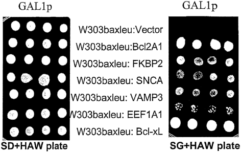
A human hippocampus cDNA library was amplified in a yeast expression vector
to obtain 1.2 x 10⁶ individual clones and transformed into W303baxleu yeast
cells
which contain an integrated Bax-expression cassette and which give "no
background" (see Figure 53). The transformants were directly screened in
plates
containing galactose as the sole carbon source to avoid growth in glucose, and
then replica-plated. (Replica plating is important since cells that grow
directly on
galactose are the ones that should contain the plasmid-bome anti-apoptotic
genes
of interest). 3.8 x 10¹⁰ individual yeast transformants were screened
(calculated
on the basis of transformants obtained on glucose plates in a control
experiment to
determine the number of cells obtained from each transformation). Plasmids
were
isolated from cells that grow in galactose, i.e. in the presence of the
functional Bax
polypeptide. Purified plasmids were retransformed into Bax-containing yeast
cells
and checked again for prevention of cell death. The polynucleotide inserts of
those plasmids that definitely prevented Bax-mediated cell death in yeast, and
which bear the anti-apoptotic polynucleotides of interest were sequenced.
As described in the Examples, performance of this screening method on a human
hippocampus cDNA library resulted in the identification of sixteen
polynucleotides (corresponding to genes or partial genes) that abrogate Bax-
mediated apoptosis and cell death in the yeast. Of the sixteen polynucleotides
thus
18
CA 02642856 2008-08-14
WO 2007/093807 PCT/GB2007/000540
identified, one was Bcl-2A1, a homologue of Bcl-2 which is a known inhibitor
of
apoptosis. The identification of Bcl-2A1 as a result of the screening method
is
clear proof-of-principle that the method is highly suitable and effective at
screening for polynucleotides that are, or that encode, inhibitors of Bax-
mediated
apoptosis.
A fourth aspect of the invention tlius provides a method of screening for a
polynucleotide that is or that encodes an inhibitor of Bax-mediated apoptosis,
the
method comprising:
(a) providing a library of polynucleotides in yeast plasmid vectors;
(b) transforming the library of polynucleotides into yeast cells as
defined in the first or second aspects of the invention;
(c) plating the transformed yeast under conditions that allow
expression of the functional Bax polypeptide and of the polynucleotides in the
yeast plasmid vectors; and
(d) identifying one or more yeast colonies that grow under the
conditions of step (c),
wherein growth of a yeast colony indicates that the polynucleotide in the
yeast
plasmid vector is, or encodes, an inhibitor of Bax-mediated apoptosis.
Preferably, the library of polynucleotides is a eDNA library, which may be
generated from sources such as human brain tissue, a tissue or cells that are
involved in diabetes, a tissue that is involved in rheumatoid arthritis, or
from a cell
line. The cDNA library may also be made from cancer tissue, heart tissue,
muscle
tissue, or a viral or bacterial genome.
Typically, the library of polynucleotides in the yeast plasmid vectors is
under the
control of an inducible promoter. Suitably, the inducible promoter can be a.
tetracycline-inducible promoter, a methionine-inducible promoter, a galactose-
inducible promoter, the ADH2 alcohol dehydrogenase promoter (repressed in
glucose, induced when glucose is exhausted and ethanol is made), or the CUPI
metallothionein promoter (induced in the presence of Cu2}, Znz). Typically,
the
galactose-inducible promoter is GALl or GAL10.
19
CA 02642856 2008-08-14
WO 2007/093807 PCT/GB2007/000540
Many suitable yeast plasmid vectors are known in the art and include pYES2
(Stratagene), and other 2-micron (multi-copy) or centromere (single-copy)
yeast
shuttle vectors known in the art.
It is preferred if transforming step (b) is a high efficiency transformation.
Typically, plating step (c) comprises incubating the plated yeast cells at 30
C in
one of the known standard yeast growth media, with galactose as the sole
carbon
source, for at least 72 hours, and possibly up to 7 days. Clearly, if the
polynucleotides in the yeast plasmid vectors are under the control of a
promoter
that is inducible by an agent other than galactose, that inducing agent is
also
included.
In an alternative embodiment, the Bax polypeptide may be under the control of
the
ADH2 promoter rather than a GAL promoter, for example, to screen for proteins
that abrogate the Bax-mediated block of cell growth in petites (that grow
without
functional mitochondria).
As is immediately apparent to a person of skill in the art of yeast genetics,
the
method may further comprise one or more of the following steps, and typically
all
three.
(e) isolating yeast cells from a yeast colony identified in step (d);
(f) isolating the yeast plasmid vector from the yeast colony identified
in step (d) or from the yeast cells isolated in step (e); and
(g) sequencing the polynucleotide (i.e. the insert) from the yeast
plasmid vector isolated in step (f).
As described below in the examples, the method typically further comprises the
step of:
(h) retesting the polynucleotide from the plasmid vector present in a
yeast colony identified in step (d), or a polypeptide encoded by said
polynucleotide, for the ability to inhibit Bax-mediated apoptosis in a model
of
CA 02642856 2008-08-14
WO 2007/093807 PCT/GB2007/000540
apoptosis.
Additionally or alternatively, the method may further comprise the step of:
(i) modifying the polynucleotide from the plasmid vector present in a
yeast colony identified in step (d), and testing the modified polynucleotide,
or a
polypeptide encoded by said modified polynucleotide, for the ability to
inhibit
Bax-mediated apoptosis in a model of apoptosis.
Further, the method may also comprise the step of:
(j) identifying the polynucleotide from a yeast colony identified in
step (d) based upon the sequence data obtained in step (g), and testing a
polynucleotide that corresponds to the identified polynucleotide, or a
polypeptide
encoded by said corresponding polynucleotide, for the ability to inhibit Bax-
mediated apoptosis in a model of apoptosis.
Since the polynucleotide obtained from a yeast colony identified in step (d)
may
not encode the full-length naturally occurring polypeptide, by a
polynucleotide
that "corresponds to" the identified polynucleotide, we include a full-length
version of the identified polynucleotide that encodes the full-length
naturally
occurring polypeptide. Also, since the gene corresponding to the identified
polynucleotide may encode several polypeptide isoforms, by a polynucleotide
that
"corresponds to" the identified polynucleotide, we include a polynucleotide
that
encodes a different isoform of the naturally occurring polypeptide. In
addition,
since the identified polynucleotide may possess sequence differences from the
naturally-occurring polynucleotide sequence, typically if the eDNA library was
obtained from a cell line, by a polynucleotide that "corresponds to" the
identified
polynucleotide, we include the naturally occurring polynucleotide. Further,
since
the polynucleotide may be isolated from a non-human cDNA library, by a
polynucleotide that "corresponds to" the identified polynucleotide we include
a
homologue of the identified polynucleotide from another species, and
preferably a
human homologue.
Suitable models of apoptosis include yeast cell models, mammalian cell models,
21
CA 02642856 2008-08-14
WO 2007/093807 PCT/GB2007/000540
and in vivo models of apoptosis. The yeast cell model for testing whether a
polynucleotide has the ability to inhibit Bax-mediated apoptosis in a cell may
be
one as described above in the first aspect of the invention, such as
WT303baxleu.
The mammalian cell model for testing whether a polynucleotide has the ability
to
inhibit Bax-mediated apoptosis in a cell may be almost any mammalian cell or
cell
line. As described below in the Examples, the HEK293 cell line was chosen
since
the anti-apoptotic genes identified from a hippocampal cDNA library are
expected
to be effective in neuronal cells, and HEK293 is suggested to have some
neuronal
features (at least, it is routinely used as a replacement of typical neuronal
cells
which are difficult to use).
For example, cells can be transfected with an indicator plasmid carrying a
reporter
gene encoding an indicator molecule, and the degree of cell death or apoptosis
can
be measured by detection of the expressed indicator molecule. For example, the
degree of apoptosis in cells transfected with an indicator plasmid expressing
the
indicator E. coli (3-galactosidase can be determined by a(3-galactosidase
ELISA
(Boehringer Mannheim), in accordance with the manufacturer's recommendations.
Also, the degree of apoptotic activity can be determined by visually scoring,
under
a microscope, blue cells expressing (3-galactosidase, after staining them with
X-
gal. As another example, the degree of apoptosis in cells transfected with an
indicator plasmid expressing Green Fluorescent Protein can be determined by
measuring the fraction of fluorescent cells in the total cell population,
using a flow
cytometer (FACScan, Becton-Dickinson) or fluorescent microscope.
Also, DNA degradation, indicative of apoptosis, can be examined by exposing
the
cells to anti-Fas antibody in the presence of CHX. Thereafter, the DNA in the
cells is extracted and purified using standard protocols. Any methods
detecting
cell death or apoptosis can be used such as those described below or in
Sellers et
al (1994); Telford et al (1994); and Poirier, Ed. (1997) Apoptosis Techniques
and
Protocols, Humana Press, Totowa, NJ, USA.
The time course of apoptosis can be analysed by measuring the level of
expression
of phosphatidylserine on the cell surface, as detected, for example, with FITC-
22
CA 02642856 2008-08-14
WO 2007/093807 PCT/GB2007/000540
labeled Annexin V, and/or by a dye-exclusion test using propidium iodide.
These
two tests can be performed using a commercially available kit, for example,
the
ApoAlert Annexin V Apoptosis kit (Clontech), in accordance with the
manufacturer's recommendations, and using a flow cytometer (FACScan, Becton-
Dickinson) or fluorescent microscope.
As an in vivo model of apoptosis in neurodegeneration, one uses chemicals
(which
results in PD or AD) to induce apoptosis in mouse or rat brain. As an in vivo
model of apoptosis in cancer, one injects tumour cells that result in tumour
formation and then determines if any agent would induce apoptosis (i.e.
shrinkage
of the tumour).
As would be appreciated by the person skilled in the art, the method may
further
comprise the step of formulating a polynucleotide or polypeptide which has the
ability to inhibit Bax-mediated apoptosis in an in vivo model of apoptosis
into a
pharmaceutically acceptable composition.
As described herein, the above method of screening for a polynucleotide that
is or
that encodes an inhibitor of Bax-mediated apoptosis, was performed on a human
hippocampus cDNA library. Sixteen polynucleotides (corresponding to genes or
partial genes) that abrogate Bax-mediated cell death in the yeast system of
apoptosis were identified and are listed in Table 1.
Table 1: "Bax antagonists" identified in the human hippocampus
# Identified Gene Brief Description SEQ ID
No
1 Bcl-2A1 Homologue of Bcl-2. Known to be expressed in the 4
hippocampus.
2 a-Synuclein Mutant a-Synuclein forms play a major role in PD and 5
(SNCA) AD. The role of the wild-type protein is unclear.
3 FKBP2 An endoplasmic reticulum resident FK506 binding 6
(FKBP-13) protein. Highly overproduced during protein
misfolding in the ER.
4 EEF1A1 Eukaryotic translation elongation factor 1 al. 7
Re orted to be involved in oncogenic transformation.
23
CA 02642856 2008-08-14
WO 2007/093807 PCT/GB2007/000540
Acts as a dominant oncogene in prostate carcinoma
VAMP3 Vesicle-associated membrane protein 3 (cellubrevin) 8
6 SNAP25 Synaptosomal-associated protein 9
7 RIMS3 Regulates synaptic membrane exocytosis 10
8 RAB40B A member of the RAS oncogene family 11
9 HMGCS1 3-hydroxy-3-metlrylglutaryl-coenzyme A synthase 1 12
(soluble)
SCD5 Stearoyl-CoA desaturase 5 13
11 Atp2a2 ATPase. Ca2+ transporting, cardiac muscle, slow 14
twitch 2
12 HRMTILI hnRNP methyltransferase-like 1. Has a homologue in 15
S. cerevisiae.
13 Clone A sequence from human chromosome 3p of unknown 16
RP11-605M1 function
14 Clone A sequence from human chromosome 22q11.22-12.2 17
CTA-373H7 of unknown function
Isolate WH6967 A sequence from the genome of human mitochondrial 18
isolate WH6967 of unknown function
16 Isolate S 1216 A sequence from the genome of human mitochondrial 19
isolate S 1216 of unknown function
Bcl-2A1, SNCA, FKBP2 (FKBP-13), EEF1A1, and VAMP3 were tested in a
mammalian system of apoptosis, and were each confirmed as having activity as
an
inhibitor of Bax-mediated apoptosis. Bcl-2A1 has previously been reported to
be
5 an inhibitor of apoptosis. It is therefore reasonable to conclude that the
other
eleven polynucleotides identified as a result of the screening method also
are, or
encode, inhibitors of Bax-mediated apoptosis in mammals.
A fifth aspect of the invention thus provides a method of combating Bax-
mediated
10 apoptosis in a cell, the method comprising administering to the cell a
polypeptide
selected from FKBP2, SNCA, EEFlA1, VAMP3, SNAP25, RIMS3, RAB40B,
HMGCSl, SCD5, ATP2A2, HRMTILI, and a polypeptide encoded by a
polynucleotide comprising SEQ ID No: 16, SEQ ID No: 17, SEQ ID No: 18 or
SEQ ID No: 19, or an anti-apoptotic derivative of any of these polypeptides,
or a
15 polynucleotide that encodes any of said polypeptides or derivatives.
24
CA 02642856 2008-08-14
WO 2007/093807 PCT/GB2007/000540
Typically, the method is performed in vitro.
Typically, the cell is a mammalian cell, such as a human cell, although as is
clear
from the Examples, the cell may be from other species such as yeast. The cell
may be from an established cell line, a primary cell culture, or a cell which
is
present in a tissue ex vivo.
Alternatively, the method may be performed in vivo.
The polypeptides, the anti-apoptotic derivatives thereof, and polynucleotides
that
encode any of said polypeptides or derivatives, have a clear utility as
research
tools in the study of apoptosis. They also have utility in methods of
screening for
further therapeutic agents that modulate apoptosis, as described below.
A sixth aspect of the invention provides the use of a polypeptide selected
from
FKBP2, SNCA, EEF1A1, VAMP3, SNAP25, RIMS3, RAB40B, HMGCSl,
SCD5, ATP2A2, HRMTILI, and a polypeptide encoded by a polynucleotide
comprising SEQ ID No: 16, SEQ ID No: 17, SEQ ID No: 18 or SEQ ID No: 19,
or an anti-apoptotic derivative of any of these polypeptides, or a
polynucleotide
that encodes any of said polypeptides or derivatives, in the preparation of a
medicament for combating Bax-mediated apoptosis in a cell.
By "combating Bax-mediated apoptosis in a cell" we mean inhibiting or
preventing Bax-mediated apoptosis in a cell.
It is appreciated that the polypeptides, derivatives and polynucleotides of
the fifth
aspect and the medicament of the sixth aspect of the invention may be suitable
for
combating any apoptosis where mitochondrial dysfunction occurs since the
apoptotic effect of Bax could be mimicked by other proteins or by
exogenous/endogenous chemicals. For example, staurospaurine is known to
mimic the effects of Bax. Thus the polypeptides, derivatives and
polynucleotides
CA 02642856 2008-08-14
WO 2007/093807 PCT/GB2007/000540
of the fifth aspect and the medicament of the sixth aspect of the invention
may be
suitable for combating staurospaurine induced cell death.
"Derivatives" of any given polypeptide may be made using protein chemistry
techniques, for example using partial proteolysis (either exolytically or
endolytically), or by de novo synthesis. Alternatively, the derivatives may be
made by recombinant DNA technology. Suitable techniques for cloning,
manipulation, modification and expression of nucleic acids, and purification
of
expressed proteins, are well known in the art and are described for example in
Sambrook et al (2001) "Molecular Cloning, a Laboratory Manual", 3`d edition,
Sambrook et al (eds), Cold Spring Harbor Laboratory Press, Cold Spring Harbor,
NY, USA.
Suitable "anti-apoptotic derivatives" of these polypeptides include fragments
thereof and modifications of the full-length polypeptide or fragment thereof,
that
have the ability to inhibit Bax-mediated apoptosis in a cell.
By "modifications" of a given polypeptide we include amino acid insertions,
deletions and substitutions, either conservative or non-conservative, at one
or more
positions. Such modifications may be called analogues of the given
polypeptide. By
"conservative substitutions" is intended combinations such as Gly, Ala; Val,
Ile, Leu;
Asp, Glu; Asn, Gln; Ser, Thr; Lys, Arg; and Phe, Tyr. Such modifications may
be
made using the methods of protein engineering and site-directed mutagenesis,
as
described in Sambrook et al 2001, supra.
Further-modifications of a given polypeptide include the addition of NH2 or
COOH
terminal tags that would allow entry of proteins into cells.
The hippocampus is a region of the brain that is known to suffer from
apoptosis in
neurodegenerative disease. Since the anti-apoptotic polynucleotides were
identified from a human hippocampus cDNA library, it is also reasonable to
conclude that each of the identified anti-apoptotic polynucleotides, or the
anti-
apoptotic polypeptides that they encode, may have utility in combating
26
CA 02642856 2008-08-14
WO 2007/093807 PCT/GB2007/000540
neurodegenerative disease.
A seventh aspect of the invention thus provides a polypeptide selected from
FKBP2, SNCA, VAMP3, SNAP25, RIMS3, RAB40B, HMGCSl, SCD5,
HRMTILI, and a polypeptide encoded by a polynucleotide comprising SEQ ID
No: 16, SEQ ID No: 17, SEQ ID No: 18 or SEQ ID No: 19, or an anti-apoptotic
derivative of any of these polypeptides, or a polynucleotide that encodes any
of
said polypeptides or derivatives, for use in medicine.
1o An eighth aspect of the invention provides a pharmaceutical composition
comprising a polypeptide selected from FKBP2, SNCA, VAMP3, SNAP25,
RIMS3, RAB4OB, HMGCSI, SCD5, HRMTILI, and a polypeptide encoded by a
polynucleotide comprising SEQ ID No: 16, SEQ ID No: 17, SEQ ID No: 18 or
SEQ ID No: 19, or an anti-apoptotic derivative of any of these polypeptides,
or a
polynucleotide that encodes any of said polypeptides or derivatives, and a
pharmaceutically acceptable carrier or excipient.
In addition, a number of other disorders or conditions are associated with
inappropriate Bax-mediated apoptosis in cells or tissues, such as
cardiovascular
cells in cardiovascular disease (Reeve et al, 2005), synovial cells in
rheumatoid
arthritis (Baier et al, 2003), and pancreatic B-cells in diabetes (Cnop et al,
2005;
Millet et al, 2005).
A ninth aspect of the invention thus provides a method of combating a disease
or
condition in a patient selected from a neurodegenerative disease or condition,
cardiovascular disease, rheumatoid arthritis and diabetes, the method
comprising
administering to the patient a polypeptide selected from FKBP2, SNCA, EEF1A1,
VAMP3, SNAP25, RIMS3, RAB40B, HMGCS1, SCD5, ATP2A2, HRMTIL1,
and a polypeptide encoded by a polynucleotide comprising SEQ ID No: 16, SEQ
ID No: 17, SEQ ID No: 18 or SEQ ID No: 19, or an anti-apoptotic derivative of
any of these polypeptides, or a polynucleotide that encodes any of said
polypeptides or derivatives.
27
CA 02642856 2008-08-14
WO 2007/093807 PCT/GB2007/000540
Cardiovascular disease is a leading cause of death worldwide. Loss of function
or
death of cardiomyocytes is a major contributing factor to cardiovascular
disease
which is a leading cause of death worldwide. Cell death in conditions such as
heart failure and myocardial infarction is associated with apoptosis.
Apoptotic
pathways have been well studied in non-myocytes and it is thought that similar
pathways exist in cardioinyocytes. These pathways include death initiated by
ligation of membrane-bound death receptors, release of pro-apoptotic factors
from
mitochondria or stress at the endoplasmic reticulum. The key regulators of
apoptosis include inhibitors of caspases (IAPs), the Bcl-2 family of proteins,
growth factors, stress proteins, calcium and oxidants. The highly organized
and
predictive nature of apoptotic signalling means it is amenable to
manipulation. A
thorough understanding of the apoptotic process would facilitate intervention
at
the most suitable points, alleviating myocardium decline and dysfunction
(Reeve
et al, 2005).
By "combating" a disease, disorder or condition in a patient we mean treating,
preventing, or ameliorating the symptoms of, that particular disorder or
condition.
Neurodegenerative diseases or disorders that can be treated using the
therapeutic
methods and uses of the present invention include stroke, spinal cord trauma,
head
injury, spinal muscular atrophy (SMA), motor neuron disease including
amyotrophic lateral sclerosis (ALS), Alzheimer's disease (AD), Parkinson's
disease (PD) and Huntington's disease (HD).
A tenth aspect of the invention thus provides the use of a polypeptide
selected
from FKBP2, SNCA, EEFIAI, VAMP3, SNAP25, RIMS3, RAB40B, HMGCSI,
SCD5, ATP2A2, HRMTILI, and a polypeptide encoded by a polynucleotide
comprising SEQ ID No: 16, SEQ ID No: 17, SEQ ID No: 18 or SEQ ID No: 19,
or an anti-apoptotic derivative of any of these polypeptides, or a
polynucleotide
that encodes any of said polypeptides or derivatives, in the preparation of a
medicament for combating a disease or condition in a patient selected from a
neurodegenerative disease or condition, cardiovascular disease, rheumatoid
arthritis and diabetes.
28
CA 02642856 2008-08-14
WO 2007/093807 PCT/GB2007/000540
FKBP2
FKBP2 (also known as FK506-Binding protein 2 and FKBP13) is a member of a
family of proteins which bind the immunosuppressant drugs, FK506 and
rapamycin. The FKBP2 gene is 3 16 in length and contains six exons and is
located at human chromosome 11q13.1-q13.3 (DiLella et al, 1992). Partaledis &
Berlin (1993) describe the FKB2 gene of S. cerevisiae which encodes a
homologue of human FKBP-13 having 57% sequence identity with human FKBP-
13, and suggest that FKB2/FKB13 plays a role in protein trafficking in the
endoplasmic reticulum (ER).
According to Genbank Accession No NP 476433, the protein encoded by the
FKB2 gene is a member of the immunophilin protein family, which plays a role
in
immunoregulation and basic cellular processes involving protein folding and
trafficking. The FKB2 encoded protein is a cis-trans prolyl isomerase that
binds
the immunosuppressants FK506 and rapamycin. It is thought to function as an ER
chaperone and may also act as a component of membrane cytoskeletal scaffolds.
This gene has two alternatively spliced transcript variants that encode the
same
isoform. Multiple polyadenylation sites have been described for this gene, but
the
full-length nature of this gene has not been determined. FKBP2 has a signal
peptide at residues 1-21 (as defmed in NP 476433), and the mature polypeptide
is
at residues 22-142. FKBP2 is a peptidylprolyl isomerase (EC 5.2.1.8).
To the best of the inventor's knowledge, FKBP2 has not been associated with
apoptosis.
Thus the invention includes combating Bax-mediated apoptosis in a cell by
administering to the cell a FKBP2 polypeptide, or an anti-apoptotic derivative
thereof, or a polynucleotide that encodes said polypeptide or derivative.
Similarly, to the best of the inventor's knowledge, FKBP2 has not been
associated
with any neurodegenerative disorder. Indeed, according to Online Mendelian
Inheritance in Man (OMIM, Reference No. 186946) FKBP2 has not been
29
CA 02642856 2008-08-14
WO 2007/093807 PCT/GB2007/000540
associated with any disease state and, as far as the inventor is aware, FKBP2
has
not been used therapeutically.
Thus the invention includes an FKBP2 polypeptide, or an anti-apoptotic
derivative
thereof, or a polynucleotide that encodes said polypeptide or derivative, for
use in
medicine.
The invention also includes combating a neurodegenerative disease in a patient
by
administering to the patient a FKBP2 polypeptide, or an anti-apoptotic
derivative
thereof, or a polynucleotide that encodes said polypeptide or derivative.
By "FKBP2 polypeptide" we include the meaning of a gene product of the human
FKBP2 gene, including naturally occurring variants thereof. The eDNA sequence
corresponding to a human FKBP2 mRNA is found in Genbank Accession No
NM 057092 (variant 2) and NM 004470 (variant 1). Transcript variant 2 has a
distinct exon at the 5' UTR compared to variant 1, although the coding region
is
the same in both variants. Human FKBP2 polypeptide includes the amino acid
sequence found in Genbank Accession No NP 476433, and naturally occurring
variants thereof.
Suitable "anti-apoptotic derivatives" of FKBP2 include anti-apoptotic
fragments
of FKBP2. By "an anti-apoptotic fragment" of FKBP2 we mean any portion of
the FKBP2 polypeptide that has the ability to inhibit Bax-mediated apoptosis
in a
cell. Typically, the fragment has at least 30% of the anti-apoptotic activity
of full-
length human FKBP2. It is more preferred if the fragment has at least 50%,
preferably at least 70% and more preferably at least 90% of the activity of
full-
length human FKBP2. Most preferably, the fragment has 100% or more of the
anti-apoptotic activity of full-length human FKBP2.
Suitable "anti-apoptotic derivatives" of FKBP2 also include modifications of
full-
length FKBP2, or a fragment thereof, that have the ability to inhibit Bax-
mediated
apoptosis in a cell. Preferably, the modified FKBP2 or modified FKBP2 fragment
retains at least 80%, or at least 85% or at least 90% sequence identity with
full-
CA 02642856 2008-08-14
WO 2007/093807 PCT/GB2007/000540
length FKBP2, or the respective FKBP2 fragment. More preferably, the modified
FIOP2 or modified FKBP2 fragment has at least 91%, or at least 92%, or at
least
93%, or at least 94% or at least 95% sequence identity, and yet more
preferably at
least 96%, or at least 97%, or at least 98% or at least 99% sequence identity
with
full-length FKBP2, or the respective FKBP2 fragment. Preferably, the modified
FKBP2 or modified FKBP2 fragment retains at least 30% of the anti-apoptotic
activity of full-length human FKBP2. It is more preferred if the modified
FKBP2
or FKBP2 derivative has at least 50%, preferably at least 70% and more
preferably
at least 90% of the activity of full-length human FKBP2. Most preferably, the
modified FKBP2 or modified FKBP2 fragment has 100% or more of the anti-
apoptotic activity of full-length human FKBP2.
By FKBP2 we also include a homologous gene product from FKBP2 genes from
other species. By "homologous gene product" we include an FKBP2 polypeptide
having at least 80% sequence identity with the human FKBP2 amino acid
sequence in Genbank Accession No NP_476433. More preferably, a homologous
gene product includes an FKBP2 polypeptide having at least 85% or at least 90%
sequence identity with human FKBP2. Yet more preferably, a homologous gene
product includes an FKBP2 polypeptide having at least 91%, or at least 92%, or
at
least 93%, or at least 94%, or at least 95%, or at least 96%, or at least 97%,
or at
least 98% sequence identity with human FKBP2. Most preferably, a homologous
gene product includes an FKBP2 polypeptide having at least 99% sequence
identity with the human FKBP2 amino acid sequence. It is appreciated that for
applications in which FKBP2 is administered to a non-human subject, the FKBP2
is preferably from the same species as the subject. If the FKBP2 is
administered
to a human subject, the FKBP2 is preferably human FKBP2, or an anti-apoptotic
fragment or variant thereof.
Although there is only 47% sequence identity (perfect match) and 69% sequence
homology (accepting conserved residues) between the FKBP2 proteins from
humans and yeast, the human FKBP2 can complement inactivating FKBP2
mutations in yeast cells (data not included).
31
CA 02642856 2008-08-14
WO 2007/093807 PCT/GB2007/000540
By a polynucleotide encoding FKBP2 we include the cDNA encoding the human
FKBP2 polypeptide and naturally occurring variants thereof cDNA sequences
encoding a human FKBP2 mRNA are found in Genbank Accession Nos.
NM 057092 and NM 004470. We also include other sequences which, by virtue
of the degeneracy of the genetic code, also encode the FKBP2 polypeptide.
The polynucleotide (SEQ ID No: 6) encoding FKBP2 that was initially identified
by the inventor was identified as a full-length fragment. This gene was re-
cloned
by PCR from a cDNA library and was C-terminally tagged for protein expression.
FKBP2 is a peptidylprolyl isomerase (EC 5.2.1.8; also known as Peptidyl-prolyl
cis-trans isomerase). There are three distinct families of peptidylprolyl
isomerases: the cyclophilins, the FKBPs, and another family that includes
parvulin
from E. coli. The three families are structurally unrelated and can be
distinguished
by being inhibited by cyclosporin A, FK-506 and 5-hydroxy-1,4-naphthoquinone,
respectively (http://www.chem.qmul.ac.uk/iubmb/enzyme/EC5/2/1/8.html).
Without wishing to be bound by any theory, the inventor considers that since
FKBP2 is a peptidylprolyl isomerase (EC 5.2.1.8; also known as peptidyl-prolyl
cis-trans isomerase), it is possible that its peptidylprolyl isomerase
activity
contributes to its anti-apoptotic activity. Thus other peptidylprolyl
isomerases,
and especially other FKPBs, as well as polynucleotides encoding them, may also
be useful in combating apoptosis. This is particularly unexpected since over-
expression of cyclophilin D, a peptidylprolyl isomerase, is known to enhance
the
apoptotic process (Li et al, 2004). Moreover, inhibition of peptidylprolyl
isomerases (also known as immunophilins) has been used as a target for
prevention of neurodegeneration.
a-Synuclein (SNCA)
SNCA is a member of the synuclein family, which also includes beta- and gamma-
synuclein. Synucleins are abundantly expressed in the brain and alpha- and
beta-
synuclein inhibit phospholipase D2 selectively. SNCA may serve to integrate
presynaptic signalling and membrane trafficking. Defects in SNCA have been
32
CA 02642856 2008-08-14
WO 2007/093807 PCT/GB2007/000540
implicated in the pathogenesis of Parlcinson disease (PD). SNCA peptides are a
major component of amyloid plaques in the brains of patients with Alzheimer's
disease (AD). Two alternatively spliced transcripts of SNCA have been
identified.
To the best of the inventor's knowledge, SNCA has not previously been shown to
be an inhibitor of Bax-mediated apoptosis.
Thus the invention includes combating Bax-mediated apoptosis in a cell by
administering to the cell a SNCA polypeptide, or an anti-apoptotic derivative
thereof, or a polynucleotide that encodes said polypeptide or derivative.
Similarly, although SNCA has been associated with both AD and PD (OMIM
Reference No. 163890), to the best of the inventor's knowledge, there has been
no
previous suggestion to use an SNCA polypeptide, or an anti-apoptotic
derivative
thereof, or a polynucleotide that encodes said polypeptide or derivative, in
combating a neurodegenerative disease in a patient. Indeed, as far as the
inventor
is aware, SNCA has not been used therapeutically.
Thus the invention includes an SNCA polypeptide, or an anti-apoptotic
derivative
thereof, or a polynucleotide that encodes said polypeptide or derivative, for
use in
medicine.
The invention also includes combating a neurodegenerative disease in a patient
by
administering to the patient a SNCA polypeptide, or an anti-apoptotic
derivative
thereof, or a polynucleotide that encodes said polypeptide or derivative.
In an embodiment, the invention includes combating a neurodegenerative disease
other than AD of PD in a patient by administering to the patient a SNCA
polypeptide, or an anti-apoptotic derivative thereof, or a polynucleotide that
encodes said polypeptide or derivative.
33
CA 02642856 2008-08-14
WO 2007/093807 PCT/GB2007/000540
By "SNCA polypeptide" we include the meaning of a gene product of the human
SNCA gene, including naturally occurring variants thereof. Two alternative
transcript variants encoding different protein isoforms have been described
for the
human SNCA gene. A cDNA sequence corresponding to a human SNCA mRNA
is found in Genbank Accession No NM 000345 (isoform NACP140) and in
Genbank Accession No NM 007308 (isoform NACP112). The NACP140
transcript is the longer transcript and encodes the longer NACP 140 isoform
(Genbank Accession No NP000336). The NACP112 transcript lacks an alternate
in-frame segment, compared to variant NACP 140, resulting in a shorter protein
(isoform NACP 112; Genbank Accession No NP_009292) that has a distinct C-
terminus, compared to isoform NACP140. The amino acid sequence of human
SNCA polypeptides includes the sequences found in Genbank Accession Nos
NP 000336 and NP_009292, and naturally occurring variants thereof. The SNCA
sequence identified in the screening experiments described herein was from the
NACP 140 variant.
Suitable "anti-apoptotic derivatives" of SNCA include anti-apoptotic fragments
of
SNCA. By "an anti-apoptotic fragment" of SNCA we mean any portion of a
SNCA polypeptide that has the ability to inhibit Bax-mediated apoptosis in a
cell.
Typically, the fragment has at least 30% of the anti-apoptotic activity of the
SNCA polypeptide of SEQ ID No: 5. It is more preferred if the fragment has at
least 50%, preferably at least 70% and more preferably at least 90% of the
activity
of the SNCA polypeptide of SEQ ID No: 5. Most preferably, the fragment has
100% or more of the anti-apoptotic activity of the SNCA polypeptide of SEQ ID
No: 5.
Suitable "anti-apoptotic derivatives" of SNCA also include modifications of
full-
length SNCA, or a fragment thereof, that have the ability to inhibit Bax-
mediated
apoptosis in a cell. Preferably, the modified SNCA or modified SNCA fragment
retains at least 80%, or at least 85% or at least 90% sequence identity with
full-
length SNCA, or the respective SNCA fragment. More preferably, the modified
SNCA or modified SNCA fragment has at least 91%, or at least 92%, or at least
93%, or at least 94% or at least 95% sequence identity, and yet more
preferably at
34
CA 02642856 2008-08-14
WO 2007/093807 PCT/GB2007/000540
least 96%, or at least 97%, or at least 98% or at least 99% sequence identity
with
full-length SNCA, or the respective SNCA fragment. Preferably, the modified
SNCA or modified SNCA fragment retains at least 30% of the anti-apoptotic
activity of full-length human SNCA. It is more preferred if the modified SNCA
or
SNCA derivative has at least 50%, preferably at least 70% and more preferably
at
least 90% of the activity of full-length human SNCA. Most preferably, the
modified SNCA or modified SNCA fragment has 100% or more of the anti-
apoptotic activity of full-length human SNCA.
By SNCA we also include a homologous gene product from SNCA genes from
other species. By "homologous gene product" we include an SNCA polypeptide
having at least 80% sequence identity with the human SNCA amino acid
sequences in Genbank Accession Nos NP_000336 and NP009292. More
preferably, a homologous gene product includes an SNCA polypeptide having at
least 85% or at least 90% sequence identity with a human SNCA. Yet more
preferably, a homologous gene product includes an SNCA polypeptide having at
least 91%, or at least 92%, or at least 93%, or at least 94%, or=at least 95%,
or at
least 96%, or at least 97%, or at least 98% sequence identity with human SNCA.
Most preferably, a homologous gene product includes an SNCA polypeptide
having at least 99% sequence identity with a human SNCA amino acid sequence
in Genbank Accession No NP 000336 or NP_009292. It is appreciated that for
applications in which SNCA is administered to a non-human subject, the SNCA is
preferably from the same species as the subject. If the SNCA is administered
to a
human subject, the SNCA is preferably human SNCA, or an anti-apoptotic
fragment or variant thereof.
By a polynucleotide encoding SNCA we include the cDNA encoding the human
SNCA polypeptide and naturally occurring variants thereof cDNA sequences
encoding a human SNCA inRNA are found in Genbank Accession Nos.
N1V1 000345 and NM 007308. We also include other sequences which, by virtue
of the degeneracy of the genetic code, also encode the SNCA polypeptide. An
example of a polynucleotide encoding an anti-apoptotic fragment of SNCA is
listed in SEQ ID No: 5.
CA 02642856 2008-08-14
WO 2007/093807 PCT/GB2007/000540
The polynucleotide (SEQ ID No: 5) encoding SNCA that was initially identified
by the inventor is believed to be a full-length fragment: the insert was
sequenced
from the 3' end and the last few nucleotides in the read were N's. A full-
length
transcript was subsequently cloned by PCR from the same hippocampal cDNA
library used for the screening, and the Bax-rescuing ability of the initially
isolated
clone and of the full-length PCR fragment was the same.
EEF 1 A
The eukaryotic elongation factor 1 A (eEF 1 A, formerly known as EF 1 alpha)
is a
key factor in protein synthesis, where it promotes the transfer of
aminoacylated
tRNAs to the A site of the ribosome. Two differentially expressed isoforms of
eEF1A, designated eEF1A-1 and eEF1A-2, are found in mammals. In humans,
the eEF1A-1 and eEF1A-2 isoforms have 92.6% sequence identity (see Figure
Is 67).
According to NP_001393, the EEF1A1 gene encodes an isoform of the alpha
subunit of the elongation factor-1 complex, which is responsible for the
enzymatic
delivery of aminoacyl tRNAs to the ribosome. This isoform (alpha 1) is
expressed
in brain, placenta, lung, liver, kidney, and pancreas, whereas the other
isoform
(alpha 2) is expressed in brain, heart and skeletal muscle. The alpha 1
isoform is
identified as an autoantigen in 66% of patients with Felty syndrome. The eEF1A
gene is located at human chromosome 6q14, but has been found to have multiple
copies on many chromosomes, some of which, if not all, represent different
pseudogenes. The alpha subunit of elongation factor-1 (EEF1A) is involved in
the
binding of aminoacyl-tRNAs to 80S ribosomes. During the process, GTP is
hydrolyzed into GDP. To perform this function, EEF1A has domains that bind
guanine nucleotides, 80S ribosomes, and aminoacyl-tRNAs. Also, EEF1A
interacts with the beta subunit of EEF1 to exchange bound GDP for GTP. The
primary structure of human EEF1A was determined by Brands et al (1986).
Ditzel et al (2000) identified EEFlAl as an autoantibody in 66% of patients
with
Felty syndrome, a disorder characterized by the association of rheumatoid
36
CA 02642856 2008-08-14
WO 2007/093807 PCT/GB2007/000540
arthritis, splenomegaly, and peripheral destruction of neutrophils leading to
neutropaenia.
According to the review by Lamberti et al (2004), EEF1A is involved in
apoptosis
(without specifying whether this is EEFlAl or EEFlA2, or both). Lamberti et al
also reports that EEF1A2 plays a role in the protection of caspase 3-mediated
apoptosis. Based on the anti-apoptotic effects of EEF1A2, Thornton et al
(2003)
suggest that EEF1A1 may also inhibit apoptosis. Talapatra et al (2001)
identified
EEF1A1 in a screen for genes that protect against apoptosis caused by
interleukin
3 withdrawal.
To the best of the inventor's knowledge, EEF1A1 has not been associated with
Bax-mediated apoptosis.
Thus the invention includes combating Bax-mediated apoptosis in a cell by
administering to the cell a EEFlAl polypeptide, or an anti-apoptotic
derivative
thereof, or a polynucleotide that encodes said polypeptide or derivative.
Similarly, to the best of the inventor's knowledge, EEFlAl has not been
associated with any neurodegenerative disorder.
Thus the invention also includes combating a neurodegenerative disease in a
patient by administering to the patient an EEF1A1 polypeptide, or an anti-
apoptotic derivative thereof, or a polynucleotide that encodes said
polypeptide or
derivative.
By "EEF1A1 polypeptide" (eukaryotic translation elongation factor 1 al) we
include the meaning of a gene product of the human EEF1A1 gene, including
naturally occurring variants thereof. A cDNA sequence corresponding to a human
EEF1A1 mRNA is found in Genbank Accession No NM 001402. Human
EEF1A1 polypeptide includes the amino acid sequence found in Genbank
Accession No NP_001393, and naturally occurring variants thereof.
37
CA 02642856 2008-08-14
WO 2007/093807 PCT/GB2007/000540
Suitable "anti-apoptotic derivatives" of EEF1A1 include anti-apoptotic
fragments
of EEF1A1. By "an anti-apoptotic fragment" of EEF1Al we mean any portion of
the EEFIAI polypeptide that has the ability to inhibit Bax-mediated apoptosis
in a
cell. Typically, the fragment has at least 30% of the anti-apoptotic activity
of full-
length human EEF1Al. It is more preferred if the fragment has at least 50%,
preferably at least 70% and more preferably at least 90% of the activity of
full-
length human EEF 1 A 1. Most preferably, the fragment has 100% or more of the
anti-apoptotic activity of full-length human EEF1A1.
Suitable "anti-apoptotic derivatives" of EEF1Al also include fragments of full-
length EEF1A1, or modifications of the full-length polypeptide of fragment
thereof, that have the ability to inhibit Bax-mediated apoptosis in a cell.
Preferably, the modified EEF1A1 or modified EEFIAl fragment retains at least
93% sequence identity with full-length EEFlAl, or the respective EEFlAl
fragment. More preferably, the modified EEF1A1 or modified EEFlAl fragment
has at least 94% or at least 95% sequence identity, and yet more preferably at
least
96%, or at least 97%, or at least 98% or at least 99% sequence identity with
full-
length EEF1A1, or the respective EEF1A1 fragment. Preferably, the modified
EEF1A1 or modified EEF1A1 fragment retains at least 30% of the anti-apoptotic
activity of full-length human EEF1A1. It is more preferred if the modified
EEFlAl or EEF1A1 derivative has at least 50%, preferably at least 70% and more
preferably at least 90% of the activity of full-length human EEF1A1. Most
preferably, the modified EEFIAl or modified EEF1A1 fragment has 100% or
more of the anti-apoptotic activity of full-length human EEF1A1.
By EEF1A1 we also include a homologous gene product from EEF1A1 genes
from other species. By "homologous gene product" we include an EEF1A1
polypeptide having at least 93% sequence identity with the human EEF1A1 amino
acid sequence in Genbank Accession No NP_001393. More preferably, a
homologous gene product includes an EEF1A1 polypeptide having at least 94%,
or at least 95%, or at least 96%, or at least 97%, or at least 98% sequence
identity
with human EEF1A1. Most preferably, a homologous gene product includes an
EEFlAl polypeptide having at least 99% sequence identity with the human
38
CA 02642856 2008-08-14
WO 2007/093807 PCT/GB2007/000540
EEFlAl amino acid sequence. It is appreciated that for applications in which
EEF1A1 is administered to a non-human subject, the EEF1Al is preferably from
the same species as the subject. If the EEFlAl is administered to a human
subject, the EEFlA1 is preferably human EEFlAl, or an anti-apoptotic fragment
or variant thereof.
By a polynucleotide encoding EEF1A1 we include the cDNA encoding the human
EEF1A1 polypeptide and naturally occurring variants thereof. A cDNA sequence
encoding a human EEFIAl mRNA is found in Genbank Accession No
NM 001402. We also include other sequences which, by virtue of the degeneracy
of the genetic code, also encode the EEF1A1 polypeptide. An example of a
polynucleotide encoding an anti-apoptotic fragment of EEF1A1 is listed in SEQ
ID No: 7 (and the full-length EEF1A1 was subsequently cloned for confirmation
of its effects in yeast and in HEK293 cells).
VAMP3
VAMP3 is a member of the vesicle-associated membrane protein (VAMP)/
synaptobrevin family. VAMPs, together with syntaxins, and the 25-kD
synaptosomal-associated protein (SNAP25), are the main components of a protein
complex involved in the docking and/or fusion of synaptic. vesicles with the
presynaptic membrane. Because of VAMP3's high homology to other known
VAMPs, its broad tissue distribution, and its subcellular localisation, the
protein
encoded by this gene was considered to be the human equivalent of the rodent
cellubrevin. In platelets the protein resides on a compartment that is not
mobilized
to the plasma membrane on calcium or thrombin stimulation
To the best of the inventor's knowledge, VAMP3 has not been associated with
apoptosis.
Thus the invention includes combating Bax-mediated apoptosis in a cell by
administering to the cell a VAMP3 polypeptide, or an anti-apoptotic derivative
thereof, or a polynucleotide that encodes said polypeptide or derivative.
39
CA 02642856 2008-08-14
WO 2007/093807 PCT/GB2007/000540
VAMP3 was found to be one of 25 genes within a locus on human chromosome
lp36 that is involved in early-onset recessive parlcinsonism (van Duijn et al,
2001).
However, VAMP3 has not been directly associated with any neurodegenerative
disease or condition. Indeed, according to OMIM Reference No 603657,
VAMP3 has not been directly associated with any disease state and, as far as
the
inventor is aware, VAMP3 has not been used therapeutically.
Thus the invention includes a VAMP3 polypeptide, or an anti-apoptotic
derivative
thereof, or a polynucleotide that encodes said polypeptide or derivative, for
use in
1 o medicine.
Thus the invention also includes combating a neurodegenerative disease or
condition in a patient by administering to the patient a VAMP3 polypeptide, or
an
anti-apoptotic derivative thereof, or a polynucleotide that encodes said
polypeptide
or derivative.
By VAMP3 polypeptide (vesicle-associated membrane protein 3) we include the
meaning of a gene product of the human VAMP3 gene, including naturally
occurring variants thereof. A cDNA sequence corresponding to a human VAMP3
mRNA is found in Genbank' Accession No NM 004781. Human VAMP3
polypeptide includes the amino acid sequence found in Genbank Accession No
NP_004772, and naturally occurring variants thereof.
Suitable "anti-apoptotic derivatives" of VAMP3 include anti-apoptotic
fragments
of VAMP3. By "an anti-apoptotic fragment" of VAMP3 we mean any portion of
the VAMP3 polypeptide that has the ability to inhibit Bax-mediated apoptosis
in a
cell. Typically, the fragment has at least 30% of the anti-apoptotic activity
of full-
length human VAMP3. It is more preferred if the fragment has at least 50%,
preferably at least 70% and more preferably at least 90% of the activity of
full-
length human VAMP3. Most preferably, the fragment has 100% or more of the
anti-apoptotic activity of full-length human VAMP3.
CA 02642856 2008-08-14
WO 2007/093807 PCT/GB2007/000540
Suitable "anti-apoptotic derivatives" of VAMP3 also include fragments of full-
length VAMP3, or modifications of the full-length polypeptide of fragment
thereof, that have the ability to inhibit Bax-mediated apoptosis in a cell.
Preferably, the modified VAMP3 or modified VAMP3 fragment retains at least
90% sequence identity with full-length VAMP3, or the respective VAMP3
fragment. More preferably, the modified VAMP3 or modified VAMP3 fragment
has at least 91%, 92%, 93%, 94% or 95% sequence identity, and yet more
preferably at least 96%, 97%, 98% or 99% sequence identity with full-length
VAMP3, or the respective VAMP3 fragment. Preferably, the modified VAMP3
or modified VAMP3 fragment retains at least 30% of the anti-apoptotic activity
of
full-length human VAMP3. It is more preferred if the modified VAMP3 or
VAMP3 derivative has at least 50%, preferably at least 70% and more preferably
at least 90% of the activity of full-length human VAMP3. Most preferably, the
modified VAMP3 or modified VAMP3 fragment has 100% or more of the anti-
apoptotic activity of full-length human VAMP3.
By VAMP3 we also include a homologous gene product from VAMP3 genes from
other species. By "homologous gene product" we include an VAMP3 polypeptide
having at least 90% sequence identity with the human VAMP3 amino acid
sequence in Genbank Accession No NP 004772. More preferably, a homologous
gene product includes an VAMP3 polypeptide having at least 95%, or at least
96%, or at least 97%, or at least 98% sequence identity with human VAMP3.
Most preferably, a homologous gene product includes an VAMP3 polypeptide
having at least 99% sequence identity with the human VAMP3 amino acid
sequence. It is appreciated that for applications in which VAMP3 is
administered
to a non-human subject, the VAMP3 is preferably from the same species as the
subject. If the VAMP3 is administered to a human subject, the VAMP3 is
preferably human VAMP3, or an anti-apoptotic fragment or variant thereof.
By a polynucleotide encoding VAMP3 we include the cDNA encoding the human
VAMP3 polypeptide and naturally occurring variants thereof. A cDNA sequence
encoding a human VAMP3 mRNA is found in Genbank Accession No
41
CA 02642856 2008-08-14
WO 2007/093807 PCT/GB2007/000540
NM 004781. We also include other sequences which, by virtue of the degeneracy
of the genetic code, also encode the VAMP3 polypeptide.
An example of a polynucleotide encoding an anti-apoptotic fragment of VAMP3
is listed in SEQ ID No: 8.
The polynucleotide (SEQ ID No: 8) encoding VAMP3 was isolated in the yeast
screen as a full-length fragment.
to SNAP25
Synaptic vesicle membrane docking and fusion is mediated by SNAREs (soluble
N-ethylmaleimide-sensitive factor attachment protein receptors) located on the
vesicle membrane (v-SNAREs) and the target membrane (t-SNAREs). The
assembled v-SNARE/t-SNARE complex consists of a bundle of four helices, one
of which is supplied by v-SNARE and the other three by t-SNARE. For t-
SNAREs on the plasma membrane, the protein syntaxin supplies one helix and the
protein encoded by SNAP25 contributes the other two. Thus the gene product of
SNAP25 is a presynaptic plasma membrane protein involved in the regulation of
neurotransmitter release.
To the best of the inventor's knowledge, SNAP25 has not been associated with
apoptosis.
Thus the invention includes combating Bax-mediated apoptosis in a cell by
administering to the cell a SNAP25 polypeptide, or an anti-apoptotic
derivative
thereof, or a polynucleotide that encodes said polypeptide or derivative.
To the best of the inventor's knowledge, SNAP25 has not been associated with
any neurodegenerative disease or condition. Indeed, according to OMIM
Reference No. 600322, SNAP25 has not been associated with any disease state
and, as far as the inventor is aware, SNAP25 has not been used
therapeutically.
Thus the invention includes a SNAP25 polypeptide, or an anti-apoptotic
derivative
42
CA 02642856 2008-08-14
WO 2007/093807 PCT/GB2007/000540
thereof, or a polynucleotide that encodes said polypeptide or derivative, for
use in
medicine.
The invention also includes combating a neurodegenerative disease or condition
in
a patient by administering to the patient a SNAP25 polypeptide, or an anti-
apoptotic derivative thereof, or a polynucleotide that encodes said
polypeptide or
derivative.
By SNAP25 (synaptosomal-associated protein 25 kDa) we include the meaning of
a gene product of the human SNAP25 gene, including naturally occurring
variants
thereof. Two alternative transcript variants encoding different protein
isoforms
have been described . for the human SNAP25 gene. A cDNA sequence
corresponding to a human SNAP25 mRNA (variant 1) is found in Genbank
Accession No NM 003081. Variant 1 contains 8 exons and encodes isoform
SNAP25A. A cDNA sequence corresponding to human SNAP25 mRNA (variant
2) is found in Genbank Accession No NM 130811 and contains an alternate exon
5 as compared to transcript variant 1. This results in an isoform SNAP25B that
is
the same length, and contains the same N- and C-termini as isoform SNAP25A,
but has 9 amino acid residue differences internally. The amino acid sequence
of
human SNAP25A and SNAP25B polypeptides includes the sequences found in
Genbank Accession Nos. NP_003072 and NP_570824, respectively, and naturally
occurring variants thereof. The SNAP25 sequence identified in the screening
experiments described herein was from isoform B.
Suitable "anti-apoptotic derivatives" of SNAP25 include anti-apoptotic
fragments
of SNAP25. By "an anti-apoptotic fragment" of SNAP25 we mean any portion of
the SNAP25A or SNAP25B polypeptide that has the ability to inhibit Bax-
mediated apoptosis in a cell. Typically, the fragment has at least 30% of the
anti-
apoptotic activity of the SNAP25 polypeptide fragment encoded by the
polynucleotide of SEQ ID No. 9. It is more preferred if the fragment has at
least
50%, preferably at least 70% and more preferably at least 90% of the anti-
apoptotic activity of the SNAP25 polypeptide encoded by the polynucleotide SEQ
43
CA 02642856 2008-08-14
WO 2007/093807 PCT/GB2007/000540
ID No. 9. Most preferably, the fragment has 100% or more of the anti-apoptotic
activity of the SNAP25 polypeptide encoded by the polynucleotide SEQ ID No. 9.
Suitable "anti-apoptotic derivatives" of SNAP25 also include modifications of
full-length SNAP25A or SNAP25B, or modifications of the fragment thereof, that
have the ability to inhibit Bax-mediated apoptosis in a cell. Preferably, the
modified SNAP25A, SNAP25B, or modified fragment thereof retains at least 90%
sequence identity with the full-length SNAP25A, SNAP25B, or the respective
fragment thereof. More preferably, the modified SNAP25A, SNAP25B, or
modified fragment thereof has at least 91%, 92%, 93%, 94% or 95% sequence
identity, and yet more preferably at least 96%, 97%, 98% or 99% sequence
identity with the full-length SNAP25A or SNAP25B, or the respective fragment.
Preferably, the modified SNAP25 or modified fragment thereof retains at least
30% of the anti-apoptotic activity of the SNAP25 polypeptide encoded by the
polynucleotide SEQ ID No. 9. It is more preferred if the modified SNAP25 or
modified derivative has at least 50%, preferably at least 70% and more
preferably
at least 90% of the anti-apoptotic activity of the SNAP25 polypeptide encoded
by
the polynucleotide SEQ ID No. 9. Most preferably, the modified SNAP25 or
modified fragment thereof has 100% or more of the anti-apoptotic activity of
the
SNAP25 polypeptide encoded by the polynucleotide SEQ ID No. 9.
By SNAP25 we also include a homologous gene product from SNAP25 genes
from other species. By "homologous gene product" we include an SNAP25
polypeptide having at least 90% sequence identity with the human SNAP25 amino
acid sequences in Genbank Accession Nos. NP 003072 and NP 570824. More
preferably, a homologous gene product includes an SNAP25 polypeptide having
at least 91%, or at least 92%, or at least 93%, or at least 94%, or at least
95%, or at
least 96%, or at least 97%, or at least 98% sequence identity with human
SNAP25A or SNAP25B. Most preferably, a homologous gene product includes
an SNAP25 polypeptide having at least 99% sequence identity with the human
SNAP25A or SNAP25B amino acid sequence. It is appreciated that for
applications in which SNAP25 is administered to a non-human subject, the
SNAP25 is preferably from the same species as the subject. If the SNAP25 is
44
CA 02642856 2008-08-14
WO 2007/093807 PCT/GB2007/000540
administered to a human subject, the SNAP25 is preferably a human SNAP25
isoform, or an anti-apoptotic fragment or variant thereof
By a polynucleotide encoding SNAP25 we include the cDNA encoding the human
SNAP25 polypeptide and naturally occurring variants thereof cDNA sequences
encoding human SNAP25 are found in Genbank Accession Nos. NM 003081 and
NM 13081 l. We also include other sequences which, by virtue of the degeneracy
of the genetic code, also encode a SNAP25 polypeptide. An example of a
polynucleotide encoding an anti-apoptotic fragment of SNAP25 is listed in SEQ
ID No: 9, which is a near full-length fragment, missing the nucleotides coding
for
the first few amino acids.
RIMS3
RIMS3 is described in Wang et al (2000) and Wang & Sudhof (2003).
RIM1 is a putative effector protein for Rab3s, synaptic GTP-binding proteins.
RIMl is localized close to the active zone at the synapse, where it interacts
in a
GTP-dependent manner with Rab3 located on synaptic vesicles. RIM2, is highly
homologous to RIMl and also expressed primarily in brain. Like RIM1, RIM2
contains an N-terminal zinc fmger domain that binds to Rab3 as a function of
GTP, a central PDZ domain, and two C-terminal C(2) domains that are separated
by long alternatively spliced sequences. The 3'-end of the RIM2 gene produces
an
independent mRNA that encodes a smaller protein referred as NIM2. NIM2 is
composed of a unique N-terminal sequence followed by the C-terminal part of
RIM2. Data bank searches identified a third RIM/NIM-related gene, which
encodes a NIM isoform referred to as NIM3 (RIMS3). NIMs, like RIMs, regulate
exocytosis. The combination of conserved and variable sequences in RIMs and
NIMs indicates that the individual domains of these proteins provide binding
sites
for interacting molecules during exocytosis.
To the best of the inventor's knowledge, RIMS3 has not been associated with
apoptosis.
Thus the invention includes combating Bax-mediated apoptosis in a cell by
CA 02642856 2008-08-14
WO 2007/093807 PCT/GB2007/000540
administering to the cell a RIMS3 polypeptide, or an anti-apoptotic derivative
thereof, or a polynucleotide that encodes said polypeptide or derivative.
To the best of the inventor's knowledge, RIMS3 has not been associated with
any
neurodegenerative disease or condition. Indeed, as far as the inventor is
aware,
RIMS3 has not been associated with any disease state and has not previously
been
used therapeutically.
Thus the invention includes a RIMS3 polypeptide, or an anti-apoptotic
derivative
lo thereof, or a polynucleotide that encodes said polypeptide or derivative,
for use in
medicine.
Thus the invention also includes combating a neurodegenerative disease or
condition in a patient by administering to the patient a RIMS3 polypeptide, or
an
anti-apoptotic derivative thereof, or a polynucleotide that encodes said
polypeptide
or derivative.
By "RIMS3 polypeptide" (regulating synaptic membrane exocytosis 3; also
known as Rab-3 interacting molecule 3 and Nim3) we include the meaning of a
gene product of the human RIMS3 gene, including naturally occurring variants
thereof. A cDNA sequence corresponding to a human RIMS3 mRNA is found in
Genbank Accession No NM 014747. Human RIMS3 polypeptide includes the
amino acid sequence found in Genbank Accession No NP_055562, and naturally
occurring variants thereof.
Suitable "anti-apoptotic derivatives" of RIMS3 include anti-apoptotic
fragments
of RIMS3. By "an anti-apoptotic fragment" of RIMS3 we mean any portion of
the RIMS3 polypeptide that has the ability to inhibit Bax-mediated apoptosis
in a
cell. Typically, the fragment has at least 30% of the anti-apoptotic activity
of the
RIMS3 polypeptide fragment encoded by the polynucleotide of SEQ ID No. 10. It
is more preferred if the fragment has at least 50%, preferably at least 70%
and
more preferably at least 90% of the anti-apoptotic activity of the RIMS3
polypeptide encoded by the polynucleotide SEQ ID No. 10. Most preferably, the
46
CA 02642856 2008-08-14
WO 2007/093807 PCT/GB2007/000540
fragment has 100% or more of the anti-apoptotic activity of the RIMS3
polypeptide encoded by the polynucleotide SEQ ID No. 10.
Suitable "anti-apoptotic derivatives" of RiMS3 also include modifications of
full-
length RIMS3, or modifications of the fragment thereof, that have the ability
to
inhibit Bax-mediated apoptosis in a cell. Preferably, the modified RIMS3 or
modified fragment of RIMS3 retains at least 90% sequence identity with the
full-
length RIMS3, or the respective fragment thereof. More preferably, the
modified
RIMS3, or modified fragment thereof has at least 91%, 92%, 93%, 94% or 95%
sequence identity, and yet more preferably at least 96%, 97%, 98% or 99%
sequence identity with the full-length RIMS3, or the respective RIMS3
fragment.
Preferably, the modified RIMS3 or modified fragment thereof retains at least
30%
of the anti-apoptotic activity of the RIMS3 polypeptide encoded by the
polynucleotide SEQ ID No. 10. It is more preferred if the modified RIMS3 or
modified derivative has at least 50%, preferably at least 70% and more
preferably
at least 90% of the anti-apoptotic activity of the RIMS3 polypeptide encoded
by
the polynucleotide SEQ ID No. 10. Most preferably, the modified RIMS3 or
modified fragment thereof has 100% or more of the anti-apoptotic activity of
the
RIMS3 polypeptide encoded by the polynucleotide SEQ ID No. 10.
By RIMS3 we also include a homologous gene product from RIMS3 genes from
other species. By "homologous gene product" we include an RIMS3 polypeptide
having at least 80% sequence identity with the human RIMS3 amino acid
sequence in Genbank Accession No NP_055562. More preferably, a homologous
gene product includes an RIMS3 polypeptide having at least 85% or at least 90%
sequence identity with human RIMS3. Yet more preferably, a homologous gene
product includes a RIMS3 polypeptide having at least 91%, or at least 92%, or
at
least 93%, or at least 94%, or at least 95%, or at least 96%, or at least 97%,
or at
least 98% sequence identity with human RIMS3. Most preferably, a homologous
gene product includes an RIMS3 polypeptide having at least 99% sequence
identity with the human RIMS3 amino acid sequence. It is appreciated that for
applications in which RIMS3 is administered to a non-human subject, the RIMS3
is preferably from the same species as the subject. If the RIMS3 is
administered
47
CA 02642856 2008-08-14
WO 2007/093807 PCT/GB2007/000540
to a human subject, the RIMS3 is preferably human RIMS3, or an anti-apoptotic
fragment or variant thereof.
By a polynucleotide encoding RIMS3 we include the cDNA encoding the human
RIMS3 polypeptide and naturally occurring variants thereof. A cDNA sequence
encoding human RIMS3 is found in Genbanle Accession No NM014747. We
also include other sequences which, by virtue of the degeneracy of the genetic
code, also encode a RIMS3 polypeptide.
1o The polynucleotide (SEQ ID No: 10) encoding RIMS3 that was isolated in the
yeast screen was full length.
RAB40B
RAB40B is a member of the RAS oncogene family (also known as RAR or
SEC4L). Many members of the Ras superfamily of GTPases have been implicated
in the regulation of haematopoietic cells, with roles in growth, survival,
differentiation, cytokine production, chemotaxis, vesicle-trafficking, and
phagocytosis. The Ras superfamily of proteins now includes over 150 small
GTPases (distinguished from the large, heterotrimeric GTPases, the G-
proteins). It
comprises six subfamilies, the Ras, Rho, Ran, Rab, Arf, and Kir/Rem/Rad
subfamilies. They exhibit remarkable overall amino acid identities, especially
in
the regions interacting with the guanine nucleotide exchange factors that
catalyze
their activation. The evolution of the Rab family of small GTP-binding
proteins
has been described by Pereira-Leal & Seabra (2001).
Regulation of the endocytic and exocytic vesicle transport pathways is
essential
for maintaining the structural and functional organization of
oligodendrocytes.
Vesicle transport pathways are regulated by the concerted control of: (1) the
formation of the carrier vesicle in the donor compartment; (2) the movement of
the vesicle along elements of the cytoskeleton; and (3) the targeting and
fusion of
the vesicle into the acceptor compartment. The mechanisms that regulate these
events are conserved among eukaryotic cells. Rab proteins are key components
in
mechanisms that regulate vesicle formation, vesicle movement, and vesicle
48
CA 02642856 2008-08-14
WO 2007/093807 PCT/GB2007/000540
targeting. Each member of the Rab protein family regulates a specific vesicle
trafficking pathway.
To the best of the inventor's knowledge, R.AB40B has not been associated with
apoptosis.
Thus the invention includes combating Bax-mediated apoptosis in a cell by
administering to the cell a RAB40B polypeptide, or an anti-apoptotic
derivative
thereof, or a polynucleotide that encodes said polypeptide or derivative.
To the best of the inventor's knowledge, RAB40B has not been associated with
any neurodegenerative disease or condition. Indeed, as far as the inventor is
aware, RAB40B has not been associated with any disease state and has not
previously been used therapeutically.
Thus the invention includes a RAB40B polypeptide, or an anti-apoptotic
derivative thereof, or a polynucleotide that encodes said polypeptide or
derivative,
for use in medicine.
The invention also includes combating a neurodegenerative disease or condition
in
a patient by administering to the patient a RAB40B polypeptide, or an anti-
apoptotic derivative thereof, or a polynucleotide that encodes said
polypeptide or
derivative.
By "RA.B40B polypeptide" we include the meaning of a gene product of the
human RAB40B gene, including naturally occurring variants thereof. A cDNA
sequence corresponding to a human RAB40B mRNA is found in Genbank
Accession No NM 006822. Human RAB40B -polypeptide includes the amino
acid sequence found in Genbank Accession No NP_006813, and naturally
occurring variants thereof.
Suitable "anti-apoptotic derivatives" of RAB40B include anti-apoptotic
fragments
of RAB40B. By "an anti-apoptotic fragment" of RAB40B we mean any portion
49
CA 02642856 2008-08-14
WO 2007/093807 PCT/GB2007/000540
of the RAB40B polypeptide that has the ability to inhibit Bax-mediated
apoptosis
in a cell. Typically, the fragment has at least 30% of the anti-apoptotic
activity of
the RAB40B polypeptide fragment encoded by the polynucleotide of SEQ ID No.
11. It is more preferred if the fragment has at least 50%, preferably at least
70%
and more preferably at least 90% of the anti-apoptotic activity of the RAB40B
polypeptide encoded by the polynucleotide SEQ ID No. 11. Most preferably, the
fragment has 100% or more of the anti-apoptotic activity of the RAB40B
polypeptide encoded by the polynucleotide SEQ ID No. 11.
Suitable "anti-apoptotic derivatives" of RAB40B also include modifications of
full-length RAB40B, or modifications of the fragment thereof, that have the
ability to inhibit Bax-mediated apoptosis in a cell. Preferably, the modified
RAB40B or modified fragment of RAB40B retains at least 90% sequence identity
with the full-length RAB40B, or the respective fragment thereof. More
preferably, the modified RAB40B, or modified fragment thereof has at least 91
%,
92%, 93%, 94% or 95% sequence identity, and yet more preferably at least 96%,
97%, 98% or 99% sequence identity with the full-length RAB40B, or the
respective R.AB40B fragment. Preferably, the modified RAB40B or modified
fragment thereof retains at least 30% of the anti-apoptotic activity of the
RAB40B
polypeptide encoded by the polynucleotide SEQ ID No. 11. It is more preferred
if
the modified RAB40B or modified derivative has at least 50%, preferably at
least
70% and more preferably at least 90% of the anti-apoptotic activity of the
RAB40B polypeptide encoded by the polynucleotide SEQ ID No. 11. Most
preferably, the modified RAB40B or modified fragment thereof has 100% or more
of the anti-apoptotic activity of the RAB40B polypeptide encoded by the
polynucleotide SEQ ID No. 11.
By RAB40B we also include a homologous gene product from RAB40B genes
from other species. By "homologous gene product" we include an R.AB40B
polypeptide having at least 90% sequence identity with the human RAB40B
amino acid sequence in Genbank Accession No NP_006813. More preferably, a
homologous gene product includes an RAB40B polypeptide having at least 91%,
or at least 92%, or at least 93%, or at least 94%, or at least 95%, or at
least 96%,
CA 02642856 2008-08-14
WO 2007/093807 PCT/GB2007/000540
or at least 97%, or at least 98% sequence identity with human RAB40B. Most
preferably, a homologous gene product includes an RAB40B polypeptide having
at least 99% sequence identity with the human RAB40B amino acid sequence. It
is appreciated that for applications in which RAB40B is administered to a non-
human subject, the RAB40B is preferably from the same species as the subject.
If
the RAB40B is administered to a human subject, the RAB40B is preferably
human R.AB40B, or an anti-apoptotic fragment or variant thereof.
By a polynucleotide encoding RAB40B we include the cDNA encoding the
human RAB40B polypeptide and naturally occurring variants thereof. ,A cDNA
sequence encoding human RAB40B is found in Genbank Accession No
NM 006822. We also include other sequences which, by virtue of the degeneracy
of the genetic code, also encode a RAB40B polypeptide. An example of a
polynucleotide encoding an anti-apoptotic fragment of RAB40B is listed in SEQ
ID No: 11. However, the full-length RAB40B is more effective at rescuing yeast
cells from Bax-mediated apoptosis than this fragment of SEQ ID No: 11.
HMGCS 1
HMGCS1 (3-hydroxy-3-methylglutaryl-coenzyme A synthase 1) is a cytoplasmic
enzyme that condenses acetyl-CoA with acetoacetyl-CoA to form HMG-CoA,
which is the substrate for HMG-CoA reductase.
Acetyl-CoA + H20 + acetoacetyl-CoA C* (S)-3-hydroxy-3-methylglutaryl-CoA +
CoA.
HMG coA synthase is also involved in the pathway of production of mevalonate
from HMG-CoA prior to the synthesis of sterols such as cholesterol and
isoprenoids. The gene for the mitochondrial HMG coA synthase is a target for
PPAR (peroxisome proliferator-activated receptor) and this receptor mediates
the
induction of this gene by fatty acids. Human cytoplasmic 3-hydroxy-3-
methylglutaryl coenzyme A synthase has been described by Rokosz (1994).
To the best of the inventor's knowledge, HMGCS1 has not been associated with
apoptosis.
51
CA 02642856 2008-08-14
WO 2007/093807 PCT/GB2007/000540
Thus the invention includes combating Bax-mediated apoptosis in a cell by
administering to the cell a HMGCS 1 polypeptide, or an anti-apoptotic
derivative
thereof, or a polynucleotide that encodes said polypeptide or derivative.
Although there is a linlc between HMG CoA reductase and AD, there is no known
link between HMG CoA synthase and AD. To the best of the inventor's
knowledge, HMGCS 1 has not been associated with any neurodegenerative disease
or condition. To the best of the inventor's knowledge, HMGCS1 has not been
associated with any neurodegenerative disease or condition. Indeed, according
to
OMIM Reference No 142940, HMGCS 1 has not been associated with any disease
state and, as far as the inventor is aware, HMGCS 1 has not been used
therapeutically.
Thus the invention includes a HMGCS1 polypeptide, or an anti-apoptotic
derivative thereof, or a polynucleotide that encodes said polypeptide or
derivative,
for use in medicine.
The invention also includes combating a neurodegenerative disease or condition
in
a patient by administering to the patient a HMGCS1 polypeptide, or an anti-
apoptotic derivative thereof, or a polynucleotide that encodes said
polypeptide or
derivative.
By HMGCS 1 polypeptide we include the meaning of a gene product of the human
HMGCSI gene, including naturally occurring variants thereof. A cDNA sequence
corresponding to a human HMGCS 1 mRNA is found in Genbank Accession No
NM 002130. Human HMGCS1 polypeptide includes the amino acid sequence
found in - Genbank Accession No NP_002121, and naturally occurring variants
thereof.
Suitable "anti-apoptotic derivatives" of HMGCSI include anti-apoptotic
fragments of HMGCS 1. By "an anti-apoptotic fragment" of HMGCS 1 we mean
any portion of the HMGCSl polypeptide that has the ability to inhibit Bax-
52
CA 02642856 2008-08-14
WO 2007/093807 PCT/GB2007/000540
mediated apoptosis in a cell. Typically, the fragment has at least 30% of the
anti-
apoptotic activity of the HMGCS 1 polypeptide fragment encoded by the
polynucleotide of SEQ ID No. 12. It is more preferred if the fragment has at
least
50%, preferably at least 70% and more preferably at least 90% of the anti-
apoptotic activity of the HMGCS 1 polypeptide encoded by the polynucleotide
SEQ ID No. 12. Most preferably, the fragment has 100% or more of the anti-
apoptotic activity of the HMGCS 1 polypeptide encoded by the polynucleotide
SEQ ID No. 12.
Suitable "anti-apoptotic derivatives" of HMGCSI also include modifications of
full-length HMGCS 1, or modifications of the fragment thereof, that have the
ability to inhibit Bax-mediated apoptosis in a cell. Preferably, the modified
HMGCS 1 or modified fragment of HMGCS 1 retains at least 90% sequence
identity with the full-length human HMGCS 1(as defined in NP_002121), or the
respective fragment thereof. More preferably, the modified HMGCS 1, or
modified fragment thereof has at least 91%, 92%, 93%, 94% or 95% sequence
identity, and yet more preferably at least 96%, 97%, 98% or 99% sequence
identity with the full-length HMGCS1, or the respective HMGCSI fragment.
Preferably, the modified HMGCS 1 or modified fragment thereof retains at least
30% of the anti-apoptotic activity of the HMGCS1 polypeptide encoded by the
polynucleotide SEQ ID No. 12. It is more preferred if the modified HMGCS 1 or
modified derivative has at least 50%, preferably at least 70% and more
preferably
at least 90% of the anti-apoptotic activity of the HMGCS 1 polypeptide encoded
by
the polynucleotide SEQ ID No. 12. Most preferably, the modified HMGCS 1 or
modified fragment thereof has 100% or more of the anti-apoptotic activity of
the
HMGCS 1 polypeptide encoded by the polynucleotide SEQ ID No. 12.
By HMGCS 1 we also include a homologous gene product from HMGCS 1 genes
from other species. By "homologous gene product" we include an HMGCS 1
polypeptide having at least 80% sequence identity with the human HMGCSI
amino acid sequence in Genbank Accession No NP 002121. More preferably, a
homologous gene product includes an HMGCS 1 polypeptide having at least 85%
or at least 90% sequence identity with human HMGCS 1. Yet more preferably, a
53
CA 02642856 2008-08-14
WO 2007/093807 PCT/GB2007/000540
homologous gene product includes a HMGCS1 polypeptide having at least 91%,
or at least 92%, or at least 93%, or at least 94%, or at least 95%, or at
least 96%,
or at least 97%, or at least 98% sequence identity with human HMGCS 1. Most
preferably, a homologous gene product includes an HMGCS 1 polypeptide having
at least 99% sequence identity with the human HMGCS 1 amino acid sequence. It
is appreciated that for applications in which HMGCS 1 is administered to a non-
human subject, the HMGCS1 is preferably from the same species as the subject.
If the HMGCSI is administered to a human subject, the HMGCS1 is preferably
human HMGCS 1, or an anti-apoptotic fragment or variant thereof.
By a polynucleotide encoding HMGCS 1 we include the cDNA encoding the
human HMGCS 1 polypeptide and naturally occurring variants thereof. A cDNA
sequence encoding human HMGCS 1 is found in Genbank Accession No
NM 002130. We also include other sequences which, by virtue of the degeneracy
of the genetic code, also encode a HMGCS1 polypeptide. An example of a
polynucleotide encoding an anti-apoptotic fragment of HMGCSI is listed in SEQ
ID No: 12, which is a partial 3' sequence of HMGCS 1.
SCD5
SCD5 (stearoyl-CoA desaturase 5; previously known as Acyl-CoA Desaturase 4
isoform a; ACOD4), as with other stearoyl-CoA desaturases (SCD; EC 1.14.99.5)
catalyses the committed step in the biosynthesis of monounsaturated fatty
acids
from saturated fatty acids. This reaction involves the introduction of a cis-
double
bond between carbons 9 and 10 in a spectrum of methylene-interrupted fatty
acyl-
CoAs.
To the best of the inventor's knowledge, SCD5 has not been associated with
apoptosis.
Thus the invention includes combating Bax-mediated apoptosis in a cell by
administering to the cell a SCD5 polypeptide, or an anti-apoptotic derivative
thereof, or a polynucleotide that encodes said polypeptide or derivative.
54
CA 02642856 2008-08-14
WO 2007/093807 PCT/GB2007/000540
To the best of the inventor's knowledge, SCD5 has not been associated with any
neurodegenerative disease or condition. Indeed, according to OMIM Reference
No 608370, SCD5 has not been associated with any disease state and, as far as
the
inventor is aware, SCD5 has not been used therapeutically.
Thus the invention includes a SCD5 polypeptide, or an anti-apoptotic
derivative
thereof, or a polynucleotide that encodes said polypeptide or derivative, for
use in
medicine.
The invention also includes combating a neurodegenerative disease or condition
in
a patient by administering to the patient a SCD5 polypeptide, or an anti-
apoptotic
derivative thereof, or a polynucleotide that encodes said polypeptide or
derivative.
By "SCD5 polypeptide" we include the meaning of a gene product of the human
SCD5 gene, including naturally occurring variants thereof. A cDNA sequence
corresponding to a human SCD5 mRNA is found in Genbank Accession No.
BC048971.1. Human SCD5 polypeptide includes the amino acid sequence found
in Genbank Accession No. AAH48971, and naturally occurring variants thereof.
Suitable "anti-apoptotic derivatives" of SCD5 include anti-apoptotic fragments
of
SCD5. By "an anti-apoptotic fragment" of SCD5 we mean any portion of the
SCD5 polypeptide that has the ability to inhibit Bax-mediated apoptosis in a
cell.
Typically, the fragment has at least 30% of the anti-apoptotic activity of the
SCD5
polypeptide fragment encoded by the polynucleotide of SEQ ID No. 13. It is
more
preferred if the fragment has at least 50%, preferably at least 70% and more
preferably at least 90% of the anti-apoptotic activity of the SCD5 polypeptide
encoded by the polynucleotide SEQ ID No. 13. Most preferably, the fragment has
100% or more of the anti-apoptotic activity of the SCD5 polypeptide encoded by
the polynucleotide SEQ ID No. 13.
Suitable "anti-apoptotic derivatives" of SCD5 also include modifications of
full-
length SCD5, or modifications of the fragment thereof, that have the ability
to
inhibit Bax-mediated apoptosis in a cell. Preferably, the modified SCD5 or
CA 02642856 2008-08-14
WO 2007/093807 PCT/GB2007/000540
modified fragment of SCD5 retains at least 90% sequence identity with the full-
length human SCD5 (as defined in AAH48971), or the respective fragment
thereof. More preferably, the modified SCD5, or modified fragment thereof has
at
least 91%, 92%, 93%, 94% or 95% sequence identity, and yet more preferably at
least 96%, 97%, 98% or 99% sequence identity with the full-length SCD5, or the
respective SCD5 fragment. Preferably, the modified SCD5 or modified fragment
thereof retains at least 30% of the anti-apoptotic activity of the SCD5
polypeptide
encoded by the polynucleotide SEQ ID No. 13. It is more preferred if the
modified SCD5 or modified derivative has at least 50%, preferably at least 70%
and more preferably at least 90% of the anti-apoptotic activity of the SCD5
polypeptide encoded by the polynucleotide SEQ ID No. 13. Most preferably, the
modified SCD5 or modified fragment thereof has 100% or more of the anti-
apoptotic activity of the SCD5 polypeptide encoded by the polynucleotide SEQ
ID
No. 13.
By SCD5 we also include a homologous gene product from SCD5 genes from
other species. By "homologous gene product" we include an SCD5 polypeptide
having at least 80% sequence identity with the human SCD5 amino acid sequence
in Genbank Accession No. AAH48971. More preferably, a homologous gene
product includes an SCD5 polypeptide having at least 85% or at least 90%
sequence identity with human SCD5. Yet more preferably, a homologous gene
product includes a SCD5 polypeptide having at least 91%, or at least 92%, or
at
least 93%, or at least 94%, or at least 95%, or at least 96%, or at least 97%,
or at
least 98% sequence identity with human SCD5. Most preferably, a homologous
gene product includes an SCD5 polypeptide having at least 99% sequence
identity
with the human SCD5 amino acid sequence. It is appreciated that for
applications
in which SCD5 is administered to a non-human subject, the SCD5 is preferably
from the same species as the subject. If the SCD5 is administered to a human
subject, the SCD5 is preferably human SCD5, or an anti-apoptotic fragment or
variant thereof.
By a polynucleotide encoding SCD5 we include the cDNA encoding the human
SCD5 polypeptide and naturally occurring variants thereof. A cDNA sequence
56
CA 02642856 2008-08-14
WO 2007/093807 PCT/GB2007/000540
encoding human SCD5 is found in Genbank Accession No. BC048971.1. We also
include other sequences which, by virtue of the degeneracy of the genetic
code,
also encode a SCD5 polypeptide. An example of a polynucleotide encoding an
anti-apoptotic fragment of SCD5 is listed in SEQ ID No: 13.
ATP2A2
ATP2A2 (also known as ATP2B and SERCA2) encodes one of the SERCA Ca2+-
ATPases (EC 3.6.3.8), which are intracellular pumps located in the
sarcoplasmic
or endoplasmic reticula of muscle cells. This enzyme catalyzes the hydrolysis
of
ATP coupled with the translocation of calcium from the cytosol to the
sarcoplasmic reticulum lumen, and is involved in regulation of the
contraction/relaxation cycle.
According to Shull et al (2003), plasma membrane Ca2+-transporting ATPases
(PMCAs) extrude Ca2+ from the cell, and sarco(endo)plasmic reticulum Ca2+-
ATPases (SERCAs) and secretory pathway Ca2+-ATPases (SPCAs) sequester Ca2}
in intracellular organelles. However, the specific physiological functions of
individual isoforms are less well understood. This information is beginning to
emerge from studies of mice and humans carrying null mutations in the
corresponding genes. Mice with targeted or spontaneous mutations in plasma
membrane Ca2+-ATPase isoform 2 (PMCA2) are profoundly deaf and have a
balance defect due to the loss of PMCA2 in sensory hair cells of the inner
ear. In
humans, mutations in SERCAI (ATP2A1) cause Brody disease, an impairment of
skeletal muscle relaxation; loss of one copy of the SERCA2 (ATP2A2) gene
causes Darier disease, a skin disorder; and loss of one copy of the SPCAl
(ATP2C1) gene causes Hailey-Hailey disease, another skin disorder. In the
mouse, SERCA2 null mutants do not survive to birth, and heterozygous SERCA2
mutants have impaired cardiac performance and a high incidence of squamous
cell
cancers. SERCA3 null mutants survive and appear healthy, but endothelium-
dependent relaxation of vascular smooth muscle is impaired and Ca2+ signalling
is
altered in pancreatic beta cells. The diversity of phenotypes indicates that
the
various Ca2+-transporting ATPase isoforms serve very different physiological
functions.
57
CA 02642856 2008-08-14
WO 2007/093807 PCT/GB2007/000540
Darier's disease is a rare cutaneous disease with an autosomal dominant mode
of
inheritance. Greasy papules and plaques arise on the seborrheic areas and in
the
flexures and almost all patients have nail abnormalities. It is characterised
by loss
of adhesion between epidermal cells and abnormal keratinisation, and
acantholysis
and dyskeratosis are the typical histological findings. The underlying defect
is a
result of mutations in the ATP2A2 gene on chromosome 12q23-24 that encodes
for a sarco/endoplasmic reticulum calcium ATPase (SERCA 2). Acantholysis is
thought to result from desmosome breakdown. Darier's disease (also known as
Darier-White disease and keratosis follicularis) is an example of a dominantly
inherited disease caused by haplo-insufficiency. Oral retinoids are the most
effective treatment but their adverse effects are troublesome. Topical
retinoids,
topical corticosteroids, surgery, and laser surgery have their advocates but
evidence for efficacy is sparse (Cooper & Burge, 2003). Darier's disease is
sometimes co-morbid with mood disorders such as bipolar disorder (Kato, 2001).
The role of ATP2A2 in Darier's disease is reviewed by Foggia & Hovnanian
(2004).
According to Dhitavat et al (2004), in Darier's disease, loss of desmosomal
adhesion triggers anoikis, a type of apoptosis characterized by cell
detachment in
keratinocytes. The apoptosis was considered to be secondary to changes in Ca2+
transport activity. In support of their hypothesis, Dhitavat et al referred to
the
fmding by Jackisch et al (2000) that inhibition of SERCA pumps with
thapsigargin triggers apoptosis in a variety of epithelial cells.
Prasad et al (2005) describes that haploinsufficiency of ATP2A2 predisposes
mice
to squamous cell tumours. However, in clear contrast to the findings of
Dhitavat
et al, Prasad et al refer to Qu et al (2004) and state that "ER stress induced
by
thapsigargin, a SERCA2 inhibitor, has been shown to prevent p53-mediated
apoptosis".
Thus, to the best of the inventor's knowledge, ATP2A2 has not been directly
associated with Bax-mediated apoptosis.
58
CA 02642856 2008-08-14
WO 2007/093807 PCT/GB2007/000540
Accordingly, the invention includes combating Bax-mediated apoptosis in a cell
by administering to the cell an ATP2A2 polypeptide, or an anti-apoptotic
derivative thereof, or a polynucleotide that encodes said polypeptide or
derivative.
Despite the co-morbidity of Darier's disease with bipolar disorder, to the
best of
the inventor's knowledge, ATP2A2 has not been directly associated with any
neurodegenerative disease or condition. Indeed, in the context of this
invention,
bipolar disorder is not considered to be a neurodegenerative disease or
condition.
Thus the invention also includes combating a neurodegenerative disease or
condition in a patient by administering to the patient a ATP2A2 polypeptide,
or an
anti-apoptotic derivative thereof, or a polynucleotide that encodes said
polypeptide
or derivative.
By "ATP2A2 polypeptide" we include the meaning of a gene product of the
human ATP2A2 gene, including naturally occurring variants thereof. Two
alternative transcript variants encoding different protein isoforms have been
described for the human ATP2A2 gene. A cDNA sequence corresponding to a
human ATP2A2 mRNA (variant 1) is found in Genbank Accession No
NM 170665. A cDNA sequence corresponding to a human ATP2A2 mRNA
(variant 2) is found in Genbank Accession No NM 001681. Transcript variant 1
(NM 170665) encodes the longer isoform 1 of the ATP2A2 protein. Transcript
variant 2 (NM001681) uses alternative splice sites in the 3' end of the coding
region resulting in an earlier termination codon than variant 1. Isoform 2 has
a
shorter and distinct C-terminus compared to isoform 1. The amino acid sequence
of human ATP2A2 variant 1 and ATP2A2 variant 2 polypeptides includes the
sequences found in Genbank Accession Nos. NP_733765 and NP 001672,
respectively, and naturally occurring variants thereof.
Suitable "anti-apoptotic derivatives" of ATP2A2 include anti-apoptotic
fragments
of ATP2A2. By "an anti-apoptotic fragment" of ATP2A2 we mean any portion of
the ATP2A2 polypeptide, isoform 1 or isoform 2, that has the ability to
inhibit
59
CA 02642856 2008-08-14
WO 2007/093807 PCT/GB2007/000540
Bax-mediated apoptosis in a cell. Typically, the fragment has at least 30% of
the
anti-apoptotic activity of the ATP2A2 polypeptide fragment encoded by the
polynucleotide of SEQ ID No. 14. It is more preferred if the fragment has at
least
50%, preferably at least 70% and more preferably at least 90% of the anti-
apoptotic activity of the ATP2A2 polypeptide encoded by the polynucleotide SEQ
ID No. 14. Most preferably, the fragment has 100% or more of the anti-
apoptotic
activity of the ATP2A2 polypeptide encoded by the polynucleotide SEQ ID No.
14.
Suitable "anti-apoptotic derivatives" of ATP2A2 also include modifications of
full-length ATP2A2 (whether isoform 1 or isoform 2), or modifications of the
fragment thereof, that have the ability to inhibit Bax-mediated apoptosis in a
cell.
Preferably, the modified ATP2A2 or modified fragment of ATP2A2 retains at
least 90% sequence identity with the full-length human ATP2A2 (as defined in
NP_733765 or NP_001672), or the respective fragment thereof. More preferably,
the modified ATP2A2, or modified fragment thereof has at least 91%, 92%, 93%,
94% or 95% sequence identity, and yet more preferably at least 96%, 97%, 98%
or 99% sequence identity with the full-length ATP2A2, or the respective ATP2A2
fragment. Preferably, the modified ATP2A2 or modified fragment thereof retains
at least 30% of the anti-apoptotic activity of the ATP2A2 polypeptide encoded
by
the polynucleotide SEQ ID No. 14. It is more preferred if the modified ATP2A2
or modified derivative has at least 50%, preferably at least 70% and more
preferably at least 90% of the anti-apoptotic activity of the ATP2A2
polypeptide
encoded by the polynucleotide SEQ ID No. 14. Most preferably, the modified
ATP2A2 or modified fragment thereof has 100% or more of the anti-apoptotic
activity of the ATP2A2 polypeptide encoded by the polynucleotide SEQ ID No.
14. -
By ATP2A2 we also include a homologous gene product from ATP2A2 genes
from other species. By "homologous gene product" we include an ATP2A2
polypeptide having at least 90% sequence identity with the human ATP2A2 amino
acid sequence in Genbank Accession Nos. NP 733765 or NP_001672. More
preferably, a homologous gene product includes an ATP2A2 polypeptide having
CA 02642856 2008-08-14
WO 2007/093807 PCT/GB2007/000540
at least 91%, or at least 92%, or at least 93%, or at least 94%, or at least
95%, or at
least 96%, or at least 97%, or at least 98% sequence identity with human
ATP2A2. Most preferably, a homologous gene product includes an ATP2A2
polypeptide having at least 99% sequence identity with the humaii ATP2A2 amino
acid sequence. It is appreciated that for applications in which ATP2A2 is
administered to a non-human subject, the ATP2A2 is preferably from the same
species as the subject. If the ATP2A2 is administered to a human subject, the
ATP2A2 is preferably human ATP2A2, or an anti-apoptotic fragment or variant
thereof.
By a polynucleotide encoding ATP2A2 we include the cDNA encoding the human
ATP2A2 polypeptide and naturally occurring variants thereof. A cDNA sequence
encoding human ATP2A2 is found in Genbank Accession Nos. N1VI 170665 and
NM 001681. We also include other sequences which, by virtue of the degeneracy
of the genetic code, also encode a ATP2A2 polypeptide. An example of a
polynucleotide encoding an anti-apoptotic fragment of ATP2A2 is listed in SEQ
ID No: 14.
HRMT1 L1
HRIVITILI (heterogeneous nuclear ribonucleoprotein methyltransferase-like 1;
also known as hmtl-like 1; protein arginine n-methyltransferase 2; prmt2) is
an
arginine methyltransferase which may act on RNA-binding proteins such as
heterogeneous nuclear ribonucleoproteins.
To the best of the inventor's knowledge, HRMTIL1 has not been associated with
apoptosis.
Thus the invention includes combating Bax-mediated apoptosis in a cell by
administering to the cell an HRMTILI polypeptide, or an anti-apoptotic
derivative thereof, or a polynucleotide that encodes said polypeptide or
derivative.
To the best of the inventor's knowledge, HRMTILI has not been associated with
any neurodegenerative disease or condition. Indeed, according to OMIM
61
CA 02642856 2008-08-14
WO 2007/093807 PCT/GB2007/000540
Reference No 601961, HRMTILI has not been associated with any disease state
and, as far as the inventor is aware, has not previously been used
therapeutically.
Thus the invention includes a HRMTILI polypeptide, or an anti-apoptotic
derivative tliereof, or a polynucleotide that encodes said polypeptide or
derivative,
for use in medicine.
The invention also includes combating a neurodegenerative disease or condition
in
a patient by administering to the patient an HRMTILI polypeptide, or an anti-
apoptotic derivative thereof, or a polynucleotide that encodes said
polypeptide or
derivative.
By "HRMTILI polypeptide" we include the meaning of a gene product of the
human HRMTILI gene, including naturally occurring variants thereof. Two
alternative transcript variants encoding the same protein isoform have been
described for the human HRMTILI gene. A cDNA sequence corresponding to a
human HRMTILI mRNA (variant 1), which is the longer transcript, is found in
Genbank Accession No NM 206962. A cDNA sequence corresponding to a
human HRMTILI mRNA (variant 2) is found in Genbank Accession No
NM 001535. Variant 2 differs in the 5' UTR compared to variant 1. The amino
acid sequence of human HRMTILI polypeptide includes the sequence found in
Genbank Accession Nos. NP_996845 and NP_001526, and naturally occurring
variants thereof.
Suitable "anti-apoptotic derivatives" of HRMTILI include anti-apoptotic
fragments of HRMTILI. By "an anti-apoptotic fragment" of HRMTILI we mean
any portion of the HRMTIL1 polypeptide that has the ability to inhibit Bax-
mediated apoptosis in a cell. Typically, the fragment has at least 30% of the
anti-
apoptotic activity of the HRMTILI polypeptide fragment encoded by the
polynucleotide of SEQ ID No. 15. It is more preferred if the fragment has at
least
50%, preferably at least 70% and more preferably at least 90% of the anti-
apoptotic activity of the HRMTILI polypeptide encoded by the polynucleotide of
SEQ ID No. 15. Most preferably, the fragment has 100% or more of the anti-
62
CA 02642856 2008-08-14
WO 2007/093807 PCT/GB2007/000540
apoptotic activity of the HRMTILI polypeptide encoded by the polynucleotide of
SEQ ID No. 15.
Suitable "anti-apoptotic derivatives" of HRMTILI also include modifications of
full-length HRMTILI, or modifications of the fragment thereof, that have the
ability to inhibit Bax-mediated apoptosis in a cell. Preferably, the modified
HRMTILI or modified fragment of HRMTILI retains at least 90% sequence
identity with the full-length human HRMTILI (as defined in NP_996845), or the
respective fragment thereof. More preferably, the modified HRMTILl, or
modified fragment thereof has at least 91%, 92%, 93%, 94% or 95% sequence
identity, and yet more preferably at least 96%, 97%, 98% or 99% sequence
identity with the full-length HRMTILI, or the respective HRMTILI fragment.
Preferably, the modified HRMTILI or modified fraginent thereof retains at
least
30% of the anti-apoptotic activity of the HRMTILI polypeptide encoded by the
polynucleotide of SEQ ID No. 15. It is more preferred if the modified HRMTILI
or modified derivative has at least 50%, preferably at least 70% and more
preferably at least 90% of the anti-apoptotic activity of the HRMTILI
polypeptide
encoded by the polynucleotide SEQ ID No. 15. Most preferably, the modified
HRMT1.L1 or modified fragment thereof has 100% or more of the anti-apoptotic
activity of the HRMTILI polypeptide encoded by the polynucleotide SEQ ID No.
15.
By HRMTILI we also include a homologous gene product from HRMTILI
genes from other species. By "homologous gene product" we include an
HRMTILI polypeptide having at least 80% sequence identity with the human
HRMTILI amino acid sequence in Genbank Accession No NP_996845. More
preferably, a homologous gene product includes an HRMTILl polypeptide having
at least 85% or at least 90% sequence identity with human HRMTILI. Yet more
preferably, a homologous gene product includes a HRMTILI polypeptide having
at least 91%, or at least 92%, or at least 93%, or at least 94%, or at least
95%, or at
least 96%, or at least 97%, or at least 98% sequence identity with human
HRMTILI. Most preferably, a homologous gene product includes an HRMTILI
polypeptide having at least 99% sequence identity with the human HRMTILI
63
CA 02642856 2008-08-14
WO 2007/093807 PCT/GB2007/000540
amino acid sequence. It is appreciated that for applications in which HRMTIL1
is
administered to a non-human subject, the HRMTlLl is preferably from the same
species as the subject. If the HRMTlLl is administered to a human subject,
the,
HRMTILI is preferably human HRMTILI, or an anti-apoptotic fragment or
variant thereof.
By a polynucleotide encoding HRMTlLl we include the cDNA encoding the
human HRMTlLl polypeptide and naturally occurring variants thereof. A cDNA
sequence encoding human HRMTIL1 is found in Genbank Accession Nos.
1o NM 206962 and NM 001535. We also include other sequences which, by virtue
of the degeneracy of the genetic code, also encode a HRMTlLl polypeptide. An
example of a polynucleotide encoding an anti-apoptotic fragment of HRMTILI is
listed in SEQ ID No: 15, which is a nearly full-length HRMTlLl polynucleotide.
The sequence from human chromosome 3p of previously unknown function
The polynucleotide from human chromosome 3p as defined in SEQ ID No: 16
(see Figure 63) is present on clone RP11-605M1 map 3p (Genbank Accession No
AC087091) and on clone Homo sapiens 3 BAC RP11-114K9 (Genbank Accession
No AC011609). The length of the complete sequence of clone RP11-605M1 is
153013 nucleotides, and SEQ ID No: 16 is from nucleotides 91339-91958. The
length of the complete sequence of clone RP11-114K9 is 184680 nucleotides, and
SEQ ID No: 16 is from nucleotides 79443-80063. Two open reading frames are
present within SEQ ID No: 16 (indicated by the arrows in Figure 63). ORF-2 is
found at position 98-217 bp of SEQ ID No: 16, and ORF-3 is found at position
260-361 bp of SEQ ID No: 16, and these ORFs would encode the following two
short polypeptides (SEQ ID Nos: 20 and 21, respectively):
Protein-ORF-2: MLRNLGLVAF VLEKLFLEDE SAVHVILQVM WHLFFKLKM
Protein-ORF-3: MCQEFKNVSY WCVCALFQES TFSVFQLTAP FPS
To the best of the inventor's knowledge, neither the polynucleotide from human
chromosome 3p as defmed in SEQ ID No: 16, nor a polypeptide encoded by the
polynucleotide of SEQ ID No: 16, nor a naturally-occurring polypeptide
64
CA 02642856 2008-08-14
WO 2007/093807 PCT/GB2007/000540
comprising the polypeptide encoded by the polynucleotide of SEQ ID No: 16,
have previously been associated with apoptosis.
Thus the invention includes combating Bax-mediated apoptosis in a cell by
administering to the cell a polypeptide encoded by the polynucleotide of SEQ
ID
No: 16, or a naturally-occurring polypeptide comprising the polypeptide
encoded
by the polynucleotide of SEQ ID No: 16, or an anti-apoptotic derivative
thereof,
or a polynucleotide that encodes said polypeptide or derivative.
To the best of the inventor's knowledge, neither the polynucleotide from human
chromosome 3p as defined in SEQ ID No: 16, nor a polypeptide encoded by the
polynucleotide of SEQ ID No: 16, nor a naturally-occurring polypeptide
comprising the polypeptide encoded by the polynucleotide of SEQ ID No: 16,
have previously been associated with any neurodegenerative disease or
condition.
Indeed, as far as the inventor is aware, such polypeptides have not been
associated
with any disease state and have not previously been used therapeutically.
Thus the invention includes a polypeptide encoded by the polynucleotide of SEQ
ID No: 16, or a naturally-occurring polypeptide comprising the polypeptide
encoded by the polynucleotide of SEQ ID No: 16, or an anti-apoptotic
derivative
thereof, or a polynucleotide that encodes said polypeptide or derivative, for
use in
medicine.
The invention includes combating a neurodegenerative disease or condition in a
patient by administering to the cell a polypeptide encoded by the
polynucleotide of
SEQ ID No: 16, or a naturally-occurring polypeptide comprising the polypeptide
encoded by the polynucleotide of SEQ ID No: 16, or an anti-apoptotic
derivative
thereof, or a polynucleotide that encodes said polypeptide or derivative.
Suitable "anti-apoptotic derivatives" include anti-apoptotic fragments of a
polypeptide encoded by the polynucleotide of SEQ ID No: 16, or anti-apoptotic
fragments of a naturally-occurring polypeptide comprising the polypeptide
encoded by the polynucleotide of SEQ ID No: 16, or anti-apoptotic fragments of
a
CA 02642856 2008-08-14
WO 2007/093807 PCT/GB2007/000540
polypeptide encoded by a naturally-occurring polynucleotide transcript
comprising SEQ ID No: 16. By "an anti-apoptotic fragment" we mean any
portion of the polypeptide that has the ability to inhibit Bax-mediated
apoptosis in
a cell. Typically, the fragment has at least 30% of the anti-apoptotic
activity of
the polypeptide fragment encoded by the polynucleotide of SEQ ID No. 16. It is
more preferred if the fragment has at least 50%, preferably at least 70% and
more
preferably at least 90% of the anti-apoptotic activity of the polypeptide
fragment
encoded by the polynucleotide of SEQ ID No. 16. Most preferably, the fragment
has 100% or more of the anti-apoptotic activity of the polypeptide fragment
encoded by the polynucleotide of SEQ ID No. 16.
Suitable "anti-apoptotic derivatives" also include modifications of a
polypeptide
encoded by the polynucleotide of SEQ ID No: 16, or of a full-length naturally-
occurring polypeptide comprising the polypeptide encoded by the polynucleotide
of SEQ ID No: 16, or of a polypeptide encoded by a naturally-occurring
polynucleotide transcript comprising SEQ ID No: 16, or modifications of the
fragment thereof, that have the ability to inhibit Bax-mediated apoptosis in a
cell.
Preferably, the corresponding section of the modified derivative retains at
least
90% sequence identity with the polypeptide encoded by the polynucleotide of
SEQ ID No: 16, or the respective fragment thereof. More preferably, the
corresponding section of the modified derivative, or modified fragment
thereof,
has at least 91%, 92%, 93%, 94% or 95% sequence identity, and yet more
preferably at least 96%, 97%, 98% or 99% sequence identity with the
polypeptide
encoded by the polynucleotide of SEQ ID No: 16, or the respective fragment
thereof. Preferably, the modified polypeptide or modified fragment thereof
retains
at least 30% of the anti-apoptotic activity of the polypeptide encoded by the
polynucleotide of SEQ ID No. 16. It is more preferred if the modified
polypeptide
or modified fragment has at least 50%, preferably at least 70% and more
preferably at least 90% of the anti-apoptotic activity of the polypeptide
encoded
by the polynucleotide SEQ ID No. 16. Most preferably, the modified polypeptide
or modified fragment thereof has 100% or more of the anti-apoptotic activity
of
the polypeptide encoded by the polynucleotide SEQ ID No. 16.
66
CA 02642856 2008-08-14
WO 2007/093807 PCT/GB2007/000540
It is appreciated that for applications in which this gene/polypeptide is
administered to a subject, it is preferably from the same species as the
subject,
particularly if the subject is a human subject.
The sequence from human chromosome 22g 11.22-12.2 of previously unknown
function
The polynucleotide from human chromosome 22q11.22-12.2 as defined in SEQ
ID No: 17 (see Figure 64) is present on clone CTA-373H7 (Genbank Accession
No Z99774). The length of the complete sequence of clone CTA-373H7 is 73239
nucleotides, and SEQ ID No: 17 is from nucleotides 41798-42499. Four open
reading frames are present within SEQ ID No: 17 (indicated by the arrows in
Figure 64), two of which encode polypepeptides of 45 residues, while the other
two encode polypeptides of 30 amino acid residues. These four ORFs are located
at the following positions within SEQ ID No: 17: ORF-8: 149-241; ORF-4: 153-
290; ORF-9: 431-523; and ORF-5: 565-702.
SEQ ID No: 17 has strong homology to Homo sapiens cDNA FLJ45323 fis, clone
BRHIP3006390 (Genbank Accession No AK127256), at nucleotides 4445-5146
of 5292.
To the best of the inventor's knowledge, neither the polynucleotide from human
chromosome 22q11.22-12.2 as defined in SEQ ID No: 17, nor a polypeptide
encoded by the polynucleotide of SEQ ID No: 17, nor a naturally-occurring
polypeptide comprising the polypeptide encoded by the polynucleotide of SEQ ID
No: 17, have previously been associated with apoptosis.
Thus the invention includes combating Bax-mediated apoptosis in a cell by
administering to the cell a polypeptide encoded by the polynucleotide of SEQ
ID
No: 17, or a naturally-occurring polypeptide comprising the polypeptide
encoded
by the polynucleotide of SEQ ID No: 17, or an anti-apoptotic derivative
thereof,
or a polynucleotide that encodes said polypeptide or derivative.
67
CA 02642856 2008-08-14
WO 2007/093807 PCT/GB2007/000540
Also, to the best of the inventor's lcnowledge, neither the polynucleotide
from
human chromosome 22q11.22-12.2 as defined in SEQ ID No: 17, nor a
polypeptide encoded by the polynucleotide of SEQ ID No: 17, nor a naturally-
occurring polypeptide comprising the polypeptide encoded by the polynucleotide
of SEQ ID No: 17, have previously been associated with any neurodegenerative
disease or condition. Indeed, as far as the inventor is aware, such
polypeptides
have not been associated with any disease state and have not previously been
used
therapeutically.
Thus the invention includes a polypeptide encoded by the polynucleotide of SEQ
ID No: 17, or a naturally-occurring polypeptide comprising the polypeptide
encoded by the polynucleotide of SEQ ID No: 17, or an anti-apoptotic
derivative
thereof, or a polynucleotide that encodes said polypeptide or derivative, for
use in
medicine.
The invention includes combating a neurodegenerative disease or condition in a
patient by administering to the cell a polypeptide encoded by the
polynucleotide of
SEQ ID No: 17, or a naturally-occurring polypeptide comprising the polypeptide
encoded by the polynucleotide of SEQ ID No: 17, or an anti-apoptotic
derivative
thereof, or a polynucleotide that encodes said polypeptide or derivative.
Suitable "anti-apoptotic derivatives" include anti-apoptotic fragments of a
polypeptide encoded by the polynucleotide of SEQ ID No: 17, or anti-apoptotic
fragments of a naturally-occurring polypeptide comprising the polypeptide
encoded by the polynucleotide of SEQ ID No: 17, or anti-apoptotic fragments of
a
polypeptide encoded by a naturally-occurring polynucleotide transcript
comprising SEQ ID No: 17. By "an anti-apoptotic fragment" we mean any
portion of the polypeptide that has the ability to inhibit Bax-mediated
apoptosis in
a cell. Typically, the fragment has at least 30% of the anti-apoptotic
activity of
the polypeptide fragment encoded by the polynucleotide of SEQ ID No. 17. It is
more preferred if the fragment has at least 50%, preferably at least 70% and
more
preferably at least 90% of the anti-apoptotic activity of the polypeptide
fragment
encoded by the polynucleotide of SEQ ID No. 17. Most preferably, the fragment
68
CA 02642856 2008-08-14
WO 2007/093807 PCT/GB2007/000540
has 100% or more of the anti-apoptotic activity of the polypeptide fragment
encoded by the polynucleotide of SEQ ID No. 17.
Suitable "anti-apoptotic derivatives" also include modifications of a
polypeptide
encoded by the polynucleotide of SEQ ID No: 17, or of a full-length naturally-
occurring polypeptide comprising the polypeptide encoded by the polynucleotide
of SEQ ID No: 17, or of a polypeptide encoded by a naturally-occurring
polynucleotide transcript comprising SEQ ID No: 17, or modifications of the
fragment thereof, that have the ability to inhibit Bax-mediated apoptosis in a
cell.
Preferably, the corresponding section of the modified derivative retains at
least
90% sequence identity with the polypeptide encoded by the polynucleotide of
SEQ ID No: 16, or the respective fragment thereof. More preferably, the
corresponding section of the modified derivative, or modified fragment
thereof,
has at least 91%, 92%, 93%, 94% or 95% sequence identity, and yet more
preferably at least 96%, 97%, 98% or 99% sequence identity with the
polypeptide
encoded by the polynucleotide of SEQ ID No: 17, or the respective fragment
thereof. Preferably, the modified polypeptide or modified fragment thereof
retains
at least 30% of the anti-apoptotic activity of the polypeptide encoded by the
polynucleotide of SEQ ID No. 17. It is more preferred if the modified
polypeptide
or modified fragment has at least 50%, preferably at least 70% and more
preferably at least 90% of the anti-apoptotic activity of the polypeptide
encoded
by the polynucleotide SEQ ID No. 17. Most preferably, the modified polypeptide
or modified fragment thereof has 100% or more of the anti-apoptotic activity
of
the polypeptide encoded by the polynucleotide SEQ ID No. 17.
It is appreciated that for applications in which this gene/polypeptide is
administered to a subject, it is preferably from the same species as the
subject,
particularly if the subject is a human subject.
The sequence from the genome of human mitochondrial isolate WH6967 of
previouslYunknown function
Homo sapiens mitochondrion isolate WH6967 (Genbank Accession No
AY255145), has a complete genome length of 16558 nucleotides. The
69
CA 02642856 2008-08-14
WO 2007/093807 PCT/GB2007/000540
polynucleotide as defined in SEQ ID No: 18 (see Figure 65) is from nucleotides
8492-9143. Two open reading frames are present within SEQ ID No: 18
(indicated by the arrows in Figure 65) at the following locations: ORF-3: 25-
168,
and ORF-5: 535-651.
To the best of the inventor's knowledge, neither the polynucleotide from the
genome of human mitochondrial isolate WH6967 as defined in SEQ ID No: 18,
nor a polypeptide encoded by the polynucleotide of SEQ ID No: 18, nor a
naturally-occurring polypeptide comprising the polypeptide encoded by the
polynucleotide of SEQ ID No: 18, have previously been associated with
apoptosis.
Thus the invention includes combating Bax-mediated apoptosis in a cell by
administering to the cell a polypeptide encoded by the polynucleotide of SEQ
ID
No: 18, or a naturally-occurring polypeptide comprising the a polypeptide
encoded by the polynucleotide of SEQ ID No: 18, or an anti-apoptotic
derivative
thereof, or a polynucleotide that encodes said polypeptide or derivative.
Although certain mitochondrial proteins are known to be associated with
disease
states, to the best of the inventor's knowledge, neither the polynucleotide
from the
genome of human mitochondrial isolate WH6967 as defined in SEQ ID No: 18,
nor a polypeptide encoded by the polynucleotide of SEQ ID No: 18, nor a
naturally-occurring polypeptide comprising the polypeptide encoded by the
polynucleotide of SEQ ID No: 18, have previously been associated with any
neurodegenerative disease or condition. Indeed, as far as the inventor is
aware,
such polypeptides have not been associated with any disease state and have not
previously been used therapeutically.
Thus the invention includes a polypeptide encoded by the polynucleotide of SEQ
ID No: 18, or a naturally-occurring polypeptide comprising the a polypeptide
encoded by the polynucleotide of SEQ ID No: 18, or an anti-apoptotic
derivative
thereof, or a polynucleotide that encodes said polypeptide or derivative, for
use in
medicine.
CA 02642856 2008-08-14
WO 2007/093807 PCT/GB2007/000540
Thus the invention includes combating a neurodegenerative disease or condition
in
a patient by administering to the cell a polypeptide encoded by the
polynucleotide
of SEQ ID No: 18, or a naturally-occurring polypeptide comprising the a
polypeptide encoded by the polynucleotide of SEQ ID No: 18, or an anti-
apoptotic
derivative thereof, or a polynucleotide that encodes said polypeptide or
derivative.
Suitable "anti-apoptotic derivatives" include anti-apoptotic fragments of a
polypeptide encoded by the polynucleotide of SEQ ID No: 18, or anti-apoptotic
fragments of a naturally-occurring polypeptide comprising the polypeptide
encoded by the polynucleotide of SEQ ID No: 18, or anti-apoptotic fragments of
a
polypeptide encoded by a naturally-occurring polynucleotide transcript
comprising SEQ ID No: 18. By "an anti-apoptotic fragment" we mean any
portion of the polypeptide that has the ability to inhibit Bax-mediated
apoptosis in
a cell. Typically, the fragment has at least 30% of the anti-apoptotic
activity of
the polypeptide fragment encoded by the polynucleotide of SEQ ID No. 18. It is
more preferred if the fragment has at least 50%, preferably at least 70% and
more
preferably at least 90% of the anti-apoptotic activity of the polypeptide
fragment
encoded by the polynucleotide of SEQ ID No. 18. Most preferably, the fragment
has 100% or more of the anti-apoptotic activity of the polypeptide fragment
encoded by the polynucleotide of SEQ ID No. 18.
Suitable "anti-apoptotic derivatives" also include modifications of a
polypeptide
encoded by the polynucleotide of SEQ ID No: 18, or of a full-length naturally-
occurring polypeptide comprising the a polypeptide encoded by the
polynucleotide of SEQ ID No: 18, or of a polypeptide encoded by a naturally-
occurring polynucleotide transcript comprising SEQ ID No: 18, or modifications
of the fragment thereof, that have the ability to inhibit Bax-mediated
apoptosis in a
cell. Preferably, the corresponding section of the modified derivative retains
at
least 90% sequence identity with the polypeptide encoded by the polynucleotide
of SEQ ID No: 186, or the respective fragment thereof. More preferably, the
corresponding section of the modified derivative, or modified fragment
thereof,
has at least 91%, 92%, 93%, 94% or 95% sequence identity, and yet more
71
CA 02642856 2008-08-14
WO 2007/093807 PCT/GB2007/000540
preferably at least 96%, 97%, 98% or 99% sequence identity with the
polypeptide
encoded by the polynucleotide of SEQ ID No: 18, or the respective fragment
thereof. Preferably, the modified polypeptide or modified fragment thereof
retains
at least 30% of the anti-apoptotic activity of the polypeptide encoded by the
polynucleotide of SEQ ID No. 18. It is more preferred if the modified
polypeptide
or modified fragment has at least 50%, preferably at least 70% and more
preferably at least 90% of the anti-apoptotic activity of the polypeptide
encoded
by the polynucleotide SEQ ID No. 18. Most preferably, the modified polypeptide
or modified fragment thereof has 100% or more of the anti-apoptotic activity
of
the polypeptide encoded by the polynucleotide SEQ ID No. 18.
It is appreciated that for applications in which this gene/polypeptide is
administered to a subject, it is preferably from the same species as the
subject,
particularly if the subject is a human subject.
The seguence from the genome of human mitochondrial isolate S 1216 of
previously unknown function
Homo sapiens mitochondrion isolate S 1216 (Genbank Accession No AY289093),
has a complete genome length of 16569 nucleotides. The polynucleotide as
defined in SEQ ID No: 19 (see Figure 66) is from nucleotides 7717-8283. Two
open reading frames are present within SEQ ID No: 19 (indicated by the arrows
in
Figure 66) at the following locations: ORF-2: 178-279, and ORF-1: 355-462.
To the best of the inventor's knowledge, neither the polynucleotide from the
genome of human mitochondrial isolate S1216 as defined in SEQ ID No: 19, nor a
polypeptide encoded by the polynucleotide of SEQ ID No: 19, nor a naturally-
occurring polypeptide comprising the polypeptide encoded by the polynucleotide
of SEQ ID No: 19, have previously been associated with apoptosis.
Thus the invention includes combating Bax-mediated apoptosis in a cell by
administering to the cell a polypeptide encoded by the polynucleotide of SEQ
ID
No: 19, or a naturally-occurring polypeptide comprising the a polypeptide
encoded by the polynucleotide of SEQ ID No: 19, or an anti-apoptotic
derivative
72
CA 02642856 2008-08-14
WO 2007/093807 PCT/GB2007/000540
thereof, or a polynucleotide that encodes said polypeptide or derivative.
Although the mitochondrial genome is known to be affected in certain disease
states, to the best of the inventor's knowledge, neither the polynucleotide
from the
genome of human mitochondrial isolate S 1216 as defined in SEQ ID No: 19, nor
a
polypeptide encoded by the polynucleotide of SEQ ID No: 19, nor a naturally-
occurring polypeptide comprising the a polypeptide encoded by the
polynucleotide of SEQ ID No: 19, have previously been associated with any
neurodegenerative disease or condition. Indeed, as far as the inventor is
aware,
such polypeptides have not been associated with any disease state and have not
previously been used therapeutically.
Thus the invention includes a polypeptide encoded by the polynucleotide of SEQ
ID No: 19, or a naturally-occurring polypeptide comprising the polypeptide
encoded by the polynucleotide of SEQ ID No: 19, or an anti-apoptotic
derivative
thereof, or a polynucleotide that encodes said polypeptide or derivative, for
use in
medicine.
The invention includes combating a neurodegenerative disease or condition in a
patient by administering to the cell a polypeptide encoded by the
polynucleotide of
SEQ ID No: 19, or a naturally-occurring polypeptide comprising the a
polypeptide
encoded by the polynucleotide of SEQ ID No: 19, or an anti-apoptotic
derivative
thereof, or a polynucleotide that encodes said polypeptide or derivative.
To the best of the inventor's knowledge, neither the polynucleotide from the
genome of human mitochondrial isolate S1216 as defmed in SEQ ID No: 19, nor a
polypeptide encoded by the polynucleotide of SEQ ID No: 19, nor a naturally-
occurring polypeptide ~comprising the a polypeptide encoded by the
polynucleotide of SEQ ID No: 19, have previously been associated with
apoptosis
or with any neurodegenerative disorder.
Suitable "anti-apoptotic derivatives" include anti-apoptotic fragments of a
polypeptide encoded by the polynucleotide of SEQ ID No: 19, or anti-apoptotic
73
CA 02642856 2008-08-14
WO 2007/093807 PCT/GB2007/000540
fragments of a naturally-occurring polypeptide comprising the a polypeptide
encoded by the polynucleotide of SEQ ID No: 19, or anti-apoptotic fragments of
a
polypeptide encoded by a naturally-occurring polynucleotide transcript
comprising SEQ ID No: 19. By "an anti-apoptotic fragment" we mean any
portion of the polypeptide that has the ability to inhibit Bax-mediated
apoptosis in
a cell. Typically, the fragment has at least 30% of the anti-apoptotic
activity of
the polypeptide fragment encoded by the polynucleotide of SEQ ID No. 19. It is
more preferred if the fragment has at least 50%, preferably at least 70% and
more
preferably at least 90% of the anti-apoptotic activity of the polypeptide
fragment
encoded by the polynucleotide of SEQ ID No. 19. Most preferably, the fragment
has 100% or more of the anti-apoptotic activity of the polypeptide fragment
encoded by the polynucleotide of SEQ ID No. 19.
Suitable "anti-apoptotic derivatives" also include modifications of a
polypeptide
encoded by the polyriucleotide of SEQ ID No: 19, or of a full-length naturally-
occurring polypeptide comprising the a polypeptide encoded by the
polynucleotide of SEQ ID No: 19, or of a polypeptide encoded by a naturally-
occurring polynucleotide transcript comprising SEQ ID No: 19, or modifications
of the fragment thereof, that have the ability to inhibit Bax-mediated
apoptosis in a
cell. Preferably, the corresponding section of the modified derivative retains
at
least 90% sequence identity with the polypeptide encoded by the polynucleotide
of SEQ ID No: 19, or the respective fragment thereof. More preferably, the
corresponding section of the modified derivative, or modified fragment
thereof,
has at least 91%, 92%, 93%, 94% or 95% sequence identity, and yet more
preferably at least 96%, 97%, 98% or 99% sequence identity with the
polypeptide
encoded by the polynucleotide of SEQ ID No: 19, or the respective fragment
thereof. Preferably, the modified polypeptide or modified fragment thereof
retains
at least 30% of the anti-apoptotic activity of the polypeptide encoded by the
polynucleotide of SEQ ID No. 19. It is more preferred if the modified
polypeptide
or modified fragment has at least 50%, preferably at least 70% and more
preferably at least 90% of the anti-apoptotic activity of the polypeptide
encoded
by the polynucleotide SEQ ID No. 19. Most preferably, the modified polypeptide
74
CA 02642856 2008-08-14
WO 2007/093807 PCT/GB2007/000540
or modified fragment thereof has 100% or more of the anti-apoptotic activity
of
the polypeptide encoded by the polynucleotide SEQ ID No. 19.
It is appreciated that for applications in which this gene/polypeptide is
administered to a subject, it is preferably from the same species as the
subject,
particularly if the subject is a human subject.
It is appreciated that further modifications of the above polypeptides useful
in the
fifth to tenth aspects of the invention may include a compound comprising a
lo polypeptide or a fragment thereof, or the polynucleotide encoding the
polypeptide
or a fragment thereof, as described above, together with another agent.
Preferably,
the compound retains at least 30% of the anti-apoptotic activity of the
respective
human polypeptide as described above. It is more preferred if the compound has
at least 50%, preferably at least 70% and more preferably at least 90%, or
100% or
more, of the anti-apoptotic activity of the respective human polypeptide as
described above. The agent may be fused to, combined with, bound to, connected
to, or otherwise associated with the said polypeptide or polynucleotide, as is
well
known in the. art.
Agents that may be useful include targeting moieties that can target the
polypeptides or the polynucleotides to a target tissue, such as, for example,
the
hippocampus. Suitable targeting moieties include protein transduction domains
such as HIV-Tat, antennapedia, PTD-5 and lysine homoplymers.
An eleventh aspect of the invention provides a method of increasing Bax-
mediated
apoptosis in a cell, the method comprising contacting the cell with an
inhibitor or
an antagonist of a polypeptide selected from FKBP2, SNCA, EEFIAI, VAMP3,
SNAP25, RIMS3, RAB40B, HMGCSI, SCD5, ATP2A2, HRMTILI, and a
polypeptide encoded by a polynucleotide comprising SEQ ID No: 16, SEQ ID No:
17, SEQ ID No: 18 or SEQ ID No: 19.
Typically, the method is performed in vitro.
CA 02642856 2008-08-14
WO 2007/093807 PCT/GB2007/000540
Typically, the cell is a mammalian cell, such as a human cell, although as is
clear
from the examples, the cell may from other species such as yeast. The cell may
be
from an established cell line, a primary cell culture, or a cell which is
present in a
tissue ex vivo.
Alternatively, the method may be performed in vivo.
A twelfth aspect of the invention provides the use of an inhibitor or an
antagonist
of a polypeptide selected from FKBP2, SNCA, EEF1A1, VAMP3, SNAP25,
lo RIMS3, RAB40B, HMGCSI, SCD5, ATP2A2, HRMTILI, and a polypeptide
encoded by a polynucleotide comprising SEQ ID No: 16, SEQ ID No: 17, SEQ ID
No: 18 or SEQ ID No: 19, in the preparation of a medicament for increasing Bax-
mediated apoptosis in a cell of a patient.
A thirteenth aspect of the invention provides an inhibitor or an antagonist of
a
polypeptide selected from FKBP2, SNCA, EEFlAl, VAMP3, SNAP25, RIMS3,
RAB40B, HMGCS1, SCD5, ATP2A2, HRMTILI, and a polypeptide encoded by
a polynucleotide comprising SEQ ID No: 16, SEQ ID No: 17, SEQ ID No: 18 or
SEQ ID No: 19, or a polynucleotide that encodes any of said inhibitors or
antagonists, for use in medicine.
A fourteenth aspect of the invention provides a pharmaceutical composition
comprising an inhibitor or an antagonist of a polypeptide selected from FKBP2,
SNCA, EEF1A1, VAMP3, SNAP25, RIMS3, RAB40B, HMGCSI, SCD5,
ATP2A2, HRMTILI, and a polypeptide encoded by a polynucleotide comprising
SEQ ID No: 16, SEQ ID No: 17, SEQ ID No: 18 or SEQ ID No: 19, or a
polynucleotide that encodes any of said inhibitors or antagonists, and a
pharmaceutically acceptable carrier or excipient.
Increasing Bax-mediated apoptosis may be useful in combating cancers, in
particular brain tumours. Cancer cells are immortal because normal cells fail
to
undergo apoptosis when they ought to. By finding apoptosis inhibitors from
specific tissues, one would be able to treat cancer by down-regulating
expression
76
CA 02642856 2008-08-14
WO 2007/093807 PCT/GB2007/000540
of proteins that inhibit the process of apoptosis.
A fifteenth aspect of the invention provides a method of combating cancer in a
patient, the method comprising administering to the patient an inhibitor or an
antagonist of a polypeptide selected from FKBP2, SNCA, EEF1A1, VAMP3,
SNAP25, RIMS3, RAB40B, HMGCS1, SCD5, ATP2A2, HRMTILI, and a
polypeptide encoded by a polynucleotide comprising SEQ ID No: 16, SEQ ID No:
17, SEQ ID No: 18 or SEQ ID No: 19, or a polynucleotide that encodes any of
said inhibitors or antagonists.
A sixteenth aspect of the invention provides the use of an inhibitor or an
antagonist of a polypeptide selected from FKBP2, SNCA, EEF1A1, VAMP3,
SNAP25, RIMS3, RAB40B, HMGCS1, SCD5, ATP2A2, HRMTIL1, and a
polypeptide encoded by a polynucleotide comprising SEQ ID No: 16, SEQ ID No:
17, SEQ ID No: 18 or SEQ ID No: 19, or a polynucleotide that encodes any of
said inhibitors or antagonists, in the preparation of a medicament for
combating
cancer in a patient.
Suitable inhibitors or antagonists of the above-listed polypeptides include
antibodies that selectively bind to the said polypeptide. Suitable antibodies
can be
made by the skilled person using technology long-established in the art.
Antibodies which inhibit the anti-apoptotic activity of the above-listed
polypeptides, are especially preferred for the anti-cancer therapeutics, and
they
can be selected for this activity using methods -well known in the art and
described
herein.
By an antibody that selectively binds a specified polypeptide we mean that the
antibody molecule binds that polypeptide with a greater affinity = than for an
irrelevant polypeptide, such as human serum albumin (HSA). Preferably, the
antibody binds the specified polypeptide with 5, or at least 10 or at least 50
times
greater affinity than for an irrelevant polypeptide. More preferably, the
antibody
molecule binds the specified polypeptide with at least 100, or at least 1,000,
or at
least 10,000 times greater affinity than for the an irrelevant polypeptide.
Such
77
CA 02642856 2008-08-14
WO 2007/093807 PCT/GB2007/000540
binding may be determined by methods well lcnown in the art, such as one of
the
Biacore systems. Preferably, the antibody molecule selectively binds the
specified polypeptide without significantly binding other polypeptides in the
body.
It is preferred if the antibodies have an affinity for their target epitope on
the
specified polypeptide of at least 10"$ M, although antibodies with higher
affinities
may be even more preferred.
By "antibody" or "antibody molecule" we include not only whole
immunoglobulin molecules as is well known in the art but also fragments
thereof
lo such as Fab, F(ab')2, Fv and other fragments tliereof that retain the
antigen-
binding site. Similarly the term antibody includes genetically engineered
derivatives of antibodies such as single chain Fv molecules (scFv) and single
domain antibodies (dAbs) comprising isolated V domains. The term also includes
antibody-like molecules which may be produced using phage-display techniques
or other random selection techniques for molecules which bind to the specified
polypeptide or to particular regions of it. Thus, the term antibody includes
all
molecules which contain a structure, preferably a peptide structure, which is
part
of the recognition site (i.e. the part of the antibody that binds or combines
with the
epitope or antigen) of a natural antibody. A general review of the techniques
involved in the synthesis of antibody fragments which retain their specific
binding
sites is to be found in Winter & Milstein (1991) Nature 349, 293-299.
By "ScFv molecules" we mean molecules wherein the VH and VL partner domains
are linked via a flexible. oligopeptide. Engineered antibodies, such as ScFv
antibodies, can be made using the techniques and approaches described in
Huston
et al (1988) "Protein engineering of antibody binding sites: recovery of
specific
activity in an anti-digoxin single chain Fv analogue produced in E. coli",
Proc.
Natl. Acad. Sci. USA, 85, pp.5879-5883, and in A. Pluckthun, (1991) "Antibody
engineering; Advances from use of E. coli expression systems", Bio/technology
9(6): 545-51.
The advantages of using antibody fragments, rather than whole antibodies, are
several-fold. The smaller size of the fragments may lead to improved
78
CA 02642856 2008-08-14
WO 2007/093807 PCT/GB2007/000540
pharmacological properties, such as better penetration to the target site.
Effector
functions of whole antibodies, such as complement binding, are removed. Fab,
Fv,
ScFv and dAb antibody fragments can all be expressed in and secreted from E.
coli,
thus allowing the facile production of large amounts of the fragments. Whole
antibodies, and F(ab')2 fragments are "bivalent". By "bivalent" we mean that
the
antibodies and F(ab')2 fragments have two antigen combining sites. In
contrast, Fab,
Fv, ScFv and dAb fragments are monovalent, having only one antigen combining
site.
It is preferred if the antibody is a monoclonal antibody. In some
circumstance,
particularly if the antibody is going to be administered repeatedly to a human
patient, it is preferred if the monoclonal antibody is a human monoclonal
antibody
or a humanised monoclonal antibody. Suitable monoclonal antibodies which are
reactive as described herein may be prepared by known techniques, for example
those disclosed in "Monoclonal Antibodies; A manual of techniques ", H Zola
(CRC Press, 1988) and in "Monoclonal Hybridoma Antibodies: Techniques and
Application ", SGR Hurrell (CRC Press, 1982).
It is preferred if the antibody is a humanised antibody. Suitably prepared non-
human antibodies can be "humanised" in known ways, for example by inserting
the CDR regions of mouse antibodies into the framework of human antibodies.
Humanised antibodies can be made using the techniques and approaches described
in Verhoeyen et al (1988) Science, 239, 1534-1536, and in Kettleborough et al,
(1991) Protein Engineering, 14(7), 773-783. Completely human antibodies may
be produced using recombinant technologies. Typically large libraries
comprising
billions of different antibodies are used. In contrast to the previous
technologies
employing chimerisation or humanisation of e.g. murine antibodies this
technology does not rely on immunisation of animals to generate the specific
antibody. Instead the recombinant libraries comprise a huge number of pre-made
antibody variants wherein it is likely that the library will have at least one
antibody specific for any antigen. Thus, using such libraries, an existing
antibody
having the desired binding characteristics can be identified. In order to find
the
good binder in a library in an efficient manner, various systems where
phenotype
79
CA 02642856 2008-08-14
WO 2007/093807 PCT/GB2007/000540
i.e. the antibody or antibody fragment is linlced to its genotype i.e. the
encoding
gene have been devised. The most commonly used such system is the so called
phage display system where antibody fragments are expressed, displayed, as
fusions with phage coat proteins on the surface of filamentous phage
particles,
while simultaneously carrying the genetic information encoding the displayed
molecule (McCafferty et al, 1990, Nature 348: 552-554). Phage displaying
antibody fragments specific for a particular antigen may be selected through
binding to the antigen in question. Isolated phage may then be amplified and
the
gene encoding the selected antibody variable domains may optionally be
transferred to other antibody formats, such as e.g. full-length
immunoglobulin, and
expressed in high amounts using appropriate vectors and host cells well known
in
the art.
The format of displayed antibody specificities on phage particles may differ.
The
most commonly used formats are Fab (Griffiths et al, 1994. EMBO J. 13: 3245-
3260) and single chain (scFv) (Hoogenboom et al, 1992, J Mol Biol. 227: 381-
388) both comprising the variable antigen binding domains of antibodies. The
single chain format is composed of a variable heavy domain (VH) linked to a
variable light domain (VL) via a flexible linker (US 4,946,778). Before use as
a
therapeutic agent, the antibody may be transferred to a soluble format e.g.
Fab or
scFv and analysed as such. In later steps the antibody fragment identified to
have
desirable characteristics may be transferred into yet other formats such as
full-
length antibodies.
WO 98/32845 and Soderlind et al (2000) Nature BioTechnol. 18:852-856 describe
technology for the generation of variability in antibody libraries. Antibody
fragments derived from this library all have the same framework regions and
only
differ in their CDRs. Since the framework regions are of germline sequence the
immunogenicity of antibodies derived from the library, or similar libraries
produced using the same technology, are expected to be particularly low
(Soderlind et al, 2000). This property is of great value for therapeutic
antibodies,
reducing the risk that the patient forms antibodies to the administered
antibody,
thereby reducing risks for allergic reactions, the occurrence of blocking
CA 02642856 2008-08-14
WO 2007/093807 PCT/GB2007/000540
antibodies, and allowing a long plasma half-life of the antibody. Thus, when
developing therapeutic antibodies to be used in humans, modern recombinant
library technology (Soderlind et al, 2001, Comb. Chem. & High Throughput
Screen. 4: 409-416) is now used in preference to the earlier hybridoma
technology.
Other suitable inhibitors or antagonists of the above-listed polypeptides
include
siRNA, antisense polynucleotides and ribozyme molecules that are specific for
the
polynucleotides encoding the above listed polypeptides, and which prevent
their
expression.
Small interfering RNAs are described by Hannon et al. Nature, 418 (6894): 244-
51 (2002); Bruiunelkamp et al., Science 21, 21 (2002); and Sui et al., Proc.
Natl
Acad. Sci. USA 99, 5515-5520 (2002). RNA interference (RNAi) is the process of
sequence-specific post-transcriptional gene silencing in animals initiated by
double-stranded (dsRNA) that is homologous in sequence to the silenced gene.
The mediators of sequence-specific mRNA degradation are typically 21- and 22-
nucleotide small interfering RNAs (siRNAs) which, in vivo, may be generated by
ribonuclease III cleavage from longer dsRNAs. 21-nucleotide siRNA duplexes
have been shown to specifically suppress expression of both endogenous and
heterologous genes (Elbashir et al (2001) Nature 411: 494-498). In mammalian
cells it is considered that the siRNA has to be comprised of two complementary
21mers as described below since longer double-stranded (ds) RNAs will activate
PKR (dsRNA-dependent protein kinase) and inhibit overall protein synthesis.
Duplex siRNA molecules selective for a polynucleotide encoding any of the
above
listed polypeptides can readily be designed by reference to its cDNA sequence.
For example, they can be designed by reference to the cDNA sequences in the
Genbank Accession Nos. listed above, or to the cDNA sequences identified as a
result of the screen described in the Examples (SEQ ID Nos: 5-19 in Figures 62-
66). Typically, the first 21-mer sequence that begins with an AA dinucleotide
which is at least 120 nucleotides downstream from the initiator methionine
codon
is selected. The RNA sequence perfectly complementary to this becomes the
first
81
CA 02642856 2008-08-14
WO 2007/093807 PCT/GB2007/000540
RNA oligonucleotide. The second RNA sequence should be perfectly
complementary to the first 19 residues of the first, with an additional UU
dinucleotide at its 3' end. Once designed, the synthetic RNA molecules can be
synthesised using methods well known in the art.
siRNAs may be introduced into cells in the patient using any suitable method,
such as those described herein. Typically, the RNA is protected from the
extracellular environment, for example by being contained within a suitable
carrier or vehicle. Liposome-mediated transfer is preferred. It is
particularly
preferred if the oligofectamine method is used.
Antisense nucleic acid molecules selective for a polynucleotide encoding any
of
the above listed polypeptides can readily be designed by reference to its cDNA
or
gene sequence, as is known in the art. Antisense nucleic acids, such as
oligonucleotides, are single-stranded nucleic acids, which can specifically
bind to
a complementary nucleic acid sequence. By binding to the appropriate target
sequence, an RNA-RNA, a DNA-DNA, or RNA-DNA duplex is formed. These
nucleic acids are often termed "antisense" because they are complementary to
the
sense or coding strand of the gene. Recently, formation of a triple helix has
proven possible where the oligonucleotide is bound to a DNA duplex. It was
found that oligonucleotides could recognise sequences in the major groove of
the
DNA double helix. A triple helix was formed thereby. This suggests that it is
possible to synthesise a sequence-specific molecules which specifically bind
double-stranded DNA via recognition of major groove hydrogen binding sites. By
binding to the target nucleic acid, the above oligonucleotides can inhibit the
function of the target nucleic acid. This could, for example, be a result of
blocking the transcription, processing, poly(A)addition, replication,
translation, or
promoting inhibitory mechanisms of the cells, such as promoting RNA
degradations.
Antisense oligonucleotides are prepared in the laboratory and then introduced
into
cells, for example by microinjection or uptake from the cell culture medium
into
the cells, or they are expressed in cells after transfection with plasmids or
82
CA 02642856 2008-08-14
WO 2007/093807 PCT/GB2007/000540
retroviruses or other vectors carrying an antisense gene. Antisense
oligonucleotides were first discovered to inhibit viral replication or
expression in
cell culture for Rous sarcoma virus, vesicular stomatitis virus, herpes
simplex
virus type 1, simian virus and influenza virus. Since then, inhibition of mRNA
translation by antisense oligonucleotides has been studied extensively in cell-
free
systems including rabbit reticulocyte lysates and wheat germ extracts.
Inhibition
of viral function by antisense oligonucleotides has been demonstrated ex vivo
using oligonucleotides which were complementary to the AIDS HIV retrovirus
RNA (Goodchild, J. 1988 "Inhibition of Human Immunodeficiency Virus
Replication by Antisense Oligodeoxynucleotides", Proc. Natl. Acad. Sci. (USA)
85(15), 5507-11). The Goodchild study showed that oligonucleotides that were
most effective were complementary to the poly(A) signal; also effective were
those targeted at the 5' end of the RNA, particularly the cap and 5N
untranslated
region, next to the primer binding site and at the primer binding site. The
cap, 5'
untranslated region, and poly(A) signal lie within the sequence repeated at
the
ends of retrovirus RNA (R region) and the oligonucleotides complementary to
these may bind twice to the RNA.
Typically, antisense oligonucleotides are 15 to 35 bases in length. For
example,
20-mer oligonucleotides have been shown to inhibit the expression of the
epidermal growth factor receptor mRNA (Witters et al., Breast Cancer Res Treat
53:41-50 (1999)) and 25-mer oligonucleotides have been shown to decrease the
expression of adrenocorticotropic hormone by greater than 90% (Frankel et al.,
J
Neurosurg 91:261-7 (1999)). However, it is appreciated that it may be
desirable to
use oligonucleotides with lengths outside this range, for example 10, 11, 12,
13, or.
14 bases, or 36, 37, 38, 39 or 40 bases.
Antisense polynucleotides may be administered systemically. Alternatively, and
preferably, the inherent binding specificity of polynucleotides characteristic
of base
pairing is enhanced by limiting the availability of the polynucleotide to its
intended
locus in vivo, permitting lower dosages to be used and miniunising systemic
effects.
Thus, polynucleotides may be applied locally to the cancer achieve the desired
effect.
The concentration of the polynucleotides at the desired locus is much higher
than if
83
CA 02642856 2008-08-14
WO 2007/093807 PCT/GB2007/000540
the polynucleotides were administered systemically, and the therapeutic effect
can be
achieved using a significantly lower total amount. The local high
concentration of
polynucleotides enhances penetration of the targeted cells and effectively
blocks
translation of the target nucleic acid sequences.
It will be appreciated that antisense agents also include larger molecules
which bind
to polynucleotides (mRNA or genes) encoding any of the above listed
polypeptides
and substantially prevent expression of the protein. Thus, antisense molecules
which
are substantially complementary to the respective mRNA is also envisaged.
The larger molecules may be expressed from any suitable genetic construct and
delivered to the patient. Typically, the genetic construct which expresses the
antisense molecule comprises at least a portion of the cDNA or gene
operatively
linked to a promoter which can express the antisense molecule in the cell.
Preferably, the genetic construct is adapted for delivery to a human cell.
Ribozymes are RNA or RNA-protein complexes that cleave nucleic acids in a site-
specific fashion. Ribozymes have specific catalytic domains that possess
endonuclease activity. For example, a large number of ribozymes accelerate
phosphoester transfer reactions with a high degree of specificity, often
cleaving
only one of several phosphoesters in an oligonucleotide substrate. This
specificity
has been attributed to the requirement that the substrate bind via specific
base-
pairing interactions to the internal guide sequence ("IGS") of the ribozyme
prior to
chemical reaction.
Ribozyme catalysis has primarily been observed as part of sequence-specific
cleavage/ligation reactions involving nucleic acids. For example, US Patent No
5,354,855 reports that certain ribozymes can act as endonucleases with a
sequence
specificity greater than that of known ribonucleases and approaching that of
the
DNA restriction enzymes. Thus, sequence-specific ribozyme-mediated inhibition
of gene expression may be particularly suited to therapeutic applications, and
may
be designed by reference to the cDNA sequences listed in the Genbank Accession
Nos. given above, or by reference to SEQ ID Nos: 5-19.
84
CA 02642856 2008-08-14
WO 2007/093807 PCT/GB2007/000540
Further agents that inhibit transcription of the genes encoding any of the
above
listed polypeptides can also be designed, for example using an engineered
transcription repressor described in Isalan et al (Nat Biotechnol, 19(7): 656-
60
(2001)) and in Urnov (Biochern Pharrnacol, 64 (5-6): 919 (2002)).
Additionally,
they can be selected, for example using the screening methods described in
later
aspects of the invention.
The therapy (treatment) may be on humans or animals. Preferably, the methods
of
the inventions are used to treat humans.
The various therapeutic agents described above in the fifth to the sixteenth
aspects
of the invention are typically formulated for administration to an individual
as a
pharmaceutical composition, i.e. together with a pharmaceutically acceptable
carrier or excipient.
By "pharmaceutically acceptable" is included that the formulation is sterile
and
pyrogen free. Suitable pharmaceutical carriers are well known in the art of
pharmacy.
The carrier(s) must be "acceptable" in the sense of being compatible with the
therapeutic agent and not deleterious to the recipients thereof. Typically,
the carriers
will be water or saline which will be sterile and pyrogen free; however, other
acceptable carriers may be used.
In an embodiment, the pharmaceutical compositions or formulations of the
invention
are for parenteral administration, more particularly for intravenous
administration,
for example by injection. Formulations suitable for parenteral administration
include aqueous and non-aqueous sterile injection solutions which may contain
anti-oxidants, buffers, bacteriostats and solutes which render the formulation
isotonic with the blood of the intended recipient; and aqueous and non-aqueous
sterile suspensions which may include suspending agents and thickening agents.
CA 02642856 2008-08-14
WO 2007/093807 PCT/GB2007/000540
In an alternative preferred embodiment, the pharmaceutical composition is
suitable for topical administration to a patient.
Preferably, the formulation is a unit dosage containing a daily dose or unit,
daily sub-
dose or an appropriate fraction thereof, of the active ingredient.
The therapeutic agent may be administered orally or by any parenteral route,
in the
form of a pharmaceutical formulation comprising the active ingredient,
optionally
in the form of a non-toxic organic, or inorganic, acid, or base, addition
salt, in a
pharmaceutically acceptable dosage form. Depending upon the disorder and
patient to be treated, as well as the route of administration, the
compositions may
be administered at varying doses.
As used herein, the term "therapeutic agent" includes polypeptides, and
polynucleotides and other molecules, except where the context demands
otherwise. For example, in some circumstances, the term "therapeutic agent"
refers to only polypeptides or refers only to polynucleotides, and this will
be clear
from the context.
In human therapy, the therapeutic agent can be administered alone but will
generally be administered in admixture with a suitable pharmaceutical
excipient,
diluent or carrier selected with regard to the intended route of
administration and
standard pharmaceutical practice.
For example, the therapeutic agent can be administered orally, buccally or
sublingually in the form of tablets, capsules, ovules, elixirs, solutions or
suspensions, which may contain flavouring or colouring agents, for immediate-,
delayed- or controlled-release applications. The compound of invention may
also
be administered via intracavernosal injection.
Such tablets may contain excipients such as microcrystalline cellulose,
lactose,
sodium citrate, calcium carbonate, dibasic calcium phosphate and glycine,
disintegrants such as starch (preferably corn, potato or tapioca starch),
sodium
86
CA 02642856 2008-08-14
WO 2007/093807 PCT/GB2007/000540
starch glycollate, croscarmellose sodiuin and certain complex silicates, and
granulation binders such as polyvinylpyrrolidone, hydroxypropylmethylcellulose
(HPMC), hydroxy-propylcellulose (HPC), sucrose, gelatin and acacia.
Additionally, lubricating agents such as magnesium stearate, stearic acid,
glyceryl
behenate and talc may be included.
Solid compositions of a similar type may also be employed as fillers in
gelatin
capsules. Preferred excipients in this regard include lactose, starch, a
cellulose,
milk sugar or high molecular weight polyethylene glycols. For aqueous
suspensions and/or elixirs, the therapeutic agents may be combined with
various
sweetening or flavouring agents, colouring matter or dyes, with emulsifying
and/or suspending agents and with diluents such as water, ethanol, propylene
glycol and glycerin, and combinations thereof.
The therapeutic agent can also be administered parenterally, for example,
intravenously, intra-arterially, intraperitoneally, intrathecally,
intraventricularly,
intrasternally, intracranially, intra-muscularly or subcutaneously, or they
may be
administered by infusion techniques. They are best used in the form of a
sterile
aqueous solution which may contain other substances, for example, enough salts
or glucose to make the solution isotonic with blood. The aqueous solutions
should
be suitably buffered (preferably to a pH of from 3 to 9), if necessary. The
preparation of suitable parenteral formulations under sterile conditions is
readily
accomplished by standard pharmaceutical techniques well-known to those skilled
in the art.
Formulations suitable for parenteral administration include aqueous and non-
aqueous
sterile injection solutions which may contain anti-oxidants, buffers,
bacteriostats and
solutes which render the formulation isotonic with the blood of the intended
recipient; and aqueous and non-aqueous sterile suspensions which may include
suspending agents and thickening agents. The formulations may be presented in
unit-dose or multi-dose containers, for example sealed ampoules and vials, and
may
be stored in a freeze-dried (lyophilised) condition requiring only the
addition of the
sterile liquid carrier, for example water for, injections, immediately prior
to use.
87 .
CA 02642856 2008-08-14
WO 2007/093807 PCT/GB2007/000540
Extemporaneous injection solutions and suspensions may be prepared from
sterile
powders, granules and tablets of the kind previously described.
For oral and parenteral administration to human patients, the daily dosage
level of
the therapeutic agent will usually be from 1 to 1000 mg per adult (i.e. from
about
0.0 15 to 15 mg/lcg), administered in single or divided doses.
Thus, for example, the tablets or capsules of the therapeutic agent may
contain
from 1 mg to 1000 mg of active agent for administration singly or two or more
at
a time, as appropriate. The physician in any event will determine the actual
dosage which will be most suitable for any individual patient and it will vary
with
the age, weight and response of the particular patient. The above dosages are
exemplary of the average case. There can, of course, be individual instances
where higher or lower dosage ranges are merited and such are within the scope
of
this invention.
The therapeutic agent may be administered intranasally or by inhalation and
are
conveniently delivered in the form of a dry powder inhaler or an aerosol spray
presentation from a pressurised container, pump, spray or nebuliser with the
use of
a suitable propellant, e.g. dichlorodifluoromethane, trichlorofluoromethane,
dichlorotetrafluoro-ethane, a hydrofluoroalkane such as 1,1,1,2-
tetrafluoroethane
(HFA 134A3 or 1,1,1,2,3,3,3-heptafluoropropane (HFA 227EA3), carbon dioxide
or other suitable gas. In the case of a pressurised aerosol, the dosage unit
may be
determined by providing a valve to deliver a metered amount. The pressurised
container, pump, spray or nebuliser may contain a solution or suspension of
the
active compound, e.g. using a mixture of ethanol and the propellant as the
solvent,
which may additionally contain a lubricant, e.g sorbitan trioleate. Capsules
and
cartridges (made, for example, from gelatin) for use in an inhaler or
insufflator
may be formulated to contain a powder mix of a therapeutic agent and a
suitable
powder base such as lactose or starch.
Aerosol or dry powder formulations are preferably arranged so that each
metered
dose or "puff' contains at least 1 mg of the therapeutic agent for delivery to
the
88
CA 02642856 2008-08-14
WO 2007/093807 PCT/GB2007/000540
patient. It will be appreciated that the overall daily dose with an aerosol
will vary
from patient to patient, and may be administered in a single dose or, more
usually,
in divided doses throughout the day.
Alternatively, the therapeutic agent can be administered in the form of a
suppository or pessary, or they may be applied topically in the form of a
lotion,
solution, cream, ointment or dusting powder. The therapeutic agents may also
be
transdermally administered, for example, by the use of a skin patch. They may
also be administered by the ocular route, particularly for treating diseases
of the
eye: Eye diseases that may be treated according to the invention include
glaucoma, retinitis pigmentosa, cataract formation, retinoblastoma, retinal
ischemia, and diabetic retinopathy.
For ophthalmic use, the therapeutic agent can be formulated as micronised
suspensions in isotonic, pH adjusted, sterile saline, or, preferably, as
solutions in
isotonic, pH adjusted, sterile saline, optionally in combination with a
preservative
such as a benzylalkonium chloride. Alternatively, they may be formulated in an
ointment such as petrolatum.
For application topically to the skin, the therapeutic agent can be formulated
as a
suitable ointment containing the active compound suspended or dissolved in,
for
example, a mixture with one or more of the following: mineral oil, liquid
petrolatum, white petrolatum, propylene glycol, polyoxyethylene
polyoxypropylene compound, emulsifying wax and water. Alternatively, they can
be formulated as a suitable lotion or cream, suspended or dissolved in, for
example, a mixture of one or 'more of the following: mineral oil, sorbitan
monostearate, a polyethylene glycol, liquid paraffin, polysorbate 60, cetyl
esters
wax, cetearyl alcohol, 2-octyldodecanol, benzyl alcohol and water.
Formulations suitable for topical administration in the mouth include lozenges
comprising the active ingredient in a flavoured basis, usually sucrose and
acacia or
tragacanth; pastilles comprising the active ingredient in an inert basis such
as gelatin
89
CA 02642856 2008-08-14
WO 2007/093807 PCT/GB2007/000540
and glycerin, or sucrose and acacia; and mouth-washes comprising the active
ingredient in a suitable liquid carrier.
For veterinary use, a therapeutic agent is administered as a suitably
acceptable
formulation in accordance with normal veterinary practice and the veterinary
surgeon will determine the dosing regimen and route of administration which
will
be most appropriate for a particular animal.
In an embodiment, polypeptides may be delivered using an injectable sustained-
release drug delivery system. These are designed specifically to reduce the
frequency of injections. An example of such a system is Nutropin Depot which
encapsulates recombinant human growth hormone (rhGH) in biodegradable
microspheres that, once injected, release rhGH slowly over a sustained period.
The polypeptide can be administered by a surgically implanted device that
releases the drug directly to the required site. For example, Vitrasert
releases
ganciclovir directly into the eye to treat CMV retinitis. The direct
application of
this toxic agent to the site of disease achieves effective therapy without the
drug's
significant systemic side-effects.
Electroporation therapy (EPT) systems can also be employed for the
administration of polypeptides. A device which delivers a pulsed electric
field to
cells increases the permeability of the cell membranes to the drug, resulting
in a
significant enhancement of intracellular drug delivery.
Polypeptides can also be delivered by electroincorporation (EI). EI occurs
when
small particles of up to 30 microns in diameter on the surface of the skin
experience electrical pulses identical or similar to those used in
electroporation.
In EI, these particles are driven through the stratum comeum and into deeper
layers of the skin. The particles can be loaded or coated with drugs or genes
or
can simply act as "bullets" that generate pores in the skin through which the
drugs
can enter.
CA 02642856 2008-08-14
WO 2007/093807 PCT/GB2007/000540
An alternative method of polypeptide delivery is the ReGel injectable system
that
is thermo-sensitive. Below body temperature, ReGel is an injectable liquid
while
at body temperature it immediately forms a gel reservoir that slowly erodes
and
dissolves into known, safe, biodegradable polymers. The active drug is
delivered
over time as the biopolymers dissolve.
Polypeptide pharmaceuticals can also be delivered orally. The process employs
a
natural process for oral uptake of vitamin B12 in the body to co-deliver
proteins
and peptides. By riding the vitamin B12 uptake system, the protein or peptide
can
move through the intestinal wall. Complexes are synthesised between vitamin
B12
analogues and the drug that retain both significant affmity for intrinsic
factor (IF)
in the vitamin B12 portion of the complex and significant bioactivity of the
drug
portion of the complex.
Polynucleotides may be administered by any effective method, for example,
parenterally (e.g. intravenously, subcutaneously, intramuscularly) or by oral,
nasal or
other means which permit the oligonucleotides to access and circulate in the
patient's
bloodstream. Polynucleotides administered systemically preferably are given in
addition to locally administered polynucleotides, but also have utility in the
absence
of local administration. A dosage in the range of from about 0.1 to about 10
grams
per administration to an adult human generally will be effective for this
purpose.
The polynucleotide may be administered as a suitable * genetic construct as is
described below and delivered to the patient where it is expressed. Typically,
the
polynucleotide in the genetic construct is operatively linked to a promoter
which can
express the compound in the cell. The genetic constructs of the invention can
be
prepared using methods well known in the art, for example in Sambrook et al
(2001).
Although genetic constructs for delivery of polynucleotides can be DNA or RNA,
it is preferred if they are DNA.
Preferably, the genetic construct is adapted for delivery to a human cell.
91
CA 02642856 2008-08-14
WO 2007/093807 PCT/GB2007/000540
Means and methods of iiitroducing a genetic construct into a cell in an animal
body are known in the art. For example, the constructs of the invention may be
introduced into cells by any convenient method, for example methods involving
retroviruses, so that the construct is inserted into the genome of the cell.
For
example, in Kuriyama et al (1991, Cell Struc. and Func. 16, 503-510) purified
retroviruses are administered. Retroviral DNA constructs comprising a
polynucleotide as described above may be made using methods well known in the
art. To produce active retrovirus from such a construct it is usual to use an
ecotropic psi2 packaging cell line grown in Dulbecco's modified Eagle's medium
(DMEM) containing 10% foetal calf serum (FCS). Transfection of the cell line
is
conveniently by calcium phosphate co-precipitation, and stable transformants
are
selected by addition of G418 to a final concentration of 1 mg/ml (assuming the
retroviral construct contains a neo gene). Independent colonies are isolated
and
expanded and the culture supernatant removed, filtered through a 0.45 m pore-
size filter and stored at -70 C. For the introduction of the retrovirus into
tumour
cells, for example, it is convenient to inject directly retroviral supernatant
to which
10 g/ml Polybrene has been added. For tumours exceeding 10 mm in diameter it
is appropriate to inject between 0.1 ml and 1 ml of retroviral supernatant;
preferably 0.5 ml.
Alternatively, as described in Culver et al (1992, Science 256, 1550-1552),
cells
which produce retroviruses are injected. The retrovirus-producing cells so
introduced are engineered to actively produce retroviral vector particles so
that
continuous productions of the vector occurred within the tumour mass in situ.
Thus, proliferating epidermal cells can be successfully transduced in vivo if
mixed
with retroviral vector-producing cells.
Targeted retroviruses are also available for use in the invention; for
example,
sequences conferring specific binding affinities may be engineered into pre-
existing viral env genes (see Miller & Vile (1995) Faseb J. 9, 190-199, for a
review of this and other targeted vectors for gene therapy).
92
CA 02642856 2008-08-14
WO 2007/093807 PCT/GB2007/000540
Other methods involve simple delivery of the construct into the cell for
expression
therein either for a limited time or, following integration into the genome,
for a
longer time. An example of the latter approach includes liposomes (Nassander
et
al (1992) Cancef= Res. 52, 646-653).
Other methods of delivery include adenoviruses carrying external DNA via an
antibody-polylysine bridge (see Curiel (1993) Prog. Med. Vif ol. 40, 1-18) and
transferrin-polycation conjugates as carriers (Wagner et al (1990) Proc. Natl.
Acad. Sci. USA 87, 3410-3414). In the first of these methods a polycation-
antibody complex is formed with the DNA construct or other genetic construct
of
the invention, wherein the antibody is specific for either wild-type
adenovirus or a
variant adenovirus in which a new epitope has been introduced which binds the
- antibody. The polycation moiety binds the DNA via electrostatic interactions
with
the phosphate backbone. The adenovirus, because it contains unaltered fibre
and
penton proteins, is internalised into the cell and carries into the cell with
it the
DNA construct of the invention. It is preferred if the polycation is
polylysine.
In an alternative method, a high-efficiency nucleic acid delivery system that
uses
receptor-mediated endocytosis to carry DNA macromolecules into cells is
employed. This is accomplished by conjugating the iron-transport protein
transferrin to polycations that bind nucleic acids. Human transferrin, or the
chicken homologue conalbumin, or combinations thereof is covalently linked to
the small DNA-binding protein protamine or to polylysines of various sizes
through a disulphide linkage. These modified transferrin molecules maintain
their
ability to bind their cognate receptor and to mediate efficient iron transport
into
the cell. The transferrin-polycation molecules form electrophoretically stable
complexes with DNA constructs or other genetic constructs of the invention
independent of nucleic acid size (from short oligonucleotides to DNA of 21
kilobase pairs). When complexes of transferrin-polycation and the DNA
constructs or other genetic constructs of the invention are supplied to the
tumour
cells, a high level of expression from the construct in the cells is expected.
93
CA 02642856 2008-08-14
WO 2007/093807 PCT/GB2007/000540
High-efficiency receptor-mediated delivery of the DNA constructs or other
genetic constructs of the invention using the endosome-disruption activity of
defective or chemically inactivated adenovirus particles produced by the
methods
of Cotten et al (1992) Proc. Natl. Acad. Scf. USA 89, 6094-6098 may also be
used.
This approach appears to rely on the fact that adenoviruses are adapted to
allow
release of their DNA from an endosome without passage through the lysosome,
and in the presence of, for example transferrin linked to the DNA construct or
other genetic construct of the invention, the construct is taken up by the
cell by the
same route as the adenovirus particle. This approach has the advantages that
there
is no need to use complex retroviral constructs; there is no permanent
modification
of the genome as occurs with retroviral infection; and the targeted expression
system is coupled with a targeted delivery system, thus reducing toxicity to
other
cell types.
It will be appreciated that "naked DNA" and DNA complexed with cationic and
neutral lipids may also be useful in introducing the DNA of the invention into
cells of the individual to be treated. Non-viral approaches to gene therapy
are
described in Ledley (1995, Human Gene TheNapy 6, 1129-1144).
Alternative targeted delivery systems are also known such as the modified
adenovirus system described in WO 94/10323 wherein, typically, the DNA is
carried within the adenovirus, or adenovirus-like, particle. Michael et al
(1995)
Gene Therapy 2, 660-668 describes modification of adenovirus to add a cell-
selective moiety into a fibre protein. Mutant adenoviruses which replicate
selectively in p53-deficient human tumour cells, such as those described in
Bischoff et al (1996) Science 274, 373-376 are also useful for delivering the
genetic construct of the invention to a cell. Thus, it will be appreciated
that a
fiirther aspect of the invention provides a virus or virus-like particle
comprising a
genetic construct of the invention. Other suitable viruses, viral vectors or
virus-
like particles include lentivirus and lentiviral vectors, HSV, adeno-assisted
virus
(AAV) and AAV-based vectors, vaccinia and parvovirus.
94
CA 02642856 2008-08-14
WO 2007/093807 PCT/GB2007/000540
Methods of delivering polynucleotides to a patient are well known to a person
of
skill in the art and include the use of immunoliposomes, viral vectors
(including
vaccinia, modified vaccinia, and adenovirus), and by direct delivery of DNA,
e.g.
using a gene-gun and electroporation. Furthermore, methods of delivering
polynucleotides to a target tissue of a patient for treatment are also well
Ienown in
the art.
For applications involving therapeutic treatment of neurodegenerative disease,
it
may be preferred to administer the therapeutic agent or formulation directly
to the
brain of the patient, or to a specific region of the brain such as the
hippocampus.
Methods of targeting and delivering therapeutic agents directly to specific
regions
of the body, including the brain, are well known to a person of skill in the
art. For
example, US Patent No 6,503,242 describes an implanted catheter apparatus for
delivering therapeutic agents directly to the hippocampus
In one embodiment, therapeutic agents including vectors can be distributed
throughout a wide region of the CNS by injection into the cerebrospinal fluid,
e.g.,
by lumbar puncture (See e.g., Kapadia et al (1996) Neurosurg 10: 585-587).
Alternatively, precise delivery of the therapeutic agent into specific sites
of the
brain, can be conducted using stereotactic microinjection techniques. For
example, the subject being treated can be placed within a stereotactic frame
base
(MRI-compatible) and then imaged using high resolution MRI to determine the
three-dimensional positioning of the particular region to be treated. The MRI
images can then be transferred to a computer having the appropriate
stereotactic
software, and a number of images are used to determine a target site and
trajectory
for microinjection of the therapeutic agent. The software translates the
trajectory
into three-dimensional coordinates that are precisely registered for the
stereotactic
frame. In the case of intracranial delivery, the skull will be exposed, burr
holes
will be drilled above the entry site, and the stereotactic apparatus used to
position
the needle and ensure implantation at a predetermined depth. The therapeutic
agent can be delivered to regions of the CNS such as the hippocampus, cells of
the
spinal cord, brainstem, (medulla, pons, and midbrain), cerebellum,
diencephalon
CA 02642856 2008-08-14
WO 2007/093807 PCT/GB2007/000540
(thalamus, hypothalamus), telencephalon (corpus stratium, cerebral cortex, or
within the cortex, the occipital, temporal, parietal or frontal lobes), or
combinations, thereof. In another embodiment, the therapeutic agent is
delivered
using other delivery methods suitable for localised delivery, such as
localised
permeation of the blood-brain barrier.
Viral and non-viral vectors can be delivered to cells of the central nervous
system.
In an einbodiment, the invention uses adeno-associated viral (AAV) vectors for
delivery of a polynucleotide that is or that encodes a therapeutic agent.
Methods
of producing viral vectors such as AAV vectors are well known in the art (see,
Sambrook et al, 2001, supra).
AAV vectors can be constructed using known techniques to provide at least the
operatively-linked components of control elements including a transcriptional
initiation region, a polynucleotide that is, or that encodes, a therapeutic
agent, a
transcriptional termination region, and at least one post-transcriptional
regulatory
sequence. The control elements are selected to be functional in the targeted
cell.
US Patent Application No 20050025746 describes delivery systems for localised
delivery of an adeno-associated virus vector (AAV) vector encoding a
therapeutic
agent to a specific region of the brain that is associated with
neurodegenerative
diseases, such as the hippocampus, subthalamic nucleus of the basal ganglia,
mesaphilia and thalamus.
Central nervous system (CNS) specific promoters such as, neuron-specific
promoters (e.g., the neurofilament promoter (Byrne and Ruddle, 1989) and glial
specific promoters (Morii et al, 1991) are preferably used for directing
expression
of a polynucleotide preferentially in cells of the CNS. Preferably, the
promoter is
tissue specific and is essentially not active outside the central nervous
system, or
the activity of the promoter is higher in the central nervous system than in
other
cells or tissues. For example, a promoter specific for the spinal cord,
brainstem,
(medulla, pons, and midbrain), cerebellum, diencephalon (thalamus,
hypothalamus), telencephalon (corpus stratium, cerebral cortex, or within the
96
CA 02642856 2008-08-14
WO 2007/093807 PCT/GB2007/000540
cortex, the occipital, temporal, parietal or frontal lobes), or combinations,
thereof.
The promoter may be specific for particular cell types, such as neurons or
glial
cells in the CNS. If it is active in glial cells, it may be specific for
astrocytes,
oligodentrocytes, ependymal cells, Schwann cells, or microglia. If it is
active in
neurons, it may be specific for particular types of neurons, e.g., motor
neurons,
sensory neurons, or interneurons. Preferably, the promoter is specific for
cells in
particular regions of the brain, for example, the cortex, stratium, nigra and
hippocampus.
Suitable neuronal specific promoters include, but are not limited to, neuron
specific enolase (NSE; Olivia et al (1991); GenBank Accession No; X51956), and
human neurofilament light chain promoter (NEFL; Rogaev et al (1992); GenBank
Accession No: L04147). Glial specific promoters include, but are not limited
to,
glial fibrillary acidic protein (GFAP) promoter (Morii et al (1991); GenBank
Accession No:M65210), S100 promoter (Morii et al (1991); GenBank Accession
No: M65210) and glutamine synthase promoter (Van den et al (1991); GenBank
Accession No: X59834). In a preferred embodiment, the gene is flanked upstream
(i.e., 5') by the neuron specific enolase (NSE) promoter. In another preferred
embodiment, the gene of interest is flanked upstream (i.e., 5') by the
elongation
factor 1 alpha (EF) promoter. A hippocampus specific promoter that might be
used is the hippocampus specific glucocorticoid receptor (GR) gene promoter.
Alternatively, for treatment of cardiovascular disease, Svensson et al (1999)
describes the delivery of recombinant genes to cardiomyocytes by
intramyocardial
injection or intracoronary infusion of cardiotropic vectors, such as
recombinant
adeno-associated virus vectors, resulting in transgene expression in murine
cardiomyocytes in vivo. Melo et al (2004) review gene and cell-based therapies
for heart disease. An alternative preferred route of administration is via a
catheter
or stent. Stents represent an attractive alternative for localized gene
delivery, as
they provide a platform for prolonged gene elution and efficient transduction
of
opposed arterial walls. This gene delivery strategy has the potential to
decrease
the systemic spread of the viral vectors and hence a reduced host immune
response. Both synthetic and naturally occurring stent coatings have shown
97
CA 02642856 2008-08-14
WO 2007/093807 PCT/GB2007/000540
potential to allow prolonged gene elution with no significant adverse
reaction.
(Sharif et al, 2004). Preferably, the polynucleotide encoding the antibody
molecule is operatively linked to targeting and/or regulatory sequences that
direct
expression of the antibody to the arteries, and preferably the arterial walls.
Thus
the polynucleotide allows generation of the specific antibodies within the
affected
individual. Suitable targeting and regulatory sequences are known to the
skilled
person.
It may be desirable to be able to temporally regulate expression of the
polynucleotide in the cell. Thus, it may be desirable that expression of the
polynucleotide is directly or indirectly (see below) under the control of a
promoter
that may be regulated, for example by the concentration of a small molecule
that
may be administered to the patient when it is desired to activate or, more
likely,
repress (depending upon whether the small molecule effects activation or
repression of the said promoter) expression of the antibody from the
polynucleotide. This may be of particular benefit if the expression construct
is
stable, i.e.. capable of expressing the antibody (in the presence of any
necessary
regulatory molecules), in the cell for a period of at least one week, one,
two, three,
four, five, six, eight months or one or more years. Thus the polynucleotide
may
be operatively linked to a regulatable promoter. Examples of regulatable
promoters include those referred to in the following papers: Rivera et al
(1999)
Proc Natl Acad Sci USA 96(15), 8657-62 (control by rapamycin, an orally
bioavailable drug, using two separate adenovirus or adeno-associated virus
(AAV)
vectors, one encoding an inducible human growth hormone (hGH) target gene,
and the other a bipartite rapamycin-regulated transcription factor); Magari et
al
(1997) J Clin Invest 100(11), 2865-72 (control by rapamycin); Bueler (1999)
Biol
Chem 380(6), 613-22 (review of adeno-associated viral vectors); Bohl et al
(1998) Blood 92(5), 1512-7 (control by doxycycline in adeno-associated
vector);
Abruzzese et al (1996) JMoI Med 74(7), 379-92 (review of induction factors,
e.g..
hormones, growth factors, cytokines, cytostatics, irradiation, heat shock and
associated responsive elements).
Another method of targeting therapeutic agents to cells of the pancreas for
treating
98
CA 02642856 2008-08-14
WO 2007/093807 PCT/GB2007/000540
diabetes, to synovial cells for treating rheumatoid arthritis, or to specific
cancers,
is to conjugate the apoptosis inhibitory polypeptides to antibodies raised
against
antigens that are highly produced by the relevant cells in these disease
states.
A seventeenth aspect of the invention provides a method of identifying a
compound that is able to modulate expression of a gene selected from FKBP2,
SNCA, EEF1A1, VAMP3, SNAP25, RIMS3, RAB40B, HMGCSl, SCD5,
ATP2A2, HRMTIL1, and a gene whose transcribed message has a cDNA
sequence comprising SEQ ID No: 16, SEQ ID No: 17, SEQ ID No: 18 or SEQ ID
No: 19, the method comprising:
providing a cell comprising a reporter gene operably linked to a promoter
and/or regulatory portion from a gene selected from FKBP2, EEF1A1, SNCA,
VAMP3, SNAP25, RIMS3, RAB40B, HMGCS1, SCD5, ATP2A2, HRMTILI,
and a gene whose transcribed message has a cDNA sequence comprising SEQ ID
No: 16, SEQ ID No: 17, SEQ ID No: 18 or SEQ ID No: 19.
contacting the cell with a candidate compound; and
measuring expression of the reporter gene,
wherein a change in the expression of the reporter gene in response to the
candidate compound identifies a compound that is able to modulate expression
of
the gene selected from FKBP2, SNCA, EEF1A1, VAMP3, SNAP25, RIMS3,
RAB40B, HMGCSI, SCD5, ATP2A2, HRMTILI, and a gene whose transcribed
message has a cDNA sequence comprising SEQ ID No: 16, SEQ ID No: 17, SEQ
ID No: 18 or SEQ ID No: 19.
An increase in the expression of the reporter gene indicates the identified
compound is able to increase expression of the respective polypeptide from its
naturally-occurring promoter in vivo.
A decrease in the expression of the reporter gene indicates the identified
compound is able to decrease expression of the respective polypeptide from its
naturally-occurring promoter in vivo.
99
CA 02642856 2008-08-14
WO 2007/093807 PCT/GB2007/000540
Many reporter genes are known in the art and include (3-GAL, GFP, and Aequorin
(which luminescent upon increase of intracellular calcium).
By "a gene selected from FKBP2, SNCA, EEFlAl, VAMP3, SNAP25, RIMS3,
RAB40B, HMGCS1, SCD5, ATP2A2, HRMTILI, and a gene whose transcribed
message has a cDNA sequence comprising SEQ ID No: 16, SEQ ID No: 17, SEQ
ID No: 18 or SEQ ID No: 19" we mean the natural genomic sequence which when
transcribed is capable of encoding the polypeptide. The natural genomic
sequence
of the genes may contain introns.
A promoter is an expression control element formed by a DNA sequence that
permits binding of RNA polymerase and transcription to occur. Methods for the
determination of the sequence of the promoter region of a gene are well known
in
the art. The presence of a promoter region may be determined by identification
of
known motifs, and confirmed by mutational analysis of the identified sequence.
Preferably, the promoter sequence is located in the region between the
transcription start site and 5kb upstream (5') of the transcription start site
of the
gene selected from FKBP2, SNCA, EEF1A1, VAMP3, SNAP25, RIMS3,
RAB40B, HMGCS1, SCD5, ATP2A2, HRMTILI, and a gene whose transcribed
message has a cDNA sequence comprising SEQ ID No: 16, SEQ ID No: 17, SEQ
ID No: 18 or SEQ ID No: 19. More preferably, it is located in the region
between
the transcription start site and 3kb or 2 kb or 1 kb or 500bp upstream (5') of
the
start site, and still more preferably it is located within the 250 bp upstream
(5') of
the transcription start site.
Regulatory regions, or transcriptional elements such as enhancers, are less
predictable than promoters in their location relative to a gene. However, many
motifs indicative of regulatory regions are well characterised and such
regions
affecting the level of transcription of the relevant gene can usually be
identified on
the basis of these motifs. The function of such a region can be demonstrated
by
well-known methods such as mutational analysis and in vitro DNA-binding assays
including DNA footprinting and gel mobility shift assays.
100
CA 02642856 2008-08-14
WO 2007/093807 PCT/GB2007/000540
Regulatory regions influencing the transcription of a gene selected from
FKBP2,
SNCA, EEFlAl, VAMP3, SNAP25, RIMS3, RAB40B, HMGCS1, SCD5,
ATP2A2, HRMTILI, and a gene whose transcribed message has a eDNA
sequence comprising SEQ ID No: 16, SEQ ID No: 17, SEQ ID No: 18 or SEQ ID
No: 19, are likely to be located within the region between the transcription
start
site and 20 kb or 10 kb or 7 lcb 5 kb or 3 lcb, or more preferably 1 lcb 5'
upstream
of the relevant transcription start site, or can be located within introns of
the gene.
The genomic sequence of the human SNCA gene can be found in human PAC
lo 27M07, and the 1'0.7-kb DNA fragment upstream of the a-synuclein
translation
start site (positions 19040-29776) can be used as a functional promoter
sequence
(Touchman et al, 2001). The SNCA Gene Accession No is AF163864, and the
transcription start site is at position 29777within this PAC. Additional
suitable
promoter and/or regulatory portions from the human SNCA gene can be readily
identified by reference to the sequence data and the above positional
infonnation
regarding the location of promoter and regulatory regions within the genomic
sequence.
The genomic sequence of the human FKBP2 gene (Homo sapiens chromosome 11
genomic contig) can be found in Genbank Accession No NT 033903, and the
complete genomic sequence (exons and introns) begins at position 9314208
within
this genomic sequence. Thus suitable promoter and/or regulatory portions from
the FKBP2 gene can be readily identified by reference to the sequence data and
the above positional information regarding the location of promoter and
regulatory
regions within the genomic sequence.
The genomic sequence of the human EEF1A1 gene can be found in Genbank
Accession No AL603910 (human DNA sequence from clone RP11-505P4 on
chromosome 6), and the complete genomic sequence (exons and introns) begins at
position 113397 (complement) within this genomic sequence. Thus suitable
promoter and/or regulatory portions from the human EEF1A1 gene can be readily
identified by reference to the sequence data and the above positional
information
s.:,.
101
CA 02642856 2008-08-14
WO 2007/093807 PCT/GB2007/000540
regarding the location of promoter and regulatory regions within the genomic
sequence.
The genomic sequence of the human VAMP3 gene can be found in Genbank
Accession No Z98884 (human DNA sequence from clone RP3-467L1 on
chromosome lp36.21-36.33), and the transcription start site is at position
50661
within this genomic sequence. Thus suitable promoter and/or regulatory
portions
from the human VAMP3 gene can be readily identified by reference to the
sequence data and the above positional information regarding the location of
promoter and regulatory regions within the genomic sequence.
The genomic sequence of the human SNAP25 gene can be found in Genbank
Accession No NT 011520 (Homo sapiens chromosome 22 genomic contig), and
the complete genomic sequence (exons and introns) begins at position 9347813
within this genomic sequence. Thus suitable promoter and/or regulatory
portions
from the human SNAP25 gene can be readily identified by reference to the
sequence data and the above positional information regarding the location of
promoter and regulatory regions within the genomic sequence.
The genomic sequence of the human RIMS3 gene can be found in Genbank
Accession No AL031289 (human DNA sequence from clone RP4-739H11 on
chromosome lp33-34.2) and the complete genomic sequence (exons and introns)
begins at position 56606 within this genomic sequence. Thus suitable promoter
and/or regulatory portions from the human RIMS3 gene can be readily identified
by reference to the sequence data and the above positional information
regarding
the location of promoter and regulatory regions within the genomic sequence.
The genomic sequence of the human RAB40B gene can be found in Genbank
Accession No NT 086886 (Homo sapiens chromosome 17 genomic contig), and
the complete genomic sequence (exons and introns) begins at position 17680884
within this genomic sequence. Thus suitable promoter and/or regulatory
portions
from the human =RAB40B gene can be readily identified by reference to the
= 102
CA 02642856 2008-08-14
WO 2007/093807 PCT/GB2007/000540
sequence data and the above positional information regarding the location of
promoter and regulatory regions within the genomic sequence.
Additional sequence upstream of the human HMGCSI coding region can be found
in Genbank Accession No BC000297, which is an extension of the mRNA
sequence. The coding region begins at position 106 within this sequence.
Suitable promoter and/or regulatory portions from the human HMGCSI gene may
be located in the first 105 nucleotides of the sequence in Genbank Accession
No
BC000297. In any event, additional information on the human HMGCS 1
1o promoter regions can readily be identified by the skilled person by
reference to the
sequence data and the above positional information regarding the location of
promoter and regulatory regions within the genomic sequence.
Additional sequence upstream of the human SCD5 coding region can be found in
Genbank Accession No NM 001037582, which is an extension of the mRNA
sequence. The coding region begins at position 236 within this sequence.
Suitable promoter and/or regulatory portions from the human SCD5 gene may be
located in the first 235 nucleotides of the sequence in Genbank Accession No
NM 001037582. In any event, additional information on the human SCD5
promoter regions can readily be identified by the skilled person by reference
to the
sequence data and the above positional information regarding the location of
promoter and regulatory regions within the genomic sequence.
The genomic sequence of the human ATP2A2 gene can be found in Genbank
Accession No NT 009775 (Horno sapiens chromosome 12 genomic contig).
Suitable promoter and/or regulatory portions from the human ATP2A2 gene can
be readily identified by reference to the sequence data and the above
positional
information regarding the location of promoter and regulatory regions within
the
genomic sequence.
The genomic sequence of the human HRMTIL1 gene can be found in Genbank
Accession No AP001761 (Homo sapiens genomic DNA, chromosome 21q), and
the complete genomic sequence (exons and introns) begins at position 558
within
103
CA 02642856 2008-08-14
WO 2007/093807 PCT/GB2007/000540
this genomic sequence. Thus suitable promoter and/or regulatory portions from
the human HRMT1L1 gene can be readily identified by reference to the sequence
data and the above positional information regarding the location of promoter
and
regulatory regions within the genomic sequence.
Typically, an increase in the expression of the reporter gene indicates the
compound is an inhibitor of Bax-mediated apoptosis, while a decrease in the
expression of the reporter gene indicates the compound is a promoter of Bax-
mediated apoptosis.
The invention thus includes a method of identifying a compound that is able to
modulate Bax-mediated apoptosis in a cell, the method comprising identifying a
compound that modulates expression of a gene selected from FKBP2, SNCA,
EEF1A1, VAMP3, SNAP25, RIMS3, R.AB40B, HMGCS1, SCD5, ATP2A2,
HRMTILI, and a gene whose transcribed message has a cDNA sequence
comprising SEQ ID No: 16, SEQ ID No: 17, SEQ ID No: 18 or SEQ ID No: 19,
as described above in the seventeenth aspect of the invention, and testing the
identified compound in an assay for Bax-mediated apoptosis. Suitable cell-
based
and in vivo assays of apoptosis are described above and in the Examples.
In an eighteenth aspect, the invention provides a further method of
identifying a
compound that is able to modulate Bax-mediated apoptosis, the method
comprising:
providing a cell that expresses a functional Bax polypeptide under the
control of an inducible promoter, and that expresses a polypeptide selected
from
FKBP2, SNCA, EEFIAI, VAMP3, SNAP25, RIMS3, RAB40B, HMGCSI,
SCD5, ATP2A2, HRMTILI, and a polypeptide encoded by a polynucleotide
comprising SEQ ID No: 16, SEQ ID No: 17, SEQ ID No: 18 or SEQ ID No: 19
under the control of its naturally-occurring promoter and/or regulatory
sequence(s);
contacting the cell with a candidate compound under conditions that
induce expression of the functional Bax polypeptide; and
performing an apoptosis assay,
104
CA 02642856 2008-08-14
WO 2007/093807 PCT/GB2007/000540
wherein a change in the level of apoptosis in comparison to a cell not
contacted
with the candidate compound indicates a compound that is able to modulate Bax-
mediated apoptosis.
Suitable cell-based and in vivo assays of apoptosis are described above and in
the
Examples.
The inventor considers that an increase in the level of apoptosis indicates
that the
compound inhibits expression of the polypeptide from its naturally-occurring
promoter, and hence is a pro-apoptotic compound. A decrease in the level of
apoptosis indicates that the compound enhances or promotes expression of the
polypeptide from its naturally-occurring promoter, and hence is an inhibitor
of
Bax-mediated apoptosis.
In this aspect of the invention, the cell is typically a mammalian cell,
preferably a
human cell. The cell is preferably from a cell line, that has been transfected
with a
construct that comprising a polynucleotide that encodes a functional Bax
polypeptide under the control of an inducible or constitutive promoter. It is
preferred if the cell is a brain cell, and more preferably a hippocampus cell.
Suitable cell lines include SH-SYSY and PC3 cells. In addition, rat and mouse
hippocampus derived primary cells are commercially available.
It is preferred if the cell expresses the polypeptide selected from FKBP2,
SNCA,
EEF1A1, VAMP3, SNAP25, RIMS3, R.AB40B, HMGCSI, SCD5, ATP2A2,
HRMTILI, and a polypeptide encoded by a polynucleotide comprising SEQ ID
No: 16, SEQ ID No: 17, SEQ ID No: 18 or SEQ ID No: 19 under the control of its
naturally-occurring promoter and/or regulatory sequence(s), in the absence of
the
candidate compound. In this way, expression of the polypeptide can be
upregulated or downregulated by the candidate compound, leading to a decrease
or an increase in the levels of Bax-mediated apoptosis, respectively.
Alternatively, the cell may not express the polypeptide selected from FKBP2,
SNCA, EEF1A1, VAMP3, SNAP25, RIMS3, RAB40B, HMGCS1, SCD5,
105
CA 02642856 2008-08-14
WO 2007/093807 PCT/GB2007/000540
ATP2A2, HRMTILI, and a polypeptide encoded by a polynucleotide comprising
SEQ ID No: 16, SEQ ID No: 17, SEQ ID No: 18 or SEQ ID No: 19 under the
control of its naturally-occurring promoter and/or regulatory sequence(s), in
the
absence of the candidate compound. In this way, only upregulation of the
expression of the polypeptide by the candidate compound can detected, thus
identifying a compound that inhibits Bax-mediated apoptosis.
A nineteenth aspect of the invention provides a method of screening for a
compound that binds to a polypeptide selected from FKBP2, SNCA, EEF1A1,
lo VAMP3, SNAP25, RIMS3, RAB40B, HMGCS1, SCD5, ATP2A2, HRMTILI,
and a polypeptide encoded by a polynucleotide comprising SEQ ID No: 16, SEQ
ID No: 17, SEQ ID No: 18 or SEQ ID No: 19, or a suitable anti-apoptotic
derivative thereof, the method comprising:
contacting the polypeptide with a candidate compound;
detecting the presence of a complex containing the polypeptide and the
candidate compound; and
optionally, identifying any compound bound to the polypeptide.
Preferably the FKBP2, SNCA, EEF1A1, VAMP3, SNAP25, RIMS3, R.AB40B,
2o HMGCS1, SCD5, ATP2A2 and HRMTILI polypeptides, and the polypeptide
encoded by a polynucleotide comprising SEQ ID No: 16, SEQ ID No: 17, SEQ ID
No: 18 or SEQ ID No: 19, are human polypeptides as described above. Suitable
anti-apoptotic derivatives of these polypeptides are also as described above
It is appreciated that a compound which binds to a polypeptide selected from
FKBP2, SNCA, EEFlA1, VAMP3, SNAP25, RIMS3, RAB40B, HMGCS1,
SCD5, ATP2A2, HRMTILI, and a polypeptide encoded by a polynucleotide
comprising SEQ ID No: 16, SEQ ID No: 17, SEQ ID No: 18 or SEQ ID No: 19,
or a suitable anti-apoptotic derivative thereof, may modulate the activity of
the
polypeptide. Typically, the compound which binds to the polypeptide will
inhibit
or antagonise the activity of the polypeptide. However, in some case, the
compound which binds to the polypeptide will enhance or promote the activity
of
the polypeptide.
106
CA 02642856 2008-08-14
WO 2007/093807 PCT/GB2007/000540
In a preferred embodiment, the candidate compound is itself a peptide or
polypeptide.
Suitable peptides or polypeptides that bind to these polypeptides may be
identified
using methods known in the art. One method, disclosed by Scott and Smith
(1990)
Science 249, 386-390 and Cwirla et al (1990) Proc. Natl. Acad. Sci. USA 87,
6378-
6382, involves the screening of a vast library of filamentous bacteriophages,
such as
M13 or fd, each member of the library having a different peptide fused to a
protein
on the surface of the bacteriophage. Those members of the library that bind to
the
polypeptide are selected using an iterative binding protocol, and once the
phages that
bind most tightly have been purified, the sequence of the peptide may be
determined
simply by sequencing the DNA encoding the surface protein fusion. Another
method that can be used is the NovaTope (TM) system commercially available
from
Novagen, Inc., 597 Science Drive, Madison, WI 53711. The method is based on
the
creation of a library of bacterial clones, each of which stably expresses a
small
peptide derived from a candidate protein in which the binding peptide is
believed to
reside. The library is screened by standard lift methods using the antibody or
other
binding agent as a probe. Positive clones can be analysed directly by DNA
sequencing to determine the precise amino acid sequence of the binding
peptide.
Further methods using libraries of beads conjugated to individual species of
peptides
as disclosed by Lam et al (1991) Nature 354, 82-84 or synthetic peptide
combinatorial libraries as disclosed by Houghten et al (1991) Nature 354, 84-
86 or
matrices of individual synthetic peptide sequences on a solid support as
disclosed by
Pirrung et al in US 5143854 may also be used to identify peptides that bind.
It will be appreciated that screening assays which are capable of high
throughput
operation will be particularly preferred. Examples may include cell based
assays
and protein-protein binding assays. An SPA-based (Scintillation Proximity
Assay;
Amersham International) system may be used. For example, an assay for
identifying a compound capable of modulating the activity of a protein kinase
may
be performed as follows. Beads comprising scintillant and a polypeptide that
may
107
CA 02642856 2008-08-14
WO 2007/093807 PCT/GB2007/000540
be phosphorylated may be prepared. The beads may be mixed with a sample
comprising the protein kinase and 32P-ATP or 33P-ATP and with the test
compound. Conveniently this is done in a 96-well format. The plate is then
counted using a suitable scintillation counter, using lcnown parameters for
32P or
33P SPA assays. Only 32P or 33P that is in proximity to the scintillant, i.e.
only that
bound to the polypeptide, is detected. Variants of such an assay, for example
in
which the polypeptide is immobilised on the scintillant beads via binding to
an
antibody, may also be used.
Other methods of detecting polypeptide/polypeptide interactions include
ultrafiltration with ion spray mass spectroscopy/HPLC methods or other
physical
and analytical methods. Fluorescence Energy Resonance Transfer (FRET)
methods, for example, well known to those skilled in the art, may be used, in
which binding of two fluorescent labelled entities may be measured by
measuring
the interaction of the fluorescent labels when in close proximity to each
other.
Alternative methods of detecting binding of a polypeptide to macromolecules,
for
example DNA, RNA, proteins and phospholipids, include a surface plasmon
resonance assay, for example as described in Plant et al (1995) Analyt Biochem
2o 226(2), 342-348. Methods may make use of a polypeptide that is labelled,
for
example with a radioactive or fluorescent label.
A fiu-ther method of identifying a compound that is capable of binding to a
polypeptide selected from FKBP2, SNCA, EEF1A1, VAMP3, SNAP25, RIMS3,
RAB40B, HMGCS1, SCD5, ATP2A2, HRMTILI, and a polypeptide encoded by
a polynucleotide comprising SEQ ID No: 16, SEQ ID No: 17, SEQ ID No: 18 or
SEQ ID No: 19, or a suitable anti-apoptotic derivative thereof, is one where
the
polypeptide is exposed to the compound and any binding of the compound to the
said polypeptide is detected and/or measured. The binding constant for the
binding of the compound to the polypeptide may be determined. Suitable methods
for detecting and/or measuring (quantifying) the binding of a compound to a
polypeptide are well known to those skilled in the art and may be performed,
for
example, using a method capable of high throughput operation, for example a
108
CA 02642856 2008-08-14
WO 2007/093807 PCT/GB2007/000540
chip-based method. Newer VLSIPSTM technology has enabled the production of
extremely small chips that contain hundreds of thousands or more of different
molecular probes. These biological chips or arrays have probes arranged in
arrays,
each probe assigned a specific location. Biological chips have been produced
in
which each location has a scale of, for example, ten microns. The chips can be
used to determine whether candidate compounds interact with any of the probes
on the chip. After exposing the array to candidate compounds under selected
test
conditions, scanning devices can examine each location in the array and
determine
whether a candidate compound has interacted with the probe at that location.
Biological chips or arrays are useful in a variety of screening techniques for
obtaining information about either the probes or the candidate compounds. For
example, a library of peptides can be used as probes to screen for drugs. The
peptides can be exposed to a receptor, and those probes that bind to the
receptor
can be identified (US 5,874,219).
Another method of targeting proteins that bind and modulate the activity of
these
polypeptides is the yeast two-hybrid system (Fields & Song (1989) Nature 340:
245-246), where the polypeptides of the invention can be used to "capture"
binding proteins.
It is desirable to identify compounds that may modulate the activity of a
polypeptide selected from FKBP2, SNCA, EEF1A1, VAMP3, SNAP25, RTMS3,
RAB40B, HMGCSI, SCD5, ATP2A2, HRMTILI, and a polypeptide encoded by
a polynucleotide comprising SEQ ID No: 16, SEQ ID No: 17, SEQ ID No: 18 or
SEQ ID No: 19, in vivo. Thus it will be understood that reagents and
conditions
used in the method may be chosen such that the interactions between a
polypeptide selected from FKBP2, SNCA, EEF1A1, VAMP3, SNAP25, RIMS3,
RAB40B, HMGCSI, SCD5, ATP2A2, HRMTIL1, and a polypeptide encoded by
a polynucleotide comprising SEQ ID No: 16, SEQ ID No: 17, SEQ ID No: 18 or
SEQ ID No: 19, or a suitable anti-apoptotic derivative thereof, and the
interacting
polypeptide are substantially the same as between a naturally occurring
polypeptide and a naturally occurring interacting polypeptide in vivo.
109
CA 02642856 2008-08-14
WO 2007/093807 PCT/GB2007/000540
Alternatively, the methods may be used as "library screening" methods, a term
well known to those skilled in the art. Thus, for example, the screening
methods
of the invention may be used to detect (and optionally identify) a
polynucleotide
capable of expressing a polypeptide activator of a polypeptide selected from
FKBP2, SNCA, EEF1A1, VAMP3, SNAP25, RIMS3, RAB40B, HMGCS1,
SCD5, ATP2A2, HRMTILI, and a polypeptide encoded by a polynucleotide
comprising SEQ ID No: 16, SEQ ID No: 17, SEQ ID No: 18 or SEQ ID No: 19, in
vivo. Aliquots of an expression library in a suitable vector may be tested for
the
ability to give the required result.
It will be appreciated that in the screening methods described herein, the
identified
compound may be a drug-like compound or lead compound for the development
of a drug-like compound.
The term "drug-like compound" is well known to those skilled in the art, and
may
include the meaning of a compound that has characteristics that may make it
suitable for use in medicine, for example as the active ingredient in a
medicament.
Thus, for example, a drug-like compound may be a molecule that may be
synthesised by the techniques of organic chemistry, less preferably by
techniques
of molecular biology or biochemistry, and is preferably a small molecule,
which
may be of less than 5000 daltons and which may be water-soluble. A drug-like
compound may additionally exhibit features of selective interaction with a
particular protein or proteins and be bioavailable and/or able to penetrate
target
cellular membranes, but it will be appreciated that these features are not
essential.
The term "lead compound" is similarly well known to those skilled in the art,
and
may include the meaning that the compound, whilst not itself suitable for use
as a
drug (for example because it is only weakly potent against its intended
target, non-
selective in its action, unstable, poorly soluble, difficult to synthesise or
has poor
bioavailability) may provide a starting-point for the design of other
compounds
that may have more desirable characteristics.
110
CA 02642856 2008-08-14
WO 2007/093807 PCT/GB2007/000540
Hence, an embodiment of the screening methods of the invention provides a
method of identifying a drug-like compound or lead compound for the
development of a drug-like compound that modulates the activity of a
polypeptide
selected from FKBP2, SNCA, EEFlA1, VAMP3, SNAP25, RIMS3, R.AB40B,
HMGCS1, SCD5, ATP2A2, HRMTILI, and a polypeptide encoded by a
polynucleotide comprising SEQ ID No: 16, SEQ ID No: 17, SEQ ID No: 18 or
SEQ ID No: 19, in vivo, the method comprising contacting a test compound with
the polypeptide or a suitable anti-apoptotic variant thereof, and determining
whether an activity of the polypeptide is changed compared to the activity of
the
polypeptide or the anti-apoptotic derivative thereof in the absence of the
test
compound.
The screening methods of the seventeenth, eighteenth and nineteenth aspects of
the invention may further comprise testing identified compounds for the
ability to
inhibit Bax-mediated apoptosis in a model of apoptosis, or in a suitable
disease
model.
Additionally or alternatively, the screening methods of the seventeenth,
eighteenth
and nineteenth aspects of the invention may further comprise modifying the
identified compounds and testing the modified compounds in a model of
apoptosis, or in a suitable disease model.
Suitable models of apoptosis are described above and in the Examples.
Cell cultures from animal models of Alzheimer's disease that can.be used for
screening and testing drug efficacy are described in US Patent Application No
20050172344. An animal model simulating neurologic disease, particularly AD,
is described in US Patent Application No 20050102708. Further animal models of
neurodegenerative diseases, including animal models of Alzheimer's disease,
animal models of Huntington's disease, rodent and primate models of
Parkinson's
disease, and genetic/non-genetic Dysmyelination models, are described by
Boulton, Baker & Butterworth (1992) in Animal Models of Neurological Disease,
I: Neurodegenerative Diseases (Human Press, ISBN: 0-89603-208-6).
111
CA 02642856 2008-08-14
WO 2007/093807 PCT/GB2007/000540
Suitable animal models for diabetes, cancer and cardiovascular disease are
well
known in the art and include chemically-induced diabetes models; nude mice and
SCID mouse models for cancer where mouse or human cancer cells are injected
into the animals; and ventricular hypertrophy pressure overload models for
cardiovascular disease.
As would be appreciated by the person skilled in the art, the method may
further
comprise formulating a compound which has the ability to inhibit Bax-mediated
apoptosis in a model of apoptosis, and especially in an in vivo model, into a
pharmaceutically acceptable composition.
Similarly, the method may further comprise formulating a compound which has
the therapeutic utility in a disease model, and especially in an in vivo
nzodel, into a
pharmaceutically acceptable composition.
All of the documents referred to herein are incorporated herein, in their
entirety,
by reference.
The listing or discussion of a prior-published document in this specification
should not necessarily be taken as an acknowledgement that the document is
part
of the state of the art or is common general knowledge.
The invention will now be described in more detail with the aid of the
following
Figures and Examples.
Figure Legends
Figure 1 is a schematic illustration of construct pBGALIMs.
Figure 2 is a schematic illustration of construct yIPLEUG1.
Figure 3 is a schematic illustration of construct yIPADEG1.
112
CA 02642856 2008-08-14
WO 2007/093807 PCT/GB2007/000540
Figure 4 is a schematic illustration of construct yIPTRPGI.
Figure 5 is a schematic illustration of construct yIPHISGI.
Figure 6 is a schematic illustration of construct iLEUBAX.
Figure 7 is a schematic illustration of construct iADEBAX.
Figure 8 is a schematic illustration of construct iTRPBAX.
Figure 9 is a schematic illustration of construct iHISBAX.
Figure 10 is a schematic illustration of construct pcDNA3. 1 /Bax.
Figure 11 is a schematic illustration of construct pIRESEGFP/BAX.
Figure 12 is a schematic illustration of construct pSYI214.
Figure 13 is a schematic illustration of construct pRS426/GALIMS.
Figure 14 is a schematic illustration of construct pRS426/PGKMS.
Figure 15 is a schematic illustration of construct pRS416/GALIMS.
Figure 16 is a schematic illustration of construct pRS416/PGKMS.
Figure 17 is a schematic illustration of construct 426/GALBc12.
Figure 18 is a schematic illustration of construct 426/GALBcI-xL.
Figure 19 is a schematic illustration of construct 426/PGKBcl2.
113
CA 02642856 2008-08-14
WO 2007/093807 PCT/GB2007/000540
Figure 20 is a schematic illustration of construct 426/PGKBcI-xL.
Figure 21 is a schematic illustration of construct 416GAL/Bcl2.
Figure 22 is a schematic illustration of construct 416GAL/Bcl-xL.
Figure 23 is a schematic illustration of construct 416PGK/Bc12.
Figure 24 is a schematic illustration of construct 416PG.K/Bcl-xL.
Figure 25. Comparison of rescue of Bax-mediated inhibition of cell growth
by Bcl-2 and Bcl-xL borne on 2-micron and centromere plasmids (Figures 17
to 24). The Bcl-2 and Bcl-xL genes are introduced into different Bax strains
(see
Table 4) by transformation or via mating. Different observations are made for
different Bax strains, plasmids, and their combinations.
Panel A: Spot tests on SD or SG.
Panel B: The score for "growth" is obtained via (background time - growth
time)/growth time, in which "growth time" is the days all the positive spots
are
observed on SG medium. "Background" implies strain transformed with the
empty plasmids (Figures 13 to 16). The "background time" is scored by the days
which the first false-positive colony is observed.
Compared with the other strains, W303baxleu has low background and high
growth. The two different promoters, GALI and PGKI, do not make any
significant difference in any of these strains, P>0.05.
Figure 26 is a schematic illustration of construct B1uKS/p21-HA.
Figure 27 is a schematic illustration of construct Blu/Bc12A1-HA.
Figure 28 is a schematic illustration of construct Blu/FKBP2-HA.
Figure 29 is a schematic illustration of construct Blu/SNCA-HA.
114
CA 02642856 2008-08-14
WO 2007/093807 PCT/GB2007/000540
Figure 30 is a schematic illustration of construct B1u/VAMP3-HA.
Figure 31 is a schematic illustration of construct Blu/EEF1Al-HA.
Figure 32 is a schematic illustration of construct Blu/Bclxl-HA.
Figure 33 is a schematic illustration of construct pcD/Bcl2A 1 -HA.
Figure 34 is a schematic illustration of construct pcD/FKBP2-HA.
Figure 35 is a schematic illustration of construct pcD/SNCA-HA.
Figure 36 is a schematic illustration of construct pcD/VAMP3-HA.
Figure 37 is a schematic illustration of construct pcD/EEF lAl -HA.
Figure 38 is a schematic illustration of construct pcD/BclxL-HA.
Figure 39 (A) is a schematic illustration of construct pSYE224.
Figure 39 (B) is a schematic illustration of construct pYES2 (Invitrogen).
Figure 40 is a schematic illustration of construct pSYE239.
Figure 41 is a schematic illustration of construct 224/Bc12Al-HA.
Figure 42 is a schematic illustration of construct 224/FKBP2-HA.
Figure 43 is a schematic illustration of construct 224/SNCA-HA.
Figure 44 is a schematic illustration of construct 224/VAMP3-HA.
Figure 45 is a schematic illustration of construct 224/EEF 1 A 1 -HA.
115
CA 02642856 2008-08-14
WO 2007/093807 PCT/GB2007/000540
Figure 46 is a schematic illustration of construct 224/BclxL-HA.
Figure 47 is a schematic illustration of construct 239Bc12Al-HA.
Figure 48 is a schematic illustration of construct 239/FKBP2-HA.
Figure 49 is a schematic illustration of construct 239/SNCA-HA.
lo Figure 50 is a schematic illustration of construct 239/VAMP3-HA.
Figure 51 is a schematic illustration of construct 239/EEF1A1-HA.
Figure 52 is a schematic illustration of construct 239/BclxL-HA.
Figure 53. After transformation into the Bax-strain W303bax1eu, the rescuing
ability of these genes is tested. Spot tests are done by spotting 5g1 aliquots
(i.e.
identical number of cells) onto SD and SG plates. The cells that co-express
the
novel genes and BAX grow to different degrees on SG plates. This is in
contrast to
the cells that harbour ornly B,4X but contain the empty vector. As expected,
these
cells do not grow at all. Similar results are obtained with the novel genes
being
expressed under the control of the GAL1 and PGKI promoters.
Figure 54 is a representative picture of immunocytochemistry for the presence
of
HA-tagged proteins.
Figure 55. Panel A shows the percentage of dead cells for the indicated times
after Bax-transfection. Panel B shows the relative percentages of viable cells
on
the third day after Bax or vector transfection.
Figure 56. Ability to rescue from Bax-mediated death by the novel proteins.
Viable cells (trypan blue negative cells) with or without the rescuing
proteins are
counted under a light microscope after Bax-transfection, using a
haemocytometer.
116
CA 02642856 2008-08-14
WO 2007/093807 PCT/GB2007/000540
Bax-mediated death is calculated according to the formula shown above.
Compared to cell lines which contain the vector alone, the cells that express
the
newly found anti-apoptotic genes show a significant decrease in Bax-mediated
death (P < 0.01). The percentage of cells in each cell line undergoing Bax-
mediated death, in the absence or presence of the newly found anti-apoptotic
genes, is annotated in the figure.
Figure 57. Quantitative TUNEL-FACS analysis of apoptosis in HEK293 cells
expressing human Bcl-2A1, FKBP2, SNCA, VAMP3, EEFIAl and Bcl-xL
stably after Bax-transfection. Cells are cultured and assayed as described in
Example 6 and DNA fragmentation (that results in apoptosis) is detected by
flow
cytometry. The percentages of cells that have undergone apoptosis, in the
absence
and presence of the newly found anti-apoptotic proteins, are annotated in the
figure. H202-treated cells are taken as a positive control since H202 is known
to
induce profound apoptosis in all eukaryotic cells. The y-axis stands for
fluorescence intensity of FITC-dUTP, whereas the x-axis represents DNA
content.
Figure 58. The novel genes inhibit Bax-induced Cytochrome c release. On
the third day post-transfection, the cells are labelled with an anti-
cytochrome c
monoclonal antibody followed by the Alexa 488-conjugated goat anti-mouse IgG.
The labelled cells are visualised by fluorescence microscopy using FITC-
specific
filters. The punctate cytochrome c labelling pattern represents the
mitochrondrial
localisation of cytochrome c while the diffuse labelling pattern represents a
cytosolic localisation of it. An intense labelling of cytosolic cytochrome c
is
observed with cells after Bax-transfection but not containing the Bax-rescuing
proteins. On the other hand, the cell lines which co-express Bcl-2A1, FKBP2,
SNCA, VAMP3, EEFlAl or Bcl-xL together with Bax display a decrease of
cytosolic cytochrome c localization pattern.
Figure 59. The novel Bax-rescuing genes inhibit Bax-induced 0T. loss. Bax
is transfected into HEK293 cell lines with or without novel gene expression.
The
transfectants are incubated for 72h as described. OTm is assessed by flow-
cytometry with DiOC6(3) staining. HEK293 treated with H202 triggers the
117
CA 02642856 2008-08-14
WO 2007/093807 PCT/GB2007/000540
negative or positive control. In the flow-cytometric profiles, the y-axis
stand for
the events analyzed (10,000 ); the x-axis refer to the log of fluorescence
intensity;
the percentages refer to A'IIm loss.
Figure 60. The novel genes decrease Bax-induced intracellular ROS. The
expression of BcI-2A1, FKBP2, SNCA, VAMP3, EEFIAI and BcI-xL leads to
decreased fluorescence-intensity compared to cells which express Bax alone
with
the empty vector (Vector). The parent HEK293 cells transfected with
pcDNA3.1(+) is the negative control and is represented as 100% (CTL), while
H202-treated HEK293 is the positive control.
Figure 61. Polynucleotide sequences of GALIMS construct, XhoI-SacI fragment
(SEQ ID No: 1); and the human Bax alpha BglII-XbaI fragment synthesised using
yeast-biased codons (SEQ ID No: 2); and the polypeptide sequence encoded by
human Bax alpha BgIII-XbaI fragment (SEQ ID No: 3).
Figure 62. Polynucleotide sequences of "hits" obtained from the yeast screen
for
inhibitors of Bax-mediated apoptosis. The sequences listed are SCNA (SEQ ID
No: 5), FKBP2 (SEQ ID No: 6), SNAP25 (SEQ ID No: 9), EEF1A1 (SEQ ID No:
7), VAMP3 (SEQ ID No: 8), RIMS3 (SEQ ID NO: 10), RAB40B (SEQ ID No:
11), HMGCSI (SEQ ID NO: 12), SCD5 (SEQ ID No: 13), ATP2A2 (SEQ ID No:
14) and HRMTILI (SEQ ID No: 15). For ATP2A2, the fragment isolated was
-3200bp which has been only partially sequenced. The sequences that were
obtained using 5' and 3' primers match the reference database sequence in the
regions shown.
Figure 63. The sequence of the polynucleotide from human chromosome 3p of
previously unknown function as defined in SEQ ID No: 16. As indicated by the
arrows, two open reading frames are present within SEQ ID No: 16. Restriction
enzyme sites are also indicated.
Figure 64. The sequence of the polynucleotide from human chromosome
22q11.22-12.2 of previously unknown function as defined in SEQ ID No: 17. As
118
CA 02642856 2008-08-14
WO 2007/093807 PCT/GB2007/000540
indicated by the arrows, four open reading frames are present within SEQ ID
No:
17. Restriction enzyme sites are also indicated.
Figure 65. The sequence of the polynucleotide from human mitochondrial isolate
WH6967 of previously unknown function as defined in SEQ ID No: 18. As
indicated by the arrows, two open reading frames are present within SEQ ID No:
18. Restriction enzyme sites are also indicated.
Figure 66. The sequence of the polynucleotide from human mitochondrial isolate
S1216 of previously unknown fu.nction as defined in SEQ ID No: 19. As
indicated by the arrows, two open reading frames are present within SEQ ID No:
19. Restriction enzyme sites are also indicated.
Figure 67. A comparison of the amino acid sequences of human EEFlAl (SEQ
ID No: 22) and human EEF1A2, showing 92.6% sequence identity (SEQ ID No:
23).
Example 1: Generation of DNA Constructs
Generation of DNA Constructs that Allow Expression of Pro-Apoptotic Bax
Constructs For Inducible Expression of Bax in Yeast fi=om Different
Chromosomal
Loci
Yeast integrative vectors pRS305 (with the Saccharomyces cerevisiae LEU2 gene
as selective marker), pRS402 (with the S. cerevisiae ADE2 gene as selective
marker) and pRS404 (with the S. cerevisiae TRPI gene as selective marker) are
purchased from ATCC (LGC Promochem, Middlesex, UK).
The .Xhol-SacI GALlp-MS fragment, containing the S. cerevisiae GALI gene
promoter fragment and the SUC2 gene terminator fragment (S)which is preceded
119
CA 02642856 2008-08-14
WO 2007/093807 PCT/GB2007/000540
by a c-rnyc Tag fragment(M), is isolated from the construct pBGAL1MS (a
pBlueScript KS(+) derivative; Figure 1; Sequence of GALIMS shown as SEQ ID
No: 1 in Figure 61) and is ligated to theXhol-Sacl digested pRS305, pRS402 and
pRS404 vectors that allows integration at the LEU2, ADE2, TRPl and HIS3
chromosomal loci. The four resultant plasmids yIPLEUG1 (Figure 2), yIPADEG1
(Figure 3), yIPTRPG1 (Figure 4) and yIPHISG1 (Figure 5) have unique BamHI,
Spel and/or AbaI restriction sites in between the 3'-end of the GAL1 promoter
and
the 5'-end of the c-myc tag linked to the SUC2 terminator.
The plasmids yIPLEUGl (Figure 2), yIPADEGl (Figure 3), yIPTRPGl (Figure 4)
and yIPHISGl (Figure 5) are digested with BanaHI and Spel and are individually
ligated to the BgIII-XbaI fragment of the Bax gene which is isolated from the
plasmid pUC19A/Bax-MS (where the BAX gene, SEQ ID No: 2, has been
synthesised using yeast-biased codons (Bennetzen & Hall, 1982)). The resultant
BAX-harbouring plasmids are named iLEUBAX (Figure 6) iADEBAX (Figure 7),
iTRPBAX (Figure 8), and iHISBAX (Figure 9). All four plasmids have unique
restriction sites in their yeast selection markers that allow linearisation of
these
plasmids for integration into the respective chromosomal loci.
Constructs for constitutive expression of Bax in mammalian cells
Subcloning of the BAX gene isolated from SHSY-5Y neuroblastoma cells in
pcDNA3.1(+)
An EcoRI -XbaI fragment of the BAX gene is cloned from the total mRNA,
isolated from the human neuroblastoma cell line SHSY-5Y, using reverse-
transcriptase mediated (RT) PCR. The sequence of the isolated BAX gene upon
translation corresponds exactly to the Bax-alpha protein, reported in the NCBI
database. This fragment is used for further subcloning in pBluescriptKS(+)
(Stratagene) digested with EcoRI and XbaI to obtain the plasmid pKS(+)/EcoRI-
Xbal/Bax.
120
CA 02642856 2008-08-14
WO 2007/093807 PCT/GB2007/000540
The EcoRI -XbaI Bax gene insert, encoding full-length BAX gene that includes
the
Start and Stop codons, is isolated from the construct pKS(+)/EcoRI-XbaI/Bax
and
is ligated to EcoRI-Xbal-digested vector pcDNA3.1(+) (Invitrogen, Paisley, UK)
to obtain pcDNA3.1Bax (Figure 10).
Subcloning of the BAX gene isolated from SHSY-5Y neuroblastoma cells in
pIRES2-GFP
The plasmid pIRES2-EGFP (Clontech Laboratories, Inc., Oxford, UK) is a
bicistronic vector which allows rapid and efficient selection of positive
clones that
express human Bax protein together with the selection marker, enhanced green
fluorescent protein (EGFP), so that virtually all transfected cells that
express
EGFP (monitored by fluorescence microscopy) also express the Bax protein. The
pIRES2-EGFP vector contains the internal ribosome entry site (IRES) of the
encephalomyocarditis virus (ECMV), which permits the translation of two open
reading frames from one messenger RNA (Clontech Laboratories). Ribosomes
can enter the bicistronic mRNA either at the 5' end to translate BAX or at the
ECMV IRES to translate the selection marker EGFP.
The EcoRI XbaI BAX gene insert, encoding full-length BAX gene isolated from
SHSY-5Y (see 1.1.2.1) and which includes Start (ATG) and Stop (TAG) codons,
is isolated from the construct pKS(+)/EcoRI-Xba1/Bax and is ligated to EcoRl-
XbaI-digested vector pSP73 (Promega) to obtain the plasmid pSP73/EcoRI-
Xbal/Bax.
A BglII-XhoI BAX gene fragment is isolated from pSP73/EcoRI-XbaI/Bax (the
BgIII site is upstream of EcoRI andXhoI is downstream ofXbal in pSP73) and is
ligated to the BglII-Sall-digested pIRES2-EGFP vector. The resultant plasmid
pIRES2-EGFP/Bax (Figure 11) allows the simultaneous expression of Bax and
3o EGFP.
121
CA 02642856 2008-08-14
WO 2007/093807 PCT/GB2007/000540
Generation of Other DNA Constrncts for the Yeast Apoptosis Screen (YAS) that
Allow Expression ofAnti-apoptotic Proteit:s
.Xhol-Sacl-fragments of GAL1p-MS fragment (i.e. GALl promoter together with a
SUC2 terminator fragment preceded by a c-myc tag sequence that ends with a
Stop codon) and PGKp-MS fragment (i.e. PGKI promoter together with a SUC2
terminator fragment preceded by a c-myc tag sequence that ends with a Stop
codon) are isolated from the constructs pBGAL1MS (Figure 1) and pSYI214
(Figure 12). The fragments are ligated to Xyiol-Sacl digested vectors pRS416
and
pRS426 (both plasmids are obtained from ATCC) to obtain the plasmids
pRS426/GALIMS (Figure 13), pRS426/PGKMS (Figure 14), pRS416/GALIMS
(Figure 15) and pRS416/PGKMS (Figure 16). The plasmid pRS416 is a single-
copy centromeric vector whereas pRS426 is a multi-copy 2-micron vector.
BglII-XbaI-digested fragments encoding human Bcl-2 and human Bcl-xL genes,
isolated from plasmids pSP73Bg1II-XbaI/Bc12 and pSP73/Bg1II-Xbal/Bcl-xL, are
ligated into BamHI-SpeI-digested pRS426/GAL1pMS or pRS426/PGKpMS to
obtain the plasmids 426GAL/Bcl2 (Figure 17), 426GAL/Bcl-xL (Figure 18),
426PGK/Bcl2 (Figure 19), 426PGK/Bc1-xL (Figure 20).
Similarly, BamHI-XbaI-digested fragments encoding human Bcl-2 and human
Bcl-xL genes are ligated into BamHI-SpeI-digested pRS416/GAL1pMS or
pRS416/PGKpMS to obtain the plasmids 416GALBcl2 (Figure 21),
416GALBc1-xL (Figure 22), 416PGK/Bc12 (Figure 23), 416PGK/Bc1-xL (Figure
24).
122
CA 02642856 2008-08-14
WO 2007/093807 PCT/GB2007/000540
Example 2: Optimisation of the yeast screening system (YAS) for the
selection of novel anti-apoptotic genes from the human hippocampus
Yeast growth conditions
YPD (1% Bacto yeast extract, 2% Bacto peptone and 2% glucose) is a rich
medium. Synthetic minimal medium (SC) consists of 0.67% yeast nitrogen base
(Difco) and 2% glucose (SD) or 2% galactose (SG). Appropriate growth
supplements (adenine, histidine, lysine, leucine, uracil or tryptophan) are
added to
SD or SG for growth of the specific strains depending on the plasmids carried
by
the strains. The supplements are kept as stock solutions and are added to the
medium in appropriate volumes. Given below are the concentrations of the stock
solutions and the volume of stock solutions necessary for preparing a litre of
medium. Final concentration for adenine uracil, tryptophan, histidine is 20
mg/l,
lysine, tyrosine is 30 mg/l and leucine is 100 mg/l.
Establishment ofyeast anti-apoptotic screening system
A number of parallel experiments have been performed to select the best yeast
strain for the yeast apoptosis screen (YAS).
Bax integration strains
Four yeast Saccharomyces cerevisiae strains are used for integrating the human
BAX gene (SEQ ID No: 2), chemically synthesised using yeast-biased codons, at
different chromosomal loci of the yeast genome. The yeast strains are:
(1) HT444 (MAT-a, leu2-3, leu2-2, 112 his4-519, ura3, lys2),
(2) W303a (.MAT-a ade2-1, trpl-1, leu2-3, leu2-112, his3-11, his3-15,
ura3-1),
(3) W303alpha (MAT-alpha, ade2-1, trpl-1', leu2-3, leu2-112, his3-11,
his3-15, ura3-1) and
(4) JL20 (MAT-a leu2-3, leu2-2, leu2-112, his4-519, adel -100, ura3-52).
123
CA 02642856 2008-08-14
WO 2007/093807 PCT/GB2007/000540
The plasmids used for integration at different chromosomal loci are:
(1) iLEUBAX (Figure 6)
(2) iADEBAX (Figure 7)
(3) iTRPBAX (Figure 8)
(4) iHISBAX (Figure 9)
The integrations yield single copies of the BAX expression cassette, under the
control of the inducible GAL] promoter, at yeast chromosomal loci where the
LEU2, ADE2, TRPl and HIS3 genes are naturally situated.
The resultant yeast strains obtained after integration are listed in Table 2.
124
CA 02642856 2008-08-14
WO 2007/093807 PCT/GB2007/000540
Table 2: Yeast Strains bearing a BAX expression cassette or an empty
plasmid in different chromosomal loci of the yeast genome
Parent Yeast Strain Plasmid used for Integration Resultant Yeast Strain Name
HT444 iLEUBAX (Figure 6) HT444baxleu
HT444 yIPLEUG1 (Figure 2) HT444leu
HT444 iADEBAX (Figure 7) HT444baxade
HT444 yIl'ADEGl (Figure 3) HT444ade
HT444 iTRPBAX (Figure 8) HT444baxtrp
HT444 yIPTRPG1 (Figure 4) HT444trp
HT444 iHISBAX (Figure 9) HT444baxhis
HT444 yIPHISG1 (Figure 5) HT444his
W303a iLEUBAX (Figure 6) W303baxleu
W303a yIPLEUGI (Figure 2) W3031eu
W303a iADEBAX (Figure 7) W303baxade
W303a yIPADEG1 (Figure 3) W303ade
W303a iTRPBAX (Figure 8) W303baxtrp
W303a yIPTRPG1 (Figure 4) W303trp
W303a iHISBAX (Figure 9) W303baxhis
W303a yIPHISG1 (Figure 5) W3031his
W303alpha iLEUBAX (Figure 6) W303alpbaxleu
W303alpha yIPLEUG1 (Figure 2) W303alpleu
W303alpha iADEBAX (Figure 7) W303alpbaxade
W303alpha yIPADEGl (Figure 3) W303alpade
W303alpha iTRPBAX (Figure 8) W303alpbaxtrp
W303alpha yIPTRPGl (Figure 4) W303alptrp
W303alpha iHISBAX (Figure 9) W303alpbaxhis
W303alpha yIPHISG1 (Figure 5) W303alphis
JL20 iLEUBAX (Figure 6) JL20baxleu
JL20 yIPLEUG1 (Figure 2) JL20leu
JL20 iADEBAX (Figure 7) JL20baxade
JL20 yIPADEGI (Figure 3) JL20ade
JL20 iTRPBAX (Figure 8) JL20baxtrp
JL20 yIPTRPGl (Figure 4) JL20trp
JL20 iHISBAX (Figure 9) JL20baxhis
JL20 yIPHISGl (Figure 5) JL20his
125
CA 02642856 2008-08-14
WO 2007/093807 PCT/GB2007/000540
Screening for an optimal BAX bearing strain for screening
Ideally, all strains should grow in SD (i.e. minimal growth medium containing
glucose together with appropriate growth supplements) but should not grow in
SG
(minimal growth medium containing galactose together with appropriate growth
supplements) since galactose will induce Bax protein expression and thereby
stop
cell growth and kill yeast cells. All strains listed in Table 2 fail to grow
as much
as control strains (that bear the empty plasmid and no BAX) in galactose under
normal growth conditions (72h at 30 C) when _106 cells are plated out on a 10
cm
diameter plate. However, in order to have a stringent selection system for
screening potential anti-apoptotic genes from a cDNA library made from any
human tissue of choice, one ought to have a strain that does not show any
growth
when relatively large number of cells are plated (i.e. >108 cells are plated
on a 10
cm diameter plate) and incubated for 72h at 30 C, as would be done during the
screening procedure. Unexpectedly, the strain W303baxleu is found to be
ideally
suited for screening as indicated from the results in Table 3. It shows no
growth
in galactose even after 7 days of incubating >108 cells at 30 C.
126
CA 02642856 2008-08-14
WO 2007/093807 PCT/GB2007/000540
Table 3: Selection of a Yeast Strain for YAS
Yeast Strains after Growth in SD, Growth in SG: 106 Growth in SG: >10$
Integration 72h, 30 C cells, 72h, 30 C cells, 72h, 30 C
HT444baxleu ++++ - +
HT444leu ++++ ++++ +++++
HT444baxade ++++ -/+ +
HT444ade ++++ ++++ +++++
HT444baxtrp ++++ +
HT444trp ++++ ++++ +++++
HT444baxhis ++++ -/+ +
HT444his -H-++ ++-I-+ +++++
W303baxleu -b-I-++ - -
W303leu ++++ ++++ +++++
W303baxade ++++ - -/+
W303ade ++++ ++++ ++ ~-I +
W303baxtrp ++++ /+ +
W303trp ++++ ++++ +++++
W303baxhis ++++ - +
W3031his ++++ ++++ +++++
W303alpbaxleu ++++ - -/+
W303alpleu ++++ ++++ +++++
W303alpbaxade ++++ /+ +
W3 03 alpade ++++ ++++ +++++
W303atpbaxtrp ++++ +
W303alptrp ++++ +-H-+ +++
W303alpbaxhis -H-++ /+ +
W303alphis ++++ ++++ +++++
JL20baxleu ++++ - +
JL20leu ++++ ++++ ++-I-++
JL20baxade ++++ -/+ +
JL20ade ++++ ++++ +++++
JL20baxtrp ++++ -/+ +
JL20trp ++++ ++++ ++++
JL20baxhis ++++ -/+ +
7L20his ++++ ++++ +++++
127
CA 02642856 2008-08-14
WO 2007/093807 PCT/GB2007/000540
Rescue of Bax-mediated yeast cell killing
Human Bcl-xL and Bcl-2 are lcnown anti-apoptotic proteins. The human Bcl-2
and Bcl-xL genes have been subcloned in centromeric (single-copy) and 2-micron
(multi-copy) vectors, under the control of GALl (strong inducible) and PGKl
(strong constitutive) promoters (plasmids depicted in Figure 17 to Figure 24).
All
plasmids carry the URA3 gene for selection in yeast. The plasmids are
transformed in the Bax-bearing strain, W303baxleu and also W303alpha. The
latter transformations are performed solely for the purpose of a mating
experiment
where the BAX gene is harboured on a strain with the Mat-a mating type whereas
the rescuing anti-apoptotic genes Bcl-2 and Bcl-xL are borne by another strain
of
the opposite Mat-alpha mating type.
For transformation of anti-apoptotic gene bearing plasmids directly into a BAX-
bearing yeast strain, 5x 10⁷ cells of the W303baxleu strain is harvested and
the
cell pellet is washed to renlove the YPD medium used for the overnight
culture.
100 g denatured salmon sperm DNA (Sigma) and 0.5 g plasmid DNA (bearing
the anti-apoptotic genes Bcl-2 or Bcl-xL; Figures 17 to 24) are added to the
pelleted cells followed by 500 1 of PEG (polyethylene glycol)-3500 solution
(40% PEG; 0.1M lithium acetate pH 7.5; 10mM Tris-HCl pH 7.5; 1mM EDTA
pH 7.5) and DMSO is added to a final concentration of 5% (v/v). After the
incubation on a Thermo-mixer (Eppendorf) for 15 min, 400 rpm at 25 C, cells
are
heat shocked for 15 min at 42 C in 10% ethanol (final concentration). The
cells
are resuspended in 500 l of TE pH7.5 after washing twice with the same volume
of TE. 200 1 bf resuspended cells are spread directly onto SD and SG plates
carrying the necessary amino acid and nucleoside supplements. The plates are
incubated at 30 C for 3 days (SD) and around 20 days (SG).
The plasmids bearing human Bcl-2 and Bcl-xL genes (Figures 17 to 24) are
transformed into the W303alpha (Mat-alpha) yeast strain so that transformants
can
be mated with the W303baxleu cells and thereby their rescuing ability of BAX
after mating can be gauged. The transformants of Bcl-2 and Bcl-xL bearing
128
CA 02642856 2008-08-14
WO 2007/093807 PCT/GB2007/000540
plasmids (URA3) are initially grown in SD medium that contained leucine,
histidine, adenine and tryptophan for 3 days at 30 C. A tiny loopful of cells
from
6 individual colonies from each transformation is mixed in 100 l TE. Similar
number of cells from W303baxleu (Mat-a, BAX") are added to the Mat-alpha cell
mix (Bcl-2+ or Bcl-xL+) and are mixed thoroughly. The mixture is plated onto a
YPD plate, incubated for 24h at 30 C, and then selected on SD plates that
contain
only histidine, adenine and tryptophan.
All transformants (directly or after mating) are tested for their ability to
abrogate
1o Bax-mediated inhibition of cell growth. The strains are compared with
regard to
(a) their rescue efficiency and (b) growth speed. The results are depicted in
Table
4 and Figure 25.
Table 4: Bax strains used to determine the best possible combination for
screening of cDNA libraries derived from any human tissue, cell or a genome
from any organism
Name Description Supplements added
to SD/SG*
HT444baxleu HT444::pRS305/GALIp-Bax (LEU2) HKU
W303baxleu W303a:: pRS305/GALlp-Bax (LEU2) HAWU
W303alpbaxleu W303alpha:: pRS305/GALlp-Bax (LEU2) HAWU
JL20baxleu JL20:: pRS305/GALlp-Bax (LEU2) HAU
W303baxade W303a::pRS402/GAL1p-Bax (ADE2) LHWU
W303baxtrp W3 03 a::pRS404/GAL 1 p-Bax (TRPI) LHAU
W303alpha W303alpha LHAWU
* A= adenine, H= histidine, K= lysine, L= leucine, U= uracil, W= tryptophan
Example 3: Screening of anti-apoptotic genes in human hippocampus
Establishing and amplifying the human hippocampal cDNA library
Human hippocampus cDNA Library was custom synthesised by BioCat GmbH
(Heidelberg, Germany). The cDNA library is made from whole human adult
normal brain hippocampus mRNA. First-strand cDNA synthesis is performed
129
CA 02642856 2008-08-14
WO 2007/093807 PCT/GB2007/000540
using an oligo(dT)-Notl primer and M-MLV-RNase H-reverse transcriptase. The
second strand synthesis is carried out with a random linker primer and Klenow
Exo-DNA polymerase. For directional cloning, the blunt-ended cDNA is first
digested with Not I and then size-fractionated on a 1.3% agarose gel.
Following
elution of cDNAs greater than 0.6 kb the cDNA was ligated into the NotI and
BsaBI digested yeast expression vector pSYE224 (Figure 39). The plasmid cDNA
library is obtained as glycerol stock. In order to maintain all the genetic
information of this library, a method that uses semi-solid amplification is
employed. 1000 ml of autoclaved 2x LB agarose (0.27% SeaPrep agarose (FMC),
2% Tryptone, 1%Yeast Extract, 1% NaCI in double-distilled H20) is cooled in a
37 C water bath for 2h. 1.22x10⁶ primary cDNA transformants (that represents
the glycerol stock solution) with 0.2 g ampicillin is added and mixed
thoroughly
on a stirrer plate for 2 min. After incubation on ice for lh, the culture
bottle is
gently moved into a gravity flow incubator set at 30 C and incubated for 45h
without disturbance. The cells are then harvested to prepare purified DNA
using
the QIAGEN plasmid Maxi-prep kit. Glycerol stock solutions are kept before
DNA purification.
High efficiency tf=ansformation of the hippocampal cDNA Library and screening
for Bax-resistant transformants
A culture of the yeast strain W303baxleu in SD (containing histidine, adenine,
uracil and trytophan) liquid medium at mid-logarithmic phase is harvested and
washed with 100 mM lithium acetate solution. To a pellet that contains 1 X 10⁹
cells, the transformation mix is added. The transformation mix contains 240 l
PEG-3500 (50% w/v, Sigma), 36 l 1.0 M lithium acetate, 50 l single-stranded
DNA (freshly boiled, 2.0 mg/ml, Sigma), 33 l sterile double-distilled H20 and
1
l (1 g/ l) cDNA library DNA. The mixture is thoroughly mixed before use.
The cell suspension is incubated with shaking at 30 C for 30 min. The
transformants are harvested and are selected directly on galactose-containing
synthetic dropout medium that lacks leucine and uracil but contains histidine,
adenine and trytophan (i.e. SG+HAW). An aliquot of the transformation mixture
is also spread on a SD+HAW plate to determine transformation efficiency. The
130
CA 02642856 2008-08-14
WO 2007/093807 PCT/GB2007/000540
transformants on this SD+HAW plate are later screened on a SG+HAW by replica
plating using sterile velvet cloth cut into square pieces. The transformation
is
repeated several times until individual transformants are obtained that are
greater
than 3 times the number of representative clones present in the original cDNA
library. In this way, one can be sure that one has screened in the yeast
apoptosis
assay (YAS) all cDNAs present in the cDNA library. The Bax-resistant colonies
from SG+HAW plates are collected individually.
Plasmid DNA isolation fi ~ om Bax-resistant transfoi mants
Bax-resistant colonies from SG+HAW plates are picked and grown in 1 ml YPD
liquid medium overnight at 30 C. The cells are harvested by centrifugation in
a
microfuge and are resuspended in 500 l of 1 M sorbitol, 0.1 M Na2EDTA (pH
7.5). After incubation with 20 l of 2.5 mg/ml Zymolyase 100T (Fisher
Scientific) for lh at 37 C, the cells are centrifuged and resuspended in 500
1 of
50mM Tris-Cl (pH 7.5), 20mM Na2EDTA with 50 l of 10% SDS. The mixture
is incubated for 30 min at 65 C with shaking at 300 rpm. After addition of 200
1
of 5 M potassium acetate, the cells are further incubation on ice for lh. The
cells
are then centrifuged at 13,000 rpm for 5 min and the supernatant is
precipitated in
one volume of 100% isopropanol. The air-dried pellet is resuspended in 300 1
of
TE (pH 7.5) and incubated with 75 g of RNase followed by addition of 30 l of
3
M sodium acetate and 200 l of 100% isopropanol. The pelleted DNA is
resuspended in 30 1 of TE (pH 7.5) and transformed into competent DH5a
bacterial cells. Thus the plasmid DNA from all yeast colonies, that bear the
Bax-
rescuing gene, is amplified via bacterial cells and purified using QIAGEN
plasmid
Mini-prep kit. These plasmids, in theory, should be bearing novel Bax-
antagonists
(i.e. novel anti-apoptotic genes) that are present in the human hippocampus.
Elimination offalse positives fi om the Bax rescue screen
All plasmids isolated from Bax-resistant yeast cells are transformed back into
W303baxleu as described in Example 2 above and are plated on SD+HAW plates.
Transformants are tested for their genuine Bax-rescuing ability by plating -
100
131
CA 02642856 2008-08-14
WO 2007/093807 PCT/GB2007/000540
cells from 10 individual colonies from each transformation onto SD+HAW and
SG +HAW plates. The plates are incubated at 30 C for 2-5 days. The plasmid
transformed cells which grow on both SD and SG plates are considered spot-test
positive and therefore bear a plasmid that genuinely rescues Bax-mediated cell
growth inhibition. Spot-test negative plasmids are treated as the false-
positives.
These plasmids bear genes that do not rescue Bax and are therefore eliminated
from the experiment. -
Yeast cells that survive on SG plates are checked for the presence of the Bax
gene
to be absolutely sure that the spot-test positive plasmids indeed do contain
anti-
apoptotic genes. PCR is performed in a 100 l reaction volume containing 100
pmol of each primer, 0.8 mM dNTPs, 2 mM MgC12, and 2.5 U Taq Polymerase
(ABG). After an initial denaturation period of 7 min at 99 C, 30 cycles are
performed, consisting of denaturation at 95 C for 1 min, annealing at 60 C for
1
min 30 sec and extension at 72 C for 3 min. 25 gl of the reactions are loaded
on a
1% TAE agarose gel and visualized by ethidium bromide staining. The primers
used to identify Bax are:
5'primer: 5'-GGAAGATCTATGGACGGTTCCGGT GAACAA-3' (SEQ ID No:
24).
3'primer: 5'-CTAGTCTAGAACCCATCTTCTTCCAGATGGTC-3' (SEQ ID
No: 25).
Sequencing the anti-apoptotic genes
23 plasmids, that demonstrate Bax-rescuing function, are subjected to DNA
sequencing reactions using ABI's BigDye terminator sequencing chemistry
(PNACL DNA sequencing service, Leicester University). The primers used for
sequencing the inserts, identified in pSYE224 (Figure 39) clones, are
5'primer: 5'-CCTCTATACTTTAACGTCAAGGAG-3' (SEQ ID No: 26).
3'primer: 5'-CGTGAATGTAAGCGTGACATAAC-3' (SEQ ID No: 27).
132
CA 02642856 2008-08-14
WO 2007/093807 PCT/GB2007/000540
The homology of the DNA sequence is determined using BLAST, a program
supported by the National Center for Biotechnology Information (NCBI) at
http://www.ncbi.nlm.nih.gov/BLAST/
Results
In the YAS screen that identifies Bax-resistant colonies, 106 colonies out of
3.808x10⁶ transformants survive on SG plates. After re-testing to identify
false
positives, 23 plasmids (21.70%) remained that still demonstrate Bax-rescuing
function. The genes are listed above in Table 1.
HA-tagged DNA sequences of the proteins #1 to #5 have been used further for
expression in yeast and mammalian cells.
Bcl-2A1 is a known Bax antagonist. We are not aware of any report that any of
the other genes listed in Table 1 have an anti-apoptotic function.
Example 4: Expression constructs for the novel anti-apoptotic genes
identified from the human hippocampal cDNA library
The basic plasmid containing HA-tag
The basic plasmid B1uKS/p21-HA (Figure 26) is made by subcloning an EcoRI-
AyioI fragment that contains p2lwAFl/clpl gene (henceforth, p21) and the DNA
sequence that codes for HA-tag in Xhol-EcoRl-digested pBlueScriptKS(+) vector
(Stratagene). The p21 WAFlicpl gene is linked to the HA-tag sequence by the
SaII
restriction site. The HA-tag sequence is followed by a Stop codon.
The above plasmid B1uKS/p21-HA is digested with Spel-Sall or BamHI-SalI to
subclone Spel-SaII or BainHI-SalI anti-apoptotic gene fragments of interest
(i.e.
novel genes or the known gene Bcl-xL which is used as a control) that would
allow them to be tagged with the HA DNA sequence at the 3'-end. The gene
133
CA 02642856 2008-08-14
WO 2007/093807 PCT/GB2007/000540
fragments do not contain a 3'-Stop codon. The Stop codon is at the 3'-end of
the
HA sequence.
Mammalian/Yeast Expression Constructs
Full-length Spel-Xhol fragments of hunian Bcl-2A1, FKBP2, SNCA (alpha-
synuclein), VAMP3, and EEF1A1 are amplified by PCR from the same human
hippocampus cDNA Library from which they were originally identified as anti-
apoptotic gene sequences. The amplified fragments are cloned into the Spel-
Sall-
digested B1uKS/p21-HA vector, which is devoid of p21. The sense primers, used
for PCR, contain 6 codons of complementarity to the- 5' end of the coding
sequence, a Spel restriction site and a consensus translational start sequence
(i.e.
the Kozak sequence, GCCACC). The antisense primers, used for PCR, contain 6
codons complementary to the 3' end of the coding sequence and an .Xhol
restriction site. All primers were synthesised by Invitrogen (Paisley, UK) and
are
listed in Table 5.
Following restriction enzyme digestion, the SpeI-XhoI PCR fragments are cloned
into the B1uKS/p21-HA vector which is first digested with Spel and SaII and
the
vector devoid of p21 is then isolated. The resultant plasmids obtained are
named
Blu/Bc12A1-HA (Figure 27), Blu/FKBP2-HA (Figure 28), Blu/SNCA-HA (Figure
29), Blu/VAMP3-HA (Figure 30) and Blu/EEF1A1-HA (Figure 31). The BgIII-
Xhol digested insert encoding full-length human Bcl-xL is isolated from the
construct pSP73Bg1II-XbalBcl-xL and ligated to BamHI-Sall-digested
BIuKS/p21-HA vector fragment to obtain the plasmid Blu/BclxL-HA (Figure 32).
The above pBlueScript-derived plasmids (Figures 27 to 32) are digested with
SpeI-Xhol and HA-tagged full-length Bcl-2A1, FKBP2, SNCA, VAMP3, EEF1A1
and Bcl-xL gene fragments are isolated. They are subcloned into the Nhel-A6iol
digested mammaliancell expression vector pcDNA3.1 (+) (Invitrogen, Paisley,
UK). The resultant plasmids are named pcDBc12A1-HA. (Figure 33),
pcD/FKBP2-HA (Figure 34), pcD/SNCA-HA (Figure 35), pcD/VAMP3-HA
(Figure 36), pcD/EEF1A1-HA (Figure 37) and pcD/BclxL-HA (Figure 38).
134
CA 02642856 2008-08-14
WO 2007/093807 PCT/GB2007/000540
The pBlueScript-derived plasmids (Figures 27 to 32) are digested with Spel-
.k7zol
and HA-tagged full-length Bcl-2A1, FKBP2, SNCA, VAMP3, EEF1A1 and Bcl-
xL gene fragments are isolated. They are also subcloned into the SpeI-XhoI
digested 2-micron, multi-copy yeast expression vectors pSYE224 (Figure 39),
pSYE239 (Figure 40). The plasmids pSYE224 and pSYE239 contain a GAL1
promoter (GALlp) and a PGK1 promoter (PGKlp), respectively. Both plasmids
contain the ZIRA3 gene as an auxotrophic selection marker.
The plasmids derived from pSYE224 (Figure 39A), bearing the HA-tagged anti-
apoptotic genes under the control of the GALI promoter, are named as
224Bc12A1-HA (Figure 41), 224/FKBP2-HA (Figure 42), 224/SNCA-HA
(Figure 43), 224/VAMP3-HA (Figure 44), 224/EEF1Al-HA (Figure 45) and 224/
BclxL-HA (Figure 46).
The plasmids derived from pSYE239 (Figure 40), bearing the HA-tagged anti-
apoptotic genes under the control of the PGKI promoter, are named as
239Bc12Al-HA (Figure 47), 239/FKBP2-HA (Figure 48), 239/SNCA-HA
(Figure 49), 239/VAMP3-HA (Figure 50), 239/ EEFlAl-HA (Figure 51) and
239BclxL-HA (Figure 52).
It is appreciated that the Invitrogen vector pYES2 (Figure 39B) can be
employed
in place of pSYE224. We have used the Spel-Notl sites for uni-directional
cloning
of our cDNA library into pSYE224. NotI is an 8-bp restriction site (with a 5'-
overhang) and a rare cutter. We intend to introduce the 8-bp Pacl restriction
site
(with a 3'-overhang), which is also a rare cutter like NotI, into pYES2 for
uni-
directional cloning purposes. Thereafter, the Pacl site would be used for uni-
directional cloning instead of the 6-bp restriction site Spel.
135
CA 02642856 2008-08-14
WO 2007/093807 PCT/GB2007/000540
Table 5: Primers used for cloning the five novel anti-apoptotic genes
obtained from screening the human hippocampal cDNA library (SEQ ID
Nos: 28-37, respectively).
Name Sequence
YAN001 5'-Bc12A1 5'-GGACTAGTGCCACCATGACAGACTGTGAATTTGG-3'
YAN002 3'-Bc12A1 5'-CCGCTCGAGACAGTATTGCTTCAGGAGAG-3'
YAN003 5'-FKBP2 5'-GGACTAGTGCCACCATGAGGCTGAGCTGGTTCCGGGTCC-3'
YAN004 3'-FKBP2 5'- CCGCTCGAGCAGCTCAGTTCGTCGCTCTATTTTGAGC-3'
YAN005 5'-a-synuclein 5'-GGACTAGTGCCACCATGGATGTATTCATGAAAGGACTTTC-3'
YAN006 3'-a-synuclein 5'-CCGCTCGAGGGCTTCAGGTTCGTAGTCTTGATACCC-3'
YAN007 5'-VAMP3 5'- GGACTAGTGCCACCATGTCTACAGGTCCAACTGCTGCCAC-3'
YAN008 3'-VAMP3 5'-CCGCTCGAGTGAAGAGACAACCCACACGATGATG-3'
5'-GGACTAGTGCCACCATGGGAAAGGAAAAGACTCATATCAA
YAN009 5'-ETEFIAI CATTG-3'
YAN010 3'-ETEFIAI 5'-CCGCTCGAGTTTAGCCTTCTGAGCTTTCTGGGCAG-3'
Example 5: The anti-apoptotic function of selected genes in yeast
Full-length human Bcl-2A1, FKBP2, SNCA, VAMP3, and EEF1A1 are amplified
by PCR from the same human hippocampus cDNA Library to construct the
plasmids represented in Figs. 41 to Figure 45 and Figure 47 to Figure 51 which
contain all the genes as a HA-Tag either under the control of the GALI
promoter
(Figure 41 to Figure 45) or under the control of the PGKI promoter (Figs. 47
to
Figure 51). These plasmids are transfonned into W303baxleu as described in
Example 2 above to analyse their anti-apoptotic function in yeast.
Bax-rescuing ability of the five novel genes
Spot tests
Small scoopful of cells from individual colonies (i.e. transformants), that
harbour
the novel genes, are resuspended in 200 l water and 5 l aliquots of these
transformants are spotted onto SD and SG plates that contain the appropriate
136
CA 02642856 2008-08-14
WO 2007/093807 PCT/GB2007/000540
supplements.
Iiicf eased colony forming ability against Bax by novel genes
Novel anti-apoptotic genes allow survival of yeast from Bax-mediated death.
The full-length genes are amplified from the human hippocampal cDNA library
and inserted downstream of the GAL1 and PGI{I promoters in yeast expression
plasmids pSYE224 (Figure 39) and pSY239 (Figure 40). All genes have an HA-
tag at their 3'-ends. Representative results for genes driven by the GALI
promoter
are shown (i.e. pSYE224 derivatives; Figs. 41 to Figure 46) in Figure 53.
Similar
results are obtained with pSYE239 derivatives (i.e. Figures 47 to 52).
Example 6: The anti-apoptotic function of selected 14enes in mammalian cells
Mammalian Cell Culture assay
Cells
Human embryonic kidney cells (HEK293, ATCC CRL-1573, LGC Promochem,
Middlesex, UK) are grown in Dulbecco's modified Eagle's medium (DMEM;
Gibco BRL, Grand Island, NY, USA) supplemented withlO% Fetal Bovine Serum
(FBS; Bio Whittaker, Walkersville, MD, USA) and 2 mM L-glutamine in a
humified atmosphere of 5% CO2 at 37 C in 25 or 75 cm2 tissue culture flasks
(Coming, NY 14831, USA). For the analysis of stable expression of protein,
cells
were cultured in the same medium containing G418 (GIBCO BRL) at a
concentration of 1 mg/ml to select for transfected cells. For
immunocytochemical
analysis, the cells are grown on the cover-slips for 24-48h.
Trypsination
Cells are detached for subculture or Nucleofector-mediated transfection
(Amaxa,
Cologne, Germany) at 70-80% confluency with 0.05% trypsin, 0.04% EDTA in
phosphate buffered saline (PBS) at 37 C. After 5 min, trypsination is stopped
with
137
CA 02642856 2008-08-14
WO 2007/093807 PCT/GB2007/000540
a two-fold amount of growth medium and transferred into a new flask or 6-well
plates with cover-slips.
Transient Transfection
DNA for transfection was prepared using the QIAGEN Endotoxin-free plasmid
Maxi kit (QIAGEN, C-12362, West Sussex, UK) according to the instruction of
the manufacturer. For Nucleofector-mediated transfection, either parent HEK293
cells or its transfectants stably expressing genes of interest are washed with
PBS
and aspirated to discard PBS. The growth medium is aspirated, cultured
adherent
cells are washed once with phosphate buffered saline (PBS) and immediately
detached with trypsin/EDTA, and centrifuged for 5 min at 170x g. 1X10⁶ cells
are
resuspended in 100 1 Nucleofector Solution V (Amaxa, Cologne, Germany).
Afterwards, 5 g of either pcDNA3.1(+) (Invitrogen) or pIRES2-EGFP (Clontech,
Palo Alto, CA, USA) vector DNA or vectors containing the fragment of Bax
(Figure 10, Figure 11) are added, the mixture is transferred into an
electroporation
cuvette and placed in the Nucleofector device (Amaxa). Program Q-01 is used
for
transfection. (Further details are proprietary information of Amaxa
Biosystems,
Cologne, Germany). Immediately after Nucleofection, the suspension is
transferred into T-25 (Coming), containing 4 ml pre-warmed culture medium.
Medium is changed after 15-20h. After 24h, the efficiency of transfected cells
is
determined by counting the number of cells positive for eGFP under a
fluorescence microscope in 10 randomly selected fields.
Establislznzent of cell lines stably expressing human Bcl-2A1, FKBP2, SNCA,
YAMP3, EEFIAI and Bcl-xL
Clone selection by G418 resistance
The plasmid DNA derived from pcDNA3.1 (+) and carrying HA-tagged full-
length Bcl-2A1, FKBP2, SNCA, VAMP3, EEFlAl and Bcl-xL gene fragments
(Figures 33 to 38) and pcDNA3.1 vector are transfected into HEK293 cells (see
Exainple 2). The transfectants are seeded onto a 100-mm tissue culture dish
138
CA 02642856 2008-08-14
WO 2007/093807 PCT/GB2007/000540
(Corning) containing 8 ml pre-warmed culture medium. After incubation for 2
days, the transfected cells are selected for G418 resistance in medium
containing 1
mg/ml G418 (Gibco/BRL Life Technologies). After 10-15 days, the surviving
single colonies (G418-resistant colonies) are picked up using cloning rings
and
transferred to 24-well dishes with further selection in medium containing 200
mg/ml G418. The expression of human Bcl-2A1, FKBP2, SNCA, VAMP3,
EEFlAl and Bcl-xL is determined using anti-HA-fluorescein (Roche, Penzberg,
Germany) using immunocytochemistry. The homogeneity of the transfectants is
assured by repeated subcloning.
Isolation of stable clones
Transient transfection efficiencies are typically 90%, respectively.
Stable transformants are designated as HEK293Bcl2Al, HEK293/ FKBP2,
HEK293/ SNCA, HEK293/ VAMP3, HEK293/EEF1A1 and HEK293/Bcl-xL,
which express HA-tagged Bcl-2A1, FKBP2, SNCA, VAMP3, EEF1A1 and Bcl-
xL, respectively. As a mock control, pcDNA3.1(+) is introduced into HEK293
and named HEK293pc0 (Table 6).
Table 6: Characteristics of transfectants
No. of Clones that are Clones that are
Name of Protein G418 anti-HA. Western blot
transformant Expressed resistant fluorescence positive
clones positive
HEK293pcO --- 3 --- ---
HEK293Bc12A1 Bcl-2A1 14 Clone 3, 4 Clone 3, 4
HEK293/ FKBP2 FKBP2 10 Clone 3, 5 Clone 3, 5
HEK293/ SNCA SNCA 6 Clone 2, 3, 4, 5 Clone 2, 3, 4, 5
HEK293/ VAMP3 VAMP3 10 Clone 2, 4 Clone 2, 4
HEK293/EEF1Al EEFIAl 10 Clone 2, 5 Clone 2, 5
BEK293/Bcl-xL Bcl-xL 10 Clone 1, 5 Clone 1, 5
ImmunocytochemistrX
139
CA 02642856 2008-08-14
WO 2007/093807 PCT/GB2007/000540
Immunocytochemistry (ICH) is an important method for identification of
proteins
in cells and in tissues. Approximately 24 - 48 h after the transfected cells
are
seeded into six-well plates containing sterilized coverslips, which are pre-
coated
with 0.001% poly-L-lysine (Sigma) , the cells are washed with pre-warmed PBS
twice and fixed with 4% paraformaldehyde (PFA) (Sigma)/PBS at 37 C for 15
min. The cells are washed twice and permeabilised using 0.3% Triton X-
100/10 mM sodium phosphate/0.5 M NaCl, pH 7.4 at room temperature for 5 min.
Then the cells are incubated for 1 h with a fluorescein -conjugated monoclonal
antibody against the HA-tagged recombinant proteins. (Roche, Penzberg,
Germany (1:50 dilution). After washing in PBS containing 10. g/100ml DAPI for
5 min at RT, the cells on the cover-slips are mounted on glass slides using
the
anti-fade mounting medium (0.1% P-phenylenediamine (PPD) (Sigma) in
glycerol. Slides are then analyzed by Olympus IX-70 fluorescence microscopy
with Analysis 5.5 B software and the images are captured by an Optronics
digital
imaging camera. Cells are counterstained with DAPI to identify the nuclear
DNA.
A representative picture is shown in Figure 54.
Table 6 summarises the results obtained from immunocytochemistry which
confirms the presence of HA-tagged proteins.
Western blotting
20 g of total protein from cell lysates are fractionated using 10% PAGE. A
monoclonal antibody that recognises the HA-tag is used to illuminate the HA-
tagged proteins.
Table 6 summarises the results obtained from Western blotting which confirms
the
presence of HA-tagged proteins.
Bax-mediated cell death in HEK293
140
CA 02642856 2008-08-14
WO 2007/093807 PCT/GB2007/000540
After transfection of Bax, using plasmids pcDNA3.1/Bax (Figure 10) and
pIRESEGFP/Bax (Fig.11), into HEK293, the cell viability is determined using
trypan blue exclusion assay to detect the Bax-mediated cell death.
Experiment
HEK293 cells are transfected with pcDNA3.1Bax (Figure 10), the control
plasmid cDNA3.1 (Invitrogen), pIRESEGFP/Bax (Figure 11) and the control
plasmid pIRES2-EGFP (Clontech). Each transfection is plated equally on to 6
1o wells of a 6-well plate. Exactly the same amount of cells and plasmid DNA
is
utilized for each transfection. The transfectants are incubated as described
in
Example 6 above. The medium is changed after 15-20h. For the first 5 days
after
transfection, on each day all 6 wells of the same transfectants are harvested
individually. The cells are rinsed, gently scraped, pelleted by
centrifugation,
resuspended in 500 l of culture medium containing 0.1 % trypan blue, loaded
into
a haemocytometer, and examined by light microscopy. Viable and nonviable
(blue cells) cells are then counted, and the scores obtained indicate dead
cells as a
percentage of total cells. The relative survival (i.e. viable) rate is also
calculated.
This experiment is repeated 3 times.
Results
Cell death occurs in HEK293 after Bax-transfection. Exclusion of trypan blue
dye by viable cells is evaluated under a light microscope using a
haemocytometer
after transient transfection of the Bax containing plasmids pcDNA3.1Bax
(Figure
10) and pIRESEGFPBax (Figure 11). The results clearly show a decrease in cell
viability. The cultured cells are harvested and counted to determine the
percentage of dead cells for the indicated times after Bax-transfection
(Figure 55,
Panel A). Cells are counted from the first day to the fifth day. The results
are
compared with cells transfected with the empty vector (i.e. pcDNA3.1 and
pIRES2-EGFP). Cells transfected with pcDNA3.1/Bax (Figure 10) and
pIRESEGFPBax (Figure 11; labelled in Figure 55 as pIRES2-EGFP/Bax) show
an increase in the percentage of dead cells (P < 0.001). It seems that cell
death is
141
CA 02642856 2008-08-14
WO 2007/093807 PCT/GB2007/000540
independent of the vector used (P > 0.05). Panel B (Figure 55) shows the
relative
percentages of viable cells on the third day after Bax or vector transfection.
Anti-apoptotic function in human cells of tlze novel genes selected by YAS
Inhibition of Bax-mediated death by the proteins expressed fi om the novel
genes
A similar trypan blue exclusion assay is used to determine the capacity of Bcl-
2A1, FKBP2, SNCA, VAMP3, EEF1A1 and Bcl-xL proteins to protect HEK293
from Bax-mediated death.
pIRES2-EGFP/Bax is transfected into Bcl-2A1, FKBP2, SNCA, VAMP3,
EEF1A1 Bcl-xL and vector transfected stable cell lines. The vector-
transfection is
done as negative control. The exact same amount of cells and plasmid DNA is
utilized for each transfection. The transfectants are incubated as described
above
in Example 6. The medium is changed after 15-20h and the transfection
efficiency is calculated. After incubation for 3 days, the viable cells are
counted
as described above. The score obtained yields the percentage of cells
undergoing
Bax-mediated death and is calculated according to the following formula. The
experiments are repeated 6 times and the results are depicted in Figure 56.
Bax-mediated cell death (%)=[(Vo-VBaX) / (FtXVo)] x100%
Vo stands for the number of viable cells without Bax that is, after vector-
transfection; VBax stands for the number of viable cells after transfection
with Bax
alone; Ft stands for the average transfection efficiency.
Detection ofDNA Fragmentation by TUNEL Assay
Bax-induced apoptosis is quantified by TUNEL (terminal
deoxynucleotidyltransferase [TdT]-mediated dUTP nick end labelling) using
FACS (fluorescence-activated cell sorting) analysis.
142
CA 02642856 2008-08-14
WO 2007/093807 PCT/GB2007/000540
pIRES2-EGFP/Bax is transfected into the HEK293 cells which stably express
human Bcl-2A1, FKBP2, SNCA, VAMP3, EEFlAl and Bcl-xL. After incubation
for 3 days, the transfectants are harvested and washed in PBS. Then the cells
fixed with freshly prepared 2% paraformaldehyde in PBS for 60 min at 15-25 C.
After washed again with PBS, the cells are permeabilised with 1% Triton X-100
in
1% sodium citrate for 2 min on ice, followed by washing twice with PBS. Cells
are then incubated with an Apotag (Roche, East Sussex, UK) enzymatic reaction
mixture containing terminal deoxynucleotidyl transferase and FITC-dUTP for 60
min at 37 C in a humidified atmosphere in the dark according to the
manufacturer's instructions before a final wash in PBS and resuspension in
PI/RNase solution (0.001% Propidium iodide, 0.05% RNase A in PBS). FACS
analysis is then performed using Beckmann-Coulter flow cytometer (Epic-Altra)
equipped with a 488 nm Argon Laser as the light source. Propidium iodide
(DNA) fluoresces at 623 nm (which determines the total number of cells) and
FITC at 520 nm (which determines apoptotic cells). A total of 10,000 events of
interest are analyzed. The results are depicted in Figure 57.
Determination of Cytochrome c Release fi=om MitochondNia by In situ
immunofluorescence
It has been shown that the pro-apoptotic Bax releases cytochrome c from the
mitochondria in order to induce apoptosis. The newly found anti-apoptotic
proteins from the human hippocampus using the yeast screen should prevent
cytochrome c release in human cells if they are truly anti-apoptotic.
'25
pcDNA3.1/Bax is transfected into the HEK293 cells which stably expresses
human Bcl-2A1, FKBP2, SNCA, VAMP3, EEF1A1, Bcl-xL and empty vector
(see above). A certain amount of transfected cells are directly seeded into
six-well
plates containing sterilized cover-slips, which are pre-coated with 0.00 1%
poly-L-
lysine (Sigma). After incubation for 3 days, cytochrome c is labelled using
SelectFX TMAlexa Fluor 488 Cytochrome c Apoptosis Detection Kit (Molecular
Probes) following the instruction of the manufacturer. Briefly, the cells are
grown
143
CA 02642856 2008-08-14
WO 2007/093807 PCT/GB2007/000540
on cover-slips and are washed with pre-warmed PBS twice and fixed with freshly
prepared 4% paraformaldehyde (PFA) (Sigma)/PBS at 37 C for 15 min. The cells
are washed twice and permeabilised using 0.3% Triton X-100/10 mM sodium
phosphate/0.5 M NaCl, pH 7.4 at room temperature for 5 min. Samples are
blocked with the blocking solution (10% heat-inactivated goat serum in PBS
after
washed twice with PBS). Primary Anti-cytochrome c mouse IgGI is 500-fold
diluted in this blocking solution and incubated with the cells on slides in a
wet box
for lh at room temperature. Samples are rinsed twice with the blocking
solution
and incubated with the Alexa 488 goat anti-mouse secondary antibody (Molecular
1o Probes) in the dark for 30 min at room temperature. Samples are rinsed
three
times in PBS and mounted on glass slides using the anti-fade mounting medium
(0.1% P-phenylenediamine (PPD) (Sigma) in glycerol). Slides are then analyzed
by Olympus IX-70 fluorescence microscopy and the images are captured by an
Optronics digital imaging camera. The results are shown in Figure 58.
Measurement of mitochondrial membrane potential
The mitochondrial membrane potential (ALI'm ) is quantified by flow cytometric
analysis using 3,3'dihexyloxacarbocyanine iodide [DiOC6(3)] -stained cells.
The
assay utilizes a lipophilic cationic fluorescent dye DiOC6(3) that is
transported
into the mitochondria by the negative mitochondrial membrane potential and
thus
is concentrated within the mitochondrial matrix. Decreased mitochondrial
membrane potential leads to reduced cellular fluorescence.
pcDNA3.1/Bax is transfected into the HEK293 cells which stably expresses
human Bcl-2A1, FKBP2, SNCA, VAMP3, EEFIAI, Bcl-xL and empty vector. A
certain amount of transfected cells are directly seeded into six-well plates
containing sterilized cover-slips, which are pre-coated with 0.001% poly-L-
lysine
(Sigma). Cells are stained with DiOC6(3) (40 nM in PBS; Molecular Probes,
Inc.)
for 15 min at 37 C and washed once with PBS, followed by analysis on a flow-
cytometer (Beckmann-Coulter Epic-Altra; excitation at 488 nm, emission at 525
nm). The results are shown in Figure 59.
144
CA 02642856 2008-08-14
WO 2007/093807 PCT/GB2007/000540
Intracellular generation of ReactilJe Oxygen Species (ROS)
Bax-mediated ROS production is monitored using a fluorescent dye, 2',7'-
dichlorodihydrofluorescein diacetate (H2DCFDA; Molecular Probes, Paisley,
UK). This dye is a stable nonpolar compound that readily diffuses into cells
and
yields DCFH. Intracellular H202 or other low-molecular-weight peroxides, in
the
presence of peroxidases oxidise DCFH to the highly fluorescent compound DCF.
Thus the fluorescence emitted by DCF directly reflects the overall oxidative
status
of a cell (Wang & Joseph, 1999).
pcDNA3.1/Bax is transfected into the HEK293 cells which stably expresses
human Bcl-2A1, FKBP2, SNCA, VAMP3, EEF1A1, Bcl-xL and empty vector.
On the fourth day after Bax transfection, the medium is aspirated and the
cultured
cells are washed twice with physiological buffer (140 mM NaC1, 5 mm KCI, 1
mM CaCl2, 1.2 mM NaHP04, 5 mM glucose and 20 mM Hepes, pH 7.4) and
immediately detached with trypsin/EDTA, then incubated in 2 ml of cell culture
medium without FBS but containing 5 M H2DCFDA for 30 min in 5% C02/95%
air at 37 C. The cells are washed twice with PBS to remove the extra-cellular
H2DCFDA. Then 1 x 10⁶ cells are resuspended in 1 ml of PBS. 100 l of the
cells
from each treatment are loaded into the 96- well plate with black sides.
Cellular
fluorescence is detected at wavelengths of 492 nm (excitation) and 520 nm
(emission). The relative fluorescence-intensity is monitored using a SynergyTM
HT Multi-Detection Microplate Reader (Bio-Tek Instruments, Winooski,
Vermont, USA). The values from 10 independent measurements are calculated
using the Bio-Tek KC4 software to give a mean value. The results are depicted
in Figure 60.
In the above experiments, data are expressed as mean SD unless otherwise
indicated. We use SPSS 12.0 software for all statistical calculations. In all
tests,
same experiment is repeated at least 3 times (n_>3) and the difference is
considered
to be statistically significant if the p value is < 0.05.
145
CA 02642856 2008-08-14
WO 2007/093807 PCT/GB2007/000540
References
Baier et al (2003) "Apoptosis in rheumatoid arthritis". Curr Opin Rlieumatol.
15(3): 274-9.
Bennetzen & Hall (1982) "Codon selection in yeast". JBiol Chem. 257(6): 3026-
31.
Brands et al (1986) The primary structure of the alpha subunit of human
elongation factor 1: structural aspects of guanine-nucleotide-binding sites.
Europ.
J. Biochem. 155: 167-171, 1986.
Budihardo et al (1999) "Biochemical pathways of caspase activation during
apoptosis". Annu Rev Cell Dev Biol. 15: 269-90.
Burtnick et al (2004) "Structure of the N-terminal half of gelsolin bound to
actin:
roles in severing, apoptosis and FAF". EMBO J. 23(14): 2713-22.
Byrne and Ruddle (1989) Proc. Natl. Acad. Sci. USA 86: 5473-5477
Cardoso & Oliveira (2003) "Inhibition of NF-kB renders cells more vulnerable
to
apoptosis induced by amyloid beta peptides". Free Radic Res. 37(9): 967-73.
Chong et al (2005) "Activating Akt and the brain's resources to drive cellular
survival and prevent inflammatory injury". Histol Histopathol. 20(1): 299-315.
Cnop et al (2005) "Mechanisms of Pancreatic {beta}-Cell Death in Type 1 and
Type 2 Diabetes: Many Differences, Few Similarities". Diabetes. 54 Suppl 2:
S97-S107.
Cooper & Burge (2003) "Darier's disease: epidemiology, pathophysiology, and
management." Am JClin Dermatol. 4(2): 97-105
Cory et al (2003) "The Bcl-2 family: roles in cell survival and oncogenesis.
Oncogene 22(53): 8590-607.
Davies (2000) "Neurotrophins: more to NGF than just survival." Curr Biol.
10(10): R374-6
Dhitavat et al (2004) Calcium pumps and keratinocytes: lessons from Darier's
disease and Hailey-Hailey disease. British Journal ofDermatology 150: 821-828.
DiLella et al (1992) "Chromosomal band assignments of the genes encoding
human FKBP12 and FKBP13". Biochem. Biophys. Res. Commun. 189(2): 819-23.
Distelhorst & Shore (2004) "Bcl-2 and calcium: controversy beneath the
surface".
Oncogene. 23(16): 2875-80.
Ditzel et al (2000) Cloning and expression of a novel human antibody--antigen
pair associated with Felty's syndrome. Proc. Nat. Acad. Sci. USA 97: 9234-
9239.
146
CA 02642856 2008-08-14
WO 2007/093807 PCT/GB2007/000540
Fan et al (1996) Mutations in the RNA polymerase II transcription machinery
suppress the hyperrecombination mutant hprl delta of Saccharomyces cerevisiae.
Genetics. 142(3): 749-59.
Foggia & Hovnanian (2004) Calcium pump disorders of the skin. Arn. J. Med
Genet. C (Semin. Med. Genet) 131C: 20-3 1.
Greenhalf et al (1996) Role of mitochondria and C-terminal membrane anchor of
Bcl-2 in Bax induced growth arrest and mortality in Saccharomyces cerevisiae.
FEBS Lett. 380(1-2): 169-75.
Hajra & Liu (2004) "Apoptosome dysfunction in human cancer". Apoptosis. 9(6):
lo 691-704.
Halliwell & Whiteman (2004) "Measuring reactive species and oxidative damage
in vivo and in cell culture: how should you do it and what do the results
mean?"
Br J Phat=macol. 142(2): 231-55.
Harms et al (2004) "Neuronal gelsolin prevents apoptosis by enhancing actin
depolymerization". Mol Cell Neurosci. 25(1): 69-82.
Heaton et al (2003) "Effects of ethanol on neurotrophic factors, apoptosis-
related
proteins, endogenous antioxidants, and reactive oxygen species in neonatal
striatum: relationship to periods of vulnerability". Brain Res Dev Brain Res.
140(2): 237-52.
Jackisch et al (2000) Delayed micromolar elevation in intracellular calcium
precedes induction of apoptosis in thapsigargin-treated breast cancer cells.
Clin
Cancer Res 6: 2844-50.
Johnston (1987) "A model fungal gene regulatory mechanism: the GAL genes of
Saccharomyces cerevisiae". Microbiol Rev. 51(4): 458-76.
Johnston (2005) "Excitotoxicity in perinatal brain injury". Brain Pathol.
15(3):
234-40.
Kalivendi et al (2004) Alpha-synuclein up-regulation and aggregation during
MPP+ induced apoptosis in neuroblastoma cells: intermediacy of transferrin
receptor iron and hydrogen peroxide. JBiol Chem 279(15): 15240-7.
Kato (2001) "Molecular genetics of bipolar disorder" Neurosci Res. 40(2): 105-
13.
Kirkland et al (2002) "A Bax-induced pro-oxidant state is critical for
cytochrome
c release during programmed neuronal death". JNew=osci. 22(15): 6480-90.
Lamberti et al (2004) The translation elongation factor 1A in tumorigenesis,
signal
transduction and apoptosis: Review article. Amino Acids 26: 443-448.
Leroy et al (2005) "Protein kinase C zeta associates with death inducing
signalling
complex and regulates Fas ligand-induced apoptosis". Cell Signal.17(9): 1149-
57.
147
CA 02642856 2008-08-14
WO 2007/093807 PCT/GB2007/000540
Li et al (2004) "Cyclophilin-D promotes the mitochondrial permeability
transition
but has opposite effects on apoptosis and necrosis". Biochem J. 383(1): 101-9.
Ligr et al (1998) Mammalian Bax triggers apoptotic changes in yeast. FEBS
Lett.
438(1-2): 61-65.
Manon et al (1997) Release of cytochrome c and decrease of cytochrome c
oxidase in Bax-expressing yeast cells, and prevention of these effects by
coexpression of Bcl-xL. FEBS Lett. 415(1): 29-32.
Matsuyama et al (1999) Yeast as a tool for apoptosis research. Curr Opin
Microbiol. 2(6): 618-23.
1o Melo LG et al, (2004) Gene and cell-based therapies for heart disease.
FASEB J.
18(6): 648-63.
Millet et al (2005) "Targeted expression of the anti-apoptotic gene CrmA to
NOD
pancreatic islets protects from autoimmune diabetes. Targeted expression of
the
anti-apoptotic gene CrmA to NOD pancreatic islets protects from autoimmune
diabetes". JAutoirnmun. Dec 6; [Epub ahead of print]
Morii et al (1991) Biochem. Biophys Res. Commun. 175: 185-191
Mundle (2005) "Par-4: A common facilitator/enhancer of extrinsic and intrinsic
pathways of apoptosis". Leuk Res. Nov 10; [Epub ahead of print]
Olivia et al (1991) Genomics 10: 157-165
Partaledis & Berlin (1993) The FKB2 gene of Saccharomyces cerevisiae,
encoding the immunosuppressant-binding protein FKBP-13, is regulated in
response to accumulation of unfolded proteins in the endoplasmic reticulum.
Proc
Natl Acad Sci USA. 90(12): 5450-4.
Pereira-Leal & Seabra (2001) Evolution of the Rab family of small GTP-binding
proteins. JMoI Biol. 313(4): 889-901.
Poirier, Ed. (1997) Apoptosis Techniques and Protocols, Humana Press, Totowa,
NJ, USA.
Pong (2003) "Oxidative stress in neurodegenerative diseases: therapeutic
implications for superoxide dismutase mimetics". Expert Opin Biol Ther. 3(1):
127-39.
Poon et al (2004) "Free radicals and brain aging". Clin Geriati= Med. 20(2):
329-
59.
Prasad et al (2005) Haploinsufficiency of Atp2a2, Encoding the
Sarco(endo)plasmic Reticulum Ca2+-ATPase Isoform 2 Caa+ Pump, Predisposes
Mice to Squamous Cell Tumors via a Novel Mode of Cancer Susceptibility.
Cancer Res 65(19): 8655-61.
148
CA 02642856 2008-08-14
WO 2007/093807 PCT/GB2007/000540
Priault et al (2003) Yeast as a tool to study Bax/mitochondrial interactions
in cell
death. FEMS Yeast Res. 4(1): 15-27.
Qu et al (2004) Endoplasmic reticulum stress induces p53 cytoplasmic
localization and prevents p53-dependent apoptosis by a pathway involving
glycogen synthase kinase-3h. Genes Dev 18: 261-77.
Reeder & Lang (1994) "The mechanism of transcription termination by RNA
polymerase I". Mol Microbiol. 12(1): 11-5.
Reeve et al (2005) "Don't lose heart--therapeutic value of apoptosis
prevention in
the treatment of cardiovascular disease." J Cell Mol Med. 9(3): 609-22.
Rogaev et al (1992) Hum. Mol. Genet. 1: 781
Rokosz et al (1994) "Human cytoplasmic 3-hydroxy-3-methylglutaryl coenzyme
A synthase: expression, purification, and characterization of recombinant wild-
type and Cys129 mutant enzymes. Arch. Biochem. Biophys. 312 (1): 1-13.
Rothstein (1983) One-step gene disruption in yeast. Meth. Enzymol. 101: 202-
11.
Rouaux et al (2004) "Targeting CREB-binding protein (CBP) loss of function as
a
therapeutic strategy in neurological disorders". Biochem Pharmacol. 68(6):
1157-
64.
Savolainen et al (1998) "Interactions of excitatory neurotransmitters and
xenobiotics in excitotoxicity and oxidative stress: glutamate and lead".
Toxicol
Lett. 102-103: 3 63 -7.
Sellers et al (1994) J. Inzmunol. Meth. 172, 255-264.
Sharif F, et al (2004) "Current status of catheter- and stent-based gene
therapy."
Cardiovasc Res. 64(2): 208-16.
Shull et al (2003) Physiological functions of plasma membrane and
intracellular
Ca2+ pumps revealed by analysis of null mutants. Ann N YAcad Sci. 986: 453-60.
Soane & Fiskum (2005) "Inhibition of mitochondrial neural cell death pathways
by protein transduction of Bcl-2 family proteins". J Bioenerg Biomembr. 37(3):
179-90.
Sreedhar & Csermely (2004) "Heat shock proteins in the regulation of
apoptosis:
new strategies in tumor therapy: a comprehensive review". Pharmacol Ther.
101(3): 227-57.
Sullivan et al (2005) "Mitochondrial permeability transition in CNS trauma:
cause
or effect of neuronal cell death?" JNeurosci Res 79(1-2): 231-9.
Sung et al (2005) "The pleiotropy of telomerase against cell death". Mol
Cells.
19(3): 303-9.
149
CA 02642856 2008-08-14
WO 2007/093807 PCT/GB2007/000540
Svensson EC, et al. (1999) Efficient and stable transduction of cardiomyocytes
after intramyocardial injection or intracoronary perfusion with recombinant
adeno-
associated virus vectors. Circulation. 99: 201-5.
Talapatra & Thompson (2001) Growth factor signalling in cell survival:
implications for cancer treatment. JPharmacol Exp Ther 298(3): 873-8.
Talapatra et al (2001) Elongation factor-1 alpha is a selective regulator of
growth
factor withdrawal and ER stress-induced apoptosis. Cell Death Differ 9: 856-
861.
Telford et al (1994) J. Immunol. Meth. 172, 1-16,
l0 Thomas & Rothstein (1989) Elevated reconlbination rates in
transcriptionally
active DNA. Cell 56: 619-630.
Thornton et al (2003) Not just for housekeeping: protein initiation and
elongation
factors in cell growth and tumorigenesis. JMoI Med 81: 536- 548
Touchman et al (2001) Human and Mouse a-Synuclein Genes: Comparative
Genomic Sequence Analysis and Identification of a Novel Gene Regulatory
Element. Genome Research 11(1): 78-86
Valko et al (2005) "Metals, toxicity and oxidative stress". Curr Med Chem.
12(10): 1161-208.
Van den et al (1991) Biochem. Biophys. Acta. 2: 249-25 1.
van Duijn et al (2001) PARK7, a novel locus for autosomal recessive early-
onset
parkinsonism, on chromosome lp36. Am. J. Hum. Genet. 69: 629-634
Veal et al (2003) Ybpl is required for the hydrogen peroxide-induced oxidation
of the Yapl transcription factor. JBiol Chem. 278(33): 30896-904. Erratum in:
J
Biol Chem. (2004) 279(47): 49562.
Voth et al (2005) ACE2, CBK1, and BUD4 in budding and cell separation.
Eukaryot Cell. 4(6): 1018-28.
Wang & Joseph (1999) "Quantifying cellular oxidative stress by
dichlorofluorescein assay using microplate reader." Free Radic Biol Med. 27(5-
6):
612-6.
Wang & Sudhof (2003) Genomic definition of RIM proteins: evolutionary
amplification of a family of synaptic regulatory proteins. Genornics.
81(2):126-37.
Wang et al (2000) The RIM/NIM family of neuronal C2 domain proteins.
Interactions with Rab3 and a new class of Src homology 3 domain proteins.
JBiol
Chem. 275(26): 20033-44.
150
CA 02642856 2008-08-14
WO 2007/093807 PCT/GB2007/000540
Xu & Reed (1998) Bax inhibitor-1, a mammalian apoptosis suppressor identified
by functional screening in yeast. Mol Cell. 1(3): 337-46.
Yamamoto & Behl (2001) "Human Nck-associated protein 1 and its binding
protein affect the metabolism of beta-amyloid precursor protein with Swedish
mutation". Neurosci Lett. 316(1): 50-4.
Yano et al (2005) "Functional proteins involved in regulation of intracellular
Ca(2+) for drug development: role of calcium/calmodulin-dependent protein
kinases in ischemic neuronal death". JPharmacol Sci. 97(3): 351-4.
Zweifel et al (2005) "Functions and mechanisms of retrograde neurotrophin
signalling". Nat Rev Neurosci. 6(8): 615-25.
151