Note: Descriptions are shown in the official language in which they were submitted.
CA 02643106 2008-08-20
WO 2008/023274 PCT/IB2007/003610
METHOD OF FORMING A CORNEAL POCKET
CROSS-REFERENCE TO RELATED APPLICATIONS
[001] The present application is a continuation-in-part of US application no.
10/980,717 filed November 3, 2004 and also claims the benefit of U.S.
Provisional Patent Application No. 60/775,607, filed on Feb. 21, 2006.
BACKGROUND OF THE INVENTION
[002] The present invention generally relates to methods of forming a
corneal pocket to receive intracorneal refractive lenses and, more
particularly to
configurations of corneal pockets to receive such lenses.
[003] Intraocular or intracorneal refractive lenses provide a viable
alternative
to spectacles and extra-ocular contact lenses for correcting deficiencies in
visual acuity and refractive errors. Intraocular lenses (IOLs) of the prior
art
typically comprise an optical portion for refraction and a haptic portion for
supporting the IOL in the anterior or posterior chamber of the eye. All or
part of
an IOL may be constructed from a deformable or flexible material. A
deformable IOL has the advantage that it can be inserted in the eye via a
smaller incision than an incision required to insert a non-deformable or rigid
IOLs of comparable dimensions. Larger incisions in the eye have many
disadvantages, including longer patient recovery times, astigmatism and
increased risk of infection.
[004] However, the flexible nature of deformable IOLs typically presents
problems both in maneuvering the IOL during an insertion procedure, and in
retaining the IOL in the correct position within the eye. To prevent the risk
of
damage or necrosis of ocular tissue following contact with, or penetration by,
a
portion of an IOL, rigid and/or pointed structures should be avoided.
[005] Nevertheless, in an attempt to anchor the IOL in place within the eye,
-1-
CA 02643106 2008-08-20
WO 2008/023274 PCT/IB2007/003610
prior art IOLs have used clasps, pointed tips, and the like which penetrate
iris
tissue. For example, US Patent No. 6,755,859 B2 to Hoffmann et al. discloses
an intraocular lens having an optical portion and two or more haptic elements
for supporting the optic portion in the eye via a tissue clasp on each haptic
element. US Patent Application Publication No. US 2002/0103537 Al (Willis et
al.) discloses an intraocular lens having an optic and a haptic, wherein the
distal
end of the haptic includes a pointed tip constructed and arranged to penetrate
the iris. In a second embodiment of Willis et al., the intraocular lens is
attached
to the iris by a staple.
[006] US Patent Application Publication No. US 2004/0085511 Al (Uno et
al.) discloses an intraocular lens having at least one pore near the center of
the
optical part of the lens, and a plurality of grooves in the back surface of
the lens
in a region that will make contact with the crystalline lens. The grooves
allow
fluid to flow towards the pores, and the pores allow fluid to flow through the
lens. The intraocular lens of Uno et al. may also have circumferentially
spaced
protrusions, arranged in the boundary between the optical part and the support
part of the lens, in an attempt to separate the optical part of the IOL from
the
crystalline lens. The diameter of the pores is restricted by potential
deterioration
in optical characteristics of the optical portion, e.g., reflection of light
incident on
the periphery of the pores. The location of the protrusions is limited by
their
potential to interfere with or restrict deformability of the lens for
insertion in the
eye.
[007] US Patent No. 6,106,553 to Feingold discloses an intraocular lens
having a shape that is predetermined with respect to a shape of the
crystalline
lens to form a spacing between at least part of the IOL and the crystalline
lens.
For example, the radius of arc of the posterior surface of an optic portion of
the
IOL may be smaller than the radius of arc of the posterior surface of a body
portion of the IOL, so that the optic portion has a vaulted relationship to
the
anterior surface of the crystalline lens in the location of the pupil. In this
relationship (e.g., Figure 28 of the '553 patent), the body portion of the IOL
is in
-2-
CA 02643106 2008-08-20
WO 2008/023274 PCT/IB2007/003610
contact with the crystalline lens at a position radially outward from the
pupil.
The IOL may have a circular groove that allows circulation of fluid in the eye
(Figures 20 and 21 of the '553 patent).
[008] Intracorneal refractive lenses offer a number of advantages for
correcting deficiencies in visual acuity. An intracorneal lens may be inserted
into an opening in the cornea of an eye having visual abnormalities. Some
previous cornea-based techniques have involved the surgical reshaping of inner
portions of the cornea to correct visual deficiencies. However, such surgical
reshaping is not reversible, resulting in some risk of creating permanent
visual
aberrations for the patient. In contrast, the procedures used with
intracorneal
lenses are reversible. Also, in typical surgical corneal reshaping surgery an
entire flap of the cornea is lifted to permit access for further surgical
modification
of the cornea. In the surgery used to insert intracorneal lenses, a flap of
the
cornea is not lifted, but rather a pocket is formed in the corneal tissue,
which
leaves more of the corneal surface intact thereby simplifying healing.
Nevertheless, the surgical preparation of such a pocket for an intracorneal
lens
is difficult to perform accurately. Also, some lenses which are available for
such
vision correction are not entirely satisfactory for a variety of reasons,
including a
tendency to shift out of position after placement, to impair transcorneal gas
diffusion, to be excessively thick, or to be unable to correct presbyopis or
astigmatism.
[009] US Patent 6,599,305 to Feingold discloses a corneal-pocket keratome
device to create a corneal pocket and a lens to be inserted and retained in
the
corneal pocket to effect correction. The corneal-pocket keratome creates a
pocket of precise dimensions in the cornea. The corneal-pocket keratome
includes a drive unit having cutting head elements which contact the subject
eye during corneal pocket formation, and also includes a blade assembly that
oscillates laterally while extending forward into the cornea to form the
pocket.
Intracorneal lenses are also disclosed in US Patent 6,599,305 which may
include a feature to impede accidental lens movement after the lens is
disposed
-3-
CA 02643106 2008-08-20
WO 2008/023274 PCT/IB2007/003610
within the corneal pocket, such features may include a swelling after
insertion or
a circumferential irregularity.
[0010] As can be seen, there is a need for a method for correcting visual
abnormalities through surgical implantation of an appropriate corrective lens
within the cornea in a precisely predictable and repeatable manner and in such
a way that the lens will remain properly positioned and oriented. There is
also a
need for a method of correcting visual abnormalities which can be reversed and
which enables correction of a wide range of visual abnormalities. There is a
further need for a method of inserting an intracorneal lens, such that it can
be
held at a desired intracorneal location, without penetrating or damaging other
ocular tissue. There is also a need for a method of creating a corneal pocket
and configurations of such pocket that effectively position and hold a variety
of
intracorneal lenses.
SUMMARY OF THE INVENTION
[0011] In one aspect of the present invention, there is provided a method for
correcting vision of a patient, comprising providing a refractive intracorneal
lens,
wherein the intracorneal lens comprises an optical portion and a haptic
portion,
and wherein the haptic portion is corrugated; forming a corneal pocket in an
eye
of the patient; and inserting the intracorneal lens in the corneal pocket.
[0012] In yet another aspect of the present invention, there is provided a
method for correcting vision of a patient comprising providing a refractive
intracorneal lens; forming with a laser a corneal pocket in a cornea of the
patient; and inserting the lens in the corneal pocket.
[0013] In a further aspect of the present invention, a corneal pocket
comprises an arced portion; and a straight portion adjacent the arced portion.
[0014] In an additional aspect of the present invention, a corneal pocket in a
cornea comprises a straight portion extending across an entirety of the
cornea.
[0015] These and other features, aspects, and advantages of the present
-4-
CA 02643106 2008-08-20
WO 2008/023274 PCT/IB2007/003610
invention will become better understood with reference to the following
drawings, description, and claims.
BRIEF DESCRIPTION OF THE DRAWINGS
[0016] Figure 1 is a sectional view of the anterior portion of an eye having a
corrugated lens disposed within the cornea of the eye, according to an
embodiment of the invention;
[0017] Figure 2 is a sectional view of the anterior portion of an eye having
an
lens disposed within the cornea of the eye, according to another embodiment of
the invention;
[0018] Figure 3 schematically represents a series of steps involved in a
method for inserting an lens in the cornea of a patient, according to another
embodiment of the invention;
[0019] Figure 4 is a cross-sectional view of a corneal pocket according to an
embodiment of the present invention; and
[0020] Figures 5A-51 depict exemplary configurations of a corneal pocket
according to various embodiments of the present invention.
DETAILED DESCRIPTION OF THE INVENTION
[0021] The following detailed description is of the best currently
contemplated
modes of carrying out the invention. The description is not to be taken in a
limiting sense, but is made merely for the purpose of illustrating the general
principles of the invention, since the scope of the invention is best defined
by
the appended claims.
[0022] Broadly, the present invention relates to methods for correction of a
visual deficiency of a patient. The present invention also relates to methods
for
insertion of an intracorneal lens in a corneal pocket in a patient's eye. The
present invention still further relates to corneal pockets configured to
facilitate
-5-
CA 02643106 2008-08-20
WO 2008/023274 PCT/IB2007/003610
the insertion of an intracorneal lens therein.
[0023] In contrast to the prior art, in some embodiments of the present
invention a variety of different corneal pocket shapes may be employed to
further enhance the ease of insertion of an intracorneal lens, and to maximize
the surface of the cornea which is left intact thereby facilitating healing.
[0024] Figure 1 is a sectional view of the anterior portion of an eye 100"'
having a corrugated intracorneal lens 10 disposed therein, according to an
embodiment of the invention. In the embodiment of the invention shown in
Figure 1, lens 10 may be disposed within cornea 110, which may partially
enclose the anterior chamber 114. Also shown in Figure 1 is an iris 108. . As
an example, lens 10 may be inserted within cornea 110 following formation of a
corneal pocket, which may be formed, e.g., using a laser or corneal-pocket
keratome device as disclosed in the aforementioned US Patent No. 6,599,305,
the disclosure of which is incorporated by reference herein in its entirety.
For
insertion in cornea 110, lens 10 may have a haptic portion 30, which may be
opaque and may be located outside the optical zone of the eye 100"', whereby
interference with the vision of the patient by haptic portion 30 may be
avoided.
In an alternative embodiment, the haptic portion may be eliminated, whereby
lens 10/10" may consist essentially of optical portion 20 (see, e.g., Figure
2).
[0025] Optical portion 20 is not restricted to the configuration shown in the
drawings, but may have various shapes, such as circular or oval, wherein
optical portion 20 may be elongated in the horizontal direction (x axis) or
shortened in the vertical direction (y axis). In some embodiments, optical
portion 20 may have a doughnut-like configuration. The size and shape of the
lens 10 may, in some cases, determine the size and shape of the corneal
pocket. Various embodiment of such cornea pockets are described below and
shown in Figures 5A-51.
[0026] The lens 10 preferably may be formed of a biocompatible material that
permits sufficient gas diffusion to allow adequate oxygenation of internal eye
tissues. Such materials may include silicone, hydrogels, urethanes or
acrylics.
-6-
CA 02643106 2008-08-20
WO 2008/023274 PCT/IB2007/003610
It also may be desirable that the lens be made of a hydrophilic material which
swells somewhat when hydrated. Such materials, for example, hydrogels, are
well known and are used in some present contact lenses.
[0027] The optical characteristics of optical portion 20 may be selected for
correcting various visual deficiencies, including without limitation: myopia
(short
sightedness), hypermetropia (long sightedness), presbyopia and astigmatis. As
an example, optical portion 20 may have a diopter power or value in the range
of from +15 to -30. Optical portion 20 may be customized for a particular
patient
to provide optical characteristics to correct a specific visual defect of a
patient.
Optical portion 20 may be multi-focal. Lens 10 may also be provided as an off-
the-shelf unit with pre-determined optical characteristics. It is to be
understood
that the present invention is not limited to treatment of the aforementioned
visual defects, and that treatment of other eye conditions is also within the
scope of the invention.
[0028] Haptic portion 30 may surround optical portion 20 and may be
corrugated. Haptic portion 30 may vary in the number and configuration of
corrugations and may be adapted for supporting optical portion 20 and for
holding lens 10 in a desired position in the cornea 110. Typically, the number
of
corrugations within haptic portion 30 may be in the range of from 1 to about
5.
In general, a larger number of corrugations of haptic portion 30 may lead to
increased flexibility and increased deformability in the horizontal direction.
Further, haptic portion 30 may have corrugations oriented in directions other
than those shown in Figure 1. Increased rigidity of haptic portion 30 may
provide improved retention of lens 10 in a desired intracorneal location.
Deformation of lens 10 allows its insertion into the cornea of a patient via a
small (about 3 mm) incision. Corrugation of haptic portion 30 may cause lens
10 to behave like a spring when distorted, from a relaxed configuration to a
flexed configuration.
[0029] Figure 2 is a sectional view of the anterior portion of an eye 100"'
having a lens 10" disposed therein, according to another embodiment of the
-7-
CA 02643106 2008-08-20
WO 2008/023274 PCT/IB2007/003610
invention. Lens 10" may comprise an optical portion 20. At least a portion of
haptic portion 30 (e.g., Figure 1) may be missing or removed in lens 10". As
an
example, lens 10" may have either no haptic portion, or a vestigial haptic
portion
30 (Figure 2). Lens 10" may be inserted within cornea 110 following formation
of a corneal pocket, for example, as described with reference to Figure 1.
Optical portion 20 in the embodiment of Figure 2 may have various features,
characteristics, and elements in common with other embodiments of the present
invention as described hereinabove. For example, an optical portion 20 for
insertion in cornea 110 may have a doughnut-like configuration comprising a
peripheral optic zone 22 having optical power and an inner non-optic zone 24
having no optical power.
[0030] Figure 3 schematically represents a series of steps involved in a
method 400 for inserting a lens in the cornea of a patient, according to
another
embodiment of the invention, wherein step 402 may involve providing a lens.
The lens, e.g., an intracorneal lens provided in step 402, may have features
generally as described hereinabove. As an example, the lens provided in step
402 may include an optical portion having a peripheral optic zone having
optical
power, and an inner non-optic zone having no optical power. In some
embodiments, the lens provided in step 402 may lack a haptic portion, e.g.,
the
lens may consist essentially of an optical portion. The lens may be inserted
in
the cornea for correcting vision of the patient. The lens provided in step 402
may be adapted to be deformable in order to facilitate insertion of the lens
in the
cornea.
[0031] Step 404 may involve forming an incision in the cornea of the eye.
Step 404 may involve forming a corneal flap or a corneal pocket. The formation
of corneal flaps and corneal pockets are known in the art of eye surgery. As
an
example, a corneal flap may be formed using a laser. The laser may be used
and guided under computer control, as is well known in the art. A corneal flap
may be formed by methods similar to those used during LASIK (laser-assisted
in-situ keratomileusis) procedures. A corneal pocket may be formed by
-8-
CA 02643106 2008-08-20
WO 2008/023274 PCT/IB2007/003610
tunneling in the cornea, for example, using a microkeratome having an
oscillating metal blade. A corneal-pocket keratome device was disclosed in US
Patent No. 6,599,305, the disclosure of which is incorporated by reference
herein in its entirety. In alternative embodiments, a corneal pocket may be
formed by a laser. Alternatively, a corneal pocket may be formed manually by
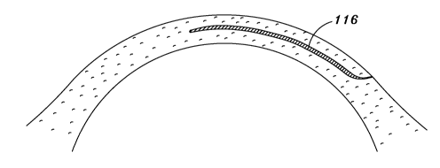
the surgeon using hand-held instruments. An exemplary corneal pocket 116
formed by an incision is depicted in cross-section in Figure 4.
[0032] Step 406 may involve inserting the lens in the cornea (see, for
example, Figure 2). In alternative embodiments, step 406 may involve inserting
the lens within a corneal pocket. Step 406 may further involve temporarily
deforming the lens preparatory to introducing the lens into the eye. The lens
may be deformed by rolling, folding, and the like. The lens of the invention
may
have prescribed memory characteristics that allow the lens to return to its
original size and configuration after insertion in the eye, while retaining
the
optical characteristics of the optical portion. Corrugations of the haptic
portion
of the lens may facilitate maneuvering the lens during step 406 by providing
rigidity to the lens and by allowing the surgeon to grasp the haptic portion
by the
corrugations. As described above, the lens may be made of a hydrophilic
material which swells when hydrated. The lens may be inserted fully hydrated
to elastically fit into a corneal pocket, or while at least partly dehydrated
such
that subsequent hydration helps secure the fit in the pocket.
[0033] Various configurations of corneal pockets may be employed in the
present invention, such as those pockets 116 depicted in Figures 5A to 51
which
are top views of the pockets 116 formed in the cornea 110. The various
configurations are adapted to be used with lenses of various shapes and sizes.
The corneal pockets 116 also are configured to facilitate the insertion of the
lens
and to minimize the size of the incision for improved post-surgical healing of
the
cornea.
[0034] The corneal pockets shown in Figures 5A to 5G and 51 include an
arced portion 118 near a center of the pocket, in addition to a straight
portion
-9-
CA 02643106 2008-08-20
WO 2008/023274 PCT/IB2007/003610
120 adjacent to the arced portion 118. In Figure 5H, the corneal pocket
includes a straight portion 122 extending across the entirety of the cornea.
[0035] In conjunction with the corneal pocket, a release area 124 (depicted in
Figures 5D-5H as arced lines) may be optionally provided in the cornea 110 so
as to provide a means of expansion and contraction when inserting and
positioning the lens in the corneal pocket. The arcuate incision in
combination
with the pocket may also reduce the induced astigmatism Release areas 124
may comprise incisions made into the cornea 110. These arcuate incisions are
made by the laser at the same time as the pocket being created, so that the
patient may not need to come back to the surgeon to enhance its vision due to
astigmatism.
[0036] As can be appreciated by those skilled in the art, the present
invention
may provide a method for correcting the vision of a patient with a refractive
intracorneal lens that may be easily inserted into a corneal pocket. The
intracorneal lens may be effectively positioned and held in place by the use
of
corrugated haptic region. A variety of corneal pocket configurations may be
used accommodate various corneal lens shapes and sizes.
[0037] It should be understood, of course, that the foregoing relates to
exemplary embodiments of the invention and that modifications may be made
without departing from the spirit and scope of the invention as set forth in
the
following claims.
-10-