Note: Descriptions are shown in the official language in which they were submitted.
CA 02643163 2008-08-20
WO 2007/100651 PCT/US2007/004712
TEMPERATURE-ADJUSTED ANALYTE DETERMINATION
FOR BIOSENSOR SYSTEMS
BACKGROUND
[0011 Biosensor systems usually provide an analysis of one or more
analytes in
biological fluids. The analysis typically includes a quantitative
determination of the analyte
in the biological fluid. The analysis is useful in the diagnosis and treatment
of physiological
abnormalities. For example, the determination of the glucose level in blood is
important to
diabetic individuals who frequently check their blood glucose level to
regulate diet and/or
medication. For other individuals, the monitoring of uric acid, lactate,
cholesterol, bilirubin,
and the like may be important.
[002] Biosensor systems may be implemented using bench-top, portable, and
other
measuring devices. The portable devices may be hand-held and usually include a
measuring
device and a sensor strip. Typically, a sample of a biological fluid is
introduced to the sensor
strip, which is disposed in the measuring device for analysis. Biosensor
systems may be
designed to analyze one or more analytes and may use different volumes of
biological fluids.
Some biosensor systems may analyze a single drop of whole blood (W13), such as
from 1-15
microliters (aL) in volume.
[003] Biosensor systems usually measure an output signal to 'determine the
analyte
concentration in a sample of the biological fluid. The output signal is
generated from an
oxidation/reduction or redox reaction of the analyte. An enzyme or similar
species may be
added to the sample to enhance the redox reaction. The output signal may be an
electric
signal, light, or light converted to an electric signal. A biosensor system
may generate the
output signal using an optical sensor system or an electrochemical sensor
system.
[004] In optical systems, the analyte concentration is determined by
measuring light
that has interacted with a light-identifiable species, such as the analyte or
a reaction or
product formed from a chemical indicator reacting with the analyte redox
reaction. An
incident excitation beam from a light source is directed toward the sample.
The light-
identifiable species absorbs or shifts the wavelength of a portion of the
incident beam, thus
altering the wavelength or reducing the intensity of the incident beam. A
detector collects
and measures the attenuated or wavelength-altered incident beam, which is the
output signal.
In other optical systems, the chemical indicator fluoresces or emits light in
response to the
CA 02643163 2008-08-20
WO 2007/100651 PCT/US2007/004712
2
=
analyte redox reaction when illuminated by the excitation beam. A detector
collects and
measures the light, which is the output signal.
[005] In electrochemical systems, the analyte concentration is determined
by
measuring an electrical signal, such as a current or potential. Typically, the
analyte
undergoes the redox reaction when an excitation signal is applied to the
sample. The
excitation signal usually is an electrical signal, such as a current or
potential. The redox
reaction generates an output signal in response to the excitation signal. The
output signal
usually is an electrical signal, such as a current or potential, which may be
measured and
correlated with the concentration of the analyte.
[006] In electrochemical systems, the measuring device usually has
electrical
contacts that connect with electrical conductors in the sensor strip. The
electrical connectors
are connected by the conductors to electrodes that extend into the sample of
the biological
fluid. The measuring device applies the excitation signal through the
electrical contacts to
the electrical conductors, which convey the excitation signal into the sample
through the
electrodes.' The redox reaction of the analyte generates an output signal in
response to the
=.= ,
= = excitation signal. The measuring device determines the analyte doneenti-
ation in 'response to
V the 'output signal. Examples of portable measuring devices- include the
Ascensia Breeze
and Elite meters of Bayer Corporation; the Precision biosensors available
from Abbott in
Abbott Park, Illinois; Accucheck biosensors available from Roche in
Indianapolis, Indiana;
and OneTouch Ultra biosensors available from Lifescan in Milpitas,
California. Examples
of bench-top measuring devices include the BAS 100B Analyzer available from
BAS
Instruments in West Lafayette, Indiana; the CH Instruments' Electrochemical
Workstation
available from CH Instruments in Austin, Texas; the Cypress Electrochemical
Workstation
available from Cypress Systems in Lawrence, Kansas; and the EG&G
Electrochemical
Instrument available from Princeton Research Instruments in Princeton, New
Jersey.
[007] Sensor strips may include reagents that react with the analyte
in the sample of
biological fluid. The reagents include an ionizing agent for facilitating the
redox of the
analyte, as well as any mediators or other substances that assist in
transferring electrons
between the analyte and the conductor. The ionizing agent may be an analyte
specific
enzyme, such as glucose oxidase or glucose dehydrogenase, to catalyze the
oxidation of
glucose in a WE sample. The reagents may include a binder that holds the
enzyme and
mediator together. In optical systems, the reagents include the chemical
indicator along with
CA 02643163 2008-08-20
WO 2007/100651 PCT/US2007/004712
3
another enzyme or like species to enhance the reaction of the chemical
indicator with the
analyte or products of the analyte redox reaction.
[008] Most biosensor systems use correlation or calibration equations to
determine
the analyte concentration in a sample of a biological fluid. Correlation
equations represent
the relationship between output signals and analyte concentrations. From each
correlation
equation, an analyte concentration may be calculated for a particular output
signal. The
correlation equations are dependent on the temperature of the sample. The
output signal for a
particular analyte concentration may change due to the effect of temperature
on the redox
reaction Of the analyte, enzyme kinetics, diffusion, and the like. A
correlation equation may
be needed for each possible sample temperature in order to calculate the
analyte
concentration from an output signal at a particular sample temperature.
[009] To reduce the number of correlation equations used in the sample
analysis,
many biosensor systems attempt to provide analyte concentrations using one or
more
correlation equations for a particular reference temperature. The analyte
concentration at a
. ..sample :temperature usually is compensated for the difference between the
. sample
sl = ...e..,temperatiire :.and the reference temperature to provide an.
analyte concentration! at. ;the . .
: :reference teiiiperature. : =
[0010] Some biosensor systems compensate for temperature by changing
the output
signal prior to calculating the analyte concentration from a correlation
equation. The output
signal usually is multiplied by a temperature correction coefficient or the
like. The
temperature-corrected output signal is used to determine the analyte
concentration. Biosensor
systems using a temperature-corrected output signal are described in U.S. Pat.
Nos. 4,750,496
and 6,576,117.
[0011] Other biosensor systems compensate for temperature by
changing the analyte
concentration calculated by the correlation equation. The analyte
concentration calculated
from the correlation equation usually undergoes a temperature correction
procedure to
provide a temperature-corrected analyte concentration. Biosensor systems
using a
temperature-corrected analyte concentration are described in U.S. Pat. Nos.
5,366,609;
5,508,171; and 6,391,645.
[0012] Additional biosensor systems compensate for temperature by
changing the
output signal prior to calculating the analyte concentration from a
correlation equation and/or
by changing the analyte concentration calculated by the correlation equation.
Biosensor
=
CA 02643163 2008-08-20
WO 2007/100651 PCT/US2007/004712
4
systems using a temperature-corrected output signal and/or a temperature-
corrected analyte
concentration are described in U.S. Pat. Nos. 4,431,004 and 5,395,504.
[0013] While these temperature compensation methods balance various
advantages
and disadvantages, none are ideal. These methods may not fully incorporate
various effects
of different sample temperatures on the redox reaction of the analyte, the
enzyme and
mediator kinetics, and diffusion. These methods may not adequately address
effects of
different analyte concentrations on enzyme kinetics and diffusion at different
sample
temperatures. These methods also may not adequately address effects of
different analyte
concentrations on the redox reaction at different sample temperatures. In
addition, the
changes to the output signal and/or the calculated analyte concentration may
introduce or
magnify errors related to the determination of the analyte concentration from
the output
signal.
[0014] Accordingly, there is an ongoing need for improved biosensor
systems,
= especially those that may provide increasingly accurate and precise
analyte concentrations at
a reference. temperature. The systems, devices, and methods of the present
invention
overcome atleast one of the disadvantages associated with conventional
biosensor systems. = . -
= = =
SUMMARY
[0015] The present invention provides a biosensor system that determines
the analyte
concentration in a sample of a biological fluid from an output signal
generated by a redox
reaction of the analyte. The biosensor system adjusts a correlation between
analyte
concentrations and output signals at a reference temperature to determine
analyte
concentrations from output signals at other temperatures. The biosensor system
uses the
temperature-adjusted correlation to determine the analyte concentration from
an output signal
at a sample temperature.
[0016] In a method for determining an analyte concentration in a sample
of a
biological fluid, the sample temperature is determined. An output signal is
generated in
response to a redox reaction of an analyte in the sample. A correlation
between analyte
concentrations and output signals at a reference temperature is adjusted in
response to
temperature. The analyte concentration is determined from the temperature-
adjusted
correlation and the output signal at the sample temperature.
[0017] In a method for adjusting a correlation between analyte
concentrations and
output signals at a reference temperature in response to temperature, the
correlations between
CA 02643163 2008-08-20
WO 2007/100651 PCT/US2007/004712
analyte concentrations and output signals are determined for a reference
temperature and at
least one other temperature. The normalized temperature functions of slope and
intercept are
developed for the correlation of the reference temperature. The correlation of
the reference
temperature is adjusted in response to the normalized temperature functions of
slope and
intercept.
[0018] A biosensor for determining an analyte concentration in a
biological fluid
includes a measuring device and sensor strip. The measuring device has a
processor
connected to a sensor interface and a temperature sensor. The sensor strip has
a sample
interface on a base. The sample interface is adjacent to a reservoir formed by
the base. The
processor adjusts a correlation between analyte concentrations and output
signals at a
reference temperature in response to a sample temperature from the temperature
sensor. The
processor determines an analyte concentration from the temperature-adjusted
correlation in
response to an output signal from the sample interface.
[0019] The following definitions are included to provide a clearer and
more
consistent understanding of the specification and claims..
[0020], .:.:"Analyte" -is defined as one or more substances present in a
sample. An .
analysis determines the presence and/or concentration of the analyte present
in the sample.
[0021] "Sample" is defined as a composition that may contain an unknown
amount of
the analyte. Typically, a sample for electrochemical analysis is in liquid
form, and preferably
the sample is an aqueous mixture. A sample may be a biological sample, such as
blood,
urine, or saliva. A sample also may be a derivative of a biological sample,
such as an extract,
a dilution, a filtrate, or a reconstituted precipitate.
[0022] "Conductor" is defined as an electrically conductive substance that
remains
stationary during an electrochemical analysis.
[0023] "Accuracy" is defined as how close the amount of analyte measured
by a
sensor system corresponds to the true amount of analyte in the sample.
Accuracy may be
expressed in terms of the bias of the sensor system's analyte reading in
comparison to a
reference analyte reading. Larger bias values reflect less accuracy.
[0024] "Precision" is defined as how close multiple analyte measurements
are for the
same sample. Precision may be expressed in terms of the spread or variance
among multiple
measurements.
[0025] "Redox reaction" is defined as a chemical reaction between two
species
involving the transfer of at least one electron from a first species to a
second species. Thus, a
CA 02643163 2008-08-20
WO 2007/100651 PCT/US2007/004712
6
redox reaction includes an oxidation and a reduction. The oxidation half-cell
of the reaction
involves the loss of at least one electron by the first species, while the
reduction half-cell
involves the addition of at least one electron to the second species. The
ionic charge of a
species that is oxidized is made more positive by an amount equal to the
number of electrons
removed. Likewise, the ionic charge of a species that is reduced is made less
positive by an
amount equal to the number of electrons gained.
[0026] "Mediator" is defined as a substance that may be oxidized or
reduced and that
may transfer one or more electrons. A mediator is a reagent in an
electrochemical analysis
and is not the analyte of interest, but provides for the indirect measurement
of the analyte. In
a simplistic system, the mediator undergoes a redox reaction in response to
the oxidation or
reduction of the analyte. The oxidized or reduced mediator then undergoes the
opposite
reaction at the working electrode of the sensor strip and is regenerated to
its original
oxidation number.
[0027] "Binder" is defined as a material that provides physical support
and
containment to the reagents while having chemical compatibility with the
reagents.
[0028] !fSteady-state", iS defined as when the change of a signal with
respect to its -
independent input variable (time, etc.).is substantially constant, such as
within 10 or 5%.
[0029] "Transient point" is defined as the value of a signal obtained as a
function of
time when an increasing rate of diffusion transitions into a relatively
constant rate of
diffusion. Before the transient point, the signal is rapidly changing with
time. Similarly,
after the transient point, the rate of signal decay becomes relatively
constant, thus reflecting
the relatively constant rate of diffusion.
[0030] "Handheld device" is defined as a device that may be held in a
human hand
and is portable. An example of a handheld device is the measuring device
accompanying
Ascensia0 Elite Blood Glucose Monitoring System, available from Bayer
HealthCare, LLC,
Elkhart, IN.
BRIEF DESCRIPTION OF THE DRAWINGS
[0031] The invention may be better understood with reference to the
following
drawings and description. The components in the figures are not necessarily to
scale,
emphasis instead being placed upon illustrating the principles of the
invention. Moreover, in
the figures, like referenced numerals designate corresponding parts throughout
the different
views.
CA 02643163 2008-08-20
WO 2007/100651 PCT/US2007/004712
7
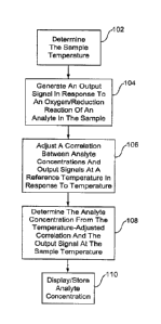
[0032] FIG. 1 represents a method for determining an analyte concentration
in a
sample of a biological fluid.
[0033] FIG. 2 represents a method for adjusting a correlation between
analyte
concentrations and output signals at a reference temperature in response to a
sample
temperature.
[0034] FIG. 3 is a graph illustrating correlations between analyte
concentrations and
output signals.
[0035] FIG. 4 is a graph illustrating normalized slopes as a function of
temperature
for correlations between glucose concentrations in whole blood and current for
an assay time
of 7 seconds.
[0036] FIG. 5 is a graph illustrating normalized intercepts as a function
of
temperature for correlations between glucose concentrations in whole blood and
current for
an assay time of 7 seconds.
[0037] FIG. 6 is a graph illustrating the normalized slopes as a function
of
temperature for correlations between glucose concentrations in whole blood and
current for
several assay times. =
[0038] FIG. 7 is a graph illustrating. .the nonnalized intercepts as a
function of
temperature for correlations between glucose concentrations in whole blood and
current for -
several assay times.
[0039] FIG. 8 is a graph illustrating the bias from a reference
temperature of
calculated glucose concentrations without any adjustment for temperature.
[0040] FIG. 9 is a graph illustrating the bias from a reference
temperature of
calculated glucose concentrations with adjustment for temperature.
[0041] FIG. 10 is a graph illustrating the temperature function of current
from a
glucose sensor with normalized slope and intercept.
[0042] FIG. 11 is a graph illustrating the temperature coefficient
function for the
normalized current of FIG. 10 in relation to temperature.
[0043] FIG. 12 depicts a schematic representation of a biosensor that
determines an
anal yte concentration in a sample of a biological fluid.
CA 02643163 2008-08-20
WO 2007/100651 PCT/US2007/004712
8
DETAILED DESCRIPTION
[0044] A
biosensor system that determines an analyte in a sample of a biological fluid
is described. The biosensor system determines the analyte concentration from
an output
signal generated by an oxidation/reduction or redox reaction of the analyte.
The system
adjusts a correlation equation for determining analyte concentrations from
output signals at
one temperature to determining analyte concentrations from output signals at
other
temperatures, such as the sample temperature. The temperature-adjusted
correlations
improve the accuracy and precision of the biosensor system in determining the
analyte
concentration of the sample. The biosensor system may determine analyte
concentrations
from output signals at a sample temperature using a temperature-adjusted
correlation
equation for a reference temperature. The
correlation equations between analyte
concentrations and output signals may be represented graphically,
mathematically, a
combination thereof, or the like. The correlation equations may be represented
by a program
number (PNA) table, another look-up table, or the like. The biosensor system
may be utilized
to determine analyte concentrations. such as glucose, uric acid, lactate,
cholesterol, bilirubin,
and the like.
[0045] FIG.
1 represents a method for determining an analyte concentration in a
sample of a biological fluid. In 102, the sample temperature is determined. In
104, an output
signal is generated in response to an oxidation/reduction reaction of the
analyte in the sample.
In 106, a correlation between analyte concentrations and output signals at a
reference
temperature is adjusted in response to temperature. In 108, the analyte
concentration is
determined from the temperature-adjusted correlation and the output signal at
the sample
temperature. In 110, the analyte concentration is displayed and may be stored
for future
reference.
[0046] In
102 of FIG. 1, the sample temperature may be determined using various
techniques. The sample temperature may be measured using a thermister,
thermometer, or
other temperature sensing device. The sample temperature may be calculated
from the output
signal of an electrochemical reaction in the sample. The sample temperature
may be assumed
to be the same or similar to a measurement of the ambient temperature or the
temperature of a
device implementing the biosensor system. Other techniques may be used to
determine the
sample temperature.
CA 02643163 2008-08-20
WO 2007/100651 PCT/US2007/004712
9
[0047] in 104 of FIG. 1, an output signal is generated in response to an
oxidation/reduction or redox reaction of an analyte in the sample. The output
signal may be
generated using an optical sensor system, an electrochemical sensor system, or
the like.
[0048] Optical sensor systems generally measure the amount of light
absorbed or
generated by the reaction of a chemical indicator with the analyte redox
reaction. An enzyme
may be included with the chemical indicator to enhance the reaction kinetics.
The output
signal or light from an optical system may be converted into an electrical
signal such as
current or potential.
[0049] In light-absorption optical systems, the chemical indicator
produces a reaction
product that absorbs light_ A chemical indicator such as tetrazolium along
with an enzyme
such as diaphorase may be used. Tetrazolium usually forms forrnazan (a
chromagen) in
response to the redox reaction of the analyte. An incident excitation beam
from a light source
is directed toward the sample. The light source may be a laser, a light
emitting diode, or the
like. The incident beam may have a wavelength selected for absorption by the
reaction
product. As the incident beam passes through the. sample, ..the reaction
product absorbs a
portion of the incident beam, thus attenuating or reducing the intensity of
the incident beam.
The incident beam may be reflected back from or = transmitted,:through the
sample to a
detector. The detector collects and measures the attenuated incident beam
(output signal).
The amount of light attenuated by the reaction product is an indication of the
analyte
concentration in the sample.
[0050] In light-generated optical systems, the chemical detector
fluoresces or emits
light in response to the analyte redox reaction. A detector collects and
measures the
generated light (output signal). The amount of light produced by the chemical
indicator is an
indication of the analyte concentration in the sample.
[0051] Electrochemical systems apply an input signal to the sample of the
biological
fluid. The input signal may be a potential or current and may be constant,
variable, or a
combination thereof such as when an AC signal is applied with a DC signal
offset. The input
signal may be applied as a single pulse or in multiple pulses, sequences, or
cycles. The
analyte undergoes a redox reaction when the input signal is applied to the
sample. An
enzyme or similar species may be used to enhance the redox reaction of the
analyte. A
mediator may be used to maintain the oxidation state of the enzyme. The redox
reaction
generates the output signal that may be measured constantly or periodically
during transient
and/or steady-state output. Various electrochemical processes may =be used
such as
CA 02643163 2014-02-26
- 10 -
amperometry, coulometry, voltammetry, or the like. Gated amperometry and gated
voltammetry also may be used.
[0052] In amperometry, a potential or voltage is applied to a sample of
the biological
fluid. The redox reaction of the analyte generates a current in response to
the potential. The
current is measured over time to quantify the analyte in the sample.
Amperometry generally
measures the rate at which the analyte is oxidized or reduced to determine the
analyte
concentration in the sample. Biosensor systems using amperometry are described
in U.S. Pat.
Nos. 5,620,579; 5,653,863; 6,153,069; and 6,413,411.
[0053] In coulometry, a potential is applied to a sample of the biological
fluid to
exhaustively oxidize or reduce the analyte within the sample. The potential
generates a
current that is integrated over the time of oxidation/reduction to produce an
electrical charge
representing the analyte concentration. Coulometry generally captures the
total amount of
analyte within the sample. A biosensor system using coulometry for whole blood
glucose
measurement is described in U.S. Pat. No. 6,120,676.
[0054] In voltammetry, a varying potential is applied to a sample of
biological fluid.
The redox reaction of the analyte generates current in response to the applied
potential. The
current is measured over time to quantify the analyte in the sample.
Voltammetry generally
measures the rate at which the analyte is oxidized or reduced to determine the
analyte
concentration in the sample. Additional information about voltammetry may be
found in
"Electrochemical Methods: Fundamentals and Applications" by A.J. Bard and L.R.
Faulkner,
1980.
[0055] In gated amperometry and gated voltammetry, pulsed excitations are
used as
described in U.S. Pat. App. Pub. Nos. 2008/0173 552A1 and 2008/0179 197A1.
[0056] In 106 of FIG. 1, a correlation between analyte concentrations and
output
signals at a reference temperature is adjusted in response to temperature. The
correlation
may be represented by a correlation or calibration equation that calculates
analyte
concentrations from output signals at the reference temperature. The
correlation equation for
the reference temperature is adjusted to calculate analyte concentrations in
response to output
signals at other temperatures such as the sample temperature. The correlation
equation may
be for a reference temperature of 25 C. Correlation equations for other
reference
temperatures may be used.
CA 02643163 2008-08-20
WO 2007/100651 PCT/US2007/004712
11
[0057] The correlation equation may be implemented to manipulate the
output signal
for determination of the analyte concentration. The correlation equation also
may be
implemented as a program number assignment (PNA) table of the slope and
intercept for the
correlation equation, another look-up table, or the like for comparison with
the electrical
output signal to determine the analyte concentration.
[0058] The effect of temperature on the correlation or calibration
equations is
responsive to the behavior of diffusion and enzymatic reactions during the
redox reaction.
For example, temperature affects the oxidation and diffusion of glucose in a
sample of whole
blood. In addition, temperature affects the diffusion of optically active
molecules.
[0059] The correlation equations may be linear or near linear, and may be
described
by a second order polynomial. In a general form, the correlation equation can
be represented
as follows:
[0060] OS = d ,,* A" + d õ *A + ...+ d,* +d * A +
do (1).
[0061] Where A is the analyte concentration, OS is the output signal, and
coefficients
t1,1, d2, d1, and do describe a temperature dependent weighing factor for each
term of the
= "biosensor response.
(0062] * = The correlation equation may be described by ihe reverse
expression, Where
the analyte concentration is expressed as a function of the output signal.
This reduces the
need to solve an nth order equation in order to find the analyte
concentration. Thus, the
correlation equation for analyte concentration may be represented as follows:
[0063] A= cõ * OS" + c,,1 * OS"-I + ...+ c, *OS2 + c, * OS + c, (2).
[0064] Where en, 0n.1, e2, ei, and co are coefficients that describe a
temperature
dependent weighing factor for each term of the biosensor response. The analyte
concentration, A, may be glucose in a sample of whole blood. The output signal
may be the
current or potential of an electrochemical system, the absorbance or %-
transmission of an
optical system, or the like.
[0065] The correlation equation may be represented by a 2"d order
response between
analyte concentration and output signals as follows:
[0066] A = c2* OS 2 + c1 * OS + c, (3).
[0067] The correlation equation may be represented by a linear response
between
analyte concentration and output signals as follows:
[0068] AR= CI* OST + Co= OSTIST Inty. I S7- (4).
CA 02643163 2008-08-20
WO 2007/100651 PCT/US2007/004712
12
[0069] Where c1 = 1/ST, co Int-r/ST, and where AR is the analyte
concentration at a
reference temperature, OST is the output signal, ST is the product of a slope
at the reference
temperature and a normalized temperature function of the slope, and Int-r is
the product of an
intercept at the reference temperature and a normalized temperature function
of the intercept.
[0070] Equation (4) may be rewritten to express the output signal in
response to the
analyte concentration as follows:
[0071] OS, _ST *AR + Int, (5)-
[0072] Where OST is the output signal at another temperature such as
the sample
temperature. AR is the analyte concentration at the reference temperature, ST
can be expressed
as a product of a constant and a normalized temperature function of the slope,
and Int-r can be
expressed as a product of a constant and a normalized temperature function of
the intercept.
[0073] Equation (5) indicates that the output signal, OST, is a
function of temperature
in terms of the temperature effect on slope, ST, and intercept, IntT, under
the analyte
concentration, AR. The slope, ST, and intercept, Int-r, adjust the slope and
intercept of a
. .corr. elation.equation at a reference temperature using normalized
temperature functions of the ,
= slope and intercept. The' temperature-adjusted slope and intercept of the
cOrrelation for the
reference leinperature may be used with an output signal at another
temperature, such as the -
sample temperature, to calculate an analyte concentration.
[0074] Accordingly, the correlation equation (5) may be rewritten to
calculate analyte
concentrations using the temperature-adjusted slope and intercept of the
correlation for the
reference temperature and output signals at another temperature, as follows:
OS hitT
[0075] A T ¨
ST (6).
[0076] Where AR is the analyte concentration at the reference
temperature, OST is the
output signal at the other temperature, Intl- is the intercept of the
correlation for the reference
temperature adjusted by a normalized temperature function for the intercept in
response to the
other temperature, and ST is the slope of the correlation for the reference
temperature adjusted
by a normalized temperature function for the slope in response to the other
temperature.
[0077] The slope of the correlation for the reference temperature is
adjusted in
response to the sample temperature, as follows:
[0078] ST = SR * f (T) (7).
CA 02643163 2008-08-20
WO 2007/100651 PCT/US2007/004712
13
[0079] Where SR is the slope of the correlation for the reference
temperature and f(T)
is a temperature function that adjusts the slope for the sample temperature.
[0080] The temperature function of slope, f(T), adjusts the slope of
the correlation for
the reference temperature to the slope of a correlation for another
temperature. The
temperature-adjusted slope may be used to calculate the analyte or glucose
concentration
using an output signal or current generated at the other temperature.
To develop the
temperature function of slope, f(T), the slopes of correlations for other
temperatures are
normalized to the slope of the correlation for the reference temperature. The
normalized
slope of a correlation for a particular temperature is a unitless coefficient
that adjusts the
slope of the correlation for the reference temperature to the slope of the
correlation for the
particular temperature. The normalized slope of the correlation for the
reference temperature
is essentially one, indicating there is little or no adjustment to the slope
of the correlation for
the reference temperature. The normalized slopes are analyzed graphically
and/or
mathematically such as with a regression analysis to develop the temperature
function of
slope, f(T). Another normalization method may be used to develop the
temperature function.
[0081] . The, ;.temperature function of slope, f(T), may be a second
order polynomial
= such as follows: =
[0082] f(T) = a2T2 + a,T + a, (8).
[0083] Where T is the sample temperature and a2, al, and ao are
coefficients of a
regression analysis representing the normalized slopes. While represented as a
polynomial,
the temperature function of slope, f(T), may be represented as a constant, an
exponential,
trigonometric, or other function, a combination thereof, and the like.
[0084] The intercept of the correlation for the reference temperature
is adjusted in
response to the sample temperature, as follows:
[0085] Intr Int *g(T) (9)-
[0086] Where IntR is the intercept of the correlation for the
reference temperature and
g(T) is a temperature function that adjusts the intercept for the sample
temperature.
[0087] The temperature function of intercept, g(T), adjusts the
intercept of the
correlation for the reference temperature to the intercept of a correlation
for another
temperature. The temperature-adjusted intercept may be used to calculate the
analyte or
glucose concentration using an output signal or current generated at the other
temperature.
To develop the temperature function of intercept, g(T), the intercepts of
correlations ,for
CA 02643163 2008-08-20
WO 2007/100651
PCT/US2007/004712
14
different temperatures are normalized to the intercept of the correlation for
the reference
temperature. The normalized intercept of a correlation for a particular
temperature is a
unitless coefficient that adjusts the intercept of the correlation for the
reference temperature
to the intercept of the correlation for the particular temperature. The
normalized intercept of
the correlation for the reference temperature is essentially one, indicating
there is little or no
adjustment to the intercept of the correlation for the reference temperature.
The normalized
intercepts are analyzed graphically and/or mathematically such as with a
regression analysis
to develop the temperature function of intercept, g(T). Another normalization
method may
be used to develop the temperature function.
[0088] The temperature function of intercept, g(T), may be a second order
polynomial
such as follows:
[0089] g(T)= b2T2 +biT +bo (10).
[0090] Where T is the sample temperature and b2, hi, and bo are
coefficients of a
regression analysis representing the normalized intercepts. While
represented as a
polynomial, the temperature function of intercept, g(T), may be represented as
a constant, an
=
exponential, trigonometric, orother function, a combination thereof, and the
like.
[0091] In 108 of FIG. 1, the analyte concentration of the sample is
determined from
the temperature-adjusted correlation equation (6) and the output signal at the
sample
temperature. The temperature functions of slope and intercept, f(T) and g(T),
are calculated
using equations (8) and (10), respectively. ST and IntT, the slope and
intercept of the
correlation for the reference temperature adjusted in response to the sample
temperature, are
calculated using equations (7) and (9), respectively_
[0092] In 110 of FIG. 1, the analyte concentration calculated using
temperature-
adjusted correlation equation (6) and the output signal at the sample
temperature may be
displayed or stored for future reference.
[0093] The effect of changes in the slope and intercept on analyte
concentration in
relation to temperature changes may be analyzed. Temperature coefficients
define the change
in a parameter in relation to the change in temperature. For parameters such
as analyte
concentration, slope, and intercept, temperature coefficients may be defined
as follows:
[0094] aA z = aAl A AAI A
(11).
ST AT
OS/S AS/S
[0095) ex, = (12).
ST AT
CA 02643163 2008-08-20
WO 2007/100651 PCT/US2007/004712
obit/int Milt I Int
[0096] a = (13).
im
aT AT
[0097] Where aA, as, and aka are the temperature coefficients of the
analyte
concentration, slope, and intercept respectively, A is the analyte
concentration, S is the slope,
Int is the intercept, and T is temperature.
[0098] For a constant input signal such as current, the relative change
in the analyte -
concentration, A, in relation to changes in the slope, S. and intercept, Int,
may be given as
follows using the analyte calculation equation (6) as follows:
dAaAdS + aA
[0099] dint (14).
DS alnt
[00100] dA raA ds+ aA dintl/A
(15).
A [US aInt
[00101] ¨ =
aA OS ¨ Int( 11S)= ¨ ¨A
(16).
as - s
[00102] aA= 1/S (17).
aitzt = = = ' = = ' =
[00103] Where ds is an output signal such as current.
[00104] Substituting equations (16) and ( 17) into. equation (15), gives
the following
relationships for the relative change in an analyte concentration such as
glucose:
dA dS dint
[00105] = (18).
A s (s*
AA = AS Alnt AS IntIS1* AInti
[00106] ¨ (19).
A s (s * A)=
S LAil Int j
[00107] Substituting the temperature coefficients from equations (11),
(12), and (13)
and translating equation (19) provides the following relationships:
&4/A = AS1S rintIS1 AIntl Intl
[00108] (20).
AT AT L Ail AT
AA/ A [00109] = aA [Int/S1 as * aim
(21).
AT A
[00110] Equation (21) indicates that the effect of the temperature
coefficient of slope is
equivalent to the analyte concentration, but is opposite in magnitude.
However, the effect of
the temperature coefficient of intercept may be smaller in magnitude,
depending on the slope,
intercept, and analyte concentration being measured.
CA 02643163 2008-08-20
WO 2007/100651 PCT/US2007/004712
16
[00111] For an analyte such as glucose in whole blood, the effect of
changes in the
intercept temperature coefficient on the glucose temperature coefficient is
small at higher
glucose concentrations. If the ratio of intercept to slope, Int/S, is 50 and
the glucose
concentration is 150 mg/dL, only one-third of the intercept temperature
coefficient has an
effect on the glucose temperature coefficient (the effect of temperature on
the temperature
coefficient of the glucose concentration includes only one-third of the effect
of temperature
on the temperature coefficient of the intercept). At lower glucose
concentrations, the effect
of the intercept temperature coefficient on the glucose temperature
coefficient is more visible.
If the ratio of intercept to slope, Int/S, is 50 and the glucose concentration
is 50 mg/c1L, all of
the intercept temperature coefficient has an effect on the glucose temperature
coefficient (the
effect of temperature on the temperature coefficient of the glucose
concentration includes all
of the effect of temperature on the temperature coefficient of the intercept).
A smaller Int/S
ratio reduces the effect of intercept temperature coefficient on the glucose
temperature
coefficient.
[00112] FIG. 2 represents a method for adjusting a correlation between
analyte
concentrations and output signals at.a reference temperature in response to
temperature. In
202, the correlations between analyte concentrations and output signals are
determined for a
reference temperature and at least one other temperature. In 204, normalized
temperature
functions are developed of slope and intercept for the correlation of the
reference
temperature. In 206, the correlation of the reference temperature is adjusted
in response to
the normalized temperature functions of slope and intercept. This method may
be used with
the method described in relation to FIG. 1, a similar method, or otherwise.
[00113] In 202 of FIG. 2, correlations between analyte concentrations and
output
signals are determined for a reference temperature and at least one other
temperature. The
output signals may be generated by an electrochemical reaction of the analyte
in the sample
as previously discussed. For each temperature, output signals are generated
experimentally
by electrochemical reactions at different analyte concentrations. The
experimental results are
analyzed to develop a correlation between the analyte concentrations and the
output signals
for each temperature.
[00114] FIG. 3 is a graph illustrating correlations between analyte
concentrations and
output signals. In this illustration, each output signal is the current
generated from an
electrochemical reaction, such as gated amperometry. The analyte
concentrations are glucose
concentrations in whole blood. Correlations between current and glucose
concentrations are
CA 02643163 2008-08-20
WO 2007/100651
PCT/US2007/004712
17
graphically shown for a reference temperature of 250 C and two other
temperatures -- 10 C
and 40 C. While the correlation at 25 C was selected as the reference
temperature,
correlations at other temperatures (including those not shown) may be selected
as the
reference temperature. While the illustration is directed toward particular
features, such as
the number of correlations, output signals, analyte concentrations,
temperatures, and the like,
the illustration is not meant to limit the scope, application, implementation,
or the like.
[00115] Each of the graphical correlations is linear and may be represented
by a
correlation equation having a general form as follows:
/ ¨
[00116] G Int (22).
[00117] Where G is the glucose concentration, I is the current, Int is the
intercept of
the correlation line with the y-axis, and S is the slope of the correlation
line. While linear
relationships are shown for the correlations between the glucose concentration
and the
current, other correlations may have other relationships, such as polynomial,
exponential,
trigonometric, a combination thereof, and the like.
[00118] In 204 of FIG. 2, normalized temperature. funetions are developed
of slope and
intercept for the correlation of the reference. temperature. The temperature
functions adjust
the slope and intercept of the correlation for the reference temperature to
the slope and
intercept of a correlation for another temperature. The temperature-adjusted
slope and
intercept may be used to calculate the analyte or glucose concentration using
an output signal
or current generated at the other temperature.
[00119] To develop the temperature functions, the slopes and intercepts are
normalized
to the slope and intercept of the correlation for the reference temperature.
The normalized
slope of a correlation for a particular temperature is a unitless coefficient
that adjusts the
slope of the correlation for the reference temperature to the slope of the
correlation for the
particular temperature. The normalized intercept of a correlation for a
particular temperature
is a unitless coefficient that adjusts the intercept of the correlation for
the reference
temperature to the intercept of the correlation for the particular
temperature. Both the
normalized slope and normalized intercept of the correlation for the reference
temperature are
essentially one, indicating there is little or no adjustment to the slope and
intercept of the
correlation for the reference temperature. Other normalization methods may be
used.
[00120] The normalized slopes of the correlations may be used to generate a
temperature function of the slope, f(T), graphically and/or mathematically
using a regression
CA 02643163 2008-08-20
WO 2007/100651 PCT/US2007/004712
18
analysis or the like. The temperature function of the slope, f(T), from a
regression analysis
may be a second order polynomial such as follows:
[00121] f (T)= a,T2 a1T a0 (23).
[00122] Where T is the sample temperature and a2, a1, and ao are
coefficients of a
regression analysis representing the normalized slopes. While represented as a
polynomial,
the regression analysis may represent the temperature function of the slope,
f(T), as another
function.
[00123] The normalized intercepts of the correlations may be used to
generate a
temperature function of the intercept, g(T), graphically and/or mathematically
using a
regression analysis or the like. The temperature function of the intercept,
g(T), from a
regression analysis may be a second order polynomial such as follows:
[00124] g(T)= b,T2 b1T b0 (24).
[00125] Where T is the sample temperature and b2, b1, and bo are
coefficients of a
regression analysis representing the normalized intercepts. While represented
as a
polynomial, the regression analysis may represent the temperature function of
the intercept,
,as another function.
= [00126] FIG. 3 illustrates that correlations between current
and glucose at 10 C, 25
C, and 40 C calculate the same glucose concentration, G25, from currents,
Ego, i25, and i10,
which are generated by electrochemical reactions of the analyte in the sample
at those
respective temperatures. The slopes and intercepts of the correlations may be
normalized to
the slope and intercept of the correlation for the reference temperature of 25
C. The
normalized slopes and intercepts of the correlations may be used to generate
the temperature
function of the slope, f(T), and the temperature function of the intercept,
g(T).
[00127] FIGS. 4 and 5 are graphs illustrating the normalized slopes and
intercepts,
respectively, as a function of temperature for correlations between glucose
concentrations in
whole blood and current. The correlations were generated from electrochemical
reactions
using gated amperometry with an assay time of 7 seconds (sec). The normalized
slopes and
intercepts are from correlations at 10 C, 200 C, 250 C, 300 C, and 400 C. The
normalized
slopes and intercepts were normalized to the slope and intercept of a
correlation at a reference
temperature of 25 C. While these illustrations are directed toward particular
features such
as normalized slopes, temperatures, and the like, the illustrations are not
meant to limit the
scope, application, implementation, or the like.
CA 02643163 2008-08-20
WO 2007/100651 PCT/US2007/004712
19
[00128] In FIG. 4, a regression analysis of the normalized slopes
generates a
temperature function of the slope, f(T), as follows:
[00129] f (T)= ¨0.00005765* T2 + 0.01453 *7' + 0.6703 (25).
[00130] The temperature function of the slope, f(T), shown in equation
(25) may be
used to adjust the slope of the correlation for the reference temperature of
250 C to the slope
of a correlation for another temperature, such as a sample temperature. T is
the other
temperature. The temperature-adjusted slope may be used to calculate the
glucose
concentration using a current generated at the other temperature.
Other temperature
functions of the slope may be used.
[00131] In FIG. 5, a regression analysis of the normalized intercepts
generates a
temperature function of the intercept, g(T), as follows:
[0.0132] g(T) = 0.0001023 *T2 + 0.01389* T +1.284 (26).
[00133] The temperature function of the intercept, g(T), shown in
equation (26) may
be used to adjust the intercept of the correlation for the reference
temperature of 25 C to the
. intercept of a correlation for another temperature, such as a sample
temperature. T is the
= =
other temperature. The temperature-adjusted intercept may be used to calculate
the glucose
.concentration using a current generated at the other temperature. Other
temperature functions
for the intercept may be used.
[00134] The separate temperature functions for slope and intercept may be
used with a
program number assignment (PNA) table of the slope and intercept of the
correlation for the
reference temperature. In addition, the normalized slope and intercept provide
a range in
which the intrinsic temperature properties of a biosensor system may be
independent of the
output signal or current magnitude generated by the electrochemical reaction.
The intrinsic
temperature properties usually depend on the sensor strip design and
manufacturing. A
biosensor system may change the temperature functions and/or correlation
equation(s) in
response to the sensor strip type and batch used. The temperature function and
correlation
equation changes may be made by changing PNA table when a different or new
sensor strip
is used.
[00135] FIGS. 6 and 7 are graphs illustrating the normalized slopes and
intercepts,
respectively, as a function of temperature for correlations between glucose
concentrations in
whole blood and current. The correlations were generated from electrochemical
reactions
using gated amperometry with assay times of 5.5 sec, 7 sec, 8.5 sec, 10 sec,
11.5 sec, 13 sec,
CA 02643163 2008-08-20
WO 2007/100651 PCT/US2007/004712
and 14.5 sec. The normalized slopes and intercepts are from correlations at 10
C, 20 C,
C, 300 C, and 40 C. The normalized slopes and intercepts were normalized to
the slope
and intercept of a correlation at a reference temperature of 25 C. While
these illustrations
are directed toward particular features, such as normalized slopes,
temperatures, and the like,
the illustrations are not meant to limit the scope, application,
implementation, or the like.
[00136] FIGS. 6 and 7 illustrate normalized slopes and intercepts for
electrochemical
reactions using gated amperometry with multiple assay times. In determining
temperature
functions for normalized slopes and intercepts in electrochemical methods
based on multiple
pulses, there are multiple calibration points in the individual pulses of a
pulse sequence. By
using currents generated at different temperatures and different times in
different pulses,
slopes and intercepts from the different temperatures can be normalized to the
slope and
intercept at 25 C. The normalized slopes and intercepts may be represented
graphically
and/or mathematically as a function of temperature. The mathematical
representation may be
by a regression analysis that generates a second order polynomial. In multiple
pulse methods,
there may be many calibration points in a time range such as from 5.5 sec. to
7, 8.5, and 10
sec. Within this range, the intrinsic temperature property of a biosensor
should be consistent -
=
if the-reagents are sufficiently hydrated.
= .
[00137] In FIG. 6, the temperature functions of the normalized slopes
essentially
overlap each other except for the 5.5 sec. assay time, which reflects the
intrinsic consistency
of the temperature sensitivity of the biosensor system. In addition, the
temperature functions
of the normalized slopes are quite symmetrical with respect to the reference
temperature of
25 C. The normalized slopes at 10 C are about 20% smaller than the normalized
slope at
25 C. The normalized slopes at 40 C are about 20% larger than the normalized
slope at
25 C.
[00138] In FIG. 7, the temperature functions for normalized intercepts are
very similar
for assay times between 5.5 sec. and 10 sec. At longer times, the temperature
effect on the
normalized intercept becomes larger.
[00139] In 206 of FIG. 2, the correlation of the reference temperature is
adjusted in
response to the normalized temperature functions of slope and intercept. The
correlation
between analyte concentrations and output signals for the reference
temperature is as follows:
G = [00140] ¨ Int (27).
SR
CA 02643163 2008-08-20
WO 2007/100651 PCT/US2007/004712
21
[00141] Where GR is the analyte concentration at the reference temperature,
iR is the
output signal at the reference temperature, IntR is the intercept of the
correlation for the
reference temperature, and SR is the slope of the correlation for the
reference temperature.
[00142] The correlation for the reference temperature represented by
equation (27)
may be adjusted in response to a sample temperature. Analyte concentrations at
the reference
temperature may be calculated using temperature-adjusted slopes and intercepts
of the
correlation for the reference temperature and output signals at a sample
temperature, as
follows:
GR =ir ¨ Int
[00143] (28).
S
[00144] Where GR is the analyte concentration at the reference temperature,
iT is the
output signal at the sample temperature, IntT is the intercept of the
correlation for the
reference temperature adjusted in response to the sample temperature, and ST
is the slope of
the correlation for the reference temperature adjusted for the sample
temperature.
[00145] The slope of the correlation for the reference temperature adjusted
in response
to The sample temperature, ST, may be calculated as follows:
[00146] ST = S R' * f (T) = = (29).
[00147] Where SR is the slope of the correlation for the reference
temperature and f(T)
is a temperature function that adjusts the slope for the sample temperature.
[00148] The intercept of the correlation for the reference temperature
adjusted in
response to the sample temperature, IntT , may be calculated as follows:
[00149] mt7. int, * g(T) (30).
[00150] Where IntR is the intercept of the correlation for the reference
temperature and
g(T) is a temperature function that adjusts the intercept for the sample
temperature.
[00151] The correlation for the reference temperature adjusted in response
to a sample
temperature as represented by equation (28) may be rewritten by substituting
equations (29)
and (30) for ST and Intl-, as follows:
[00152] G ¨ (Int * g(T))
(S R f (T )) (31).
[00153] Where GR is the analyte concentration at the reference
temperature, IT is the
output signal at the sample temperature, IntR is the intercept for the
correlation of the
reference temperature, g(T) is the normalized temperature function for
intercept, SR is the
CA 02643163 2008-08-20
WO 2007/100651 PCT/US2007/004712
22
slope for the correlation of the reference temperature, and f(T) is the
normalized temperature
function for slope.
[00154] The correlation for the reference temperature adjusted in
response to a sample
temperature as represented by equation (31) may be rewritten for use with the
examples
illustrated in FIGS. 3-5, as follows:
[00155] G25T - (Int25 * 0.00005765*T2 + 0.01453* T 0.6703))
(S25* (0.0001023* T2 + 0.01389* T +1.284 (32).
[00156] Where G25 is the analyte concentration at the reference
temperature of 25 C,
iT is the output signal at the sample temperature, Int25 is the intercept of
the correlation for the
reference temperature of 25 C, S25 is the slope of the correlation for the
reference
. temperature of 25 C, and T is the sample temperature.
[00157] FIGS. 8 and 9 are graphs illustrating the glucose bias values
from a reference
temperature as a function of temperature. FIG. 8 is a graph illustrating the
bias of calculated
glucose concentrations without any adjustment for temperature. FIG. 9 is a
graph illustrating
the bias of calculated glucose concentrations with adjustment for temperature
as described
previously. These graphs illustrate the .percent bias from a reference
temperature of 25 C =
for plasma glucose concentrations 0.E56.9 mg/dL, 114.0 mg/dL, and 432.9 mg/dL
in whole.
blood. The analysis was generated from electrochemical reactions using gated
amperometry
with an assay time of 7 sec at sample temperatures of 10 C, 20 C, 25 C, 30
C, and
40 C. While the illustrations are directed toward particular features such as
temperatures,
glucose concentrations, and the like, the illustrations are not meant to limit
the scope,
application, implementation, or the. like.
[00158] In FIGS. 8 and 9, the percent bias values at 10 C, 20 C, and 25
C for the
56.9 mg/dL glucose concentration show little if any change after the
temperature adjustment,
especially the percent bias value at 10 C. FIG. 8 indicates that the glucose
concentrations
from a correlation without temperature compensation generally have a negative
bias at
temperatures below the reference temperature of 25 C. FIG. 8 also indicates
that glucose
concentrations from a correlation without temperature adjustment generally
have a positive
bias at temperatures above the reference temperature of 25 C. FIG. 9
indicates that the
percent bias values converge to a narrower range of about +1- 5 percent when
correlations
with the temperature adjustment are used.
[00159] The temperature coefficient function of any particular parameter
may be used
to further show the internal consistency of the temperature function for
adjusting correlation
CA 02643163 2008-08-20
WO 2007/100651 PCT/US2007/004712
23
equations between analyte concentrations and output signals. The temperature
coefficient
(the intrinsic property) of the output signal, OS, may be defined as follows:
[00160] a aos/os a ln.(0S)
os (33).
or or
[00161] Where aos, is the temperature coefficient of the output signal, OS
is the output
signal, and T is temperature.
[00162] FIGS. 10 and 11 are graphs illustrating the effect on. the
temperature
coefficient function of the temperature-adjusted correlation equations between
analyte
concentrations and output signals. FIG. 10 illustrates the temperature
function of current
from a glucose sensor with normalized slope and intercept. FIG. 11 illustrates
the
temperature coefficient function for the normalized current of FIG. 10 in
relation to
temperature. The normalized current and temperature coefficients (TempCo) are
in response
to glucose concentrations of 50 mgAIL, 100 medL, 200 mg/dL, 400 mg/dL, and 600
rrig/dL.
In FIG. 10, the current at 25 C should be equal to the glucose value according
to equation (5)
for the normalized slope and intercept. FIG. 11 indicates that the temperature
coefficients
are functions of temperature -- the lower , the temperature, the higher the
temperature
, .
coefficient. Within .the temperature range of.. about 100 C through about 40
C, the
=. .
temperature coefficient ranges from about 1.85 %/' C through about 0.75 %/ C.
In
addition, the temperature coefficient functions are independent of glucose
concentration.
While the illustrations are directed toward particular features such as
temperature, glucose
concentrations, and the like, the illustrations are not meant to limit the
scope, application,
implementation, or the like.
[00163] FIG. 12 depicts a schematic representation of a biosensor 1200 that
determines
an analyte concentration in a sample of a biological fluid. Biosensor 1200
includes a
measuring device 1202 and a sensor strip 1204, which may be implemented as a
bench-top
device, a portable or hand-held device, or the like. The measuring device 1202
and the
sensor strip 1204 may be adapted to implement an electrochemical sensor
system, an optical
sensor system, a combination thereof, or the like. The biosensor 1200 adjusts
a correlation
for determining analyte concentrations from output signals at one temperature
to determining
analyte concentrations from output signals at other temperatures, such as a
sample
temperature as previously discussed. The temperature-adjusted correlations
improve the
accuracy and precision of the biosensor 1200 in determining the analyte
concentration of the
sample. The biosensor 1200 may be utilized to determine analyte
concentrations, including
CA 02643163 2008-08-20
WO 2007/100651 PCT/US2007/004712
24
those of glucose, uric acid, lactate, cholesterol, bilirubin, and the like.
While a particular
configuration is shown, the biosensor 1200 may have other configurations,
including those
with additional components.
[00164] The sensor strip 1204 has a base 1206 that forms a reservoir 1208
and a
channel 1210 with an opening 1212. The reservoir 1208 and the channel 1210 may
be
covered by a lid with a vent. The reservoir 1208 defines a partially-enclosed
volume (the
cap-gap). The reservoir 1208 may contain a composition that assists in
retaining a liquid
sample such as water-swellable polymers or porous polymer matrices. Reagents
may be
deposited in the reservoir 1208 and/or channel 1210. The reagents may include
one or more
enzymes, binders, mediators, and like species. The reagents may include a
chemical
indicator for an optical system. The sensor strip 1204 also may have a sample
interface 1214
disposed adjacent to the reservoir 1208. The sample interface 1214 may
partially or
completely surround the reservoir 1208. The sensor strip 1204 may have other
configurations.
[00165] In an optical sensor system, the sample:interface 1214 has an
optical portal or
aperture for viewing the sample. The optical portal May be covered by an
essentially
transparent material. The sample interface may haye optical portals on
opposite sides of the
reservoir 1208.
[00166] In an electrochemical system, the sample interface 1214 has
conductors
connected to a working electrode and a counter electrode. The electrodes may
be
substantially in the same plane. The electrodes may be separated by greater
than 200 or 250
1.1M and may be separated from the lid by at least 100 um. The electrodes may
be disposed
on a surface of the base 1206 that forms the reservoir 1208. The electrodes
may extend or
project into the cap-gap formed by the reservoir 1208. A dielectric layer may
partially cover
the conductors and/or the electrodes. The sample interface 1214 may have other
electrodes
and conductors.
[00167] The measuring device 1202 includes electrical circuitry 1216
connected to a
sensor interface 1218 and a display 1220. The electrical circuitry 1216
includes a processor
1222 connected to a signal generator 1224, a temperature sensor 1226, and a
storage medium
1228.
[00168] The signal generator 1224 provides an electrical input signal to
the sensor
interface 1218 in response to the processor 1222. In optical systems, the
electrical input
signal may be used to operate or control the detector and light source in the
sensor interface
CA 02643163 2008-08-20
WO 2007/100651 PCT/US2007/004712
1218. In electrochemical systems, the electrical input signal may be
transmitted by the sensor
interface 1218 to the sample interface 1214 to apply the electrical input
signal to the sample
of the biological fluid. The electrical input signal may be a potential or
current and may be
constant, variable, or a combination thereof, such as when an AC signal is
applied with a DC
signal offset. The electrical input signal may be applied as a single pulse or
in multiple
pulses, sequences, or cycles. The signal generator 1224 also may record an
output signal
from the sensor interface as a generator-recorder.
[0 0 169] The temperature sensor 1226 determines the temperature of the
sample in the
reservoir of the sensor strip 1204. The temperature of the sample may be
measured,
calculated from the output signal, or assumed to be the same or similar to a
measurement of
the ambient temperature or the temperature of a device implementing the
biosensor system.
The temperature may be measured using a thermister, thermometer, or other
temperature
sensing device. Other techniques may be used to determine the sample
temperature.
[00 1 70] The storage medium 1228 may be a magnetic, optical, or
semiconductor
memory, another computer readable storage device, or the like.- The storage
medium 1228
may be a fixed memory device or a removable memory device such as a memory
card.
[0 0 1 71] The processor 1222 implerrients the'analyte analysis and data
treatment using
computer readable software code and data stored in the storage medium 1228.
The processor
1222 may start the analyte analysis in response to the presence of sensor
strip 1204 at the
sensor interface 1218, the application of a sample to the sensor strip 1204,
in response to user
input, or the like. The processor 1222 directs the signal generator 1224 to
provide the
electrical input signal to the sensor interface 1218. The processor 1222
receives the sample
temperature from the temperature sensor 1226. The processor 1222 receives the
output signal
from the sensor interface 1218. The output signal is generated in response to
the redox
reaction of the analyte in the sample. The output signal may be generated
using an optical
system, an electrochemical system, or the like. The processor 1222 determines
analyte
concentrations from output signals at a sample temperature using a temperature-
adjusted
correlation equation for a reference temperature as previously discussed. The
results of the
analyte analysis are output to the display 1220 and may be stored in the
storage medium
1228.
[00 1 72] The correlation equations between analyte concentrations and
output signals
may be represented graphically, mathematically, a combination thereof, or the
like. The
correlation equations may be represented by a program number (PINIA) table,
another look-up
26
table, or the like that is stored in the storage medium 1228. Instructions
regarding
implementation of the analyte analysis may be provided by the computer
readable software
code stored in the storage medium 1228. The code may be object code or any
other code
describing or controlling the functionality described herein. The data from
the analyte
analysis may be subjected to one or more data treatments, including the
determination of
decay rates, K constants, slopes, intercepts, and/or sample temperature in the
processor 1222.
[00173] In electrochemical systems, the sensor interface 1218 has contacts
that connect
or electrically communicate with the conductors in the sample interface 1214
of the sensor
strip 1204. The sensor interface 1218 transmits the electrical input signal
from the signal
generator 1224 through the contacts to the connectors in the sample interface
1214. The
sensor interface 1218 also transmits the output signal from the sample through
the contacts to
the processor 1222 and/or signal generator 1224.
[00174] In light-absorption and light-generated optical systems, the
sensor interface
1218 includes a detector that collects and measures light. The detector
receives light from the
liquid sensor through the optical portal in the sample interface 1214. In a
light-absorption
optical system, the sensor interface 1218 also includes a light source such as
a laser, a light
emitting diode, or the like. The incident beam may have a wavelength selected
for absorption
by the reaction product. The sensor interface 1218 directs an incident beam
from the light
source through the optical portal in the sample interface 1214. The detector
may be
positioned at an angle such as 45 to the optical portal to receive the light
reflected back from
the sample. The detector may be positioned adjacent to an optical portal on
the other side of
the sample from the light source to receive light transmitted through the
sample.
[00175] The display 1220 may be analog or digital. The display may be an
LCD
display adapted to displaying a numerical reading.
[00176] In use, a liquid sample for analysis is transferred into the cap-
gap formed by
the reservoir 1208 by introducing the liquid to the opening 1212. The liquid
sample flows
through the channel 1210 into the reservoir 1208, filling the cap-gap while
expelling the
previously contained air. The liquid sample chemically reacts with the
reagents deposited in
the channel 1210 and/or reservoir 1208.
[00177] The sensor strip 1204 is disposed adjacent to the measuring device
1202.
Adjacent includes positions where the sample interface 1214 is in electrical
and/or optical
communication with the sensor interface 1218. Electrical communication
includes the
transfer of input and/or output signals between contacts in the sensor
interface 1218 and
CA 2643163 2017-07-20
27
conductors in the sample interface 1214. Optical communication includes the
transfer of
light between an optical portal in the sample interface 1214 and a detector in
the sensor
interface 1218. Optical communication also includes the transfer of light
between an optical
portal in the sample interface 1214 and a light source in the sensor interface
1218.
[00178] The processor 1222 receives the sample temperature from the
temperature
sensor 1226. The processor 1222 directs the signal generator 1224 to provide
an input signal
to the sensor interface 1218. In an optical system, the sensor interface 1218
operates the
detector and light source in response to the input signal. In an
electrochemical system, the
sensor interface 1218 provides the input signal to the sample through the
sample interface
1214. The processor 1222 receives the output signal generated in response to
the redox
reaction of the analyte in the sample as previously discussed.
[00179] The processor 1222 determines the analyte concentration of the
sample. The
measuring device adjusts the correlation between analyte concentrations and
output signals at
a reference temperature in response to the sample temperature. The analyte
concentration is
determined from the temperature-adjusted correlation and the output signal at
the sample
temperature. In 110, the analyte concentration is displayed and may be stored
for future
reference.
[00180] Without limiting the scope, application, or implementation, the
methods and
systems previously described may be implemented using the following algorithm:
[00181] Stepl: Turn on meter power
[00182] Step 2: Perform biosensor Self-test
[00183] Step 3: Perform standardization of biosensor electronics
[00184] Step 4: Measure temperature, T
[00185] Step 5: Check temperature range
[00186] if (T > TH,) then, Set Error Mode, "Temperature too high"
[001871 if (T <T1,) then, Set Error Mode, "Temperature too low"
[00188] Step 6: Apply input signal to sample
[00189] Step 7: Measure output signal, i
[00190] Step 8: Look up slope and intercept in program number
assignment
(PNA) table
[00191] S = Slope value for current
[00192] Int = Intercept for current
CA 2643163 2017-07-20
CA 02643163 2008-08-20
WO 2007/100651 PCT/US2007/004712
28
[00193] Step 9: Adjust slope and intercept for temperature effect.
[00194] ST = S * (a2*Ti2 ai*T1+ ao)
[00195] Int-r = Int*(b2*T 12 b I *T: 130
[00196] Step 10: Calculate glucose concentration at 25 C
i
[00197] G25 =
sr
[00198] Step 11: Check for extreme glucose levels
[00199] if (G25> Gina)) then, Set Error Mode, "Glucose too high"
[00200] Step 12: Display result
[00201] A program number assignment (PNA) table that may be used in the
algorithm
is given in Table I below. The constants that may be used in the algorithm are
given in Table
II below. Other PNA tables and/or constants may be used.
[00202]
PNA # code slope of PNA # code slope of PNA #' code slope of PNA # code slope
of
table,# = co1umn1 table # column table # column table #
column =
. 8.028 8.498 8.995 .:9.522; .
=
intercept intercept intercept
intercept
1 1 310.04 18 18 310.62 - 34 35 311.24 49
52 311.90
2 2 330.11 19 19 331.87 35 36 333.73 50
53 335.71
3 3 350.18 20 20 353.11 36 37 356.22 51
54 359.51
4 4 370.25 21 21 374.36 37 38 378.71 52
55 383.32
5 - 390.32 22 22 395.60 38 39 401.20 53 56
407.12
6 6 410.39 23 23 416.85 39 40 423.69 54 57 430.92
7 7 430.46 24 24 438.09 40 41 446.17 55 58 454.73
8 8 450.53 25 25 459.34 41 42 468.66 56 59 478.53
9 9 470.60 - 26 26 480.58 42 43 491.15 57
60 502.34
10 - 490.67 27 27 501.83 43 44 513.64 58 61
526.14
11 11 510.74 28 28 523.07 44 45 536.13 59
62 549.95
12 12 - 530.81 29 29 544.32 45 46 558.62
60 63 573.75
13 13 550.88 30 30 565.56 46 47 581.11 61
64 597.56 -
14 14 - 570.95 31 31 586.81 47 48 603.59
62 65 621.36
15 591.02 32 32 608.05 48 49 626.08 66
16 16 611.09 33 33 629.30 50 67
17 17 631.16 34 51 68
CA 02643163 2008-08-20
WO 2007/100651 PCT/US2007/004712
29
[00203] TABLE I
[00204]
CONSTANT DESCRIPTION VALUE UNITS
Invalid Temperature High 50 C
TL o Invalid Temperature Low 5 C
az coefficient, slope temperature function -5.765e-5 --
a1 coefficient, slope temperature function 0.01453 --
a,, coefficient, slope temperature function 0.6703
b2 coefficient, intercept temperature function 1.023
hi coefficient, intercept temperature function -0.01389
--
coefficient, intercept temperature function 1. 284
Gmax maximum allowable glucose concentration 1500 mg/dL
[00205] TABLE II
[00206] , While various embodiments of the invention have been described,
it will be
, apparent ,to those-of ordinary skill in the art that other embodiments
and implementations are
possible within the scope of the invention.
=