Note: Descriptions are shown in the official language in which they were submitted.
CA 02644115 2008-09-05
WO 2007/103379
PCT/US2007/005706
METHOD FOR ENDOTHELIAL CELL EXTRACTION FROM
ADIPOSE TISSUES
TECHNICAL FIELD OF THE INVENTION
[01] This invention is related to the area of cell culture and purification.
In
particular, it relates to endothelial cell culture and purification.
BACKGROUND OF INVENTION
[02] For many applications in cellular therapies and tissue engineering, it is
necessary to obtain endothelial cells from patient tissue for transplantation
back into that same patient. In order to minimize manufacturing costs, as well
as to minimize any potential changes that occur to endothelial cells during
culture, it is advantageous to isolate large numbers of endothelial cells
quickly, within several hours. In order for such endothelial cells to be
practically useful, it is often necessary to obtain. large numbers of cells
(e.g., >
1 million), and to have fairly high purity of isolated cells (e.g., > 90%
endothelial identity). In addition, it is desirable to minimize time of
isolation,
which improves cellular viability and increases the ease of use.
[03] Bypass surgery is a common treatment for coronary and peripheral vascular
disease, which is the largest cause of mortality in both the USA and Europe
(1,2). In 2004, 427,000 coronary bypass surgeries have been performed (2).
The patency of autologous vein grafts is better than those with prosthetic
grafts in bypass surgery; however, up to 30% of patients don't have suitable
veins for bypass procedure. In these patients, small diameter prosthetic
grafts
are used, which results in comparatively high failure rate. The large
difference
in patency between prosthetic and auto logous vein grafts could partially be
attributed to a lack of endothelial cells (EC), which prevent thrombogenicity,
on the luminal surface of prosthetic grafts (3, 4). Therefore, developing
strategy to successfully seed EC on prosthetic vascular grafts would most
likely improve the patency observed for these grafts.
[04] EC seeding may be carried out in either single-stage or two-stage
procedure
(5). Two-stage seeding involving expansion of limited EC in vitro, which may
take 4-5 weeks (6), therefore, is not appropriate for urgent patient care. In
1
CA 02644115 2008-09-05
WO 2007/103379
PCT/US2007/005706
addition, expansion of EC in vitro requires GLP facility with high cost.
Single-stage seeding is to isolate large number of EC and then immediately
seed on prosthetic graft. Several investigators have tried to develop a single
stage seeding procedure (5). Adipose tissue has been reported to contain
abundant microvascular endothelial cells (MVEC) with easy Accessibility (7,
8). Seeding vascular grafts with adipose-derived EC has enhanced patency in
animal models (9-11), however, the results of clinical trials in human have
been disappointing (12, 13). The reason could be that humans, unlike canines,
lack self-endotheliazation capacity. In addition, contaminating non-
endothelial cells isolated form adipose tissue contribute to intimal
hyperplasia
(14-17). Therefore, finding a quick and consistent method to isolated large
number of EC with high purity is critical for the success of small diameter
prosthetic graft implantation.
[05] Generally speaking, two types of methods have been used to purify EC from
different tissues. Positive selection involves application of magnetic beads
conjugated with EC specific antibodies or molecules such as platelet
endothelial cell adhesion molecule (PCAM/CD31), CD34, ve-cadherin
(CD144), or Ulex europaeus agglutinin-1 (UEA-1) (18-20); negative depletion
employs specific antibodies against non-endothelial cells to exclude cells
such
as fibroblasts or monocytes (21). Cells selected positively using CD34
Dynabeads or selected negatively using anti-fibroblast and monocytes
antibodies were about 87% and 71% CD31-positive, respectively. However,
EC recovery using CD34 beads is only about 24% (19). In addition, EC is
usually isolated using crude collagenase, which shows substantial lot
variation
and needs validation each time with changing lots (22).
[06] There is a continuing need in the art for faster and more successful
endothelial
cell recovery and seeding, for example, for in vivo uses.
BRIEF SUMMARY OF THE INVENTION
[07] A first embodiment of the invention provides a method of preparing
endothelial cells from adipose tissue. Adipose tissue from a liposuction
procedure is washed and cells are collected from the tissue. The cells are
2
CA 02644115 2014-11-21
enzymatically treated with a purified preparation of collagenase. The
preparation is depleted in pepsin, trypsin, and thermolysin. In one
embodiment, the preparation includes purified dispase (known in the
prior art as neutral protease). The treated cells are
sorted by contacting with magnetic beads comprising a first antibody specific
for an antigen selected from a first group consisting of: CD31, CD34, CD144,
and CD146, or an antigen selected from a second group consisting of CD14,
CD45, and F19. Cells which are bound to said magnetic beads ake collected if
the antibody is specific for an antigen in the first group, and cells which
are
not bound to said magnetic beads are collected if the antibody is specific for
an antigen in the second group.
[08] A second embodiment of the invention is a method for assaying endothelial
cell preparations for suitability for seeding in vascular grafts. An
endothelial
cell preparation is seeded in a culture medium suitable for endothelial cells
for
3.days or less. The culture medium is in a vessel having a surface coated with
an extracellular matrix protein. The amount of cells in the preparation which
adhere to the surface is determined.
[09] Another embodiment of the invention is a population of endothelial cells
isolated from adipose tissue. The population has the following properties:
>80 % of cells in the population are viable;
>80 % of cells in the population are endothelial
cells;
>50 % of cells in the population adhere to a
substrate = within 24 hours of seeding on the
substrate.
[10] Yet another embodiment of the invention is a prosthetic vascular graft
comprising endothelial cells seeded onto its lumen. The endothelial cells
adhere to the lumen at a density of >50,000 cells/cm2.
[111 . These and other embodiments of the invention will be apparent to one of
skill
in the art upon reading the detailed description.
=
3
CA 02644115 2014-11-21
=
= BRIEF DESCRIPTION OF THE DRAWINGS
[12] Fig. I. Cell yields with different enzymes for different incubation time
[13] Fig. 2. Cell viability with different enzymes for different incubation
times.
[14] Fig. 3. Percentage of EC after digestion
[15] Fig. 4. CD31. purified EC were characterized using FACS analysis.
[16] Fig. 5. Purified EC show cobble stone morphology in culture
[17] Fig. 6A. Purified EC stain vWF positive in culture. Fig. 68 shows nuclear
staining of cells.
= [18] Fig. 7. (Fig. 7A) Surface of non-treated engineered vascular graft,
smooth
collagenous surface, no endothelial cells present. (Fig. 7B) CD31 microbead
purified EC attached to fibronectin coated, engineered vascular graft in 16hr.
Graft is produced from decellularization of a tissue-engineered artery. These
purified EC cells attach to fibronectin coated engineered vascular grafts very
rapidly. About 50% of the surface area has been covered with purified EC in
about 16hr. The approximate cell density in this example is 110,000 cells/cm2
[19] Fig. 8. Adipose-derived endothelial cells from fat, seeded for 16 hours
onto
engineered vascular graft. Dense cell seeding is evident. In addition, cell
spreading Onto the graft surface, indicative of firm adhesion, is noted by
arrowheads.
[20] Fig. 9A-9D. CD31+ cells were cultured in DMEM/10%FBS/MVGS on da y4.
These cells were isolated from A: LB1; B: LB2; C: LB3; D: crude collagenase
type I digestion with Fig. 9A: LB1; Fig. 9B: crude collagenase type I; Fig.
9C: LB2; Fig. 9D: LB3
=
DETAILED DESCRIPTION OF THE INVENTION
121] We describe a cell isolation method using purified collagenase types I
and II,
TM
combined with purified dispase (known in the prior art as neutral protease),
to maximize
enzyme variation and cellular
CA 02644115 2008-09-05
WO 2007/103379
PCT/US2007/005706
=
damage during isolation. This purified enzyme formulation contributes to
high viability and high plating efficiency of cells isolated by this method.
We
use CD31 microbeads to enrich fat-derived EC cells. As high as 84% of
CD144 positive EC can be achieved. We found that EC purity after CD31
selection is related to the percentage of EC before selection. Purified EC
express EC specific markers, such as CD31, CD34, CD144 and CD146 and
they are negative in CD105, CD133, CD117 and CD141 expression or weak
expressors thereof. These purified populations of EC from fat display very
high viability and rapid attachment rate. These two characteristics of
viability
and plating, combined with high purity make this isolation method truly
applicable to the clinic, in contrast to other methods that have been
reported,
which result in either low purity or low viability/plating efficiency. The EC
cells which are isolated from fat many not have all the same properties as EC
isolated from the vasculature.
[22] Endothelial cells reside in all tissues of the body, in the form of
microvascular
and large vessel endothelium. In order to isolate cells from various tissues,
it
is typical to disaggregate the tissue, and then to select the endothelial
cells
from the balance of the cells that are also resident within the tissue. The
present invention concerns a method of obtaining endothelial cells from
tissues, which involves a disaggregation step, and an endothelial cell
selection
step. In some embodiments of the present invention, a centrifugation or other
separation step may be utilized before or after disaggregation, to facilitate
obtaining the endothelial cells.
[23] For disaggregation of the tissue, several techniques may be used. In one
embodiment, mechanical agitation and/or physical mincing may be employed
to break up tissue architecture. Straining the tissue, or forcing through a
sieve,
may also disaggregate bulk tissues. Vigorous stirring may also be used.
Alternatively (or in addition), proteases such as bacterial collagenase,
elastase,
or dispase may be utilized to break up the extracellular matrix that contains
the
tissue cells. Purified collagenase can be used, particularly those that are
depleted in pepsin, trypsin, and/or therrnolysin. Ion chelators such as EDTA
may be utilized, which bind divalent cations that mediate cellular adhesion to
matrix, thereby freeing cells from their surrounding proteins. All of these
techniques may be performed at room temperature, or at temperatures higher
CA 02644115 2014-01-06
WO 2007/103379
FCT/US2007/005706
than room temperature, such as 37 C, which may maximize the activity of
various proteases. The pH of the incubating solution may be varied to
increase the disaggregadon of the tissue, with typical pH values ranging from
4.0¨ 10Ø The times for application of these treabnents can vary from 1
minute to as long as 24 hours, depending on the tissue density and strength of
the extracellular matrix.
[24) In some embodiments, a centrifugation step can be utilized after the
disaggregation step as a means of collecting the cells. Centrifugation may
occur in any type of standard buffer, or may occur in specialized
centrifugation gradients solutions such as Ficoll gradients. Centrifugation
steps may be particularly advantageous with tissues wherein the surrounding
tissue has a different physical density than the endothelial cells. For
example,
endothelial isolation from adipose tissue or from bone marrow, both of which '
contain a high density of fat cells, can be improved by centrifugation.
Centrifugation can separate low-density fat cells from higher density
endothelial cells. However, in general, centrifugation alone is not sufficient
. for selective endothelial isolation from tissues. This is because other
cells
types, such as fibroblasts and perictyes, can have similar densities to
endothelial cells. Hence, even if centrifugation is employed, a subsequent
purification step is generally necessary to achieve high endothelial purity,
for
example of > 80, 85,90, or 95 %. Tissue and cells can be washed in any cell-
suitable buffer(s) including phosphate buffered saline, for example. Cell
culture media may also be used. The washed tissue and/or cells can be
decanted after settling or centrifugation to separate types of cells and
cellular
debris that migrates to different phases.
[25) As an additional step in endothelial cell isolation, cellular selection
is
employed. Selection may be "positive," in that endothelial cell
characteristics =
or markers are utilized to select the cells, or may be "negative," in that
characteristics of other cell types within the tissue or centrifuged pellet
may be
utilized to exclude or remove those other cell types from the endothelial
cells.
Types of sorting procedures that are compatible with the present invention
include magnetic bead isolation (MACS), fluorescence activated cell sorting
(FACS), and elutriation. Examples of endothelial-specific markers that may
be used for selection include the surface receptors for vascular endothelial
6
CA 02644115 2008-09-05
WO 2007/103379
PCT/US2007/005706
growth factor (VEGF), vascular endothelial cadherin (VE-Cadherin), platelet
endothelial adhesion molecule (PECAM, or CD-31), CD34 (Ligand for CD62
(L-selectin)), surface lectins (which are bound by UEA-1), von Willebrand
factor, P-selectin, E-selectin, vascular endothelial cell adhesion molecule
(VCAM-1), CD144 (Cadherin-5, VE-cadherin), CD146 (MCAM, MUC18, S-
endo), and intercellular adhesion molecule (ICAM-1). Of these, those most
advantageous for the present invention may include those that are expressed
on the surface of non-activated endothelial cells, and would include CD-31, =
VE-cadherin, VEGF receptor, and lectins. These lists are merely exemplary
and not limiting.
[26] Examples of negative selective markers would be those surface markers of
other cell types within the tissue, and would be therefore somewhat tissue-
specific. Many CD markers are compatible with present invention, especially
those that recognize contaminating cell types in tissues, e.g., fibroblasts.
As a
specific example, fibroblasts, pericytes and smooth muscle cells express the
surface receptor for platelet-derived growth factor (PDGF receptor), and this
marker can be used to select cells for exclusion or removal from the
endothelial cell population. Particular antigens which can be targeted as a
negative selection markers include CD14, CD45, and F19.
[27] If either FACS or magnetic bead cell sorting are used for cellular
selection,
then either one or some combination of the above types of positive or negative
markers would be used to effect selection. In general, specific antibodies or
other binding molecules for the cell-specific marker would be either bound to
a fluorophore to allow FACS, or would be bound to magnetic beads to allow
for cell separation by MACS. Either positive or negative selection may be
utilized, or, in some embodiments, a combination of both positive and
negative selection may be utilized. Alternatively, selection with several
markers, positive and/or negative, may be utilized. Alternatively, elutriation
can be used as a selection method; this method relies on specific size and
density characteristics of the endothelial cells and other cells in the
tissue. The
specific range of endothelial sizes and densities, which may differ only
slightly from sizes and densities of other cell types in the tissue, can be
utilized to select the endothelial cells from the remaining cell types in the
tissue.
7
CA 02644115 2008-09-05
WO 2007/103379 PCT/US2007/005706
[28] In addition, it is envisioned that one particular method of selection
does not
preclude the use of additional methods. In other words, several methods of
endothelial cell selection may be used either concurrently or in sequence,
within the scope of the present invention. Certain steps can also be repeated
to
achieve better purification or yield.
[29] Remarkably, the endothelial cell preparations of the present invention
are =
highly and quickly adherent to appropriate substrates. Thus, we have
observed that adherence to substrates occurs at a high rate and density and
within a short period of time. Adherence can be assessed for example, at 12
hours, at 18 hours, at 24 hours, at 36 hours, at 48 hours, and/or at 72 hours
post seeding. We have observed significant rates of adherence at times as
short as these. The adherence can be evaluated on a vessel surface, such as a
slide or culture vessel, or on a vascular graft. The quick adherence to
substrates ("stickiness") and the high viability and the endothelial purity
are
among the signature properties of the populations of the present invention.
Appropriate substrates are typically coated with extracellular matrix
proteins.
These may include collagen, fibronectin, and gelatin.- Populations of the
present invention achieve at least 50 % adherence at 12 hours, at 18 hours, at
24 hours, at 36 hours, at 48 hours, and/or at 72 hours post seeding.
[30] Populations of endothelial cells according to the present invention are
highly
viable. Without being bound by any theory or mechanism, it is believed that
prior art disaggregation methods were so harsh that viability and adhesiveness
was adversely affected. The present populations are at least 50, 60, 70, 80,
or
even 90 percent viable.
[31] Populations of endothelial cells according to the present invention are
highly
pure. Using modern standards for assessing the identity of endothelial cells,
i.e., using appropriate antigenic markers as described above, the present
populations are at least about 50, 60, 70, 80, or even 90 percent endothelial
cells. Such markers for endothelial cells include CD31+, CD34 , CD144 ,
CD146+, CD133-, CD45- , CD117-, and/or CD141-. Moreover, endothelial
cells can be characterized by their ability to secrete tPA and prostacyclin in
culture. All of these markers may not be equally well expressed in endothelial
8
CA 02644115 2008-09-05
WO 2007/103379
PCT/US2007/005706
cell preparations. Again, without intending to be bound by any theory, it is
postulated that prior art populations were unsuccessful for their intended
purpose due to low ratios of real endothelial cells and/or low viability.
[32] The combined results of high endothelial cell purity, high viability, and
high
adherence permits a liposuction sample to be taken from a patient to receive a
vascular graft, to quickly process the sample so that the resulting cells can
be
used to populate the lumen of the vascular graft, and implant the vascular
graft
to the same patient within the same day or two or three days. Moreover, these
properties permit the colonization by the endothelial cells of the lumen at a
density of >50,000 cells/cm2, >75,000 cells/cm2, >85,000 cells/cm2, >95,000
cells/cm2, >105,000 cells/cm2, or >110,000 cells/cm2, Such high cell densities
will increase the patency and decrease the failure of vascular grafts.
Example I
[33] Subcutaneous fat tissue was obtained from liposuction or other surgical
procedures. Fat tissue was subjected to mincing and then to bacterial
collagenase to effect disaggregation. Disaggregated tissue was then
centrifuged to separate adipose cells from other cell types (including
endothelial cells), which reside in the centrifuged pellet. Re-suspension of
the
centrifuged pellet was followed by antibody-based selection for endothelial-
specific markers. Fluorescence activated cell sorting for the endothelial
surface molecule VE-cadherin was utilized_ Under these conditions, each
gram of fat tissue produced between 1.0¨ 1.2 million cells in the centrifuged
pellet after disaggregation. Cell sorting for VE-cadherin resulted in =
endothelial cell selection. We have found that the endothelial cell content in
the centrifuged cell pellet is approximately 15-20 %. This translates to
approximately 200,000 endothelial cells per gram of fat. The cellular
viability
of the endothelial cells, as assessed by 7AAD staining, is typically 85-90%.
Hence, if 10 grams of subcutaneous fat are obtained from a given patient
(corresponding to 2 teaspoons), approximately 2 million living endothelial
cells are obtained, which is a sufficient number to line the inside of a
Vascular
graft, such as might be used for bypass surgery. The purity of the isolated
9
CA 02644115 2008-09-05
WO 2007/103379
PCT/US2007/005706
endothelial cells, as assessed by repeat fluorescence activated cell sorting,
was
approximately 90%.
Example 2
[34] We tested Liberase Blendzymes (Roche Diagnostics, Indianapolis, Indiana)
for digesting liposuction tissue and releasing endothelial cells (EC).
Liberase =
Blendzymes consist of purified collagenase and other proteases and different
batches have the same enzyme activity. Therefore, variation resulting from
enzyme lots can be avoided. Cell yield (number of cells/gm fat) and cell
viability (measured using 7-AAD by FACS) are shown in Fig. 1 and Fig. 2.
Average cell yields are similar between LB1 and LB3 with the same
incubation time (30min), about 5x10⁵ cells per gram of fat. Longer incubation
with LB3 (40min) almost doubles the cell yield (1x10⁶) with comparable cell
viability.
[35] Percentage of EC as a fraction of the total non-adipocyte cells released
from
fat tissue is similar using different Liberase Blendzyme enzymes and digestion
times, as shown. (Fig. 3.) These cells are not purified by any means after
. enzymatic digestion, but merely characterized by FACS sorting for
endothelial-specific markers. These data show that, following enzymatic
digestion, the non-adipocyte cell population is mixed, with a fraction of
endothelial cells that are mixed with other cell types. Without purification
of
this mixed cell population that results from enzymatic digestion, this mixed
population is not optimal for seeding onto a vascular graft, since
contaminating fibroblasts and other cell types could contribute to intimal
hyperplasia and graft failure.
. [36] Mixed cell populations derived after enzymatic digestion are purified
using
positive selection with CD31 microbeads. Purified EC express CD31, CD34,
CD144, and CD146; and they are CD105 and CD141 negative (Fig..4). The
= endothelial purity of this population is greater than 80%, as assessed by
CD144=(VE-cadherin) expression, a marker that is highly specific for
endothelium.
CA 02644115 2008-09-05
WO 2007/103379
PCT/US2007/005706
[37] Purified EC grow in culture, show typical EC cobble stone morphology
(Fig.
5), and stain von Willebrand factor-positive with inunnocytochemistry (Fig.
6). Von Willebrand factor (vWF) is a marker for highly differentiated
endothelium, showing the high functionality of the EC isolated with this
technique.
[38] The purified EC cells attach to fibronectin-coated, engineered, vascular
grafts
in about 10 hr. The graft is produced from decellularization of a tissue-
engineered artery. The decellularized graft luminal surface, without
endothelium, is smooth andproteinaceous (Fig. 7A). The purified EC cells
attach to fibronectin-coated engineered vascular grafts very rapidly. About 50
% of the surface area has been covered with purified EC in about 16 hr. The
approximate cell density in this example is 110,000 cellskm2. (Fig. 7B).
[39] Positive selection using anti-CD31 microbeads has been tested for
enrichment
of EC. Percentage of cells .expressing CD144, an EC specific marker, was
used to measure the purity of EC. EC purity after CD31 enrichment is directly
related to the percentage of EC before enrichment (Table 1). The highest EC
purity observed with this method is about 87%. This reflects the significant
variation among individuals regarding EC percentage before purification.
11
CA 02644115 2008-09-05
WO 2007/103379
PCT/US2007/005706
Table I. EC purity after enrichment is dependent on EC% prior to enrichment
Enrichment EC% before EC% after
methods enrichment enrichment
CD31,
autoMACS 2.91 17.2
9.87 56.2
10 40
12 58.8
13.3 41.2
15.9 52.4
21.4 84
28.5 86.7
42.1 73.3
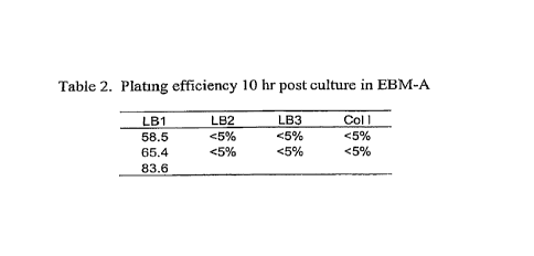
[40] Cells released from LB1 digestion had the highest plating efficiency
after
culture in EBM-A media for 10 hrs (Table 2, FIG. 9A-9D). The plating
efficiency is at least 58% from LB1 digestion. This is significantly higher
than that observed with LB2 or LB3. Importantly, the plating efficiency is
much higher than with crude collagcnase that is utilized by others. One key
aspect of the invention is the rapid plating (in this case, in 10 hours or
less) of
a high fraction of purified EC from adipose tissue. The underlying reason that
Cells released using LB1 and then purified have such a high plating efficiency
could be that dispase (in LB1) is a more gentle enzyme than thennolysin (in
LB3), and cells digested with LB I may as a result retain receptors necessary
for cell attachment. In addition, non-purified collagenase may damage cells
also, and reSult in a low plating efficiency as compared to highly purified
forms of enzyme.
12
CA 02644115 2008-09-05
WO 2007/103379
PCT/US2007/005706
Table 2. Plating efficiency 10 hr post culture in EBM-A
LB1 LB2 LB3 Coil
58.5 <5% <5% <5%
65.4 <5% <5% <5%
83.6
Table 3: EC recovery after CD31 microbead purification
EC
Starting EC% before CD31* EC% after recovery'
cells (106) purification collection (106) purification
(%)
15.5 2.91 2.2 17.2 0.84
22.4 9.87 2.7 56.2 0.69
43 13.3 8.1 41.2 0.58
49 10 4.8 40 0.39
51 12 6.7 58.8 0.64
23.4 21.4 4.44 84 0.74
48.6 15.9 9.3 52.4 0.63
55 28.5 13.44 86.9 0.75
15 42.1 3.36 73.3 0.39
50 28.5 13.44 86.7 0.82
Mean 0.65
STD EV 0.16
Example 3- Materials and Methods
1. Materials
1.1. Tissue
Adipose tissue samples obtained from liposuction aspirates or dissected
subcutaneous
fat tissue
1.2. Reagents
1.2.1. Phosphate Buffer Saline without calcium and magnesium (PBS (1X)) or
Hanks'
Balanced Salt Buffer without calcium and magnesium (HBSS (1x)) (Gibco)
1.2.2. M199 media (Gibco)
1.2.3. EBM-2 media (Clonetics)
EC recovery (%) = (starting cell number x EC% before purification)/(CD31
positive
collection x EC% after purification) x 100
13
CA 02644115 2008-09-05
WO 2007/103379
PCT/US2007/005706
1.2.4.) RPMI (Gibco)
= 1.2.5. Bovine Serum Albumin (BSA) (Miltenyi Biotech)
1.2.6. Liberase Blendzyrne 1 and Liberate Blendzyme 3 (Roche)
1.2.7. Collagenase type I (Sigma)
1.2.8. L-glutamine (Invitrogen)
1.2.9. Hydrocortisone (Sigma)
1.2.10. Dibutyryl cyclic AMP (Sigma)
1.2.11. Penicillin-Streptomycin Solution (100x Sigmal)
1.2.13. Trypsin-EDTA (0.25 mg/ml) (Invitrogen)
1.2.14. Ethylenediamine-tetraacetic acid disodium salt (EDTA) (Sigma)
1.2.15. Fetal Bovine Serum (FBS) qualified, heat inactivated (US) (Gibco
1.2.16. CD31 MicroBeads (Miltenyi Biotech)
1.2.17. FcR blocking reagent (Miltenyi Biotech)
1.2.18. CD 31-Fitc (BD)
1.2.19. CD 45-APC (BD)
1.2.20. CD105 Fite (Chemicon)
1.2.21. CD117 APC (BD)
1.2.22. CD133 PE (BD)
1.2.23. Thrombomodulin (CD 141-PE, BD)
1.2.24. Ve-cadherin-PE (CD 144-PE, eBiosciences)
1.2.25. CD146-PE (BD)
1.2.26. Uea-l-fite (Biomeda)
1.2.27. 7-AAD (BD)
1.2.28. vWF (Von Willebrand Factor) antibody (Dako)
14
CA 02644115 2014-01-06
WO 2007/103379
PCT/US2007/005706
=
1.2.29.4% paraformaldebyde (USB)
TM
1.2.30. Triton X-100 (Sigma)
TM
1.231. Tween-20 (Sigma)
1.2.32. 4',6-diamidino-2-phenylindo1e, dihydrochloride (DAPI) (Molecular
Probes)
1.3. Supplies
1.3.1. 500 ml plastic centrifugation bottles (Coming)
1.3.2. 0.2 pm filter units (Nalgene)
1.33. 50 ml conical tubes (Corning)
1.3.4. 15 conical tubes (Corning)
133.5 ml polystyrene tubes (BD)
1.4 Equipment
1.4.1. AutoMACS Separator is benchtop automated magnetic cell sorters
(Miltenyi
Biotech)
1.4.2. BD FACSCalibur system (BD)
1:4.3. Titer Plate Shaker (Lab-Line Instruments)
1.4.3. Centrifuge (Beckman)
1.4.4. Biosafety Hood
1.4.5. CO2 Incubator (NUAIR)
1.4.6. Inverted microscope ¨Nikon Eclipse TS100 with Epi-Fluorescence
Attachment
(Mercury Lamp Illuminator model name: C-SHO) (Nikon Instruments Incorporation,
Melville, NY) and equipped with a camera photometric cool-snap Nilcon)
=
CA 02644115 2008-09-05
WO 2007/103379
PCT/US2007/005706
1.5. Media stock solution
All the media solutions are filtered through a 0.2 um filter unit and Frozen
down in
50m1 tube in -20 C..
1.5.1. EBM-A medium
EBM-2 base medium (CLOTECH)
20% FBS
L-glutamine (0.292 mg/ml)
Hydrocortisone (1 pg/m1)
Di cAMP (0.25 mg/ml)
1% Penicillin-Streptomycin Antibiotic solution
1.5.2. DMEM BASED medium
DMEM (Invitrogen)
10% PBS
lx MVGS (Cascade Biologics)
Penicillin-Streptomycin
16
CA 02644115 2008-09-05
WO 2007/103379
PCT/US2007/005706
2. Methods
After transportation to the laboratory, the liposuction sample is immediately
processed to isolate MVEC. If sample can't be processed immediately, store it
at
room temperature and process it in 24 hrs. Before performing the experiment,
warm
up the water bath to 37 C.
All the following procedures are performed in Biosafety Hoods
2.1. Isolation
2.1. 1. Warm up buffer 500 or more of PBS or HBSS with 0.1% glucose. Line the
surface of the biosafety hood with a disposable bench protector.
2.1.2. Prepare digestion solution (0.75 u/ml Liberase Blendzyme I (LB1) or
Liberase
Blendzyme 3(LB3) or 4mg/m1 collagenase type I in PBS or HBSS with 4mg/m1 BSA
and 0.1% glucose), and warm it in the 37 C waterbath.
2.1.3. Warm M199 and EC media in the 37 C waterbath
2.1.4. Prepare RPMI/1% FBS/2mM EDTA and keep it at 4 C
2.1.5. To maintain optimal sterile conditions, open the surgical container
used for
liposuction procedure under the biosafety hood.
Dispense 200 ml of adipose tissue in 500 ml centrifuge bottles (Coming). Add
an
equal volume of warm PBS or HBSS. Agitate to wash the tissue and then allow
phase
separation for 3-5 min. Aspirate the infranatant solution (lower liquid
phase).
The wash is repeated several times until a clear infranatant solution is
obtained
(usually 3-4 times).
2.1.6. Centrifuge cells at 1500-2000 rpm for 5-15 min after last wash.
Aspirate the
infranatant solution. Measure adipose tissue and add about lml of warm LB I or
LB3,
17
CA 02644115 2008-09-05
WO 2007/103379
PCT/US2007/005706
or collagenase type I solution per 1 g of fat. Tight the lid and place them on
a shaker
in a 37 C incubator with gentle shaking for 30-40min. Mix tissue manually
every 10
mm.
2.1.7..Add 150 ml of warm M199 media and centrifuge cells at 1200rpm for 5 mm.
2.1.8-After spinning, EC as well as other cells will form a pellet at the
bottom of the
bottle or tube (this will usually include a layer of dark red cells).
Carefully remove the
top layer of oil and fat, the primary adipocytes (a yellow layer of floating
cells), and
the underlying layer of digestion solution. Leave behind a small volume of
collagenase solution above the pellet so that the cells are not disturbed.
2.1.9. Suspend the cells in 10 ml of warm M199 media and filter cells with a
70um
cell strainer. Centrifuge the cells at 1200 rpm in an appropriate centrifuge
for 5
minutes at room temperature.
2.1.10. Aspirate the remaining media. When aspirating, the tip of the pipette
should
aspirate from the top so that the oil is removed as thoroughly as possible.
The cell
pellet should be at the bottom of the tubes.
2.1.11. Resuspend the cells with 10 ml of cold RPM1/1% FBS/2mM EDTA mediuin
in each tube. Pool the cells in one 50 ml conical tube. Filter cells through a
70um
stainer.
2.1.12. Remove 150 ul cell suspension and count cells using Sysmex.
2.2. EC enrichment with CD31 microbeads
2.2.1. Transfer 25 x 106 -50 x 106 cells to a 15 ml conical tube and
centrifuge cells at
1200 rpm for 5 minutes at room temperature.
2.2.2. Aspirate off the supernatant and suspend the cells to a maximum
concentration
of 10 x 106 cells per 60 ul of RPMI/1% FBS/2mM EDTA medium. Add 20u1 of FcR
18
CA 02644115 2008-09-05
WO 2007/103379
PCT/US2007/005706
blocking reagent per 10 x 106 cells. Mix briefly, and then add 20 ul of CD31-
MicroBeads per 10 x 106 cells. Incubate cells for 15 min at 4 C.
2.2.3. After incubation, rinse cells with RPMI/1% FBS/2mM EDTA medium and
centrifuge cells at 300xg for 10 min.
2.2.4. Suspend cell pellet in 2 ml of RPM1/1% FBS/2m.M EDTA medium.
2.2.5. Load cells on autoMACS and separated using program POSSELD.
2.2.6. Collect CD31 positive and negative cells.
2.2.7. Transfer 150 ul of either CD31 positve or negative cells to a micro
centrifuge
tube and count cells using Sysmex.
2.2.8. Characterize cells before and after CD31 separation using using
Fluorescence
Activated Cell Sorting (FACS). Viability is measured using FACS by staining
cells
with 7-AAD.
=
2.3 Plating efficiency:
2.3.1. Seed CD31 purified EC with a density of 2-5x 10⁵/cm2 in ECM coated,
such as
fibronectin, geletin, collagen I, with EC media such as EBM-A or
MEM/10%FBS/MVGS and cultured in 5% CO2, 37C incubator.
2.3.2. Next day, shake plate gently and collect media containing unattached
cells in a
tube. Rinse cells with PBS and collect cells in the same tube. Add fresh media
to the
plate and place back to incubator.
2.3.3. Centrifuge collected media 1500rpm for 5 mm.
2.3.4. Aspirate media and suspend cells in 200 to 500 ul PBS. Vigorously pipet
cells
and count cells using Sysmem.
2.3.5. Plating efficiency = (number of total seeded cells¨ number of floating
cells)/total seeded cells).
19
CA 02644115 2008-09-05
WO 2007/103379
PCT/US2007/005706
2.4. Cell characterization
Cells before and after CD31 microbeads separation as well as cultured for 48
hr are
collected and characterized by FACS analysis for CD31, CD34, CD45, CD141 and
CD144, CD146 expression. Some cells from 96 well plates were fixed with 4%
paraformaldehyde and stained with vWF, eNOS or CD31.
2.5. Seed EC on prosthetic grafts
A 0.5 x 0.5 cm piece of HumacyteTM engineered grafts were coated with human
fibronectin (10Oug/m1) in 6-well plate for 1-8hr at 37 C. CD31 microbead
selected
EC (1 x 10⁶-5 x 10⁶) were then added in the well and incubated for 10-16 hr in
the
incubator. The grafts were fixed in formalin and scanning electron microscopy
(SEM) was performed on these grafts.
CA 02644115 2014-01-06
References:
1. American Heart Association - Heart Disease and Stroke Statistics. 2006
Update.
http://www.americanheart.org/downloadable/heart/1166711577754HS_StatsInsid
eText.pdf
2. Pomposelli FB Jr, Arora S, Gibbons GW, Frykberg R, Smalcowslci P, Campbell
DR, Freeman DV, LoGerfo FW. Lower extremity arterial reconstruction in the
very elderly: successful outcome preserves not only the limb but also
residential
status and ambulatory function. J Vasc Surg. 1998; 28(2):215-25.
3. Veith ET, Moss CM, Sprayregen S, Montefusco C. Preoperative saphenous
venography in arterial reconstructive surgery of the lower extremity. Surgery.
1979; 85(3):253-6.
4. Veith FJ, Gupta SK, Ascer E, White-Flores S, Samson RH, Scher LA, Towne JB,
Bernhard VM, Bonier P, Flinn WR, et al. Six-year prospective multicenter
randomized comparison of autologous saphenous vein and expanded
polytetrafluoroethylene grails in infrainguinal arterial reconstructions.
J Vasc Surg. 1986; 3(1):104-14.
5. Tiwari A, Salacinski HJ, Hamilton G, Seifalian AM. Tissue engineering of
vascular bypass grails: role of endothelial cell extraction. Eur J Vase
Endovasc
Surg. 2001; 21(3):193-201. Review.
6. Zilla P. Fasol R, Dudeck U, Siedler S, Preiss P. Fischlein T, Muller-
Glauser W,
Baitella G, Sanan D, Odell J, et al. In situ cannulation, microgrid follow-up
and
low-density plating provide first passage endothelial cell masscultures for in
vitro
lining.
.1 Vasc Surg. 1990; 12(2):180-9.
7. Jarrell BE, Williams SK, Stokes G, Hubbard FA, Carabasi RA, Koolpe E,
Greener D, Pratt K, Moritz MI, Radomski J, et al. Use of freshly isolated
capillary
endothelial cells for the immediate establishment of a monolayer on a vascular
graft at surgery.
Surgery. 1986; 100(2):392-9.
21
CA 02644115 2014-01-06
8. Williams SK, Jarrell BE, Rose DG, Pontell J, Kapelan BA, Park PIC, Carter
TL.
Human microvessel endothelial cell isolation and vascular graft sodding in the
operating room. Ann Vasc Surg. 1989 Apr; 3(2):146-52.
9. Pasic M, Muller-Glauser W, von Segesser L, Odermatt B, Lachat M, Turina M.
Endothelial cell seeding improves patency of synthetic vascular grafts: manual
versus automatized method. Eur J Cardiothorac Surg. 1996; 10(5):372-9.
10. Williams SIC, Rose DG, Jarrell BE. Microvascular endothelial cell sodding
of
ePTTE vascular grafts: improved patency and stability of the cellular lining.
J Biomed Mater Res. 1994 Feb; 28(2):203-12.
11. Williams SK, Jarrell BE, Kleinert LB. Endothelial cell transplantation
onto
polymeric arteriovenous grafts evaluated using a canine model.
J Invest Surg. 1994 Nov-Dec; 7(6):503-17.
12. Park PK, Jarrell BE, Williams SIC, Carter TL, Rose DG, Martinez-Hernandez
A,
Carabasi RA 3rd. Thrombus-free, human endothelial surface in the midregion of
a
Dacron vascular graft in the splanchnic venous circuit--observations after
nine
months of implantation. J Vasc Surg. 1990; 11(3):468-75.
13. Meerbaum SO. Sharp WV, Schmidt SP. Lower extremity revascularization with
polytetrafluoroethylene grafts seeded with microvscular endothelial cells. In:
Zilla, P, Fasol, R. Callow, A. editors. Applied Cardiovascular Biology 1990-
91.
International Society For Applied Cardiovascular Biology. 2nd ed. Basel:
ICarger,
1992, pp107-119 et al.
14. Sharp WV, Schmidt SP, Meerbaum SO, Pippert TR. Derivation of human
microvascular endothelial cells for prosthetic vascular graft seeding. Ann
Vasc
Surg. 1989; 3(2):104-7.
15. Stemetti AV, Hunter WJ, Schultz RD, Sugimoto JT, Blair EA, Hacker K,
Chasan
P, Valentine J. Seeding with endothelial cells derived from the microvessels
of the
omentum and from the jugular vein: a comparative study. J Vasc Surg. 1988;
7(5):677-84.
16. Arts CH, Hedeman Joosten PP, Blankensteijn JD, Staal FJ, Ng PY, Heijnen-
Snyder GJ, Sixma JJ, Verhagen HJ, de Groot PG, Eikelboom BC. Contaminants
from the transplant contribute to intimal hyperplasia associated with
22
CA 02644115 2014-01-06
microvascular endothelial cell seeding.
Eur J Vase Endovasc Surg. 2002; 23(1):29-38.
17. Arts CH, Blankensteijn JD, Heijnen-Snyder GJ, Verhagen RI, Hedeman Joosten
PP, Sixma JJ, Eikelboom BC, de Groot PG. Reduction of non-endothelial cell
contamination of microvascular endothelial cell seeded grafts decreases
thrombogenicity and intimal hyperplasia. Eur J Vasc Endovasc Surg. 2002;
23(5):404-12.
18. Hewett PW, Murray JC. Irnmunomagnetic purification of human microvessel
endothelial cells using Dynabeads coated with monoclonal antibodies to PECAM-
1. Eur J Cell Biol. 1993; 62(2):451-4.
19. Arts CH, de Groot P, Heijnen-Snyder GJ, Blankensteijn JD, Eikelboom BC,
Slaper-Cortenbach IC. Application of a clinical grade CD34-mediated method for
the enrichment of microvascular endothelial cells from fat tissue.
Cytotherapy.
2004; 6(1):30-42.
20. Miebach S, Grau S, Hummel V, Rieckmann P, Tonn JC, Goldbrunner RH.
Isolation and culture of microvascular endothelial cells from gliomas of
different
WHO grades.
J Neurooncol. 2006; 76(1):39-48.
21. Arts CH, Heijnen-Snyder GJ, Joosten PP, Verhagen HJ, Eikelboom BC, Sixma
JJ,
de Groot PG. A novel method for isolating pure microvascular endothelial cells
from subcutaneous fat tissue ideal for direct cell seeding. Lab Invest. 2001;
81(10):1461-5.
22. Williams SK, McKenney S, Jarrell BE. Collagenase lot selection and
purification
for adipose tissue digestion. Cell Transplant. 1995; 4(3):281-9
23