Note: Descriptions are shown in the official language in which they were submitted.
CA 02644133 2015-10-27
- 1 -
REVOLVING TISSUE SAMPLE HOLDER FOR BIOPSY
DEVICE
BACKGROUND
Biopsy samples have been obtained in a variety of ways in various medical
procedures using
a variety of devices. Biopsy devices may be used under stereotactic guidance,
ultrasound
guidance, MRI guidance, or otherwise. Merely exemplary biopsy devices are
disclosed in
U.S. Pat. No. 5,526,822, entitled "Method and Apparatus for Automated Biopsy
and
Collection of Soft Tissue," issued June 18, 1996; U.S. Pat. No. 6,086,544,
entitled "Control
Apparatus for an Automated Surgical Biopsy Device," issued July 11, 2000; U.S.
Pub. No.
2003/0109803, entitled "MRI Compatible Surgical Biopsy Device," published June
12, 2003;
U.S. Pub. No. 2007/0118048, entitled "Remote Thumbwheel for a Surgical Biopsy
Device,"
published May 24, 2007; U.S. Provisional Patent Application Serial No.
60/869,736, entitled
"Biopsy System," filed December 13, 2006; and U.S. Provisional Patent
Application Serial
No. 60/874,792, entitled "Biopsy Sample Storage," filed December 13, 2006.
While several
systems and methods have been made and used for obtaining a biopsy sample, it
is believed
that no one prior to the inventors has made or used the invention described in
the appended
claims.
SUMMARY OF THE INVENTION
In one embodiment, there is provided a tissue sample holder for collecting
tissue samples
obtained using a biopsy device. The biopsy device has a cutter defining a
cutter lumen along
an axis. The tissue sample holder comprises:
(a) a rotatable manifold, wherein the manifold is in fluid communication
with a conduit, wherein the manifold is configured to redirect fluid,
wherein the manifold is configured to communicate a vacuum from the
conduit to the cutter lumen;
(b) a plurality of discrete tissue sample chambers associated with the
manifold, wherein each of the tissue sample chambers is configured to
receive at least one tissue sample communicated through the cutter
lumen;
CA 02644133 2015-10-27
- 2 -
(c) a cover member configured to removably cover the manifold and the
tissue sample chambers, wherein the manifold and tissue sample
chambers are configured to rotate within the cover member; and
(d) a tissue sample holder rotation mechanism, wherein the tissue sample
holder rotation mechanism is operable to rotate the manifold to
successively index the tissue sample chambers relative to the cutter
lumen.
In another embodiment, there is provided a tissue sample holder for collecting
tissue samples
obtained using a biopsy device. The biopsy device has a conduit and a cutter
defining a cutter
lumen along an axis. The tissue sample holder comprises:
(a) a rotatable manifold, wherein the manifold is in fluid communication
with the conduit, wherein the manifold is configured to redirect fluid
from a first axial direction to a lateral direction and back to a second
axial direction, opposite to the first axial direction, to the cutter lumen;
(b) a plurality of discrete tissue sample chambers associated with the
manifold, wherein each of the tissue sample chambers is configured to
receive at least one tissue sample communicated through the cutter
lumen; and
(c) a tissue sample holder rotation mechanism, wherein the tissue sample
holder rotation mechanism is operable to rotate the manifold to
successively index the tissue sample chambers relative to the cutter
lumen.
In another embodiment, there is provided a tissue sample holder for collecting
tissue
samples obtained using a biopsy device, wherein the biopsy device has a
conduit and a
cutter defining a cutter lumen along an axis, wherein the tissue sample holder
comprises:
(a) a rotatable manifold defining a longitudinal axis,
wherein the
rotatable manifold is rotatable about the longitudinal axis, wherein
the rotatable manifold comprises a plurality of tissue sample
chambers, wherein the tissue sample chambers are radially spaced
from the longitudinal axis and are angularly spaced from each
other about the longitudinal axis, wherein the tissue sample
CA 02644133 2015-10-27
,
- 2a -
chambers are oriented substantially parallel to the longitudinal axis;
(b) a longitudinal passage disposed substantially parallel to and radially
offset from the plurality of tissue sample chambers; and
(c) a tissue sample holder rotation mechanism, wherein the tissue sample
holder rotation mechanism is operable to rotate the manifold about
the longitudinal axis to successively index the tissue sample
chambers relative to the cutter lumen;
wherein the cutter lumen, the longitudinal passage, and at least one of the
tissue sample
chambers are in fluid communication such that the cutter lumen, the
longitudinal
passage, and the at least one of the tissue sample chambers together define a
pneumatic path; and
wherein the at least one of the tissue sample chambers is positioned in the
pneumatic path
between the cutter lumen and the longitudinal passage.
In another embodiment, there is provided a tissue sample assembly for
collecting tissue
samples obtained using a biopsy device having a translatable tissue sample
cutter
defining a cutter lumen extending along an axis, wherein the tissue sample
assembly
comprises:
(a) a manifold rotatable about a manifold axis, the manifold
axis being
substantially parallel to and offset from the axis of the cutter
lumen, and the rotatable manifold comprising a plurality of
chambers, wherein the chambers are radially spaced from the
manifold axis and angularly spaced from each other about the
longitudinal axis; and
(b) a plurality of tissue sample receiving members releasably carried
by the rotatable manifold, each tissue sample receiving member
having a plurality of tissue sample compartments, each tissue
sample compartment for receiving and holding a tissue sample, and
each tissue sample compartment having a plurality of vacuum
openings extending therethrough;
wherein the number of chambers exceeds the number of tissue sample receiving
members, and wherein the cutter lumen and the at least one of the chambers
CA 02644133 2015-10-27
- 2b -
together define a pneumatic path for providing vacuum through the vacuum
openings in
one of the tissue sample compartments.
In another disclosed embodiment, there is provided a tissue sample holder for
collecting
tissue samples obtained using a biopsy device. The biopsy device has a conduit
and a cutter
defining a cutter lumen along an axis. The tissue sample holder comprises:
CA 02644133 2008-11-19
- 3 -
(a) a rotatable manifold, wherein the rotatable manifold comprises a
plurality of radially extending fins;
(b) a plurality of tissue sample compartments, wherein each of the tissue
sample compartments is positioned at least partially between
corresponding fins of the manifold, wherein each of the tissue sample
compartments is configured to hold a tissue sample captured by the
cutter; and
(c) a tissue sample holder rotation mechanism, wherein the tissue sample
holder rotation mechanism is operable to rotate the manifold to
successively index the tissue sample compartments relative to the
cutter lumen.
BRIEF DESCRIPTION OF THE DRAWINGS
While the specification concludes with claims which particularly point out and
distinctly
claim the invention, it is believed the present invention will be better
understood from the
following description of certain examples taken in conjunction with the
accompanying
drawings, in which like reference numerals identify the same elements and in
which:
FIG. 1 depicts a schematic view of an exemplary biopsy system;
FIG. 2 depicts a perspective view of an exemplary assembled biopsy device, for
use in a
stereotactic setting;
FIG. 3 depicts an exploded view of the biopsy device of FIG. 2, with the probe
detached from
the holster;
FIG. 4 depicts a perspective view of an exemplary assembled biopsy device, for
use in an
ultrasound setting;
FIG. 5 depicts an exploded view of the biopsy device of FIG. 4, with the probe
detached from
the holster;
FIG. 6 depicts a top perspective view of a probe portion of the biopsy device
of FIG. 3;
CA 02644133 2008-11-19
- 4 -
FIG. 7 depicts a bottom perspective view of the probe portion of FIG. 6;
FIG. 8 depicts a top perspective view of the probe portion of FIG. 6, with a
top cover
removed;
FIG. 9 depicts a bottom perspective view of the probe portion of FIG. 6, with
a base
removed;
FIG. 10 depicts a lateral cross-sectional view of the probe portion of FIG. 6,
taken along a
longitudinal plane;
FIG. 11 depicts a perspective view of a needle component of the probe portion
of FIG. 6;
FIG. 12 depicts a partial perspective view of the probe portion of FIG. 6,
showing a needle
hub assembly;
FIG. 13 depicts a partial perspective view of the probe portion of FIG. 6,
showing a needle
hub assembly with a needle manifold removed;
FIG. 14 depicts a partial, cross-sectional view of a cutter rotation and
translation mechanism
of the probe portion of FIG. 6, taken along a longitudinal plane;
FIG. 15 depicts a front perspective view of an exemplary tissue sample holder;
FIG. 16 depicts the tissue sample holder of FIG. 15, with a cup and other
components
removed;
FIG. 17 depicts the tissue sample holder of FIG. 15, with a tissue sample tray
removed;
FIG. 18 depicts a rear view of the tissue sample holder of FIG. 15;
FIG. 19 depicts a rear view of the tissue sample holder of FIG. 15, with a cup
and other
components removed;
FIG. 20 depicts a perspective view of an engagement member;
FIG. 21 depicts an exploded view of an applier and the tissue sample holder of
FIG. 15;
CA 02644133 2008-11-19
- 5 -
FIG. 22 depicts a perspective view of the applier of FIG. 21 inserted in the
tissue sample
holder of FIG. 15;
FIG. 23 depicts a perspective view of a holster of the biopsy device of FIG.
2;
FIG. 24 depicts a top view of the holster of FIG. 23, with a top cover
removed;
FIG. 25 depicts a side view of the holster of FIG. 23, with side panels
removed;
FIG. 26 depicts another side view of the holster of FIG. 23, with side panels
removed;
FIG. 27 depicts a partial view of the holster of FIG. 23, showing an exemplary
needle
rotation mechanism;
FIG. 28 depicts a partial view of the holster of FIG. 23, showing an exemplary
needle firing
mechanism;
FIG. 29 depicts a partial view of the holster of FIG. 23, showing an exemplary
needle firing
mechanism in a cocked configuration;
FIG. 30 depicts a partial view of the holster of FIG. 23, showing an exemplary
cutter drive
mechanism;
FIG. 31 depicts a partial view of the holster of FIG. 23, showing an exemplary
tissue holder
rotation mechanism;
FIG. 32 depicts another partial view of the holster of FIG. 23, showing an
exemplary tissue
holder rotation mechanism;
FIG. 33 depicts a bottom perspective view of the probe portion of the biopsy
device of FIG.
4;
FIG. 34 depicts a top perspective view of the probe portion of FIG. 33, with a
top cover
removed;
FIG. 35 depicts a bottom perspective view of the probe portion of FIG. 33,
with a base
removed;
CA 02644133 2008-11-19
- 6 -
FIG. 36 depicts a partial perspective view of the probe portion of FIG. 33,
showing a needle
hub assembly;
FIG. 37 depicts a partial perspective view of the probe portion of FIG. 33,
showing a needle
hub assembly with a needle manifold removed;
FIG. 38 depicts a front perspective view of an exemplary tissue sample holder,
with a cup and
other components removed;
FIG. 39 depicts the tissue sample holder of FIG. 38, with a tissue sample tray
removed;
FIG. 40 depicts a rear view of the tissue sample holder of FIG. 38, with a cup
and other
components removed;
FIG. 41 depicts a front perspective view of a holster of the biopsy device of
FIG. 4;
FIG. 42 depicts a rear perspective view of the holster of FIG. 41;
FIG. 43 depicts a top view of the holster of FIG. 41, with a top cover
removed;
FIG. 44 depicts a partial view of the holster of FIG. 41, showing an exemplary
cutter drive
mechanism;
FIG. 45 depicts a partial view of the holster of FIG. 41, showing an exemplary
tissue holder
rotation mechanism;
FIG. 46 depicts a perspective view of an exemplary vacuum control module and
exemplary
vacuum canister;
FIG. 47 depicts the vacuum control module of FIG. 46 with the vacuum canister
of FIG. 46
separated therefrom;
FIG. 48 depicts a perspective view of the vacuum canister of FIG. 46;
FIG. 49 depicts a top view of the vacuum canister of FIG. 46;
FIG. 50 depicts a top view of the vacuum canister of FIG. 46, with tubes
engaged with a top
portion of the canister;
CA 02644133 2008-11-19
- 7 -
FIG. 51 depicts a cross-sectional view of the canister of FIG. 46, taken along
a longitudinal
plane;
FIG. 52 depicts a rear perspective view of the vacuum control module of FIG.
46;
FIG. 53 depicts the vacuum control module of FIG. 46, with an outer casing
removed;
FIG. 54 depicts a perspective view of a vacuum canister port assembly of the
vacuum control
module of FIG. 46;
FIG. 55 depicts a front view of the vacuum canister port assembly of FIG. 54;
FIG. 56 depicts a rear view of the vacuum canister port assembly of FIG. 54;
FIG. 57 depicts a cross-sectional view of the vacuum canister port assembly of
FIG. 54;
FIG. 58 depicts a cross-sectional view of the vacuum canister port assembly of
FIG. 54 with
the vacuum canister of FIG. 46 inserted therein;
FIG. 59 depicts a perspective, cross-sectional view of an exemplary tube;
FIG. 60 depicts a schematic flow diagram showing an exemplary rotation
sequence of a
tissue sample holder;
FIG. 61 depicts an exemplary sequence of the position of a cutter within a
cannula, relative to
fluid communication being provided through lateral and axial vacuum tubes, in
an exemplary
"sample" cycle;
FIG. 62 depicts an exemplary sequence of the position of a cutter within a
cannula, relative to
fluid communication being provided through lateral and axial vacuum tubes, in
an exemplary
"clear probe" cycle;
FIG. 63 depicts an exemplary sequence of the position of a cutter within a
cannula, relative to
fluid communication being provided through lateral and axial vacuum tubes, in
an exemplary
"position" cycle;
CA 02644133 2008-11-19
- 8 -
FIG. 64 depicts an exemplary sequence of the position of a cutter within a
cannula, relative to
fluid communication being provided through lateral and axial vacuum tubes, in
an exemplary
"aspirate" cycle;
FIG. 65 depicts an exemplary sequence of the position of a cutter within a
cannula, relative to
fluid communication being provided through lateral and axial vacuum tubes, in
an exemplary
"smart vac" cycle;
FIG. 66 depicts an exemplary "status" page of an exemplary user interface for
a biopsy
system;
FIG. 67 depicts an exemplary "probe" page of an exemplary user interface for a
biopsy
system;
FIG. 68 depicts an exemplary "system" page of an exemplary user interface for
a biopsy
system; and
FIG. 69 depicts an exemplary user interface that may be applied to a portion
of a biopsy
device.
DETAILED DESCRIPTION
The following description of certain examples of the invention should not be
used to limit the
scope of the present invention. Other examples, features, aspects,
embodiments, and
advantages of the invention will become apparent to those skilled in the art
from the
following description, which is by way of illustration, one of the best modes
contemplated for
carrying out the invention. As will be realized, the invention is capable of
other different and
obvious aspects, all without departing from the invention. Accordingly, the
drawings and
descriptions should be regarded as illustrative in nature and not restrictive.
As shown in FIG. 1, an exemplary biopsy system (2) includes a biopsy device
(100, 101) and
a vacuum control module (400). As shown in FIGS. 2-3, biopsy device (100)
comprises a
probe (102) and a holster (202). Similarly, as shown in FIGS. 4-5, biopsy
device (101)
comprises a probe (103) and a holster (302). As will be described in greater
detail below,
each probe (102, 103) is separable from its corresponding holster (202, 302).
Use of the term
CA 02644133 2008-11-19
- 9 -
"holster" herein should not be read as requiring any portion of probe (102,
103) to be inserted
into any portion of holster (202, 302). Indeed, in some variations of biopsy
devices (100,
101), probe (102, 103) may simply sit on holster (202, 302). In some other
variations, a
portion of holster (202, 302) may be inserted into probe (102, 103).
Furthermore, in some
biopsy devices (100, 101), probe (102, 103) and holster (202, 302) may be of
unitary or
integral construction, such that the two components cannot be separated. Still
other suitable
structural and functional relationships between probe (102, 103) and holster
(202, 302) will
be apparent to those of ordinary skill in the art in view of the teachings
herein.
Some variations of biopsy devices (100, 101) may include one or more sensors
(not shown),
in probe (102, 103) and/or in holster (202, 302), that is/are configured to
detect when probe
(102, 103) is coupled with holster (202, 302). Such sensors or other features
may further be
configured to permit only certain types of probes (102, 103) and holsters
(202, 302) to be
coupled together. In addition or in the alternative, such sensors may be
configured to disable
one or more functions of probes (102, 103) and/or holsters (202, 302) until a
suitable probe
(102, 103) and holster (202, 302) are coupled together. Of course, such
sensors and features
may be varied or omitted as desired.
By way of example only, probe (102, 103) may be provided as a disposable
component,
while holster (202, 302) may be provided as a reusable component. Vacuum
control module
(400) is provided on a cart (not shown) in the present example, though like
other components
described herein, a cart is merely optional. Among other components described
herein, a
footswitch (not shown) and/or other devices may be used to provide at least
some degree of
control of at least a portion of biopsy system (2). Conduits (200) provide
communication of
power (e.g., electrical, pneumatic, etc.), control signals, saline, vacuum,
and venting from
vacuum control module (400) to biopsy device (100, 101). Each of these
components will be
described in greater detail below.
I. Exemplary Probe for Stereotactic Use
As shown in FIGS. 6-14, probe (102) comprises a needle portion (10) and a body
portion
(112). Body portion (112) comprises a cover member (114) and a base member
(116). A
tissue sample holder (140) is removably secured to base member (116), though
tissue sample
CA 02644133 2015-10-27
- 10 -
holder (140) may alternatively be secured to cover member (114) or some other
component.
As will be described in greater detail below, a pair of tubes (402, 404) are
coupled with probe
(102).
A. Exemplary Needle
In the present example, needle portion (10) comprises an outer cannula (12)
having a tissue
piercing tip (14) and a transverse tissue receiving aperture (16) located
proximally from the
tissue piercing tip (14). Tissue piercing tip (14) is configured to penetrate
tissue without
requiring a high amount of force, and without requiring an opening to be
preformed in the
tissue prior to insertion of tip (14). Suitable configurations for tissue
piercing tip (14) will be
apparent to those of ordinary skill in the art in view of the teachings
herein. For instance, as
shown in FIG. 11, tip (14) of the present example is part of a needle piece
(18), which is
formed of a stamped piece of metal. In particular, needle piece (18) is
stamped to form tip
(14) and wall (30), which will be described in greater detail below. A
plurality of openings
(32), including venting openings (34) are formed through wall. Various ways in
which fluid
may be communicated through openings (32, 34) will be described in greater
detail below,
with reference to FIGS. 61-65. Needle piece (18) is then twisted such that tip
(14) and wall
(30) are substantially perpendicular to one another. Needle piece (18) is then
inserted into
cannula (12), with tip (14) protruding through a slot formed in the distal end
of cannula (12).
A tissue stop (26) is provided immediately proximal to tip (14). Still other
ways in which tip
(14) may be formed, including alternative techniques, materials, and
configurations, will be
apparent to those of ordinary skill in the art in view of the teachings
herein.
The interior of outer cannula (12) of the present example defines a cannula
lumen (20) and a
vacuum lumen (40), with a wall (30) separating the cannula lumen (20) from the
vacuum
lumen (40). A plurality of external openings (22) are formed in outer cannula
(12), and are in
fluid communication with vacuum lumen (40). Examples of openings that are
similar to
external openings (22) are disclosed in U.S. Pub. No. 2007/0032742, entitled
"Biopsy Device
with Vacuum Assisted Bleeding Control," published February 8, 2007. Of course,
as with
other components described herein, external openings (22) are merely optional.
CA 02644133 2015-10-27
- 11 -
In some embodiments, wall (30) extends a substantial amount of the length of
needle portion
(10). In other embodiments, wall (30) proximally extends just past the region
where the
distal end of a cutter (50), which will be described below, terminates in
needle portion (10).
For instance, cannula lumen (20) may be sized and configured such that, with
cutter (50)
disposed therein, a gap exists between the exterior of cutter (50) and at
least a portion of the
interior of cannula (12). Such a gap may provide a vacuum lumen (40) along the
length of
cannula (12) proximal to the proximal end of wall (30). Still other ways in
which a vacuum
lumen (40) may be provided will be apparent to those of ordinary skill in the
art in view of
the teachings herein.
In the present example, a plurality of transverse openings (32, 34) are formed
through wall
(30) to provide fluid communication between cannula lumen (20) and vacuum
lumen (40).
As will be described in greater detail below, vacuum, saline, and/or
pressurized air may be
communicated from vacuum lumen (40) to cannula lumen (20) via transverse
openings (32,
34).
B. Exemplary Cutter
A hollow cutter (50) is disposed within cannula lumen (20). The interior of
cutter (50)
defines a cutter lumen (52), such that fluid and tissue may be communicated
through cutter
(50) via cutter lumen (52). As will be described in greater detail below,
cutter (50) is
configured to rotate within cannula lumen (20) and translate axially within
cannula lumen
(20). In particular, cutter (50) is configured to sever a biopsy sample from
tissue protruding
through transverse aperture (16) of outer cannula (12). As will also be
described in greater
detail below, cutter (50) is further configured to permit severed tissue
samples (4) to be
communicated proximally through cutter lumen (52). Merely illustrative
examples of such
severing and proximal communication are described in U.S. Pat. No. 5,526,822,
though any
other suitable structures or techniques may be used for severing and/or
communicating tissue
samples (4) within a biopsy system (2).
Cutter (50) may be subject to various treatments or configurations in order to
facilitate
proximal communication of tissue samples (4) through cutter lumen (52). For
instance, the
CA 02644133 2008-11-19
- 12 -
surface finish inside of cutter (50), defining cutter lumen (52), may be
subject to shot peening
(e.g., with glass beads, sodium bicarbonate, etc.) to reduce adhesion between
tissue and cutter
(50). In addition, or in the alternative, the interior of cutter (50),
defining cutter lumen (52),
may be subject to acid etching and/or plasma etching to reduce adhesion
between tissue and
cutter (50). In addition, or in the alternative, a hydrolubricous material or
other non-stick
coating may be applied to the interior of cutter (50), defining cutter lumen
(52), to reduce
friction between tissue and cutter (50). In addition, or in the alternative,
the interior of cutter
(50), defining cutter lumen (52), may be subject to a rifling surface cut.
Other suitable
treatments for the interior of cutter (50) will be apparent to those of
ordinary skill in the art in
view of the teachings herein. Alternatively, the interior of cutter (50) may
be subject to no
treatment at all in some embodiments.
In an alternate embodiment of cutter (50), a distal portion of cutter (50) has
an inner diameter
and outer diameter that are less than the inner diameter and outer diameter of
a proximal
portion of cutter (50). For instance, the distal-most inch of cutter (50) may
provide a neck
down region (not shown), which transitions into a region having a greater
diameter along the
remaining, proximal length of cutter (50). Such a neck down configuration may
reduce tissue
compression as a tissue sample (4) moves proximally through cutter lumen (52).
The distal
end of outer cannula (12) may also have a complimentary neck down region that
is either the
same length as, shorter than, or longer than a neck down region of cutter
(50). Other suitable
lengths of a neck down region in cutter (50) and/or outer cannula (12) will be
apparent to
those of ordinary skill in the art in view of the teachings herein.
In another alternative embodiment of cutter (50), a plurality of raised
surfaces are provided,
extending inwardly within the interior of cutter (50), running the length of
cutter (50). Such
raised surfaces may be configured to reduce tissue surface contact with the
interior of cutter
(50).
In yet another alternative embodiment of cutter (50), an inner sleeve (not
shown) may be
provided within the distal end interior of cutter (50). For instance, such an
inner sleeve may
have a length of approximately 0.15 inches or any other suitable length. The
distal end of
cutter (50) may be chamfered after such an inner sleeve is inserted, such that
chamfered
CA 02644133 2008-11-19
- 13 -
cutter (50) end and the chamfered sleeve end collectively provide a sharp edge
for severing
tissue. As a severed tissue sample (4) travels proximally through cutter lumen
(52), it will
encounter a greater inner diameter of cutter lumen (52) as soon as the tissue
sample (4) passes
the proximal end of the inner sleeve. This increase in effective diameter may
reduce
compression of the tissue sample (4), thereby improving transport reliability
of the tissue
sample (4). Still other suitable variations of cutter (50) will be apparent to
those of ordinary
skill in the art in view of the teachings herein.
C. Exemplary Needle Hub
As shown in FIGS. 12-13, a needle hub (60) is secured to outer cannula (12),
and comprises a
thumbwheel (62) and a sleeve portion (64) extending proximally from thumbwheel
(62).
Needle hub (60) of the present example is overmolded about a proximal portion
of outer
cannula (12), though needle hub (60) may be formed and/or secured relative to
outer cannula
(12) using any other suitable techniques (e.g., set screws, adhesives, etc.).
Furthermore,
while needle hub (60) of the present example is formed of a plastic material,
any other
suitable material or combination of materials may be used.
Sleeve portion (64) of the present example comprises an annular projection
(66), a
longitudinal slot (68), and a transverse opening (70), which is formed near
the proximal end
of sleeve portion (64). One or more additional transverse openings (70) (e.g.,
diametrically
opposed transverse openings (70)) may also be provided in sleeve portion (64).
A pair of o-
rings (72) are positioned such that one o-ring (72) is proximal to transverse
opening (70) and
another o-ring (72) is distal to transverse opening (70). As will be described
in greater detail
below, transverse opening (70) is in fluid communication with the interior
defined by needle
hub (60), which is also in fluid communication with vacuum lumen (40) of outer
cannula
(12). Other suitable configurations for sleeve portion (64) will be apparent
to those of
ordinary skill in the art in view of the teachings herein.
Thumbwheel (62) is operable to rotate outer cannula (12) about its
longitudinal axis, relative
to cover member (114) and base member (116). For instance, thumbwheel (62) may
be used
to orient aperture (16) to a number of desired orientations about the
longitudinal axis defined
by outer cannula (12). Such multiple orientations may be desirable, by way of
example only,
CA 02644133 2015-10-27
- 14 -
to obtain a plurality of tissue samples (4) from a biopsy site, without
requiring the needle
portion (10) to be removed from the patient during the acquisition of such a
plurality of tissue
samples (4). An illustrative example of such rotation and acquisition of
multiple tissue
samples (4) is disclosed in U.S. Pat. No. 5,526,822. Other ways in which
multiple tissue
samples (4) may be obtained at various locations will be apparent to those of
ordinary skill in
the art in view of the teachings herein. For instance, rotation of outer
cannula (12) may be
motorized or automated, such as using any of the components described in
greater detail
below, or using any other suitable components or techniques. As another non-
exhaustive
example, an entire biopsy device (101) may be rotated during acquisition of
tissue samples
(4), without necessarily removing biopsy device (101) from the patient during
such rotation
and tissue sample (4) acquisition, to obtain tissue samples (4) from various
orientations about
the longitudinal axis defined by outer cannula (12).
It will also be appreciated that other structures may be used to perform
manual rotation of
outer cannula (12). In particular, and as shown in FIG. 12-13, an exposed gear
(74) may be
engaged with outer cannula (12). In this example, gear (74) is slid onto the
proximal end of
sleeve portion (64). A radially inwardly extending projection (not shown) of
gear (74) is
configured to mate with slot (68) of sleeve portion (64), such that gear (74)
rotates unitarily
with sleeve portion (64) while being movable longitudinally along sleeve
portion (64). With
sleeve portion (64) being unitarily engaged with outer cannula (12), rotation
of gear (74) will
further cause rotation of cannula (12) for reorienting aperture (16). Gear
(74) is further
configured to engage with a complimentary exposed gear (206) of holster (202),
as will be
described in greater detail below. In particular, gear (74) is configured to
mesh with gear
(206) such that gear (206) can impart rotation to gear (74), thereby rotating
outer cannula
(12). Some exemplary structures and techniques for selectively causing gear
(206) to rotate
will be discussed in greater detail below, while others will be apparent to
those of ordinary
skill in the art in view of the teachings herein.
It will also be appreciated in view of the teachings herein that the
orientation of aperture (16)
may be indicated on a graphical user interface. For instance, one or more
sensors may be
operable to detect the orientation of aperture (16), and communicate
indicative data to a
CA 02644133 2008-11-19
- 15 -
=
processor. The processor may be in Communication with a display (e.g., display
screen
(702), described below, etc.) to provide visual indication of aperture (16)
orientation. Other
ways in which the orientation of aperture (16) may be indicated to a user will
be apparent to
those of ordinary skill in the art in view of the teachings herein.
Alternatively, orientation of
aperture (16) may be not indicated to a user.
D. Exemplary Needle Manifold
As shown in FIG. 12, a needle manifold (80) is provided about sleeve portion
(64). Needle
manifold (80) is fixed relative to base member (116) in this example. Needle
manifold (80)
is in fluid communication with tube (402), such that tube (402) may
communicate saline, a
vacuum, atmospheric air, and/or pressurized air, etc., to needle manifold
(80), as will be
described in greater detail below. Needle manifold (80) is further in fluid
communication
with the interior of sleeve portion (64), via transverse opening (70). 0-rings
(64) are
configured to maintain a fluid seal between needle manifold (80) and sleeve
portion (64),
even as sleeve portion (64) translates longitudinally relative to needle
manifold (80), such as
during firing of needle (10) as will be described in greater detail below; and
even during
rotation of sleeve portion (64) about its longitudinal axis. A seal (not
shown) is also provided
at the proximal end of sleeve portion (64), at the interface between sleeve
portion (64) and
cutter (50). Needle manifold (80), sleeve portion (64), and outer carmula (12)
are thus
configured and arranged such that saline, a vacuum, atmospheric air, and/or
pressurized air,
etc. that is communicated via tube (402) to needle manifold (80) will be
communicated to
vacuum lumen (40) via transverse opening (70). Of course, any other suitable
structures or
arrangements may be used to communicate saline, a vacuum, atmospheric air,
and/or
pressurized air, etc. from tube (402) to vacuum lumen (40).
E. Exemplary Cutter Rotation and Translation Mechanism
In the present example, and as shown in FIG. 14, body portion (112) of probe
(102)
comprises a cutter rotation and translation mechanism (120), which is operable
to rotate and
translate cutter (50) within outer cannula (12). Cutter rotation and
translation mechanism
(120) comprises a sleeve (122) unitarily secured to cutter (50), a nut member
(124), and a
gear (138). In the present example, sleeve (122) is formed of plastic
overmolded about
CA 02644133 2008-11-19
- 16 -
cutter (50), though any other suitable materials may be used, and sleeve (122)
may be secured
relative to cutter (50) using any other suitable structures or techniques
(e.g., set screws, etc.).
Nut member (124) is secured relative to base member (116), and has internal
threads (126).
A portion of sleeve (122) has external threads (128) that are configured to
engage with
threads (126) of nut member (124). Threads (126, 128) are configured such
that, as sleeve
(122) rotates relative to nut member (124), sleeve (122) will longitudinally
translate relative
to nut member (124), depending on the direction of such relative rotation. By
way of
example only, threads (126, 128) may be configured to have a pitch that
provides
approximately 40-50 threads per inch. Such a thread pitch may provide a ratio
of cutter (50)
rotation to cutter (50) translation that is ideal for severing tissue.
Alternatively, any other
thread pitch may be used. With sleeve (122) being unitarily secured to cutter
(50) in the
present example, longitudinal translation of sleeve (122) relative to nut
member (124) will
result in the same translation of cutter (50).
Another portion of sleeve (122) has a plurality of external flats (130), which
are configured to
engage with a complimentary plurality of internal flats (132) of gear (138).
Gear (138) is
positioned coaxially about sleeve (122) and cutter (50). Flats (130, 132) are
configured such
that rotation of gear (138) causes rotation of sleeve (122). With sleeve (122)
being unitarily
secured to cutter (50) in the present example, rotation of gear (138) and
sleeve (122) will
result in the same rotation of cutter (50). Flats (130, 132) are further
configured such that
sleeve (122) may translate longitudinally relative to gear (138) (e.g., the
fit between sleeve
(122) and gear (138) is not so tight as to prevent such translation). It will
therefore be
appreciated that, as gear (138) rotates, given the relative configurations of
threads (126, 128)
and flats (130, 132), such rotation of gear (138) will simultaneously result
in rotation and
longitudinal translation of sleeve (122), which will in turn result in
simultaneous rotation and
longitudinal translation of cutter (50).
In the present example, gear (138) is partially exposed through base member
(116), and is
configured to mate with a complimentary exposed gear (208) of holster (202),
as will be
described in greater detail below. In particular, gear (138) is configured to
mesh with gear
(208) such that gear (208) can impart rotation to gear (138), thereby
activating cutter rotation
and translation mechanism (120). As will be described in greater detail below,
gear (208) is
CA 02644133 2008-11-19
- 17 -
in communication with a motor (272) that is within holster (202). In the
present example,
gears (138, 208) and threads (126, 128) are configured such that each
revolution of motor
(272) results in approximately 0.00012 inches of translation of cutter (50).
Of course, any of
these components may have other configurations that result in any other
suitable ratio of
cutter (50) translation to motor (272) rotation.
It will be appreciated in view of the teachings herein that cutter rotation
and translation
mechanism (120) described above is merely exemplary, and that translation
and/or rotation of
cutter (50) may alternatively be provided in various other ways. For instance,
biopsy probe
(102) may include a motor (not shown) or other device, such that biopsy probe
(102) lacks
exposed gear (138). Alternatively, any suitable structure other than exposed
gear (138) (e.g.,
a rack, etc.) may be used to receive communication of motion or energy from
some other
component, in order to rotate and/or translate cutter (15). Furthermore,
cutter rotation and
translation mechanism (120) may be configured such that more than one exposed
gear (138)
is present (e.g., one gear (138) for receiving translation motion, and another
gear (138) for
receiving rotation motion, etc.). In other merely illustrative alternatives,
translation and/or
rotation of cutter (50) may be performed at least in part by pneumatic
actuators (not shown),
pneumatic motors (not shown), or a variety of other components. Furthermore,
it will be
appreciated that pneumatic components may be combined with other mechanical
components
and/or electro-mechanical components in order to translate and/or rotate
cutter (50).
Base member (116) further comprises a cutter passage (54), through which the
proximal end
of cutter (50) is disposed. A seal (56) is provided at the distal interface of
cutter (50) and
cutter passage (54), to prevent escape of a vacuum or fluid between the outer
surface of cutter
(50) and the inner surface of the distal end of cutter passage (54). Cutter
passage (54) is sized
such that, as cutter (50) translates during use of biopsy device (100), the
distal end of cutter
(50) remains within cutter passage (54). Of course, any other suitable
structures or
configurations may be used.
F. Exemplary "Sharps Reduction" Variation
In the present example, needle portion (10) and cutter (50) are configured to
be removable
from biopsy probe (102), such as after a session of use of biopsy device
(100). In particular,
CA 02644133 2008-11-19
- 18 -
base member (116) of body portion (112) of biopsy probe (102) comprises a
release tab
(118), which is resiliently movable relative to base member (116) via an arm
(119). Release
tab (118) is configured to restrict axial movement of needle portion (10) by
restricting axial
movement of gear (74), which is engaged with sleeve portion (64) of hub (60)
as noted
above, when release tab (118) is in a default position. Of course, the
engagement between
and configurations of gear (74) and sleeve portion (64) will permit some
degree of axial
movement of needle portion (10), such as for firing of needle portion (10),
even while release
tab (118) is in a default position. However, when release tab (118) is
sufficiently depressed,
such as by a user, release tab will provide clearance for gear (74) to be
moved distally of base
member (116). In other words, with release tab (118) sufficiently depressed,
the entirety of
needle portion (10), including the entirety of needle hub (60) and gear (74),
may be axially
pulled distally from body portion (112) of biopsy probe (102); such that the
entirety of needle
portion (10), including the entirety of needle hub (60) and gear (74), may be
completely
separated from body portion (112).
It will be appreciated in view of the disclosure herein that, with the
entirety of needle portion
(10), including the entirety of needle hub (60) and gear (74), completely
separated from body
portion (112), cutter (50) will still be extending from body portion (112). To
remove cutter
(50) from body portion, a user may simply "unscrew" cutter (50) from body
portion (112). In
particular, the user may grip a portion of needle (50) protruding from body
portion (112) and
rotate needle (50) relative to body portion (112) while pulling distally on
cutter (50). Such
rotation and pulling of cutter (50) may cause interaction of threads (126,
128) that ultimately
results in threads (128) passing completely distally past threads (126). With
threads (128)
passing completely distally past threads (126), no other components of body
portion (112)
will substantially constrain cutter (50) in the axial direction, such that
cutter (50) may be
pulled distally completely from body portion (112) without further rotation.
In other words,
after sufficient rotation of cutter (50) relative to body portion (112),
cutter (50) may be
completely separated from body portion (112). It will be appreciated in view
of the teachings
herein that sleeve (122) and needle manifold (80) may be configured such that
sleeve (122)
may be axially passed completely through needle manifold (80). Gear (138) may
essentially
remain in its place as sleeve (122) and the rest of cutter (50) is pulled
axially relative thereto.
CA 02644133 2008-11-19
- 19 -
Other suitable relationships between components to provide, permit, or
facilitate removability
of needle portion (10) and cutter (50) from body portion (112) will be
apparent to those of
ordinary skill in the art in view of the teachings herein.
While a release tab (118) and other components have been described as
providing and/or
permitting complete removability of needle portion (10) and cutter (50) from
body portion
(112), it will be appreciated in view of the teachings herein that such
removability may be
provided using a variety of other structures and techniques. For instance, in
some
embodiments, tab (118) or some other feature is configured to break away from
base member
(116) when engaged with sufficient force, permitting removal of the entirety
of needle
portion (10), including the entirety of needle hub (60) and gear (74). In yet
another alternate
embodiment, probe (102) is configured such that, when needle portion (10) and
needle hub
(60) are manually angulated relative to rest of body portion (112), a
retention feature located
in base member (116) is disengaged, allowing the entirety of needle portion
(10), including
the entirety of needle hub (60) and gear (74), to be removed axially from body
portion (112).
Still other components, features, and techniques for providing, permitting, or
facilitating
removability of needle portion (10) and cutter (50) from body portion (112)
will be apparent
to those of ordinary skill in the art in view of the teachings herein.
It will also be appreciated that such removability may reduce the amount of
"sharps"
provided by biopsy device (100). In particular, to the extent that sharp
device components
that have been exposed to bodily fluids need to be disposed of in a manner
different from
disposal of other waste (e.g., placed in a "sharps bin" as opposed to a
regular trash bin), the
complete removability of needle portion (10) and cutter (50) from body portion
(112) may
permit the needle portion (10) and cutter (50) to be handled in accordance
with "sharps"
waste disposal procedure without requiring the remainder of body portion (112)
to be subject
to the same waste disposal. In other words, and by way of example only, after
a use of
biopsy device (100), the needle portion (10) and cutter (50) may be removed
from body
portion (112) and placed in a "sharps bin," while the remainder of body
portion (112) may be
placed in a regular trash bin.
G. Exemplary Tissue Sample Holder Manifold
CA 02644133 2008-11-19
- 20 -
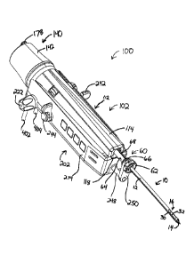
As shown in FIGS. 15-19, a tissue sample holder (140) is provided at the end
of body portion
(112) of probe (102). Tissue sample holder (140) comprises a cup (142), a
manifold (144),
and a plurality of trays (160). Manifold (144) includes a central recess
(146), a plurality of
longitudinal passages (148), a plurality of chambers (150) defined by radially
extending walls
(152), and plurality of radial passages (154). Each longitudinal passage (148)
is substantially
in fluid isolation relative to every other longitudinal passage (148).
However, each radial
passage (154) is substantially in fluid communication with every other radial
passage (154)
via an annular passage (not shown) located within the rear of manifold (144).
Alternatively,
each radial passage (154) may be substantially in fluid isolation relative to
every other radial
passage (154). In the present example, each longitudinal passage (148) is in
fluid
communication with a corresponding one of each radial passage (154). In
particular, each
longitudinal passage (148) terminates proximally in a corresponding radial
passage (154).
In addition, each radial passage (154) is in fluid communication with a
corresponding one of
each chamber (150), via a respective pair of openings (156). Accordingly, it
will be
appreciated that each longitudinal passage (148) is in fluid communication
with a
corresponding chamber (150), via a corresponding radial passage (154) and pair
of openings
(156). In particular, the radial position of each longitudinal passage (148)
relative to central
recess (146) corresponds with the radial position of the associated radial
passage (154), pair
of openings (156), and chamber (150). Of course, any other suitable structures
or
configurations for manifold (144) may be used.
In some variations, a screen, mesh, or other component is provided on or in
manifold (144),
or elsewhere within tissue sample holder (140), to prevent passage of tissue
into or through
certain openings or gaps. In other variations, such components are omitted.
H. Exemplary Tissue Sample Trays
Trays (160) of the present example are configured to be placed on manifold
(144), and to
receive tissue samples (4) as will be described in greater detail below. Each
tray (160) may
be rigid, and may be preformed to have a generally arcuate configuration.
Alternatively,
trays (160) may be formed of a flexible material, such that trays (160) may be
bent to
conform to the curvature of manifold (144). Alternatively, trays (160) may
comprise one or
CA 02644133 2008-11-19
-21 -
more joints, such that portions of trays (160) may bend or flex at such
joints. Still other
suitable configurations may be used.
Each tray (160) of the present example has a base portion (162) and a
plurality of hollow wall
portions (164). Hollow wall portions (164) define chambers (166). By way of
example only,
each chamber (166) may be configured to receive a single tissue sample (4)
captured by
cutter (50). Alternatively, chambers (166) may be configured such that each
chamber (166)
may hold more than one tissue sample (4). Manifold (144) and chambers (166) of
the present
example are further configured such that blood, saline, and/or other fluids
may pass through a
chamber (166) and exit through tube (404), even if a tissue sample (4) is
within such a
chamber (166). In other words, chamber (166) will permit fluids to pass around
a tissue
sample (4).
As shown, the underside of each hollow wall portion (164) is configured to
receive a wall
(152) of manifold (144). Wall portions (164) and walls (152) are configured
such that a gap
is provided between each base portion (162) and manifold (144) when trays
(160) are placed
on manifold (144). As is also shown, each hollow wall portion (164) has a
generally tapered
configuration, though any other suitable configuration may be used. In
addition, trays (160)
have a plurality of openings (168) that are formed, in sets, through the base
portion (162)
within each chamber (164). Accordingly, each chamber (166) of trays (160) is
in fluid
communication with an associated chamber (150) of manifold (144) via openings
(168).
Each longitudinal passage (148) of manifold (144) is therefore in fluid
communication with a
corresponding chamber (166) of trays (160). It will therefore be appreciated
that, when tube
(404) is placed in fluid communication with a given longitudinal passage
(148), tube (404)
will be in fluid communication with the chamber (166) that is associated with
that
longitudinal passage (148).
In the present example, manifold (144) and trays (160) provide eighteen
chambers (150, 166).
Alternatively, any other number of chambers (150, 166) (i.e., more or less
than eighteen) may
be provided. For instance, in one variation, manifold (144) provides three
chambers (150),
and three trays (160) are used that each have only one chamber (166). In yet
another
variation, a single tray (160) is used. For instance, a single tray (160) may
provide a single
CA 02644133 2008-11-19
- 22 -
large chamber (166) or any suitable number of chambers (166). Other suitable
numbers of
chambers (150, 166) and ways in which such chambers (150, 166) may be provided
will be
apparent to those of ordinary skill in the art in view of the teachings
herein. Furthermore,
manifold (144) and trays (160) may have any suitable shape.
Each tray (160) may further comprise one or more types of markings or other
indicia to
distinguish one chamber (166) from another chamber (166). For instance, a
number or other
distinguishing marking may be provided on or near each chamber (166), such as
in relief
form, in recessed form, or otherwise. In another embodiment, a radiopaque
marker is
provided on or near each chamber (166). For instance, an entire tray (160)
that is carrying
one or more tissue samples (4) may be placed under X-ray for evaluation, and
the radiopaque
marker associated with each chamber (166) (and hence, associated with each
tissue sample
(4)), may be visible in the image obtained using X-ray. In other words, tissue
samples (4)
need not necessarily be removed from trays (160) in order to take an X-ray or
radiograph
image of tissue samples (4). Furthermore, trays (160) may be dropped directly
into formalin
or any other liquid with tissue samples (4) still on trays (160). In addition,
trays (160) may be
placed in a sleeve or container, etc., individually or in groups, to protect
tissue samples (4)
and/or to ensure that tissue samples (4) stay in trays (160) or for other
purposes. Such a
sleeve or container may be flexible, rigid, or have other properties. By way
of example only,
a sleeve or other container may be flat, and may be configured to flatten out
a flexible tray
(160) that is inserted therein. Other structures and techniques that may be
used with trays
(160), such as after tissue samples (4) are communicated to trays (160) will
be apparent to
those of ordinary skill in the art in view of the teachings herein.
Cup (142) is configured to engage bayonets (134) of base member (116), such
that cup (142)
may be removed from or secured to base member (116) upon sufficient rotation
of cup (142)
relative to base member (116). In addition, an o-ring (136) is provided about
base member
(116) to provide a seal between base member (116) and cup (142). Of course,
any other
suitable structures may be used to provide engagement of cup (142) with base
member (116)
and/or to provide a seal between base member (116) and cup (142). Cup (142) is
also formed
of a transparent material in the present example, enabling the user to
visually inspect tissue
samples (4) in tissue sample holder (140) while tissue sample holder (140) is
still coupled
CA 02644133 2008-11-19
- 23 -
with base member (116). For instance, a user may inspect tissue samples (4)
for color, size,
and density (e.g., to the extent that chamber (166) is full of saline, etc.).
It will also be appreciated in view of the teachings herein that the
removability of cup (142)
and trays (160) may permit a user to harvest a relatively large number of
tissues samples in a
relatively short period of time. Furthermore, the removability of cup (142)
and trays (160)
may permit a user to remove unsatisfactory tissue samples (4) from tissue
sample holder
(140) (e.g., using tweezers, etc.) and then re-couple trays (160) and cup
(142) for further
sampling. Other ways in which the removability and other properties of tissue
sample holder
(140) of the present example may be utilized will be apparent to those of
ordinary skill in the
art in view of the teachings herein.
I. Exemplary Rotation and Alignment of Manifold
Manifold (144) of the present example is configured to rotate relative to base
member (116),
as will be described in greater detail below. Manifold (144) of the present
example is further
configured such that each longitudinal passage (148) may be selectively
aligned with a port
(406) that is in fluid communication with tube (404). Such alignment of a
longitudinal
passage (148) and port (406) will place the aligned longitudinal passage (148)
in fluid
communication with tube (404), such that induction of a vacuum within tube
(404) will effect
induction of a vacuum within longitudinal passage (148), as well as within the
chamber (166)
associated with that longitudinal passage (148). In addition, manifold (144)
and trays (160)
of the present example are configured such that each chamber (166) may be
selectively
placed in fluid communication with cutter lumen (52). It will therefore be
appreciated that a
vacuum in tube (404) may induce a vacuum in cutter lumen (52), with the vacuum
being
communicated via port (406), an associated longitudinal passage (148), an
associated radial
passage (154), an associated pair of openings (156), an associated chamber
(150), an
associated set of openings (168), and an associated chamber (166). Of course,
there are a
variety of other ways in which a vacuum may be induced within a cutter lumen
(52), and any
other suitable structures or techniques may be used. Furthermore, pressurized
air, a liquid
(e.g., saline), or any other fluid may be communicated in either direction
through the above-
mentioned components in lieu of or in addition to a vacuum being induced
therein.
CA 02644133 2008-11-19
- 24 -
A gear (170) is engaged with manifold (144) of the present example. In
particular, gear (170)
has a shaft (172) that is inserted within central recess (146) of manifold
(144). The shaft
(172) has a flat (174) that is configured to engage a complimentary flat (147)
of central recess
(146). Engagement of flats (174, 147) is such that gear (170), shaft (172),
and manifold (144)
rotate unitarily. Alternatively, gear (170) and manifold (144) may have any
other suitable
configurations or relationships. Nevertheless, gear (170) of the present
example may be used
to rotate manifold (144), which will in turn permit selective alignment of
longitudinal
passages (148) with port (406), in addition to contemporaneously permitting
selective
alignment of chambers (166) with cutter lumen (52). In particular, and as will
be described in
greater detail below, gear (170) is configured to mesh with a complimentary
gear (210) of
holster (202), such that gear (210) may be used to impart rotation to gear
(170). Such
rotation may be used to selectively (e.g., consecutively) align chambers (166)
with cutter
lumen (52), to successively collect a discrete tissue sample (4) in each
chamber (166) during
use of biopsy device (100). Furthermore, such collection of tissue samples (4)
may be
performed without having to withdraw and re-insert needle portion (10)
relative to patient
during such a process.
J. Exemplary "Parking Pawl"
Body portion (112) of the present example further comprises an engagement
member (180),
which is secured to base member (116). As shown in FIG. 20, engagement member
(180)
comprises a pawl portion (182) having teeth (184). Pawl portion (182) is
resiliently urged for
teeth (184) to engage with gear (170). In particular, engagement of teeth
(184) of pawl
portion (182) with gear (170) prevents rotation of gear (170) (and hence,
prevents rotation of
manifold (144)). Accordingly, pawl portion (182) is configured to prevent
rotation of
manifold (144) when pawl portion (182) is in a default position. In the
present example, pawl
portion (182) is in the default position when biopsy probe (102) is not
coupled with a holster
(202). However, when biopsy probe (102) is coupled with a holster (202), a
boss (212) on
holster (202) is configured to engage pawl portion (182). In particular, boss
(212) on holster
(202) is configured to disengage pawl portion (182) from gear (170) when
biopsy probe (102)
is coupled with a holster (202), such that pawl portion (182) will no longer
prevent rotation of
gear (170) or manifold (144) when biopsy probe (102) is coupled with a holster
(202). When
CA 02644133 2008-11-19
- 25 -
biopsy probe (102) is removed from holster (202), the resilience of engagement
member
(180) urges pawl portion (182) back to the default position, such that pawl
portion (182) will
again prevent rotation of gear (170) and manifold (144).
When biopsy probe (102) is packaged for shipment from a manufacturing
facility, or in other
situations, tissue sample holder (140) may be configured such that a
predetermined chamber
(166) is aligned with cutter lumen (52). With pawl portion (182) maintaining
such alignment
to the time when biopsy probe (102) is coupled with a holster (202) for a
first use, software or
control logic that is used to control biopsy device (100) may "safely assume"
that the
predetermined chamber (166) is aligned with cutter lumen (52), and may control
biopsy
device (100) accordingly. Furthermore, if biopsy probe (102) is removed from
holster (202)
during a tissue sample (4) acquisition procedure, software or control logic
that is used to
control biopsy device (100) may "remember" which chamber (166) was last
aligned with
cutter lumen (52), to the extent that software tracks which chamber (166) is
being or has been
used during a procedure. If biopsy probe (102) is recoupled with holster (202)
to continue
the procedure, the software or control logic may continue to control biopsy
device (100)
based on the chamber (166) that the software "remembered." Alternatively, a
user may
specify that a new biopsy probe (102) has been coupled with holster (202),
which may result
in the software or control logic again "assuming" that the predetermined
chamber (166) is the
one that is aligned with the cutter lumen (52).
While a pawl portion (182) has been described as a structure selectively
preventing the
rotation of gear (170) and manifold (144), it will be appreciated that any
other alternative
structures may be used for such purposes. By way of example only, a Geneva
wheel
mechanism (not shown) may be used as an alternative mechanism for rotating
manifold (144)
and maintaining the rotational position of manifold (144) between intentional
rotations. For
instance, gear (170) may be substituted with a Geneva driven wheel (not
shown), while gear
(210) may be substituted with a Geneva drive wheel (not shown). Other suitable
alternatives
for rotating manifold (144) and/or maintaining the rotational position of
manifold (144) will
be apparent to those of ordinary skill in the art in view of the teachings
herein. In addition, it
will be appreciated that a biopsy device (100) may lack a pawl portion (182)
or other rotation
CA 02644133 2015-10-27
- 26 -
prevention feature altogether, such that a manifold (144) may freely rotate
when biopsy probe
(102) is not coupled with a holster (202).
K. Exemplary Dedicated Passage
As shown in FIGS. 16-17, 19, and 21, tissue sample holder (140) of the present
example has
a passage (158) formed through manifold (144). Passage (158) extends
longitudinally,
completely through manifold (144), and is offset from but parallel with the
central axis
defined by manifold (144). Like chambers (166), passage (158) is configured to
be
selectively aligned with cutter lumen (52). However, unlike chambers (166),
passage (158) is
not in fluid communication with any of longitudinal passages (148) or radial
passages (154).
In other versions, passage (158) may be provided in fluid communication with
one or more
longitudinal passages (148) and/or radial passages (154).
Passage (158) of the present example is configured to permit instruments
and/or liquids, other
materials, etc., to be passed through manifold (144) and through cutter lumen
(52). For
instance, passage (158) may be used to insert an instrument for deploying one
or more
markers at a biopsy site, via cutter lumen (52) and via outer cannula (12),
out through
aperture (16). A merely exemplary marker applier that may be inserted through
passage
(158) may include the MAMMOMARK biopsy site marker applier, by Ethicon Endo-
Surgery, Inc. of Cincinnati, Ohio. Other suitable marker applier devices that
may be inserted
through passage (158) may include any of those described in U.S. Patent No.
7,047,063; U.S.
Patent No. 6,996,433; U.S. Patent No. 6,993,375; or U.S. Pub. No.
2005/0228311. Any of
such appliers, including variations of the same, may be introduced through
passage (158) to
deploy one or more markers at a biopsy site, via aperture (16), while needle
portion (10)
remains inserted in a patient (e.g., shortly after biopsy samples are
extracted from the patient,
etc.). Such marker deployment may be accomplished even while tissue samples
(4) reside
within tissue sample holder (140), secured to biopsy probe (102).
Alternatively, such marker
appliers may be inserted directly into cutter lumen (52) with tissue sample
holder (140) being
removed from biopsy probe (102).
CA 02644133 2008-11-19
- 27 -
As noted above, biopsy probe (102) may be initially provided with a
predetermined chamber
(166) being aligned with cutter lumen (52) by default. However, in other
versions, biopsy
probe (102) is initially provided with passage (158) being aligned with cutter
lumen (52) by
default. Furthermore, to the extent that a user desires having passage (158)
aligned with
cutter lumen (52) during use of biopsy device (100), after manifold (144) has
been rotated
during such use, the controls may be used to command manifold (144) to rotate
to align
passage (158) with cutter lumen (52).
Cup (142) further comprises an opening (176) and a hatch (178). Opening (176)
is
configured to be aligned with passage (158) when cup (142) is secured to base
member (116),
such as by rotating manifold (144) to align passage (158) with opening (176).
Hatch (178) is
configured to selectively cover opening (176). For instance, hatch (178) may
be configured
to seal opening (176) when hatch (178) covers opening (176). Hatch (178) may
further be
configured to permit a user to "peel back" hatch (178) and/or pivot hatch
(178) in order to
gain access to opening (176) and passage (158). It will be appreciated in view
of the
disclosure herein that hatch (178) may be substituted or supplemented with a
variety of
alternative structures, including but not limited to a removable stopper or
other structure.
L. Exemplary Medicine Applier
As shown in FIGS. 21-22, an applier (90) may be coupled with biopsy probe
(102) via
opening (176) in cup (142) and passage (158) in manifold (144). In this
example, applier
(90) comprises a hollow shaft portion (92) and a luer lock portion (94). Shaft
portion (92) is
sized and configured such that, when applier (90) is inserted through opening
(176) and
through passage (158), shaft portion (92) creates a seal with cutter lumen
(52) (e.g., through
engagement with the inner surface of cutter lumen (52)). Shaft portion (92)
and luer lock
portion (94) may thereby be placed in fluid communication with cutter lumen
(52). By way
of example only, a syringe (not shown) or other device may be coupled with
luer lock portion
(94). A therapeutic agent may thus be injected from such a syringe, through
applier (90),
through cutter lumen (52), through outer cannula (12), and out through
aperture (16) to reach
a biopsy site. Such injections may be made before or after tissue samples (4)
are acquired
using biopsy device (100), and may be made while needle portion (10) remains
inserted in the
CA 02644133 2008-11-19
- 28 -
patient. Other suitable ways in which an applier (90) may be used, as well as
alternative
ways in which an applier (90) may be configured, will be apparent to those of
ordinary skill
in the art in view of the teachings herein. By way of example only, applier
(90) may
alternatively be inserted directly into cutter lumen (52) with tissue sample
holder (140) being
removed from biopsy probe (102).
11. Exemplary Holster for Stereotactic Use
As shown in FIGS. 23-32, a holster (202) comprises a top cover (204), through
which a
portion of each of gears (206, 208, 210) is exposed, side panels (214, 216),
and a base
member (218). As described above, boss (212) is provided on top cover (204),
and is
configured to disengage pawl portion (182) from gear (170) when biopsy probe
(102) is
coupled with holster (202). Holster (202) of this example further comprises a
needle rotation
mechanism (220), a needle firing mechanism (240), a cutter drive mechanism
(270), and a
tissue holder rotation mechanism (280). In addition, a user interface (800) is
provided on
each side panel (214, 216). Each of these merely exemplary components will be
described in
greater detail below.
As noted above, holster (202) of the present example is configured to be
coupled with a
biopsy probe (102), such as biopsy probe (102) described above, to provide a
biopsy device
(100). In addition, holster (202) is configured to be mounted to a table,
fixture, or other
device, such as for use in a stereotactic or X-ray setting. However, it will
be appreciated in
view of the disclosure herein that holster (202) may be used in a variety of
other settings and
combinations.
A. Exemplary Needle Rotation Mechanism
In the present example, and as shown in FIG. 27, needle rotation mechanism
(220) comprises
a pair of knobs (222), each of which has a respective gear (224) in beveled
engagement with
a gear (226) on the proximal end of an elongate shaft (228). Another gear (not
shown),
which is provided on the distal end of shaft (228), is engaged with gear
(230). Gear (230) is
engaged with yet another gear (232) on the proximal end of yet another shaft
(234). The
distal end of shaft (234) has another gear (236), which is engaged with gear
(206) described
CA 02644133 2008-11-19
- 29 -
above. It will therefore be appreciated in view of the disclosure herein that
rotation of one or
both of knobs (222) will result in rotation of gear (206), with such rotation
being
communicated via gears (224, 226, 230, 236) and shafts (228, 234).
Furthermore, as also
noted above, when biopsy probe (102) is coupled with holster (202), gear (206)
will mesh
with gear (74). Thus, when biopsy probe (102) is coupled with holster (202),
rotation of one
or both of knobs (222) will cause needle portion (10) of biopsy probe (102) to
rotate. Of
course, a variety of alternative mechanisms, structures, or configurations may
be used as a
substitute or supplement for needle rotation mechanism (220). By way of
example only, a
motor (not shown) may be used to effect rotation of needle portion (10). In
other versions,
needle rotation mechanism (220) may simply be omitted altogether.
B. Exemplary Needle Firing Mechanism
As shown in FIGS. 28-29, needle firing mechanism (240) of the present example
comprises a
pair of triggers (242), buttons (244), a motor (246), a firing rod (248), and
a fork (250). Fork
(250) is configured to engage sleeve portion (64) of needle hub (60) when
biopsy probe (102)
is coupled with holster (202). For instance, fork (250) may engage sleeve
portion (64)
between thumbwheel (62) and annular projection (66). In the present example,
engagement
between fork (250) and sleeve portion (64) is such that sleeve portion (64)
(and therefore,
needle portion (10)) will translate longitudinally with fork (250). Fork (250)
is coupled with
firing rod (248), such that fork (250) will translate longitudinally with
firing rod (248).
A damper (252) with a washer (253) is provided about firing rod (248). A coil
spring (254) is
also provided about firing rod (248). In particular, coil spring (254) is
engaged with both
washer (253) and a portion of base member (218). Coil spring (254) is biased
to urge damper
(252), washer (253), and firing rod (248) distally. It will be appreciated,
however, that like
other components described herein, coil spring (254) is merely exemplary, and
a variety of
alternative components (resilient or otherwise) may be used in addition to or
in lieu of coil
spring (254).
A sled (256) and a screw gear (258) are also coupled with firing rod (248). In
particular, sled
(256) is coupled with the proximal end of firing rod (248), and is configured
to longitudinally
translate unitarily with firing rod (248). Similarly, screw gear (258) is
configured to
CA 02644133 2008-11-19
- 30 -
longitudinally translate with firing rod (248) (through at least some range of
motion), while
being prevented from rotating about firing rod (248). An outer gear (260) is
engaged with
screw gear (258). In particular, the interior (not shown) of outer gear (260)
is engaged with
the threads of screw gear (258); such that when outer gear (260) rotates
relative to screw gear
(258), such rotation causes screw gear (258) to longitudinally translate
relative to outer gear
(260). Outer gear (260) is in communication with another gear (262), which is
itself in
communication with a gear (264) that is coupled with motor (246). Accordingly,
when motor
(246) is activated to rotate, such rotation will cause screw gear (258),
firing rod (248), and
sled (256) to longitudinally translate. In other words, rotation of motor
(246) will be
communicated to outer gear (260) via gears (262, 264), and such rotation will
be converted to
longitudinal motion due to the configuration and engagement of outer gear
(260) and screw
gear (258). Of course, all of these components are merely illustrative, and
any other suitable
components, configurations, or techniques may be used to cause longitudinal
translation of
firing rod (248).
Triggers (242) of the present example are each configured to partially rotate
forward and
rearward, while buttons (244) are configured to be pressed inward. In
addition, a plurality of
switches (not shown) may be communicatively coupled with triggers (242) and/or
buttons
(244), such that the switches are selectively activated by a user when
triggers (242) are
moved forward or rearward and/or when buttons (244) are depressed. One or more
resilient
members (e.g., a spring, etc.) may be included to bias each trigger (242) to a
centered or
substantially vertical orientation. One or more resilient members (e.g., a
spring, etc.) may
also be included to bias each button (244) to an outward position. Triggers
(242) and buttons
(244) are also sealed in the present example to prevent ingress of fluid into
holster (202),
though like other features, this is merely optional.
In the present example, triggers (242) are further configured such that, when
one or both of
triggers (242) are moved rearward, such movement activates a switch that is in
communication with motor (246). Such activation causes motor (246) to rotate,
which in turn
causes firing rod (248) to longitudinally translate proximally as described
above. As will be
described in greater detail below, such rearward movement of trigger (242) may
thus cause
motor (246) to arm or "cock" the needle firing mechanism (240).
CA 02644133 2008-11-19
-31 -
Needle firing mechanism (240) of the present example further comprises a catch
(266), which
is configured to selectively engage sled (256). In particular, as firing rod
(248) and sled (256)
are longitudinally translated proximally (e.g., by rotation of motor (246)),
sled (256)
approaches catch (266). When catch (266) and sled (256) engage, catch (266) is
configured
to hold sled (256) (and therefore, firing rod (248)) in place. Catch (266) may
maintain such
position of sled (256) even after motor (246) has stopped rotating, and even
with spring (254)
urging sled (256) and firing rod (248) toward a distal position. When these
components are
in these proximal positions and configurations, needle firing mechanism (240)
may be said to
be in a "cocked" configuration. A merely exemplary cocked configuration of
needle firing
mechanism (240) is shown in FIG. 29.
It will be appreciated in view of the teachings herein that, with needle
firing mechanism (240)
in such a cocked configuration, fork (250) and needle portion (10) will be at
a proximal,
ready-to-fire position. One or more components of biopsy device (100) may be
configured to
provide an audio and/or visual indication that the needle firing mechanism
(240) is fully
cocked. For instance, biopsy device (100) may produce a distinct clicking
sound, beep, or
other audible signal; and/or a graphical user interface may provide some
visual indication that
the needle firing mechanism (240) is cocked.
In addition, holster (202) may further include one or more sensors (not shown)
or other
feature(s) configured to sense or detect when needle firing mechanism (240)
has been cocked
and/or when needle firing mechanism (240) has been fired. For instance, biopsy
system (2)
may be configured such that one or more functions of biopsy system (2) are
essentially
disabled while needle firing mechanism (240) is cocked, until needle firing
mechanism (240)
is fired. By way of example only, biopsy system (2) may prevent initiation of
a "sample"
cycle (described below), initiation of a "clear probe" cycle (described
below), or other
functions while needle firing mechanism (240) is cocked. Such functions may be
again
permitted after needle firing mechanism (240) has been fired and after needle
(10) has
reached a fully fired position. Alternatively, cocking of needle firing
mechanism (240) may
have no affect or other affects on one or more functions of biopsy system (2).
CA 02644133 2008-11-19
- 32 -
In one variation, after sled (256) has been moved into engagement with catch
(266) to cock
needle firing mechanism (240), motor (246) may reverse its rotation. In this
variation, a
proximal portion of firing rod (248) may have a longitudinal slot or recess
(not shown)
formed transversely through or in firing rod (248). Screw gear (258) may have
an internal
pin or other feature (not shown) that is configured to engage such a slot or
other feature of
firing rod (248), such that the pin or other feature of screw gear (258) is
further configured to
both prevent screw gear (258) from rotating about firing rod (248) and permit
screw gear
(258) to translate through some range of motion relative to firing rod (248).
For instance,
before needle firing mechanism (240) is cocked, such a pin or other feature of
screw gear
(258) may be positioned at or near the proximal end of a longitudinal slot or
recess of firing
rod (248); such that as motor (246) is activated to translate screw gear (258)
proximally to
cock needle firing mechanism (240), the pin or other feature engages firing
rod (248) to urge
firing rod (248) proximally with screw gear (258). Then, after sled (256) has
been moved
proximally into engagement with catch (266), motor (246) may reverse its
rotation. Such
reversal of motor (246) rotation may translate screw gear (258) distally. The
configuration of
the slot or other feature of firing rod (248) and the configuration of the pin
or other feature of
screw gear (258) may permit such distal translation of screw gear (258)
relative to firing rod
(248), leaving firing rod in a proximal cocked position. Furthermore, when
needle portion
(10) is fired as described below, the configuration of the slot or other
feature of firing rod
(248) and the configuration of the pin or other feature of screw gear (258)
may permit firing
rod (248) to translate distally relative to screw gear (258) with relative
ease during such
firing. Other suitable relationships between firing rod (248) and screw gear
(258) may be
used, including but not limited to a variation described below.
When a user is ready to fire needle portion (10), the user may push and hold
one or both of
triggers (242) forward, and may push one or both buttons (244) in while one or
both of
triggers (242) are held forward. Such actuation of trigger(s) (242) and
button(s) (244) may
cause catch (266) to release sled (256). Suitable structures and
configurations that may be
used to cause actuation of trigger(s) (242) and button(s) (244) to result in
catch (266)
releasing sled (256) will be apparent to those of ordinary skill in the art in
view of the
teachings herein. With sled (256) being so released, the resilience of spring
(254) may urge
CA 02644133 2008-11-19
-33 -
damper (252) and washer (253) (and therefore, firing rod (248), fork (250),
and needle
portion (10)) distally, thereby firing needle portion (10). Such distal motion
of needle portion
(10) may be relatively sudden, and may be performed with a force sufficient to
penetrate
tissue with tip (14) of needle portion (10).
In another variation, motor (246) does not reverse its rotation to advance
screw gear (258)
back to a distal position before needle portion (10) is fired. For instance,
screw gear (258)
may be unitarily secured to firing rod (248), and may be unable to translate
longitudinally in
either direction through any range of motion relative to firing rod (248). In
this variation, as
needle portion (10) is fired, gears (260, 262, 264) may be configured to
rotate freely, thereby
providing negligible resistance to distal motion of firing rod (248).
Alternatively, a clutch
mechanism (not shown) may be provided to disengage one or more of gears (260,
262, 264)
during firing of needle portion (10). Other ways in which a needle firing
mechanism (240)
may be configured or operated will be apparent to those of ordinary skill in
the art in view of
the teachings herein.
In the present example, triggers (242) and buttons (244) are configured such
that pushing or
actuation of buttons (244) will have no firing effect unless triggers (242)
are held forward.
Similarly, holding of triggers (242) will not cause firing of needle portion
(10) until buttons
(244) are also pressed while triggers (242) are held forward. Suitable
structures and
configurations for providing such interdependence of triggers (242) and
buttons (244) will be
apparent to those of ordinary skill in the art. For instance, buttons (244)
may rotate with
triggers (242), such that buttons (244) rotate forward with triggers (242). In
such versions,
buttons (244) and catch (266) may be configured such that actuation of buttons
(244) will not
cause catch (266) to release sled (256) unless buttons (244) are rotated
forward. In addition
or in the alternative to buttons (244) rotating with triggers (242), triggers
(242) may be
configured to lock catch (266) in place (e.g., even with buttons (244) being
actuated) until
triggers (242) are rotated forward, such that forward rotation of triggers
(242) will permit
catch (266) to be released when buttons (244) are actuated. Other ways in
which triggers
(242) and buttons (244) may be provided as interdependent for purposes of
firing (or for
other purposes) will be apparent to those of ordinary skill in the art in view
of the teachings
herein.
CA 02644133 2008-11-19
- 34 -
C. Exemplary Cutter Drive Mechanism
As shown in FIG. 30, cutter drive mechanism (270) of the present example
comprises a
motor (272) with a shaft (274) extending therefrom. Gear (208) is mounted to
shaft (274),
and is configured to rotate unitarily therewith. As noted above, a portion of
gear (208) is
exposed through top cover (204), such that gear (208) meshes with gear (138)
of cutter
rotation and translation mechanism (120) when biopsy probe (102) is coupled
with holster
(202). Accordingly, when motor (272) is activated to rotate, such rotation may
be
communicated via shaft (274) and gears (208, 138), to effect simultaneous
rotation and
translation of cutter (50) as described above. Other ways in which a cutter
drive mechanism
(270) may be configured or operated will be apparent to those of ordinary
skill in the art in
view of the teachings herein.
D. Exemplary Tissue Holder Rotation Mechanism
As shown in FIGS. 31-32, tissue holder rotation mechanism (280) of the present
example
comprises a motor (282) having a shaft (284) with a gear (286) mounted
thereto, such that
gear (286) rotates unitarily with shaft (284). Gear (286) is configured to
mesh with gear
(288), which is mounted to shaft (290). Gear (210), which has been noted
above, is also
mounted to shaft (290), at the proximal end of shaft (290). In particular,
gear (210) is
configured to mesh with gear (170) of tissue sample holder (140) when biopsy
probe (102) is
coupled with holster (202). Accordingly, when motor (282) is activated to
rotate, such
rotation may be communicated via shafts (284, 290) and gears (286, 288, 210,
170), to effect
rotation of manifold (144) as described above.
In addition, an encoder wheel (292) is coupled with shaft (290), and is
configured to rotate
unitarily therewith. Encoder wheel (292) has a plurality of slots (294) formed
therethrough.
Slots (294) fan radially outward, and are angularly spaced apart relative one
another. Of
course, slots (294) may have any other suitable configuration. A sensor (296)
is positioned
adjacent to encoder wheel (292). In particular, sensor (296) is positioned
such that slots
(294) successively pass before sensor (296) as encoder wheel (292) rotates
with shaft (290).
Sensor (296) may therefore be used to count the passage of slots (294), which
may be
translated into data indicative of the rotational position of manifold (144).
In other words,
CA 02644133 2008-11-19
- 35 -
since encoder wheel (292) and manifold (144) rotate concomitantly when biopsy
probe (102)
is coupled with holster (202) in the present example, the passage of slots
(294) past sensor
(296) during rotation of shaft (290) may be indicative of manifold (144)
rotation, and
therefore of manifold (144) position. It will be appreciated that information
indicative of
manifold position (144) may be further indicative of which particular chamber
(166) is
aligned with cutter lumen (52). Suitable uses for such information will be
apparent to those
of ordinary skill in the art in view of the teachings herein.
Suitable devices that may be used for sensor (296) will also be apparent to
those of ordinary
skill in the art in view of the teachings herein. Similarly, suitable
substitutes for encoder
wheel (292) and sensor (296) will be apparent to those of ordinary skill in
the art, including
but not limited to combinations of magnets and hall effect sensors, light
sources and
photosensors, etc. Furthermore, other ways in which a tissue holder rotation
mechanism
(280) may be configured or operated will be apparent to those of ordinary
skill in the art in
view of the teachings herein.
III. Exemplary Probe for Ultrasound Use
As shown in FIGS. 33-37, an alternative biopsy probe (103) comprises a needle
portion (350)
and a body portion (352). Body portion (352) comprises a cover member (354)
and a base
member (356). A tissue sample holder (368) is removably secured to base member
(356),
though tissue sample holder (368) may alternatively be secured to cover member
(354) or
some other component. As will be described in greater detail below, a pair of
tubes (402,
404) are coupled with probe (103). As will also be described in greater detail
below, and as
noted above, biopsy probe (103) is configured to be coupled with a holster
(302) to provide a
biopsy device (101).
A. Exemplary Needle
In the present example, needle portion (350) comprises an outer cannula (12)
having a tissue
piercing tip (14) and a transverse tissue receiving aperture (16) located
proximally from the
tissue piercing tip (14). In this example, these components are essentially
the same as the
components bearing the same names and item numbers described above, so they
will not be
CA 02644133 2008-11-19
- 36 -
described in greater detail here. In other words, the features, properties,
and components of
outer cannula (12), tip (14), and aperture (16) as described above (including
cannula lumen
(20), vacuum lumen (40), wall (30), transverse openings (32), etc.) may be the
same for
needle portion (350) as they were described above with respect to needle
portion (10). Of
course, they may alternatively be varied in any suitable way, as desired.
Similarly, cutter (50) in probe (103) may have the same relationship with
needle portion
(350) as the relationship described above between cutter (50) and needle
portion (10); as well
as all the same features, properties, and components as cutter (50) described
above in the
context of probe (102). Such aspects of cutter (50) will also therefore not be
repeated here.
B. Exemplary Needle Hub
As shown in FIGS. 36-37, a needle hub (358) is secured to outer cannula (12)
of probe (103),
and comprises a thumbwheel (62) and a sleeve portion (360) extending
proximally from
thumbwheel (62). Needle hub (358) of the present example is overmolded about a
proximal
portion of outer cannula (12), though needle hub (358) may be formed and/or
secured relative
to outer cannula (12) using any other suitable techniques (e.g., set screws,
etc.). Furthermore,
while needle hub (358) of the present example is formed of a plastic material,
any other
suitable material or combination of materials may be used.
Sleeve portion (360) of the present example comprises an annular projection
(66), a plurality
of flats (362), and a transverse opening (70), which is formed near the
proximal end of sleeve
portion (360). A pair of o-rings (72) are positioned such that one o-ring (72)
is proximal to
transverse opening (70) and another o-ring (72) is distal to transverse
opening (70). As will
be described in greater detail below, transverse opening (70) is in fluid
communication with
the interior defined by needle hub (60), as well as with vacuum lumen (40) of
outer cannula
(12). In the present example, another transverse opening (70) is formed
through sleeve
portion (360), also between o-rings (72), and opposite to the other transverse
opening (70).
Other suitable configurations for sleeve portion (360) will be apparent to
those of ordinary
skill in the art in view of the teachings herein.
CA 02644133 2008-11-19
- 37 -
Thumbwheel (62) of sleeve portion (360) is essentially the same as, and may be
operated in a
manner similar to, thumbwheel (62) of sleeve portion (64) of probe (102)
described above.
Thumbwheel (62) will therefore not be discussed in any greater detail here. Of
course,
thumbwheel (62) may alternatively be varied in any suitable way, as desired,
if not omitted
altogether, in the case of either probe (102, 103).
In the present example, an exposed gear (364) is slid onto sleeve portion
(360). In particular,
the interior of gear (364) is configured to mate with flats (362) of sleeve
portion (360), such
that gear (364) rotates unitarily with sleeve portion (360). With sleeve
portion (360) being
unitarily engaged with outer cannula (12), rotation of gear (364) will further
cause rotation of
cannula (12) for reorienting aperture (16). Gear (364) is exposed through base
member
(356), and is further configured to engage with a complimentary exposed gear
(not shown) of
a holster (not shown). In particular, gear (364) is configured to mesh with a
complimentary
exposed gear such that the complimentary gear can impart rotation to gear
(364), thereby
rotating outer cannula (12). However, in the present example, gear (364) is
not engaged with
a complimentary gear when probe (103) is coupled with holster (302). It will
therefore be
appreciated that, like other components and features described herein, gear
(364) and flats
(362) may simply be omitted if desired.
C. Exemplary Needle Manifold
As shown in FIGS. 34-36, a needle manifold (366) is provided about sleeve
portion (360).
Needle manifold (366) is fixed relative to base member (356) in this example.
Needle
manifold (366) is in fluid communication with tube (402), such that tube (402)
may
communicate saline, a vacuum, and/or pressurized air, etc., to needle manifold
(366) as will
be described in greater detail below. Needle manifold (366) is further in
fluid
communication with the interior of sleeve portion (360), via transverse
openings (70), one of
which is shown in FIG. 37. 0-rings (64) are configured to maintain a fluid
seal between
needle manifold (366) and sleeve portion (360), even as sleeve portion (360)
rotates relative
to needle manifold (366). A seal (not shown) is may also provided at the
proximal end of
sleeve portion (360), at the interface between sleeve portion (360) and cutter
(50). Needle
manifold (366), sleeve portion (360), and outer cannula (12) are thus
configured and arranged
CA 02644133 2008-11-19
- 38 -
such that saline, a vacuum, and/or pressurized air, etc. that is communicated
via tube (402) to
needle manifold (366) will be communicated to vacuum lumen (40) via transverse
openings
(70). Of course, any other suitable structures or arrangements may be used to
communicate
saline, a vacuum, and/or pressurized air, etc. from tube (402) to vacuum lumen
(40).
D. Exemplary Cutter Rotation and Translation Mechanism
In the present example, and as shown in FIGS. 34-35, body portion (350) of
probe (103)
comprises a cutter rotation and translation mechanism (120), which is operable
to rotate and
translate cutter (50) within outer cannula (12). Cutter rotation and
translation mechanism
(120) in this example has essentially the same components, features, and
operability of the
cutter rotation and translation mechanism (120) described above with respect
to probe (102).
Cutter rotation and translation mechanism (120) will therefore not be
discussed in any greater
detail here. Of course, cutter rotation and translation mechanism (120) may
alternatively be
varied in any suitable way, as desired, in the case of either probe (102,
103).
E. Exemplary "Sharps Reduction" Variation
In addition, needle portion (350) and cutter (50) of biopsy probe (103) may be
configured to
be removable from biopsy probe (103) in essentially the same manner as
described above
with respect to removability of needle portion (10) from biopsy probe (102).
For instance,
body portion (352) may include a feature similar to release tab (118), or any
other suitable
feature, to provide, permit, or facilitate removability of needle portion
(350) and cutter (50)
from body portion (352).
F. Exemplary Tissue Sample Holder Manifold
As shown in FIGS. 38-40, a tissue sample holder (368) is provided at the end
of body portion
(352) of probe (103). Tissue sample holder (368) comprises a cup (142), a
manifold (370),
and a plurality of trays (372). Manifold (370) includes a central recess
(146), a plurality of
openings (374), and a longitudinally extending sidewall (382). Sidewall (382)
only extends
for a portion of the length of manifold (370) in this example, though sidewall
(382) may
alternatively extend to any other degree as desired. Manifold (370) also
includes a plurality
of radially extending walls (380). Walls (380) and the interior surface of
sidewall (382)
CA 02644133 2008-11-19
- 39 -
define a plurality of longitudinal passages (376). Each longitudinal passage
(376) is in fluid
communication with a corresponding opening (374).
In addition, walls (380) and the exterior surface of sidewall (382) define a
plurality of
chambers (378). With sidewall (382) providing clearance (e.g., by not
extending the full
length of manifold (370)), each chamber (378) is in fluid communication with a
corresponding longitudinal passage (376). Manifold (370) is thus configured
such that each
opening (374) is in fluid communication with a corresponding chamber (378). Of
course, any
other suitable structures or configurations for manifold (370) may be used.
For instance,
manifold (144) described above with respect to biopsy probe (102) may be used
with biopsy
probe (103) in lieu of manifold (370) being used with biopsy probe (103).
Likewise,
manifold (370) may be used with biopsy probe (102) in lieu of manifold (144)
being used
with biopsy probe (102).
G. Exemplary Tissue Sample Trays
Trays (372) of the present example are configured to be placed on manifold
(370), and to
receive tissue samples (4) as will be described in greater detail below. Each
tray (372) has a
plurality of base portions (382), a plurality of hollow wall portions (384),
and a plurality of
webs (386). Base portions (392), hollow wall portions (384), and webs (386)
define
chambers (388). By way of example only, each chamber (388) may be configured
to receive
a single tissue sample (4) captured by cutter (50). Alternatively, chambers
(388) may be
configured such that each chamber (388) may hold more than one tissue sample
(4). As
shown, the underside of each hollow wall portion (384) is configured to
receive a wall (380)
of manifold (370). As is also shown, each hollow wall portion (384) has a
generally tapered
configuration, though any other suitable configuration may be used.
In addition, trays (372) have a plurality of openings (390), extending
longitudinally, formed
through the base portion (392) within each chamber (388). Openings (390)
continue,
extending radially outwardly, through a portion of each web (386).
Accordingly, with
sidewall (382) not extending the full length of manifold (370), the openings
(390) permit
fluid communication between each longitudinal passage (376) and each
corresponding
CA 02644133 2008-11-19
- 40 -
chamber (388). In other words, each opening (374) is in fluid communication
with a
corresponding chamber (388).
Each tray (372) may further comprise one or more types of markings or other
indicia to
distinguish one chamber (388) from another chamber (388). Such markings or
indicia may
be similar to the same described above with respect to chambers (166) of trays
(160).
Accordingly, discussion of such markings or indicia will not be repeated here.
Similarly, cup
(142) of tissue sample holder (368) is essentially the same as cup (142) of
tissue sample
holder (140) described above. Discussion of cup (142) will therefore not be
repeated here.
H. Exemplary Rotation and Alignment of Manifold
Manifold (370) of the present example is configured to rotate relative to base
member (356),
as will be described in greater detail below. Manifold (370) of the present
example is further
configured such that each opening (374) may be selectively aligned with a port
(not shown)
that is in fluid communication with tube (404). Such alignment of an opening
(374) and such
a port will place the aligned opening (374) in fluid communication with tube
(404), such that
induction of a vacuum within tube (404) will effect induction of a vacuum
through opening
(374), as well as within the chamber (388) associated with that opening (374).
In addition,
manifold (370) and trays (372) of the present example are configured such that
each chamber
(388) may be selectively placed in fluid communication with cutter lumen (52).
It will
therefore be appreciated that a vacuum in tube (406) may induce a vacuum in
cutter lumen
(52), with the vacuum being communicated via the above-noted port, an
associated opening
(374), an associated longitudinal passage (376), and an associated chamber
(388). Of course,
there are a variety of other ways in which a vacuum may be induced within a
cutter lumen
(52), and any other suitable structures or techniques may be used.
Furthermore, pressurized
air, a liquid (e.g., saline), or any other fluid may be communicated through
the above-
mentioned components in lieu of or in addition to a vacuum being induced
therein.
A gear (170) is engaged with manifold (370) of the present example. In
particular, gear (170)
is inserted within central recess (146) of manifold (370). Gear (170) and
central recess (146)
of manifold (370) are essentially the same in configuration and in operation
as gear (170) and
central recess (146) described above with respect to manifold (144). For
instance, gear (170)
CA 02644133 2008-11-19
-41 -
is configured to mesh with a complimentary gear (210) of holster (302), such
that gear (210)
may be used to impart rotation to gear (170). Such rotation may be used to
selectively (e.g.,
consecutively) align chambers (388) with cutter lumen (52), to successively
collect a discrete
tissue sample (4) in each chamber (388) during use of biopsy device (101).
Furthermore,
such collection of tissue samples (4) may be performed without having to
withdraw and re-
insert needle portion (350) relative to patient during such a process.
I. Exemplary "Parking Pawl"
Body portion (352) of the present example further comprises a pawl portion
(182) having
teeth (not shown). Pawl portion (182) is resiliently urged for the teeth to
engage with gear
(170). Pawl portion (182) in this context is thus essentially the same in
configuration and
operability as pawl portion (182) discussed above in the context of engagement
member
(180) of probe (102). Accordingly, the similar details on configuration,
function, operability,
etc. will not be repeated here. However, it should be noted that in the
present example, pawl
portion (182) is integral with the remainder of base member (356), rather than
being provided
as part of a separate engagement member (180). Of course, body portion (352)
may be
modified such that pawl portion (182) is provided as part of a separate piece
that is secured
relative to base member (356). Similarly, base member (116) of probe (102) may
be
modified such that pawl portion (182) is formed as an integral piece of base
member (116), in
lieu of being part of a separate engagement member (180) that is secured
relative to base
member (116). Still other variations will be apparent to those of ordinary
skill in the art in
view of the teachings herein. In addition, it will be appreciated that a
biopsy device (101)
may lack a pawl portion (182) altogether, such that a manifold (370) may
freely rotate when
biopsy probe (103) is not coupled with a holster (302).
J. Exemplary Dedicated Chamber
As shown in FIGS. 38-40, tissue sample holder (368) of the present example has
a passage
(158) formed through manifold (370). Passage (158) of manifold (370) is
essentially the
same in configuration, function, operability, etc. as passage (158) of
manifold (144) described
above. Details of passage (158) will therefore not be repeated here. However,
it will be
noted that, like passage (158) of manifold (144), passage (158) of manifold
(370) may be
CA 02644133 2008-11-19
- 42 -
used to pass instruments such as biopsy site marker deployment devices, an
applier (90),
and/or other devices or liquids, etc., into and/or through cutter lumen (52).
Similarly, biopsy
probe (103) may be initially provided with passage (158) being aligned with
cutter lumen
(52) by default.
Cup (142) of tissue sample holder (368) further comprises an opening (176) and
a hatch
(178). Cup (142), opening (176), and hatch (178) of tissue sample holder (368)
are
essentially the same in configuration, function, operability, etc. as cup
(142), opening (176),
and hatch (178) of tissue sample holder (140). Accordingly, details of cup
(142), opening
(176), and hatch (178) will not be repeated here.
IV. Exemplary Holster for Ultrasound Use
As shown in FIGS. 41-45, an alternative holster (302) comprises a top housing
member
(304), through which a portion of each of gears (208, 210) is exposed, and a
bottom housing
member (306). Boss (212) is provided on top housing member (304), and is
configured to
disengage pawl portion (182) from gear (170) when biopsy probe (103) is
coupled with
holster (302). A plurality of hook members (305) extend from top housing
member (304)
for selectively securing probe (103) to holster (302), though other structures
or techniques
may be used. Holster (302) of this example further comprises a cutter drive
mechanism (310)
and a tissue holder rotation mechanism (320). Each of these merely exemplary
components
will be described in greater detail below. Holster (302) of the present
example is configured
to be coupled with a biopsy probe (103), such as biopsy probe (103) described
above, to
provide a biopsy device (101). In addition, holster (302) is configured to be
handheld, such
that biopsy device (101) may be manipulated and operated by a single hand of a
user (e.g.,
using ultrasound guidance, etc.). However, it will be appreciated in view of
the disclosure
herein that holster (302) may be used in a variety of other settings and
combinations. By way
of example only, holster (302) may alternatively be coupled with biopsy probe
(102) instead
of biopsy probe (103). As another merely illustrative example, holster (302)
may be coupled
with a variation of biopsy probe (102) that has a modified needle hub (60)
(e.g., a needle hub
(60) that is shorter, not configured for firing needle portion (10), etc.)
A. Exemplary Cutter Drive Mechanism
CA 02644133 2008-11-19
- 43 -
As shown in FIG. 44, cutter drive mechanism (310) of the present example
comprises a
motor (312) with a shaft (314) extending therefrom. Gear (208) is mounted to
shaft (314),
and is configured to rotate unitarily therewith. As noted above, a portion of
gear (208) is
exposed through top housing member (304), such that gear (208) meshes with
gear (138) of
cutter rotation and translation mechanism (120) when biopsy probe (103) is
coupled with
holster (302). Accordingly, when motor (312) is activated to rotate, such
rotation may be
communicated via shaft (314) and gears (208, 138), to effect simultaneous
rotation and
translation of cutter (50) as described above. Other ways in which a cutter
drive mechanism
(310) may be configured or operated will be apparent to those of ordinary
skill in the art in
view of the teachings herein.
B. Exemplary Tissue Holder Rotation Mechanism
As shown in FIG. 45, tissue holder rotation mechanism (320) of the present
example
comprises a motor (322) having a shaft (324) with a gear (326) mounted
thereto, such that
gear (326) rotates unitarily with shaft (324). Gear (326) is configured to
mesh with gear
(328), which is mounted to shaft (330). Gear (210), which has been noted
above, is also
mounted to shaft (330), at the proximal end of shaft (330). In particular,
gear (210) is
configured to mesh with gear (170) of tissue sample holder (368) when biopsy
probe (103) is
coupled with holster (302). Accordingly, when motor (322) is activated to
rotate, such
rotation may be communicated via shafts (324, 330) and gears (326, 328, 210,
170), to effect
rotation of manifold (370) as described above.
In addition, an encoder wheel (292) is coupled with shaft (330), and is
configured to rotate
unitarily therewith. Encoder wheel (292) has a plurality of slots (294) formed
therethrough,
similar to slots (294) noted above. A sensor (296) is positioned adjacent to
encoder wheel
(292). In particular, sensor (296) is positioned such that slots (294)
successively pass before
sensor (296) as encoder wheel (292) rotates with shaft (290). Sensor (296) may
therefore be
used to count the passage of slots (294), which may be translated into
rotational position of
manifold (366). In other words, since encoder wheel (292) and manifold (366)
rotate
concomitantly when biopsy probe (103) is coupled with holster (302) in the
present example,
the passage of slots (294) past sensor (296) during rotation of shaft (330)
may be indicative of
CA 02644133 2008-11-19
- 44 -
manifold (366) rotation, and therefore of manifold (366) position. It will be
appreciated that
such information may be further indicative of which particular chamber (388)
is aligned with
cutter lumen (52). Suitable uses for such information will be apparent to
those of ordinary
skill in the art in view of the teachings herein. Suitable devices that may be
used for sensor
(296) will also be apparent to those of ordinary skill in the art in view of
the teachings herein.
Furthermore, other ways in which a tissue holder rotation mechanism (320) may
be
configured or operated will be apparent to those of ordinary skill in the art
in view of the
teachings herein.
C. Exemplary Illumination Features
As shown in FIGS. 41-43, holster (302) of the present example further includes
a plurality of
LEDs (308, 316, 318). In particular, a pair of LEDs (308) are provided on the
distal end of
holster (302). The light emitted by LEDs (308) is viewable through openings
formed in the
distal end of top housing member (304). LEDs (308) are positioned and
configured to act as
"headlights" for biopsy device (101), such as by illuminating a site of a
patient where needle
portion (350) is to be inserted. LEDs (308) may be continuously activated,
such as being
activated while biopsy device (101) is activated. Altematively, LEDs (308) may
be
selectively activated, such as by a switch (not shown) on holster (302), on
probe (103), on
vacuum control module (400), or otherwise. Other ways in which LEDs (308) may
be
activated, positioned, or otherwise operated or configured will be apparent to
those of
ordinary skill in the art in view of the teachings herein.
LEDs (316, 318) are provided on the proximal end of holster (302). The light
emitted by
LEDs (316, 318) is viewable through openings formed in the distal end of
bottom housing
member (306). As shown, LEDs (316) are each positioned on either side of LED
(318),
which is positioned between gear (210) and boss (212). LEDs (316) are
configured to
provide illumination of tissue sample holder (368). In particular, manifold
(370) and other
components are configured to permit illumination of tissue sample holder (368)
by LEDs
(316, 318) in this example. For instance, manifold (370), gear (170), shaft
(172), and/or other
components may be formed of a substantially transparent or substantially
translucent
material, including combinations of materials providing a combination of
transparent and/or
CA 02644133 2008-11-19
- 45 -
translucent properties. Cup (142) may also be substantially transparent or
substantially
translucent to permit a user to see at least some amount of light emitted by
LEDs (316, 318).
Suitable selections and arrangements of materials and components for
permitting illumination
of tissue sample holder (368) by LEDs (316, 318) will be apparent to those of
ordinary skill
in the art in view of the teachings herein.
It will also be appreciated that one or more of LEDs (316, 318) may be
positioned to
illuminate a particular chamber (388) within tissue sample holder (368),
rather than
illuminating the entire tissue sample holder (368). For instance, LEDs (316,
318) may be
configured to illuminate an active chamber (388), such as the chamber (388)
located in the
nine o'clock, twelve o'clock, and/or three o'clock position. Furthermore, one
or more of
LEDs (308, 316, 318) may be configured to flash or change color to indicate an
error
condition (e.g., blocked cutter lumen (52), probe (103) insufficiently coupled
with holster
(302), leakage in a tube (402, 404, 408, 410), etc.). Other ways in which LEDs
(316, 318)
may be activated, positioned, or otherwise operated or configured will be
apparent to those of
ordinary skill in the art in view of the teachings herein.
It will also be appreciated that holster (202) may be modified to include any
of LEDs (308,
316, 318). Similarly, manifold (144) and/or other components of probe (102)
may be
configured to permit manifold (144) to be illuminated by LEDs (316, 318); and
cup (142)
may be configured to permit a viewer to observe illumination of manifold (144)
in biopsy
device (100). Alternatively, any or all of LEDs (308, 316, 318) may simply be
omitted from
biopsy device (100, 101) altogether.
While LEDs (308, 316, 318) have been described in the present example as
providing
illumination, any other suitable source of light may be used, including but
not limited to an
incandescent bulb. Alternatively, a biopsy device (100, 101) may lack a source
of light
altogether.
V. Exemplary Vacuum Control Module and Canister
FIGS. 46-47 show an exemplary vacuum control module (400) and an exemplary
vacuum
canister (500). As shown, vacuum canister (500) is configured to be inserted
into vacuum
CA 02644133 2008-11-19
- 46 -
control module (400). As will be described in greater detail below, vacuum
control module
(400) is operable to induce a vacuum through vacuum canister (500), and such a
vacuum may
be communicated to biopsy probe (102, 103) as described above. Furthermore,
vacuum
canister (500) is operable to collect fluids that are communicated from biopsy
probe (102,
103) during use of biopsy probe (102, 103). Vacuum canister (500) may thus be
regarded as
providing a fluid interface between biopsy probe (102, 103) and vacuum control
module
(400).
A. Exemplary Vacuum Canister
As shown in FIGS. 48-51, vacuum canister (500) comprises a base portion (502),
a lid
portion (506), and a handle (508). Handle (508) is configured to be gripped by
a user when
user inserts vacuum canister (500) into vacuum control module (400) or
withdraws vacuum
canister (500) from vacuum control module (400), as will be described in
greater detail
below. Base portion (502) is substantially hollow, and is configured to
provide a reservoir
(504) for collection of fluids (e.g., saline, blood, etc.) communicated from
biopsy probe (102,
103).
Lid portion (506) of the present example has tracks (530) formed in its sides.
Tracks (530)
are configured to engage with rails (460) in the canister compartment (458) of
vacuum
control module (400), as will be described in greater detail below. Tracks
(530) each have a
flared portion (532) to provide guidance for tracks (530) to engage rails
(460), to thereby
facilitate insertion of vacuum canister (500) into canister compartment (458)
of vacuum
control module (400). In other embodiments, tracks (530) are provided on base
portion
(502). Alternatively, tracks (530) may be substituted or supplemented with any
other suitable
structures in any other suitable location(s), or may be simply omitted
altogether.
In the present example, lid portion (506) has a plurality of trenches (510)
formed therein. As
will be described below, trenches (510) are configured to receive tubes (402,
404, 408, 410).
A plurality of top ports (512) are formed on lid portion (506), and each top
port (512) is
configured have one of tubes (402, 404) coupled therewith. In particular, each
top port (512)
is configured to provide a path for fluid communication from a connected tube
(402, 404) to
the reservoir (504) defined by base portion (502). Lid portion (506) further
comprises a
CA 02644133 2008-11-19
- 47 -
vacuum port (514), which is configured to be placed in fluid communication
with a vacuum
source (412) in vacuum control module (400), as will be described in greater
detail below.
Vacuum port (514) includes a pair of o-rings (534) configured to provide a
seal when
engaged with a complimentary vacuum port (462) as will be described in greater
detail
below. It will be appreciated in view of the teachings herein that, when
vacuum source (412)
is used to generate a vacuum, such a vacuum may be communicated to tubes (402,
404) via
vacuum port (514), reservoir (504), and top ports (512). The vacuum may be
further
communicated to biopsy probe (102, 103) via tubes (402, 404). Lid portion
(506) also
includes a vent recess (544), configured for venting the open end of a vent
tube (410) into.
Such venting will be described in greater detail below.
Lid portion (506) also has a cap (526) that is removably secured to an access
port (528). Cap
(526) is configured to provide a seal of access port (528) during use of
biopsy system (2).
After biopsy system (2) has been used, and liquid is present in reservoir
(504), cap (526) may
be removed to gain access to reservoir (504). Of course, like other components
mentioned
herein, cap (526) and access port (528) are merely optional, and may be
varied, substituted,
supplemented, or simply omitted altogether as desired.
As best seen in FIG. 51, a float (516) is provided in a cage (518), which
extends from the
bottom of lid portion (506) into reservoir (504). While float (516) is shown
as having a
spherical shape, any other suitable shape may be used. An elastomeric funnel
member (520)
is partially disposed in and engaged with cage (518). In addition, a
hydrophobic filter (522)
is provided between the bottom of lid portion (506) and funnel member (520). A
conduit
(524) is formed in lid portion (506), providing fluid communication from
vacuum port (514)
to filter (522) and funnel member (520), and therefore, to reservoir (504).
Filter (522) is
configured to prevent communication of liquids (e.g., saline, blood, etc.)
from reservoir (504)
through conduit (524) and vacuum port (514); while permitting a vacuum to be
communicated or induced therethrough.
Float (516) has properties (e.g., density) such that it will float in a liquid
but will not be
drawn upward when a vacuum is induced within reservoir (504). In other words,
when
vacuum source (412) is activated to induce a vacuum through vacuum port (514),
float (516)
CA 02644133 2008-11-19
- 48 -
will not necessarily be drawn up against funnel member (520). The vacuum may
therefore be
communicated "around" float (516) and through funnel member (520). However, as
reservoir (504) fills with liquid, float (516) will begin to float up toward
funnel member
(520). Eventually, liquid drawn into reservoir (504) via tubes (402, 404) and
top ports (512)
may reach a level within reservoir (504) to a point where float (516) engages
furmel member
(520) in a manner sufficient to prevent fluid from passing between float (516)
and funnel
member (520). Furthermore, such engagement between float (516) and funnel
member (520)
may prevent a vacuum from being communicated to reservoir (504) by vacuum port
(514).
Such blockage of vacuum communication may be sensed within biopsy system (2),
and may
trigger some sort of notification that vacuum canister (500) is substantially
full of liquid. For
instance, a vacuum blockage may affect an automatic shutoff of vacuum source
(412). A
vacuum blockage may also trigger a visual indication on a graphical user
interface and/or an
audible signal.
Those of ordinary skill in the art will appreciate in view of the teachings
herein that filter
(522), float (516), cage (518), and funnel member (520) are all merely
exemplary. Indeed,
any other suitable devices or structures may be used in addition to or in lieu
of such
components. Alternatively, such components may be simply omitted altogether.
In other
words, the inventors contemplate that a variety of other configurations for
vacuum canister
(500) may be used, and that, like every other component of biopsy system (2)
described
herein, vacuum canister (500) need not be limited to the particular
construction that is
explicitly described herein.
B. Exemplary Tube Connection and Configuration
FIG. 50 shows an example of tubes (402, 404, 408, 410) being provided in
trenches (510).
Trenches (510) may include one or more features configured to retain tubes
(402, 404, 408,
410) within trenches (510). For instance, inwardly-directed ribs or
protrusions may be
provided near the tops of trenches (510). Alternatively, the sidewalls of
trenches (510) may
provide an interference fit; or may be slanted, such that the tops of the
sidewalls of trenches
(510) provide less clearance than the bottoms of the sidewalls. Alternatively,
an adhesive
may be used to secure tubes (402, 404, 408, 410) within trenches (510). As yet
another
CA 02644133 2008-11-19
- 49 -
variation, one or more caps, clasps, or other members may be secured over
portions of tubes
(402, 404, 408, 410) to secure tubes (402, 404, 408, 410) within trenches
(510). Other ways
in which tubes (402, 404, 408, 410) may be secured or retained within trenches
(510) will be
apparent to those of ordinary skill in the art.
A plurality of top ports (512) are formed on lid portion (506), and each top
port (512) is
configured have one of tubes (402, 404) coupled therewith. In particular, each
top port (512)
is configured to provide a path for fluid communication from a connected tube
(402, 404) to
the reservoir (504) defined by base portion (502). In one embodiment, canister
(500) is pre-
packaged with tubes (402, 404, 408, 410) already positioned in trenches (510),
in addition to
having tubes (402, 404) coupled with probe (102, 103) prior to product
packaging. In other
embodiments, canister (500) and/or probe (102, 103) may be packaged without
some or all of
tubes (402, 404, 408, 410) already connected. However, in some embodiments
where
canister (500) and probe (102, 103) come with tubes (402, 404, 408, 410) pre-
connected,
aside from inserting canister (500) in canister compartment (458) as described
below, a user
may have connection of tube (408) with a saline bag (444) as the only fluid
connection that
the user needs to make. Of course, in embodiments where saline is not used,
fluid
communication for biopsy system (2) may be ready for use as soon as the user
inserts canister
(500) into canister compartment (458).
As is shown in FIG. 1, tube (408) is fed into tube (402). As is shown in FIGS.
1 and 50, tube
(410) is also fed into tube (402). In particular, a connector (446) connects
vent tube (410)
with tube (402); and a connector (448) connects saline tube (408) with tube
(402). As shown,
connector (446) is provided adjacent to canister (500), while connector (448)
is provided near
biopsy probe (102, 103). In the present example, connectors (446, 448) simply
provide a
constantly open conduit between tubes (410, 402) and tubes (408, 402),
respectively. In other
embodiments, connectors (446, 448) may have any other suitable components
(e.g., valve,
etc.). It will be appreciated in view of the disclosure herein that the
configuration of tubes
(402, 408, 410) and connectors (446, 448) permits any of a vacuum, vent, or
saline to be
communicated through tube (402). An exemplary determination of which of these
will be
communicated through tube (402) will be described in greater detail below.
CA 02644133 2008-11-19
- 50 -
C. Exemplary Vacuum Control Module
As shown in FIGS. 46-47 and 52-58, the vacuum control module (400) of the
present
example comprises an outer casing (414), a vacuum canister slot (416), a
handle portion
(418), and a user interface (700). Outer casing (414) includes a face portion
(420), behind
which resides a display screen (702), capacitive switches (704), and a speaker
(706). Face
portion (420) is configured such that display screen (702) can be viewed
therethrough; such
that capacitive switches (704) may be activated therethrough; and such that
sounds coming
from speaker (706) can be heard therethrough. As will be described in greater
detail below,
display screen (702), switches (704), and speaker (706) may be regarded as
collectively
forming user interface (700). Outer casing (414) further comprises a top cover
(422), a
wraparound cover (424), and trim pieces (426).
Outer casing (414) is configured such that outer casing (414) is relatively
easy to clean. For
instance, surface transitions (e.g., between face portion (420), top cover
(422), a wraparound
cover (424), and trim pieces (426), etc.) are reduced. Furthermore, with
capacitive switches
(704) being provided behind face portion (420) in lieu of conventional push
buttons or other
mechanical input components, fluid ingress and dirt capture areas are reduced
if not
eliminated.
As shown in FIG. 53, vacuum control module (400) of the present example
further comprises
a base portion (428), which has a pair of upright members (430) extending
upwardly
therefrom and inwardly toward each other, meeting at handle portion (418).
Accordingly,
base portion (428), upright members (430), and handle portion (418) are
configured such that
when a user carries vacuum control module (400) by handle portion (418), the
weight of
vacuum control module (400) is borne by base portion (428) and upright members
(430). In
one embodiment, upright members (430) and handle portion are collectively
formed by a
unitary metal member fixedly secured to base member (428), such as via screws,
bolts, welds,
or using other components or techniques. Handle portion (418) may further
comprise a
plastic overmold formed about such a unitary metal member. Of course, as with
other
components described herein, upright members (430) and handle portion (418)
may be
CA 02644133 2008-11-19
- 51 -
formed in a variety of alternative ways using a variety of alternative
structures and
techniques.
With handle portion (418), vacuum control module (400) may be provided as a
substantially
portable unit. For instance, vacuum control module (400) may have a size and
weight (e.g.,
less than 10 kg) such that a single user may pick up and carry control module
(400), by
handle portion (418) or otherwise, with relative ease. Vacuum control module
(400) may
also be used with or without a cart. For instance, portability of vacuum
control module (400)
may permit it to simply be set on a tabletop or other location. Such
portability may be
desirable in MRI suite settings or in other settings.
Vacuum control module (400) of the present example also includes fans (432)
and a vent
(433), though these components may be varied or omitted. Vacuum control module
(400)
also includes a ground lug (434), a USB port (436), and an Ethernet port
(438). In addition,
vacuum control module (400) includes a cord socket (435) for connecting vacuum
control
module (400) to an AC outlet using a conventional cord, and a power switch
(439). It will be
appreciated by those of ordinary skill in the art in view of the teachings
herein that USB port
(436) and/or Ethernet port (438) may be used to couple vacuum control module
(400) with a
variety of other devices, including but not limited to a local or remote
desktop or laptop
computer, the internet, a local area network, any other network, a storage
device, or a device
associated with one or more particular imaging modalities (e.g., a pod or cart
associated with
Magnetic Resonance Imaging, etc.). Such ports (436, 438) may permit data
and/or
commands to be communicated from vacuum control module (400) to an external
device. In
addition or in the alternative, ports (436, 438) may permit data and/or
commands to be
communicated from an externai device to vacuum control module (400). Other
ways in
which ports (436, 438) may be used will be apparent to those of ordinary skill
in the art in
view of the teachings herein. Similarly, it will be appreciated that ports
(436, 438) may be
substituted, supplemented, varied, or omitted as desired.
As also shown in FIG. 53, a vacuum pump (440) is provided in vacuum control
module
(400). A muffler assembly (442) connected to vacuum pump (440) to reduce noise
generated
by vacuum pump (440). Vacuum pump (440) and muffler assembly (442) thus
collectively
CA 02644133 2008-11-19
- 52 -
provide a vacuum source (412) in the present example, though any other
suitable components
may be used. For instance, muffler assembly (442) is merely optional. Vacuum
pump (440)
and muffler assembly (442) are fixedly secured relative to base portion (428),
such as via
screws, bolts, welds, or using other components or techniques. One or more
rubber feet (not
shown) or similar components may be positioned between vacuum pump (440) and
base
portion (428) to absorb vibration generated by vacuum pump, such as to further
reduce noise.
Other ways in which noise from vacuum pump (440) may be reduced will be
apparent to
those of ordinary skill in the art in view of the teachings herein.
In the present example, saline is provided for biopsy system (2) by a
conventional saline bag
(444), which is separate from vacuum control module (400). For instance,
saline bag (444)
may be coupled with tube (408) using any suitable conventional fitting. In
other
embodiments, saline is provided from within vacuum control module (400). For
instance,
vacuum control module (400) may include a feature (not shown) that is operable
to receive a
conventional saline bag (444), with a port (not shown) for placing tube (408)
in fluid
communication with saline bag (444). Vacuum control module (400) may
alternatively
include some other type of reservoir within casing (414) for providing saline.
In other
embodiments, saline is not used at all with biopsy system (2). It will also be
appreciated that
vacuum control module (400) may also include a source of pressurized air, such
as a pump or
charged canister, etc. Such pressurized air may be communicated to a biopsy
device (100,
101) for any suitable purpose, including but not limited to communicating
pressurized air
through one or more lumens (20, 40, 52), activating a component (e.g.,
pneumatic motor or
actuator, etc.) within biopsy device (100, 101), or for any other purpose.
Still other
components that may be incorporated into or otherwise associated with vacuum
control
module (400) will be apparent to those of ordinary skill in the art in view of
the teachings
herein.
D. Exemplary Vacuum Canister Port in Control Module
As shown in FIGS. 53-58, vacuum control module (400) of the present example
further
comprises a vacuum canister port assembly (450). Vacuum canister port assembly
(450)
comprises a bracket (452), an inner casing (454), and a plurality of solenoids
(456). Bracket
CA 02644133 2008-11-19
- 53 -
(452) is configured to be fixedly secured relative to base portion (428), such
as via screws,
bolts, welds, or using other components or techniques. Heat sinks (459) are
secured to
bracket (452), as are solenoids (456) and inner casing (454).
Inner casing (454) defines a canister compartment (458), which is configured
to receive
vacuum canister (500) as noted above. In particular, rails (460) extend
inwardly from the
interior of bracket (452), through the sidewalls of inner casing (454), and
into canister
compartment (458). As described above, rails (460) are configured to engage
tracks (530) on
vacuum canister (500), to guide vacuum canister (500) as vacuum canister (500)
is inserted
into canister compartment (458). Each rail (460) has a tapered portion (460)
to facilitate
engagement with tracks (530) in the present example, though tapered portions
(460) are
merely optional. It will be appreciated in view of the disclosure herein that
rails (460) may
alternatively extend inwardly only from the sidewalls of inner casing (454)
rather than from
bracket (452). Alternatively, rails (460) may be otherwise configured or
positioned, or may
be omitted altogether.
E. Exemplary Vacuum Canister Quick-Connect
Inner casing (454) of the present example also includes a vacuum port (462). A
port coupler
(464) is provided on the exterior of inner casing (454), opposite to vacuum
port (462), and is
in fluid communication with vacuum port (462). Port coupler (464) is
configured to be
connected with a tube, hose, or other structure for fluidly coupling port
coupler (464) with
vacuum pump (440). In other words, vacuum pump (440) may be placed in fluid
communication with vacuum port (462) via a tube (not shown) connected with
port coupler
(464), such that vacuum pump (440) may draw a vacuum through vacuum port
(462).
Vacuum port (462) is configured to engage with vacuum port (514) of vacuum
canister (500)
when vacuum canister (500) is inserted into canister compartment (458). In
particular,
vacuum port (462) provides a female-shaped compliment to male-shaped vacuum
port (514).
0-rings (534) on vacuum port (514) are configured to provide sealed engagement
between
vacuum port (462) and vacuum port (514). Of course, the male-female
arrangement between
vacuum ports (462, 514) may be reversed, or some other relationship between
vacuum ports
CA 02644133 2008-11-19
- 54 -
(462, 514) may be provided. Furthermore, other variations may be used where o-
rings (534)
are substituted, supplemented, or omitted altogether.
F. Exemplary Pinching Valve System
Solenoids (456) each include a respective rod (470). Each rod (470) has a
corresponding
engagement tip (472, 474, 476, 478) secured unitarily thereto. Each solenoid
(456) is
operable to selectively move its rod (470) with tip (472, 474, 476, 478)
upward or downward
when solenoid (456) is activated, the upward or downward movement being
dependent on the
signal communicated to each solenoid (456). Rods (470) are positioned such
that, when
vacuum canister (500) is inserted in canister compartment (458), tips (472,
474, 476, 478)
may be selectively engaged with tubes (402, 404, 408, 410) through selective
activation of
solenoids (456). In particular, when vacuum canister (500) is inserted into
canister
compartment (458) of vacuum control module (400), tip (472) is positioned to
selectively
engage saline tube (408), tip (474) is positioned to selectively engage vent
tube (410), tip
(476) is positioned to selectively engage axial vacuum tube (404), and tip
(478) is positioned
to selectively engage lateral vacuum tube (402).
Recesses (536, 538, 540, 542) are formed in lid portion (506) of vacuum
canister (500), and
are configured to provide sufficient clearance for tips (472, 474, 476, 478)
to fully engage
tubes (402, 404, 408, 410). Such engagement may include tips (472, 474, 476,
478) pinching
tubes (402, 404, 408, 410) against lid portion (506) (e.g., using lid portion
(506) as an
engagement surface), to thereby prevent fluid communication through tubes
(402, 404, 408,
410).
In the present example, recess (536) is configured to permit tip (472) to
fully engage saline
tube (408), recess (538) is configured to permit tip (474) to fully engage
vent tube (410),
recess (540) is configured to permit tip (476) to fully engage axial vacuum
tube (404), and
recess (542) is configured to permit tip (478) to fully engage lateral vacuum
tube (402). Such
full engagement of tips (472, 474, 476, 478) with tubes (402, 404, 408, 410)
will serve to
prevent fluid from being communicated through fully engaged tubes (402, 404,
408, 410) in
this example. In other words, solenoids (456), rods (470), and tips (472, 474,
476, 478) may
be used to serve a valving function with respect to tubes (402, 404, 408,
410), such that
CA 02644133 2008-11-19
- 55 -
selective activation of solenoids (456) may permit or prevent communication of
fluid through
tubes (402, 404, 408, 410). Suitable combinations of permitting/preventing
fluid
communication through tubes (402, 404, 408, 410) during use of biopsy system
(2) will be
described in greater detail below.
In some variations, each solenoid (456) is engaged with one or more resilient
members (e.g.,
springs, etc.). For instance, such resilient members may be located at the
bottom of solenoids
(456), and may be used to control tolerance stack-up and match the force
profile of solenoids
(456) to the force profile of tubes (402, 404, 408, 410). Of course, such
resilient members
may be located elsewhere and may perform other functions in addition to or in
lieu of those
mentioned above. Similarly, other components may be used to control tolerance
stack-up and
match force profiles. Alternatively, such resilient members or other
components may be
simply omitted altogether.
While fluid control is provided by solenoids (456), rods (470), and tips (472,
474, 476, 478)
in the present example, it will be appreciated that fluid control may be
provided in a variety
of alternative ways. For instance, alternative valving devices or systems may
be provided
within vacuum control module (400). Alternatively, all or some valving
functions may be
performed within biopsy device (100, 102). For instance, a constant vacuum may
be
communicated to biopsy device (101, 102), and a valving member within biopsy
device (101,
102) may be operable to selectively communicate such a vacuum to vacuum lumen
(40)
and/or cutter lumen (52). In other embodiments, one or more of motors within
biopsy device
(100, 101) may be used to control a vacuum pump that is located within biopsy
device (100,
101) to provide a vacuum. Such a vacuum motor may be dedicated to controlling
such a
pump, or a preexisting motor (246, 272, 282, 312, 322) may be used to control
such a pump.
Still other ways in which communication of fluid (e.g., saline, vacuum,
venting, etc.), through
tubes (402, 404, 408, 410) or otherwise within biopsy system (2), may be
selectively
controlled or provided will be apparent to those of ordinary skill in the art
in view of the
teachings herein.
G. Exemplary Crushable Tubing
CA 02644133 2008-11-19
- 56 -
In some embodiments, and as shown in FIG. 59, tubes (402, 404, 408, 410) are
formed with a
plurality of longitudinal slits (490). In the present example, slits (490)
extend along the full
length of each of tubes (402, 404, 408, 410). In other embodiments, slits
(490) are provided
only along the portions of the lengths of tubes (402, 404, 408, 410) where
tubes (402, 404,
408, 410) will be selectively engaged by tips (472, 474, 476, 478). With tubes
(402, 404,
408, 410) being formed of a low durometer polymer with slits (490), tubes
(402, 404, 408,
410) have a relatively low resistance to being crushed by tips (472, 474, 476,
478) in a
manner sufficient for fluid communication to be stopped in a tube (402, 404,
408, 410) that is
being crushed by a tip (472, 474, 476, 478). However, tubes (402, 404, 408,
410) still have
sufficient strength to refrain from collapsing when a vacuum is induced within
tubes (402,
404, 408, 410), despite having slits (490). Tubes (402, 404, 408, 410) may
also have
sufficient thickness to provide resistance to kinking.
It will be appreciated in view of the teachings herein that slits (490) may be
formed in tubes
(402, 404, 408, 410) using a variety of techniques. For instance, when tubes
(402, 404, 408,
410) are formed using a thermoplastics extrusion process, cold knives may be
provided at the
exit of an extrusion die to cut the material while it is still hot.
Alternatively, when tubes (402,
404, 408, 410) are formed using a thermoset extrusion process, hot knives may
be provided at
the exit of an extrusion guide to cut the material while it is still green.
Alternatively, slits
(490) may be formed by cutting downstream of a curing oven or cooling bath.
Other ways in
which slits (490) may be formed will be apparent to those of ordinary skill in
the art in view
of the teachings herein. It will also be appreciated that slits (490) may have
any other
suitable configuration (e.g., number of slits (490), depth of slits (490),
length of slits (490),
selection of which tubes (402, 404, 408, 410) have slits (490), etc.). Of
course, slits (490)
may simply be omitted altogether.
Furthermore, one or more of tubes (402, 404, 408, 410) may be colored or
translucent, such
as to conceal blood that may be communicated therethrough.
H. Exemplary Motor Control
Vacuum control module (400) of the present example also includes a controller
(480)
operable to control motors (246, 272, 282, 312, 322) in holsters (202, 302).
For instance, a
CA 02644133 2015-10-27
- 57 -
single controller (480) may coordinate between motor functions on different
motors (246,
272, 282, 312, 322) that are within the same biopsy system (2). Vacuum control
module
(400) includes a port (482) for providing communication of motor control
signals and power
to motors (246, 272, 282, 312, 322) via a cable (484). In other embodiments,
motor control
signals are provided wirelessly. While holster (202) of the present example
has three motors
(246, 272, 282) and holster (302) of the present example has two motors (312,
322), the same
controller (480) and port (482) may be used to control each holster (202,
302). Alternatively,
each holster (202, 302) may have a respective dedicated port on vacuum control
module
(400).
Motors (246, 272, 282, 312, 322) may include any suitable combination of
brushed or
brushless technology. For instance, one or more of motors (246, 272, 282, 312,
322) may be
a brushless motor that uses optical commutation. In some embodiments, the use
of optical
commutation may provide a degree of immunity to high ambient magnetic fields,
such as
those that may be found in an MRI suite. A merely illustrative example of a
motor using
optical commutation is disclosed in U.S. Patent No. 5,424,625, entitled
"Repulsion Motor,"
issued June 13, 1995. Another merely illustrative example of a motor using
optical
commutation is disclosed in U.S. Patent No. 7,053,586, entitled "Brushless
Repulsion Motor
Speed Control System," issued May 30, 2006.
By way of example only, one or more of motors (246, 272, 282, 312, 322) may
include an
OPTEK 0PR5005 reflective miniature surface mount optical source/detector
sensor pair.
Suitable sensors may include those that are tranmissive and/or those that are
reflective.
Furthermore, the light that is used may be coherent (e.g., LASER) or non-
coherent (e.g.,
generated by an LED). Either visible or invisible light spectra may be used.
In the present
example, a reflective infrared (IR) sensor comprising an IR photodiode and an
IR
phototransistor is used. The optosensors are arrayed around the motor shaft in
120
increments in a circular array on a printed circuit board and in angular
alignment with the
phase coils of the motor. A flag or optical interrupter that is aligned with
magnets on the
rotor is affixed to the motor shaft that transmissive/non-reflective for half
of its permiteter
and reflective/non-transmissive over the other half. When the phase coils are
properly
CA 02644133 2008-11-19
- 58 -
aligned with the optical sensors and the optical flag is properly alighted
with the magnetic
poles on the rotor, a 600 position sensing of the rotor is possible, just as
it is with hall effect
sensors. In addition, the logic level output from the optical sensors may be
made identical to
that of the hall effect sensors, allowing interchangeability of sensing types
with control
hardware such as controller (480). Other suitable constructions for motors
(246, 272, 282,
312, 322), including those using optical commutation or otherwise, will be
apparent to those
of ordinary skill in the art in view of the teachings herein.
Controller (480) of the present example comprises a Magellan 4 axis chipset
from
Performance Motion Devices, Inc. of Lincoln, Massachusetts. In one embodiment,
controller
(480) is configured to use hall effect signals for position-based control of
any one of motors
(246, 272, 282, 312, 322). For instance, as noted above, motors (282, 322) of
the present
example are operationally coupled with encoder wheels (292) and sensors (296).
Such a
configuration may provide a three channel (A, B, and Index pulse) quadrature
encoder which,
in combination with controller (480), permits repeatability of positioning
manifold (144, 366)
within approximately 0.1 degree.
In some embodiments hall effect sensors are used to provide both commutation
and position
control of at least one of motors (246, 272, 282, 312, 322). Controller (480)
is configured to
provide a multiplexing scheme with signals provided by such hall effect
sensors and those
provided by the sensor (296), whereby sixteen differential signals are
multiplexed onto either
four or six differential lines that are coupled with port (482) and
effectively continued
through cable (484). Of course, any other suitable multiplexing scheme may be
used, to the
extent that any is used at all. Still other suitable configurations for and
methods of operating
through controller (480) will be apparent to those of ordinary skill in the
art in view of the
teachings herein.
VI. Exemplary Modes of Operation
It will be appreciated in view of the disclosure herein that there are a
variety of methods by
which biopsy system (2) may be operated. For instance, regardless of the
structures or
techniques that are used to selectively control communication of fluid (e.g.,
saline, vacuum,
venting, etc.), through tubes (402, 404, 408, 410) or otherwise within biopsy
system (2), there
CA 02644133 2008-11-19
- 59 -
are a variety of timing algorithms that may be used. Such timing algorithms
may vary based
on an operational mode selected by a user. Furthermore, there may be overlap
among
operational modes (e.g., biopsy system (2) may be in more than one operational
mode at a
given moment, etc.). In addition to fluid communication timing algorithms
being varied
based on a selected mode of operation, other operational aspects of biopsy
system (2) may
vary based on a selected operational mode. For instance, operation of tissue
sample holder
(140, 368) may vary based on a selected operational mode, as may operation of
cutter (50)
and other components of biopsy system (2). Several merely exemplary
operational modes
will be described in greater detail below, while others will be apparent to
those of ordinary
skill in the art in view of the teachings herein.
A. Exemplary Presentation of Captured Tissue Samples
One merely exemplary operational mode may include a "view sample" mode. In
this mode,
manifold (144, 366) may be configured to rotate after a tissue sample (4) is
acquired, to
present the tissue sample (4) to the operator for viewing before the user
acquires the next
tissue sample. In particular, and as shown in FIG. 60, a tissue sample (4) is
drawn into the
chamber (166, 388) that is in the twelve o'clock position when the tissue
sample (4) is
initially acquired. Manifold (144, 366) is then rotated until the tissue
sample (4) is at the
three o'clock position, thereby permitting a user to easily view the tissue
sample (4) from the
side of biopsy device (100, 101). Such rotation may occur substantially
immediately after
tissue sample (4) is drawn into chamber (166, 388). Alternatively, biopsy
system (2) may
"wait" to see if any user inputs occur within a certain time period (e.g., 2
seconds) after tissue
sample (4) has been acquired, then rotate the tissue sample (4) to the three
o'clock position
only if no user inputs have occurred within that time period.
The rotational position of manifold (144, 366) may be maintained such that
tissue sample (4)
is kept at the three o'clock position until some other user input is provided.
For instance, if a
user provides input indicating a desire to obtain another tissue sample (4),
biopsy system (2)
may rotate manifold (144, 366) to align the next available chamber (166, 388)
(e.g., a
chamber (166, 388) that is immediately adjacent to the chamber (166, 388) in
which the most
recently acquired tissue sample (4) resides) with cutter lumen (52). After the
next available
CA 02644133 2008-11-19
- 60 -
chamber (166, 388) has been aligned with cutter lumen (52), cutter (50) may be
activated to
obtain another tissue sample (4), and an axial vacuum may be used to draw this
next tissue
sample (4) into the next available chamber (166, 388). If a "clear probe" or
"aspirate" user
input is provided, manifold (144, 366) may be rotated to re-align the chamber
(166, 388) in
which tissue sample (4) resides with cutter lumen (52), and then the "clear
probe" or
"aspirate" control may be carried out as described below. Similarly, if a
"smart vac" cycle is
initiated, which will be described in greater detail below, then manifold
(144, 366) may be
rotated to re-align the chamber (166, 388) in which tissue sample (4) resides
with cutter
lumen (52), such that the "smart vac" cycle may be carried out.
An illustration of the rotation sequence of the present example is provided in
FIG. 60. As
shown in block (600) tissue sample holder (140, 368) is initially configured
such that a first
chamber (166, 388) is at the twelve o'clock position. Then, as shown in block
(602), a tissue
sample (4) is communicated to the first chamber (166, 388). With the "view
sample" mode
activated, manifold (144, 366) then rotates such that the first chamber (166,
388) is at the
three o'clock position, as shown in block (604). As shown in block (606), upon
receiving
user input to initiate another sampling cycle, manifold (144, 366) is rotated
to place a second
chamber (166, 388) at the twelve o'clock position, such that a tissue sample
(4) is then
communicated via cutter lumen (52) into the second chamber (166, 388). As
shown in block
(608), manifold (144, 366) then rotates such that the second chamber (166,
388) is at the
three o'clock position to present the second tissue sample (4) to the user. As
shown in block
(610), the process of the present example repeats for tissue sample (4)
acquisition in a third
chamber (166, 388). This process may be repeated until all chambers (166, 388)
within tissue
sample holder (140, 368) are full.
As an alternative to waiting for a user input, tissue sample (4) may be kept
in the three
o'clock position for a certain time period (e.g., 5 seconds), with the
manifold (144, 366)
being automatically rotated to align the next available chamber (166, 388)
with cutter lumen
(52), regardless of whether a user has provided an input. As another non-
limiting variation,
biopsy system (2) may keep tissue sample (4) in the three o'clock position
only for such a
time period, unless the user has provided some type of input before the
expiration of that time
period, which would cause manifold (144, 366) to be rotated as noted above.
Still other ways
CA 02644133 2008-11-19
- 61 -
in which timing and/or user inputs may be used to determine the duration for
which a tissue
sample (4) is kept in the three o'clock position will be apparent to those of
ordinary skill in
the art in view of the teachings herein. It will also be appreciated that such
rotational control
of manifold (144, 366) may be carried out at least in part by controller
(480), in combination
with feedback from encoder wheel (292) and sensor (296), or using any other
suitable
components.
Biopsy system (2) may also be configured to permit a user to select the nine
o'clock position
(or any other position) for presentation of tissue sample (4) in lieu of the
three o'clock
position noted above. Biopsy system (2) may also permit a user to disable the
"view sample"
mode, such that the only rotation of manifold (144, 366) between acquisition
of tissue
samples (4) is to align a next available chamber (166, 388) with cutter lumen.
Other
variations of biopsy system (2) may lack a "view sample" mode or similar mode,
as well as
components that might be used for such a mode, altogether.
B. Exemplary "Sample" Cycle
Another exemplary operational mode, which may overlap with the "view sample"
mode
discussed above, is a sampling mode, during which a "sample" cycle may be
initiated. An
exemplary sequence of cutter (50) position within outer cannula (12), relative
to fluid
communication provided through tubes (402, 404), in a "sample" cycle is shown
in FIG. 61.
This cycle is initiated after needle portion (10) has been inserted into the
breast of a patient.
With needle portion (10) inserted, lateral and axial vacuum are applied. In
particular,
solenoids (456) are activated such that tips (476, 478) are moved upward to
substantially
disengage tubes (402, 404), permitting a vacuum to be communicated through
tubes (402,
404). Given the fluid connection of tube (402) with needle manifold (80, 366),
as well as the
transverse openings (32) formed through wall (30), communication of a vacuum
through tube
(402) will draw a lateral vacuum relative to cannula lumen (20). Communication
of a
vacuum through tube (404) will draw an axial vacuum through cutter lumen (52),
given the
fluid connection of tube (404) to cutter lumen (52) via tissue sample holder
(140, 368) in this
example.
CA 02644133 2008-11-19
- 62 -
With the axial and lateral vacuum applied as described above, cutter (50) is
retracted axially.
Such axial retraction is performed using motor (272, 312) and cutter rotation
and translation
mechanism (120) as described above. The axial retraction of cutter (50) will
serve to "open"
aperture (16), which results in tissue prolapsing into aperture (16) under the
influence of the
above-described vacuums. Cutter (50) may dwell in a retracted position for a
certain period
of time to ensure sufficient prolapse of tissue.
Next, cutter (50) is advanced distally to sever tissue that is prolapsed
through aperture (16).
Such advancement may be accomplished by simply causing motor (272, 312) to
rotate in the
direction opposite to the direction in which motor (272, 312) rotated during
retraction of
cutter (50). In some embodiments, vacuum lumen (40) is switched from vacuum to
saline as
cutter (50) advances. For instance, solenoids (456) may move tip (478)
downward to pinch
tube (402), thereby preventing further communication of vacuum through tube
(402); and
may move tip (472) upward to substantially disengage tube (408), thereby
permitting
communication of saline through tubes (408, 402). In some other embodiments,
vacuum
lumen (40) is switched from vacuum to vent as cutter (50) advances. For
instance, solenoids
(456) may move tip (478) downward to pinch tube (402), thereby preventing
further
communication of vacuum through tube (402); and may move tip (474) upward to
substantially disengage tube (410), thereby permitting venting (e.g., into
atmosphere) through
tubes (408, 402). In still other embodiments, vacuum lumen (40) alternates
between saline
and venting. An axial vacuum continues to be communicated through cutter lumen
(52) as
cutter (50) is advanced.
As the distal end of cutter (50) passes the distal edge of aperture (16), such
that cutter (50)
"closes" aperture (16), the prolapsed tissue should be severed and at least
initially contained
within cutter lumen (52). Transverse openings (32) should be configured such
that at least
one or more of transverse openings (32) are not covered by cutter (50) when
cutter (50) has
reached a position to "close" aperture (16). With aperture (16) closed and a
vent being
provided by transverse openings (32) through tube (402), an axial vacuum being
communicated through cutter lumen (52) by tube (404) should draw the severed
tissue
sample (4) proximally through cutter lumen (52) and into a chamber (166, 388)
of tissue
sample holder (140, 368). Cutter rotation and translation mechanism (120) may
also be
CA 02644133 2008-11-19
- 63 -
controlled to cause cutter (50) to reciprocate one or more times through a
slight range of
motion at a distal position to sever any remaining portions that may have not
been completely
severed in the first pass of cutter (50).
Before tissue sample (4) is communicated proximally through cutter lumen (52),
with
aperture (16) being closed by cutter (50), vacuum lumen (40) being vented by
tubes (402,
410), and an axial vacuum being provided by tube (404) via cutter lumen (52),
cutter (50) is
retracted slightly to expose a portion of aperture (16) for a short period of
time. During this
time, saline may be provided at atmospheric pressure to vacuum lumen (40) by
tubes (402,
408). Further retraction of cutter (50) exposes more transverse openings (32),
thereby
increasing fluid communication between vacuum lumen (40) and cannula lumen
(20).
Retraction of cutter (50) also exposes the pressure of the tissue cavity (from
which tissue
sample (4) was obtained) to the distal surface of tissue sample (4). As a
result of the slight
retraction of cutter (50) in this particular example, the likelihood of
atmospheric pressure
being applied to the distal face of tissue sample (4) may be increased to help
ensure that
severed tissue sample (4) does not remain in needle portion (10) (a.k.a. a
"dry tap"). Cutter
(50) is then fully advanced distally, closing both aperture (16) and all
transverse openings
(32). Such "closure" of transverse openings (32) may ensure that if medication
is applied at
this time (between samples) to reduce pain, it will reach the breast cavity
through external
openings (22) instead of being aspirated through transverse openings (32) and
through cutter
lumen (52) and tissue sample holder (140, 368).
With the cutter (50) being completely advanced (e.g., such that all transverse
openings (32)
and aperture (16) are closed), and severed tissue sample (4) being
communicated proximally
through cutter lumen (52) and into a chamber (166, 388) by an axial vacuum
drawn by tube
(404), biopsy device (100, 101) will be in a ready state. In this ready state,
vacuum lumen
(40) is vented to atmosphere, and axial vacuum tube (404) is sealed (a.k.a.
"dead-headed").
In other words, tip (472) is pinching saline tube (408) to prevent fluid
communication
therethrough, tip (474) is substantially disengaged from vent tube (410) to
permit venting to
atmosphere therethrough, tip (476) is pinching axial vacuum tube (404) to
prevent fluid
communication therethrough, and tip (478) is pinching lateral vacuum tube
(402) to prevent
fluid communication therethrough. In this ready state, biopsy device (100,
101) is ready to
CA 02644133 2008-11-19
- 64 -
obtain another tissue sample (4), such as by initiating another sampling
sequence as described
above.
It will be appreciated that a "sample" cycle may be carried out in a variety
of alternative
ways. For instance, motion of cutter (50) may vary during the process of
acquiring a tissue
sample. Furthermore, the timing of, sequence of, and interrelationships
between lateral
vacuum, axial vacuum, venting, and saline may be varied in a number of ways.
Accordingly,
the inventors contemplate a host of other permutations of such variables, and
do not consider
the invention to be limited in any way to the merely illustrative permutations
explicitly
discussed in detail above.
C. Exemplary "Clear Probe" Cycle
It will be appreciated that, at some point during use of biopsy device (100,
101), biopsy
device (100, 101) may exhibit signs of being jammed with tissue or other
debris. Such signs
will be apparent to those of ordinary skill in the art in view of the
teachings herein. During
such times, or otherwise, it may be desirable to initiate a sequence that may
clear such tissue
or debris in order to improve the performance of biopsy device (100, 101). To
that end,
biopsy system (2) may permit a "clear probe" cycle to be initiated. A merely
exemplary
"clear probe" cycle will be described in detail below, while other variations
of a "clear probe"
cycle will be apparent to those of ordinary skill in the art in view of the
teachings herein.
FIG. 62 depicts an exemplary sequence of the position of cutter (50) within
needle portion
(10), relative to fluid communication being provided through tubes (402, 404),
in an
exemplary "clear probe" cycle.
If the "clear probe" cycle of the present example is initiated while biopsy
system (2) is in a
"view sample" mode as described above, manifold (144, 366) will be rotated
move chamber
(166, 388) from the three o'clock (or nine o'clock) position back to the
twelve o'clock
position. If biopsy system (2) is not in a "view sample" mode when the "clear
probe" cycle
of the present example is initiated, then manifold (144, 366) is not rotated.
Next, cutter (50)
retracts slightly to expose a portion of aperture (16) for a short period of
time. During this
period of exposure, air and/or saline (at atmospheric pressure) is
communicated via tube
(402). Also during this time, vacuum is provided through tube (404). Cutter
(50) then
CA 02644133 2008-11-19
- 65 -
advances to close aperture (16) without covering all of transverse openings
(32). This same
cycle is repeated additional times (e.g., one to four additional times, etc.)
to complete the
"clear probe" cycle. After the "clear probe" cycle is completed, biopsy system
(2) enters a
ready state. To the extent that a next "sample" cycle is not initiated within
a certain amount
of time (e.g., a few seconds, etc.), the "view sample" mode may be reactivated
until the next
"sample" cycle is initiated.
It will be appreciated that a "clear probe" cycle may be carried out in a
variety of alternative
ways. For instance, motion of cutter (50) may vary during the process of
clearing a probe
(102, 103). Furthermore, the timing of, sequence of, and interrelationships
between lateral
vacuum, axial vacuum, venting, and saline may be varied in a number of ways.
Accordingly,
the inventors contemplate a host of other permutations of such variables, and
do not consider
the invention to be limited in any way to the merely illustrative permutations
explicitly
discussed in detail above.
D. Exemplary "Position" Cycle
FIG. 63 depicts an exemplary sequence of the position of cutter (50) within
needle portion
(10), relative to fluid communication being provided through tubes (402, 404),
in an
exemplary "position" cycle. If a "position" cycle is initiated when aperture
(16) is closed
(e.g., when cutter (50) is advanced to a distal position) and when biopsy
device (100, 101) is
in a ready state, then cutter (50) is retracted proximally. During this time,
tube (402)
continues to be vented to atmosphere and tube (404) is sealed (a.k.a. dead-
headed) by being
pinched by tip (476).
A "position" cycle may be used in a variety of contexts. For instance, during
an ultrasound
guided procedure or other procedure, a needle (10) may be inserted into tissue
with aperture
(16) closed. To confirm the location of aperture (16) within the tissue, a
"position" cycle
may be initiated to open the aperture (16) to aid in visualizing the aperture
(16). Once the
aperture (16) location is confirmed, a "position" cycle may be initiated to
close aperture (16).
Another application of a "position" cycle may be when a marker is to be
deployed into the
tissue through cutter lumen (52) and into the tissue via aperture (16). In
this context, a
"position" cycle may be initiated to open aperture (16) to allow the tissue
marker to be
CA 02644133 2015-10-27
- 66 -
deployed into tissue via the open aperture (16). Other suitable uses for a
"position" cycle will
be apparent to those of ordinary skill in the art in view of the teachings
herein.
If a "position" cycle is initiated when aperture (16) is open (e.g., when
cutter (50) is retracted
to a proximal position) and when biopsy device (100, 101) is in a ready state,
then cutter (50)
is advanced distally to close aperture (16). During this time, tube (402)
continues to be
vented to atmosphere and tube (404) is sealed (a.k.a. dead-headed) by being
pinched by tip
(476).
A variation of the "position" cycle may be used to vary the size of aperture
(16) with cutter
(50) in a manner such that aperture (16) will not open further than a
preselected size during a
"sample" cycle. For instance, it may be desirable to "shorten" the length of
aperture (16) in
order to acquire tissue samples (4) of a relatively shorter length, to acquire
tissue samples (4)
that are relatively close to the surface of a patient's skin, or for other
purposes. Exemplary
uses of cutter (50) position to vary the size of an aperture (16) during
acquisition of tissues
samples (4) are disclosed un U.S. Pub. No. 2006/0200040, entitled "Biopsy
Device with
Variable Side Aperture," published September 7, 2006. As will be described in
greater detail
below, user interfaces (700, 800) may be used to variably select the degree to
which aperture
(16) may be opened during a "sample" cycle.
It will be appreciated that a "position" cycle may be carried out in a variety
of alternative
ways. For instance, motion of cutter (50) may vary during the process of
positioning a cutter
(50). Furthermore, the timing of, sequence of, and interrelationships between
lateral vacuum,
axial vacuum, venting, and saline may be varied in a number of ways.
Accordingly, the
inventors contemplate a host of other permutations of such variables, and do
not consider the
invention to be limited in any way to the merely illustrative permutations
explicitly discussed
in detail above.
E. Exemplary "Aspirate" Cycle
It may be desirable to remove fluids from a biopsy site during a biopsy
procedure.
Accordingly, biopsy system (2) of the present example includes an "aspirate"
cycle, which
CA 02644133 2008-11-19
- 67 -
may be used to remove such fluids or for other purposes. FIG. 64 depicts an
exemplary
sequence of the position of cutter (50) within needle portion (10), relative
to fluid
communication being provided through tubes (402, 404), in an exemplary
"aspirate" cycle.
If the "aspirate" cycle of the present example is initiated while biopsy
system (2) is in a
"view sample" mode as described above, manifold (144, 366) will be rotated
move chamber
(166, 388) from the three o'clock (or nine o'clock) position back to the
twelve o'clock
position. If biopsy system (2) is not in a "view sample" mode when the
"aspirate" cycle of
the present example is initiated, then manifold (144, 366) is not rotated.
Next, as an aspirate
button (not shown) is being actuated, or as some other user input is being
provided, cutter
(50) retracts until such actuation or input ceases. Thus, the longer the
button is depressed or
other input is provided, the more of aperture (15) is exposed by cutter (50).
In addition, as
the aspirate button is actuated or some other user input is provided, vacuum
is provided
through both of tubes (402, 404). Such vacuum is thus communicated axially
through cutter
lumen (52), and laterally (relative to cannula lumen (20)) through transverse
openings (32).
It will be appreciated that, with aperture (16) being at least partially open,
vacuum provided
through tubes (402, 404) may serve to draw fluids from the biopsy site. Such
fluids will be
deposited in vacuum canister (500) in the present example.
When the aspirate button is released, or similar user input ceases or changes,
tube (402) may
be switched from providing a lateral vacuum to providing a vent. In other
words, solenoids
(456) may be activated such that tip (478) substantially engages tube (402) to
prevent further
communication of a vacuum through tube (402), and such that tip (474)
substantially
disengages tube (410) to permit venting through tubes (410, 402). In addition,
tube (404) is
sealed (a.k.a. dead-headed) at this time, such as by tip (476) substantially
engaging tube (404)
to prevent further communication of a vacuum through tube (402). After a brief
pause (e.g., a
few seconds), cutter (50) is completely advanced distally, closing aperture
(16) and covering
transverse openings (32). Biopsy device (100, 101) is then again in a ready
state.
If aperture (16) was open (e.g., cutter (50) at least partially retracted)
when the "aspirate"
cycle was initiated, then aperture (16) will remain open during the "aspirate"
cycle, and
vacuum is provided through tubes (402, 404) during the duration of the
aspirate button being
CA 02644133 2008-11-19
- 68 -
actuated (or during the duration of some other user input being provided).
Once the aspirate
button is released (or the other user input ceases or changes), then aperture
(16) remains
open, and biopsy device (100, 101) is again in a ready state. Accordingly,
cutter (50) need
not move during an "aspirate" cycle.
It will be appreciated that a "aspirate" cycle may be carried out in a variety
of alternative
ways. For instance, motion of cutter (50) may vary during the process of
aspirating through a
probe (102, 103). Furthermore, the timing of, sequence of, and
interrelationships between
lateral vacuum, axial vacuum, venting, and saline may be varied in a number of
ways.
Accordingly, the inventors contemplate a host of other permutations of such
variables, and do
not consider the invention to be limited in any way to the merely illustrative
permutations
explicitly discussed in detail above.
F. Exemplary "Smart Vac" Cycle
There may be situations that arise during use of biopsy system (2) when needle
portion (10)
remains inserted in a patient's breast without tissue samples (4) being taken
for a certain
period of time. It may be desirable to remove fluids from a biopsy site during
such periods.
Accordingly, biopsy system (2) of the present example includes a "smart vac"
cycle, which
may be used to periodically remove such fluids during such periods or for
other purposes.
FIG. 65 depicts an exemplary sequence of the position of cutter (50) within
needle portion
(10), relative to fluid communication being provided through tubes (402, 404),
in an
exemplary "smart vac" cycle.
A "smart vac" cycle of the present example may be initiated when biopsy system
(2) has been
in a ready state for an extended period of time (e.g., one minute, thirty
seconds, other periods
of time, etc.) without any user inputs having been provided during such time.
Such a period
of dormancy may cause a "smart vac" cycle to be initiated automatically,
whereby cutter (50)
retracts slightly to expose a portion of aperture (16) during a short period
of time (e.g., a few
seconds). With cutter (50) slightly retracted, vacuum is applied through tubes
(402, 404) to
remove fluids from the biopsy site. Cutter (50) then automatically advances to
close off
aperture (16), and biopsy system (2) returns to a ready state. The "smart vac"
cycle again
CA 02644133 2008-11-19
- 69 -
automatically repeats if no further user inputs are provided within a certain
period of time
after the first "smart vac" cycle is completed. This process may be repeated
indefinitely.
In an alternate embodiment, the level of vacuum may be lower during a "smart
vac" cycle
then it is during other operational cycles. Such a lower vacuum level may be
provided in a
variety of ways. For instance, tips (476, 478) may partially pinch tubes (402,
404) to restrict
but not cut off fluid communication through tubes (402, 404). Alternatively,
operation of
vacuum pump (440) may be modified to adjust vacuum levels induced by vacuum
pump
(440). Other ways in which a vacuum level may be varied will be apparent to
those of
ordinary skill in the art in view of the teachings herein.
It will be appreciated that a "smart vac" cycle may be carried out in a
variety of alternative
ways. For instance, motion of cutter (50) may vary during the process of
removing fluids
from a biopsy site. Furthermore, the timing of, sequence of, and
interrelationships between
lateral vacuum, axial vacuum, venting, and saline may be varied in a number of
ways.
Accordingly, the inventors contemplate a host of other permutations of such
variables, and do
not consider the invention to be limited in any way to the merely illustrative
permutations
explicitly discussed in detail above.
VII. Exemplary User Interface on Vacuum Control Module
As discussed above, display screen (702), switches (704), and speaker (706)
may be regarded
as collectively forming user interface (700). In addition, as also discussed
above, face
portion (420) is configured such that display screen (702) can be viewed
therethrough; such
that capacitive switches (704) may be activated therethrough; and such that
sounds coming
from the speaker (706) can be heard therethrough. Capacitive switches (704)
are configured
such that switches (704) are activated when a user's finger comes in close
enough proximity
to switches (704). In particular, a capacitive switch (704) may generate an
electrical field,
such that the approaching finger of a user may cause a disturbance in the
electrical field that
may be detected by the approached switch (704). Capacitive switches (704) may
have
sufficient sensitivity such that a user need not even touch face portion (420)
in order to
activate a capacitive switch (704). In other words, capacitive switches (704)
may be
configured such that a user's finger need only reach certain distance from
face portion (420)
CA 02644133 2008-11-19
- 70 -
over capacitive switches (704) in order to activate switches (704). Of course,
any other
suitable "touch-free" technology (e.g., ultrawideband radar, etc.) may be used
in lieu of or in
addition to capacitive switches (704). Alternatively, other input devices
(e.g., conventional
buttons, switches, sliders, dials, etc.) may be used.
Capacitive switches (704) of the present example are supplemented with LEDs
(not shown).
In particular, an LED is positioned with respect to each capacitive switch
(704) to provide
visual feedback when the associated capacitive switch (704) is sufficiently
activated by a
user. For instance, an LED associated with each capacitive switch (704) may
remain lit by
default, and may switch to unlit when its associated capacitive switch (704)
has been
sufficiently activated. Alternatively, an LED associated with each capacitive
switch (704)
may remain unlit by default, and may switch to lit when its associated
capacitive switch (704)
has been sufficiently activated. An LED may also be used to provide visual
feedback as to
the state of vacuum control module (400). For instance, a status LED may stay
constantly lit
as vacuum control module (400) is running, and may pulse (e.g., ebb and
intensify) when
vacuum control module (400) is in a "sleep mode" (e.g., powered-on but not
being actively
used). Other ways in which LEDs or other light sources or visual indicators
may be
incorporated into vacuum control module, either in conjunction with capacitive
switches
(704) or otherwise, will be apparent to those of ordinary skill in the art in
view of the
teachings herein.
In addition, speaker (706) may emit auditory tones to reinforce feedback
associated with use
of vacuum control module (400). For instance, speaker (706) may emit a tone
when a
capacitive switch (704) has been activated. In addition, certain switches
(704) may have
certain tones or auditory patterns associated with them. Similarly, certain
selections made by
a user activating switches (704), such as the selections and operations
described in greater
detail below, may each have a distinct, associated tone or auditory pattern.
Of course,
auditory tones or patterns, or other uses for speaker (706), may be
incorporated into vacuum
control module (400) and use of the same in a variety of alternative ways.
Other aspects of user interface (700) are shown in FIGS. 66-68. In particular,
FIGS. 66-68
show a variety of exemplary screens (720, 740, 760) that may be displayed on
display screen
CA 02644133 2008-11-19
-71 -
(702). Each of these merely exemplary screens (720, 740, 760) will be
described in greater
detail below. In one embodiment, face portion (420) and display screen (702)
configured
such that the perimeter of display screen (702) cannot be seen through face
portion (420).
Furthermore, face portion (420) does not provide any definition for a
perimeter associated
with display screen (702). Thus, text, icons, and other visual indicia
displayed on display
screen (702) appears to "float" on the face of vacuum control module (400). Of
course, such
a configuration is merely optional.
As is also shown in FIGS. 66-68, capacitive switches (704) are visually
presented as buttons
(708, 710), which are vertically aligned adjacent to screens (720, 740, 760).
Buttons (708,
710) include a top button (708), which is used to cycle between the various
screens (720, 740,
760); and lower buttons (710), which are used to provide input selections
relative to an active
screen (720, 740, 760). In particular, each time top button (708) is
activated, such activation
causes display screen (702) to change from one screen (720, 740, 760) being
active to the
next screen (720, 740, 760) being active.
Each screen (720, 740, 760) has a corresponding tab (722, 740, 762) associated
therewith. In
particular, a "Status" tab (722) is associated with a status screen (720), a
"Probe" tab (742) is
associated with a probe screen (740), and a "System" tab (762) is associated
with a system
screen (760). Tabs (722, 740, 762) are arranged at the top of each
corresponding screen (720,
740, 760), and the tabs (722, 740, 762) of other screens (720, 740, 760) can
still be seen when
a given screen (720, 740, 760) is active. For instance, in FIG. 66, the status
screen (720) is
active, yet the "Probe" tab (742) and "System" tab (762) may still be seen.
However, the
"Status" tab (722) is more brightly lit than the "Probe" tab (742) and
"System" tab (762) in
FIG. 66. In FIG. 67, the probe screen (740) is active; while in FIG. 68, the
system screen
(762) is active. It will be appreciated by those of ordinary skill in the art
in view of the
teachings herein that tabs (722, 740, 762) are merely exemplary, and that tabs
(722, 740, 762)
may be incorporated into a user interface (700) in a variety of alternative
ways. In addition,
there are a variety of alternative features that may be used in addition to or
in lieu of tabs
(722, 740, 762).
A. Exemplary "Status" Screen
CA 02644133 2015-10-27
- 72 -
Referring back to FIG. 66, a merely exemplary status screen (720) includes
several visual
indicators (724, 726, 728, 730). For instance, a "view sample" indicator (724)
indicates
whether biopsy system (2) is in "view sample" mode, examples of which are
described in
greater detail above. As shown, the "view sample" indicator (724) of this
example includes
an icon shown as a circle with a slash to indicate that the "view sample" mode
is turned off.
A checkmark or other indication may be used to indicate when the "view sample"
mode is
turned on. A user may turn the "view sample" mode on or off when the probe
screen (740) is
active, as will be described in greater detail below. Of course, other
suitable visual indicators
may be used in addition to or in lieu of the circle with a slash and/or
checkmark to indicate
the status of the "view sample" mode.
A "vacuum level" indicator (726) is also provided on status screen (720). As
shown, the
"vacuum level" indicator (726) of this example includes an icon shown as a set
of ascending
bars, to indicate the vacuum level of biopsy system (2). A user may adjust the
vacuum level
of biopsy system (2) when the system screen (760) is active, as will be
described in greater
detail below. Incremental increases in the vacuum level are indicated in this
example by the
illumination of an additional bar in the set of ascending bars of "vacuum
level" indicator
(726). In other words, the number of bars that are illuminated in "vacuum
level" indicator
(726) will be indicative of the vacuum level of biopsy system (2). Of course,
any other
suitable visual indicators (e.g., a simulated needle gauge, a number, etc.)
may be used in
addition to or in lieu of ascending bars to indicate the level of vacuum
within biopsy system
(2).
A "needle aperture" indicator (728) is also provided on status screen (720).
As shown, the
"needle aperture" indicator (726) of this example includes an icon shown as a
needle end
with a brightly lit cutter. This "needle aperture" indicator (726) may be used
to indicate the
maximum distance to which cutter (50) will be retracted within needle portion
(10) during
use of biopsy system (2). For instance, as noted above in the context of a
"position" cycle, a
user may wish to restrict proximal movement of cutter (50) to restrict the
degree to which
aperture (16) will be opened within a breast. Such use of a cutter (50) to
vary the aperture
(16) opening for a biopsy procedure is described in U.S. Pub. No.
2006/0200040, entitled
"Biopsy Device with Variable Side Aperture," published September 7, 2006. A
user may
CA 02644133 2015-10-27
- 73 -
adjust this effective needle aperture (16) when the probe screen (740) is
active, as will be
described in greater detail below. The position of the cutter portion of the
icon in the "needle
aperture" indicator (726) relative to the needle portion of the icon in the
"needle aperture"
indicator (726) may be indicative of the effective needle aperture (16) set by
a user. Of
course, any other suitable visual indicators may be used in addition to or in
lieu of a rendering
of a needle and cutter end to indicate the effective needle aperture set by a
user.
A "smart vac pulse" indicator (730) is also provided on status screen (720),
to indicate
whether biopsy system (2) is in "smart vac" mode as described in greater
detail above. As
shown, the "smart vac pulse" indicator (730) of this example includes an icon
shown as
checkmark to indicate that the "smart vac pulse" mode is turned on. A circle
with a slash or
other indication may be used to indicate when the "smart vac pulse" mode is
turned off. A
user may turn the "smart vac" mode on or off when the probe screen (740) is
active, as will
be described in greater detail below. Of course, other suitable visual
indicators may be used
in addition to or in lieu of the circle with a slash and/or checkmark to
indicate the status of
the "smart vac" mode.
In view of the foregoing, status screen (720) of the present example is used
merely to indicate
the status of several variables within biopsy system (2). Status screen (720)
of this particular
example is not configured to accept user inputs to change any of these
variables or otherwise
alter the operation of biopsy system (2). Buttons (710) are not active when
status screen
(720) is active. In order to change any of the variables, a user must activate
top button (708)
in status screen (720) in order to switch active screens from status screen
(720) to probe
screen (740) or system screen (760), where the user may then provide inputs to
change
variables. In other embodiments, however, a status screen (720) may permit a
user to change
some or all variables whose status is indicated on status screen (720). Other
ways in which a
status screen (720) or other screen may be provided will be apparent to those
of ordinary skill
in the art in view of the teachings herein. In addition, in some embodiments,
a status screen
(720) is simply omitted altogether (e.g., such that only a probe screen (740)
and system
screen (760) and/or other screens are used, etc.).
CA 02644133 2008-11-19
- 74 -
B. Exemplary "Probe" Screen
Referring back to FIG. 67, a merely exemplary probe screen (740) includes
several visual
indicators (744, 746, 748, 750). For instance, an "aperture" indicator (742)
indicates the
maximum distance to which cutter (50) will be retracted within needle portion
(10) during
use of biopsy system (2). For instance, as noted above, a user may wish to
restrict proximal
movement of cutter (50) to restrict the degree to which aperture (16) will be
opened within a
breast. A user may adjust this effective needle aperture (16) by activating
the button (710)
that is next to the "aperture" indicator (742). Each time the user activates
this button (710),
biopsy system (2) will make a corresponding adjustment to the effective needle
aperture (16),
such as through controller (480). Such adjustments may be incremental, such as
to provide
an aperture (16) that is 50%, 75%, or 100% open, though other increments may
be used. In
addition, each time the user activates this button (710), the cutter portion
of the icon in the
"aperture" indicator (742) moves relative to the needle portion of the icon in
the "aperture"
indicator (742). Arrows are also shown above the needle portion of the icon to
emphasize the
maximum proximal position of the needle selected by the user. Furthermore, a
text
representation (e.g., "Sm" for small aperture (16), "Lg" for large aperture,
etc.) may be
included to further indicate the effective aperture (16) size selected by the
user.
It will be appreciated in view of the teachings herein that "aperture"
indicator (742) on probe
screen (740) is similar to "needle aperture" indicator (728) on status screen
(720), except that
"aperture" indicator (742) on probe screen (740) provides additional
information on the
effective aperture (16) length selected by the user. Furthermore, unlike
status screen (720) in
the present example, probe screen (740) permits the user to adjust the
effective aperture (16)
length by activating the button (710) that is next to "aperture" indicator
(742). Each
activation of button (710) by the user may result in an incrementally
decreased effective
aperture (16) length, until the length reaches zero, at which time a
subsequent activation of
button (710) may result in the length "flipping back" to the full aperture
(16) length. As an
alternative to permitting incremental changes in effective aperture (16)
length, user interface
(700) may permit a user to gradually change the effective aperture (16)
length, such as by
using a slider, dial, knob, etc., including by use of touch-sensitive virtual
representations
(e.g., on a touch-sensitive screen) of such input devices. Other ways in which
a user may be
CA 02644133 2008-11-19
- 75 -
permitted to adjust effective aperture (16) length will be apparent to those
of ordinary skill in
the art in view of the teachings herein. In addition, any other suitable
visual indicators may
be used in addition to or in lieu of a rendering of a needle and cutter end to
indicate the
effective needle aperture set by a user.
Probe screen (740) of the present example also includes a "view sample"
indicator (746),
which indicates whether biopsy system (2) is in "view sample" mode as
described above. As
shown, the "view sample" indicator (746) of this example includes an icon
shown as a circle
with a slash to indicate that the "view sample" mode is turned off. To turn
the "view sample"
mode on, the user may activate the button (710) next to the "view sample"
indicator (746). A
checkmark or other icon or indicator may replace the circle with a slash to
indicate that the
"view sample" mode has been turned on. To turn the "view sample" mode back
off, the user
may activate the button (710) next to the "view sample" indicator (746) again.
It will be appreciated in view of the teachings herein that "view sample"
indicator (746) on
probe screen (740) is similar to "view sample" indicator (724) on status
screen (720), except
that probe screen (740) permits the user to turn the "view sample" mode on and
off by
activating the button (710) that is next to "view sample" indicator (746). Of
course, other
suitable visual indicators may be used in addition to or in lieu of the circle
with a slash and/or
checkmark to indicate the status of the "view sample" mode.
Probe screen (740) of the present example also includes a "revolver reset"
indicator (748),
which indicates that the button (710) that is next to the "revolver reset"
indicator (748) may
be activated to reset the manifold (144, 366) position. In particular, as
noted above, encoder
wheel (292) and sensor (296) are used in some embodiments to track the
rotational position
of manifold (144, 366) during use of biopsy device (100, 101). When a user has
replaced
manifold (144, 366), such that the last chamber (166, 388) that biopsy system
(2) "thinks" is
aligned with cutter lumen (52) is no longer aligned with cutter lumen (52),
the user may
activate the button (710) that is next to the "revolver reset" indicator (748)
to indicate to
biopsy system (2) that a new manifold (144, 366) has been coupled with probe
(102, 103).
Biopsy system (2) will then "assume" that the predefined chamber (166, 388),
or the passage
(158) is aligned with cutter lumen (52). The button (710) that is next to the
"revolver reset"
CA 02644133 2008-11-19
- 76 -
indicator (748) may also be activated under other conditions, such as when a
user has
manually rotated manifold (144, 366) to align the predefined chamber (166,
388) with cutter
lumen (52).
Probe screen (740) of the present example also includes a "smart vac pulse"
indicator (750),
which indicates whether biopsy system (2) is in "smart vac" mode as described
in greater
detail above. As shown, the "smart vac pulse" indicator (750) of this example
includes an
icon shown as checkmark to indicate that the "smart vac pulse" mode is turned
on. A circle
with a slash or other indication may be used to indicate when the "smart vac
pulse" mode is
turned off. To turn the "smart vac" mode off, the user may activate the button
(710) next to
the "smart vac pulse" indicator (750). A circle with a slash or other icon or
indicator may
replace the checkmark to indicate that the "smart vac" mode has been turned
off. To turn the
"smart vac" mode back on, the user may activate the button (710) next to the
"smart vac
pulse" indicator (750) again.
It will be appreciated in view of the teachings herein that "smart vac pulse"
indicator (750) on
probe screen (740) is similar to "smart vac pulse" indicator (730) on status
screen (720),
except that probe screen (740) permits the user to turn the "smart vac" mode
on and off by
activating the button (710) that is next to "smart vac pulse" indicator (750).
Of course, other
suitable visual indicators may be used in addition to or in lieu of the circle
with a slash and/or
checkmark to indicate the status of the "smart vac" mode.
C. Exemplary "System" Screen
Referring back to FIG. 68, a merely exemplary system screen (760) includes
several visual
indicators (764, 766, 768, 770).. For instance, a "vacuum level" indicator
(764) is provided
on system screen (760). As shown, the "vacuum level" indicator (764) of this
example
includes an icon shown as a set of ascending bars, to indicate the vacuum
level of biopsy
system (2). To adjust the vacuum level of biopsy system (2), the user may
activate the button
(710) next to the "vacuum level" indicator (764). Each time the user activates
this button
(710), the vacuum level of biopsy system (2) may increase incrementally. Such
incremental
increase may be indicated by illuminating an additional bar in the set of
ascending bars of
CA 02644133 2008-11-19
- 77 -
"vacuum level" indicator (764). In other words, the number of bars that are
illuminated in
"vacuum level" indicator (764) will be indicative of the vacuum level of
biopsy system (2).
If the user activates the associated button (710) when all of the bars are
illuminated (e.g.,
which may indicate that the vacuum level is at its highest), the level of
vacuum may be
significantly decreased to the lowest level, such that only the first bar in
the set of bars is
illuminated. Thus, a user may cycle through various incremental vacuum levels
by
repeatedly activating the button (710) that is next to the "vacuum level"
indicator (764), and
these incremental changes in the vacuum level may be indicated in the set of
ascending bars
of the "vacuum level" indicator (764).
It will be appreciated that control of vacuum level, as selected by a user via
the system screen
(760), may be effected in a variety of ways. For instance, the selected vacuum
level may be
effected by changing the operation of vacuum pump (440). Alternatively, the
selected
vacuum level may be effected by changing the degree to which tips (476, 478)
disengage
tubes (402, 404) when a vacuum is to be applied through tubes (402, 404). For
instance,
solenoids (456) may be activated to release tips (476, 478) from tubes only
slightly, such that
tips (476, 478) create a restriction in tubes (402, 404) without preventing a
vacuum from
being communicated through tubes (402, 404). In another variation, an
additional valve (not
shown) or other component at any suitable location is used to vary the vacuum
level in
accordance with a user's selections.
It will be appreciated in view of the teachings herein that "vacuum level"
indicator (764) on
system screen (760) is similar to "vacuum level" indicator (764) on status
screen (720),
except that system screen (760) permits the user to change the vacuum level of
biopsy system
(2) by activating the button (710) that is next to "vacuum level" indicator
(764). Of course,
any other suitable visual indicators (e.g., a simulated needle gauge, a
number, etc.) may be
used in addition to or in lieu of ascending bars to indicate the level of
vacuum within biopsy
system (2).
System screen (760) of the present example also includes a "volume" indicator
(766). As
shown, the "volume" indicator (766) of this example includes an icon shown as
a speaker and
a set of bars that increase in size, to indicate the volume level of tones
that will be emitted by
CA 02644133 2008-11-19
- 78 -
speaker (706). To adjust the volume, the user may activate the button (710)
that is next to the
"volume" indicator (766). Each time the user activates this button (710), the
volume may
increase incrementally. Such incremental increase may be indicated by
illuminating an
additional bar in the set of ascending bars of "volume" indicator (766). In
other words, the
number of bars that are illuminated in "volume" indicator (766) will be
indicative of the
volume of tones or other sounds that will be emitted by speaker (706).
"Volume" indicator
(766) and its associated button (710) are thus similar to "vacuum level"
indicator (764) and
its associated button (710) as described above, with the exception that the
former are
associated with volume levels while the latter are associated with vacuum
levels. Of course,
any other suitable visual indicators (e.g., a simulated dial, a number, etc.)
may be used in
addition to or in lieu of a speaker and bars that increase in size to indicate
the volume level.
System screen (760) of the present example also includes a "standby" indicator
(768). As
shown, the "standby" indicator (768) of this example includes an icon shown as
a star and a
moon. To put biopsy system (2) in a standby mode, the user may activate the
button (710)
that is next to the "standby" indicator (768). In one version of standby mode,
vacuum pump
(440) is turned off, and at least some user input devices are deactivated
(e.g., user interface
(800) on holster (202, 302), a footswitch, etc.). Other variations of a
standby mode will be
apparent to those of ordinary skill in the art in view of the teachings
herein. In order to bring
biopsy system (2) out of standby mode, a user may simply activate any
capacitive switch
(704) at user interface (700), activate any switch or button on holster (202,
302), or perform
some other action.
System screen (760) of the present example also includes a "shutdown"
indicator (770). As
shown, the "shutdown" indicator (770) of this example includes an icon
representative of a
power button. To shut biopsy system (2) down, the user may activate the button
(710) that is
next to the "shutdown" indicator (770). Of course, there are a variety of
other ways in which
a user may be permitted to shut biopsy system (2) down.
While not shown in the accompanying drawings, it will be appreciated that
display screen
(702) may display a variety of other displays not explicitly described above.
By way of
example only, when cable (484) is not connected to port (482), display screen
(702) may
CA 02644133 2008-11-19
- 79 -
display a message instructing the user to connect cable (484). Similarly, when
vacuum
canister (500) is not inserted into canister compartment (458), or if a
satisfactory seal is not
obtained between vacuum ports (462, 514), display screen (702) may display a
message
instructing the user to properly insert vacuum canister (500) into canister
compartment (458).
VIII. Exemplary User Interface on Holster
In addition to or in lieu of a user interface (700) being provided by a vacuum
control module
(400), a user interface (800) may be provided on biopsy device (100, 101). For
instance, such
a user interface (800) may be provided on a probe (102, 103) and/or on a
holster (202, 302).
In the present example, a merely exemplary user interface (800) is provided on
holster (202).
Also in the present example, controls provided through user interface (700) of
vacuum
control module (400) relate more to settings of biopsy system (2), while
controls provided
through user interface (800) of holster (202) relate more to actual operation
of biopsy device
(100). It will be appreciated, however, that such roles may be reversed or
mixed. For
instance, user interface (800) may be configured to permit a user to adjust at
least some
settings of biopsy system (2), and/or user interface (700) may be configured
to permit a user
operate biopsy device (100).
Referring to FIG. 69, user interface (800) of the present example is provided
as a membrane
that is securable to either or both of side panels (214, 216). User interface
(800) may also be
provided, at least in part, as an in-mold decoration (MD). Such an IMD
configuration may
provide a seal of holster (202), such that the presence of user interface
(800) does not create
undesirable leak points. An IMD configuration may nevertheless provide
flexible areas for
user input, such as buttons (802, 803, 804, 806, 808) described below. In
other embodiments,
user interface (800) is provided, at least in part, through a double shot
molding process.
Other ways in which a user interface (800) may be provided will be apparent to
those of
ordinary skill in the art in view of the teachings herein.
User interface (800) of the present example comprises five buttons (802, 803,
804, 806, 808),
each of which will be described in greater detail below, though any other
suitable number of
buttons may be used. In some embodiments, buttons (802, 803, 804, 806, 808)
are provided
as thin film switches as part of the membrane. In other embodiments, buttons
(802, 803, 804,
CA 02644133 2008-11-19
- 80 -
806, 808) are formed in the side panel (214, 216) to which the membrane is
adhered. In still
other embodiments, buttons (802, 803, 804, 806, 808) comprise capacitive
switches. In the
present example, buttons (802, 803, 804, 806, 808) (or at least a perimeter of
buttons (802,
803, 804, 806, 808)) are lit by LEDs or other sources of light behind a
membrane. Other
ways in which buttons (802, 803, 804, 806, 808) may be provided will be
apparent to those of
ordinary skill in the art in view of the teachings herein.
Buttons (802, 803) of the present example may be actuated to advance or
retract cutter (50),
respectively. Such advancement or retraction may be used to selectively reduce
the effective
aperture (16) size, as noted above, during a sampling cycle. Alternatively, a
user may wish to
vary aperture size (16) while aspirating. Other situations in which a user may
wish to
advance or retract cutter (50) by activating buttons (802, 803) will be
apparent to those of
ordinary skill in the art in view of the teachings herein. As will be
described in greater detail
below, the cutter (50) position obtained through a user's activation of
buttons (802, 803) may
be indicated through the discrete lighted sections (812) of a cutter position
indicator (810) on
user interface (800).
Button (804) of the present example is operable to initiate a sampling cycle.
Exemplary
sampling cycles are discussed above in detail, and will therefore not be
described in greater
detail here. Suitable ways in which a button (804) may be made operable to
initiate a
sampling cycle will be apparent to those of ordinary skill in the art in view
of the teachings
herein. Furthermore, in some variations, button (804) also performs the same
function of
button (802) as described above, such that button (802) may be omitted.
Similarly, in other
variations, button (802) performs the same function as button (804) as
described above, such
that button (804) may be omitted.
Button (806) of the present example is operable to initiate a lateral vacuum
within probe
(102). For instance, actuation of button (806) may result in a vacuum being
communicated
through tube (402), which may in turn be communicated through transverse
openings (32).
Suitable ways in which a button (806) may be made operable to initiate a
lateral vacuum will
be apparent to those of ordinary skill in the art in view of the teachings
herein.
CA 02644133 2008-11-19
- 81 -
Button (808) of the present example is operable to initiate a clear probe
cycle. Exemplary
clear probe cycles are discussed above in detail, and will therefore not be
described in greater
detail here. Suitable ways in which a button (808) may be made operable to
initiate a clear
probe cycle will be apparent to those of ordinary skill in the art in view of
the teachings
herein.
User interface (800) also includes a cutter position indicator (810), which
includes a
representation of the distal end of outer cannula (12) and a plurality of
discrete lighted
sections (812). By way of example only, one or more LEDs or other sources of
light may be
used to illuminate discrete sections (812). The lighting of discrete sections
(812) may serve
to indicate the position of cutter (50) relative to aperture (16). For
instance, the last lit
discrete section (812) may indicate the distal end of cutter (50). In some
embodiments, only
those discrete sections (812) corresponding to cutter (50) position are lit,
while the remaining
discrete sections (812) are unlit. In other embodiments, those discrete
sections (812)
corresponding to cutter (50) position are lit with one color (e.g., red),
while the remaining
discrete sections (812) are lit with another color (e.g., yellow). Still other
ways in which a
cutter position indicator (810) may be used to indicate the position of cutter
(50) will be
apparent to those of ordinary skill in the art in view of the teachings
herein. In addition, there
are a variety of ways in which cutter (50) position data may be effectively
communicated to
cutter position indicator (810). By way of example only, one or more sensors
may be
communicatively coupled with cutter (50), cutter rotation and translation
mechanism (120),
and/or cutter drive mechanism (270).
User interface (800) also includes an icon (814) indicating an needle cocking
direction for
trigger (242), as well as an icon (816) indicating an unlocking direction for
trigger (242).
Ways in which trigger (242) may be used to cock and fire (e.g., in conjunction
with actuation
of button (244)) needle portion (10) are described in greater detail above.
Icons (814, 816)
may simply provide visual indications of the directions for rotating trigger
(242) to
accomplish such actions.
In addition, user interface (800) includes an error light (820). Error light
(820) may be
selectively lit under a variety of conditions. For instance, error light (820)
may be lit when a
CA 02644133 2008-11-19
- 82 -
tissue is jammed in cutter lumen (52) or elsewhere within biopsy system (2).
Error light
(820) may also provide "trouble codes" by flashing in a particular sequence or
pattern that is
associated with a particular condition. For instance, the number of times
error light (820)
flashes before repeating a flashing sequence may be varied based on error
conditions. It will
also be appreciated that other components of user interface (800) may be used
to
communicate one or more error conditions, in lieu of or in addition to error
light (820). For
instance, discrete sections (812) of cutter position indicator (810) may flash
or be selectively
lit in certain patterns or sequences to indicate certain error conditions.
Other ways in which
error conditions may be communicated to a user, via lights or otherwise, will
be apparent to
those of ordinary skill in the art in view of the teachings herein. Similarly,
ways in which
error conditions may be detected will be apparent to those of ordinary skill
in the art in view
of the teachings herein.
In versions where both sides of a holster (202, 302) have buttons (802, 803,
804, 806, 808),
biopsy system (2) may be configured to assign the first side on which a button
(802, 803, 804,
806, 808) is activated as the "active" side of the holster (202, 302).
Similarly, biopsy system
(2) may assign the first side on which a trigger (242) or button (244) s
activated as the
"active" side of the holster (202, 302). By way of example only, in versions
providing a
"view sample" mode as described above, such an assignment of an "active" side
may dictate
whether recently acquired tissue samples (4) are presented at a three o'clock
position or at a
nine o'clock position. In other words, if a user first activates a button
(244, 802, 803, 804,
806, 808) or trigger (242) on a side corresponding to the three o'clock
position of tissue
sample holder (140, 368), manifold (144, 366) may rotate to present a recently
acquired
tissue sample (4) to the user at a three o'clock position. Alternatively,
biopsy system (2) may
be configured to vary other functions in response to an assignment of an
"active" side, or may
simply not assign an "active" side at all.
It will be appreciated that a variety of components may be used to give effect
to buttons (802,
803, 804, 806, 808), lighted sections (812), and error light (820). For
instance, one or more
printed circuit boards (not shown) may be provided within holster (202). In
addition, user
interface (800) may be at least partially in communication with vacuum control
module
(400), such as via cable (484) or otherwise. Other ways in which user
interface (800) may be
CA 02644133 2008-11-19
- 83 -
incorporated into biopsy system (2), as well as other variations of user
interface (800), will be
apparent to those of ordinary skill in the art in view of the teachings
herein.
Embodiments of the present invention have application in conventional
endoscopic and open
surgical instrumentation as well as application in robotic-assisted surgery.
Embodiments of the devices disclosed herein can be designed to be disposed of
after a single
use, or they can be designed to be used multiple times. Embodiments may, in
either or both
cases, be reconditioned for reuse after at least one use. Reconditioning may
include any
combination of the steps of disassembly of the device, followed by cleaning or
replacement
of particular pieces, and subsequent reassembly. In particular, embodiments of
the device
may be disassembled, and any number of the particular pieces or parts of the
device may be
selectively replaced or removed in any combination. Upon cleaning and/or
replacement of
particular parts, embodiments of the device may be reassembled for subsequent
use either at a
reconditioning facility, or by a surgical team immediately prior to a surgical
procedure.
Those skilled in the art will appreciate that reconditioning of a device may
utilize a variety of
techniques for disassembly, cleaning/replacement, and reassembly. Use of such
techniques,
and the resulting reconditioned device, are all within the scope of the
present application.
By way of example only, embodiments described herein may be processed before
surgery.
First, a new or used instrument may be obtained and if necessary cleaned. The
instrument
may then be sterilized. In one sterilization technique, the instrument is
placed in a closed an
sealed container, such as a plastic or TYVEK bag. The container and instrument
may then be
placed in a field of radiation that can penetrate the container, such as gamma
radiation, x-
rays, or high-energy electrons. The radiation may kill bacteria on the
instrument and in the
container. The sterilized instrument may then be stored in the sterile
container. The sealed
container may keep the instrument sterile until it is opened in a medical
facility. A device
may also be sterilized using any other technique known in the art, including
but not limited to
beta or gamma radiation, ethylene oxide, or steam.
Having shown and described various embodiments of the present invention,
further
adaptations of the methods and systems described herein may be accomplished by
appropriate modifications by one of ordinary skill in the art without
departing from the scope
CA 02644133 2008-11-19
- 84 -
of the present invention. Several of such potential modifications have been
mentioned, and
others will be apparent to those skilled in the art. For instance, the
examples, embodiments,
geometrics, materials, dimensions, ratios, steps, and the like discussed above
are illustrative
and are not required. Accordingly, the scope of the present invention should
be considered in
terms of the following claims and is understood not to be limited to the
details of structure
and operation shown and described in the specification and drawings.