Note: Descriptions are shown in the official language in which they were submitted.
DEMANDES OU BREVETS VOLUMINEUX
LA PRESENTE PARTIE DE CETTE DEMANDE OU CE BREVETS
COMPREND PLUS D'UN TOME.
CECI EST LE TOME 1 DE 2
NOTE: Pour les tomes additionels, veillez contacter le Bureau Canadien des
Brevets.
JUMBO APPLICATIONS / PATENTS
THIS SECTION OF THE APPLICATION / PATENT CONTAINS MORE
THAN ONE VOLUME.
THIS IS VOLUME 1 OF 2
NOTE: For additional volumes please contact the Canadian Patent Office.
CA 02644540 2013-07-04
T CELL ASSAYS
The present invention relates to novel T cell assay methods, in particular
where T cell
responses to test antigens are increased by removal of regulatory T cells. The
present
invention also relates to novel assays where the timing of incubation with
antigens or
other samples is varied in order to optimize detection of T cell responses. In
particular, the invention relates to T cell assays with proteinaceous samples
where
optimal detection of T cell epitopes is achieved using multiple timepoint
measurements of T cell proliferation or cytokine release. In addition, the
invention
relates to T cell assays where the timing of incubation with antigen with
either
antigen-presenting cells (APCs) or T cells or both APCs and T cells is varied
in order
to optimize detection of T cell epitopes. The invention particularly relates
to T cell
assays with immunomodulatory or toxic samples which directly affect either
APCs, T
cells or both APCs and T cells. The invention has particular application for
measurement of human T cell responses to pharmaceuticals, allergens, irritants
or
other substances contacted by man.
T cell assays provide an effective method for measuring T cell responses to
antigens
and other samples, especially in humans. Such assays are considered as "ex
vivo"
assays where blood samples are taken from donors and processed such that
primary
cultures of blood cells are used directly in such assays. For
peptides and
proteinaceous samples, ex vivo human T cell assays have been used to detect
human T
cell epitopes for several purposes including evaluating the potential
immunogenicity
of such samples in man (Jones et al., .I. Interferon Cytokine Res., vol 24
(2004) p560-
572), defining T cell epitopes within a protein sequence for subsequent
inclusion in
vaccines, and defining T cell epitopes within a protein sequence for
subsequent
removal in order to avoid immunogenicity (Jones et al., J. Interferon Cytokine
Res.,
vol 24 (2004) p560-572, and Jones et al., ,I. Thromb. Haemost., vol 3 (2005)
p1-10).
Current T cell assay methods broadly involve either incubating peptide or
proteinaceous samples with a mixture of APCs and T cells prior to measurement
of T
cell responses, or incubating peptide or proteinaceous samples with APCs and
then
adding T cells prior to measurement of T cell responses. In both types of
assay,
CA 02644540 2008-09-02
WO 2007/099341 PCT/GB2007/000736
2
multiple blood samples are used individually for parallel testing of each
individual
peptide or proteinaceous sample, and T cell responses are then measured
usually at a
single time point. T cell responses are typically measured either by
incorporation of a
pulse of radioactive label such as tritiated thymidine (3HTdR) into
proliferating T
cells ("T cell proliferation") or by release of cytokines such as IL-2 from
activated T
cells ("cytokine release").
Current T cell assay methods to detect T cell epitopes are limited by one or
both of
poor sensitivity and/or by interference due to immunomodulatory or toxic
samples
which inhibit, stimulate or otherwise modify either APCs, T cells or both APCs
and T
cells. As such, current T cell assay methods may not detect some or all T cell
epitopes in certain peptide and proteinaceous samples and may not be
applicable to
measurement of T cell responses to immunomodulatory or toxic samples including
peptide and proteinaceous samples, non-proteinaceous samples including organic
molecules, and formulations of proteinaceous and non-proteinaceous samples
where
the formulation itself may be immunomodulatory or toxic.
In relation to sensitivity, a primary cause of poor sensitivity in ex vivo T
cell assays
may relate to factors in the assay mixture which reduce T cell responses to
test
antigens including cell types or factors within the assay culture or by the
test antigen
or test samples themselves. A further cause of poor sensitivity in ex vivo T
cell assays
may relate to the kinetics of T cell responses to T cell epitopes within
peptide or
proteinaceous samples whereby individual T cell epitopes may induce T cell
responses at different times. For T cell proliferation where a single time
point is used,
T cell proliferation upon addition of certain samples may, on the one hand, be
initially
rapid but then decline at the time when a pulse of radioactive label is added
such that
no significant proliferation response is detected. On the other hand, T cell
proliferation upon addition of certain other samples may be initially slow at
the time
when a pulse of radioactive label is added such that no significant
proliferation
response is detected even though subsequent proliferation becomes significant.
For
cytokine release where a single time point is used, cytokine production upon
addition
of certain samples may, on the one hand, be initially rapid but these
cytokines may be
CA 02644540 2008-09-02
WO 2007/099341 PCT/GB2007/000736
3
subsequently consumed by cells within the assay mixture such that no
significant
cytokine is detected at the single assay time point. On the other hand,
cytokine
release may be initially slow such that no significant proliferation response
is detected
at the single assay time point even though subsequent cytokine release becomes
significant. The kinetics of proliferation or cytokine release may be
influenced by a
range of factors such as allotypic variation in T cell responses between
different blood
samples, efficiency and kinetics of uptake and processing by APCs, efficiency
of
proteolysis of peptide or proteinaceous samples within APCs, strength and
frequency
of T cell epitopes within a peptide or proteinaceous sample, binding affinity
of T cell
epitopes to specific MHC class II allotypes, efficiency of recognition of
peptide-MHC
class II complexes by T cell receptors, frequency and concentration of co-
stimulatory
cell surface molecules, concentrations of co-stimulatory cytokines,
stimulation of
other cells in the assay mix such as CD8+ T cells or suppressor T cells, the
presence of
memory T cells, and the ability of some samples such as small peptides to
directly
load onto MHC class II molecules expressed on the surface of APCs.
In relation to T cell assay interference by immunomodulatory or toxic samples,
such
samples may bind directly or be taken up by APCs, T cells or both APCs and T
cells.
Such samples can down- or up-regulate the normal immunological function of
APCs
and/or T cells such that T cell epitopes or T cell responses to samples are
not detected.
Another cause of T cell assay interference by immunomodulatory samples is
through
toxicity to APCs, T cells or both APCs and T cells. Other causes of T cell
assay
interference by immunomodulatory samples include up- or down-regulation of
subsets of APCs or T cells such as up-regulation of CD8+ T cells or suppressor
T
cells.
In order to usefully exploit T cell assays for a range of applications
especially in
relation to human pharmaceuticals, there is a need for more sensitive T cell
assays
methods for optimal detection of T cell epitopes and also a need for T cell
assays
which can be used with immunomodulatory or toxic samples.
CA 02644540 2008-09-02
WO 2007/099341 PCT/GB2007/000736
4
The present invention is partly based on the discovery that removal of
regulatory T
cells from T cell assay mixtures results in substantial increases in helper T
cell
responses to test antigens. Thus, the present invention provides novel T cell
assay
methods for optimal detection of T cell epitopes where suppressor T cell are
removed
from cultures resulting in an increase in T cell responses to test antigens.
In addition,
the present invention provides novel T cell assay methods for optimal
detection of
immunogenicity in proteins that modulate T cells and/or APCs, or proteins that
have a
toxic effect on T cells and/or APC. The present invention can also be applied
to the
detection of non-proteinaceous compounds that can stimulate T cells either
directly
through the T cell receptor, or by covalently binding to proteins, or by
binding
directly to peptides bound by MHC class II molecules, or by binding directly
to MHC
class II molecules. In particular, the invention provides for methods where
regulatory
T cells which would normally down-regulate effector T cell responses are
removed
from cultures prior to measurement of responses to test antigens. In addition,
the
invention provides for methods with multiple time points of measurement where
the
time points after incubation with antigens or other samples are optimized for
detection
of T cell responses.
In the first aspect the present invention provides a method for measuring a
helper T cell
response to a test substance comprising the follows steps:
(a) isolating antigen-presenting cells (APCs) and T cells from a sample
obtained from
an organism;
(b) depleting regulatory T cells from the isolated cells;
(c) incubating said APCs and regulatory T cell-depleted cells obtained in (b)
with the
test substance; and
(d) assaying T cell responses to the test substance.
The APCs and T cells are normally obtained from a blood sample. However,
different
sources of T cells and/or APCs can be used in the invention including those
derived
from tonsils, Peyer's Patch, tumours and cell lines. In one preferred
embodiment, the
method is carried out using human peripheral blood mononuclear cells (PBMCs).
CA 02644540 2008-09-02
WO 2007/099341
PCT/GB2007/000736
As used herein the term "depletion" means elimination of some of the
regulatory T
cells. This can be done by physically removing the cells or by inhibiting or
modulating the action of the T cells. Thus the activity of the targeted T
cells is
reduced. Preferably 75%, 80%, 85%, 90%, 95%, 97%, 98%, or 99% of the targetted
5 T cell activity is removed by the depletion process.
It will be understood by those skilled in the art that, as part of the present
invention, a
range of methods for the depletion or targeting of regulatory T cells might be
used as
alternatives to the depletion of regulatory T cells by virtue of CD25 hi. It
will also be
understood that the present invention will also include methods for modulation
of the
effects of regulatory T cells in T cell assays. For depletion or targeting,
molecules
expressed on the surface of regulatory T cells may be used in conjunction with
or as
alternatives to CD25 for the depletion of these cells. Such molecules may
include but
not be limited to GITR, CTLA-4, CD103, CC chemokine receptor 4, CD62L and
CD45RA and may also include surface-associated cytokines or surface forms of
cytokines such as IL-10 and TGFP. Depletion may be achieved by several methods
including binding to specific antibodies to adsorb regulatory T cells onto a
solid
phase, or to cause the destruction or inhibition of such regulatory T cells,
or otherwise
to separate regulatory T cells from other T cells for the T cell assays. For
modulation,
molecules secreted by regulatory T cells may be prevented from such secretion
or
may be blocked/inhibited/destroyed after secretion. Such molecules may include
cytokines such as IL-10, IL-4, IL-5 and TGF13 and such molecules may be
blocked
using organic or inorganic molecules which bind to such molecules, for example
antibodies or soluble receptors, or by inhibitory nucleic acids such as siRNA,
antisense oligonucletides, or other nucleic acids delivered into regulatory T
cells or
induced within such cells. Modulation of regulatory T cell activity may also
be
achieved by targeting receptors or other surface molecules on regulatory T
cells
including but not limited to GITR, CTLA-4. CD103, CC chemokine receptor 4,
CD62L and CD45RA in such a way as to break the suppressive function of these
cells. Such inhibition of function may be achieved, for example, by specific
antibodies with an agonist function or which may block ligand-target
interactions
such that regulatory T cells are not removed but are rendered non-functional.
CA 02644540 2008-09-02
WO 2007/099341 PCT/GB2007/000736
6
Modulation of regulatory T cell activity may also be achieved by blocking the
target
receptors of molecules secreted by regulatory T cells or by blocking pathways
activated or down-regulated by such secreted molecules. Also for modulation,
regulatory T cells may be inhibited directly, for example by blocking of
transcription
factors such as foxp3 or blocking of other functions or pathways related to
regulatory
T cells. Such inhibition or blocking may be achieved by organic or inorganic
molecules, or by inhibitory nucleic acids such as siRNA, antisense
oligonucletides, or
other nucleic acids delivered into regulatory T cells or induced within such
cells. In
all cases where organic, inorganic or nucleic acid molecules are used to
inhibit the
action of or otherwise modulate regulatory T cells, where such molecules
themselves
interfere with T cell assays, such molecules will preferably be removed from
such
assays or modified to a form which will not interfere with such assays. For
example,
specific antibodies or proteins used to remove molecules secreted by
regulatory T
cells will either be selectively removed prior to T cell assays or will be
used in a
specific form which will not interfere with T cell assays. For example, for
human T
cell assays, a human form of an antibody or protein will be used to avoid T
cell
responses to the antibody or protein itself.
In the T cell assays of the present invention with test antigens that do not
modulate T
cells and/or APCs (typically proteins or peptides but also non-proteinaceous
compounds) the key steps are as follows;
(1) PBMCs are isolated from human blood samples
(2) Optionally CD8+ T cells are removed
(3) CD25hi+ T cells are depleted
(4) Cultures are incubated with test antigens at one or more concentrations
and
tested at one or more time points for T cell proliferation and/or cytokine
release
Key steps in the T cell assays of the present invention where the test
antigens do
modulate T cells and/or APCs are as follows;
(1) PBMCs are isolated from human blood samples
CA 02644540 2008-09-02
WO 2007/099341 PCT/GB2007/000736
7
(2) APCs are isolated, typically by adherence to plastic, APCs are induced to
= differentiate using cytokines and the test antigen is added to the APCs
(3) Autologous PBMCs, processed by prior depletion of CD25111+ T cells and
optionally CD8+ T cells, are mixed with the APCs
(4) Cultures are incubated with test antigens at one or more concentrations
and
tested at one or more time points for T cell proliferation and/or cytokine
release
When the test substances are peptides or proteinaceous samples or non-
proteinaceous
samples which are not immunomodulatory or toxic to APCs or T cells, blood can
used
as a source of CD4+ T cell and APCs (in the form of monocytes and dendritic
cells).
Typically a cohort of donors is selected to best represent the number and
frequency of
HLA-DR allotypes expressed in the world population or in the population under
study. Allotypes expressed in the cohort are typically >80% of those expressed
in the
population with all major HLA-DR alleles (individual allotypes with a
frequency >5%
expressed in the world population) being well represented. Alternatively
allotypes
expressed in the cohort are chosen to over-represent or to comprise HLA
allotypes
which are thought to be associated with a particular disease under study. In a
preferred embodiment of the present invention, PBMCs are prepared from blood
samples by fractionation on density gradients and are then depleted of CD8+ T
cells
and CD25hi T cells such that the remaining PBMC comprise mainly CD4+ T cells
(-70%) and APCs (monocytes 10-20% and dendritic cells 1-3%). Such CD8+ CD25111
depleted PBMC are established in cell culture and one or more peptides or
proteinaceous samples or non-proteinaceous samples are added and the cultures
incubated.
Measurement of T cell responses can then either be conducted at one fixed
timepoint,
or at multiple timepoints. These timepoints can be pre-determined by measuring
the
kinetics of T cell responses to similar samples or an optimisation substance.
The optimal conditions for an assay can be determined by using an optimisation
substance. An "optimisation substance" as used herein is a compound that is
known to
CA 02644540 2008-09-02
WO 2007/099341 PCT/GB2007/000736
8
induce T cell responses, such as individual immunomodulatory/toxic
peptides/whole
proteins, that are of a size and structure similar to the samples to be tested
or with
similar properties to the test substance. For peptides or proteinaceous
samples or non-
proteinaceous samples, one or more peptides (typically 9-40 amino acids in
length) or
whole proteins or non-proteinaceous compounds of a size and structure similar
to the
samples to be tested can be used as an optimisation substance. The
optimisation
substances are assayed and the results used to define the kinetics of typical
T cell
responses. For example, T cell responses are measured at various time points,
most
commonly between days 4 and 9 after addition of sample using one or more of a
range of different alternative assays. Once the kinetics of T cell responses
to the
optimisation substance are established, a set of assay time points can be
defined for
subsequent testing of samples. In this manner, T cell responses to test
samples can be
assayed at one or more suitable time points. Alternatively, or in addition,
two or more
concentrations can be used to establish the kinetics of T cell responses to
the
optimisation substance, and samples can then be tested at these
concentrations.
T cells response can be measured using a number of different assays such as T
cell
proliferation by incorporation of a pulse of 3HTdR (or other radioactive,
fluorescent
or chemilurninescent compounds taken up by proliferating T cells), release of
cytokines such as IL-2 or IFNy, mRNA transcription changes increased
transcription
of activation marker mRNA, Ca2+ flux, and changes in phenotypic markers
especially
markers for activated T cells. Typically, for peptides or proteinaceous
samples, T cell
responses will either be measured by incorporation of a pulse of 3HTdR at days
5, 6,
7 and 8 after addition of the sample or by measurement of cytokine release
(especially
IL-2) at 8 days after addition of the sample (or by both 3HTdR incorporation
and
cytokine release measurements). As an alternative, especially for peptides
with
highly overlapping sequences (for example 15mers from a protein sequence with
12
amino acid overlaps), incorporation of a pulse of 3HTdR and/or measurement of
cytokine release at a single timepoint, typically day 7 after addition of the
test peptide,
can be used. Adjacent overlapping peptides are likely to contain T cell
epitopes which
together enhance the sensitivity for T cell epitope detection.
CA 02644540 2008-09-02
WO 2007/099341
PCT/GB2007/000736
9
When the peptide or proteinaceous samples are immunomodulatory or toxic to
APCs
or T cells, the sample obtained from the organism is processed and the APCs
are
separated from the other cells. This is typically carried out by adherence to
plastic,
and the peptide or proteinaceous sample is then incubated with these APCs.
APCs
can be incubated with cytokines such as interleukin 4, granulocyte-macrophage
colony stimulating factor, tumor necrosis factor alpha and interleukin 1 alpha
to
induce a mature APC phenotype. Samples in standard T cell assays with pre-
fractionated APCs will usually require a sample:APC incubation time of up to
48
hours. Preferably, semi-mature APC are generated by incubation in growth
medium
containing interleukin 4 and granulocyte-macrophage colony stimulating factor
for up
to 4 days. Samples including immunomodulatory or toxic samples are then added
to
the semi-mature APC and incubated for a short time. Depending on the toxicity
or
immunomodulatory function of the sample, incubation times with semi-mature APC
can range from 3 to 10 hours. Following sample:APC incubation, exogenous
sample
is removed by repeated washing of semi-mature APC. Mature sample pulsed APCs
are then generated by incubation with a pro-inflammatory stimulus such as
tumour
necrosis factor or interleukin 1 or CD40 ligand or lipopolysaccharide.
Autologous T
cells are added, typically CD4+ CD8- CD25 hi depleted T cells prepared from
PBMCs
as above to the mature sample-pulsed APC. CD4+ CDS- CD25hi depleted T cells
are
incubated with mature sample pulsed APCs for a range of further incubation
time
points. An optimisation substance as described above can be used to establish
the
kinetics of responses with different APC incubation time points and/or
different T cell
incubation time points. The results obtained with the optimisation substance
can be
used to define a set of APC incubation and/or T cell incubation time points
for
subsequent testing of samples. In this manner, T cell responses to test
samples are
detected at one or more of the assay time points. Alternatively, or in
addition, two or
more concentrations can be used to establish the kinetics of T cell responses
to the
optimisation substance and samples can then be tested at these concentrations.
When the sample to be tested is non-proteinaceous, either of the methods above
(i.e.
methods for peptide or proteinaceous samples with or without immunomodulatory
or
CA 02644540 2008-09-02
WO 2007/099341 PCT/GB2007/000736
toxic properties) can be used depending on whether the non-proteinaceous
sample is
immunomodulatory or toxic to APCs, T cells or both.
For proteinaceous or non-proteinaceous samples which are immunomodulatory to
APCs,
5 T cells or both, an optional additional step is to directly test for the
up- or down-
regulation of phenotypic markers of, for example, T cell activation or APC
differentiation. Typical markers of T cell activation include changes in
expression of
CD69, CD25, CTLA4, GITR and measurement of intracellular Ca2+ flux. Common
phenotypic markers used to assess APC differentiation include MHC class II,
CD80 and
10 CD86, which are all highly expressed on mature APCs. These additional
steps can
provide information on the kinetics of T cell responses to test samples which
assist in
defining the assay timepoints for optimally testing for T cell responses to
test samples.
Novel ex vivo T cell assay methods of the present invention have a range of
applications especially in relation to pharmaceuticals for human use. For
proteins for
prospective use as pharmaceuticals, T cell assays of the present invention can
be used
to identify T cell epitopes within the protein sequence by testing overlapping
peptides
from the protein sequence. The location and strength of such T cell epitopes
can then
be used for assessment of the potential immunogenicity of the protein in man.
Alternatively, T cell epitopes within the protein can be subsequently removed
by
mutation of the protein sequence prior to use in man. T cell epitopes within
certain
proteins may also be identified by methods of the present invention and then
incorporated into vaccines either by inclusion of the T cell epitope sequence
(or
variant thereof) within a protein vaccine or for addition to other components
as part of
a vaccine.
Novel T cell assays of the present invention can be used for assessment of the
potential immunogenicity of a range of types of molecules including peptides,
proteins and non-proteins including organic molecules, lipids, carbohydrates
or
molecules composed of two or more different moieties including conjugates,
mixtures
and formulations. T cell assays of the present invention have broad
application in
both research, development, manufacture and clinical testing of
pharmaceuticals. In
CA 02644540 2008-09-02
WO 2007/099341
PCT/GB2007/000736
11
research, for example, T cell responses to different analogues of active
molecules can
be used to assess potential immunogenicity of these analogues in man. Such T
cell
responses can thus be used as criteria for selection of lead pharmaceuticals
for further
development. In development, for example, T cell responses to different
formulations
of the same molecule can be determined to assess potential immunogenicity of
these
formulations in man. Such T cell responses can thus be used as criteria for
selection
of the optimal formulation for clinical trials. In manufacture, for example, T
cell
responses to manufacturing batches of the same molecule can be determined to
assess
potential immunogenicity of these batches and also to assess any changes in
the
molecule between batches. Such T cell responses can be used as a quality test
for
manufacturing. In clinical testing, for example, T cell responses can be
determined
using patient blood in order, for example, to assess immunogenicity to the
pharmaceutical undergoing trials. T cell assays of the present invention could
also be
used in clinical trials to determine any MHC restriction of T cell responses
to the
pharmaceutical.
As an alternative to use in detection of T cell epitopes, T cell assay methods
of the
present invention can be used to assess potential adverse reactions to
pharmaceuticals,
preferably for human use. These adverse reactions including hypersensitivity,
allergy,
irritancy, immunosuppression, hyperimmune stimulation and injection site
reactions.
T cell assay methods of the present invention can be also used to assess
potential
adverse reactions to non-pharmaceuticals treatments such as transplantation,
to
environmental agents such as grass pollen allergens, to foodstuffs, to
cosmetics, and
to a range of industrially produced reagents such as detergents and enzymes.
It will be understood by those skilled in the art that a range of variations
in the T cell
assay methods of the present invention can be used but that these variations
will fall
within the scope of the invention, for example by using multiple assay time
points in
the analysis of T cell responses. For instance, it will be understood that
within the
scope there are a range of different methods known in the art for analysis of
T cell
responses including methods such as MHC-peptide binding which determine
individual steps towards a T cell response. As an alternative to fractionating
T cells
CA 02644540 2008-09-02
WO 2007/099341 PCT/GB2007/000736
12
and APCs as described above, other cells may be fractionated for use in T cell
assays
of the present invention. T cell assays can be performed with APCs enriched
for
Langerhan cells, different macrophage subsets or different subsets of APCs,
and/or
using or enriching for different subsets of T cells. It will also be
understood that
cytokines could be added to (or removed from) the assay mixtures of T cell
assays of
the present invention in order, for example, to enhance sensitivity or to down-
or up-
regulate specific APCs or T cells. Different formats of T cell assays can be
used in the
invention, for example recall assay formats where T cells are primed by APC
presentation of a protein or peptide and then re-challenged by the same or a
related
protein or peptide.
The following examples are provided to illustrate the invention and should not
be
considered as limiting the scope of the invention.
Example 1: Effect of CD25+ T cell depletion on T cell responses
Peripheral blood mononuclear cells were isolated from healthy
community donor buffy coats (from blood drawn within 24 hours)
obtained from National Blood Transfusion Service (Addenbrooke's
Hospital, Cambridge, UK) and according to approval granted by
Addenbrooke's Hospital Local Research Ethics Committee. PBMC were
isolated from buffy coats by Ficoll (GE Healthcare, Chalfont St Giles,
UK) density centrifugation and CD8+ T cells were depleted using CD8+
RossetteSepTM (StemCell Technologies, Vancouver, Canada). Donors
were characterized by identifying HLA-DR haplotypes using an AllsetTM
SSP-PCR based tissue-typing kit (Dynal, Wirral, UK) as well as
determining T cell responses to a control antigen Keyhole Limpet
Haemocyanin (KLH) (Pierce, Cramlington, UK), Tetanus Toxoid
(Aventis Pasteur, Lyon, France) and control peptide epitope from
Influenza HA (C32, aa 307-319).
CA 02644540 2008-09-02
WO 2007/099341
PCT/GB2007/000736
13
CD25 hi T cell depletion was carried out using anti-CD25 Microbeads
from Miltenyi Biotech (Guildford, UK) using the supplier's standard
protocol and magnet. 10 vials of each donor was thawed and cells were
resuspended in 30mls 2% inactivated human serum/PBS (Autogen
Bioclear, Caine, Wiltshire, UK). 5x10⁷ cells were transferred to 3 x 15ml
tubes with the remaining cells kept as whole PBMCs. An anti-CD25
microbeads dilution mixture was made using 300111 of beads + 4200 1 of
separation buffer (0.5% human serum/2mM EDTA/PBS). The 15m1
tubes were centrifuged and resuspended in 500111 of microbeads dilution
mixture. Tubes were then kept at 4 C for 5, 10 or 20 minutes before
separating on the column. Columns were set up by placing column in the
magnet supported on a stand, adding 2mls separation buffer to column
and allowing it to drip through. After incubation with beads 10m1
separation buffer was added and tubes were centrifuged at 1500rpm for 7
minutes. Cells were then resuspended in 500 1 of separation buffer and
added to the column followed by 2 x lml washes with separation buffer.
The flow through the column was collected in 15ml tubes and contained
the CD25 T cell depleted fraction. These cells were spun down at
1500rpm for 7 minutes and resuspended in 3m1 AIMV medium
(Invitrogen, Paisley, UK) before counting.
Cells were stained for CD4 and CD25 and cell numbers detected by
FACS. 5-10 x105 cells of each cell population were put in one well of a
96-well U bottomed plate (Greiner Bio-One, Frickenhausen, Germany).
The plate was spun down at 1200rpm for 4 minutes. Supernatant was
ejected and cells were resuspended in 50 1 antibody dilution. Antibody
dilution consisted of 1/50 dilution of FITC-labelled anti-CD4 antibody
(R&D Systems, Minneapolis, USA) + 1/25 dilution of PE-labelled anti-
CD25 antibody (R&D Systems, Minneapolis, USA) in FACS buffer (1%
human serum/0.01% Sodium azide/PBS). Control wells were also
unstained, stained with isotype controls or single stained with labelled
antibody.
CA 02644540 2008-09-02
WO 2007/099341
PCT/GB2007/000736
14
Plates were incubated on ice for 30 minutes in the dark. Plates were then
spun down at 1200rpm for 4 minutes. Supernatant was ejected and cells
were resuspended in 2000 FACS buffer. This was repeated twice and
cells were then transferred to FACS tubes. Cells were run through a
FACS Calibur (Becton Dickinson, Oxford, UK), and data collected and
analysed based on size, granularity and fluorescent tags.
Proliferation assays were carried out as follows. Whole CD8+ T cell
depleted PBMC and CD8+ CD25 hi depleted PBMC were added at 2 x 10⁵
per well in 100111 of AIMV. Using flat bottom 96 well plates, triplicate
cultures were established for each test condition. For each peptide 1001AI
was added to the cell cultures to give a final concentration of 5 M. Cells
were incubated with peptides and protein antigens for 7 days before
pulsing each well with 1mCi/m1 3HTdR (GE Healthcare, Chalfont St
Giles, UK), for 18 hours.
For the proliferation assay, a threshold of a stimulation index equal to or
greater than 2 (SI>2) was used whereby peptides inducing proliferative
responses above this threshold were deemed positive (dotted line). All
data was analysed to determine the coefficient of variance (CV), standard
deviation (SD) and significance (p<0.05) using a one way, unpaired
Student's T test. All responses shown with SI>2 were significantly
different (p<0.05)from untreated media controls.
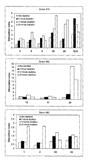
The results are shown in figure 1 which represent T cell proliferative
responses in PBMCs from three human donors (475, 440 and 462) to a
series of borderline or weak T cell epitopes (peptides 1
(PGQTATITCSGHALG), 2 (GDKFVSWYQQGSGQS),
6 (IKPEAPGCDASPEELNRYYASLRHYLNLVTRQRY),
9 (QSISNWLNWYQQKPG), 13 (KGLEWLVVIWSDGSS),
CA 02644540 2008-09-02
WO 2007/099341
PCT/GB2007/000736
17 (AASGFTFSSFGMSWV), 20
(DTAVYYCAAAGVRAEDGRVRTLPSEYTFWGQ
-GTQV), 24 (HQSLVIKLMPNITLL) and to a pair of strong T cell
epitopes (peptides 25 (PKYRNMQPLNSLKIAT) and 26
5 (TVFYNIPPMPL) and to KLH antigen. The results show an increase in
T cell responses for all peptides after depletion of CD25hi T cells.
Maximum responses were determined for all peptides following 10 or 20
minute depletion of CD25hi T cells. These results demonstrated strong
increases in T cell responses after CD25 hi T cell depletion which, in the
10 examples of peptides such as peptides 1 and 2, allowed detection of T
cell
epitopes in peptides previously scored borderline or negative for T cell
responses.
15 Example 2 ¨ Timecourse Peptide T cell Assays
Wild type (WT) and T cell epitope depleted peptides
(HLRHCLSCSKCRKEM and HARHCLSCSKCRKEM, respectively)
derived from human sTNFR1 sequence were synthesized (Pepscan
Systems, Leystad, Netherlands) and tested using the method of example 1
in which CD8+ CD25 hi T cell depleted PBMC were used to compare
peptides derived from sTNFR-1 for the capacity to stimulate T cell
responses from twenty healthy donors. Bulk cultures were established by
adding 1 ml of 2-4x10⁶/m1 CD8+ CD25 hi T cell depleted PBMC in AIM V
culture medium to each well of a 24 well plate (Greiner Bio-One,
Frickenhausen, Germany). Each peptide was tested separately against
each donor by adding lml 10 M peptide to each bulk culture (final
concentration of 51AM for a 2m1 per bulk culture). For comparison,
additional bulk cultures were established for untreated and positive
(KLH) controls. Replicate samples (of T blasts) were removed from bulk
cultures on days 6-9 and proliferation was assessed in 96 round bottom
plates The data were used to assess the magnitude and kinetics of T cell
CA 02644540 2008-09-02
WO 2007/099341
PCT/GB2007/000736
16
responses to each peptide on days 6, 7, 8 and 9 post stimulation. In
addition, the same twenty healthy donors used in the time course
proliferation assay were tested for 1L-2 production after 8 days culture
with the TNFR1 peptides using the IL-2 Elispot assay. Elispot plates were
pre-wet with 70% ethanol then coated with IL-2 capture antibody (R&D
systems, Minneapolis, USA) overnight at 4 C. The plates were washed
twice with PBS (Invitrogen, Paisley, UK) then blocked in 1% BSA/PBS
for 2 hours at room temperature. The plates were washed in PBS prior to
the addition of CD8+ CD25 hi T cell depleted PBMC at 4x10⁵ cells per
well and test samples at a final concentration of 5[1.M. After 7 days at
37 C/ 5% CO2, the plates were developed. After washing with first water
then PBS, IL-2 detection antibody (R&D systems, Minneapolis, USA) in
PBS/1% BSA was added for 2 hours at 37 C. After further washing with
PBS, streptavadin-AP (R&D systems, Minneapolis, USA) was added for
1.5 hours, the plates washed again then BCIP/NBT chromagen (R&D
systems, Minneapolis, USA) added for 30 minutes. The plates were
washed with water, dried then spot counts analysed using the
Immunospot Elispot analyzer, software version 3 (Cleveland, Ohio,
USA).
For both T cell proliferation and IL-2 Elispot assays, responses that
exceed a SI threshold of 2 (dotted line) and are significantly (p<0.05)
different than background (*) were deemed positive. The results shown
in figure 2 indicate that the WT peptide gave responses in the same three
human donors (3, 8 and 11) in both proliferation and 1L-2 Elispot assays
indicating that this peptide contains a T cell epitope. The proliferation
timecourse indicated that for these three donors, peak proliferation
responses were detected 7 days after peptide addition and, in each case,
the WT TNFR1 peptide would have been scored negative as a T cell
epitope if proliferation responses had been measured at timepoints 8 or 9
days after peptide addition. These results also show a strong correlation
between T cell responses measured by proliferation and IL-2 Elispot.
CA 02644540 2008-09-02
WO 2007/099341
PCT/GB2007/000736
17
Example 3 ¨ Timecourse Whole Protein T cell Assays
Wild type (WT) and mutant T cell epitope depleted human sTNFR1
proteins were prepared as human Fc fusion proteins as described in
WO/2004/113387 with the epitope-depleted protein having mutations
I10Q, T2OR, H23P, L56A, L108T, L110H and L149D. Proliferation and
IL-2 Elispot assays were performed as in example 2 except that lml of
sTNFR1 proteins were added to a final concentration of 1011g/ml. The
data shown in figure 3 indicate that, for the proliferation assay, significant
T cell responses were detected in donors 13 and 17 for the WT but not the
mutant T cell epitope depleted protein. Peak responses were observed at
days 8 and 9 and neither donor 13 or 17 showed any significant response
at day 6. For the IL-2 Elispot assay, both donors 13 and 17 were again
positive for T cell responses to WT but not the mutant epitope depleted
protein. In addition, donor 4 gave a significant response to WT protein in
this assay. As with example 2, the results further demonstrate the utility
of the timecourse assay in detecting T cell responses, in this case to whole
proteins. As with example 2, the results show a good correlation between
T cell responses measured by proliferation and IL-2 Elispot.
Example 4 ¨ Timecourse Immunomodulatory Protein T cell Assays
This example illustrates the invention when used to measure T cell
responses to an immunomodulatory protein, human interferon beta which
is known to upregulate inhibitory molecules on dendritics cells such as
HLA-G (Mitsdoerffer M et al J Neuroimmunol. 2005 159:155-64) and
B7-1H (Schreiner B et al J Neuroinnnunol. 2004 155:172-82). In order to
test whether linear T cell epitopes present in the sequence of IFN beta
could stimulate T cells in vitro, a modified method for loading antigen
into monocyte derived dendritic cells was developed in which the
CA 02644540 2008-09-02
WO 2007/099341
PCT/GB2007/000736
18
biological effects of IFN beta on both dendritic cells (DC) and CD4+ T
cells was minimized.
Monocytes were isolated from PBMC by adherence to tissue culture
plastic (>90% CD14+) and were cultured in 24 well plates in AIM V
medium with 5% heat inactivated human AB serum (Autogen Bioclear,
Caine, Wiltshire, UK) (growth medium) at an approximate density of
1x10⁶ per well (24 well plate). Monocytes were incubated in growth
medium containing human IL-4 (Peprotech, Rocky Hill, NJ, USA) and
GM-CSF (Peprotech, Rocky Hill, NJ, USA) for 3 days. On day 3, 44
1.1g/m1 of Betaferon (Schering AG, Berlin, Germany) were added in 0.5ml
test buffer plus 3% heat inactivated human AB serum and 25mM (final
concentration) HEPES pH 8. Control wells containing 50Kg/m1 KLH or
no antigen (untreated cells) were incubated in lml PBS+0.01% Tween 20
plus 3% heat inactivated human AB serum (standard buffer). DC were
incubated with antigen for 6 hours after which DCs were washed 6 times
to remove exogenous IFN beta. Cells were then resuspended in growth
medium containing TNF alpha (Peprotech, Rocky Hill, NJ, USA), GM-.
CSF and IL-4 overnight.
On day 4, autologous CD8+ CD25 hi depleted CD4+ T cells were isolated
by negative selection from PBMC (Dynal Human CD4+ Negative
Isolation Kit, Wirral, UK) and were then added to DCs at 1 x 10⁵ per well
in both proliferation and Elispot plates. Elispot plates were incubated for
6 days before developing (as in example 2) and proliferation plates were
incubated for 7 days before proliferation was measured by incorporation
of 3HTdR (a 6 hour pulse at 1 Ci/well).
As with example 2, for proliferation and Elipsot assays an empirical
threshold of stimulation index >2 was selected where responses above
this threshold were deemed positive. Furthermore statistical analysis was
CA 02644540 2008-09-02
WO 2007/099341
PCT/GB2007/000736
19
also performed to determine whether the responses were significantly
(p<0.05) different from untreated control (*). Additional analysis to
determine the degree of intra-assay variation included coefficient of
variance (CV).
The results, as shown in figure 4, indicate significant T cell responses in 4
out of 29 donors for the proliferation assay and the same 4 out of 29
donors for the IL-2 Elispot assay. This data shows that T cell responses
could be reproducibly demonstrated even with an immunomodulatory
protein.
Example 5 ¨ Timecourse Small Molecule T cell Assays
Carbamazepine (Novartis Pharmaceuticals UK) and an N-acetyl
iminostilbene (an analogue of carbamazepine, synthesized according to
Ying et al Journal of Allergy and Clinical Immunology 2006; 118:233-
241) were compared for the ability to stimulate T cell responses in a panel
of healthy donors. Both compounds were tested at 25 g/m1 in separate
bulk cultures for each donor according to the method of example 2.
Briefly, bulk cultures were established using 2-4x10⁶ CD8+ CD25hi T cell
depleted PBMC in each well of a 24 well plate. Replicate samples (of T
blasts) are removed from bulk cultures on days 5-8 and proliferation was
assessed in 96 well plates. The data were used to assess the magnitude
and kinetics of T cell responses to each compound.
As for example 2, a SI>2 was used as a threshold for positive responses
and data was further analyzed to determine the coefficient of variance
(CV), standard deviation (SD) and significance (p<0.05) using parametric
and non-parametric statistical analysis. Any given compound was
CA 02644540 2008-09-02
WO 2007/099341
PCT/GB2007/000736
considered to be immunogenic only if the response is statistically
significant (p<0.05) with an SI>2.
The results show that the carbamazepine metabolite N-acetyl
5 iminostilbene stimulates fewer donors than carbamazepine (known to be a
potent inducer of delayed allergic responses in patients) when tested over
a range of concentrations using the time course T cell assay method. It is
clear that using a single time point T cell assay in a large number of T cell
responses would not have been detected. Indeed the majority of T cell
10 responses against carbamazepine are induced on day 5 with only one
additional response detect on days 6 and 7. Assessment of T cell
responses against N-acetyl and carbamazepine using a single time point T
cell assay on days 6, 7 or 8 would not have discriminated any level of
immunogenicity between these two compounds.
DEMANDES OU BREVETS VOLUMINEUX
LA PRESENTE PARTIE DE CETTE DEMANDE OU CE BREVETS
COMPREND PLUS D'UN TOME.
CECI EST LE TOME 1 DE 2
NOTE: Pour les tomes additionels, veillez contacter le Bureau Canadien des
Brevets.
JUMBO APPLICATIONS / PATENTS
THIS SECTION OF THE APPLICATION / PATENT CONTAINS MORE
THAN ONE VOLUME.
THIS IS VOLUME 1 OF 2
NOTE: For additional volumes please contact the Canadian Patent Office.