Note: Descriptions are shown in the official language in which they were submitted.
CA 02644574 2014-04-11
METHODS OF PREDETERMINING THE CONTOUR OF A RESECTED BONE
SURFACE AND ASSESSING THE FIT OF A PROSTIIESIS ON TIIE BONE
CROSS REFERENCE TO RELATED APPLICATIONS
[ow 1] This application claims the same priority as US Patent No.
S.231.634.
BACKGROUND
[0002] The present disclosure relates to methods for determining an
optimal fit of a
prosthesis on a resected bone surface.
[0003] Orthopaedic procedure's for the replacement of all, or a portion
of. a patient's joint
typically require resecting, and reshaping of the bones of the joint to
receive prosthetic
components. For example. a typical total knee prosthesis has three main
components: a femoral
component for replacing at least a portion of the distal end of the femur. a
tibial component for
replacing at least a portion of the proximal end of the tibia, and a bearing
insert for replacing at
least a portion or the articulating tissue between the femur and the tibia.
Procedures for
implanting a total knee prosthesis typically involve preparing and reshaping
both the distal end
of the femur and the proximal end of the tibia prior to implanting the
prosthetic components.
The amount of bone removed may be partially. determined by the size and type
of prosthetic
components to be implanted. The size of prosthetic components may be initially
determined by
measurements taken of the knee prior to and during surgery, and the final
determination Of size
may be made after taking measurements and trialing a provisional prosthesis
during the
procedure.
CA 02644574 2008-08-28
WO 2007/109467 PCT/US2007/063949
SUMMARY
[0004] The present disclosure provides methods for predetermining a
contour of a
resected bone surface and assessing a fit of a prosthesis on the resected bone
surface. The
present disclosure also provides methods for designing prostheses to fit
discrete patient
populations as well as methods for designing customized prostheses.
[0005] In one form thereof, the present disclosure provides a method of
virtually
assessing the fit of a prosthesis for placement on a resected bone surface,
the method including
the steps of creating a two-dimensional outline of the resected bone surface;
creating a two-
dimensional outline of a first prosthesis; and comparing the two-dimensional
outline of the
resected bone surface with the two-dimensional outline of the first
prosthesis. In another form
thereof, the present disclosure provides an apparatus for virtually assessing
the fit of a prosthesis
for placement on a resected bone surface, the apparatus including a first
computer adapted to
create a two-dimensional outline of the resected bone surface; a second
computer for creating a
two-dimensional outline of a first prosthesis; and a third computer for
comparing the two-
dimensional outline of the resected bone surface with the two-dimensional
outline of the first
prosthesis. In yet another form thereof, the present disclosure provides a
method of designing a
prosthesis to substantially fit a resected bone surface based on a population
of bones, the method
including the steps of creating a plurality of two-dimensional outlines
corresponding to each
resected bone surface for each bone of the population; and determining a
contour of a bone
engaging surface of a prosthesis using the plurality of two-dimensional
outlines, wherein the
contour substantially matches the plurality of two-dimensional outlines of the
resected bone
surfaces. In still another form thereof, the present disclosure provides an
apparatus for designing
a prosthesis to substantially fit a resected bone surface based on a
population of bones, the
apparatus including a first computer for creating a plurality of two-
dimensional outlines
corresponding to each resected bone surface for each bone of the population;
and a second
computer for determining a contour of a bone engaging surface of a prosthesis
which
substantially matches the plurality of two-dimensional outlines of the
resected bone surfaces. In
one form thereof, the present disclosure provides a method of creating a
prosthesis for placement
on a resected bone surface, the method including the steps of creating a two-
dimensional outline
of the resected bone surface; and determining a contour of a bone engaging
surface of a
Page 2 of 14
CA 02644574 2008-08-28
WO 2007/109467 PCT/US2007/063949
prosthesis using the two-dimensional outline of the resected bone surface. In
another form
thereof, the present disclosure provides an apparatus for creating a
prosthesis for placement on a
resected bone surface, the apparatus including a first computer for creating a
two-dimensional
outline of the resected bone surface; and a second computer for determining a
contour of a bone
engaging surface of a prosthesis using the two-dimensional outline of the
resected bone surface.
BRIEF DESCRIPTION OF THE DRAWINGS
[0006] The above mentioned and other features of the disclosure, and the
manner of
attaining them, will become more apparent and will be better understood by
reference to the
following description of embodiments of the disclosure taken in conjunction
with the
accompanying drawings, wherein:
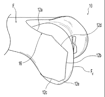
[0007] FIG. 1 is a perspective view of a digital model of the distal end
of a femur
including a virtual resection according to an exemplary method of the present
disclosure;
[0008] FIG. 2 is a perspective view of the digital model of FIG. 1,
further illustrating the
vertices of the virtual resection;
[0009] FIG. 3 is a top view of the two-dimensional outline of the femoral
resection of
FIG. 1;
[0010] FIG. 4 a perspective view of an exemplary distal femoral
prosthesis which may be
used in an exemplary method of the present disclosure;
[0011] FIG. 5 is a perspective view of the prosthesis of FIG. 4, further
illustrating the
step of virtually unfolding the prosthesis;
[0012] FIG. 6 is a top view of the two-dimensional outline of the
prosthesis of FIG. 4
after the unfolding step of FIG. 5;
[0013] FIG. 7 is an illustration of another step of the method of the
present disclosure
wherein outlines of several exemplary prostheses are compared with outlines of
several virtually
resected exemplary femurs; and
[0014] FIG. 8 is another illustration of the step shown in FIG. 7.
Page 3 of 14
CA 02644574 2008-08-28
WO 2007/109467 PCT/US2007/063949
[0015] Corresponding reference characters indicate corresponding parts
throughout the
several views. Although the drawings represent embodiments of the present
disclosure, the
drawings are not necessarily to scale and certain features may be exaggerated
in order to better
illustrate and explain the present disclosure. Although the exemplifications
set out herein
illustrate embodiments of the disclosure, the embodiments disclosed below are
not intended to be
exhaustive or to be construed as limiting the scope of the invention to the
precise forms
disclosed.
DETAILED DESCRIPTION
[0016] The present disclosure may include references to the following
terms: anterior (at
or near the front of the body, as opposed to the back of the body); posterior
(at or near the back
of the body, as opposed to the front of the body); lateral (at or near the
side of the body, farther
from the midsagittal plane, as opposed to medial); medial (at or near the
middle of the body, at or
near the midsagittal plane, as opposed to lateral); proximal (toward the
beginning, at or near the
head of the body, as opposed to distal); and distal (further from the
beginning, at or near the foot
of the body, as opposed to proximal).
[0017] Referring to FIGS. 1-8, an exemplary method of the present
disclosure may be
used to determine how a femoral prosthesis will fit on the distal end of a
femur, i.e., to assess
whether a prosthesis is of the right size and shape for the distal end of the
femur and whether the
prosthesis suitably conforms thereto. The method generally includes the steps
of obtaining a
three-dimensional (3-D) model of a bone based on an acquired image of the
bone, virtually
resecting the 3-D model of the bone, i.e., creating or simulating a resection
of the bone within a
computer or other intelligent processing device, preparing a bone profile of
the virtual resection,
creating a two-dimensional (2-D) outline or footprint of the resection from
the bone profile,
preparing an prosthesis profile, creating a 2-D outline or footprint from the
prosthesis profile,
and comparing the 2-D outlines of the bone profile and the prosthesis profile
to assess or
determine the fit of the prosthesis with the bone.
[0018] More particularly, referring to FIG. 1, 3-D digital model 10 of an
exemplary
femur F is illustrated. Digital model 10 may be obtained by obtaining a
computed tomography
("CT") scan of a femur to produce a 3-D image of the femur and converting the
3-D image to
Page 4 of 14
CA 02644574 2008-08-28
WO 2007/109467 PCT/US2007/063949
digital model 10. The conversion of the 3-D CT scan image to 3-D digital model
10 may be
performed using any suitable modeling software including, for example, Amira ,
available from
Mercury Computer Systems, Inc., of Chelmsford, Massachusetts. Digital model 10
may include
femur F having distal end Fd.
[0019] Referring still to FIG. 1, using suitable software, such as MATLAB
, available
from The MathWorks, of Natick, Massachusetts, and Unigraphics , available from
UGS Corp.,
of Plano, Texas, a virtual resection of distal end Fd of model femur F is
performed. Similar to
the resection performed in actual knee arthroplasty procedures, the virtual
resection involves
defining femoral cut planes 12a-12e on distal end Fd of model femur F. Femoral
cut planes 12a-
12e are calculated using an algorithm of the software. The algorithm
calculates femoral cut
planes 12a-12e based on a proposed, exemplary femoral prosthesis and the known
surgical
technique specified for the proposed femoral prosthesis. More particularly,
distal end Fd of
model femur F may be preliminarily measured based on the known surgical
technique and using
the software described above. The resulting measurements are used to
preliminarily select a
femoral prosthesis size and type. Resection of distal end Fd of model femur F
is determined by
the selected femoral prosthesis and involves resecting distal end Fd of femur
F to complement
and receive the prosthesis. For example, as shown in FIG. 4, model femoral
prosthesis 20 may
be preliminarily selected. Femoral prosthesis 20 is a cruciate-retaining
femoral prosthetic
component having bone engaging surface 22. Bone engaging surface 22 includes a
plurality of
intersecting planar surfaces, including anterior surface 22a, distal surface
22b, posterior surface
22c, anterior chamfer surface 22d, and posterior chamfer surface 22e.
Accordingly, as shown in
FIG. 1, the virtual resection of distal end Fd of model femur F includes
defining a plurality of
intersecting cut planes 12a-12e including anterior cut plane 12a, distal cut
plane 12b, posterior
cut plane 12c, anterior chamfer cut plane 12d, and posterior chamfer cut plane
12e, which
correspond to the plurality of intersecting planar surfaces 22a-22e of model
prosthesis 20 (FIG.
4). As illustrated in FIGS. 2 and 3, cut planes 12a-12e intersect one another
at femoral cut plane
vertices 14a-14d. More particularly, anterior cut plane 12a intersects
anterior chamfer cut plane
12d at vertex 14a. Anterior chamfer cut plane 12d intersects distal cut plane
12b at vertex 14b.
Distal cut plane 12b intersects posterior chamfer cut plane 12e at vertex 14c.
Posterior chamfer
cut plane 12e intersects posterior cut plane 12c at vertex 14d.
Page 5 of 14
CA 02644574 2008-08-28
WO 2007/109467 PCT/US2007/063949
[0020] Referring still to FIGS. 1 and 2, femoral profile 16, shown as a
dotted line, of the
virtually resected model femur F is prepared by outlining cut planes 12a-12e
extending between
cut plane vertices 14a-14d. Two-dimensional outline or footprint 18 of the
resected surface of
model femur F is then obtained, as shown in FIG. 3, by unfolding or bending
profile 16 at cut
plane vertices 14a-14d until cut planes 12a-12e are aligned in a single plane.
The suitable
software mentioned above may be used to manipulate profile 16 to create two-
dimensional
outline 18.
[0021] Referring now to FIGS. 4-6, two-dimensional outline or footprint
26 of proposed
prosthesis 20 may be made using a process similar to that described above for
outline or footprint
18 of femoral profile 16. More particularly, 3-D digital model 20 of a femoral
prosthesis may be
obtained using any known method and any suitable software, including those
described above.
As discussed above, model prosthesis 20 includes bone engaging surface 22,
which includes
anterior planar surface 22a, distal planar surface 22b, posterior planar
surface 22c, anterior
chamfer planar surface 22d, and posterior chamfer planar surface 22e. Planar
surfaces 22a-22e
intersect one another at prosthesis vertices 24a-24d. More particularly,
anterior planar surface
22a intersects anterior chamfer surface 22d at vertex 24a. Anterior chamfer
surface 22d
intersects distal planar surface 22b at vertex 24b. Distal planar surface 22b
intersects posterior
chamfer surface 22e at vertex 24c, and posterior chamfer surface 22e
intersects posterior surface
22c at vertex 24d.
[0022] Referring to FIG. 4, prosthesis profile 25 of model prosthesis 20
is prepared by
outlining the perimeter of intersecting planar surfaces 22a-22e between
prosthesis vertices 24a-
24d. Prosthesis profile 25 is represented by the heavy dashed line extending
about the perimeter
of model prosthesis 20. Turning to FIGS. 5 and 6, two-dimensional outline or
footprint 26 of
prosthesis profile 25 is created by using the suitable software to unfold or
bend profile 25 at
vertices 24a-24d until planar surfaces 22a-22e are aligned within a single
plane.
[0023] Prosthesis outline 26 may be visually compared with femur outline
18 to
determine and assess whether model prosthesis 20 is a suitable fit for model
femur 10.
Prosthesis outline 26 may be compared with femur outline 18 by superimposing
one atop the
other and observing the overlapping shapes and the differences therebetween.
Furthermore,
Page 6 of 14
CA 02644574 2008-08-28
WO 2007/109467 PCT/US2007/063949
using the suitable software mentioned above, quantitative analysis may be made
of outlines 26
and 18. For instance, measurements of outlines 26 and 18 may be taken and the
suitable
software can calculate deviations between the measurements. For example, width
measurements
of outlines 26 and 18 at the intersections of each planar surface may be taken
and/or at midpoints
of each planar surface between such intersections with other planar surfaces.
Any deviations
between outlines 26 and 18 may then be used to calculate proposed changes in
prosthesis 20 to
thereby reshape prosthesis 20 to minimize the deviations. Alternatively, any
deviations between
outlines 26 and 18 may prompt a user to select a different prosthesis 20 and
perform the same
analysis to assess the fit of the second prosthesis 20 on model femur 10.
[0024] The method described above has several useful, practical
applications. For
example, the method described above may be used to develop new and improved
existing
prosthesis designs. It is contemplated that this method may be used to survey
a large population
of subjects to develop statistics and identify trends in bone shapes, and to
adapt prosthesis sizes
and shapes accordingly. More specifically, two-dimensional footprints of
virtually resected
bones of a large population of patients may be obtained and compared to two-
dimensional
footprints of numerous available prostheses.
[0025] FIGS. 7 and 8 illustrate an exemplary application of the methods
of the present
disclosure. FIG. 7 illustrates femur footprints or outlines 18a-18d, shown as
dotted lines, taken
from a virtually resected model of a femur of four different subjects compared
with footprints or
outlines 26a-26c, shown in solid lines, taken from three different models of
available prostheses.
FIG. 8 schematically illustrates the same footprints 18a-18d, 26a-26c. The
comparison shown in
FIGS. 7 and 8 demonstrates that the prosthesis yielding footprint 26a is
larger in width W (FIG.
6) than the virtually resected bones yielding footprints 18b-18d. In an
exemplary embodiment,
outlines 18a-18d may be used to design or create a prosthesis which
substantially matches at
least some of outlines 18a-18d. For example, a prosthesis may be created or
designed which is a
best fit approximation to a plurality of outlines 18 which may be based on a
specific patient
population, such as the female population.
[0026] In an exemplary embodiment, a method of the present disclosure may
be
performed on the femurs of a large population of women to obtain
medial/lateral and
Page 7 of 14
CA 02644574 2014-04-11
anterior/posterior dimensions of the femurs and calculate ratios between the
medial/lateral and
anterior/posterior dimensions. These dimensions and calculations may be used
in designing
femoral components for use on female anatomy. In another exemplary embodiment,
a method of
the present disclosure may also be used to obtain medial/lateral and
anterior/posterior dimensions
of existing femoral components and calculate ratios between the medial/lateral
and
anterior/posterior dimensions of the femoral components. The dimensions and
calculated ratios
may then be used to compare existing femoral components to the dimensions and
calculated
ratios of the femurs of women to identify potential areas of the femoral
component where fit can
be optimized. Such a comparison is fully described in U.S. Patent Application
Serial No.
11/611,021, entitled DISTAL FEMORAL KNEE PROSTHESES, assigned to the assignee
of the
present application.
The same type of process may be performed for other populations, such as a
population of males,
various ethnic populations, populations based on age, stature-based
populations, and/or
populations based on disease progression or disease status.
[0027] In addition, the method described above may be used in guiding the
design and
manufacture of custom prostheses. For instance, a patient's femur may be
modeled, virtually
resected and footprinted as described above. The footprint could then be used
as the footprint for
forming a prosthesis. Although the method described above is exemplified with
reference to the
distal end of the femur and femoral prostheses, the methods of the present
invention may be
applied to any bone and any prosthesis.
[0028] While this invention has been described as having exemplary designs,
the present
disclosure may be further modified within the spirit and scope of this
disclosure. This
application is therefore intended to cover any variations, uses, or
adaptations of the disclosure
using its general principles. Further, this application is intended to cover
such departures from
the present disclosure as come within known or customary practice in the art
to which this
disclosure pertains.
Page 3 of 14