Note: Descriptions are shown in the official language in which they were submitted.
CA 02644721 2008-09-03
WO 2007/117849 PCT/US2007/063961
USE OF INHIBITORS OF JUN N-TERMINAL KINASES
TO TREAT GLAUCOMA
BACKGROUND OF THE INVENTION
This application claims priority to U.S. application serial number 11/394,893
filed
March 31, 2006; which is a continuation-in-part application of serial number
11/259,566,
filed October 26, 2005, which claims benefit of U.S. provisional application
serial number
60/623,755, filed October 29, 2004.
1. Field of the Invention
lo The present invention relates generally to the field of glaucoma and, more
specifically, to the use of inhibitors of Jun N-terminal kinases (JNK) to
lower intraocular
pressure and provide neuroprotection to patients suffering from glaucoma.
2. Description of the Related Art
Many pathological changes in the eye, such as glaucoma, acute ischemic optic
zs neuropathy, macular degeneration, retinitis pigmentosa, retinal detachment,
retinal tears or
holes, and other ischemic retinopathies or optic neuropathies, cause injury or
death of
retinal neurons, which can lead to loss of vision. For example, primary open-
angle
glaucoma (POAG) is a progressive disease leading to optic nerve damage and
ultimately
blindness. The cause of this disease has been the subject of extensive studies
for many
20 years, but is still not fully understood. Glaucoma results in the neuronal
degeneration of the
retina and optic nerve. Even under optimal medical care and surgical
treatment, it is still
associated with a gradual loss of retinal ganglion cells (RGC), which causes a
decline of
visual function (Van Buskirk et al. (1993); Schumer et al. (1994)).
-1-
CA 02644721 2008-09-03
WO 2007/117849 PCT/US2007/063961
An abnormal increase in intraocular pressure (IOP) is a major risk factor of
glaucoma. Currently, the only available treatment for glaucoma is to lower IOP
either by
medication or surgery. Lowering IOP is effective in slowing the development of
POAG
and delaying its damaging effects. Nonetheless, the loss of visual field in
glaucoma
s patients does not always correlate with IOP, and lowering IOP alone does not
completely
stop the disease process.
There is not one mechanism that seems sufficient alone to explain the wide
spectrum and patterns of pathological changes usually observed in glaucoma
patients. It is
probable that glaucoma involves more than one etiology and different
mechanisms are
zo manifested in different patients and/or different stages of the disease.
Some of the more
important proposals are: deprivation of neurotrophic factors, vascular
abnormality
(ischemia), and glutamate toxicity. These mechanisms eventually lead to
apoptosis of the
RGC (Clark & Pang (2002)).
The same mechanisms have been proposed to be involved in other ocular
diseases.
is For example, a decrease in neurotrophic factors is associated with a rat
model of retinitis
pigmentosa (Amendola et al. (2003)). Introduction of certain neurotrophic
factors to the
retina can reduce retinal damages related to retinitis pigmentosa (Tao et al.
(2002)), retinal
detachment (Hisatomi et al., (2002); Lewis et al. (1999)), and experimental
macular
degeneration (Yamada et al. (2001)). Retinal ischemia is involved in acute
ischemic optic
20 neuropathy, macular degeneration (Harris et al. (1999)), and other ischemic
retinopathies
or optic neuropathies. Similarly, glutamate toxicity may contribute to the
retinal damages
seen in retinal detachment (Sherry & Townes-Anderson (2000)).
-2-
CA 02644721 2008-09-03
WO 2007/117849 PCT/US2007/063961
U.S. Patent Application No. US2005/0069893 describes the measurement of JNK
gene expression as a means of diagnosing glaucoma but does not discuss the use
of
inhibitors of JNK to lower intraocular pressure or to provide neuroprotection
to a patient
suffering from glaucoma.
Currently, no available therapy for glaucoma seeks to interrupt the mechanisms
by
which the ocular tissues are damaged in the disease process. Moreover,
although a variety
of therapeutic agents have been proposed as having the ability to lower ocular
hypertension, many of these agents have associated side effects which may
render them
undesirable as ocular therapeutic agents. What is needed is a glaucoma
treatment that
zo addresses the underlying pathological cause of the disease and thereby
provides a decrease
in IOP and neuroprotection without resulting undesirable side effects
typically associated
with agents used to lower IOP.
SUMMARY OF THE INVENTION
zs The present invention overcomes these and other drawbacks of the prior art
by
providing compositions and methods for decreasing ocular hypertension and
providing
neuroprotection to patients suffering from glaucoma. The compositions and
methods
comprise at least one inhibitor of JNK to lower IOP and provide
neuroprotection.
BRIEF DESCRIPTION OF THE DRAWINGS
The following drawings form part of the present specification and are included
to
further demonstrate certain aspects of the present invention. The invention
may be better
understood by reference to these drawings in combination with the detailed
description of
specific embodiments presented herein.
-3-
CA 02644721 2008-09-03
WO 2007/117849 PCT/US2007/063961
FIG. 1. Dose-dependent effects of SP600125 on TGF(32-stimulated (24 h)
increase of fibronectin level in supematants from cultured human trabecular
meshwork
(GTM-3) cells.
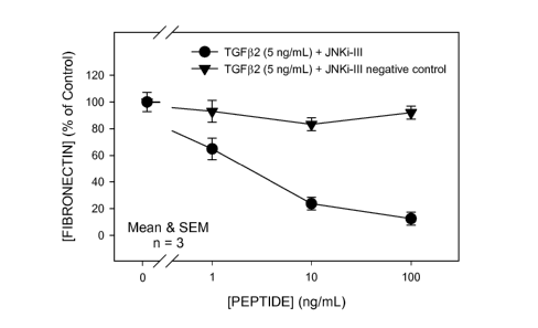
FIG 2. Response to a selective, cell-permeable peptide inhibitor (JNKi-III;
Calbiochem cat. # 420130) vs that of a cell-permeable negative (scrambled
sequence)
control peptide (Calbiochem, cat. # 420131). Both agents were tested for
effect on
TGF(32-stimulated (24 h) increase of fibronectin level in supematants from
cultured
human trabecular meshwork (GTM-3) cells.
FIG. 3. Effect of SP600125 on rat RGC survival with or without trophic
factors,
i with or without glutamate (100 M). The cells were cultured with the
respective
conditions for 3 days. Survival was quantified by counting all Thy-1 positive
healthy
cells.
FIG. 4. Effect of SP600125 on ischemia/reperfusion-induced optic neuropathy.
An optic nerve damage score of 1 represented no damage, and a score of 5
represented
total damage. *: p < 0.05 versus the vehicle-treated group by Student's t-
test.
FIG. 5. Effects of SP600125 on the survival of cultured adult rat RGC. The
cells
were treated with glutamate (100 M) with or without SP600125 for 3 days.
FIG. 6. Effects of SP600125 on the survival of cultured adult rat RGC.
Selected
trophic factors (bFGF, BDNF, CNTF) were withdrawn from all wells except the
controls.
The cells were treated with the indicated concentrations of SP600125 for 3
days. (TF =
trophic factors).
-4-
CA 02644721 2008-09-03
WO 2007/117849 PCT/US2007/063961
DETAILED DESCRIPTION OF PREFERRED EMBODIMENTS
The present invention is directed to compositions and methods for decreasing
ocular hypertension, lowering intraocular pressure and/or providing
neuroprotection to
patients suffering from glaucoma. The compositions comprise one or more
inhibitor(s) of
JNK in a pharmaceutically acceptable vehicle.
Jun N-terminal kinases (JNK) are a family of stress-activated protein kinases
comprising at least 10 isoforms created by alternative splicing of mRNA
transcripts
derived from three genes: JNKl, JNK2, and JNK3 (Gupta et al. (1996)).
Activation of
JNK is required for certain forms of stress-induced apoptosis (Tournier et al.
(2000)),
i which leads to phosphorylation of a number of transcription factors and
cellular proteins,
particularly those associated with apoptosis (e.g., Bc12, Bcl-XL, p53, etc.).
In cell culture,
activation of JNK correlates with neuronal apoptosis induced by a variety of
insults (Xia
et al. (1995); Le-Niculescu et al. (1999)). JNK3 is required for sympathetic
neuron death
following trophic factor withdrawal (Bruckner et al. (2001)). Mice deficient
in JNK3 are
zs resistant to the hippocampal neurotoxicity induced by kainic acid (Yang et
al. (1997)).
Because of these neuroprotective actions, inhibitors of JNK have been proposed
as
treatment for degenerative diseases of the brain, such as, Alzheimer's
disease, Parkinson's
disease, stroke, and ischemia-induced brain dysfunction. In addition, because
the JNK
signaling pathway also regulates the activity and metabolism of some of the
molecules
20 involved in inflammation (Manning & Mercurio (1997)), JNK inhibitors were
proposed as
treatment for immune diseases, such as rheumatoid arthritis, asthma, chronic
transplant
rejection, inflammatory bowel disease, and multiple sclerosis. Other studies
further
indicate that JNK inhibitors may be useful as potential therapeutic agents for
obesity, type
2 diabetes (Hirosumi et al. (2002)), and cancer (Adjei (2001)).
-5-
CA 02644721 2008-09-03
WO 2007/117849 PCT/US2007/063961
It is not obvious that JNK inhibitors, even with multiple pharmacological
actions
listed above, are useful in lowering intraocular pressure and providing
neuroprotection.
None of the above mentioned diseases have been shown to be associated with
glaucoma,
elevated intraocular pressure, or ocular neuroprotection. Moreover, the
usefulness of a
s drug in the brain does not predict its usefulness in the eye, since
therapeutic agents useful
for degenerative diseases in the brain do not always protect against
glaucomatous
apoptotic death of RGC or other ocular diseases. Inflammation, immune
abnormality,
diabetes, obesity, or cancer is not widely accepted as an etiology of
glaucoma, elevated
intraocular pressure or ocular neuroprotection.
zo Unexpectedly, the present inventors have discovered that inhibition of Jun
N-
terminal kinases (JNKi) significantly reduces transforming growth factor-beta2
(TGF(32)-
induced fibronectin expression by a human trabecular meshwork (TM) cell line
(FIG. 1
and FIG. 2). Fibronectin is known to be a component of the TM's extracellular
matrix
(ECM) and an over accumulation of ECM in the TM region is a hallmark of many
forms
15 of glaucoma. Such increases are believed to lead to increased resistance to
aqueous
outflow, thereby elevating intraocular pressure (IOP). JNK has also been
implicated in the
signaling pathway for TGF(3-mediated production of connective tissue growth
factor
(CTGF) (Utsugi et al. 2003). CTGF may play a role in IOP elevation in that it
is known to
increase accumulation of various ECM components, including fibronectin.
20 It has been shown that a non-peptide JNK inhibitor, SP-600125, was
protective
against glutamate-induced or trophic factor withdrawal-induced death of a rat
retinal
neuron, the RGC, in culture (see co-pending U.S. Application Serial No.
11/259.566).
The compound was also found to be protective against ischemia/reperfusion-
induced optic
neuropathy in the rat. Since deprivation of trophic factors, ischemia, and
glutamate
-6-
CA 02644721 2008-09-03
WO 2007/117849 PCT/US2007/063961
toxicity were proposed as potential mechanisms of glaucoma and various ocular
diseases,
these data indicate that non-peptide JNK inhibitors are useful as therapeutic
agents for
providing neuroprotection for ocular tissues.
As used herein, "inhibitors of JNK" refers to those compounds which can
decrease
s the activity of JNK to 50% or lower of the control value. The potential
inhibitory effect of
compounds on JNK activity can be easily evaluated by those skilled in the art.
Many JNK
activity assay kits are commercially available, e.g., Stratagene catalog #
205140, Upstate
catalog # 17-166, etc. In preferred aspects, the JNK inhibitors for use in the
compositions
and methods of the present invention may be small molecules, peptides,
peptidomimetics,
zo antibodies, etc. Most preferably, the JNK inhibitors will be small
molecules.
Examples of JNK inhibitors expected to be useful in the methods and
compositions
of the present invention include, but not are limited to, SP600125 and
pharmacologically
active compounds disclosed in patent applications numbers W0200035906,
W0200035909, W0200035921, W0200064872, W0200112609, W0200112621,
15 W0200123378, W0200123379, W0200123382, W0200147920, W0200191749,
W02002046170, W02002062792, W02002081475, W02002083648, W02003024967;
all of which are hereby incorporated by reference.
Additional preferred JNK inhibitors expected to be useful in the methods and
compositions of the invention include
-7-
CA 02644721 2008-09-03
WO 2007/117849 PCT/US2007/063961
.:.
a Cti
~;--
..~,~ ......
H~'
N
AS601245 (Ferrandi et al., 2004).
H
..,~.
aws
CEP-1347 (KT7515) (Maroney et al. 1998; Roux et al. 2002).
HO
H3CO0C11'
0 ,,H
H3Crr,..
N
N - ~
0
N .
H
K252a (Roux et al., 2002).
The methods comprise administering one or more JNK inhibitors to a human
patient to decrease ocular hypertension, or lower intraocular pressure and
provide
neuroprotection.
8-
CA 02644721 2008-09-03
WO 2007/117849 PCT/US2007/063961
The JNK inhibitors of the present invention may be contained in various types
of
pharmaceutical compositions, in accordance with formulation techniques known
to those
skilled in the art. In general, the JNK inhibitors will be formulated in
solutions or
suspensions for topical ophthalmic or intraocular administration, or as
tablets, capsules or
s solutions for systemic administration (e.g., oral or intravenous).
Oral formulations of the JNK inhibitors are preferred due to ease of
administration.
Oral formulations may be in liquid or solid form. In general, oral
formulations will
include the active JNK inhibitor and inert excipients. In general, solid
tablet or capsule
dosages will contain various excipients such as bulking agents, binding
agents, time
zo release coatings, and so on. Liquid dosages will contain carriers, buffers,
tonicity agents,
solubilizing agents, and so on.
In general, the doses utilized for the above described purposes will vary, but
will
be in an effective amount to inhibit or ameliorate retinal neuropathy. As used
herein, the
term "pharmaceutically effective amount" refers to that amount which lowers
intraocular
is pressure and inhibits or ameliorates retinal neuropathy. The JNK inhibitors
will normally
be contained in these formulations in an amount from about 0.01 to about 10.0
weight/percent. Preferable concentrations range from about 0.1 to about 5.0
weight/percent. For topical administration, these formulations are delivered
to the disease
site one to six times a day, depending on the routine discretion of the
skilled clinician.
20 Systemic administration, for example, in the form of tablets or liquid
useful for the
treatment will contain about 10-1000 mg of a JNK inhibitor, and can be taken 1-
4 times
per day depending on the discretion of the skilled clinician.
-9-
CA 02644721 2008-09-03
WO 2007/117849 PCT/US2007/063961
As used herein, the term "pharmaceutically acceptable carrier" refers to any
formulation which is safe, and provides the appropriate delivery for the
desired route of
administration of an effective amount of at least one JNK inhibitor of the
present invention.
The following examples are included to demonstrate preferred embodiments of
the
invention. It should be appreciated by those of skill in the art that the
techniques disclosed
in the examples which follow represent techniques discovered by the inventor
to function
well in the practice of the invention, and thus can be considered to
constitute preferred
modes for its practice. However, those of skill in the art should, in light of
the present
disclosure, appreciate that many changes can be made in the specific
embodiments which
zo are disclosed and still obtain a like or similar result without departing
from the spirit and
scope of the invention.
Example 1
Fibronectin Assay
Is Cultured transformed human TM cells were used in these studies. Generation
and
characterization of the GTM-3 transformed cell line has been previously
described. [Pang
IH, et al., Curr Eye Res. 1994; 13:51-63]. Maintenance growth medium consisted
of
Dulbecco's modified Eagle's medium with Glutamax I (Gibco/BRL, Grand Island,
NY)
supplemented with 10% fetal bovine serum (Hyclone, Logan, UT) and 50 g/mL
20 gentamicin (Gibco/BRL). For assay, cultures were trypsinized and seeded
into 24-well
plates (Coming Costar, Acton, MA) and allowed to grow until monolayers reached
approximately 90% confluence. Culture medium was then replaced with 0.25 mL
serum-
and antibiotic-free medium containing the appropriate test compound(s). Cells
were
-10-
CA 02644721 2008-09-03
WO 2007/117849 PCT/US2007/063961
incubated 24 h, at 5% COz and 37 C. Aliquots of culture supematants were then
assayed
for fibronectin content by ELISA.
In experiments assessing the effects of the JNK inhibitor, SP600125, the cells
were
cultured concurrently with the compound and with TGF(32 (5 ng/mL). TGF(32 is a
known
.5 inducer of fibronectin production by TM cells. SP600125 caused a dose-
dependent
reduction in the level of fibronectin in TGF(32-treated GTM-3 cell
supematants. These
results are illustrated in FIG. 1. The role of JNK was further elucidated by
use of a
selective, cell-permeable peptide inhibitor of JNK. As with SP600125, co-
incubation with
the peptide inhibitor caused a dose-dependent reduction in the level of
fibronectin in
TGF(32-treated GTM-3 cell supematants. These results contrast with a lack of
effect of a
cell-permeable negative (scrambled sequence) control peptide, thus
corroborating the
central role of JNK in these studies. Results with these peptides are
illustrated in FIG. 2.
Example 2
1.5 The following example demonstrates the protective efficacy of a JNK
inhibitor
against cytotoxic insults to retinal cells.
Rat Retinal Gan2lion Cell Survival Assay
Adult Sprague-Dawley rats were euthanized by COz asphyxiation. Their eyes
were enucleated and placed in Dulbecco's modified Eagle's medium: Nutrient
mixture F12
(1:1; DMEM/F12). The retinas were incubated in a papain solution, containing
papain (34
units/mL), DL-cysteine (3.3 mM), and bovine serum albumin (0.4 mg/ml) in
DMEM/F12,
for 25 min at 37 C. Retinal pieces were then triturated until cells were
dispersed. Cell
suspension (1.5 ml; containing approximately 4.5 x 10⁶ cells) was placed into
each of the
-11-
CA 02644721 2008-09-03
WO 2007/117849 PCT/US2007/063961
poly-D-lysine coated glass bottom culture dishes. The cells were cultured in a
culture
medium previously described by Barres et al. (1988) for 3 days in 95% air/5%
COz at
37 C.
In experiments assessing the toxicity of glutamate on cell survival, the cells
were
cultured with 100 M glutamate for 3 days. In experiments assessing the
detrimental
effect of neurotrophic factor withdrawal on cell survival, basic fibroblast
growth factor,
brain-derived trophic factor, and ciliary-derived neurotrophic factor were
removed from
the medium and cells cultured for 3 days. In experiments assessing the
potential
protective effects of a JNK inhibitor, SP600125, the cells were cultured with
the
zo compound in the presence of the glutamate or in the absence of the
indicated trophic
factors for 3 days. At the end of the 3-day culture period, the cells were
immunostained
for Thy-l, a cell surface marker for RGC, and observed under a fluorescent
microscope.
Thy-l-positive cells were counted and averaged. The results are illustrated in
FIG. 3.
FIG. 3 illustrates that the survival of RGC depended on the presence of the
is indicated neurotrophic factors, such that removal of the neurotrophic
factors (TF
Withdrawal) from the culture medium caused death of RGC to approximately 50%
of the
control group. Incubation of the cells with SP600125 significantly and
completely
protected the cells against such insult. FIG. 3 also shows that glutamate was
toxic to the
RGC, since addition of 100 M glutamate to the culture medium decreased cell
survival
20 by approximately 50%. Again, incubation of the cells with SP600125 also
significantly
and completely protected the cells against this cytotoxicity.
-12-
CA 02644721 2008-09-03
WO 2007/117849 PCT/US2007/063961
Example 3
The following example demonstrates the protective efficacy of a JNK inhibitor
against ischemia-induced optic neuropathy in the rat.
Ischemia/Reperfusion-Induced Optic Neuropathy in the Rat
s Adult Wistar rats were anesthetized and the anterior chamber of one eye of
each
animal was cannulated. The cannula was connected to a raised saline reservoir
whose
height was adjusted to produce an ocular pressure that was higher than the
systolic
pressure of the animal, which, by stopping retinal blood flow, produced
retinal ischemia.
After 60 minutes of ischemia, the intracameral cannula was removed to allow
reperfusion
io of the retina. Two weeks later, the rats were euthanized, their optic
nerves isolated, fixed
in 2% paraformaldehyde, 2.5% glutaraldehyde in 0.1 M cacodylate buffered
solution,
sectioned, and stained in 1% p-phenylenediamine in isopropanol:methanol (l :
l) prepared
as described by Hollander and Vaaland (1968). The optic nerve damage in each
optic
nerve section was ranked by an Optic Nerve Damage Score as previously reported
by
is Pang et al. (1999). In this ranking system, a score of 1 represented no
damage, and a score
of 5 represented total damage.
To test the potential protective effect of SP600125, selected animals were
treated
with a daily intraperitoneal injection of SP600125 (30 mg/kg) for 16
consecutive days
starting 2 days before ischemia was induced. The results are illustrated in
FIG. 4.
20 FIG. 4 shows that ischemia/reperfusion caused significant damage to the
optic
nerve as indicated by a dramatic increase in the optic nerve damage score. It
also
demonstrates that systemic administration of SP600125 could protect against
this ischemic
insult to the retina as shown by a significant reduction in the optic nerve
damage score.
-13-
CA 02644721 2008-09-03
WO 2007/117849 PCT/US2007/063961
Example 4
A JNK inhibitor, SP600125, was tested in cultured adult rat retinal ganglion
cells
(RGC). It was shown to protect against both glutamate-induced and trophic
factor
.5 withdrawal-induced cytotoxicity.
METHODS
A. RGC culture
Adult Sprague-Dawley rats were euthanized by COz asphyxiation. Their eyes were
enucleated and the retinas isolated. Retinal cells were treated with of papain
solution for
la 25 min at 37 C, then washed 3 times with 5 mL RGC culture medium
(Neurobasal
medium with various nutrient supplements + 1% fetal calf serum). Retinal cells
were
dispersed by trituration. Cell suspension was placed onto poly-D-lysine- and
laminin-
coated 8-well chambered culture slides. The cells were then cultured in 95%
air/5% COz
at 37 C.
15 B. Cytotoxic Insults
For glutamate-induced toxicity studies, cells were pre-treated with vehicle or
the
indicated compounds for 30 minutes, followed by 100 M glutamate for 3 days.
For trophic factor withdrawal studies, three trophic factors, basic fibroblast
growth
factor, brain-derived neurotrophic factor, and ciliary neurotrophic factor,
were removed from
20 the culture medium. Cells were cultured in this medium with the indicated
compounds for 3
days.
C. Quantification of cell survival
At the end of the incubation period, the cells were fixed and labeled for Thy-
l, a
RGC marker, by immunocytochemistry. Cell survival was quantified by manually
counting
25 Thy-l-positive healthy cells in each well.
-14-
CA 02644721 2008-09-03
WO 2007/117849 PCT/US2007/063961
RESULTS
A. Effect of SP600125 on glutamate-induced toxicity in rat RGC
It has been previously shown that glutamate was toxic to rat RGC; only 50-70%
of
cells survived after a 3-day treatment of 100 M glutamate. The glutamate-
induced toxicity
in these cells can be prevented by pretreatment with MK801. SP600125 was
protective
against this insult in a dose-dependent manner (FIG. 5).
B. Effect of SP600125 on trophic factor withdrawal-induced toxicity in RGC
Previously, it was shown that removal of the three trophic factors for 3 days
caused
death of approximately 40-50% of the cells. SP600125 was protective against
this insult in a
dose-dependent manner (FIG. 6).
Example 5
Topical compositions useful for treating glaucoma and other ocular diseases:
Component Wt.%
JNK inhibitor 0.1-5
HPMC 0.01-10
Benzalkonium Chloride 0.005-0.5
Sodium Chloride 0.5-2.0
Edetate Disodium 0.005-0.5
NaOH/HCl q.s. pH 7.4
Purified Water q.s. 100 mL
i~
The above formulation is prepared by first placing a portion of the purified
water
into a beaker and heating to 90 C. The hydroxypropylmethylcellulose (HPMC) is
then
-15-
CA 02644721 2008-09-03
WO 2007/117849 PCT/US2007/063961
added to the heated water and mixed by means of vigorous vortex stirring until
all of the
HPMC is dispersed. The resulting mixture is then allowed to cool while
undergoing
mixing in order to hydrate the HPMC. The resulting solution is then sterilized
by means
of autoclaving in a vessel having a liquid inlet and a hydrophobic, sterile
air vent filter.
The sodium chloride and the edetate disodium are then added to a second
portion
of the purified water and dissolved. The benzalkonium chloride is then added
to the
solution, and the pH of the solution is adjusted to 7.4 with O.1M NaOH/HC1.
The solution
is then sterilized by means of filtration.
SP600125 is sterilized by either dry heat or ethylene oxide. If ethylene oxide
lo sterilization is selected, aeration for at least 72 hours at 50 C is
necessary. The sterilized
compound is weighed aseptically and placed into a pressurized ballmill
container.
Sterilized glass balls are then added to the container and the contents of the
container are
milled aseptically at 225 rpm for 16 hours, or until all particles are in the
range of
approximately 5 microns.
Is Under aseptic conditions, the micronized drug suspension or solution formed
by
means of the preceding step is then poured into the HPMC solution with mixing.
The
ballmill container and balls contained therein are then rinsed with a portion
of the solution
containing the sodium chloride, the edetate disodium and benzalkonium
chloride. The
rinse is then added aseptically to the HPMC solution. The final volume of the
solution is
20 then adjusted with purified water and, if necessary, the pH of the solution
is adjusted to
pH 7.4 with NaOH/HC1.
Example 6
Preferred formulation for topical administration:
-16-
CA 02644721 2008-09-03
WO 2007/117849 PCT/US2007/063961
Component Wt.%
JNK inhibitor 0.1-5
HPMC 0.5
Benzalkonium Chloride 0.01
Sodium Chloride 0.8
Edetate Disodium 0.01
NaOH/HCl q.s. pH 7.4
Purified Water q.s. 100 mL
Example 7
Formulation for oral administration:
s Tablet:
1-1000 mg of a JNK inhibitor with inactive ingredients such as starch, lactose
and
magnesium stearate can be formulated according to procedures known to those
skilled in
the art of tablet formulation.
All of the compositions and/or methods disclosed and claimed herein can be
made
zo and executed without undue experimentation in light of the present
disclosure. While the
compositions and methods of this invention have been described in terms of
preferred
embodiments, it will be apparent to those of skill in the art that variations
may be applied
to the compositions and/or methods and in the steps or in the sequence of
steps of the
method described herein without departing from the concept, spirit and scope
of the
i~ invention. More specifically, it will be apparent that certain agents which
are both
chemically and structurally related may be substituted for the agents
described herein to
-17-
CA 02644721 2008-09-03
WO 2007/117849 PCT/US2007/063961
achieve similar results. All such substitutions and modifications apparent to
those skilled
in the art are deemed to be within the spirit, scope and concept of the
invention as defined
by the appended claims.
- 18 -
CA 02644721 2008-09-03
WO 2007/117849 PCT/US2007/063961
References
The following references, to the extent that they provide exemplary procedural
or
other details supplementary to those set forth herein, are specifically
incorporated herein
by reference.
Patents and Published Patent Applications
W0200035906
W0200035909
W0200035921
W0200064872
W0200112609
W0200112621
W0200123378
W0200123379
W0200123382
is W0200147920
W0200191749
W02002046170
W02002062792
W02002081475
W02002083648
W02003024967
-19-
CA 02644721 2008-09-03
WO 2007/117849 PCT/US2007/063961
Other Publications
Adjei, "Blocking oncogenic Ras signaling for cancer therapy," J NATL CANCER
INST
93:1062-1074 (2001).
Amendola et al., "Postnatal changes in nerve growth factor and brain derived
s neurotrophic factor levels in the retina, visual cortex, and geniculate
nucleus in
rats with retinitis pigmentosa," NEUROSCi LETT 345:37-40 (2003).
Barres et al., "Immunological, morphological, and electrophysiological
variation among
retinal ganglion cells purified by panning," NEURON 1:791-803 (1988).
Bruckner et al., "JNK3 contributes to c-Jun activation and apoptosis but not
oxidative
ia stress in nerve growth factor-deprived sympathetic neurons," J NEUROCHEM
78:298-303 (2001).
Clark & Pang, "Advances in Glaucoma Therapeutics," ExPERT OPIv EMERGIvG DRUGS
7:141-164 (2002).
Ferrandi et al., "Inhibition of c-Jun N-terminal kinase decreases
cardiomyocyte apoptosis
15 and infarct size after myocardial ischemia and reperfusion in anaesthetized
rats, "
BRIT. J. PHARM. 142:953-960 (2004).
Gupta et al., "Selective interaction of JNK protein kinase isoforms with
transcription
factors," EMBO J 15:2760-2770 (1996).
Harris et al., "Progress in measurement of ocular blood flow and relevance to
our
20 understanding of glaucoma and age-related macular degeneration," PROG
RETiNA
EYE RES 18:669-687 (1999).
Hirosumi et al, "A central role for JNK in obesity and insulin resistance,"
NATURE
420:333-337 (2002).
Hisatomi et al., "Critical role of photoreceptor apoptosis in functional
damage after
25 retinal detachment," CURR EYE RES 24:161-172 (2002).
-20-
CA 02644721 2008-09-03
WO 2007/117849 PCT/US2007/063961
Hollander and Vaaland, "A reliable staining method for semi-thin sections in
experimental
neuroanatomy," BRAiN RES 10:120-126 (1968).
Le-Niculescu et al., "Withdrawal of survival factors results in activation of
the JNK
pathway in neuronal cells leading to Fas ligand induction and cell death," MOL
s CELL BIOL 19:751-763 (1999).
Lewis et al., "Effects of neurotrophin brain-derived neurotrophic factor in an
experimental model of retinal detachment," INVEST OPHTHALMOL Vis SCi 40:1530-
1544 (1999).
Manning & Mercurio, "Transcription inhibitors in inflammation," ExP OPIv
INVEST
DRuGs 6:555-567 (1997).
Maroney et al., "Motoneuron Apoptosis is Blocked by CEP-1347 (KT 7515), a
Novel
Inhibitor of the JNK Signaling Pathway," J. NEUROSCIENCE 18(1):104-111 (1998).
Pang et al., "Preliminary characterization of a transformed self strain
derived from
human trabecular meshwork, " CURR. EYE RES. 13:51-63 (1994).
Pang et al., "Age-dependent changes in ocular morphology of a spontaneous
ocular
hypertensive mouse strain (DBA/2J)," INVEST OPHTHALMOL Vis RES 40:S671
(1999).
Roux et al., "K252a and CEP1347 are neuroprotective compounds that inhibit
mixed-
lineage kinase-3 and induce activation of Akt and ERK," J. BioL. CHEM.
277(51):49473-49480 (2002).
Schumer et al., "The nerve of glaucoma!," ARCH OPHTHALMOL 112:37-44 (1994).
Sherry & Townes-Anderson, "Rapid glutamtergic alterations in the neural retina
induced
by retinal detachment," INVEST OPHTHALMOL VIS SCi 41:2779-2790 (2000).
-21-
CA 02644721 2008-09-03
WO 2007/117849 PCT/US2007/063961
Tao et al., "Encapsulated cell-based delivery of CNTF reduces photoreceptor
degeneration in anima models of retinitis pigmentosa," INVEST OPHTHALMOL ViS
Sci 43:3292-3298 (2002).
Toumier et al., "Requirement of JNK for stress-induced activation of the
cytochrome c-
s mediated death pathway," SCIENCE 288:870-874 (2000).
Utsugi et al., Am. J. Respir. Cell Mol. Biol., 28:754-761 (2003).
Van Buskirk et al., "Predicted outcome ftom hypotensive therapy for
glaucomatous optic
neuropathy," AM J OPHTHALMOL 25:636-640 (1993).
Xia et al., "Opposing effects of ERK and JNK-p38 MAP kinases on apoptosis,"
SCIENCE
270:1326-1331 (1995).
Yamada et al., "Fibroblast growth factor-2 decreases hyperoxia-induced
photoreceptor
cell death in mice," AM J PATxoL 159:1113-1120 (2001).
Yang et al., "Absence of excitotoxicity-induced apoptosis in the hippocampus
of mice
lacking the JNK3 gene," NATURE 389:865-870 (1997).
-22-