Note: Descriptions are shown in the official language in which they were submitted.
CA 02645155 2008-09-05
WO 2007/103333 PCT/US2007/005624
ORTHOPEDIC PLATE
THIS APPLICATION IS BASED ON U.S. PROVISIONAL APPLICATION SERIAL
NO. 60/780,027 FILED ON MARCH 7, 2006
Field of the Invention
The present invention relates to an orthopedic plate for fixation of bones. In
particular
it relates to a terminal section of an orthopedic plate that is useful for
stabilization of small
bones; for example, for a tibial plate for fixation of a tibial fracture, or
for a distal radial plate
for fixation of a distal radial fracture, or other fracture areas that can
benefit from the design
of the present invention.
Background of the Invention
The ankle joint involves the intersection of the tibia, the fibula and the
tarsals and
metatarsals. The wrist is the joint formed at the intersection of the radius,
the ulna, the carpals
and the metacarpals. Both of these joints are designed to allow a great deal
of freedom in the
movement of the relevant appendage (i.e. the hand or foot). Attendant with
this relative
freedom, the joint itself is somewhat unstable, and easily subjected to trauma
resulting in
displacement or distortion within the bones of the joint, and in harm to the
bones themselves.
The wrist is the most frequently injured area of the upper extremity with
three fourths of wrist
injuries involving a fracture of the distal radius, and/or of the radius, and
the ankle is subject
to similar statistics with respect to the union of the tibia, the tarsals and
the metatarsals. These
injuries usually present in an emergency room setting, and often involve a
fall for example for
the wrist, onto an outstretched hand, and for the ankle, they involve a
misstep orito a foot
causing a rolling of the ankle. While the past conventional wisdom has
included a belief that
such injuries will tend to heal sufficiently on their own, there is often a
loss of function and an
early onset of arthritis that can be precipitated by the misdiagnosis and
improper treatment of
such injuries.
The treatments known for trauma to the extremities have included external
stabilization and fixation such as by plaster casts, external fixators, and
orthopedic plates.
Casting alone, presents the possibility of misalignment of the fragments which
can lead to
severe loss of function and early onset of arthritis, if the fracture is not
properly reduced,
CA 02645155 2008-09-05
WO 2007/103333 PCT/US2007/005624
and/or, if the fragments do not stay in a reduced state, -in particular where
the patient is not
compliant. External fixators have been demonstrated to have an efficacy, but
are
cumbersome, cosmetically unappealing, and can lead to the possibility of
infection at the
attachment sites.
In order to avoid the foregoing problems, surgeons often consider methods of
internal
fixation, which typically include wire and/or screws, and plates. One issue
presented by the
use of wires is that a construct is time-consuming to construct; and screws
alone, often do not
provide the stability required for fusion of the fragments. Plates have the
benefit of providing
a construct that is designed for ease of implantation, and at the same time
have the
disadvantage that there is a significant variety in the shape and size of
individual bones.
Further, in particular, the tibia and radius bones are relatively small so
that individual
variations are relatively more significant than in larger bones, such as the
femur, the pelvis,
and the humerus. Moreover, the flesh surrounding the ankle joint is
particularly dense with
tendons, ligaments, nerves and blood vessels all of which are less forgiving
of the intrusion of
a metal construct than muscle or fatty tissue. This is also true for the wrist
joint, particularly
on the volar (or thumb) side.
Summary of the Invention
The orthopedic plate of the present invention has a portion that is designed
to extend
longitudinally along the bone. This portion has an inferior curved surface
which faces, or in
some, but not necessarily all instances, touches the bone surface. More
specifically, the
curved surface is intended broadly to face the bone and to touch along its
surface so as to
support it on the surface of the plate (i.e. the surface facing the surface of
the bone) so much
as is allowed given the particular variations in individual bones. 'This
portion of the plate
changes this inferior curve as it advances proximally along the bone from a
shallower to a
sharper radius and further spirals downward toward the more advanced side of
the plate. The
more advanced side means the side which advances further along the
longitudinal axis.
Further, the plate in accordance with the invention has a plurality of screw
holes, including
one or more which are positioned along a central portion of the plate and
further_which
2
CA 02645155 2008-09-05
WO 2007/103333 PCT/US2007/005624
preferably includes two or more which are offset from the central portion of
the plate. The
screw holes can be threaded so as to accept screws having threaded heads which
will lock into
position, or alternatively so that a screw with a smaller rounded smooth head
can be screwed
into the bone and mesh with the internal threads of the screw holes.
In a preferred embodiment, the plate includes a set of tabs or "ears" which
are offset
from the longitudinal axis of the plate, and further which allow the placement
of screw holes
that are offset from the longitudinal axis of the plate, as well as being
offset longitudinally
from each other. This allows the plate to be contoured about the circumference
of the bone,
and for the screws to be positioned at a convergent angle to provide for
better pullout values,
i.e. such that it requires a greater force to pull the screws from the bone.
The ears may be
located at the terminal portion of the plate or somewhat more intermediate to
the terminus of
the plate, depending on the intended application. The plate may also include
an intermediate
set of ears that similarly have a pair of offset intermediate threaded screw
holes that are both
longitudinally and laterally, or radially offset from the longitudinal axis of
the plate, and
which accept screws so as to have their axes at convergent angles. Again, the
feature
provides for better pullout values, and helps to avoid interference of the
screws in the bone.
In this instance, the plate includes a central screw hole which is located
between the proximal
pair of ears, and the intermediate pair of ears. This screw hole is preferably
positioned so that
the axis forms a right angle relative to the=longitudinal axis of the plate,
and further relative to
a lateral axis through the hole. Thus, a point in the center of the central
screw hole can be
used to define the origin of the plate, and the angles of the screw and/or
pegs holes can be
referenced with X, Y, and Z coordinates relative to this central hole.
Further, the topography
of the head can be defined using this coordinate system, which permits the
manufacture of the
plate using coniputer generated imaging.
Distal to the intermediate pair of ears, the plate includes a slot which is
radiused at
either end to accept a screw having a head of the same dimensions as the
threaded screw
holes. The slot is elongated along the longitudinal axis of the plate. This
allows the plate to
be loosely attached by inserting a screw through the slot, and prior to
tightening the- plate can
3
CA 02645155 2008-09-05
WO 2007/103333 PCT/US2007/005624
be slid in the longitudinal direction to allow the plate to be optimally
positioned on the bone.
The slot also allows the bone to be viewed through the plate preferably in the
vicinity of the
fracture.
In one embodiment, the plate of the invention may include a portion or head
having a
profile which flares from the sides of the plate to a leading edge that
includes a central oblique
linking area. This embodiment is designed expressly for use in the distal
radius and the
oblique linking area is designed to help to mark the placement of the plate
relative to the
radius. The head is shaped like a heart where the lobes have been
asymmetrically truncated,
like the palm of a hand, or like a modified kidney shape. Further in this
embodiment, the head
has a complex topography in the Z direction which echoes a generalized shape
for the distal
volar surface of a radius. The lunate prominence of the head has a lower
elevation in the Z
direction than the elevation of the styloid prominence in the direction
relative to the radius.
The longitudinal axis at the center of the proximal portion of the bone
defines the Y direction,
and the X direction extends transverse in a direction in which the bone
widens. Further, the
plate includes an oblique depression, or cup, that extends from the rounded
pinky side of the
head and gradually morphs into the elevated styloid prominence in one diagonal
direction,
and rises less gradually upward into the lunate prominence on the other side
of the head.- This
distal cup undulates to define a superficial (i.e. relative to its surface)
serpentine as it links
into the proximal portion of the plate. The head preferably includes holes for
pegs or screws
which may be fixed, or variable. In other embodiments, such as for the distal
tibias, the plate
may include a corresponding head portion which is designed specifically to
support the ankle
joint at the union of the tibia, fibula, the tarsals and the metatarsals.
Similarly, designs for
other indications may include other head shapes, or none at all, so that the
plate is straight, or
even symmetrical from the top view about an axis transverse to the
longitudinal axis of the
plate. In the Z direction, the plate mimics the reverse spiral of the radial
bone as it extends
proximally away from the distal portion. Thus, the proximal portion of the
plate appears to
twist or spiral along the longitudinal axis, and includes a greater radial
bend as it extends
proximally since the bone becomes smaller and more circular in cross-section.
4
CA 02645155 2008-09-05
WO 2007/103333 PCT/US2007/005624
Brief Description of the Drawings
Figure 1 is a top view of an orthopedic plate and plate head in accordance
with a first embodiment of the invention for use on a left distal radius;
= Figure 2 is a perspective view of the plate of Figure 1 viewed from the
outer
proximal surface looking toward the head with the styloid side of the plate
downward;
Figure 3 is a cross section of the plate of Figure 1 taken along line 3-3 in
Figure 1;
Figure 3a is a detail of the central screw hole of Figure 3;
Figure 4 is a cross section of the plate of Figure 3 taken along line 4-4 in
Figure 3;
Figure 5 is a view taken from the styloid side edge of the plate of Figure 1;
Figure 6 is a view taken from the proximal edge of the plate of Figure 1;
Figure 7 is a section of the plate taken along line 7 of Figure 1;
Figure 7a is a detail of the screw hole of Figure 7;
Figure 8 is a view from the side of the lunate prominence with the plate head
in
a lowered orientation and viewing the head in partial section to illustrate
the detail of
the threads of the peg holes;
Figure 8a is a detail of the peg holes from Figure 8;
Figure 9 is a top view of the plate of Figure 1 showing the lines at which the
lateral sections, of Figures 9a through 9g;
Figure 9a is a section in the Y direction taken at line 9a of Figure 9;
Figure 9b is a section in the Y direction taken at line 9b of Figure 9;
Figure 9c is a section in the Y direction taken at line 9c of Figure 9;
Figure 9d is a section in the Y direction taken at line 9d of Figure 9;
Figure 9e is a section in the Y directian taken at line 9e of Figure 9;
Figure 9f is a section in the Y direction taken at line 9f of Figure 9;
Figure 9g is a section in the Y direction taken at line 9g of Figure 9;
Figure 10 is a top view of a second embodiment of the distal radius plate of
the
present invention;
5
CA 02645155 2008-09-05
WO 2007/103333 PCT/US2007/005624
Figure 11 is a view from the proximal portion of the radial bone showing the
plate in accordance with the present invention in position on the volar side
of the bone
and illustrating the angles for the pegs;
Figure 12 is a top view of a third embodiment of the distal radial plate of
the
present invention with fixed angle pegs, and having an extended proximal
portion;
Figure 13 is a side perspective view of the plate of Figure 12;
Figure 14 is a top view of a fourth embodiment of the distal radial plate of
the
present invention with both fixed angle and variable angle locking pegs, and
having an
different embodiment of the extended proximal portion;
Figure 15 is a side perspective view of the plate of Figure 14;
Figure 16 is a detail of the head of the distal radial plate of Figure 14
showing
the locking cam inserts in position in the peg holes of the head;
Figure 17 is a top view of the locking cam insert used in the variable axis
embodiment shown in Figures 10, 14 and 15;
Figure 18 is a cross section of the locking cam insert of Figure 17;
Figure 19 is a side view of a variable axis locking screw for use with the
embodiment shown in Figures 10, 14 and 15;
Figure 20 is a top view of the variable axis locking screw of Figure 19;
Figure 21 is a side view of a non-locking screw that can be used as part of
the
plate system of the present invention;
Figure 22 is a cross section of the non-locking screw of Figure 21 taken along
line 22 of Figure 21;
Figure 22a is a detail of the thread of Figure 22;
Figure 23 is an end view of the insertion tip of the screw of Figure 21;
Figure 24 is an end view of the torque receiving recess of the head of the
screw
of Figure 21;
Figure 25 is a side view of a locking screw that can be used as part of the
plate
system of the present invention;
6
CA 02645155 2008-09-05
WO 2007/103333 PCT/US2007/005624
Figure 26 is a cross section of the non-locking screw of Figure 25 taken along
line 26 of Figure 25;
Figure 26a is a detail of the head of Figure 26;
Figure 27 is a top perspective view of a drill guide that can be used with the
plate system of the present invention;
Figure 28 is a cross section of the drill guide of Figure 27;
Figure 29 is a top view of a short version of the orthopedic plate in
accordance
with the present invention;
Figure 30 is a first side view of the plate of Figure 29;
Figure 31 is a bottom view of the plate of Figure 29;
Figure 32 is an edge view from the distal edge of the plate of Figure 29;
Figure 33 is a edge view from the proximal edge of the plate of Figure 29; and
Figure 34 is a second side view of the plate of Figure 29.
Detailed Description of the Drawings
The present invention relates 'to an orthopedic plate that can. be used to
stabilize the
fracture of bone such as a radial bone, and in particular to the
longitudinally extending plate
portion, which tends to be placed proximally to the head, in the event that
there is one_
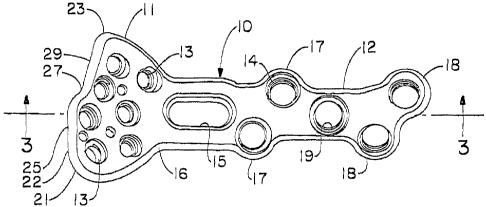
A first embodiment of the plate is shown generally at 10 in Figureq which
includes a
first, most distal portion or head 11 which has a profile from the top view
similar to the palm
of a hand, or which is shaped like a truncated heart, or a modified kidney
shape. The head 11
slopes upward in a complex and organic topography away from the more elongated
inversely
curving proximal portion 12 of the plate. The head 11 includes a plurality of
holes 13 for
pegs, which holes can be internally threaded or not, or can also include means
to provide for a
variable locking axis. The proximal plate portion 12 also includes a plurality
of holes 14 for
screws, which similarly can include internal threads, or which can be smooth,
or include
means for a variable locking axis screw. The proximal portion of the plate
also includes a slot
' 15 which is situated near the junction of the head 11 and the proximal
portion of the plate, or
7
CA 02645155 2008-09-05
WO 2007/103333 PCT/US2007/005624
the neck 16. The slot 15 can have a smooth internal edge, or can include a
textured feature,
such as grooves or tracks. The proximal portion also has two sets of tabs or
ears, an
intermediate pair 17 and a terminal pair 18 which each extend laterally from
the longitudinal
profile of the plate, and which provide for opposing screw holes that are each
offset from the
longitudinal axis of the plate and from each other along the longitudinal
axis. The central
point of a central screw hole 19 provides a point of reference or origin for
mapping in three
dimensions the topography or superficial locus of any point on the plate,
which in turn
enables the plate to be made having the complex curving fully contoured
configuration that it
does. The offset ears provide for convergence of the screws in the proximal or
plate portion
12 of the distal radius plate while still avoiding screw interference so as to
provide for
improved pullout strength as compared to a version where the proximal screws
are located
along a line, such as the longitudinal axis.
As shown in this embodiment, the head portion 11 of the plate has a complex
profile
which is rounded on either side away from the neck area 16 to form a first
prominence 21 and
a second prominence 23. The first prominence 21 has a more gradual curve than
the second
prominence and is also the more distally extending of the two prominences. It
is intended to
support the radial styloid, and thus is ternzed the styloid prominence herein.
The plate is
provided in a left-and a right version, which are mirror images of each other.
The plate is
generally, intended to be implanted on the volar side of the radius (i.e. the
top side when the
arm is supine, and the palm is pointed upward). The styloid prominence 21 is
thus on the
lateral facing side of the plate, or the thumbward side. The second prominence
23 is designed
to fit under the ridge of the lunate process, and is thus termed the "lunate"
prominence herein.
The distal edge 22 of the head 11 extends in a direction across the
longitudinal axis of the
proximal portion of the plate in three segments. A first portion 25 extends
substantially
transverse to the longitudinal axis of the plate to a point slightly more
than, or about midway
across the head of the plate. A second edge portion 27 links the first portion
25 and the third
portion 29 and extends at an oblique angle proximally toward the third edge
portion. The
edge portion 27 or oblique link helps the surgeon to gauge the placement of
the plate relative
to the lunate ridge of the radial bone.
8
CA 02645155 2008-09-05
WO 2007/103333 PCT/US2007/005624
While the plate has tapering areas, the cross sectional dimension is generally
sufficiently uniform that the contours of the top surface 30 generally mirror
the contours of
the bottom surface 31. In this context, "bottom" or "internal" is used to mean
the surface
which faces, and which may, but does not necessarily have to touch the bone,
and `top" or
"external" means the outwardly facing surface. These surfaces undulate to
mimic the shape
or topography of the radial bone. More specifically, the bottom surface of the
proximal
portion of the plate includes a concavity or radius 32 along the longitudinal
axis where the
thumb side of the plate has a greater arc than the pinky side. As might best
be viewed in
Figure 8, the pinky side of the plate, (i.e., in that view, the right side)
forms a shallow
serpentine on both the top and bottom surfaces which defines a gentle
depression or cup 33
which is followed by a rise toward the lunate prominence 23 of the head. The
cup extends
and becomes shallower as the plate surface rises toward the styloid
prominence, which has the
highest elevation in the Z direction. Thus, particularly in the proximal
portion, the plate 15 appears to spiral along the longitudinalaxis of the
radial bone. Figure 6 further illustrates this
aspect of the plate in accordance with the invention where the Y axis is taken
through a
central screw hole.
The topography of the plate is even more clearly shown in the sectional
drawings
Figures 9 through 9g which represent parallel slices taken in the Z planes at
progressive
locations along the longitudinal axis. It can be seen from these sections that
the proximal
portion 12 of the plate, as well as the head portion 11 of the plate is
substantially non-planar,
meaning that there is no significant portion of either the top surface or the
bottom surface of
either the proximal portion, or the head portion that defines a single plane.
Instead, the head
of the plate undulates from a central cup area that has a diagonal aspect from
its lowest
portion near the neck 16 of the plate on the pinky side of the head to the
highest portion at the
distal area on the styloid side of the plate. Thus, the top or exterior
surface of the head 11 has
a slightly concave area or cupped area 33 and other areas,= such as the
styloid prominence 21
and the lunate prominence 23 which are slightly convex on the top surface 30,
or which rise.
This transition can be said to cause the head to have top and bottom surfaces
30, 31 which
9
CA 02645155 2008-09-05
WO 2007/103333 PCT/US2007/005624
undulate as they transition from the proximal portion of the plate 12 to the
head portion 11.
Further, the proximal portion of the plate 12 includes a bottom surface 31
which is radiused to
fit the curve of the bone as it extends proximally from the wrist joint toward
the elbow joint.
This concave or radiused area changes as it extends along the longitudinal
axis of the plate.
In particular, the plate transitions from an area that is flatter in the
vicinity of the neck 16, and
which increases in the amount of curve as can be seen by comparing Figure 9d
through 9g
which illustrate the cross-section of the plate at progressive proximal
locations along the
longitudinal axis. This topography is mirrored in the left and right versions
of the plate,
which'are enantiomorphs (meaning that there is a three dimensional mirror
symmetry) of each
other. The plate spirals down the bone changing the internal curvature to
accommodate the
bone, and further changing the outline, or footprint that would be left on the
bone by spiraling
along the axis of the bone. Thus, the plate changes cross-section in the Z-
direction, and also
shifts in the X-Y directions to define this spiral.
The plate head 11 is further provided with a plurality of holes 40 which
receive pegs
that are implanted into the distal portion of the radius, or into fragments of
the bone. In
particular, the distal radius plate head of the present invention is presented
in two
embodiments. In a first embodiment, all of the holes define a fixed axis for
the pegs which
they receive. The pegs holes 40 include internal threads 41 which mate with
locking threads
on the head of the pegs and which therefore lock the pegs in position in the
plate and which
accordingly lock the bone relative to the plate. Of course, the plate could
include peg holes
which have no internal threads, or some combination of threaded and non-
threaded holes.
Alternatively, pegs can be used with a plate having threaded holes, where the
pegs are not
threaded at the top, but include a head that fits within the major diameter of
the internal
threads. In yet another embodiment, the pegs can be secured in the plate by
means of a
variable locking mechanism that allows the pegs to be inserted through-the
plate holes at a
selected angle and to be subsequently locked into position at that angle. It
is particularly
preferred to provide a combination of locking pegs and variable locking pegs
in the same
head.
-
CA 02645155 2008-09-05
WO 2007/103333 PCT/US2007/005624
The holes include a distal hole 42 in the styloid prominence 21, and a hole in
the
lunate prominence 45 and one or more (two in the case shown) holes 43, 44 in
the head
intermediate to the two side holes. One or more proximal set of holes is also
advantageously
provided. For example, a hole 46 may be provided under the styloid hole 42 and
a hole 48
may be provided proximal to the hole 45 in the lunate prominence 23, and a
hole 47 may be
provided between the hole 46 and the hole 48. The angles of these holes
determine the angles
of the pegs that they receive. The angles are defined on a three coordinate
matrix where 0,0,0
is the origin and is located at the center of a central screw hole in the
plate. Further, the plate
is shown as including smaller diameter holes for K wires which help with the
pIacement and
angulation of the pegs. Thus, the holes K43-K48 have similar orientations to
the holes 43-48
(where no K holes are illustrated for holes 42, 45 and 47). The angles are set
to provide for the
most common dislocation of fmgments and to provide for the optimal fixation
using the pegs.
The design contemplates a plurality of fixed peg holes, including one 42, 45
in each of
, the styloid (i.e. hole one) and lunate prominences (i.e. hole four); which
splay outward and
away from the plate such that they diverge away from one another to be capable
of locking a
styloid fragment and/or a fragment from the lunate fossa portion of the radial
bone. A second
distal-most hole 43 is located generally under the skewed linking area of the
head, which
defmes a peg axis that extends through the radius and distally toward the
scaphoid or
navicular bone and a third hole 44 is distally aligned between the two holes
of the
prominences but is slightly backed off proximally from the second hole, with a
peg axis that is
more transverse than the peg axis of the second hole. The fourth peg hole 45
is the hole of the
lunate prominence which is slightly more proximal than the first hole which is
located in the
styloid prominence. A fifth hole 46 is located in the first rounded side,
which is on the styloid
or thumbward side, toward the intersection between the proximal portion of the
plate, and the
plate head. The axis defined by this hole diverges outwardly toward the
lateral portion of the
radius (in a supine position). A sixth hole 47 is located in a central portion
of the head such as
on a longitudinal axis of the plate, and a final seventh hole 48 is located
most proximally at
the intersection of the head 11 and the proximal portion of the plate 12 at
the neck 16 of the
11
CA 02645155 2008-09-05
WO 2007/103333 PCT/US2007/005624
plate, with the axis of the peg appearing to be more or less transverse
relative to the plane
defined by the opening of the peg hole.
In a further embodiment of the invention, pegs or screws are included which
have a
variable axis with a locking mechanism. While this mechanism is illustrated
for use in the
head portion of the plate, is should be understood that it could be used in
the proximal portion
of the plate instead, or as well. Figures 10, 11, 14 through 20, illustrate
this embodiment of
the invention. In particular, one or more of the peg holes in the head portion
I 11 of the plate
110 of the first embodiment may be replaced with variable locking pegs, or the
pegs may be
reoriented. The proximal portion 112 has the same features and is the same as
previously
described. In the embodiment shown, holes 42, 45, and 47 are each replaced
with a variable
locking mechanism 142, 145, and 147. As one example of a variable locking
mechanism, the
one illustrated includes a camming mechanism on the head of the peg which
mates with a cam
locking insert that fits into and locks into a hole in the plate. The cam
locking insert includes
an anchor member that causes the cam locking insert to resist rotation as the
camming
members of the peg engage the cam raceway of the cam locking insert. Other
variable
locking mechanisms can be used with this invention, including other expansion
inserts which
mate with the holes in the plate at a variable angle, but can be locked in
position, such as by
expansion.
As an additional aspect of the invention, a distal radius plate is provided in
an
embodiment which has an elongated proximal portion. This design is illustrated
in Figures 12
through 14 and is shown with a distal head having only fixed a.ngle pegs and
having both
fixed and variable angle pegs. In the embodiment shown in Figures 12 and 13,
the plate 310
has a distal portion, or head 311 and a proximal portion 312. The head portion
includes pegs
holes 313 which can be internally threaded so as to define screw holes having
fixed axes as is
shown in Figures 12 and 13, or as is illustrated in Figure 11, and one or.more
of the fixed peg
holes may be replaced with a variable axis mechanism, that advantageously also
provides for
locking of the angle of the axis. Again, it is envisioned that the proximal
portion of the plate
is provided in a right and a left version which are mirror images from each
other.
12
CA 02645155 2008-09-05
WO 2007/103333 PCT/US2007/005624
The elongated version of the plate includes the features of the previously
described
version, with screw holes 314 in the proximal portion; an elongated slot 315
located along the
central axis of the plate, adjacent the neck 316 which is the area that links
the head 311, and
the proximal portion 312. The elongated version of the plate further includes
an intermediate
tabbed area 317 having opposing offset ears that each receive a screw through
an internally
threaded screw hole, and a terminal tabbed area 318 that includes opposing
offset ears that
likewise each includes internally threaded screw holes. This version of the
plate includes a
central screw hole 319 that defines the origin for the coordinate system of
the plate, and in
addition, there are one or more additional longitudinally aligned screw holes
320. The
elongated version of the plate has a proximal portion having a spiraling
radiused portion
similar to the shorter version except with a longer, and thus, more pronounced
spiral. The
fixed angle head 311 is the same as for the shorter version and the fixed
angle head 11 of
Figure 1, and the head 411 shown in Figures 14 and 15 is the same as the head
111 shown in
Figures 10 and 16.
A camming insert is inserted in the variable axis bore holes, which have
concavely
rounded sides to permit variable angulation of the camming insert in the bore.
The camming
insert is a generally circular or ring shaped insert 710 having an expansion
gap 712 which is
essentially a planar slice taken in the insert so as to create a gap. The
insert 710 has a top
surface 714 which is generally planar joined to a co-planar bottom surface 716
by an
outwardly curving side surface 718. There is a concentric inwardly curving
surface 720
which further includes the cam race 722, which in this case are two grooves
that spiral a
portion of the way down and around the inside surface. The grooves are open,
and preferably
only for a portion of the top 714 where the grooves are located. This open
area of the race
allows the cams to be introduced into the race. Subsequently, as the screw or
peg is turned in
the camming insert, the cam engages the cam race and causes the insert to
expand at the gap.
This action causes the insert to lock in the recess 726 in the plate which
receives the insert.
Further, the insert 712 includes a stop 724. The stop is a projection that is
received in a well
13
CA 02645155 2008-09-05
WO 2007/103333 PCT/US2007/005624
728 in the recess which retains the stop 724 and prohibits the cam insert from
turning with the
peg as it is turned relative to the plate.
Figures 19 and 20 show a variable axis locking screw 750, which has a shaft
751 with
a blunt or rounded insertion tip 790. The shaft 751 tapers throughout its
length. The screw
includes a locking head 760.. The locking head includes a pair of cam wings
770 which are
shaped to engage the race in the locking insert 710 and cause the insert to
expand radially
outwardly to form a friction fit in the bore hole and lock the camming insert
in position in the
bore. The shaft of the variable locking screw 750 is threaded with a thread
753 and having a
taper to the minor diameter 752 of the shaft 751 over the first three to five
turns of the thread
toward the insertion tip 790 while the major diameter does not taper. The head
760 further
includes a torque driving recess 780, with an optional bore 782 connected to
the torque
driving recess by= a transitional area 78, which retains the screw 750 on the
post of a
screwdriver.
Figures 21 through 24 show a non-locking proximal screw 810, which is intended
in
particular for use in the proximal portion of the plate, when it is desirable
that the screw does
not lock into the plate. The screw 810 has a shaft portion 811 having a thread
813. The
thread 813 shown in Figures 21 through 24 has. a taper in the minor diameter
812 over a
portion 815 of the= shaft 811, such as about the first three turns of the
thread. Thereafter, the
terminal portion 816 of the shaft 811 has a constant minor diameter 812. The
screw 810 has a
blunt tip 850 and a rounded head 820 having a torque driving recess 830
optionally including
a bore 832 to receive the post of a screwdriver to retain the screw on the
screwdriver. The
head has a spherically rounded lower portion 834 and a rounded upper portion
836 where the
maximum outer diameter is smaller than the inner diameter of the threaded
proximal screw
holes. The shaft 811 has a minor diameter 512 about which the thread 813
spirals. The
thread 813 includes a spiraling radial edge 514 best viewed in the thread
detail Figure 22a,
which defines the major diameter. The thread further includes a front thrust
face 815 which
forms an angle of about 20 +/- 5 to a plane transverse to the longitudinal
axis of the screw.
The trailing face 816 of the thread 813 forms an angle of about 5 +/- 2 to
the same plane.
14
CA 02645155 2008-09-05
WO 2007/103333 PCT/US2007/005624
Figures,25 through 26(a) show a locking proximal screw 860, which is intended
in
particular for use in the proximal portion of the plate when it is desirable
that the screw locks
into the plate. The screw 860 has a shaft portion 861 having a thread 863
similar to the thread
previously shown. The thread 863 shown in Figures 25 and 26 has a taper in the
minor
diameter 862 over a portion 865 of the shaft 861, such as the first three
turns of the thread.
Thereafter, the terminal portion 866 of the shaft 861 has a constant minor
diameter 862. The
head of the screw 870 includes external locking threads 883 as can be best
viewed in the head
detail in Figure 26a. These threads also include a radial edge 884, a front
thrust face 885, and
a trailing face 886. The angle of the front thrust face 885 is the same as the
angle of the
trailing face relative to a plane which transverses the longitudinal axis of
the screw, and is
about 30 +/- 5 for each angle. Thus, the locking thread =883 on the head 870
of the screw is
a symmetrical v-shaped thread when viewed in profile in cross section. The
head 870 tapers
along the longitudinal axis, in both the major and the minor diameter, by a
similar amount, as
is shown in Figure 22a. For a proximal screw, the taper is about 7 , or about
3.5 per side
when measured in cross-section. A larger screw might be used in the distal
head of the plate
and might have a taper of 20 or 10 per side in section.
The screw 860 has a blunt tip 890 and a rounded head 870 having a torque
driving
recess 880 optionally including a bore 882 to receive the post of a
screWdriver to retain the
screw on the screwdrivei-. The head has a-locking thread 884 which tapers in
both the major
and minor diameter.
Figures 27 and 28 show a drill guide for either the holes in the distal
portion, or for the
holes in the proximal portion. The drill guide 910 includes an extending
handle 912 with a
tapering linking portion 914 and a post 916 which engages the hole in the
plate. The post 916
has an internal hole for the drill bit and can have a smooth tapered surface
at the terminal end
918 which engages the hole of the plate by friction, or the post end 918 can
include threads to
lock into the internal threads of the plate and to fix the angle for the fixed
screws.
CA 02645155 2008-09-05
WO 2007/103333 PCT/US2007/005624
Figures 29 through 34 show a further embodiment of the plate in accordance
with the
invention. This plate 1010 is comparable to the other embodiments in having a
head 1011
joined to a proximal plate portion 1012. The head 1011 includes a plurality of
peg holes 1013
as previously described and the proximal portion includes a plurality of screw
holes 1014.
This embodiment of the plate has only a single set of offset tabs 1017 which
allows for
convergent screws and the plate ends in a terminus 1018.
While in accordance with the patent statutes the best mode and preferred
embodiment
have been set forth, the scope of the invention is not limited thereto, but
rather by the scope of
the attached claims.
16