Note: Descriptions are shown in the official language in which they were submitted.
CA 02646221 2008-09-17
WO 2007/107831 PCT/IB2007/000637
1
Electrostimulating ap aratus and method
The invention relates to an electrostimulating apparatus and
method.
In neurophysiology, the H reflex, or Hoffman reflex is known,
which, although it is a reflex that is very similar to the
monosynaptic reflex following a mechanical stretching of a
muscle, may also be evoked through an electric stimulation
conducted at the level of an afferent innervation. In the
recent past, the H reflex in humans has been studied widely,
as the features of the latter enable useful information to be
obtained for defining the spinal excitability in humans both
in physiological and pathological conditions. In particular,
the modulation of the H reflex has been studied following
serious clinical manifestations of a heterogeneous group of
pathologies, comprising spasticity, dystonia and
fibromyalgia. In these pathologies, an increase in spinal
excitation at the level of a single metamer or of several
metamers is recognised as a physiopathological common
denominator that is activated by various central and
peripheral influences, and the spinal excitation can be
studied in vivo in humans by evaluating carefully the H
reflex both in terms of latency and in terms of the amplitude
of the reflex with respect to the dispensed stimulation. The
H reflex is definable as the simplest of the spinal reflexes
and can be evoked by electrically stimulating type Ia
afferent fibres comprised in the muscle spindle endings. This
stimulation is followed by a transmission of the evoked
discharge afferent to the spinal cord, a production of a
synchronised postsynaptic excitatory potential that is
sufficient to discharge the motor neurons of a relevant pool
with a transmission of the reflex discharge along the axons
of the alpha-type motor neurons to the muscle. The
excitability of the spinal motor neuron depends directly on
the descending central path under the systemic influence,
which is typically at the endocrine level and is mediated by
circulating neurotransmitters, of projection of the
CA 02646221 2008-09-17
WO 2007/107831 PCT/IB2007/000637
2
peripheral reflex arch. The measurement of the minimum
latency of the H wave, combined with the amplitude, width and
threshold values of the latter, provides information on the
conduction level of the reflex arch. The amplitude of the H
reflex on the other hand enables to measure indirectly the
quantity of alpha motor neurons that have been activated
synchronously, modulated by various afferences. A weak
voluntary contraction strengthens the H reflex, increasing
the discharge of the motor neuron pool, but alters the
latency of the reflex. In non-pathological circumstances, the
H reflex can be recorded from the soleus muscle by
stimulating the tibial nerve and from the flexor carpi
radialis muscle by stimulating the median nerve through a
low-frequency stimulus.
If it is not possible to reproduce a reflected response this
can be ascribed to an afferent disturbance or to a low
central excitability. The low central excitability does not
necessarily indicate a specific pathology, as the test during
a weak muscular contraction may reveal an intact reflex path
with a normal latency. In the literature, there are reported
various attempts to reduce the hyperexcitability of the motor
neuron through Transcutaneous Electric Stimulation (TENS),
although there is no univocal consensus on the effect that
the latter could have on the Hoffman reflex.
The spinal excitability is regulated by many influences that
can be concisely classified as above the spinal cord,
systemic (due to hormones and circulating neurotransmitters),
propriospinal (intra-spinal connections) or reflected
peripheral influences.
The reflected peripheral influences in turn comprise a
combination of reflex arches, which are both monosynaptic and
oligo- or multisynaptic and are integrated at a distinct
spinal innervation level (metamer). The peripheral afferences
come from the central branch of the cells of the spinal
ganglia. The peripheral branch is connected to different
types of receptor: the muscle spindles, the tendon receptors,
CA 02646221 2008-09-17
WO 2007/107831 PCT/1B2007/000637
3
the joint receptors and various types of cutaneous receptors.
In particular, the afferences of the muscle spindles (fibres
Ia) are the afferences that determine the most direct
relations with the pool of the alpha motor neurons
interacting in the so-called "Sherrington monosynaptic
reflex". Although the Sherrington reflex model is still an
object of discussion, it can be stated that when a muscle is
stretched the primary sensory fibres, i.e. the afferent
neurons of the group Ia of the muscle spindles, respond both
to the speed and degree of extension, sending the information
at the spinal level. On the other hand, the secondary sensory
fibres, i.e. the afferent neurons of the group II, detect and
send to the central nervous system (CNS) only the information
relating to the degree of stretching. This information is
transmitted monosynaptically to the alpha motor neuron that
activates the extrafusal fibres in order to reduce the
stretching and is transmitted polysynaptically, by means of
an interneuron, to another alpha motor neuron that inhibits
the contraction in the antagonist muscle. Further, at the
same time, through two types of gamma motor neurons, known as
static and dynamic motor neurons, the CNS is able to
influence the afferences of the muscle spindles during
movement. The muscle spindle is thus definable as the most
important proprioceptor, having a fundamental role in the
movement and the control of the reflex activity. The combined
signal coming from a plurality of muscle spindles of each
muscle provides the CNS with information, generating a fine
adjustment of the muscular activation and thus acting as a
sort of servo control. At the same time, the muscle spindles
are controlled in a continuous manner by the gamma neurons
that the CNS controls separately from the alpha motor neurons
by controlling all muscle functions. The intrafusal fibres
are typically excited by the stimulation below the extrafusal
motor threshold: as soon as the motor threshold has been
exceeded, the muscle contraction activates the tendon
receptors, which provoke the effect of the muscle spindles.
CA 02646221 2008-09-17
WO 2007/107831 PCT/1B2007/000637
4
WO 02/09809 discloses an apparatus for treating muscular,
tendon and vascular pathologies by means of which a
stimulation is applied to a patient, which stimulation
comprises a series of electric pulses having a width
comprised between 10 and 40 microseconds and an intensity
that is variable in function of the impedance and conductance
of the tissue subjected to stimulation, and comprised between
100 and 170 microamperes.
WO 2004/084988 discloses an electrostimulating apparatus
owing to which it is possible, in function of the type of
electric stimulation produced and of the configuration
parameters adopted, to generate an induced bioactive
neuromodulation, which is suitable for producing vasoactive
phenomena on the microcircle and on the macrocircle. These
phenomena are in turn mediated by phenomena connected to the
direct stimulation of the smooth muscle and by essentially
catecolaminergic phenomena, by means of stimulation of the
postsynaptic receptors. The aforesaid apparatus is able to
produce specific stimulation sequences that induce
reproducible and constant neurophysiological responses. In
particular, WO 2004/084988 discloses an activating sequence
for activating the microcircle (ATMC) and a relaxing sequence
for relaxing the muscle fibre (DCTR), which are able to
stimulate various functional contingents, including the
striated muscle, the smooth muscle and the peripheral mixed
nerve. The aforesaid stimulation sequences are assembled on
three basic parameters: the width of the stimulation, the
frequency of the stimulation and the intervals of time during
which different width/frequency combinations follow. The
general operating model of the stimulation sequences reflects
the digital-analogue transduction that occurs in the
transmission of a nerve pulse.
The neuronal electric stimulation by modulation of frequency
and amplitude, or FREMST' (Frequency Rhythmic Electric
Modulation SystemT"'), disclosed in the aforesaid WO
2004/084988 and in WO 2004/067087 (incorporated herein for
CA 02646221 2008-09-17
WO 2007/107831 PCT/IB2007/000637
reference), is characterised by the use of transcutaneous
electric currents, which are produced by means of sequential
electric pulses having variable frequency and width. The
frequency may vary between 0.1 to 999 Hz, the width of the
5 stimulation is comprised between 0.1 and 40 ps and the
voltage, which is kept constantly above the perception
threshold, is comprised between 0.1 and 300 V (preferably 150
V). By suitably combining the aforesaid frequency and width
variations a specific sequence defined as DCTR is obtained,
having a relaxing effect and comprising a series of
subphases, called A, B and C. Frequency and width are
constant in the subphase A, the frequency is constant and the
width is variable in the subphase B, the frequency is
variable and the width is constant in the subphase C.
Experimental studies have enabled the effects of FREMS to be
evaluated and the capacity of the latter to evoke compound
muscle action potentials (cMAP) to be evaluated, which are
obtainable in the adductor hallucis muscle by stimulating the
posterior tibial nerve, as well as the variation in amplitude
of the aforesaid H reflex by using the latter as a
conditioning stimulus. As disclosed in WO 2004/084988, the
aforesaid experimental studies have also shown that the
greatest amplitude of the cMAPs that is obtainable (0.60
0.02 mV) is approximately 15 times less than that of the
cMAPs obtained with the known devices that dispense TENS
current, i.e. amplitudes of the order of 9 0.6 mV with
stimuli having a width typically comprised in a range of 200-
1000 }is. It has been further observed that the maximum
amplitude value of the cMAPs is obtained in the presence of a
width/frequency ratio of 0.13 (40 s/29 Hz).
A further type of sequence, called ATCM and suitably designed
for obtaining a vasoactive effect, has a prevailing action on
the motility of the microcircle, i.e. of the smooth
sphincters of the arterioles and venules of the subcutaneous
tissue. The ATCM sequence is divisible into three
subsequences, called S1, S2, S3. The subsequences S1 and S3
CA 02646221 2008-09-17
WO 2007/107831 PCT/IB2007/000637
6
are both distinguished by a frequency increase phase, with
distinct time modes. The subsequence S2 is mainly constituted
for producing variability in the width of the individual
stimuli, in a gradually increasing frequency range, in such a
way as to reduce the bioreaction, until the latter is
stabilised. More in detail, the subsequence S1, having a
relaxing effect and therefore having an effect that is very
similar to the aforesaid DCTR sequence, comprises phases in
which, after a first adaptation phase conducted at 1 Hz
frequency, the frequency is gradually increased at a constant
amplitude, thus decreasing the bioreaction in a gradual
manner. Subsequently, the frequency is increased in a much
more rapid manner until it reaches a target of 19 Hz. The
subsequence S2 is then run, which is in turn divisible into
four phases, called S2-A, S2-B, S2-C and S2-D. In the
subsequence S2, after a phase (S2-A) conducted at a constant
frequency in which the amplitude is rapidly increased until
the instant 1, the frequency is gradually increased and
consequently the bioreaction rapidly falls until the instant
2 (S2-B) . At this point the amplitude is reset that again
rises at a constant frequency until the instant 3(S2-C).
Subsequently, the frequency again increases gradually whilst
the amplitude is kept constant and, consequently, the
bioreaction gradually decreases until the instant 3(S2-D).
In this way the bioreaction is varied in a discontinuous
manner, producing points of sudden slope variation, i.e. the
points 1, 2 and 3. In practice, as disclosed in WO
2004/084988, a system is obtained producing a sequence of
vasodilations and vasocontractions with sequential increases
and decreases of haematic flow of the microcircle surrounding
the stimulation zone. These vasodilations and
vasocontractions produce a "pump" effect that is clearly
produced by neuromodulation of the sympathetic
neurovegetative system, which influences the vasoaction
through the smooth muscle of the capillary vessels and the
arterioles. In this way it can be shown that this
CA 02646221 2008-09-17
WO 2007/107831 PCT/IB2007/000637
7
subsequence, which is distinguished by alternating variations
of the rheobase, therefore produces a vasoactive effect
consisting of sequential vasodilation phases and
vasoconstriction phases. This definitely produces a draining
effect and, above all, makes the microcircle elastic and
modulates the latter around a main carrying event that
determines the average variation thereof.
An object of the invention is to improve known
electrostimulating apparatuses.
Another object is to provide an electrostimulating apparatus
that enables muscular hyperexcitability of spinal and/or
cerebral origin in a patient to be treated.
A further object is to provide an electrostimulating
apparatus and method that enables muscular hyperexcitability
of spinal and/or cerebral origin in a patient to be treated.
In a first aspect of the invention, there is provided an
electrostimulating apparatus, comprising generating means for
generating electric pulses organised in sequences having
preset values of typical parameters, said typical parameters
comprising amplitude, width and frequency of said pulses, a
plurality of stimulation channels such as to dispense said
sequences to body zones of an organism in an independent
manner, varying means suitable for varying at least one of
said typical parameters so as to substantially prevent said
organism from habituating to said electric pulses.
In a second aspect of the invention, there is provided a
method for electrostimulating an organism, comprising:
-producing a sequence of electric pulses having a relaxing
effect and a further sequence of electric pulses having a
vasoactive effect;
-dispensing said sequence to body zones of said organism, and
further dispensing said further sequence to further body
zones of said organism, said body zones and said further body
zones comprising an agonist muscle and an antagonist muscle
of a neuromuscular compartment comprised in said organism.
CA 02646221 2008-09-17
WO 2007/107831 PCT/IB2007/000637
8
These aspects of the invention are based on a new
neurophysiological effect that was found during recent
experimental studies conducted on the aforesaid FREMS. These
studies have in fact shown that the amplitude of the H reflex
sampled from the ipsilateral soleus muscle with or without
conditioning of the FREMS applied to the short adductor
hallucis muscle, is substantially decreased (by a value equal
to 50%) during FREMS stimulation. The amplitude variation of
the H reflex is significantly influenced by the variations of
the width pulse/stimulation frequency ratio (w/f), in
particular during the subphase C (r2 = 0.43; p<0.001). This
result suggested that the FREMS is actually capable of
modulating the amplitude of the H reflex, very probably
through active recruitment of the muscle spindles.
Owing to these results it has been possible to make a new
electrostimulating apparatus, by means of which a new
electrostimulating method can be carried out for treating the
spinal hyperexcitability that is secondary to cerebral or
spinal damage and is the cause of spasticity in a patient.
The new electrostimulating apparatus enables the aforesaid
FREMS to be applied, with different sequences and
simultaneously, in two antagonist neuromuscular districts of
a motor limb that are connected to the same metamer and
mutually connected through an afferent
neuron/interneuron/alpha motor neuron loop (circuit). In this
way, a synergic effect can be produced that inhibits the
hypertonic contraction, which contraction is typically caused
by the dysfunctions of the upper motor neuron and is
therefore typical of the spastic phenomena that are secondary
to cerebral or spinal damage of the central nervous system.
The invention can be better understood and implemented with
reference to the attached Figures, which illustrate an
exemplifying but non-limiting embodiment thereof, in which:
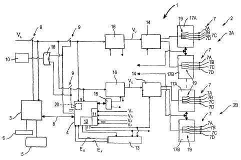
Figure 1 is a block diagram illustrating an
electrostimulating apparatus comprising a plurality of
independent stimulation channels;
CA 02646221 2008-09-17
WO 2007/107831 PCT/IB2007/000637
9
Figures 2 to 4 show electromyograms illustrating the
production of cMAP in the abductor hallucis muscle obtained
by stimulating the posterior tibial nerve with DCTR
sequences;
Figure 5 shows a potential difference/time cartesian graph,
illustrating the variation in the cMAP value during the
subphases A, B and C of a DCTR sequence;
Figure 6 shows a potential difference/ratio between pulse
width and pulse frequency cartesian graph, illustrating the
variation in the cMAP value during the application of a DCTR
sequence;
Figure 7 is a graph illustrating the amplitude of the H
reflex in the presence or absence of FREMS stimulation;
Figures 8 to 10 show cartesian graphs illustrating the
amplitude variation of the H reflex in function of the
variation in the ratio between pulse width and pulse
frequency, during three FREMS stimulation sessions;
Figure 11 shows a cartesian graph illustrating the average
amplitude variations of the H reflex in function of the
variations in the ratio between pulse width and pulse
frequency, as measured during the three FREMS stimulations of
Figures 8-10.
Figure 1 shows schematically the assembly of the circuits
comprised in an electrostimulating apparatus 1 that is able
to produce and dispense the aforesaid DCTR (relaxing)
sequences and ATMC (vasoactive) sequences comprised in FREMS
stimulation through a plurality of independent stimulation
channels 2, each of which is formed by a plurality of pairs
of transcutaneous electrodes 7.
In the embodiment of the apparatus 1 shown in Figure 1 there
are provided four stimulation channels 2, of which only two
are shown (for reasons of clarity) and are indicated by 2A,
2B.
In an embodiment that is not shown, there is provided an
apparatus 1 comprising a number of stimulation channels 2
that is greater than four.
CA 02646221 2008-09-17
WO 2007/107831 PCT/IB2007/000637
In another embodiment that is not shown, there is provided an
apparatus 1 comprising a number of stimulation channels 2
that is less than four.
The apparatus 1 comprises a first control unit 3 and a second
5 control unit 4, which interact with one another and are made
of microprocessors of known type. The first control unit 3
controls a displaying device, for example a liquid crystal
display 5, and an alphanumeric keyboard 6. By keying in on
the latter a user of the apparatus 1 can direct the operation
10 of the latter and set the parameters, which are displayable
on the display 5, of the electric stimulations to be
administered to a patient.
In an embodiment that is not shown, there is provided a
remote-control device by means of which a patient connected
to the apparatus 1 can control the operation of the latter
without interacting with the keyboard 6. This embodiment is
particularly useful inasmuch as it enables the patient to
control the apparatus 1 by acting as a sensory feedback
element relating to one or more operating parameters of the
apparatus 1. The first control unit 3 controls a safety
switch 9, which in turn controls an input supply voltage VA.
In normal operating conditions, the switch 9 is closed and a
voltage adjuster 16 (the function of which will be disclosed
below) that is comprised in each stimulation channel 2 is
thus supplied. In emergency conditions, for example in the
event of apparatus faults, the first control unit 1 opens the
switch 9 and thus interrupts the supply, to the voltage
adjuster 16. To the second control unit 4 a luminous device,
for example a LED 10 of known type, is further connected.
When a patient is connected to the apparatus 1 by means of
the electrodes 7 and the apparatus 1, supplied by the voltage
VA, administers an electric stimulation to the patient, the
LED 10 lights up, thus indicating that the patient is
subjected to the action of an electric current.
Through a serial communication interface 8, of known type,
the first control unit 3 is connected to the second control
CA 02646221 2008-09-17
WO 2007/107831 PCT/IB2007/000637
11
unit 4, which controls the production of the electric pulses
by adjusting the basic parameters thereof, i.e. amplitude,
width and frequency, and comprises an analogue-digital
converter (ADC) 11 and an integrated timing unit (ITU) 12. In
the second control unit 4 there can be housed a support 20
(that is shown by means of a dotted line) on which the data
are recorded that are necessary for the operation of the
apparatus 1, such as, for example, the data relating to the
stimulation sequences that are producible by the apparatus 1.
The support 20 is readable through data processing means
(which is not shown), of known type, comprised in the
apparatus 1 or arranged outside the apparatus 1 and
interfaced with the latter. The data processing means, if it
is comprised in the apparatus 1, may, for example, be
positioned in the second control unit 4.
In an embodiment that is not shown, the support 20 is housed
in the first control unit 3.
The analogue-digital converter 11 receives a feedback signal
(in the form of voltage) relating to the pulse amplitude, and
intervenes by producing an adjustment and/or an alarm signal
if the pulse amplitude produced by the apparatus 1 is
different from that set by the user. In particular, the
analogue-digital converter 11 receives a reference voltage VT
regulating the operation of the analogue-digital converter
11, a further reference voltage VR, which enables the correct
operation of the analogue-digital converter 11 to be checked,
and, from each of the stimulation channels 2, a feedback
voltage VF.
The integrated timing unit 12 defines the width and frequency
of the pulse by interacting with a timing control device 13.
The latter controls the width and frequency of the produced
pulse and, if one or the other of these parameters is not
correct, produces and sends a width error signal ED and/or a
frequency error signal EF, which are able to arrest the
second control unit 4.
CA 02646221 2008-09-17
WO 2007/107831 PCT/IB2007/000637
12
Similarly to what has been disclosed in relation to the first
control unit 3, also the second control unit 4 controls
safety switches 9, which are provided in a number equal to
the number of stimulation channels 2 comprised in the
apparatus 1. The safety switches 9 controlled by the first
control unit 3 and by the second control unit 4 interact with
one another and with the LED 10 through an "OR"-type logic
port 18.
The electric signals defining the frequency and width of the
pulse are produced by the integrated timing unit 12 and are
sent directly to an outlet pulses generator 14. In the
apparatus 1 the outlet pulses generators 14 and the
stimulation channels 2 are provided in equal numbers. Pulse
width is defined and adjusted by a digital-analogue converter
(DAC) 15 interacting with the second control unit 4. The
digital-analogue converter 15 produces a plurality of
electric signals defining the pulse amplitude for each single
channel 2, and each signal is sent to a voltage adjuster 16.
The apparatus 1 comprises a number of voltage adjusters 16
that is equal to the number of stimulation channels 2. An
outlet voltage VU, the value of which is comprised between 0
and 300 Volts, is produced by each voltage adjuster 16 and is
sent to a corresponding outlet pulses generator 14. Each
outlet pulses generator 14 produces a pulse having a preset,
frequency and width and sends this pulse to a pair of outlet
selectors 17A, 17B to which the electrodes 7 are connected.
The pairs of outlet selectors 17A, 17B are provided in a
number equal to the number of outlet pulses generators 14
comprised in the apparatus 1. Each outlet selector 17A, 17B
comprises a plurality of switches 19, which are provided in a
number equal to the number of electrodes 7 connected to the
selector, by means of which switches the produced pulse can
be alternatively transmitted to the corresponding electrode
7, or stopped. In each pair of outlet selectors 17A, 17B the
electrodes 7 are associated functionally so as to form four
pairs, the electrodes of each pair being indicated
CA 02646221 2008-09-17
WO 2007/107831 PCT/IB2007/000637
13
respectively as 7A, 7B, 7C and 7D. The electrodes 7 of each
pair are connected to the corresponding outlet selector 17A
or 17B.
In an embodiment that is not shown, outlet selectors 17A, 17B
are provided comprising a number of pairs of electrodes 7
greater than four.
In another embodiment that is not shown, there are provided
outlet selectors 17A, 17B comprising a number of pairs of
electrodes 7 that are less than four.
When the apparatus 1 is in use, by acting on the switches 19,
it is possible to select the electrodes 7 to which to send
the pulse produced by the outlet pulses generators 14. It is
thus possible to use independently both the pairs of
electrodes 7A-7D comprised in two or more stimulation
channels 2 and the pairs of electrodes 7A-7D comprised in a
single stimulation channel 2.
As the second control unit 4, by means of the digital-
analogue converter 15 and the integrated timing unit 12, is
able to adjust the amplitude, width and frequency of the
pulses produced in the stimulation channels 2 in an
independent manner for each channel 2, the apparatus 1 is
such as to be able to multiply the outlet pulses and space
the latter in a preset manner.
Further, the integrated timing unit 12 enables the width of
the outlet pulse to be increased in a preset manner. In
particular, it is possible to obtain a percentage increase of
the width of an electric stimulation pulse that is conducted
in a plurality of phases, after the completion of which
phases the width of the pulse remains constant. The
percentage increase of the width of the pulse, the width of
the pulse and the number of the phases are mutually
correlated by the following formula:
Ti(Nf) = To x (1 + Io~Nf
where:
Nf = Number of phase;
CA 02646221 2008-09-17
WO 2007/107831 PCT/IB2007/000637
14
Ti(Nf) = Width of stimulation pulse in function of the number
of phase;
To= Width of initial stimulation pulse;
Io = Percentage increase of pulse width.
In the embodiment of the apparatus 1 illustrated in Figure 1,
the obtainable percentage increase Io is equal to 20%, 25%,
33%, 50%, and the values expressing Nf (i.e., the number of
phases) is comprised between 0 and 9.
The integrated timing unit 12 further enables to vary in a
pseudorandom manner the length of the period of time that
elapses between two subsequent phases. In this way, it is
possible to produce stimulation sequences in which the width
of the pulses varies proportionately to the percentage
increase in a random manner. This enables phenomena of
biological accommodation to be prevented, i.e. the stimulated
tissues in a patient are prevented from habituating to the
pulses and thus becoming less sensitive to the latter.
In the embodiment of the apparatus 1 illustrated in Figure 1,
there are provided at least four periods of time that can be
generated by random numbers.
In order to prevent the aforesaid phenomena of biological
accommodation, the apparatus 1 can also act by varying the
frequency and the amplitude of the pulses. The frequency, as
previously disclosed, is adjusted by the integrated timing
unit 12, whilst the amplitude is adjusted by the digital-
analogue converter 15.
As previously disclosed, there is provided an embodiment of
the apparatus 1 equipped with a remote control, by using
which the patient may act as a sensory feedback element with
respect to operation of the apparatus 1. In fact, the patient
can be suitably instructed to vary the amplitude during the
electrostimulating treatment by acting on the digital-
analogue converter 15 through the remote control so as to
prevent the aforesaid phenomena of biological accommodation.
For example, the patient can be instructed to vary the pulse
amplitude when the pulse reaches a maximum (subjective) level
CA 02646221 2008-09-17
WO 2007/107831 PCT/IB2007/000637
of tolerability. Alternatively, the patient can be instructed
to vary the pulse amplitude when the pulse reaches the
sensitivity threshold.
In use, the apparatus 1 is connected to a patient affected by
5 spastic phenomena and at least two distinct stimulation
channels 2 are used, for example the aforesaid channels 2A
and 2B, the electrodes 7 of which are applied respectively to
a body region near the specific efferent nerve of a
hypertonic muscle (agonist muscle) and at a further body
10 region comprising the corresponding antagonist muscle. The
hypertonic muscle is then stimulated through the DCTR
relaxing sequence whilst, simultaneously, the antagonist
muscle is stimulated through the ATMC vasoactive sequence.
The latter enables a direct muscular stimulation as well as
15 an interaction with the sympathetic afferents and the
afferents of the neurovegetative system, such as to close the
circuit comprising motor neuron, interneuron and afferent
neuron. The aforesaid double, simultaneous and differentiated
stimulation inhibits the contraction of the hypertonic
agonist muscle and rhythmically excites the motor neuron that
is in synergy with the antagonist hypotonic muscle, creating
mutual inhibition through the channel of the interneuron. The
aforesaid effect of inhibition of the contraction of the
hypertonic muscle is obtained by stimulating the latter with
a sequence that is suitable for producing a phase depression
of the H reflex.
When necessary, by using a suitable number of stimulation
channels 2, and therefore a suitable number of pairs of
electrodes 7, it is possible to stimulate simultaneously more
than two body zones of the patient, in particular 4, 8 or 16
body zones. The pulses dispensed to the various body zones
may or may not have the same frequency, and may be dispensed
in a simultaneous manner or in a spaced over time, i.e.
sequential, manner.
When the apparatus 1 is used to stimulate electrically a
plurality of body zones of the patient, it is possible,
CA 02646221 2008-09-17
WO 2007/107831 PCT/IB2007/000637
16
during treatment, to select a certain number of body zones
and limit the stimulation to the latter. This is obtained by
acting on the second control unit 4 so as to exclude, for a
preset period of time, all the stimulation channels 2 except
for those relating to the body zones that it is desired to
stimulate.
All the parameters relating to the operating modes of the
apparatus 1, including the aforesaid "preferential zones"
stimulation mode, can be recorded on the aforesaid support
20, which thus enables operation of the apparatus 1 to be
programmed.
The experimental results are set out below that have led to
the creation of the electrostimulating apparatus 1 disclosed
above and the subsequent confirmations provided by the
clinical experimentation.
In order to verify the possibility of using the FREMS
stimulation in the treatment of the muscular
hyperexcitability of spinal and/or cerebral origin, sequences
of electric pulses of the aforesaid DCTR-type were used that
were produced by a LorenzTM electrostimulating apparatus. In
these DCTR sequences, the successive width variations
(between 10 and 40 ps) and frequency variations (between 1
and 39 Hz) can induce compound action potentials (cMAP) if
applied along the motor nerve of the muscle, in a similar way
to what occurs with voluntary muscle recruitment by means of
the temporal summation. In particular, it was wished to
evaluate the possibility of influencing the motor spinal
activity through a different regulation of the activation of
the various types of muscle spindle. For this purpose, the
variation of the amplitude of the H reflex was evaluated,
which was obtained by evoking the latter between the soleus
muscle and the abductor hallucis muscle, both partially
innerved at the level of the first sacral vertebra (S1).
As shown in Figures 4 to 6, it was possible to obtain the
cMAPs in the abductor hallucis muscle by stimulating the
posterior tibial nerve with DCTR sequences. The highest cMAP
CA 02646221 2008-09-17
WO 2007/107831 PCT/IB2007/000637
17
value, measured in terms of entire amplitude of the signal or
RMS (0.60 mV 0.02), was about 15 times less than the
amplitude of the cMAP obtainable with the electric
stimulators TENS of known type, which use stimuli having a
width of 200-1000 ps and produce cMAP the value of which is
equal to approximately 9-10 mV. The maximum value of RMS
amplitude of the cMAP is detectable in the presence of a w/f
ratio equal to 0.13, a value that corresponds to a pulse
frequency of 29 Hz and to a stimulus width equal to 40 ps. By
increasing further the stimulation frequency up to 39 Hz, the
w/f ratio falls to 0.10 and the.value of RMS amplitude of the
cMAP decreases slightly. As no correlation between the
absolute value of the w/f ratio and the RMS amplitude of the
cMAP can be shown, it can be assumed that the increase of the
cMAP is connected to the progression of the DCTR sequence and
not directly to the absolute value of the w/f ratio.
Figure 7 shows the amplitude of the H reflex with or without
FREMS stimulation. In absence of the latter, the H reflex
decreases progressively with a significant linear correlation
(r2 = 0.44). In the presence of FREMS stimulation, the
amplitude of the H reflex decreases immediately and remains
at low levels, but without showing any correlation (r2
=0.01). This demonstrates the possibility of obtaining the
modulation of the H reflex at the pulse frequency (f) and
pulse width (w) variations, expressed as the w/f ratio. The
results show that this pattern of stimulation induces a
direct and reproducible modulation of the excitability of the
involved spinal motor neurons. The DCTR sequence is able to
recruit cMAP in a similar manner to recruiting of
neuromuscular junctions through a series of incremental
peaks. The obtained cMAP is smaller than the cMAP that is
obtainable by means of the traditional neurophysiological
modes with a pulse width of > 100 ms. With regard to the
aforesaid recruitment of the cMAPs through FREMS stimulation,
the presence of a linear trend in the increase of the cMAP
must also be emphasised that is coherent with the incremental
CA 02646221 2008-09-17
WO 2007/107831 PCT/IB2007/000637
18
trend of width and frequency of the DCTR sequence. Actually,
more than the single variations of f and w it is the w/f
ratio that better discloses the contribution of both
variables to the intensity of the stimulus. Further, it can
be found that the correlation between the w/f ratio and the
amplitude of the H reflex is not of linear type. It can thus
be stated that the amplitude of the cMAP is determined not
only by the intensity of the stimulus but that also the
temporal stimulation sequence has great relevance. By
applying the transcutaneous electrodes of the apparatus
LorenzTM directly on the adductor hallucis muscle, the
stimulation near the muscle certainly not being identical to
the stimulation of a motor nerve, it has been demonstrated
that this mode of administration below the motor threshold,
but sequentially ordered, is able to influence the
excitability of the spinal motor neurons.
With reference to Figures 8 to 11, during the subphase C of
all the sampled FREMS stimulation cycles, a strong linear
correlation can be found between the amplitude of the H
reflex and the w/f ratio, (r2 = 0.43; P<0.001). As previously
mentioned, one of the most important systems for regulating
the spinal excitability is the reflex path that originates
from the muscle spindles and influences the excitability of
the pool of the alpha motor neurons by means of the
inhibitory interneurons. It is supposed that an electric
recruitment of muscular activity may, at a low stimulation
intensity, be more effective in activating muscle spindles
rather than the entire striated muscle following the low
activation threshold of the muscle spindles. In the absence
of FREMS stimulation, the amplitude of the H reflex shows a
spontaneous and progressive attenuation due to a traditional
accommodation mechanism. On the other hand, during FREMS
stimulation, the trend of the amplitude of the H reflex is
greatly attenuated and in a constant manner. The phase B of
the DCTR sequence is in fact distinguished by the increase in
the width of the constant frequency pulses; this is a "tonic"
CA 02646221 2008-09-17
WO 2007/107831 PCT/IB2007/000637
19
and proportional activation mode to which the nuclear bag
muscle spindles are more sensitive. It can be supposed that
the trend of the H reflex during the subphase B of the DCTR
sequence is an expression of a prevalent involvement of
nuclear-bag spindles. During the subphase C, on the other
hand, rapid and reproducible oscillations of the amplitude of
the H reflex occur, in linear correlation with the rapid
frequency increase of the pulses of the DCTR sequence. The
nuclear chain muscle spindles are preferentially activated by
high-frequency and high-variability stimuli. On the basis of
the foregoing remarks, it can be supposed that the phase C of
the DCTR sequence is preferably active on the contingent of
the nuclear chain muscle spindles. In the terminal phase of
the phase C the amplitude of the H reflex again shows an
increase although the stimulation frequency reaches the
maximum value. This is the effect of the stimulation of the
Ib receptors due to the tendon stretching during the
contraction of the muscle. Another fundamental physiological
implication of this analysis is that the effect induces a
significant persistence of the attenuation of the average of
the H reflex even after the end of the DCTR stimulation. This
persistency in suppressing the amplitude of the reflex
reflects an adaptational increase of the spinal inhibitory
activity that has never been shown in the literature.
Since this has highlighted the possibility of devising new
therapies for certain motion disorders that are distinguished
by an abnormal motor neuron excitability, the aforesaid
hypotheses were subjected to clinical experimentation. The
latter was conducted on hospitalized patients suffering from
pathologies of the upper motor neuron, such as haemiplegia,
paraplegia, quadriplegia or spastic tetraparesis. These
pathologies were a consequence of the ischemic phenomena,
central haemorrhagic (brain stroke or head injury) phenomena
or spinal cord lesions.
The therapeutic protocol consisted of simultaneously
stimulating the hypertonic muscle with DCTR sequences and
CA 02646221 2008-09-17
WO 2007/107831 PCT/IB2007/000637
the antagonist muscle with ATMC sequences. Reasonably alert
patients having a reasonable sense of space and time and a
decent or high degree of cooperation, not suffering from
fixed contractions of the joints and from grade 2-4 muscle-
5 tendon retractions on the modified Rankin Scale (mRS), were
accepted for treatment. On the other hand, patients having
an altered state of consciousness, patients who were not
very or not at all cooperative, wearers of pacemakers or
implantable defibrillators, and patients affected by
10 pathologies that were such as not to allow the use of
electrotherapies, were excluded. The patients were assessed
clinically at the moment of recruitment, at the end of the
treatment and at 15, 30 and 45 days from the end of the
therapy. For the functional assessments specific clinical
15 scales were used: Ashworth Scale, A.D.L. Index (Activities
of Daily Living according to Barthel), Rankin Scale, Spasm
Frequency Scale, Motricity Index, FIM (Functional
Independence Measure). These clinical scales enable the
degree of tone and spasticity of a patient to be assessed
20 and the possibility of the latter to perform motor functions
with the limbs, to walk independently and to be independent
in activities of daily living (ADL). For the pain
assessment, the VAS 0-100 scale was used. The patients were
subjected to a daily treatment session for 15 consecutive
sessions. At the initial assessment all the patients had a
grade 2 Ashworth spastic hypertonia of the lower limbs. At
the end of the first cycle of therapy a reduction of
hypertonia was found, with a grade 1 Ashworth average
assessment. These evidences show the clinical efficacy of
the method and of the electrostimulating apparatus that have
been previously disclosed.