Note: Descriptions are shown in the official language in which they were submitted.
CA 02646222 2008-09-16
WO 2007/108947 PCT/US2007/005927
1
NITROFURAN COMPOUNDS
FOR THE TREATMENT OF CANCER AND ANGIOGENESIS
Field of the Invention
The invention relates to the synthesis and use of nitrofuran compounds (for
example,
Nifiirtimox) to treat cancer and inhibit angiogenesis. The invention also
provides therapeutic
compositions and kits comprising nitrofuran compounds.
Background
Cancer is the second leading cause of death in the United States. Due to the
ever
increasing aging population in the United States, it is reasonable to expect
that rates of cancer
incidence will continue to grow. Cancer is currently treated using a variety
of modalities
including surgery, radiation therapy and chemotherapy. The choice of
treatments depends
upon the type, location and dissemination of the cancer. One of the advantages
of surgery
and radiation therapy is the ability to control to some extent the impact of
the therapy, and
thus to limit the toxicity to normal tissues in the body. Chemotherapy is
arguably the most
appropriate treatment for disseminated cancers such as leukemia and lymphoma
as well as
metastases. Chemotherapy is generally administered systemically and thus
toxicity to normal
tissues is a major concern. Not all tumors, however, respond to
chemotherapeutic agents and
others, although initially responsive to chemotherapeutic agents, may develop
resistance.
Thus there is a need for a better understanding of the mechanisms underlying
the formation
and progression of cancer and the development of resistance to treatment.
There is also a
need for more effective cancer treatments.
Evidence has accumulated over the past several years to support the hypothesis
that
angiogenesis promotes the growth and progression of solid tumors and
leukemias.
Angiogenesis favors the transition from hyperplasia to neoplasia i.e. the
passage from a state
of cellular multiplication to a state of uncontrolled proliferation
characteristic of tumor cells.
Angiogenesis also influences the dissemination of cancer cells throughout the
entire body
eventually leading to metastasis formation.
=
More recent evidence implicates angiogenesis in the pathogenesis of diseases
other
than cancer. For example, angiogenesis seems to provide a conduit for the
entry of
inflammatory cells into sites of chronic inflammation (e.g., Crohn's disease
and chronic
cystitis) and destroys cartilage in rheumatoid arthritis. Angiogenesis also
contributes to growth
and hemorrhage of atherosclerotic plaques, leads to intraperitoneal bleeding
in endometriosis,
CA 02646222 2008-09-16
WO 2007/108947 PCT/US2007/005927
2
and is a cause of blindness. Angiogenesis has also been implicated in the
pathogenesis of other
diseases and as a result the search for effective angiogenesis inhibitors has
intensified. Several
angiogenesis inhibitors have recently been discovered and some are currently
in clinical trials.
Neuroblastoma is a leading cause of cancer death in children; it is the most
common
extracranial solid tumor in children, and it carries a poor prognosis. Current
treatments of
intensive chemotherapy, surgery, radiation and autologous bone marrow
transplant are often
unsuccessful leaving the patients uncured, weak, and unable to tolerate more
intense
treatment. Currently most children greater than 1 year of age fail standard
therapies. Only
30% of these children survive up to 5 years after diagnosisi'2. New
advancements in
treatment strategies are therefore needed to improve the overall survival rate
in
neuroblastoma.
=
Summary of the Invention
The invention provides methods for the improved synthesis of nitrofuran
compounds,
as well as methods and compositions for the treatment of cancers and for
inhibiting
angiogenesis in mammalian, especially human, subjects. The invention is based
in part on
the serendipitous discovery that Nifurtimox, a known nitrofuran compound used
as an anti-
fungal agent, reduced the size of a neuroblastoma tumor in a patient who was
being treated -
with Nifurtimox for Chagas disease. Nifurtimox was also found to inhibit
proliferation of
neuroblastoma cells in vitro. The invention is also based in part on the
discovery that
Nifurtimox inhibits angiogenesis.
Nifurtimox belongs to a group of compounds known as nitrofurans (Figure 1).
Nitrofurans (including Nifurtimox) are nitroheterocyclic compounds, may of
which have
biological activity against protozoan and bacterial infections in mammals. 4
To date, the
nitrofurans have not been investigated for use in the treatment of human
cancers because of
observed toxic effects in veterinary animals. For example, nitrofurazone, a
veterinary
antimicrobial, was found to cause mammary and ovarian tumors in animals.5
The novel observation that Nifurtimox reduces tumor size, inhibits
proliferation of
neuroblastoma cells, and inhibits angiogenesis indicates that Nifurtimox and
other nitrofuran
compounds can be used to treat cancer and diseases or disorders that are
mediated or caused
by angiogenesis. Some of these diseases and disorders are recited herein as
targets of the
therapy.
Thus, in one aspect the invention provides a method for treating a mammalian,
preferably a human, subject having a cancer. The method comprises
administering to that
CA 02646222 2008-09-16
WO 2007/108947 PCT/US2007/005927
3
subject a nitrofuran compound in an effective amount to treat the cancer. The
cancer may be
a metastatic cancer. In preferred embodiments, it may be a solid tumor, for
example a
neuroblastoma, medulloblastoma, peripheral malignant nerve sheath tumor,
ependymoma,
craniopharyngioma, astrocytoma (juvenile pilocytic astrocytoma, subependymal
giant cell
astrocytoma, pleimorphic xanthoastrocytoma, anaplastic astrocytoma, or
gliomatosis cerebri),
meningioma, germinoma, glioma, mixed glioma, choroid plexus tumor,
oligodendroglioma
(mixed glioma (e.g., oligoastrocytoma) or anaplastic oligodendroglioma),
peripheral
neuroectodermal tumor, primitive neuroectodermal tumor (PNET), CNS lymphoma,
pituitary
adenoma, or Schwannoma. In some epecially preferred embodiments, the cancer is
a
neuroblastoma or medulloblastoma. In any embodiment, the subject may be free
of other
indication calling for treatment with the nitrofuran, i.e., it is not required
that the subject also
have Chagas disease or some other condition caused by a microbial infection
for example.
The method may additionally comprise treating the subject with chemotherapy,
surgery
and/or radiation therapy.
In another aspect, the invention provides a method for inhibiting angiogenesis
in a
mammalian, preferably a human, subject. The method comprises administering to
that
subject a nitrofuran compound in an effective amount to inhibit angiogenesis.
The. subject
may have a cancer, an ocular disease (for example, macular degeneration, a
maculopathy,
diabetic retinopathy, or retinopathy of prematurity (retrolental
fibroplasia)), a skin disease
(for example, infantile hemangioma, verruca vulgaris, psoriasis,
neurofibromatosis, or
epidermolysis bullosa), an autoimmune disease (for example, rheumatoid
arthritis), a
gynecologic disease (for example, endometrial polyp, endometriosis,
dysfunctional uterine
bleeding, ovarian hyperstimulation syndrome, polycystic ovary syndrome (PCO),
or
preeclempsia), a cardiovascular disease (for example, coronary artery disease,
ischemic
cardiomyopathy, myocardial ischemia, arteriosclerosis, atherosclerosis,
atherosclerotic plaque
neovascularization, arterial occlusive disease, ischemia, ischemic ulcers,
ischemic or post-
myocardial ischemia revascularization, peripheral vascular diseases, or
intermittent
claudication), or a gastrointestinal disease (for example, Crohn's disease and
ulcerative
colitis, Buerger Disease, thromboangiitis obliterans, arteriosclerosis
obliterans, ischemic
ulcers, multiple sclerosis, idiopathic pulmonary fibrosis, HIV infection,
plantar fasciitis, Von
Hippel-Landau Disease, CNS hemangioblastoma, retinal hemangioblastoma,
thyroiditis,
benign prostatic hypertrophy, glomerulonephritis, ectopic bone formation, or
keloids). The
cancer may be biliary tract cancer; bladder cancer; bone cancer; brain or CNS
cancer; breast
cancer; cervical cancer; choriocarcinoma; colon and rectum cancer; connective
tissue cancer;
CA 02646222 2008-09-16
WO 2007/108947 PCT/US2007/005927
4
cancer of the digestive system; endometrial cancer; esophageal cancer; eye
cancer; fibromael;
cancer of the head and neck; gastric cancer; intra-epithelial neoplasm; kidney
cancer; larynx
cancer; leukemia including acute myeloid leukemia, acute lymphoid leukemia,
chronic
myeloid leukemia, chronic lymphoid leukemia; liver cancer; lung cancer (e.g.
small cell and
non-small cell); lymphoma including Hodgkin's and Non-Hodgkin's lymphoma;
melanoma;
oral cavity cancer (e.g., lip, tongue, mouth, and pharynx); ovarian cancer;
pancreatic cancer;
prostate cancer; retinoblastoma; rhabdomyosarcoma; rectal cancer; renal
cancer; cancer of the
respiratory system; sarcoma; skin cancer; stomach cancer; testicular cancer;
thyroid cancer;
uterine cancer; cancer of the urinary system, a sarcoma, or a carcinoma. The
cancer may be a
metastatic cancer. In any embodiment, the subject may be otherwise free of any
indication
calling for treatment with the nitrofuran, for example free of Chagas disease.
The method
may additionally comprise treating the patient with chemotherapy, surgery
and/or radiation
therapy.
In another aspect, the invention provides pharmaceutical compositions for the
treatment of the foregoing diseases, disorders, or conditions. The
compositions comprise one
or more than one nitrofuran compound in admixture with a pharmaceutically
acceptable
carrier. Preferably, the nitrofuran compound is Nifurtimox, Furazolidine or
Nifuratel. More
preferably, the nitrofuran compound is Nifurtimox. In one very specific
aspect, the
composition comprises a pharmaceutical unit dosage form comprising an amount
of a
nitrofuran compound, preferably Niturtimox, effective to treat a neuroblastoma
or other
related cancer, i.e., a central nervious system cancer. Preferably the unit
dosage is about 200-
300 mg of medicament, in admixture with a. pharmaceutically acceptable
carrier. In another
very specific aspect, the compositions comprise a nitrofuran compound,
especially
Nifurtimox, and may also include ascorbic acid or buthionine sulfoximine as a
second active
ingredient or agent. These compositions may be formulated for oral,
intrathecal, intravenous,
or intramuscular administration; oral administration formulations are
preferred.
In yet another aspect, the invention is a kit comprising a nitrofuran
compound, for
example Nifurtimox, in admixture with a suitable pharmaceutically acceptable
carrier,
formulated for oral, intrathecal, intravenous, or intramuscular
administration. Oral
formulation is preferable. The kit may also include ascorbic acid or
buthionine sulfoximine
or both in effective amount(s), likewise in suitable pharmaceutically
acceptable carrier(s).
The kit may further comprise instructions for use. In some embodiments the
instructions for
use instruct the health care provider how to administer Nifurtimox.
CA 02646222 2013-09-06
54722-1
4a
In a further aspect, the present invention relates to use of a therapeutically
effective amount of Nifurtimox in the treatment of a cancer or the inhibition
of angio genesis
in a mammal.
=
CA 02646222 2008-09-16
WO 2007/108947 PCT/US2007/005927
As noted above, the compositions, their uses and the kits may additionally
include a second agent or ingredient. The second agent may be a glutathione
antagonist or
depletor. Examples of glutathione antagonists or depletors include but are not
limited to
buthionine sulfoximine, isothiocyanates, cyclophosphamide, ifosphamide,
actinomycin D, or
N-(4-hydroxyphenyl) retinamide (4-HPR). The second agent may be a pro-oxidant.
Examples of pro-oxidants include but are not limited to ascorbic acid,
hydrogen peroxide,
and hydroquinone. Pro-oxidants are known to those of ordinary skill in the
art. In some
important embodiments the pro-oxidant (e.g., ascorbic acid) is administered
simultaneously
with or before the nitrofuran compound. The second agent may be a
chemotherapeutic agent.
Examples of some important chemotherapeutic agents include but not limited to
topotecan,
organornetallics like cisplatin, paraplatin, doxorubicin, vincristine,
vinblastine, taxol and
congeners there from, actinomycin D. Exampls of these chemotherapeutic agents
are listed
below. The second agent may be a vascular disrupting agent. Examples of
vascular
disrupting agents include but are not limited to combretostatins,
isothiocyanates both
naturally occurring or synthetic derivatives and analogs thereof. The second
agent may be
an angiogenesis inhibitor. Examples of angiogenesis inhibitors include but are
not limited to
2-methoxyestradiol (2-ME), AG3340, Angiostatin, Antithrombin III, Anti-VEGF
antibody,
Batimastat, bevacizumab (avastatin), BMS-275291, CAI, Canstatin, Captopril,
Cartilage
Derived Inhibitor (CDI), CC-5013, Celecoxib (CELEBREXS), COL-3,
Combretastatin,
Combretastatin A4 Phosphate, Daheparin (FRAGINO), EMD 121974 (Cilengitide),
Endostatin, Erlotinib (TARCEVA0), gefitinib (Iressa), Genistein, Halofuginone
Hydrobromide (TEMPOSTATINTNI), Idl , Id3, IM862, imatinib mesylate, Inducible
protein
10, Interferon-alpha, Interleukin 12, Lavendustin A, LY317615 or AE-941
(NEOVASTATTm), Marimastat, Maspin, Medroxpregesterone Acetate, Meth-1, Meth-2,
Neovastat, Osteopontin cleaved product, PEX, Pigment epithelium growth factor
(PEGF),
Platelet factor 4, Prolactin fi-agment, Proliferin-related protein (PRP),
PTK787/ZK 222584,
Recombinant human platelet factor 4 (rPF4), Restin, Squalamine, SU5416,
SU6668,
Suramin, Taxol, Tecogalan, Thalidomide, Thrombospondin, TNP-470, Troponin
Vasostatin, VEG1, VEGF-Trap, and ZD6474.
The angiogenesis inhibitor may be a VEGF antagonist. In some embodiments the
VEGF antagonist is a VEGF binding molecule. The VEGF binding molecule may be a
VEGF antibody or antigen binding fragment thereof. In some embodiments the
VEGF
antagonist is NeXstar.
CA 02646222 2008-09-16
WO 2007/108947 PCT/US2007/005927
6
The therapeutic composition of the invention may be administered orally,
sublingually, buccally, intranasally, intravenously, intramuscularly,
intrathecally,
intracranially, intraperitoneally, subcutaneously, intradermally, topically,
rectally, vaginally,
intrasynovially or intravitreously.
In another aspect, the invention includes an improved method of making the
nitrofuran compound and nitrofuran analogs of the invention. This method
results in a more
efficient and less hazardous synthesis of the side chain of nitrofuran and of
nitrofuran analogs
and is described in detail in the Detailed Description.
These and other aspects of the invention, as well as various advantages and
utilities will be apparent with reference to the Detailed Description. Each
aspect of the
inventions can encompass various embodiments as will be understood.
Brief Description of the Drawings
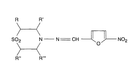
Figure 1 is the structure of the backbone of a nitrofuran compound.
Figure 2A is a histogram showing cell viability of neuroblastoma cells at
different
doses of nifurtimox. Cell viability assay: SMS-KCN, SMS-KCNR, and IMR-32 cells
were
cultured in 48 well plates (50,000 cells/well) and treated with 1 t.tg/ml, 10
pg/m1 or 20 ug/m1
of Nifurtimox for 120 hours. Cell viability was assessed using the MTS assay
as described in
Materials and Methods and expressed as percent of vehicle treated control. The
data
represents the mean SD of four replicates.
Figure 2B is a histogram showing BrdU incorporation at different doses of
nifurtimox. Cell proliferation assay: SMS-KCNR cells were cultured in 48 well
plates and
treated with 0, 1.0, 5.0, 10 and 20 tig/m1 nifurtimox for 4$ hours. DNA
synthesis was
determined by BrdU incorporation assay as described in the Example. Results
are expressed
as percentages of untreated controls and means of six replicates ( SD).
Figure 3A is a set of photographs showing the effect of nifurtimox on
neuroblastoma
cells. Sub-confluent cultures of SMS-KCNR cells were treated with 0, 1.0, 10
and 20 g/ml
nifurtimox for 96 hours. The cells were photographed using a light microscope
as described
in Materials and Methods at x100 magnification. Vehicle treated cells were
used as control.
Panels i: Vehicle control, ii: 1 12g/m1, iii: 10 pg/ml, and iv: 20
g/mlnifurtimox.
Figure 3B is a set of pictures showing apoptotic cell death of neuroblastoma
cells by
nifurtimox. SMS-KCNR Cells were cultured and incubated with 0, 1.0, 10 and 20
jig/ml of
nifurtimox for 96 hours, TUNEL assay was performed, photographed and processed
as
described in the Example. Magnification = 100x. i to iv are representative
pictures of
CA 02646222 2008-09-16
WO 2007/108947 PCT/US2007/005927
7
overlaid apoptotic stain and nuclear stain. TUNEL positive nuclei due to DNA
fragmentation
in SMS-KCNR cells indicates occurrence of apoptotic cell death by nifurtimox
treatment. (i):
Vehicle control, (ii): 1 pg/ml, (iii): 10 2g/ml, and (iv): 20 pg/ml. The
number of apoptotic
nuclei increases with increasing nifurtimox dose.
Figure 4A is a picture of a slot showing the effect of nifurtimox in caspase-3
activation in neuroblastoma cells. SMS-KCNR cells were cultured and incubated
with
increasing concentrations of nifurtimox for 96 hours. The cells were lysed,
separated on 12%
SDS PAGE, blotted on to PVDF membrane and probed with antibodies specific for
activated
caspase-3 as described in the Example. The blots were stripped and reprobed
with actin
antibodies as loading control. Upper panel - Activated Caspase-3, Lower panel -
Actin.
Figure 4B is a histogram showing the effect of nifurtimox on neuroblastoma
cell
viability in the absence or presence of Z-VAD-FMK. SMS-KCNR cells were
pretreated with
pancaspase inhibitor, Z-VAD-FMK. SMS-KCNR cells were pretreated with 50 1.1M Z-
VAD-
FMK for 90 minutes before treatment with nifurtimox (10 Wail) for 96 hours.
Cell viability
was measured by MTS assay. The reversal of cytotoxicity by pancaspase
inhibitor was
determined by comparing the viability of nifurtimox treated cells in the
presence or absence
of the pancaspase inhibitor. The values are the mean SD of quadruplicates.
Figure 5 is a picture of a Western Blot showing the effect of nifurtimox on
Akt
Phosphorylation. (A): SMS-KCNR cells were serum deprived for 18 hours, treated
with 0,
1.0, 10 and 20 p,g/m1 nifurtimox for 90 minutes and then stimulated with BDNF
(100 jig/m1)
for 10 minutes. Cells were lysed and analyzed by western blot analysis using
phospho-Akt
antibodies (upper panel). The blots were stripped and reprobed with antibodies
specific for
total Akt protein (lower panel). (B): SMS-KCNR cells were treated with
nifurtimox for 90
minutes in the presence of serum. Cells were then lysed and analyzed by
western blot
analysis using antibodies.
Figure 6 is a set of pictures showing the inhibition of angiogenesis by
nifurtimox on
microvascular sprouting in growth factor stimulated aortic ring assay. (A)
Vehicle treated
aorta without growth factor (B) Vehicle treated aorta with growth factor. (C),
(D), (E)
represent nifurtimox treatment at 1, 10 and 20 ug/ml concentrations in
presence of growth
factor.
Figure 7 is a histogram showing the inhibition of human aortic endothelial
cell
proliferation by nifurtimox in a dose dependent manner as reflected by the
decreased
incorporation of bromo deoxy uridine (BrDu) in the DNA synthesis.
CA 02646222 2008-09-16
WO 2007/108947 PCT/US2007/005927
8
Figure 8 (A-C) is a set of pictures showing the inhibition of tube formation,
a critical
step in the angiogenesis process by nifurtimox in a dose dependent manner. (A)
Vehicle
treated endothelial cells without growth factor (B) Vehicle treated
endothelial cells with
growth factor. (C) represents nifurtimox treatment at 20 ugiml concentrations
in presence of
growth factor figure and quantitative graph. (D) is a histogram showing the
quantitative
analysis of the effect of nifurtimox on fuse formations assay.
Figure 9 is a histogram showing the effect of nifurtimox in combination with
buthionine sulfoximine (BSO) on neuroblastoma cell viability. SMS KCNR cells
were
cultured overnight and then treated with 0, 1, or 10 m.g/m1 nifurtimox in
combination with
either 1, 50 or 100 1.1M BSO for 48 hours. Cell viability was measured with
Calcein AM
assay.
Figure 10 is a histogram showing the effect of nifurtimox in combination with
either
ascorbic acid or BSO on neuroblastoma cell viability. SY5Y cells were cultured
overnight
and then treated with either 10 p.g/m1 nifurtimox (N), 0.3 mM ascorbic acid
(A) or 50 1.1.M
BSO (B) alone or in combinations (NB, NA, NBA) for 24 hours. Cell viability
was measured
with Calcein AM assay. The results are expressed as a percent of vehicle
control.
Figure 11 is a histogram showing the effect of nifurtimox treatment on
neuroblastoma
xenograft mice as described in Example 12.
Figure 12 is a schematic diagram illustrating the scheme for the synthesis of
the
nitrofuran compounds of the invention as described in detail in Example 16.
Detailed Description of the Invention
The invention is based, in part, on the serendipitous discovery (as detailed
in Example
13) that the administration of 4-[5-Nitrofurfurylidene)amino]-3-
methylthiomorpholine 1,1-
dioxide, which is also referred to by it nonproprietary name "Nifurtimox",
reduces tumor
size, inhibits proliferation of neuroblastoma cells, and inhibits
angiogenesis. Thus, the
invention includes, in some aspects, administering to a subject having a
cancer a nitrofuran
compound in the form of a medicament to treat the cancer in the subject. The
invention also
includes, in some aspects, administering a nitrofuran compound to a subject to
inhibit
angiogenesis in the subject. The nitrofuran compound is administered in an
effective and
physiogically acceptable amount to treat the cancer in the subject. Although
not wishing to
be bound to any particular theory, we believe that Nifurtimox exerts its
cytotoxic effect
specifically by generating free radicals. Nifurtimox is a nitroheterocyclic
compound; its nitro
group can be reduced to the nitro anion radical in cell-free systems by
interacting with
CA 02646222 2008-09-16
WO 2007/108947
PCT/US2007/005927
9
cytochrome P-450 reductase, xanthine oxidase, ascorbate, and catecholamines.
Nitro anions
can then reduce oxygen to the superoxide anion radical and hydrogen peroxide.
In Chagas
disease, the nitro anion free radicals and oxyradicals have been shown to be
cytotoxic for the
parasite T. Cruzi. The reduction of the nitro group not only generates anion
radicals, but
interaction with catecholamines3 appears also to generate semiquinone free
radicals that
exacerbate damage to functionally important biomolecules, leading to apoptosis
of
neuroblastoma cell lines. Neuroblastoma cells are known to contain high levels
of
catecholamines, thereby potentially leading to relatively specific targeting
of these cells. The
reaction with catecholamines in neuroblastoma cell lines was confirmed by the
reduction of
cytotoxicity by pretreatment with AMPT, a tyrosine hydroxylase inhibitor that
reduces the
total amount of catecholamine stored in cells. In addition, the enhanced
sensitivity of.
- sympathetic neurons¨but not parsaympathetic neurons or non-neuronalcells--
to nifurtimox
supports this conclusion.
The term "treatment" or "treating" includes amelioration, cure or maintainence
(i.e.,
the prevention of relapse) of a disorder. Treatment after a disorder has
started aims to reduce,
ameliorate or altogether eliminate the disorder, and/or its associated
symptoms, to prevent it
from becoming worse, or to prevent the disorder from re-occuring once it has
been initially
eliminated (i.e., to prevent a relapse).
A subject means a mammalian species, including but not limited to a dog, cat,
horse,
cow, pig, sheep, goat, chicken, rodent, or primate. Subjects can be house pets
(e.g., dogs,
cats), agricultural stock animals (e.g., cows, horses, pigs, chickens, etc.),
laboratory animals
(e.g., mice, rats, rabbits, etc.), zoo animals (e.g., lions, giraffes, etc.),
but are not so limited.
Preferred subjects are human subjects. The human subject may be a pediatric,
adult or a
geriatric subject.
As used herein the terms "nitrofuran(s)" and "nitrofuran compound(s)" are
employed
interchangeably and encompass furans having a side chain containing one or
more nitrogen
atoms. As is well known in the art, furan is an unsaturated aromatic
heterocyclic compound
composed of four carbon atoms and one oxygen atom. See Ege, Organic Chemistry,
3d. Ed.,
D.C. Heath & Co., Lexington, MA (1994). Examples of nitrofurans include,
without
limitation, Nifurtimox, Furazolidine and Nifuratel.
See Raether W, Hanel H.
Nitroheterocyclic Drugs with Broad Spectrum Acitivity, Parasit Res 2003;
90:S19-S39;
Albrecht et al., J. Med. Chem. 13(4): 736 (1970); Albrecht et al.,
Arzneimittel-Forschung
(Drug Res.) 21(1): 127-31 (1971); Pozas et al., Bioorganic & Medicinal
Chemistry Letters
15: 1417-21 (2005). "Compound" includes both the synthetically prepared and
administered
CA 02646222 2008-09-16
WO 2007/108947 PCT/US2007/005927
nitrofuran compound and nitrofuran compounds produced in vivo after
administration of
another compound.
"Cancer" as used herein refers to an uncontrolled growth of cells which
interferes
with the normal functioning of the bodily organs and systems. Cancer cells
which migrate
from their original location and seed vital organs can eventually lead to the
death of the
subject through the functional deterioration of the affected organs. A cancer
cell is a cell that
divides and reproduces abnormally due to a loss of normal growth control.
Cancer cells
almost always arise from at least one genetic mutation. In some instances, it
is possible to
distinguish cancer cells from their normal counterparts based on profiles of
expressed genes
and proteins, as well as to the level of their expression. Genes commonly
affected in cancer
cells include oncogenes, such as ras, neu/HER2/erbB, myb, myc and abl, as well
as tumor
suppressor genes such as p53, Rb, DCC, RET and WT. Cancer-related mutations in
some of
these genes lead to a decrease in their expression or a complete deletion. In
others, mutations
cause an increase in expression or the expression of an activated variant of
the normal
counterpart.
The term "tumor" is usually equated with neoplasm, which literally means "new
growth" and is used interchangeably with "cancer." A "neoplastic disorder" is
any disorder
associated with cell proliferation, specifically with a neoplasm. A "neoplasm"
is an abnormal
mass of tissue that persists and proliferates after withdrawal of the
carcinogenic factor that
initiated its appearance. There are two types of neoplasms, benign and
malignant. Nearly all
benign tumors are encapsulated and are noninvasive; in contrast, malignant
tumors are almost
never encapsulated but invade adjacent tissue by infiltrative destructive
growth. This
infiltrative growth can be followed by tumor cells implanting at sites
discontinuous with the
original tumor.
A metastasis is a region of cancer cells, distinct from the primary tumor
location
resulting from the dissemination of cancer cells from the primary tumor to
other parts of the
body. At the time of diagnosis of the primary tumor mass, the subject may be
monitored for
the presence of metastases. Metastases are most often detected through the
sole or combined
use of magnetic resonance imaging (MRI) scans, computed tomography (CT) scans,
blood
and platelet counts, liver function studies, chest X-rays and bone scans in
addition to the
monitoring of specific symptoms.
The method of the invention can be used to treat cancer in a subject. In some
embodiments, the cancer is a central nervous system (CNS) cancer. Examples of
some
important CNS cancers include, but are not limited to, neuroblastoma,
medulloblastoma,
CA 02646222 2008-09-16
WO 2007/108947 PCT/US2007/005927
11
peripheral malignant nerve sheath tumor, ependymoma, chraniopharyngioma,
astrocytoma,
meningioma, germinoma, glioma, mixed glioma, choroid plexus tumor,
oligodendroglioma,
peripheral neuroectodermal tumor, primitive neuroectodermal tumor (PNET), CNS
lymphoma, pituitary adenoma, and Schwannoma. In some embodiments the
astrocytoma is
Grade I, Grade II, Grade III, or Grade IV. The astrocytoma may be a low-grade
or a high-
grade. The astrocytoma may be juvenile pilocytic astrocytoma, subependymal
giant cell
astrocytoma, pleimorphic xanthoastrocytoma, anaplastic astrocytoma, or
gliomatosis cerebri.
In some embodiments the oligodendroglioma is a mixed glioma (oligoastrocytoma)
or an
anaplastic oligodendroglioma. In one preferred embodiment, the cancer is
neuroblastoma.
Other cancers that can be treated by the methods of this invention include but
are not
limited to basal cell carcinoma, biliary tract cancer, bladder cancer, bone
cancer, brain and
CNS cancer, breast cancer, cervical cancer, choriocarcinoma, colon and rectum
cancer,
connective tissue cancer, cancer of the digestive system, endometrial cancer,
esophageal
cancer, eye cancer, fibroma, cancer of the head and neck, gastric cancer,
intra-epithelial
neoplasm, kidney cancer, larynx cancer, leukemia including acute myeloid
leukemia, acute
lymphoid leukemia, chronic myeloid leukemia, chronic lymphoid leukemia, liver
cancer,
lung cancer (e.g. small cell and non-small cell), lymphoma including Hodgkin's
and Non-
Hodgkin's lymphoma, melanoma, oral cavity cancer (e.g., lip, tongue, mouth,
and pharynx),
ovarian cancer, pancreatic cancer, prostate cancer, retinoblastoma,
rhabdomyosarcoma, rectal
cancer, renal cancer, cancer of the respiratory system, sarcoma, skin cancer,
stomach cancer,
testicular cancer, thyroid cancer, uterine cancer, cancer of the urinary
system, as well as other
carcinomas and sarcomas.
Carcinomas are cancers of epithelial origin. Carcinomas intended for treatment
with
the methods of the invention include, but are not limited to, acinar
carcinoma, acinous
carcinoma, alveolar adenocarcinoma (also called adenocystic carcinoma,
adenomyoepithelioma, cribriform carcinoma and cylindroma), carcinoma
adenomatosum,
adenocarcinoma, carcinoma of adrenal cortex, alveolar carcinoma, alveolar cell
carcinoma
(also called bronchiolar carcinoma, alveolar cell tumor and pulmonary
adenomatosis), basal
cell carcinoma, carcinoma basocellulare (also called basaloma, or basiloma,
and hair matrix
carcinoma), basaloid carcinoma, basosquamous cell carcinoma, breast carcinoma,
bronchioalveolar carcinoma, bronchiolar carcinoma, bronchogenic carcinoma,
cerebriform
carcinoma, cholangiocellular carcinoma (also called cholangioma and
cholangiocarcinoma),
chorionic carcinoma, colloid carcinoma, comedo carcinoma, corpus carcinoma,
cribriform
carcinoma, carcinoma en cuirasse, carcinoma cutaneum, cylindrical carcinoma,
cylindrical
CA 02646222 2008-09-16
WO 2007/108947 PCT/US2007/005927
12
cell carcinoma, duct carcinoma, carcinoma durum, embryonal carcinoma,
encephaloid
carcinoma, epibulbar carcinoma, epidermoid carcinoma, carcinoma epitheliale
adenoides,
carcinoma exulcere, carcinoma fibrosum, gelatiniform carcinoma, gelatinous
carcinoma,
giant cell carcinoma, gigan. tocellulare, glandular carcinoma, granulosa cell
carcinoma, hair-
matrix carcinoma, hematoid carcinoma, hepatocellular carcinoma (also called
hepatoma,
malignant hepatoma and hepatocarcinoma), Htirthle cell carcinoma, hyaline
carcinoma,
hypemephroid carcinoma, infantile embryonal carcinoma, carcinoma in situ,
intraepidermal
carcinoma, intraepithelial carcinoma, ICrompecher's carcinoma, Kulchitzky-cell
carcinoma,
lenticular carcinoma, carcinoma lenticulare, lipomatous carcinoma,
lymphoepithelial
carcinoma, carcinoma mastitoides, carcinoma medullare, medullary carcinoma,
carcinoma
melanodes, melanotic carcinoma, mucinous carcinoma, carcinoma muciparum,
carcinoma
mucocellulare, mucoepiderrnoid carcinoma, carcinoma mucosum, mucous carcinoma,
carcinoma myxomatodes, nasopharyngeal carcinoma, carcinoma nigrum, oat cell
carcinoma,
carcinoma ossificans, osteoid carcinoma, ovarian carcinoma, papillary
carcinoma, periportal
carcinoma, preinvasive carcinoma, prostate carcinoma, renal cell carcinoma of
kidney (also
called adenocarcinoma of kidney and hypernephoroid carcinoma), reserve cell
carcinoma,
carcinoma sarcomatodes, scheinderian carcinoma, scirrhous carcinoma, carcinoma
scroti,
signet-ring cell carcinoma, carcinoma simplex, small-cell carcinoma, solanoid
carcinoma,
spheroidal cell carcinoma, spindle cell carcinoma, carcinoma spongiosum,
squamous
carcinoma, squamous cell carcinoma, string carcinoma, carcinoma
telangiectaticum,
carcinoma telangiectodes, transitional cell carcinoma, carcinoma tuberosum,
tuberous
carcinoma, verrucous carcinoma, carcinoma vilosum.
Sarcomas are rare mesenchymal neoplasms that arise in bone and soft tissues.
Different types of sarcomas are recognized and these include: liposarcomas
(including
myxoid lipo sarcomas and pleiomorphic lipo sarcomas),
leiomyosarcomas,
rhabdomyosarcomas, malignant peripheral nerve sheath tumors (also called
malignant
schwannomas, neurofibrosarcomas, or neurogenic sarcomas), Ewing's tumors
(including
Ewing's sarcoma of bone, extraskeletal (i.e., non-bone) Ewing's sarcoma, and
primitive
neuroectoderrnal tumor [PNET]), synovial sarcoma, angiosarcomas,
hemangiosarcomas,
lymphangiosarcomas, Kaposi's sarcoma, hemangioendothelioma, fibrosarcoma,
desmoid
tumor (also called aggressive fibromatosis), dermatofibrosarcoma protuberans
(DFSP),
malignant fibrous histiocytoma (MFH), hemangiopericytoma, malignant
mesenchymoma,
alveolar soft-part sarcoma, epithelioid sarcoma, clear cell sarcoma,
desmoplastic small cell
tumor, gastrointestinal stromal tumor (GIST) (also known as GI stromal
sarcoma),
CA 02646222 2008-09-16
WO 2007/108947 PCT/US2007/005927
13
osteosarcoma (also known as osteogenic sarcoma)-skeletal and extraskeletal,
and
chondrosarcoma.
The cancers to be treated may be refractory cancers. As used herein, a
refractory
cancer is a cancer that is resistant to the ordinary standard of care
prescribed. These cancers
may appear initially responsive to a treatment and then recur, or they may be
completely non-
responsive to the treatment. Subjects being treated according to the invention
for a refractory
cancer therefore may have already been exposed to another treatment for their
cancer.
Alternatively, if the cancer is likely to be refractory (e.g., given an
analysis of the cancer cells
or history of the subject), then the subject may not have already been exposed
to another
treatment. Examples of refractory cancers include but are not limited to
leukemias,
melanomas, renal cell carcinomas, colon cancer, liver (hepatic) cancers,
pancreatic cancer,
Non-Hodgkin's lymphoma, and lung cancer.
The invention can also be used to treat cancers that are immunogenic. Cancers
that
are immunogenic are cancers that are known to (or likely to) express
immunogens on their
surface or upon cell death. These immunogens are in vivo endogenous sources of
cancer
antigens and their release can be exploited by the methods of the invention in
order to treat
the cancer. Examples of immunogenic cancers include malignant melanoma and
renal cell
cancer. Mantel Cell Lymphoma, follicular lymphoma, diffuse large B-cell
lymphoma, T-cell
acute lymphoblastic leukemia, Burkitt lymphoma, myeloma, immunocytoma, acute
promyelocytic leukemia, chronic myeloid/acute lymphoblastic leukemia, acute
leukemia, B-
cell acute lymphoblastic leukemia, anaplastic large cell leukemia,
myelodysplastic
syndrome/acute myeloid leukemia, Non-Hodgkin's lymphoma, chronic lymphocytic
leukemia (CLL), acute lymphoblastic leukemia (ALL). acute myelogenous leukemia
(AML),
Common (pre-B) acute lymphocytic leukemia, malignant melanoma, T-cell
lymphoma,
leukemia, B-cell lymphoma, epithelial malignancies, lymphoid malignancies,
gynecological
carcinomas, biliary adenocarcinomas, and ductal adenocarcinomas of the
pancreas.
The invention involves in some other aspects, methods for inhibiting
angiogenesis in
a subject. Angiogenesis is an abnormal rapid proliferation of endothelial
cells resulting in
persistent and unabated formation of abnormal new blood vessels
(microvessels).
Angiogenesis that continues for months or years can support the growth and
progression of
cancer and may result in damage to various organs and tissues such as, for
example, the eye,
skin, heart, blood vessels, lung, gastrointestinal tract, and the
genitourinary tract. The
methods of the invention involve administering to a subject a nitrofuran
compound in an
effective amount to inhibit the angiogenesis. The nitrofuran compound is
administered in an
CA 02646222 2008-09-16
WO 2007/108947
PCT/US2007/005927
14
effective amount to inhibit the angiogenesis in the subject. Preferably the
compound is
Niturtimox, and the subject is a human subject.
As used herein the term "inhibits angiogenesis" refers to the reduction of the
number
or density of the abnormal microvessels. A reduction of the number of abnormal
microvessels refers to decreasing the number of existing abnormal microvessels
or decreasing
the production of new microvessels. Reduction, as used herein, includes total
elimination or
eradication, as well as other decreases which do not result in total
eradication.
Angiogenesis may be assesed by various methods or techniques. The most widely
used method in clinical settings relies on histochemical or
immunohistochemical staining of
blood vessels (microvessels) in biopsies (open or needle) or specimens.
Features of
angiogenesis that may be examined include, for example, blood vessel density
and/or the
morphology and/or thickness of the perivascular cuff. Areas of microvessel
density in a
histologic biopsy or specimen are quantified. Areas of high microvessel
density ("hot spots")
may, for example, contain the most tumor cells and/or have the highest chance
of
metastasizing. One technique of determining microvessel density is by
measuring
intercapillary distance. Another method of assessing angiogenesis is measuring
perivascular
cuff thickness. An increase in the thickness of the preivascular cuff is
associated with
progression of the angiogenesis and may be indicative of disease worsening.
Angiogenesis may also be assessed by measuring blood, serum, plasma, or tissue
levels of angiogenesis (angiogenic) factors. Levels of angiogenic factors
serve as a surrogate
marker of angiogenesis. Examples of angiogenic factors that may serve as
surrogate markers
of angiogenesis include but are not limited to Angiogenin, Angiopoietin-1, Del-
1, Fibroblast
growth factors: acidic (aFGF) and basic(bFGF), Follistatin, Granulocyte colony-
stimulating
factor (G-CSF), Hepatocyte growth factor (HGF) /scatter factor (SF),
Interleukin-8 (IL-8),
Leptin, Midkine, Placental growth factor, Platelet-derived endothelial cell
growth factor (PD-
ECGF), Platelet-derived growth factor-B13 (PDGF-BB), Pleiotrophin (PTN),
Progranulin,
Proliferin, Transforming growth factor-alpha (TGF-alpha), Transforming growth
factor-beta
(TGF-beta), Tumor necrosis factor-alpha (TNF-alpha), Vascular endothelial
growth factor
(VEGF)/vascular permeability factor (VPF). Imaging techniques are also useful
for the
assesment of angiogenesis. Suitable imaging techniques or devices include non-
invasive
devices such as CT, rotational CT, micro-CT, multiple energy computed
tomography
(MECT), single detector CT (SDCT), multi-detector CT (MDCT), volumetric CT
(VCT),
MRI, micro-MR, X-ray, rotational X-ray, PET, near infrared/optical and other
non-invasive
scanning techniques and devices that may be used outside a subject's body or
inserted non-
:
CA 02646222 2008-09-16
WO 2007/108947
PCT/US2007/005927
invasively into a body cavity. Angiogenesis may also be imaged by CT
angiography (CTA),
tomosynthesis, X-ray micro-angiography, and by other techniques. One
angiogenesis
imaging technique involves the use microbubble-based contrast agents (SonoVue)
combined
with ultrasound and contrast specific imaging modalities to detect perfusion
changes on
tumor microvascular perfusion. Other angiogenesis imaging techniques include
color
Doppler and mammography. Color Doppler imaging can demonstrate angiogenesis in
tumors
such as breast cancer. Mammography may reveal the vascularized rim of a breast
tumor. A
wide range of imaging or radiologic signs may be enhanced by dyes.
Angiogenesis may also be assessed in a subject by a process that involves
introducing
at least one contrast agent into a body region of interest. For example, a
contrast agent for
detecting blood vessels may be injected into a blood vessel. A small amount of
contrast
agent may be introduced locally to enhance the detection of blood vessels in a
particular body
region of interest. Alternatively, a contrast agent may be provided in an
amount sufficient to
enhance the detection of blood vessels in a large body region or in the entire
subject body.
Structure data may be obtained for the body of the subject, or may be obtained
for one or
more target organs e.g., a lung, heart, breast, colon, etc., portion of an
organ, or another target
volume of the subject's body. A target volume can be any portion of the
subject's body. e.g.,
a limb, the abdomen, the torso, the neck, the head, or any portion thereof.
Other methods or
techniques to assess angiogenesis not described herein may be used for the
purpose of this
invention. Methods and techniques to assess angiogenesis are known to those of
ordinary
skill in the art.
The nitrofurans may be administered in combination with other therapies, such
as for
example radiation therapy, surgery, conventional chemotherapy or with a
combination of one
or more additional therapies.
The nitrofurans may be administered alone in a pharmaceutical composition, or
combined with therapeutically effective and physiologically acceptable amounts
of one or
more other active ingredients or agents. Such other active ingredients
include, but are not
limited to, glutathione antagonists, angiogenesis inhibitors, chemotherapeutic
agent(s), and
antibodies (e.g., cancer antibodies). The nitrofuran compound and the other
active
ingredients or agents may be administered simultaneously or sequentially. When
the
nitrofuran compound is administered simultaneously with another active agent
or combined
with another active ingredient, the nitrofuran compound and the other active
ingredient may
be administered in the same or separate formulations, but are administered at
the same time.
The other active agents may be administered sequentially with one another and
with the
=e CAC
CA 02646222 2008-09-16
WO 2007/108947 PCT/US2007/005927
16
nitrofuran compound when the administration of the other active agent and the
nitrofuran is
temporally separated. The separation in time between administrations may be a
matter of
minutes, hour, days, or it may be longer.
Examples of glutathione antagonists include but are not limited to buthionine
sulfoximine, cyclophosphamide, ifosphamide, actinomycin D, and N-(4-
hydroxyphenyl)
retinamide (4-HPR).
Examples of angiogenesis inhibitors include but are not limited to 2-
methoxyestradiol
(2-ME), AG3340, Angiostatin, Antithrombin III, Anti-VEGF antibody, Batimastat,
bevacizumab (avastatin), BMS-275291, CAI, Canstatin, Captopril, Cartilage
Derived
Inhibitor (CDI), CC-5013, Celecoxib (CELEBREX0), COL-3, Combretastatin,
Combretastatin A4 Phosphate, Dalteparin (FRAGINO), EMD 121974 (Cilengitide),
Endostatin, Erlotinib (TARCEVA0), gefitinib (Iressa), Genistein, Halofuginone
Hydrobromide (TEMPOSTATINTm), Idl , Id3, IM862, imatinib mesylate, Inducible
protein
10, Interferon-alpha, Interleukin 12, Lavendustin A, LY317615 or AE-941
(NEOVASTATTm), Marimastat, Maspin, Medroxpregesterone Acetate, Meth-1, Meth-2,
Neovastat, Osteopontin cleaved product, PEX, Pigment epithelium growth factor
(PEGF),
Platelet factor 4, Prolactin fragment, Proliferin-related protein (PRP),
PTK787/ZK 222584,
Recombinant human platelet factor 4 (rPF4), Restin, Squalamine, SU5416,
SU6668,
Suramin, Taxol, Tecogalan, Thalidomide, Thrombospondin, TNP-470, Troponin I,
Vasostatin, VEG1, VEGF-Trap, and ZD6474. In some embodiments the angiogenesis
inhibitor is a VEGF antagonist. The VEGF antagonist may be a VEGF binding
molecule.
VEGF binding molecules includeVEGF antibodies or antigen binding fragment(s)
thereof.
One example of a VEGF antagonist is NeXstar.
Examples of categories of chemotherapeutic agents that may be used as an
additional
active ingredient include but are not limited to DNA damaging agents and these
include
topoisomerase inhibitors (e.g., etoposide, ramptothecin, topotecan,
teniposide, mitoxantrone),
anti-microtubule agents (e.g., vincristine; vinblastine), anti-metabolic
agents (e.g., cytarabine,
methotrexate, hydroxyurea, 5-fluorouracil, floxuridine, 6-thioguanine, 6-
mercaptopurine,
fludarabine, pentostatin, chlorodeoxyadenosine), DNA alkylating agents (e.g.,
cisplatin,
mechlorethamine, cyclophosphamide, ifosfamide, melphalan, chorambucil,
busulfan,
thiotepa, carmustine, lomustine, carboplatin, dacarbazine, procarbazine), and
DNA strand
break inducing agents (e.g., bleomycin, doxorubicin, daunorubicin, idarubicin,
mitomycin C).
Chemotherapeutic agents include synthetic; semisynethetic and naturally
derived agents.
Important chemotherapeutic agents include but are not limited to Acivicin,
Aclarubicin,
CA 02646222 2008-09-16
WO 2007/108947
PCT/US2007/005927
17
Acodazole Hydrochloride, Acronine, Adozelesin, Adriamycin, Aldesleukin,
Alitretinoin,
Allopurinol Sodium, Altretamine, Ambomycin, Ametantrone Acetate,
Aminoglutethimide,
Amsacrine, Anastrozole, Annonaceous Acetogenins, Anthramycin, Asimicin,
Asparaginase,
Asperlin, Azacitidine, Azetepa, Azotomycin, Batimastat, Benzodepa, Bexarotene,
Bicalutamide, Bisantrene Hydrochloride, Bisnafide Dimesylate, Bizelesin,
Bleomycin
Sulfate, Brequinar Sodium, Bropirimine, Bullatacin, Busulfan, Cabergoline,
Cactinomycin,
Calusterone, Caracemide, Carbetimer, Carboplatin, Carmustine, Carubicin
Hydrochloride,
Carzelesin, Cedefingol, Celecoxib, Chlorambucil, Cirolemycin, Cisplatin,
Cladribine,
Crisnatol Mesylate, Cyclophosphamide, Cytarabine, Dacarbazine, DACA (N42-
(12imethyl-
amino)ethyliacridine-4-carboxamide), Dactinomycin, Daunorubicin Hydrochloride,
Daunomycin, Decitabine, Denileukin Diftitox, Dexormaplatin, Dezaguanine,
Dezaguanine
Mesylate, Diaziquone, Docetaxel, Doxorubicin, Doxorubicin Hydrochloride,
Droloxifene,
Droloxifene Citrate, Dromostanolone Propionate, Duazomycin, Edatrexate,
Eflornithine
Hydrochloride, Elsamitrucin, Enloplatin, Enpromate, Epipropidine, Epirubicin
Hydrochloride, Erbulozole, Esorubicin Hydrochloride, Estramustine,
Estramustine Phosphate
Sodium, Etanidazole, Ethiodized Oil I 131, Etoposide, Etoposide Phosphate,
Etoprine,
Fadrozole Hydrochloride, Fazarabine, Fenretinide, Floxuridine, Fludarabine
Phosphate,
Fluorouracil, 5-FdUMP, Flurocitabine, Fosquidone, Fostriecin Sodium, FK-317,
FK-973,
FR-66979, FR-900482, Gemcitabine, Gemcitabine Hydrochloride, Gemtuzumab
Ozogamicin, Gold Au 198, Goserelin Acetate, Guanacone, Hydroxyurea, Idarubicin
Hydrochloride, Ifosfamide, Ilmofosine, Interferon Alfa-2a, Interferon Alfa-2b,
Interferon
Alfa-n I, Interferon Alfa-n3, Interferon Beta- I a, Interferon Gamma- I b,
Iproplatin,
Irinotecan Hydrochloride, Lanreotide Acetate, Letrozole, Leuprolide Acetate,
Liarozole
Hydrochloride, Lometrexol Sodium, Lomustine, Losoxantrone Hydrochloride,
Masoprocol,
Maytansine, Mechlorethamine Hydrochloride, Megestrol Acetate, Melengestrol
Acetate,
Melphalan, Menogaril, Mercaptopurine, Methotrexate, Methotrexate Sodium,
Methoxsalen,
Metoprine, Meturedepa, Mitindomide, Mitocarcin, Mitocromin, Mitogillin,
Mitomalcin,
Mitomycin, Mytomycin C, Mitosper, Mitotane, Mitoxantrone Hydrochloride,
Mycophenolic
Acid, Nocodazole, Nogalamycin, Oprelvekin, Ormaplatin, Oxisuran, Paclitaxel,
Pamidronate
Disodium, Pegaspargase, Peliomycin, Pentamustine, Peplomycin Sulfate,
Perfosfamide,
Pipobroman, Piposulfan, Piroxantrone Hydrochloride, Plicamycin, Plomestane,
Porfimer
Sodium, Porfiromycin, Prednimustine, Procarbazine Hydrochloride, Puromycin,
Puromycin
Hydrochloride, Pyrazofurin, Riboprine, Rituximab, Rogletimide, Rolliniastatin,
Safingol,
Safingol Hydrochloride, Samarium/Lexidronarn, Semustine, Simtrazene,
Sparfosate Sodium,
CA 02646222 2008-09-16
WO 2007/108947
PCT/US2007/005927
18
Sparsomycin, Spirogermanium Hydrochloride, Spiromustine, Spiroplatin,
Squamocin,
Squamotacin, Streptonigrin, Streptozocin, Strontium Chloride Sr 89, Sulofenur,
Talisomycin,
Taxane, Taxoid, Tecogalan Sodium, Tegafur, Teloxantrone Hydrochloride,
Temoporfin,
Teniposide, Teroxirone, Testolactone, Thiamiprine, Thioguanine, Thiotepa,
Thymitaq,
Tiazofurin, Tirapazamine, Tomudex, TOP-53, Topotecan Hydrochloride, Toremifene
Citrate,
Trastuzumab, Trestolone Acetate, Triciribine Phosphate, Trimetrexate,
Trimetrexate
Glucuronate, Triptorelin, Tubulozole Hydrochloride, Uracil Mustard, Uredepa,
Valrubicin,
Vapreotide, Verteporfin, Vinblastine, Vinblastine Sulfate, Vincristine,
Vincristine Sulfate,
Vindesine, Vindesine Sulfate, Vinepidine Sulfate, Vinglycinate Sulfate,
Vinleurosine Sulfate,
Vinorelbine Tartrate, Vinrosidine Sulfate, Vinzolidine Sulfate, Vorozole,
Zeniplatin,
Zinostatin, Zorubicin Hydrochloride, 2-Chlorodeoxyadenosine, 2'-Deoxyformycin,
9-
aminocamptothecin, raltitrexed, N-propargy1-5,8-dideazafolic acid, 2-chloro-2'-
arabino-
fluoro-2'-deoxyadenosine, 2-chloro-2'-deoxyadenosine, anisomycin, trichostatin
A, hPRL-
G129R, CEP-751, linomide, sulfur mustard, nitrogen mustard (mechlor ethamine),
cyclophosphamide, melphalan, chlorambucil, ifosfamide, busulfan, N-methyl-N-
nitrosourea
(MNU), N, N'-Bis(2-chloroethyl)-N-nitrosourea (F3CNU), N-(2-chloroethyl)-N'-
cyclohexyl-
N-nitrosourea (CCNU), N-(2-chloroethyl)-N'-(trans-4-methylcyclohexyl-N-
nitrosourea
(MeCCNU), N-(2-chloroethyl)-N'-(diethyl)ethylphosphonate-N-nitrosourea
(fotemustine),
streptozotocin, diacarbazine (DTIC), mitozolomide, temozolomide, thiotepa,
mitomycin C,
AZQ, adozelesin, Cisplatin, Carboplatin, Ormaplatin, Oxaliplatin, C1-973, DWA
2114R,
JM216, JM335, Bis (platinum), tomudex, azacitidine, cytarabine, gemcitabine, 6-
Mere aptopurine, 6-Thioguanine, Hypoxanthine, tenipo side, 9-amino
camptothecin,
Topotecan, CPT-11, Doxorubicin, Daunomycin, Epirubicin, darubicin,
mitoxantrone,
losoxantrone, Dactinomycin (Actinomycin D), amsacrine, pyrazoloacridine, all-
trans retinol,
14-hydroxy-retro-retinol, all-trans retinoic acid, N-(4-Hydroxyphenyl)
retinamide, 13-cis
retinoic acid, 3-Methyl TTNEB, 9-cis retinoic acid, fludarabine (2-F-ara-AMP),
and 2-
chlorodeoxyadenosi ne (2 -C da).
Other chemotherapeutic agents include: 20-epi-1,25 dihydroxyvitamin D3, 5-
ethynyluracil, abiraterone, aclarubicin, acylfulvene, adecypenol, adozelesin,
aldesleukin,
ALL-TK antagonists, altretamine, ambamustine, amidox, amifostine,
aminolevulinic acid,
amrubicin, amsacrine, anagrelide, anastrozole, andrographolide, angiogenesis
inhibitors,
antagonist D, antagonist G, antarelix, anti-dorsalizing morphogenetic protein-
1, antiandrogen,
prostatic carcinoma, antiestrogen, antineoplaston, antisense oligonucleotides,
aphidicolin
glycinate, apoptosis gene modulators, apoptosis regulators, apurinic acid, ara-
CDP-DL-
,
CA 02646222 2008-09-16
WO 2007/108947
PCT/US2007/005927
19
PTBA, arginine deaminase, asulacrine, atamestane, atrimustine, axinastatin 1,
axinastatin 2,
axinastatin 3, azasetron, azatoxin, azatyrosine, baccatin III derivatives,
balanol, batimastat,
BCR/ABL antagonists, benzochlorins, benzoylstaurosporine, beta lactam
derivatives,
beta-alethine, betaclamycin B, betulinic acid, bFGF inhibitor, bicalutamide,
bisantrene,
bisaziridinylspermine, bisnafide, bistratene A, bizelesin, breflate, bleomycin
A2, bleomycin
B2, bropirimine, budotitane, buthionine sulfoximine, calcipotriol, calphostin
C, camptothecin
derivatives (e.g., 10-hydroxy- camptothecin), canarypox IL-2; capecitabine,
carboxamide-
amino-triazole, carboxyamidotriazole, CaRest M3, CARN 700, cartilage derived
inhibitor,
carzelesin, casein kinase inhibitors (ICOS), castanospermine, cecropin B,
cetrorelix, chlorins,
chloroquinoxaline sulfonamide, cicaprost, cis-porphyrin, cladribine, clomifene
analogues,
clotrimazole, collismycin A, collismycin B, combretastatin A4, combretastatin
analogue,
conagenin, crambescidin 816, crisnatol, cryptophycin 8, cryptophycin A
derivatives, curacin
A, cyclopentanthraquinones, cycloplatam, cypemycin, cytarabine ocfosfate,
cytolytic factor,
cytostatin, dacliximab, decitabine, dehydrodidemnin B, 2'deoxycoformycin
(DCF),
deslorelin, dexifosfarnide, dexrazoxane, dexverapamil, diaziquone, didemnin B,
didox,
diethylnorspermine, dihydro-5-azacytidine, dihydrotaxol, dioxamycin, diphenyl
spiromustine,
discodermolide, docosanol, dolasetron, doxifluridine, droloxifene, dronabinol,
duocarmycin
SA, ebselen, ecomustine, edelfosine, edrecolomab, eflomithine, elemene,
emitefur,
epirubicin, epothilones (A, R H, B, R = Me), epithilones, epristeride,
estramustine
analogue, estrogen agonists, estrogen antagonists, etanidazole, etoposide,
etoposide 4'-
phosphate (etopofos), exemestane, fadrozole, fazarabine, fenretinide,
filgrastim, finasteride,
flavopiridol, flezelastine, fluasterone, fludarabine, fluorodaunorunicin
hydrochloride,
forfenimex, forrnestane, fostriecin, fotemustine, gadolinium texaphyrin,
gallium nitrate,
galocitabine, ganirelix, gelatinase inhibitors, gemcitabine, glutathione
inhibitors, hepsulfam,
heregulin, hexamethylene bisacetamide, homoharringtonine (HHT), hypericin,
ibandronic
acid idarubicin, idoxifene, idramantone, ilmofosine, ilomastat,
imidazoacridones, imiquimod,
immunostimulant peptides, insulin-like growth factor-1 receptor inhibitor,
interferon
agonists, interferons, interleukins, iobenguane, iododoxorubicin, ipomeanol, 4-
, irinotecan,
iroplact, irsogladine, isobengazole, isohomohalicondrin B, itasetron,
jasplakinolide,
kahalalide F, larnellarin-N triacetate, lanreotide, leinamycin, lenograstim,
lentinan sulfate,
leptolstatin, letrozole, leukemia inhibiting factor, leukocyte alpha
interferon, leuprolide +
estrogen + progesterone, leuprorelin, levamisole, liarozole, linear polyamine
analogue,
lipophilic disaccharide peptide, lipophilic platinum compounds, lissoclinamide
7, lobaplatin,
lombricine, lometrexol, lonidamine, losoxantrone, lovastatin, loxoribine,
lurtotecan, lutetium
CA 02646222 2008-09-16
WO 2007/108947 PCT/US2007/005927
texaphyrin, lysofylline, lytic peptides, maitansine, mannostatin A,
marimastat, masoprocol,
maspin, matrilysin inhibitors, matrix metalloproteinase inhibitors, menogaril,
merbarone,
meterelin, methioninase, metoclopramide, MIF inhibitor, mifepristone,
miltefosine,
mirimostim, mismatched double stranded RNA, mithracin, mitoguazone,
mitolactol,
mitomycin analogues, mitonafide, mitotoxin fibroblast growth factor-saporin,
mitoxantrone,
mofarotene, molgramostim, monoclonal antibody, human chorionic gonadotrophin,
monophosphoryl lipid A + myobacterium cell wall sk, mopidamol, multiple drug
resistance
gene inhibitor, multiple tumor suppressor 1-based therapy, mustard anticancer
agent,
mycaperoxide B, mycobacterial cell wall extract, myriaporone, N-
acetyldinaline, N-
substituted benzamides, nafarelin, nagrestip, naloxone + pentazocine, napavin,
naphterpin,
nartograstim, nedaplatin, nemorubicin, neridronic acid, neutral endopeptidase,
nilutamide,
nisamycin, nitric oxide modulators, nitroxide antioxidant, nitrullyn, 06-
benzylguanine,
octreotide, okicenone, oligonucleotides, onapristone, ondansetron,
ondansetron, oracin, oral
cytokine inducer, ormaplatin, osaterone, oxaliplatin, oxauriomycin, paclitaxel
analogues,
paclitaxel derivatives, palauamine, palmitoylrhizoxin, pamidronic acid,
panaxytriol,
panomifene, parabactin, pazelliptine, pegaspargase, peldesine, pentosan
polysulfate sodium,
pentostatin, pentrozole, perflubron, Perfosfamide, perillyl alcohol,
phenazinomycin,
phenylacetate, phosphatase inhibitors, picibanil, pilocarpine hydrochloride,
pirarubicin,
piritrexim, placetin A, placetin B, plasminogen activator inhibitor, platinum
complex,
platinum compounds, platinum-triamine complex, podophyllotoxin, porfimer
sodium,
porfiromycin, propyl bis-acridone, prostaglandin J2, proteasome inhibitors,
protein A-based
immune modulator, protein kinase C inhibitor, protein kinase C inhibitors,
microalgal,
protein tyrosine phosphatase inhibitors, purine nucleoside phosphorylase
inhibitors,
purpurins, pyrazoloacridine, pyridoxylated hemoglobin polyoxyethylene
conjugate, raf
antagonists, raltitrexed, ramosetron, ras farnesyl protein transferase
inhibitors, ras inhibitors,
ras-GAP inhibitor, retelliptine demethylated, rhenium Re 186 etidronate,
rhizoxin, ribozymes,
RII retinamide, rogletimide, rohitukine, romurtide, roquinimex, rubiginone B
1, ruboxyl,
safingol, saintopin, SarCNU, sarcophytol A, sargramostim, Sdi 1 mimetics,
semustine,
senescence derived inhibitor 1, sense oligonucleotides, signal transduction
inhibitors, signal
transduction modulators, single chain antigen binding protein, sizofiran,
sobuzoxane, sodium
borocaptate, sodium phenylacetate, solverol, somatomedin binding protein,
sonermin,
sparfosic acid, spicamycin D, spiromustine, splenopentin, spongistatin 1,
squalamine, stem
cell inhibitor, stem-cell division inhibitors, .stipiamide, stromelysin
Mhibitors, sulfinosine,
superactive vasoactive intestinal peptide antagonist, suradista, suramin,
swainsonine,
CA 02646222 2008-09-16
WO 2007/108947
PCT/US2007/005927
21
synthetic glycosaminoglycans, tallimustine, tamoxifen methiodide,
tauromustine, tazarotene,
tecogalan sodium, tegafur, tellurapyryliurn, telomerase inhibitors,
temoporfin, temozolomide,
teniposide, tetrachlorodecaoxide, tetrazomine, thaliblastine, thalidomide,
thiocoraline,
thrombopoietin, thrombopoietin mimetic, thymalfasin, thymopoietin receptor
agonist,
thymotrinan, thyroid stimulating hormone, tin ethyl etiopurpurin,
tirapazamine, titanocene
dichloride, topotecan, topsentin, toremifene, totipotent stem cell factor,
translation inhibitors,
tretinoin, triacetyluridine, triciribine, trimetrexate, triptorelin,
tropisetron, turosteride, tyrosine
kinase inhibitors, tyrphostins, UBC inhibitors, ubenimex, urogenital sinus-
derived growth
inhibitory factor, urokinase receptor antagonists, vapreotide, variolin B,
vector system,
erythrocyte gene therapy, velaresol, veramine, verdins, verteporfin,
vinorelbine, vinxaltine,
vitaxin, vorozole, zanoterone, zeniplatin, zilascorb, and zinostatin
stimalamer.
Other chemotherapeutic agents include: Antiproliferative agents (e.g.,
Piritrexim
Isothionate), Antiprostatic hypertrophy agent (e.g., Sitogluside), Benign
prostatic hyperplasia
therapy agents (e.g., Tamsulosin Hydrochloride), Prostate growth inhibitor
agents (e.g.,
Pentomone), and Radioactive agents: Fibrinogen 1 125, Fludeoxyglucose F 18,
Fluorodopa F
18, Insulin I 125, Insulin 1131, Iobenguane 1123, Iodipamide Sodium 1131,
Iodoantipyrine I
131, Iodocholesterol I 131, Iodohippurate Sodium I 123, Iodohippurate Sodium I
125,
Iodohippurate Sodium 1131, Iodopyracet 1125, Iodopyracet 1131, Iofetamine
Hydrochloride
1123, Iomethin 1125, Iomethin 1131, Iothalamate Sodium 1125, Iothalamate
Sodium 1131,
Iotyrosine I 131, Liothyronine I 125, Liothyronine I 131, Merisoprol Acetate
Hg 197,
Merisoprol Acetate Hg 203, Merisoprol Hg 197, Methyl Iodobenzo Guanine (MIBG-
I131 or
MIBG-I123), Selenomethionine Se 75, Technetium Tc 99m Antimony Trisulfide
Colloid,
Technetium Tc 99m Bicisate, Technetium Tc 99m Disofenin, Technetium Tc 99m
Etidronate, Technetium Tc 99m Exametazime, Technetium Tc 99m Furifosmin,
Technetium
Tc 99m Gluceptate, Technetium Tc 99m Lidofenin, Technetium Tc 99m Mebrofenin,
Technetium Tc 99m Medronate, Technetium Tc 99m Medronate Disodium, Technetium
Tc
99m Mertiatide, Technetium Tc 99m Oxidronate, Technetium Tc 99m Pentetate,
Technetium
Tc 99m Pentetate Calcium Trisodium, Technetium Tc 99m Sestamibi, Technetium Tc
99m
Siboroxime, Technetium Tc 99m Succimer, Technetium Tc 99m Sulfur Colloid,
Technetium
Tc 99m Teboroxime, Technetium Tc 99m Tetrofosmin, Technetium Tc 99m Tiatide,
Thyroxine 1125, Thyroxine 1131, Tolpovidone 1131, Triolein 1125, and Triolein
1131.
MIBG-I131 and MIBG-1123 are especially preferred chemotherapeutic agents for
co-
administration with the nitrofuran containing pharmaceutical compositions of
the invention.
CA 02646222 2008-09-16
WO 2007/108947
PCT/US2007/005927
22
Another category of chemotherapeutic agents is anti-cancer Supplementary
Potentiating Agents, including: Tricyclic anti-depressant drugs (e.g.,
imipramine,
desipramine, amitryptyline, clomipramine, trimipramine, doxepin,
nortriptyline, protriptyline,
amoxapine and maprotiline), non-tricyclic anti-depressant drugs (e.g.,
sertraline, trazodone
and citalopram), Ca++ antagonists (e.g., verapamil, nifedipine, nitrendipine
and caroverine),
Calmodulin inhibitors (e.g., prenylamine, trifluoroperazine and clomipramine),
Amphotericin
B, Triparanol analogues (e.g., tamoxifen), antiarrhythmic drugs (e.g.,
quinidine),
antihypertensive drugs (e.g., reserpine), Thiol depleters (e.g., buthionine
and sulfoximine)
and Multiple Drug Resistance reducing agents such as Cremaphor EL.
Other chemotherapeutic agents include: annonaceous acetogenins, asimicin,
rolliniastatin, guanacone, squamocin, bullatacin, squamotacin, taxanes,
paclitaxel,
gemcitabine, methotrexate FR-900482, FK-973, FR-66979, FK-317, 5-FU, FUDR,
FdUMP,
Hydroxyurea, Docetaxel, discodermolide, epothilones, vincristine, vinblastine,
vinorelbine,
meta-pac, irinotecan, SN-38, 10-0H campto, topotecan, etoposide, adriamycin,
flavopiridol,
Cis-Pt, carbo-Pt, obleomycin, mitomycin C, mitliramycin, capecitabine,
cytarabine, 2-C1-
2'deoxyadenosine, Fludarabine-PO4, mitoxantrone, mitozolomide, Pentostatin,
and Tomudex.
One important class of chemotherapeutic agents are taxanes (e.g., paclitaxel
and
docetaxel). Nitrofuran compounds in combination with tamoxifen or aromatase
inhibitor
arimidex (i.e., anastrozole) are particularly useful for breast and
gynecological cancers.
Examples antibodies that can be used as other active ingredients according to
the
invention include but are not limited to anti-CD20 mAb (monoclonal antibody),
rituximab,
RituxanTM, anti-CD20 mAb, tositumomab Bexxar, anti-HER2, trastuzumab,
HerceptinTM,
anti-HER2, MDX-210, anti-CA125 mAb, oregovomab, 1343.13, OvarexTM, Breva-Rex,
AR54, GivaRex, ProstaRex, anti-EGF receptor mAb, IMC-C225, ErbituxTM, anti-EGF
receptor mAb, MDX-447, gemtuzumab ozogamicin, Mylotarg, CMA-676, anti-CD33
(Wyeth
Pharmaceuticals), anti-tissue factor protein (TF), (Sunol), ior-c5, c5, anti-
EGF receptor mAb,
MDX-447, anti-17-1A mAb, edrecolomab, Panorex, anti-CD20 mAb (Y-90 labeled),
ibritumomab tiuxetan (IDEC-Y2B8), Zevalin, anti-idiotypic mAb mimic of
ganglioside GD3
epitope, BEC2, anti-HLA-Dr10 mAb (131 I LYM-1), OncolymTM, anti-CD33 humanized
mAb (SMART M195), ZamylTM. anti-CD52 humAb (LDP-03), CAMPATH, anti-CD1 mAb,
ior t6, anti-CAR (complement activating receptor) mAb, MDX-11, humanized
bispecific
mAb conjugates (complement cascade activators), MDX-22, 0V103 (Y-90 labeled
antibody),
celogovab, OncoScintTM, anti-17-1A mAb, 3622W94, anti-VEGF (RhurnAb-VEGF),
CA 02646222 2008-09-16
WO 2007/108947 PCT/US2007/005927
23
bevacizumab, AvastinTM, anti-TAC (IL-2 receptor) humanized Ab (SMART),
daclizumab,
Zenapax, anti-TAG-72 partially humanized bispecific Ab, MDX-220, anti-
idiotypic mAb
mimic of high molecular weight proteoglycan (I-Mel-I), MELIMMUNE-1, anti-
idiotypic
mAb mimic of high molecular weight proteoglycan (I-Mel-2), MELIMMUNE-2, anti-
CEA
Ab (hMN14), CEACideTM, PretargetTm radioactive targeting agents, hmAbH11 scFv
fragment
(NovomAb-02), H11 scFv, anti-DNA or DNA-associated proteins (histones) mAb and
conjugates, TNT (e.g. CotaraTm), Gliomab-H mAb, GNI-250 mAb, anti-EGF receptor
mAb,
EMD-72000, anti-CD22 humanized Ab, LymphoCide, Non-Hodgkin's anti-CD33 mAb
conjugate with calicheamicin (CMA 676), gemtuzumab ozogamicin, MylotargTm,
Monopharm-C, colon, anti-idiotypic human mAb to GD2 ganglioside, 4B5,
melanoma, anti-
EGF receptor humanized Ab, ior egf/r3, anti-ior c2 glycoprotein mAb, ior c5,
BABS
(biosynthetic antibody binding site) proteins, anti-FLK-2/FLT-3 mAb, mAb/small-
molecule
conjugate, TAP (tumor-activated prodrug), anti-GD-2 bispecific mAb, MDX-260,
antinuclear
autoantibodies (binds nucleosomes), ANA Ab, anti-HLA-DR Ab (SMART 1D10 Ab),
RemitogenTM, SMART ABL 364 Ab, anti-CEA 1131-labeled mAb, ImmuRA1T-CEA.
Other antibodies that can be used according to the invention include anti-TNFa
antibody such as infliximab (Remicade) and etanercept (Enbrel) for rheumatoid
arthritis and
Crohn's disease, palivizuma, anti-RSV antibody for pediatric = subjects,
bevacizumab,
alemtuzumab, Campath-1H, BLyS-mAb, fSLE; anti-VEGF2, anti-Trail receptor; B3
mAb,
m170 mAb, mAB BR96, and Abx-Cbl mAb. The invention embraces a number of
classes of
antibodies and fragments thereof including but not limited to antibodies
directed to cancer
antigens (as described above), cell surface molecule, stromal cell molecules,
extracellular
matrix molecules, and tumor vasculature associated molecules.
A cell surface molecule is a molecule that is expressed at the surface of a
cell. In
addition to an extracellular domain, it may further comprise a transmembrane
domain and a
cytoplasmic domain. Examples include HER 2, CD20, CD33, EGF receptor, HLA
markers
such as HLA-DR, CD52, CD1, CEA, CD22, GD2 ganglioside, FLK2/FLT3, VEGF, VEGFR,
and the like.
A stromal cell molecule is a molecule expressed by a stromal cell. Examples
include
but are not limited to FAP and CD26.
An extracellular matrix molecule is a molecule found in the extracellular
matrix.
Examples include but are not limited to collagen, glycosaminoglyeans (GAGs),
proteoglycans, elastin, fibronectin and laminin.
CA 02646222 2008-09-16
WO 2007/108947 PCT/US2007/005927
24
A tumor vasculature associated molecule is a molecule expressed by vasculature
of a
tumor (i.e., a solid cancer rather than a systemic cancer such as leukemia).
As with a cancer
antigen, a tumor vasculature associated molecule may be expressed by normal
vasculature
however its presence on vasculature of a tumor makes it a suitable target for
anti-cancer
therapy. In some instances, the tumor vasculature associated molecule is
expressed at a
higher level in tumor vasculature than it is in normal vasculature. Examples
include but are
not limited to endoglin (see U.S. Pat. No. 5,660,827), ELAM-1, VCA_M-1, ICAM-
1, ligand
reactive with LAM-1, MHC class II antigens, aminophospholipids such as
phosphatidylserine
and phosphatidylethanolarnine (as described in U.S. Pat. No. 6,312,694),
VEGFR1 (Flt-1)
and VEGFR2 (KDR/F1k-1), and other tumor vasculature associated antigens such
as those
described in U.S. Pat. No. 5,776,427. Antibodies to endoglin are described in
U.S. Pat. No.
5,660,827 and include TEC-4 and TEC-11, and antibodies that recognize
identical epitopes to
these antibodies. Antibodies to aminophOspholipids are described in U.S. Pat.
No. 6,312,694.
Antibodies that inhibit VEGF are described in U.S. Pat. No. 6,342,219 and
include 2C3
(ATCC PTA 1595). Other antibodies that are specific for tumor vasculature
include
antibodies that react to a complex of a growth factor and its receptor such as
a complex of
FGF and the FGFR or a complex of TGFI3 and the TGFOR. Antibodies of this
latter class are
described in U.S. Pat. No. 5,965,132, and include GV39 and 0V97.
It is to be understood that the antibodies embraced by the invention include
those
recited explicitly herein and also those that bind to the same epitope as
those recited herein.
Also useful in the invention are antibodies such as the following, all of
which are
commercially available:
Apoptosis Antibodies:
BAX Antibodies: Anti-Human Bax Antibodies (Monoclonal), Anti-Human Bax
Antibodies (Polyclonal), Anti-Murine Box Antibodies (Monoclonal), Anti-Murine
Box
Antibodies (Polyclonal);
Fas/Fas Ligand Antibodies: Anti-Human Fas / Fas Ligand Antibodies, Anti-Murine
Fas/Fas Ligand Antibodies Granzyrne Antibodies Granzyme B Antibodies;
BCL Antibodies: Anti Cytochrome C Antibodies, Anti-Human BCL Antibodies
(Monoclonal), Anti-Human be! Antibodies (Polyclonal), Anti-Murine be!
Antibodies .
(Monoclonal), Anti-Murine bc1 Antibodies (Polyclonal)
Miscellaneous Apoptosis Antibodies: Anti TRADD, TRAIL, TRAFF, DR3
Antibodies Anti-Human Fas / Fas Ligand Antibodies Anti-Murine Fas / Fas Ligand
Antibodies;
CA 02646222 2008-09-16
WO 2007/108947
PCT/US2007/005927
Miscellaneous Apoptosis Related Antibodies: BIM Antibodies: Anti Human, Murine
bim Antibodies (Polyclonal), Anti-Human, Murine bim Antibodies (Monoclonal);
PARP Antibodies: Anti-Human PARP Antibodies (Monoclonal), Anti-Human PARP
Antibodies (Polyclonal) Anti-Murine PARP Antibodies;
Caspase Antibodies: Anti-Human Caspase Antibodies (Monoclonal), Anti-Murine
Caspase Antibodies;
Anti-CD Antibodies: Anti-CD29, PL18-5 PanVera, Anti-CD29, PL4-3 PanVera,
Anti-CD41a, PT25-2 PanVera, Anti-CD42b, PL52-4 PanVera, Anti-CD42b, GUR20-5
PanVera, Anti-CD42b, WGA-3 PanVeraAnti-CD43, 1D4 PanVera, Anti-CD46, MCP75-6
PanVera, Anti-CD61, PL11-7 PanVera, Anti-CD61, PL8-5 PanVera, Anti-CD62/P-
slctn,
PL7-6 PanVera, Anti-CD62/P-slan, WGA-1 PanVera, Anti-CD154, 5F3 PanVera; and
anti-
CD1, anti-CD2, anti-CD3, anti-CD4, anti-CD5, anti-CD6, anti-CD7, anti-CD8,
anti-CD9,
anti-CD10, anti-CD11, anti-CD12, anti-CD13, anti-CD14, anti-CD15, anti-CD16,
anti-CD17,
anti-CD18, anti-CD19, anti-CD20, anti-CD21, anti-CD22, anti-CD23, anti-CD24,
anti-CD25,
anti-CD26, anti-CD27, anti-CD28, anti-CD29, anti-CD30, anti-CD31, anti-CD32,
anti-CD33,
anti-CD34, anti-CD35, anti,CD36, anti-CD37, anti-CD38, anti-CD39, anti-CD40
anti-CD41,
anti-CD42, anti-CD43, anti-CD44, anti-CD45, anti-CD46, anti-CD47, anti-CD48,
anti-CD49,
anti-CD50, anti-CD51, anti-CD52, anti-CD53, anti-CD54, anti-CD55, anti-CD56,
anti-CD57,
anti-CD58, anti-CD59, anti-CD60, anti-CD61, anti-CD62, anti-CD63, anti-CD64,
anti-CD65,
anti-CD66, anti-CD67, anti-CD68, anti-CD69, anti-CD70, anti-CD71, anti-CD72,
anti-
CD73, anti-CD74, anti-CD75, anti-CD76, anti-CD77, anti-CD78, anti-CD79, anti-
CD80,
anti-CD 81, anti-CD82, anti-CD83, anti-CD84, anti-CD85, anti-CD86, anti-CD87,
anti-CD88,
anti-CD89, anti-CD90, anti-CD91, anti-CD92, anti-CD93, anti-CD94, anti-CD95,
anti-CD96,
anti-CD97, anti-CD98, anti-CD99, anti-CD100, anti-CD101, anti-CD102, anti-
CD103, anti-
CD104, anti-CD105, anti-CD106, anti-CD107, anti-CD108, anti-CD109, anti-CD110,
anti-
CD111, anti-CD112, anti-CD113, anti-CD114, anti-CD115, anti-CD116, anti-CD117,
anti-
CD118, anti-CD119, anti-CD120, anti-CD121, anti-CD122, anti-CD123, anti-CD124,
anti-
CD125, anti-CD126, anti-CD127, anti-CD128, anti-CD129, anti-CD130, anti-CD131,
anti-
CD132, anti-CD133, anti-CD134, anti-CD135, anti-CD136, anti-CD137, anti-CD138,
anti-
CD139, anti-CD140, anti-CD141, anti-CD142, anti-CD143, anti-CD144, anti-CD145,
anti-
CD146, anti-CD147, anti-CD148, anti-CD149, anti-CD150, anti-CD151, anti-CD152,
anti-
CD153, anti-CD154, anti-CD155, anti-CD156, anti-CD157, anti-CD158, anti-CD159,
anti-
CD160, anti-CD161, anti-CD162, anti-CD163, anti-CD164, anti-CD165, anti-CD166,
anti-
CD167, anti-CD168, anti-CD! 69, anti-CD170, anti-CD171, anti-CD172, anti-
CD173, anti-
CA 02646222 2008-09-16
WO 2007/108947 PCT/US2007/005927
26
CD174, anti-CD175, anti-CD176, anti-CD177, anti-CD178, anti-CD179, anti-CD180,
anti-
CD181, anti-CD182, anti-CD183, anti-CD184, anti-CD185, anti-CD186, anti-CD187,
anti-
CD188, anti-CD189, anti-CD190, anti-CD191, anti-CD192, anti-CD193, anti-CD1
94, anti-
CD195, anti-CD196, anti-CD197, anti-CD198, anti-CD199, anti-CD200, anti-CD201,
anti-
CD202, anti-CD203, anti-CD204, anti-CD205, anti-CD206, anti-CD207, anti-CD208,
anti-
CD209, anti-CD210, anti-CD211, anti-CD21 2, anti-CD213, anti-CD214, anti-
CD215, anti-
CD216, anti-CD217, anti-CD218, anti-CD219, anti-CD220, anti-CD221, anti-CD222,
anti-
CD223, anti-CD224, anti-CD225, anti-CD226, anti-CD227, anti-CD228, anti-CD229,
anti-
CD230, anti-CD231, anti-CD232, anti-CD233, anti-CD234, anti-CD235, anti-CD236,
anti-
CD237, anti-CD238, anti-CD239, anti-CD240 anti-CD241, anti-CD242, anti-CD243,
anti-
CD244, anti-CD245, anti-CD246, anti-CD247, anti-CD248, anti-CD249, anti-CD250,
and
the like.
Human Chemokine Antibodies: Human CNTF Antibodies, Human Eotaxin
Antibodies, Human Epitherlial Neutrophil Activating Peptide-78, Human Exodus
Antibodies,
Human GRO Antibodies, Human 11CC-1 Antibodies, Human 1-309 Antibodies, Human
1P-10
Antibodies, Human 1-TAC Antibodies, Human LIF Antibodies, Human Liver-
Expressed
Chemokine Antibodies, Human lymphotoxin Antibodies, Human MCP Antibodies,
Human
MIP Antibodies, Human Monokine Induced by IFN-gamma Antibodies, Human NAP-2
Antibodies, Human NP-1 Antibodies, Human Platelet Factor-4 Antibodies, Human
RANTES
Antibodies, Human SDF Antibodies, Human TECK Antibodies;
Murine Chemokine Antibodies: Human B-Cell Attracting Murine Chemokine
Antibodies, Chemokine-1 Antibodies, Murine Eotaxin Antibodies, Murine Exodus
Antibodies, Murine GCP-2 Antibodies, Murine KC Antibodies, Murine MCP
Antibodies,
Murine M1P Antibodies, Murine RANTES Antibodies, Rat Chemokine Antibodies, Rat
Chemokine Antibodies, Rat CNTF Antibodies, Rat GRO Antibodies, Rat MCP
Antibodies,
Rat MIP Antibodies, Rat RANTES Antibodies;
Cytokine / Cytokine Receptor Antibodies: Human Biotinylated Cytokine /
Cytokine
Receptor Antibodies, Human IFN Antibodies, Human IL Antibodies, Human Leptin
Antibodies, Human Oneostatin Antibodies, Human TNF Antibodies, Human TNF
Receptor
Family Antibodies, Murine Biotinylated Cytokine / Cytokine Receptor
Antibodies, Murine
IFN Antibodies, Murine IL Antibodies, Murine TNF Antibodies, Murine TNF
Receptor
Antibodies; anti-CCR4 antibody;
Rat Cytokine / Cytokine Receptor Antibodies: Rat Biotinylated Cytokine /
Cytokine
Receptor Antibodies, Rat IFN Antibodies, Rat IL Antibodies, Rat TNF
Antibodies;
CA 02646222 2008-09-16
WO 2007/108947 PCT/US2007/005927
27
ECM Antibodies: Collagen / Procollagen, Laminin, Collagen (Human), Laminin
(Human), Procollagen (Human), Vitronectin / Vitronectin Receptor, Vitronectin
(Human),
Vitronectin Receptor (Human), Fibronectin / Fibronectin Receptor, Fibronectin
(Human),
Fibronectin Receptor (Human);
Growth Factor Antibodies: Human Growth Factor Antibodies, Murine Growth Factor
Antibodies, Porcine Growth Factor Antibodies;
Miscellaneous Antibodies: B aculovirus Antibodies, Cadherin Antibodies,
Complement Antibodies, Clq Antibodies, VonWillebrand Factor Antibodies, Cre
Antibodies,
HIV Antibodies, Influenza Antibodies, Human Leptin Antibodies , Murine Leptin
Antibodies, Murine CTLA-4 Antibodies, Human CTLA-4 Antibodies, P450
Antibodies,
RNA Polymerase Antibodies;
Neurobio Antibodies: Amyloid Antibodies, GFAP Antibodies, Human NGF
Antibodies , Human NT-3 Antibodies , Human NT-4 Antibodies:
Still other antibodies can be used in the invention and these include
antibodies listed
in references such as the MSRS Catalog of Primary Antibodies, and Linscott's
Directory.
In some preferred embodiments of the invention, the antibodies are Avastin
(bevacizumab), BEC2 (mitumomab), Bexxar (tositumomab), Campath (alemtuzumab),
CeaVac, Herceptin (trastuzumab), IMC-C225 (centuximab), LymphoCide
(epratuzumab),
MIX-210, Mylotarg (gemtuzumab ozogamicin), Panorex (edrecolomab),. Rituxan
(rituximab), Theragyn (pemtumomab), Zamyl, and Zevalin (ibritumomab
tituxetan). The
invention also covers antibody fragments thereof.
In some preferred embodiments, the cancer antigen is VEGF, Anti-idiotypic mAb
(GD3 ganglioside mimic), CD20, CD52, Anti-idiotypic mAb (CEA mimic), ERBB2,
EGFR,
CD22, ERBB2 X CD65 (fcyRI), EpCam, PEM and CD33.
The invention encompasses the use of both antibodies and antibody fragments.
The
antibodies may be monoclonal or polyclonal, and can be prepared by
conventional
methodology. They may further be isolated or present in an ascites fluid. Such
antibodies
can be further manipulated to create chimeric or humanized antibodies as will
be discussed in
greater detail below.
Significantly, as is well-known in the art, only a small portion of an
antibody
molecule, the paratope, is involved in the binding of the antibody to its
epitope (see, in
general, Clark, W.R. (1986) The Experimental Foundations of Modern Immunology
Wiley &
Sons, Inc., New York; Roitt, I. (1991) Essential Immunology, 7th Ed.,
Blackwell Scientific
Publications, Oxford). The pFc' and Fc regions, for example, are effectors of
the complement
CA 02646222 2008-09-16
WO 2007/108947 PCT/US2007/005927
28
cascade but are not involved in antigen binding. An antibody from which the
pFc'_region has
been enzymatically cleaved, or which has been produced without the pFc'
region, designated
an F(ab1)2 fragment, retains both of the antigen binding sites of an intact
antibody. Similarly,
an antibody from which the Fe region has been enzymatically cleaved, or which
has been
produced without the Fe region, designated an Fab fragment, retains one of the
antigen
binding sites of an intact antibody molecule. Proceeding further, Fab
fragments consist of a
covalently bound antibody light chain and a portion of the antibody heavy
chain denoted Fd.
The Fd fragments are the major determinant of antibody specificity (a single
Fd fragment
may be associated with up to ten different light chains without altering
antibody specificity)
and Fd fragments retain epitope-binding ability in isolation.
Within the antigen-binding portion of an antibody, as is well-known in the
art, there
are complementarity determining regions (CDRs), which directly interact with
the epitope of
the antigen, and framework regions (FRs), which maintain the tertiary
structure of the
paratope (see, in general, Clark, 1986; Roitt, 1991). In both the heavy chain
Fd fragment and
the light chain of IgG immunoglobulins, there are four framework regions (FR1
through .FR4)
separated respectively by three complementarity determining regions (CDR1
through CDR3).
The CDRs, and in particular the CDR3 regions, and more particularly the heavy
chain CDR3,
are largely responsible for antibody specificity.
It is now well-established in the art that the non-CDR regions of a mammalian
antibody may be replaced with similar regions of co-specific or heterospecific
antibodies
while retaining the epitopic specificity of the original antibody. This is
most clearly
manifested in the development and use of "humanized" antibodies in which non-
human
CDRs are covalently joined to human FR and/or Fc/pFc1 regions to produce a
functional
antibody. Thus, for example, PCT International Publication Number WO 92/04381
teaches
the production and use of humanized murine RSV antibodies in which at least a
portion of the
murine FR regions has been replaced by FR regions of human origin. Such
antibodies,
including fragments of intact antibodies with antigen-binding ability, are
often referred to as
"chimeric" antibodies. Commercial sources of humanized or chimeric antibodies
include
GenPharm, Xenotech, AbGenix and CellGeneSys.
Thus, as will be apparent to one of ordinary skill in the art, the present
invention also
provides for F(abP)2, Fab, Fv and Fd fragments; chimeric antibodies in which
the Fe and/or
FR and/or CDR1 and/or CDR2 and/or light chain CDR3 regions have been replaced
by
homologous human or non-human sequences; chimeric F(ab')2 fragment antibodies
in which
the FR and/or CDR1 and/or CDR2 and/or light chain CDR3 regions have been
replaced by
CA 02646222 2008-09-16
WO 2007/108947 PCT/US2007/005927
29
homologous human or non-human sequences; chimeric Fab fragment antibodies in
which the
FR and/or CDR1 and/or CDR2 and/or light chain CDR3 regions have been replaced
by
homologous human or non-human sequences; and chimeric Fd fragment antibodies
in which
the FR and/or CDR1 and/or CDR2 regions have been replaced by homologous human
or non-
human sequences. The present invention also includes so-called single chain
antibodies.
The nitrofuran compounds are administered in therapeutically effective and
physiologically acceptable amounts, which is the amount physiologically
tolerable to the
subject that is necessary or sufficient to realize the desired beneficial
biologic effect, in this
case the treatment of cancer or the inhibition of angiogenesis. A biologically
beneficial effect
can, for example, be measured by determining the physiological effects of the
treatment
following administration .of the treatment. The biologically beneficial effect
may be the
amelioration and or absolute elimination of symptoms resulting from the
disorder being
treated, or the inhibition of angiogenesis in the disorder being treated as
evidenced, for
example, by a reduction in the number of microvessels (e.g., abnormal
microvessels) on
imaging.
The therapeutically effective and physiologically acceptable amount may vary
depending upon the particular compound(s) or combination of compounds and/or
therapies
used. It can also vary depending on such factors as the condition (e.g.
cancer) being treated,
the size of the subject, or the severity of the disease or condition. One of
ordinary skill in the
art can empirically determine the effective amount of a particular nitrofuran
compound or
combination without necessitating undue experimentation. Combined with the
teachings
provided herein, by choosing among the various compounds and weighing factors
such as
potency, relative bioavailability, patient body weight, severity of adverse
side-effects and
preferred mode of administration, an effective prophylactic or therapeutic
treatment regimen
can be planned which does not cause substantial toxicity and yet is entirely
effective to treat
the particular subject.
In some instances, a sub-therapeutic dosage of either the nitrofuran compound
or the
second agent, or a sub-therapeutic dosage of both, is used to treat the
subject. For example,
when a nitrofuran compound is used together with an anti-cancer agent, the
nitrofuran
compound and the anti-cancer agent may be administered in sub-therapeutic
doses and still
produce a desirable therapeutic effect. A "sub-therapeutic dose" as used
herein refers to a
dosage which is less than that dosage which would produce a therapeutic result
in the subject
if administered in the absence of the other agent. Thus, the sub-therapeutic
dose of an anti-
cancer agent is one which would not produce the same or a substantially
similar therapeutic
CA 02646222 2008-09-16
WO 2007/108947 PCT/US2007/005927
result in the subject in the absence of the administration of nitrofuran
compound.
Therapeutic doses of anti-cancer agents are known in the field of medicine.
These doses have
been extensively described in references such as Remington's Pharmaceutical
Sciences, 18th
ed., 1990, or the Physician Desktop Reference; as well as many other medical
references
relied upon by the medical profession as guidance for the treatment of cancer
and are well
known in the art.
For any compound described herein a therapeutically effective amount may be
initially determined from in vitro assays such as cell culture assays.
Therapeutically effective
amounts can also be determined in animal studies. For instance, the effective
amount of a
nitrofuran compound with or without a second agent can be assessed using in
vivo assays of,
for example, tumor regression and/or prevention of tumor formation. Relevant
animal
models include, for example, assays in which malignant cells are injected into
the animal
subjects, usually in a defined site. Generally, a range of nitrofuran compound
doses are
administered into the animal. Inhibition of the growth of a tumor following
the injection of
the malignant cells is indicative of the ability to reduce the risk of
developing a cancer.
Inhibition of further growth (or reduction in size) of a pre-existing tumor is
indicative of the
ability to treat the cancer.
The applied dose of both agents can be adjusted based on the relative
bioavailability
and potency of the administered compound(s). Adjusting the dose to achieve
maximal
efficacy based on the methods described above and other methods are well
within the
capabilities of the ordinarily skilled artisan.
Subject doses of the compounds described herein typically range from about 0.1
jig to
30,000 mg, more typically from about 1 jig/day to 20,000 mg, even more
typically from
about 10 jig to 15,000 .mg, and most typically from about 100 jig to 10,000
pg. Stated in
terms of subject body weight, typical dosages range from about 0.1 jig to 200
mg/kg/day,
more typically from about 0.5 to 150 mg/kg/day. In some important embodiments,
the
compound is administered in amounts from about 1 to 100 mg/kg/day. In some
other
important embodiments, the compound is administered in an amount of 10-60
mg/kg/day.
A "routine schedule" as used herein, refers to a predetermined designated
period of
time. The routine schedule may encompass periods of time which are identical
or which
differ in length, as long as the schedule is predetermined. For instance, the
routine schedule
may involve 2, 3, 4, or 6 administrations per day, administration on a daily
basis, every two
days, every three days, every four days, every five days, every six days, a
weekly basis, a
CA 02646222 2008-09-16
WO 2007/108947 PCT/US2007/005927
31
monthly basis or any set number of days or weeks there-between, every two
months, three
months, four months, five months, six months, seven months, eight months, nine
months, ten
months, eleven months, twelve months, etc. Alternatively, the predetermined
routine
schedule may involve administration on a daily basis for the first week,
followed by a
monthly basis for several months, and then every three months after that. Any
particular
combination would be covered by the routine schedule as long as it is
determined ahead of
time that the appropriate schedule involves administration on a certain day.
The compounds of the invention may be administered in pharmaceutically
acceptable
carriers, or in the context of a vector or delivery system. An example of a
chemical/physical
vector of the invention is a colloidal dispersion system. Colloidal dispersion
systems include
lipid-based systems including oil-in-water emulsions, micelles, mixed
micelles, and
liposomes. A preferred colloidal system of the invention is a liposome.
Liposomes are
artificial membrane vessels which are useful as a delivery vector in vivo or
in vitro. It has
been shown that large unilamellar vessels (LUV), which range in size from 0.2 -
4.0 1.1,M can
encapsulate large macromolecules. RNA, DNA and intact virions can be
encapsulated within
the aqueous interior and be delivered to cells in a biologically active form
(Fraley, et al.,
Trends Biochem. Sci., (1981) 6:77).
Liposomes may be targeted to a particular tissue by coupling the liposome to a
specific ligand such as a sugar, glycolipid, or protein. Ligands which may be
useful for
targeting a liposome to a cell include, but are not limited to intact or
fragments of molecules
which interact with cell specific receptors and molecules, such as antibodies,
which interact
with the cell surface markers of cells. Such ligands may easily be identified
by binding
assays well known to those of skill in the art. In still other embodiments,
the liposome may
be targeted to the cancer by coupling it, for example, to one of the
immunotherapeutic
antibodies discussed earlier. Additionally, the vector may be coupled to .a
nuclear targeting
peptide, which will direct the vector to the nucleus of the host cell.
Liposomes are commercially available from Gibco BRL, for example, as
LIPOFECTINTm and LIPOFECTACETm, which are formed of cationic lipids such as
3 dioleyloxy)-propyli-N, N, N-trimethylammonium chloride (DOTMA) and dimethyl
dioctadecylammonium bromide (DDAB). Methods for making liposomes are well
known in
the art and have been described in many publications. Liposomes also have been
reviewed
by Gregoriadis, G. in Trends in Biotechnology, (1985) 3:235-241.
CA 02646222 2008-09-16
WO 2007/108947 PCT/US2007/005927
32
In another embodiment, the chemical/physical vector is a biocompatible
microsphere
that is suitable for delivery, such as oral or mucosal delivery. Such
microspheres are
disclosed in Chickering et al., Biotech. And Bioeng., (1996) 52:96-101 and
Mathiovvitz et al.,
Nature, (1997) 386:.410-414 and PCT Patent Application W097/03702.
Both non-biodegradable and biodegradable polymeric matrices can be used to
deliver
the nitrofuran compound and/or the second agent to the subject. Biodegradable
matrices are
preferred. Such polymers may be natural or synthetic polymers. The polymer is
selected
based on the period of time over which release is desired, generally in the
order of a few
hours to a year or longer. Typically, release over a period ranging from
between a few hours
and three to twelve months is most desirable. The polymer optionally is in the
form of a
hydrogel that can absorb up to about 90% of its weight in water and further,
optionally is
cross-linked with mUlti-valent ions or other polymers.
The polymeric matrix preferably is in the form of a microparticle such as. a
microsphere (wherein the agents are dispersed throughout a solid polymeric
matrix) or a
microcapsule (wherein the agents are stored in the core of a polymeric shell).
Other forms of
the polymeric matrix for containing the agents include films, coatings, gels,
implants, and
stents. The size and composition of the polymeric matrix device is selected to
result in
favorable release kinetics in the tissue into which the matrix is introduced.
The size of the
polymeric matrix further is selected according to the method of delivery which
is to be used,
typically injection into a tissue or administration of a suspension by aerosol
into the nasal
and/or pulmonary areas. Preferably when an aerosol route is used the polymeric
matrix and
the nitrofuran compound are encompassed in a surfactant vehicle. The polymeric
matrix
composition can be selected to have both favorable degradation rates and also
to be formed of
a material which is bioadhesive, to further increase the effectiveness of
transfer when the
matrix is administered to a nasal and/or pulmonary surface that has sustained
an injury. The
matrix composition also can be selected not to degrade, but rather, to release
by diffusion
over an extended period of time. In some preferred embodiments, the nitrofuran
compound is
administered to the subject via an implant.
Bioadhesive polymers of particular interest include bioerodible hydrogels
described
by H.S. Sawhney, C.P. Pathak and J.A. Hubell in Macromolecules, (1993) 26:581-
587, the
teachings of which are incorporated herein, polyhyaluronic acids, casein,
gelatin, glutin,
polyanhydrides, polyacrylic acid, alginate, chitosan, poly(methyl
methacrylates), poly(ethyl
rn ethacryl ates), poly(butylmethacrylate), poly(isobutyl
methacrylate),
poly(hexylmethacrylate), poly(isodecyl methacrylate), poly(laurel
methacrylate), poly(phenyl
CA 02646222 2008-09-16
WO 2007/108947 PCT/US2007/005927
33
methacrylate), poly(methyl acrylate), poly(isopropyl acrylate), poly(isobutyl
acrylate), and
poly(octadecyl acrylate).
The compositions and methods of the invention in certain instances may be
useful for
replacing existing surgical procedures or drug therapies, although in most
instances the
present invention is useful in improving the efficacy of existing therapies
for treating such
conditions. Accordingly combination therapy may be used to treat the subjects
that are
. .
=
undergoing or that will undergo a treatment for cancer. For example, the
agents may be
administered to a subject in combination with another anti-proliferative
(e.g., an anti-cancer)
therapy. Suitable anti-cancer therapies include surgical procedures to remove
the tumor
mass, chemotherapy or localized radiation. The other anti-proliferative
therapy may be
administered before, concurrent with, or after treatment with the agent of
tile litkention. There
may also be a delay of several hours, days and in some instances weeks between
the
administration of the different treatments, such that the agent may be
administered before or
after the other treatment. In some embodiments, the nitrofuran compound may be
administered with or without the other anti-proliferative treatment (e.g.,
prior to surgery,
radiation or chemotherapy), although the timing is not so limited.
The nitrofuran compound can also be administered in combination with non-
surgical,
anti-proliferative (e.g., anti-cancer) drug therapy. In one embodiment, the
agent may be
administered in combination with an anti-cancer agent such as a cytostatic
compound. A
cytostatic compound is a compound (e.g., a nucleic acid, a protein) that
suppresses cell
-
growth and/or proliferation. In some embodiments, the cytostatic compound is
directed
towards the malignant cells of a tumor. In yet other embodiments, the
cytostatic compound is
one which inhibits the growth and/or proliferation of vascular smooth muscle
cells or
fibroblasts.
According to the methods of the invention, the nitrofuran or nitrofuran analog
may be
administered prior to, concurrent with, or following other anti-cancer
agent(s). The
administration schedule may involve administering the different agents in an
alternating
fashion. In other embodiments, the combination therapy of the invention may be
delivered
before and during, or during and after, or before and after treatment with
other therapies. In
some cases, the agent is administered more than 24 hours before the
administration of the
other anti-proliferative treatment. In other embodiments, more than one anti-
proliferative
therapy may be administered to a subject. For example, the subject may receive
the agents of
the invention, in combination with both surgery and at least one other anti-
proliferative
CA 02646222 2008-09-16
WO 2007/108947 PCT/US2007/005927
34
compound. Alternatively, the agent may be administered in combination with
more than one
anti-cancer agent.
The nitrofuran compound can be combined with other therapeutic agents such as
adjuvants to enhance immune responses. The nitrofuran or nitrofuran analog and
other
therapeutic agent(s) may be administered simultaneously or sequentially. When
the other
therapeutic agents are administered simultaneously they can be administered in
the same or
separate formulations, but are administered at the same time. The
administration of the other
therapeutic agents and the nitrofuran or nitrofuran analog can also be
temporally separated,
meaning that the therapeutic agents are administered at a different time,
either before or after,
the administration of the nitrofuran or nitrofuran analog. The separation in
time between the
administration of these compounds and agents may be a matter of minutes or it
may be
longer. Other therapeutic agents include but are not limited to nucleic acid
adjuvants, non-
nucleic acid adjuvants, cytokines, non-immunotherapeutic antibodies, antigens,
etc.
A nucleic acid adjuvant is an adjuvant that is a nucleic acid. Examples
include
immunostimulatory nucleic acid molecules such as those containing CpG
dinucleotides, as
described in U.S. Patents US 6,194,388 B 1, issued February 27, 2001, US
6,207,646 81,
issued March 27, 2001, and US 6,239,116 Bl, issued May 29, 2001.
A "non-nucleic acid adjuvant" is any molecule or compound except for the
immunostimulatory nucleic acids described herein which can stimulate the
humoral and/or
cellular immune response. Non-nucleic acid adjuvants include, for instance,
adjuvants that
create a depo effect, immune-stimulating adjuvants, adjuvants that create a
depo effect and
stimulate the immune system and mucosal adjuvants.
An "adjuvant that creates a depo effect" as used herein is an adjuvant that
causes an
antigen, such as a cancer antigen present in a cancer vaccine, to be slowly
released in the
body, thus prolonging the exposure of immune cells to the antigen. This class
of adjuvants
includes but is not limited to alum (e.g., aluminum hydroxide, aluminum
phosphate); or
emulsion-based formulations including mineral oil, non-mineral oil, water-in-
oil or oil-in-
water-in oil emulsion, oil-in-water emulsions such as Seppic ISA series of
Montanide
adjuvants (e.g., Montanide ISA 720, AirLiquide, Paris, France); MF-59 (a
squalene-in-water
emulsion stabilized with Span 85 and Tween 80; Chiron Corporation, Emeryville,
CA; and
PRO VAX (an oil-in-water emulsion containing a stabilizing detergent and a
micelle-forming
agent; IDEC Pharmaceuticals Corporation, San Diego, CA).
An "immune stimulating adjuvant" is an adjuvant that causes activation of a
cell of
the immune system. It may, for instance, cause an immune cell to produce and
secrete
CA 02646222 2008-09-16
WO 2007/108947 PCT/US2007/005927
cytokines. This class of adjuvants includes but is not limited to saponins
purified from the
bark of the Q. saponaria tree, such as QS21 (a glycolipid that elutes in the
21st peak with
HPLC fractionation; Antigenics, Inc., Waltham, MA); poly [di
(carboxylatophenoxy)
phosphazene (PCPP polymer; Virus Research Institute, USA); derivatives of
lipopolysaccharides such as monophosphoryl lipid A (MPL; Ribi ImmunoChem
Research,
Inc., Hamilton, MT), muramyl dipeptide (MDP; Ribi) and threonyl-muramyl
dipeptide (t-
MDP; Ribi); 0M-174 (a glucosamine disaccharide related to lipid A; OM Phanna
SA,
Meyrin, Switzerland); and Leishmania elongation factor (a purified Leishmania
protein;
Corixa Corporation, Seattle, WA).
"Adjuvants that create a depo effect and stimulate the immune system" are
those
compounds which have both of the above- identified functions. This class of
adjuvants
includes but is not limited to ISCOMS (Immunostimulating complexes which
contain mixed
saponins, lipids and form virus-sized particles with pores that can hold
antigen; CSL,
Melbourne, Australia); SB-AS2 (SmithKline Beecham adjuvant system #2 which is
an oil-in-
water emulsion containing MPL and QS21: SmithKline Beecham Biologicals [SB13],
Rixensart, Belgium); SB-AS4 (SmithKline Beecham adjuvant system #4 which
contains
alum and MPL; SBB, Belgium); non-ionic block copolymers that form micelles
such as CRL
1005 (these contain a linear chain of hydrophobic polyoxpropylene flanked by
chains of
polyoxyethylene; Vaxcel, Inc., Norcross, GA); and Syntex Adjuvant Formulation
(SAF, an
oil-in-water emulsion containing Tween 80 and a nonionic block copolymer;
Syntex
Chemicals, Inc., Boulder, CO).
A "non-nucleic acid mucosal adjuvant" as used herein is an adjuvant other than
an
immunostimulatory nucleic acid that is capable of inducing a mucosal immune
response in a
subject when administered to a mucosal surface in conjunction with an antigen.
Mucosa'
adjuvants include but are not limited to Bacterial toxins: e.g., Cholera toxin
(CT), CT
derivatives including but not limited to CT B subunit (CTB) (Wu et al., 1998,
Tochikubo et
al., 1998); CTD53 (Val to Asp) (Fontana et al., 1995); CTK97 (Val to Lys)
(Fontana et al.,
1995); CTK104 (Tyr to Lys) (Fontana et al., 1995); C'TD53/K63 (Val to Asp, Ser
to Lys)
(Fontana et al., 1995); CTH54 (Arg to His) (Fontana et al., 1995); CTN107 (His
to Asn)
(Fontana et al., 1995); CTE114 (Ser to Glu) (Fontana et al., 1995); CTE112K
(Glu to Lys)
(Yamamoto et al., 1997a); CTS61F (Ser to Phe) (Yamamoto et al., 1997a, 1997b);
CTS106
(Pro to Lys) (Douce et al., 1997, Fontana et al., 1995); andCTK63 (Ser to Lys)
(Douce et al.,
1997, Fontana et al., 1995), Zonula occludens toxin, zot, Escherichia coli
heat-labile
enterotoxin, Labile Toxin (LT), LT derivatives including but not limited to LT
B subunit
CA 02646222 2008-09-16
WO 2007/108947 PCT/US2007/005927
36
(LTB) (Verweij et al., 1998); LT7K (Arg to Lys) (Komase et al., 1998, Douce et
al., 1995);
LT61F (Ser to Phe) (Komase et al., 1998); LT112K (Glu to Lys) (Komase et al.,
1998);
LT118E (Gly to Gin) (Komase et al., 1998); LT146E (Arg to Glu) (Komase et al.,
1998);
LT192G (Arg to Gly) (Komase et al., 1998); LTK63 (Ser to Lys) (Marchetti et
al., 1998,
Douce et at., 1997, 1998, Di Tommaso et al., 1996); and "LTR72 (Ala to Arg)
(Giuliani et al.,
1998), Pertussis toxin, PT. (Lycke et al., 1992, Spangler BD, 1992, Freytag
and Clemments,
1999, Roberts et al., 1995, Wilson et al.; 1995) including PT-9K/129G (Roberts
et al., 1995,
Cropley et al., 1995); Toxin derivatives (see below) (Holmgren et al., 1993,
Verweij et al.,
1998, Rappuoli et al., 1995, Freytag and Clements, 1999); Lipid A derivatives
(e.g.,
monophosphoryl lipid A, MPL) (Sasaki et al., 1998, Vancott et al., 1998;
Muramyl Dipeptide
(MDP) derivatives (Fukushima et al., 1996, Ogawa et al., 1989, Michalek et
al., 1983,
Morisaki et al., 1983); Bacterial outer. membrane proteins (e.g., outer
surface protein A
(OspA) lipoprotein of Borrelia burgdorferi, outer membrane protein of
Neisseria
meningitidis) (Marinaro et al., 1999, Van de Verg et al., 1996); Oil-in-water
emulsions (e.g.,
MF59) (Barchfield et al., 1999, Verschoor et al., 1999, O'Hagan, 1998);
Aluminum salts
(Isaka et al., 1998, 1999); and Saponins (e.g., QS21) Aquila
Biopharmaceuticals, Inc.,
Worcester, MA) (Sasaki et al., 1998, MacNeal et al., 1998), ISCOMS, MF-59 (a
squalene-in-
water emulsion stabilized with Span 85 and Tween 80; Chiron Corporation,
Emeryville, CA);
the Seppic ISA series of Montanide adjuvants (e.g., Montanide ISA 720;
AirLiquide, Paris,
France); PROVAX (an oil-in-water emulsion containing a stabilizing detergent
and a micell-
forming agent; IDEC Pharmaceuticals Corporation, San Diego, CA); Syntext
Adjuvant
Formulation (SAF; Syntex Chemicals, Inc., Boulder,
CO);
poly[di(carboxylatophenoxy)phosphazene (PCPP polymer; Virus Research
Institute, USA)
and Leishmania elongation factor (Corixa Corporation, Seattle, WA).
The invention further provides -kits that comprise the compounds and/or agents
of the
invention and optionally instructions of use thereof. The compounds and/or
agents may be
present in parenteral forms (e.g., for intravenous, intramuscular, or
intrathecal administration)
or in oral forms such as tablets, pills, capsules, caplets and the like. The
kit may further
contain a second active ingredient or agent either formulated together with
the nitrofuran or
formulated separately. The unit dosages provided in each form will depend upon
whether the
nitrofuran compund is used together with or in the absence of the second
ingredient or agent.
The kit may optionally comprise a housing such as a box or a bag. Instructions
for use may
be supplied separately from the dispensing unit or housing or they may be
imprinted on one
or both. The compounds and/or agents may be provided in a one a day dispensing
unit such
CA 02646222 2008-09-16
WO 2007/108947 PCT/US2007/005927
37
as a blister pack or dial pack type dispenser, preferably with days of the
week or day of the
month (e.g., 1, 2, 3, 4, etc.) printed on the -dispenser. If the compounds
and/or agents are to
be administered every other day or twice (or more) a day, the dispensing unit
can be modified
=
accordingly.
The following examples are provided to illustrate specific instances of the
practice of
the present invention. They are not intended to limit the scope of the
invention. As will be
apparent to one of ordinary skill in the art, the present invention will find
application in a
variety of methods and compositions. In the examples, the therapeutic
potential of
nifurtimox on neuroblastoma using three neuroblastoma cell lines (SMS-KCNR,
SMS-KCN,
and IMR-32) which are representative of the Type 3 childhood form of
neuroblastomI6, is
demonstrated. Withotit intending to be bound by any particular theory, it is
believed that the
molecular mechanism of action of nifurtimox involves formation of free
radicals within
cells9'17. Cellular damage caused by free radicals result in induction of
apoptosis leading to
the observed toxicity by nifurtimox18-20. Earlier studies have shown that the
treatment of
T.cruzi epimastigotes with nifurtimox decreased cell viability which was
associated with
increased ultrastructural damage in the ce1110'21. A similar cytotoxic effect
was observed by
nifurtimox on SMS-KCNR, SMS-KCN and IMR-32 neuroblastoma cell lines as
described in
Example 4. Among the different neuroblastoma cell lines used, SMS-KCNR was
found to be
most sensitive to nifurtimox and further studies were carried out using this
cell line.
Nifurtimox showed a dose and time dependent decrease in cell viability (Figure
2A).
Decrease in cell viability may be due to cell death due to cytotoxicity of the
drug or lack of
proliferation. Our studies demonstrated dose dependent decrease in cell
proliferation using
BrdU assay as detailed in Example 5. The cytotoxic effect of nifurtimox was
evident during
microscopic examination as described in Example 6, during which, the cells
were found to be
rounding up and floating suggestive of apoptosis (Figure 3A). TUNEL assay was
performed
to confirm apoptosis. In this assay, the DNA termini generated following
fragmentation, as
in the case of apoptosis, were labeled with fluorescent deoxy-thymidine analog
using the
terminal deoxynucleotide transferase enzyme. Increased number of terminal ends
in the
DNA leads to increased labeling which in turn is reflected in the increased
fluorescence
signal22. Increased TUNEL staining was observed with nifurtimox treatment
(Figure 3B)
which indicated that the drug caused apoptosis in the neuroblastoma cells.
Caspases are a central component of apoptotic machinery and caspase-3 is an
executioner caspase that is activated by several anti-cancer drugs23. As
detailed in Example
9, upon treatment with nifurtimox, there was strong activation of caspase-3.
This is shown by
CA 02646222 2008-09-16
WO 2007/108947 PCT/US2007/005927
38
an immunoblot of treated cells with antibodies specific to activated caspase-3
(Figure 4A).
Caspase-3 activation was dose dependent which corresponded with increased
apoptosis
marked by increased TUNEL staining of the treated cells.
The role of caspases in the nifurtimox induced apoptosis was corroborated by
using
caspase inhibitors. Pretreatment of cells with Z-VAD-FMK, a pan-caspase
inhibitor24, which
has the ability to inhibit a broad range of caspases effectively, reversed
nifurtimox induced
apoptosis in the neuroblastoma cells (Figure 4B). This reversal of apoptosis
by Z-VAD-FMK
confirmed the involvement of caspases in the nifurtimox induced apoptosis of
neuroblastoma
cells. The pan-caspase inhibitor, Z-VAD-FMK, has been used by others to
confirm caspase
mediated apoptosis by Flavopiridol treated neuroblastoma cell lines25.
TrkB is a receptor tyrosine kinase and is a high affinity receptor for
neurotrophins.
Activation of TrkB by neurotropins, similar to other Trks, has been implicated
in
differentiation as well as in suppressing apoptosis in neuronal cells26. TrkB
is expressed
almost exclusively in biologically unfavorable neuroblastomas2'3 BDNF binding
to TrkB
leads to the activation of the Ras/MAPK and PI3KJAkt signaling pathways. The
Akt
pathway has been shown to be critical for cell survival and resistance to
chemotherapy3'15.
As detailed in Example 3, when SMS-KCNR cells were stimulated with BDNF,
phosphorylation of Akt due to activation of the TrkB receptor was inhibited in
the presence
of nifurtimox (Figure 6A). This suggests that nifurtimox may act by inhibiting
TrkB
signaling. This finding is significant because the TrkB-BDNF pathway promotes
cell
survival and is known to protect cells from DNA damaging therapeutic agents in
neuroblastoma cells leading to the development of chemoresistanee3'18'27.
Therefore drugs
that inhibit this pathway are valuable in treating neuroblastoma.
Additionally, nifurtimox
decreased phosphorylation of Akt (Figure 6B) suggesting that Akt may be a
direct target of
nifurtimox.
Similar to Akt, ERK1/2 phosphorylation is considered to promote survival and
is
activated by BDNF/TrkB pathway in neuroblastoma26. However, treatment of SMS-
KCNR
cells with nifurtimox did not alter the phosphorylation of ERK 1/2
phosphorylation (data not
shown). These differential effects of nifurtimox likely reflect the unique
functions of each
pathway. For example, it has been reported that Akt mediates the promotion of
cell survival
induced by BDNF, where as, ERK1/2 mediates BDNF induced cell
differentiation28. These
studies indicate that nifurtimox can be a therapeutic agent to combat
neuroblastoma.
Tested in vivo, Niurtimox treatment resulted in a significant decrease in
tumor size in
mice. See Example 12. In humans, Nifurtimox treatment, alone or in combination
with other
CA 02646222 2008-09-16
WO 2007/108947 PCT/US2007/005927
39
therapies, resulted in tumor regression without toxic side effects. Other
researchers have
found that Nifurtimox has many side effects in adults including
nausea/vomiting, myalgia,
weakness, headache, parasthesias, polyneuritis, psychotic disorders, and
seizures& and have
determined that it is much better tolerated in children, as seen in a study of
67 children using
nifiutimox for Chagas disease". However in our study described in detail in
Examples 13
and 14, no patients have been excluded from the study due to adverse druel
t.
In conclusion, the examples show that Nifurtimox is cytotoxic to neuroblastoma
cells.
It inhibits proliferation and induces apoptosis in neuroblastoma. Apoptosis is
mediated by
the activation of caspase-3. Nifurtimox suppressed basal and TrkB mediated
Alct
phosphorylation suggesting inhibition of TrkB signaling. Thus, nifurtimox is a
potent
cytotoxic and apoptotic agent and can be a therapeutic agent to combat
neuroblastoma.
Examples
Example 1: Reagent Preparation
Nifurtimox (from Bayer, Germany) was dissolved in dimethyl sulfoxide (DMSO) as
a
20 mg/ml stock and stored in aliquots at -20 C. zVAD-fmk (Calbiochem La Jolla
CA) was
dissolved in DMSO at a concentration of 10 mM and stored at -20 C. Brain-
derived
neurotrophic factor (BDNF) was dissolved in sterile water (100 ptg/m1) and
stored at -80 C
(Santa Cruz Biotechnology, Santa Cruz, CA). Antibodies to cleaved caspase-3
and,
phosphorylated and total forms of Akt, were obtained from Cell Signaling
Technology,
Beverly MA, secondary anti-rabbit antibody coupled to HRP was from Amersham
Pharmacia
Biotech, Piscataway, NJ, and propidium Iodide was from Sigma Chemical Co., St
Louis,
MO.
Example 2.- Cell Culture and Treatment
The human neuroblastoma cell lines IMR-32 (ATCC), SMS-KCN, SMS-KCNR
(from John Mans, CHOP, Philadelphia, PA) were maintained in RPMI 1640 -media
supplemented with 10% fetal bovine serum, 100 units/ml penicillin and 100
ughnl
streptomycin at 37 C in a humidified incubator. Cells were grown in 6 well
plates or 100
mm dishes to 75% confluency and serum deprived (RPMI 1640 with 0.1% BSA)
overnight
before treatment. Cells were treated with Nifurtimox (0, 1, 10, or 20 g/ml)
for 24 hours for
caspase activation studies or for 2 hours and stimulated with 100 p.g/m1 BDNF
(Santa Cruz
Biotechnology, CA) for 10 minutes for analysis of Trk signaling.
CA 02646222 2008-09-16
WO 2007/108947 PCT/US2007/005927
Example 3: Nifurtimox suppresses Aid phosphorylation and TrkB mediated
signaling
SMS-KCNR cells are known to express the TrkB receptor14. BDNF specifically
stimulates the TrkB receptor on neuroblastoma cells and leads to
phosphorylation of AktT15
which plays a vital role in cell survival, ultimately leading to
chemoresistance3'15. To
understand the signaling pathways mediating the antiproliferative and
cytotoxic effects of
nifurtimox in neuroblastoma cells, TrkB and Akt signaling in SMS-KCNR cells
was
determined. Stimulation of SMS-KCNR cells with BDNF in the absence of
nifurtimox
resulted in an increased phosphorylation of Akt (Figure 6A, Lane 1); however,
addition of
nifurtimox (10 and 20 p,g/m1) significantly inhibited Akt phosphorylation as
indicated by
decreased band intensities (Figure 6A, Lanes 3 and 4). Total Akt protein
levels were not
altered by nifurtimox (Figure 6A). Furthermore, nifurtimox inhibited serum
stimulated Akt
phosphorylation (Figure 6B). In the presence of serum, there were high levels
of Akt
activation (Figure 6B, Lane 1); however, addition of nifurtimox abrogated the
serum
stimulated activation of Akt. At 1.0 and 101.tg/m1 doses, nifurtimox
completely inhibited the
phosphorylation of Akt (Figure 6B, Lanes 2 and 3). These studies clearly
showed that
nifurtimox inhibits Akt signaling in neuroblastoma. On the other hand,
phosphorylation of
ERK1/2 (another downstream target of TrkB) was not affected. There was no
change in the
phosphorylation of ERK1/2 indicating that nifurtimox may not affect ERK
mediated
signaling (data not shown).
Example 4: Nifurtimox Suppresses the Cell Viability of Neuroblastoma Cells
The growth inhibitory effect of nifurtimox on SMS-KCN, SMS-KCNR and 1MR-32
neuroblastoma cell lines was determined by MTS assay. Cell viability was
measured using
the CellTiter 96 AQ One Solution Cell Proliferation Assay kit (Promega,
Madison W1)4.
Cells were cultured (50,000 per well) in 48 well plates for 24 hours and
treated with
increasing concentrations of nifurtimox (0, 1, 10, and 20 p.g/m1) for 24, 48,
72 and 96 hours.
Vehicle treated cells were used as control (0.001% DMSO). At the end of the
incubation
period, MT S reagent (3 -(4,5-dimethylthiazol-2-y1)-5 -(3-
carboxymethoxypheny1)-2-(4-
sulfopheny1)-2H-tetrazolium.)12 was added to each well in fresh media to a
final concentration
of 0.5 mg/m1 and incubated for 4 hours. Absorbance was measured at 490 tun
using a
microplate reader (Multiskan RC, Fisher Scientific Pittsburgh, PA). Cell
viability is
represented as the mean percentage +/- SD of absorbance before and after
treatment.
As shown in Figure 2A, nifurtimox inhibited the growth of these cell lines
significantly in a time and dose-dependent manner and was most pronounced in
SMS-KCNR
_ _ _
CA 02646222 2008-09-16
WO 2007/108947 PCT/US2007/005927
41
cells. The cell viability was decreased to 37% (SMS-KCNR), 59% (SMS-KCN), 78%
(IMR-32) after 120 hours of treatment at 20 g/m1 nifurtimox. IC50 values, the
concentration
required to reach 50% inhibition of growth, for these cell lines were
determined to = be
14.7 ug/m1 for SMS-KCNR, 25.4 jig/m1 for SMS-KCN and 52.9 jig/m1 for 1MR-32.
These
studies clearly show that nifurtimox is cytotoxic to neuroblastoma cell lines.
Of importance
is that such cytotoxic effects of nifurtimox are not observed with normal
epithelial cells in
culture'.
Example 5: Nifurtimox Inhibits Neuroblastoma Cell Proliferation and DNA
synthesis
Cell viability is a sum total of events that include proliferation and cell
death. First,
we tested the ability of nifurtimox to inhibit DNA synthesis as a measure of
its effect on
proliferation. DNA synthesis was determined by measuring BrdU incorporation
into DNA as
a surrogate for proliferation.
The effect of nifurtimox on proliferation of neuroblastoma cell lines and
endothelial
cells was determined by BrdU (bromodeoxyuridine) incorporation assay during
DNA
synthesis6'13 following manufacturer's instructions (Roche Molecular
Biochemicals,
Indianapolis IN). SMS-KCNR cells (50,000 per well) were cultured in 48 well
plates for 24
hours, serum deprived for 18 hours and treated with nifurtimox (0, 1, 10, and
20 jig/m1) for
24 hours. Cells were labeled with 10 M BrdU for 6 hours. The cells were fixed
and the
color was developed using peroxidase conjugated anti-BrdU antibody and TMB
(3,3',5,5'-
tetramethylbenzidine) chromogenic substrate. Absorbance was measured at 450
run using a
microplate reader (Multiskan RC, Fisher Scientific, Pittsburgh, PA). In this
assay, the color
intensity correlates directly to the amount of BrdU incorporation which in
turn reflects
proliferation. The results are expressed as percentage of BrdU incorporation
(+/-SD) and are
shown in Figure 2B. BrdU incorporation decreased with the increase in drug
concentration
indicating that nifurtimox inhibited DNA synthesis. A 50% inhibition in DNA
synthesis was
observed at 18.1 jig/m1 nifurtimox. BrdU incorporation studies demonstrated
that nifurtimox
is a potent inhibitor of neuroblastoma cell proliferation. The results are
shown in Figure 7.
When treated with nifurtimox, there was a dose dependent inhibition of DNA
synthesis in
HEC; a 50% inhibition was observed at 10 g/m1 nifurtimox, over 80% inhibition
was
observed at 20 Orli concentration nifurtimox and 30% inhibition was observed
at
concentrations as low as 5 jig/ml ofnifurtimox. The BrdU incorporation studies
demonstrate
that nifurtimox is a potent inhibitor of HEC proliferation.
CA 02646222 2008-09-16
WO 2007/108947 PCT/US2007/005927
42
Example 6: Nifurtimox Induces Apoptosis of Neuroblastoma Cells
The cytotoxic effect of nifurtimox in neuroblastoma cells was further
investigated to
understand underlying mechanisms. SMS-KCNR cells were grown to 60% confluency
in 48
well plates, incubated with increasing concentrations of nifurtimox for 96
hours. Vehicle
treated cells were used as control. Following the treatment morphological
features were
initially observed with light microscopy using inverted microscope (Nikon
Eclipse TS100,
Japan) fitted with a Fuji digital camera (Japan). The cells were washed with
PBS, fixed with
paraformaldehyde, permeabilized with Triton X-100 at room temperature, labeled
with
fluorescein-12-dUTP using terminal deoxynucleotidyl transferase and
counterstained with
propidium iodide (5 g/ml). Negative controls (without terminal transferase)
were included
in each experiment. Apoptosis was detected by fluorescence microscopy (Nikon
Eclipse
TE2000-E fitted with a cooled CCD camera, Japan). Propidium iodide staining
was used to
detect both non-apoptotic and apoptotic cells and fluorescein staining was
used to detect the
apoptotic cells.
Microscopic examination of nifurtimox treated cells revealed morphological
alterations such as decreased axonal length, rounding and floating which are
suggestive of
apoptosis (Figure 3A). The morphological changes were progressively pronounced
with
increased nifurtimox concentration. At 1 pg/m1 nifurtimox, a decrease in axon
length was
apparent when compared to vehicle treated cells (Figure 3 A-u and A-I
respectively). At
fig/ml nifurtimox, the decrease in axon length was more prominent and was
associated
with rounding of cells (Figure 3A-iii). At 20 ug/m1 nifurtimox, the cells were
rounded up
and floating with a few cells remained attached to the plate. (Figure 3A-iv).
Of importance is
that these morphologic apoptotic changes occurred in a dose dependent manner.
TUNEL (terminal deoxynucleotidyltransferase-mediated deoxyuridine 5`-
triphosphate
(dUTP) nick-end labeling) assay was used to confirm the induction of apoptosis
by
nifurtimox. The terminal nucleotide transferase reaction was used to identify
cell nuclei
containing fragmented DNA in nifurtimox treated SMS-KCNR cells (0-20 g/m1)
for 96
hours. The nuclei were identified by red propidium iodide staining and TUNEL
positive
nuclei were identified by yellow spots in the PI and TUNEL overlay. The TUNEL
assay
shows a dose dependent increase in apoptosis in the SMS-KCNR cells after 72
hours of
treatment with nifurtimox. With increasing drug concentration, increased
apoptotic signal is
seen in the nuclei of the cells (Figure 3B).
CA 02646222 2008-09-16
WO 2007/108947 PCT/US2007/005927
43
Example 7: Western Blot Analysis
After treatment, the cells were collected by scraping and resuspended in 200
of E
Buffer (10 mM Tris pH 7.6, 50 mM NaC1, 5 mM EDTA, 50 mM NaF, 0.1 mM NaVO4, 1%
Triton, 10 ug/ml Aprotinin, 10 ug/ml Leupeptin, ABSF) and incubated on ice for
20 minutes
to lyse the cells. Cell lysates were sonicated for 10 seconds and centrifuged
for 20 minutes at
14,000 rpm at 4 C. Protein concentration was determined with BioRad protein
estimation kit
(BioRad, Hercules, CA). Cell lysates were electropheresed on a 12% SDS-PAGE
and blotted
onto PVDF (Polyvinylidene fluoride) membranes. The blots were blocked with 5%
non-fat
dry milk (BioRad, Hercules, CA) in PBST for 1 hour. The blots were probed with
antibodies
specific for cleaved caspase-3 and phosphorylated and total forms of Akt and
ERK1/2. The
protein bands were visualized using horseradish peroxidase-conjugated
secondary antibodies
(Amersham-Pharmacia Biotech, Piscataway, NJ) followed by enhanced
chemiluminescence
(Upstate, Waltham, MA) and documented using BioRad, Gel Document System, GDS
8000
(BioRad, Hercules CA). Blots were stripped and probed with actin antibody as
an internal
loading control (Sigma Chemical Company, St. Louis, MO).
Example 8: BrdU incorporation assay of HEC
The effect of nifurtimox on proliferation was assessed by measuring BrdU
incorporation during DNA synthesis. Human Endothelial Cells (HEC) (1 X 10⁴
cells) were
cultured in 96 well plates, serum deprived for 16 hours and treated with 0,
0.15, 0.3, 0.6125,
1.25, 2.5, 5 10 and 20 pt.g/m1 in Medium 200 in the presence or absence of 50
ng/ml VEGF
for 72 hours. BrdU (10 p.M final concentration) was added to the cells and re-
incubated for
further 6 hours. The cells were fixed and the color was developed using anti-
BrdU-POD
antibody and TMB chromogenic substrate. The assay was performed according to
the
manufacturer's instructions. In this colorimetric cell proliferation assay,
the color intensity
correlates directly to the amount of BrdU incorporated into the DNA which in
turn represents
proliferation. The results are expressed as percentage BrdU incorporation.
Example 9: Role of caspase-3 in apoptosis by Nifurtimox; Caspase inhibition
studies
Confirmation that nifurtimox treatment induces apoptosis was obtained by
demonstrating activation of caspase-3, an initiator of the apoptotic cascade.
SMS-KCNR
cells were treated with 0, 1, 10 and 2011g/m1 nifurtimox for 24 hours and
activation of
caspase-3 was determined by immunoblot analysis using antibodies that
recognize activated
forms of caspase-3. The cell viability was measured by MTS assay as above.
Minimal or
CA 02646222 2008-09-16
WO 2007/108947 PCT/US2007/005927
44
basal level of caspase-3 activation was observed in vehicle treated cells
(Figure 4A).
Addition of nifurtimox resulted in a dose dependent and strong activation of
Caspase 3 as
evidenced by the appearance of 19 and 17 lcDa bands (Figure 4A). Activation of
caspase-3
by nifurtimox corroborated the increase in apoptosis shown by morphological
observations
and TUNEL assay (Figure 3A, 313).
To evaluate the critical role played by caspases in nifurtimox induced
apoptosis in
neuroblastoma cells, Z-VAD-FMK, a pancaspase inhibitor that irreversibly binds
to the
catalytic sites of caspases 1 through 9, was used. Inactivation of caspases
results in inhibition
of caspase mediated apoptosis. SMS-KCNR cells were pretreated with 50 p.M
pancaspase
inhibitor, Z-VAD-FMK for 90 minutes and then treated with nifurtimox (10
g/m1) for 96
hours. SMS-KCNR cells were pretreated with 50 p.M Z-VAD-FMK for 90 minutes
before
treatment with nifurtimox (10 ug/m1 for 96 hours). This dose of 10 pg/m1
nifurtimox was
chosen because it is closer to IC50 value (Figure 2A). The cell viability was
measured 'by
MTS assay. Nifurtimox decreased the viability of SMS-KCNR cells by 50%
compared to the
vehicle treated control. However, addition of Z-VAD-FMK resulted in the
reversal of the
cytotoxic activity of nifurtimox (Figure 4B). Pretreatment with 50 RM Z-VAD-
FMK for 90
minutes increased cell viability to 90% in the presence of nifurtimox. These
studies clearly
showed that the cytotoxic effect of nifurtimox was reversed by pan-caspase
inhibitor, Z-
VAD-FMK suggesting that the observed apoptotic effect of nifurtimox was
mediated by
caspases.
=
Example 10: Islifurtimox inhibits angiogenesis- Tube assay
First, we tested the effect of nifurtimox on tube formation on the Matrigel
matrix.
Stimulation with growth factors leads to morphological differentiation of
endothelial cells
and formation of tube like structures on matrices. The endothelial tube
formation assay was
performed in Matrigel bed. Briefly, Matrigel bed was prepared by using pre-
cooled pipettes,
plates and tubes on ice. Growth factor-reduced Matrigel was thawed overnight
at 4 C and
mixed to homogeneity, Culture plates (48-well) were coated with 0.1 ml of
Matrigel and
allowed to gelatinize at 37 C for 30 minutes. HEC (2 X 10⁴) per well were
seeded on the
Matrigel bed and cultured in EBM-2 basal media containing nifurtimox (0, 10 or
20 jig/ml) in
the presence or absence of VEGF (50 ng/ml) for 8 hours. Capillary networks
were
photographed using a phase-contrast microscope (Nikon Eclipse TS100, fitted
with Fuji
digital camera, Japan), and the number of tubes was quantified by counting the
branching
points from four quadrants of each well.
CA 02646222 2008-09-16
WO 2007/108947 PCT/US2007/005927
On Matrigel substratum, endothelial cells form aggregates, and then aggregated
cells
start to sprout and fuse to form tube-like structures. In the presence of
growth factor HEC
formed organized elongated tube-like structures resembling capillaries with an
extensive
network. (Figure 8A). However, in the absence of growth factor (control), no
such
organized structures were observed (Figure 8B). In the presence of VEGF,
nifurtimox
exhibited a marked inhibitory effect on the formation of tube-like structures
by HEC. Tube
formation was diminished with formation of incomplete network of capillary
like structure
(Figure 8C). Nifurtimox showed 50% ( 7%) inhibition of tube formation at 20
pg/m1
respectively. These findings suggest that nifurtimox inhibits the tube
formation step in
angiogenesis
Example ii: Nifurtimox inhibits angiogenesis- Aortic ring assay
Next, we tested the ability of nifurtimox to inhibit growth factor induced
aortic
capillary sprouts using rat aortic rings (explants) embedded in Matrigel beds.
Matrigel bed
mimics physiological extracellular matrix representing its natural composition
and
architecture. Due to these features, Matrigel enables several cell types,
including endo-thelial
cells, to maintain in culture their in vivo phenotype and 3-dimensional
organization. Aortic
arches were removed from euthanized rats and immediately transferred to a
culture dish
containing ice-cold serum-free media. The peri-aortic fibroadipose tissue was
carefully
removed with fine microdissection forceps and iridectomy scissors paying
special attention
not to damage the aortic ..wall. Aortic rings (1 mm thick) were sectioned and
extensively
rinsed in 5 consecutive washes of Medium 200. Ring shaped explants of rat
aorta were then
embedded in Matrigel beds in 48 well plates, treated with 1, 10 or 20 ig/m1
nifurtimox in the
presence or absence of growth factors and incubated at 37 C in a tissue
culture incubator.
The explants were examined every second day with a Nikon Eclipse TS100
inverted
microscope at an appropriate magnification and photographed at the end of 9th
day. On the
ninth day, the capillary sprouting were quantified by grading and recording
the extent of
sprouting directly reflecting angiogenesis using Nikon Eclipse TS100
fluorescent microscope
(Japan) and Fuji digital camera.
The aortic rings cultured in serum free media showed little or no sprouting
(Figure
7A). When the aortic rings were cultured in the presence of growth factors
(complete media),
sprouting of microvessels were initially noticed in on day 3-4, with the
number and length of
microvessels increasing with prolonged culture time. Microvessel outgrowths
arose from the
edges of the implanted ring. The initially linear sprouts of HEC progressively
branched,
CA 02646222 2008-09-16
WO 2007/108947 PCT/US2007/005927
46
anatomized, and gave rise to a complex microvascular network. A thick
capillary network of
branching microvessels with tubes and loops developed from the periphery of
the explant,
which was found to spread towards the edges of the well (Figure 7B). In
contrast, this
microvessel formation was dramatically decreased in a dose dependent manner
when
nifurtimox was added to complete media. A marked delay in outgrowth of the
sprouts from
the explants with a regression in both the number of microvessels and the
number of branches
was observed in nifurtimox treated aortic rings. At lower doses (1 and 10
Itg,/m1) of
nifurtimox, the capillary network was sparse and incomplete (Figure 7C and D)
and at higher
doses of nifurtimox (20 H.g/m1) there was complete inhibition of micro-
vascular sprouting
(Figure 7E). The endothelial nature of the microvessels was demonstrated by
staining the
aortic rings with Dil- Ac-LDL (Data not shown). Dil-Ac-LDL is selectively
taken up by
endothelial cells and does not impair their growth, survival or functions. In
accordance with
their endothelial origin, the tubular outgrowths were fluorescent and
corresponds to the phase
contrast micrographs. Analysis of the out-growing microvessels revealed that
nifurtimox
strongly inhibited angiogenesis.
Example 12: Decrease in Tumor Size in Mice
Xenograft experiments using 107 SMS-KCNR cells injected into nude mice and
treated with or without 150mg/kg/day of nifurtimox in food pellets for 30
consecutive days.
Upon harvest, tumor size was measured. The results are shown in Figure 11. A
greater than
three fold decrease in tumor size was seen (1.14 grams versus 0.3 grams).
Example 13: Treatment of a Human Patient having neuroblastoma and Chagas
disease
A five year old female patient with progressive refractory neuroblastoma being
treated with the conventional chemotherapeutics Cyclophosphamide
(250mg/m2/dose in 50
ml/m2 NS, infused over 30 minute) and Topotecan (0.75mg/m2/dose in 50 ml NS or
D5W,
infused over 30 minutes ) acquired Chagas disease from a blood transfusion.
Chagas disease
is a parasitic disease caused by Trypanasoma cruzi endemic to South America.
Although
nifurtimox is not currently approved by FDA in the US for Chagas or any other
diseases, it
was obtained through the Center for Disease Control (CDC) in order to treat
the Chagas
disease in the patient.
The patient was started on a 15-20mg/kg/day dose of Nifurtimox, by oral tablet
three
times daily; and the patient's tumor subsequently regressedl. When in
remission and off all
medications the patient relapsed, was restarted on nifurtimox,
cyclyphosphamide and
CA 02646222 2008-09-16
WO 2007/108947
PCT/US2007/005927
47
topotecan, and again demonstrated tumor regression. There were no significant
side effects
or evidence of significant organ toxicity in this patient.
Example 14: Treatment of Human Patients having neuroblcistotna only
Three additional patients with relapsed neuroblastoma (but not with Chagas
disease)
were treated with a 20 mg/kg/day (in three doses) of Nifurtimox alone or in
combination with
additional treatments as described below. All showed tumor regression and
tolerated their
treatment regimen well, with no significant side effects or evidence of organ
toxicity.
Age Ascorbic Acid Cyclophosphamide Topotecan
Patient 4 none
Beginning. day 15, Beginning day 15,
A
250mg/e/dose in 50 0.75mg/m2/dose in 50 ml
ml/m2 NS, infused over NS or D5W, infused over
30 minute; daily for 5 30 minutes; daily for 5
consecutive days every consecutive days every 21
21 days, for 3 cycles days, for 3 cycles
Patient 6 5 grams twice 250mg/m2/dose in 50 0.75mg/m2/dose in 50 ml
weekly,
1.V. ml/m2 NS, infused over NS or D5W, infused over
increased up to 30 minute; daily for 5 30 minutes; daily for 5
25 grams twice consecutive days every consecutive days every 21
weekly 21 days, for 3 cycles
days, for 3 cycles
Patient 9 Beginning day 250mg/m2/dose in 50 0.75mg/m2/dose in 50 ml
22, 5
grams ml/m2 NS, infused over NS or D5W, infused over
twice weekly 30 minute; daily for 5 30 minutes; daily for 5,
increased up to consecutive days every consecutive days every 21
30 grams twice 21 days, for 3 cycles days, for 3 cycles
weekly
Patient A presented with multiply relapsed neuroblastoma and was started on
Nifurtimox at
the dosage described above. After two weeks the patient's quality of life had
improved and
tumor stabilization was observed. After four months of treatment with
Nifurtimox co-
administered with cyclophosphamide and topotecan, significant and continuous
tumor
regression was observed. After two months on the treatment regimen, Patient B
had
normalization of tumor markers; bone marrow aspirate was clear and negative
for disease and
MIBG scan, which had multiple spots upon presentation, showed one spot
remaining. Patient
C had an initial tumor marker VMA of 22 and bone marrow positive for tumor. By
Day 21
prior to the start of ascorbic acid co-administration, the patient's tumor
marker VMA had '
decreased to 17 and the patient's bone marrow was negative. After 3 cycles of
treatment, the
patient's tumor marker VMA had further decreased to 13.
_ _
CA 02646222 2008-09-16
WO 2007/108947 PCT/US2007/005927
48
Example 15: Dose Toxicity Studies in Human Patients
The standard therapeutic dose in children for the treatment of Chagas disease
is 15-20
mg/kg/day. Toxicity at these doses is generally mild. For dose toxicity
studies, escalating
dosages as set forth in the following table are used.
Dose Escalation
Level Number of Patients/group Dose (mg/kg/day)
1 3 20
2 3 30 =
3 3 40
4 3 50
3 60
On days 1-21, the patients are treated with Nifurtimox alone in oral dosages
three
times per day. Thereafter, Cyclophosphamide and topotecan are added to the
Nifurtimox
treatment regimen Each cycle of chemotherapy is given over 5 consecutive days
every 21
days as follows: Prehydration with 500 ml/m2 D5W 1/4 NS over 30-60 minutes,
along with
antiemetic therapy. Cyclophosphamide, 250mg/m2/dose in 50 ml/m2 NS, infused
over 30
minute. Topotecan , 0.75mg/m2/dose in 50 ml NS or D5W, infused over 30
minutes.
FILGRASTIM, 5 micrograms/kg sc daily is given beginning 24-48 hours after the
completion of day 5 chemotherapy and until neutrophil recovery. PEG-filgrastim
6 mg Sc
may be substituted for patients >40 kg at the discretion of the treating
physician.
This 21 day cycle is repeated for a total of 3 times with re-evaluation prior
to each
cycle. Re-evaluation will include CBC, LDH, ferritin, urine catecholamines,
MRI of primary
site, and MIBG scan, plus bone marrow aspiration/biopsy if positive at
beginning of protocol.
Treatment beyond the 4 cycles of therapy is left up to the discretion of the
treating physician.
Example 16: Improved Synthesis of Nitrofurans (specifically Nifurtimox)
An improved, more efficient, and less hazardous synthesis of the nitrofuran
side chain
was accomplished and is illustrated in Figure 12 and described below.
Referring to Figure 12, the synthesis of diol (1) was achieved with base
promoted
nucleophilic addition of mercaptoethanol to propylene oxide at reflux in
ethanol as solvent.
As known by those skilled in the art, this reaction is achievable with
solvents other than
ethanol and with bases other than sodium ethoxide.
CA 02646222 2013-09-06
=
54722-1
49
11-1 NMR (CDCI3): 5 3.868 (1H), 3.734(213), 2.725(413), 2.465(21-1),
1.222(3H)MS (FAB): 159 [M+Nar
The synthesis of sulfone diol (2) was achieved with hydrogen peroxide solution
under
catalytic amounts of acids, such as phosphoric acid, at reflux. The solvent
was removed under
reduced pressure or under stream of air, N2 or argon and then dried under high
vacuum.
11-1 NMR( CDC13): 5 4.48 (1H), 4.12 (213), 3.31 (313), 3,13 (11-1), 1.31 (31-
1);
MS (FAB):191 [M+Naj
The cyclization of sulfone diol (2) ( I eq.) to compound (3) was achieved by
base
promoted hydrazine insertion with tBoc-NHNH2 (1.25 eq) at reflux overnight.
The reaction
mixture was extracted with ethyl acetate and concentrated under reduced
pressure to afford a
semi solid product.
MS (FAB): 165.2 flv1+1-1] +, 187 [M+Na] +.
. =
The synthesis of Nifurtimox was achieved by condensation of compound (3) (1.1
eq.)
with 5-nitro-2-furaldehyde (1 eq.) at reflux, for periods of up to 24 his, in
alcoholic solvents
such as ethyl alcohol, preferably anhydrous. The reaction mixture was filtered
hot or cold
under suction and afforded the desired product.
11-1 NMR (CDC13+CD30D): 5 7.40(11-1), 7.32(1H), 6.67(1H), 4.16-4.23(1H),
3.98-4.04(1H), 3.64-3.74(1I-1),2.80-3.02(4H) and 1.45-1.47(d, 3H)
All of the compositions and methods disclosed and claimed herein can be made
and
executed without undue experimentation in light of the disclosure. Although
the compositions
and methods of the invention have been described in terms of preferred
embodiments, it will
be apparent to those having ordinary skill in the art that variation may be
made to the compositions
and methods without departing from the scope of the claimed invention. For
example, certain agents
and composition that are chemically related may be substituted for the agents
described herein if the
same or similar results would be achieved. All such similar substitutes and
modifications apparent to
those skilled in the art are deemed to be within the scope of the claimed
invention.
CA 02646222 2013-09-06
54722-1
In case of conflict with any references cited herein, this specification
including definitions
will control. In addition the material, methods and examples are illustrative
only and not intended
to be limiting.
References:
1. Brodeur GM, Pritchard .1, Berthold F, et al., Revisions of the
international criteria for
neuroblastoma diagnosis, staging, and response to treatment. J Clin Oncol
1993; 11:1466-
1477.
2. Brodeur GM, Maris J1\4, Yamashiro DJ, Hogarty MD, White PS. Biology and
Genetics of Human Neuroblastomas. J Pediatr Hematol Oncol 1997; 19:93-101.
3. Ho R, Eggert A, Hishilci T. et al., Resistance to Chemotherapy Mediated
by TrkB in
Neuroblastomas. Cancer Res 2002; 62:6462-6466.
4. Raether W, Hanel H. Nitroheterocyclic Drugs with Broad 'Spectrum
Acitivity, Parasit
Res 2003; 90319-S39.
5. Hiralci Y, Sekine A, Nabeshi H, Midorikawa K, Murata M, Kumagai Y,
Kawanishi S.
Mechanism of carcinogenesis induced by a veterinary antimicrobial drug,
nitrofurazone, via
oxidative DNA damage and cell proliferation. Cancer Letters 2004; 215:141-
150..
6. Castro J, Diaz De Toranzo EG. Toxic Effects of Nifurtimox and
Benznidazole, Two
Drugs used against American Trypanosomiasis (Chagas' Disease). Biomed Environ
Sci
1988; 1:19-33.
7. Faundez M, Pino L, Letelier P. et al., Buthionine sulfoximine increases
the toxicity of
nifurtimox and benznidazole to Trypanosoma cruzi. Antimiciob Agents Chemother
2005;
49:126-30.
8. Docampo R, Stoppani AOM. Generation of Superoxide anion and Hydrogen
Peroxide induced by Nifurtimox in Ttypanosoma cruzi. Arch Biochem Biophys
1979;
197:317-321.
9. Montalto do Mecca M, Diaz EG, Castro JA. Nifurtimox biotransformation to
reactive
metabolites or nitrite in liver subcellular fractions and model systems.
Toxicol Lett 2002;
136:1-8.
10. Muelas-Serrano S, Perez-Serrano J, Gomez-Barrio A, Aran VJ, Rodriguez-
Caabeiro
F. Untrastsuctural Alterations Induced by Nifurtimox and another Nitro
Derivative on
Epimastigotes of Ttypanasoma crui. Parasitol Res 2002; 88:97-101.
CA 02646222 2008-09-16
WO 2007/108947 PCT/US2007/005927
51
11. Solari A, Ortiz S. Soto A, et al., Treatment of Trypanosoma cnizi-
infected children
with Nifurtimox: a 3 year follow-up by PCR. J Antimicrob Chemother 2001;
48:515-519.
12. MaLich G, Markovic B, Winder C, The sensitivity and specificity of the
MTS
tetrazolium assay for detecting the in vitro cytotoxicity of 20 chemicals
using human cell
lines. Toxicology 1997; 124:179-192.
13. Gratzner HG. Monoclonal antibody to 5-bromo- and 5-iodo-deoxyuridine: A
new
reagent for detection of DNA replication. Science 1982;218:474-475.
14. Feng X, Jiang H, Baik JC, Edgar C, Eide FR BDNF Dependence in
Neuroblastoma.. J
Neurosci Res 2001;64(4):355-63.
15. Jaboin 3, Kim CJ, Kaplan DR, Thiele CJ. Brain-derived Neurotrophic
Factor
Activation of TrkB Protects Neuroblastoma Cells from Chemotherapy-induced
Apoptosis via
Phosphatidylinositol 3'-Kinase Pathway. Cancer Res 2002;62:6756-6763.
16. Reynolds CP, Biedler JL, Spengler BA, Reynolds DA, Eoss RA, Frenkel EP,
Smith
RG, Characterization of human neuroblastoma cell lines established before and
after therapy.
J Natl Cancer Inst 1986;76:375-87.
17. Moreno SNJ, Mason RP, Docampo R. Reduction of Nifurtimox and
Nitrofurantoin to
Free Radical Metabolites by Rat Liver Mitochondria. J Biol Chem 1984;259:6298-
6305.
18. Hileman EO, Liu J, Albitar M, Keating MJ, Huang P. Intrinsic Oxidative
Stress in
Cancer Cells: a Biochemical Basis for Therapeutic Selectivity. Cancer
Chemother Pharmacol
2004;53:209-219.
19. Behrend L, Henderson G, Zwacka RM. Reactive Oxygen Species in Oncogenic
Transformation. Biochem Soc Trans 2003;31:1441-1444.
20. Carreras MC, Franco MC, Peralta JG, Poderoso IL Nitric Oxide, complex
I, and the
Modulation of Mitochondrial Reactive Oxygen Species in Biology and Disease.
Mol Aspects
Med 2004;25:125-139.
21. Muelas-Serrano S, Le-Senne A, Fernandez-Portillo C, Nogal JJ, Ochoa C,
Gomez-
Barrio A. In Vitro and In Vivo Anti-Trypanosoma cruzi Activity of a Novel
Nitro-derivative.
Mem lust Oswaldo Cruz 2002;97:553-557.
22. Attanasio A, Schiffer D. Ultrastnactural Delection of DNA Strand Breaks
by in situ
End-labeling Techniques. J Pathol 1995;176:27-35.
23. Thornberry NA, Lazebnik Y. Caspases: enemies within. Science1998;281:
1312-
1316.
24. Van Noorden CJ. The History of Z-VAD-FMK, a Tool for Understanding the
Significance of Caspase Inhibition. Acta Histochem 2001;103:241-251.
CA 02646222 2008-09-16
WO 2007/108947 PCT/US2007/005927
52
25. Puppo M, Pastorino S, Melillo G, Pezzolo A, Varesio L, Bosco MC.
Induction of
Apoptosis by Flavopiridol in Human Neuroblastoma Cells is Enhanced under
Hypoxia and
Associated with N-myc Proto-Oncogene Down Regulation. Clin Cancer Res
2004;10:8704-
8719.
26. Hetman M, Xia Z. Signaling pathways mediating anti-apoptotic action of
neurotrophins. Acta Neurobiol Exp 2000;60:531-545.
27. Middlemas DS, Kihl BK, Zhou J, Zhu X. Brain-derived Neurotrophic Factor
promotes Survival and Chemoprotection of Human Neuroblastoma Cells. J Biol
Chem
1999;274:16451-16460.
28. Encinas M, Iglesias M, Llecha N, ComeIla JX. Extracellular-regulated
kinases and
phosphatidylinositol 3-kinase are involved in brain-derived neurotrophic
factor-mediated
survival and neuritogenesis of the neuroblastoma cell line SH-SY5Y. J
Neurochem
1999:73:1409-21.
=