Note: Descriptions are shown in the official language in which they were submitted.
CA 02647147 2015-08-25
SPECIFICATION
TO WHOM IT MAY CONCERN:
Be it known that Lee E. Goldstein, a U.S. Citizen of Marblehead, MA, Norman C.
Ford, a U.S. Citizen of Amherst, MA, Leo T. Chylack, Jr., a U.S. Citizen of
Duxbury, MA, Paul D. Hartung, a U.S. Citizen of Acton, MA, Marc D. Friedman a
U.S. Citizen of Needham, MA, Evan A. Sherr, a U.S. Citizen of Ashland, MA, and
Steven D. Fantone, a U.S. Citizen of Lynnfield, MA have an invention entitled
OCULAR IMAGING of which the following description in connection with the
accompanying figures is a specification.
CA 02647147 2015-08-25
OCULAR IMAGING
RELATED APPLICATIONS
This application claims priority United States provisional patent application
number 60/791,288, filed April 11, 2006.
BACKGROUND
It is always desirable to detect diseases early in their progress. Early
detection
enables early treatment which has generally been proven to yield a higher
success rate in
treating various diseases. Recently, it has been discovered that analyzing
peoples' eyes,
and in particular the lenses of the eyes, can yield indications of various
types of diseases. =
For example, measurements taken of light scattering within the eye has been
shown to
provide useful diagnostic information to detect and monitor the progress of
diseases such
as Alzheimer Disease {AD}. This disease in particular has recently been shown
to cause
changes in the supra-nuclear region of the lens of the eye. Since this region
is only a
fraction of a millimeter thick, measurements of this region, to be useful,
need to be very
accurate in the information for the position of the measurement. This is
especially true
because the human eye is in almost constant motion even when a patient is
fixating on an
illuminated target.
It has been shown that the presence of or an increase in the amount of
aggregate
in the supranuclear and/or cortical lens regions of a test mammal's eye
compared to .a
normal control value indicates that the test mammal is suffering from, or is
at risk of,
developing a neurodegenerative disease such as an amyloidogenic disorder.
Amyloidogenic disorders include AD, Familial AD, Sporadic AD, Creutzfeld-Jakob
disease, variant Creutzfeld-Jakob disease, spongiform encephalopathies, Prion
diseases
(including scrapie, bovine spongiform encephalopathy, and other veterinary
prionopathies), Parkinson's disease, Huntington's disease (and trinucleotide
repeat =
diseases), amyotrophic lateral sclerosis, Down's Syndrome (Trisomy 21), Pick's
Disease
2
CA 02647147 2008-09-24
WO 2007/120755
PCT/US2007/009009
(Frontotemporal Dementia), Lewy Body Disease, neurodegeneration with brain
iron
accumulation (Hallervorden-Spatz Disease), synucleinopathies (including
Parkinson's
disease, multiple system atrophy, dementia with Lewy Bodies, and others),
neuronal
intranuclear inclusion disease, tauopathies (including progressive
supranuclear palsy,
Pick's disease, corticobasal degeneration, hereditary frontotemporal dementia
(with or
without Parkinsonism), and Guam amyotrophic lateral sclerosis/parkinsonism
dementia
complex). These disorders may occur alone or in various combinations.
Aggregate
analysis is also useful to detect Transmissible Spongiform Encephalopathies
(TSEs),
which are prion-mediated diseases characterized by fatal spongiforrn
neurodegeneration
of the brain and are associated with severe and fatal neurological signs and
symptoms.
TSE prionopathies include Creutzfeld-Jacob Disease (CJD); new variant,
Creutzfeld-
= Jacob Disease (nv-CJD); Gertsmarm-Straussler-Scheinker syndrome; fatal
familial
insomnia; Kuru; Alpers Syndrome; Bovine Spongiform Encephalopathy (BSE);
scrapie;
and chronic wasting disease (CVVD).
= SUMMARY
In general, in some aspects, the invention provides a system for performing
quasi-
elastic light scattering and fluorescent ligand scanning on a subject's eye.
The system
may include a light source configured to transmit light toward the subject's
eye, a lens
configured to focus light sent from the source and scattered by the subject's
eye, a
measurement reflector disposed to receive at least a portion of the focused
light and
configured to reflect a first portion of the received light, a camera
configured and
disposed to receive the first portion of the received light and configured to
provide indicia
of an image corresponding to the first portion of the received light, and a
processor
coupled to the camera and configured to analyze intensities of light in the
image to
determine a location of a reference point corresponding to an interface of a
portion of the
eye.
The reference point may correspond to: an interface of a lens capsule of the
eye;
an interface between the lens capsule and an anterior chamber of the eye; one
of a
posterior lens capsule interface; an air-cornea interface; a cornea-aqueous
interface, and
3
CA 02647147 2008-09-24
WO 2007/120755
PCT/US2007/009009
an interface of a retina of the eye. Moreover, the light source and processor
may be
configured to perform fluorescent ligand scanning. To that end, the system may
be
configured such that light scattered only at substantially 900 relative to a
path of the light
entering the subject's eye is collected and analyzed.
Further, implementations of the invention may also include one or more of the
following features:
= A light source configured to transmit infrared light.
= A measurement reflector including a mirror configured to reflect the
first portion
of the received light, where the mirror defines an opening configured to allow
a
second portion of the received light to pass unreflected by the mirror.
= A correlator coupled to the reflector to receive the second portion of
the received
light, which may be used to correlate measured scattered light intensity over
time.
= A processor configured to actuate the measurement reflector such that the
second
portion of the received light corresponds to light scattered from a selected
portion
of the eye relative to the reference point.
= A processor coupled to the correlator and configured to analyze indicia
of the
second portion of the received light.
= A processor configured to provide an indication of the presence of
material
associated with a medical condition of the subject based on the indicia of the
second portion of the received light and a location in the eye from which the
second portion of the received light was scattered.
Further, implementations of the invention may include one or more of the
following features:
= A Processor configured to analyze light intensities in the image to
determine
locations of regions in the eye relative to the reference point.
= A processor configured to associate the light intensities in the image
.with regions
from which the light associated with the light intensities was scattered.
= A processor configured to determine locations of a supra-nucleus, a
nucleus, and a
cortex of the eye.
4
CA 02647147 2008-09-24
WO 2007/120755
PCT/US2007/009009
The system, according to some embodiments, may further include a display
coupled to the processor where the processor is configured to cause the
processor to
display an ellipse in the image and a light source configured to transmit a
pencil beam
and/or a fan beam of light. To that end, a processor may be configured to:
adjust a size
and position of the ellipse relative to the image; to analyze light
intensities in the image to
determine a location of an iris of the eye and to size and position the
ellipse over the iris
in the image; and/or configured to adjust the size of the ellipse in response
to input from a
user of the system.
In general, in another aspect, the invention provides a diagnostic light
scattering
= 10 method including transmitting a pencil beam of light into a subject's
eye, acquiring light
from the pencil beam scattered by the subject's eye, and analyzing the
acquired scattered
light to determine a location of a reference point corresponding to an
interface of a
portion of the eye.
The step of analyzing may include: determining the reference point as a point
corresponding to an interface of a lens capsule of the eye; and evaluating
intensity of =
light scattered by the eye to determine first and second regions of high
intensity along a
line of propagation of the pencil beam. To that end, the first and second
regions may be
separated by a relatively large third region substantially free of scattered
light from the
pencil beam, where the second region being further along the line of
propagation from a
source of the pencil beam and determined to correspond to the lens capsule.
The
analyzing may include determining the reference point as a point corresponding
to one of
an interface between the lens capsule and an anterior chamber of the eye, a
posterior lens =
capsule interface, an air-cornea interface, a cornea-aqueous interface, and an
interface of
a retina of the eye. The analyzing may further include determining locations
of a cortex,
a supra-nucleus, and/or a nucleus of the eye.
Method aspects of the invention may further include analyzing intensity of
light
scattered from a selected portion of the eye relative to the reference point
to determine a
physical property of material at the selected portion, and providing an
indication of the
physical property of the material at the selected portion. The step of
providing the
5
CA 02647147 2008-09-24
WO 2007/120755
PCT/US2007/009009
indication may include providing an indication of presence of aggregates in a
supra-
nucleus of the eye.
The method aspects of the invention may further include forming an image from
the acquired light, reflecting the pencil beam before the acquiring,
determining an actual
position of a particular portion of the acquired light in the image relative
to a desired
position of the particular portion of the acquired light, and altering the
reflecting to
reduce a separation of the actual position and the desired position of the
particular portion
of the acquired light.
The acquiring may include acquiring light scattered by the eye only at
approximately 900 relative to a direction of propagation of the pencil beam.
Some method aspects of the invention may include transmitting of a fan beam of
light into the subject's eye, acquiring light from the fan beam scattered by
the subject's
eye, forming an image of the eye from the acquired fan beam light scattered by
the .
subject's eye, and superimposing an ellipse on the image approximating a size
and
location of an iris of the eye in the image. The superimposing may be done
automatically
by a computer, and may be formed by the computer through analysis by the
computer of
light intensities in the image.
Some method aspects include quasi-elastic light scattering and may be
performed
using a device. Such methods may further include performing fluorescent ligand
scanning using the same device. The step of performing fluorescent ligand
scanning may
include illuminating the subject's eye, measuring first data of fluorescence
of the eye
before introducing an imaging agent into the eye, introducing the imaging
agent into the
eye, measuring second data of fluorescence of the eye after introducing the
imaging agent
into the eye, and comparing the first and second data.
In general, in another aspect, the invention provides a system for diagnostic
imaging of a subject's eye the system includes a light source configured to
transmit light
by stimulated emission of radiation, an optical scanning device configured to
produce a
vertical fan beam of light from the light source and linearly sweep the
vertical fan beam
from side to side, a first lens configured to focus light sent from the
optical scanning
device to create an virtual image plane that is coplanar with a subject's line
of sight and is
6
CA 02647147 2008-09-24
WO 2007/120755 PCT/US2007/009009
a vertical cross-sectional plane through a portion of the subject's eye, a
second lens
configured to focus light sent from the optical scanning device and scattered
by the
subject's eye to create a sharp focus plane which coincides with the virtual
image plane
of the subject's eye, a first measurement reflector disposed to receive at
least a portion of
the focused light and configured to reflect a first portion of the received
light, a first
camera configured and disposed to receive the first portion of the received
light and
configured to provide indicia of an image corresponding to the first portion
of the
received light, and a processor coupled to the camera and configured to
analyze
intensities of light in the image to determine a location of a reference point
corresponding
to an interface of a portion of the eye wherein the linear sweep of the
vertical fan beam
from side to side by the optical scanning device traverses the vertical fan
beam of light in
an!:1 out along the virtual image plane of the subject's eye.
Implementations of the invention may include one or more of the following
features. The reference point corresponds to an interface of a lens capsule of
the eye.
The reference point corresponds to an interface between the lens capsule and
an anterior
chamber of the eye. The reference point corresponds to one of a posterior lens
capsule
interface, an air-cornea interface, a cornea-aqueous interface, and an
interface of a retina
of the eye. The light source and processor are configured to perform
fluorescent ligand
scanning. The light source is configured to transmit infrared light. The
system is
configured such that light scattered only at substantially 90 relative to a
path of the light '
entering the subject's eye is collected and analyzed.
Further, implementations of the invention may include one or more of the
following features. The measurement reflector includes a mirror configured to
reflect the
first portion of the received light, the mirror defining an opening configured
to allow a
second portion of the received light to pass unreflected by the mirror, the
system further
comprising a correlator coupled to the reflector to receive the second portion
of the
received light and to correlate measured scattered light intensity over time.
The
processor is configured to actuate the measurement reflector such that the
second portion
of the received light corresponds to light scattered from a selected portion
of the eye
relative to the reference point, and wherein the processor is coupled to the
correlator and
7
CA 02647147 2008-09-24
WO 2007/120755
PCT/US2007/009009
configured to analyze indicia of the second portion of the received light. The
processor is
configured to provide an indication of the presence of material associated
with a medical
condition of the subject based on the indicia of the second portion of the
received light
and a location in the eye from which the second portion of the received light
was
scattered. The system further comprises a second measurement reflector
disposed to
receive at least a portion of the focused light and configured to reflect a
second portion of
the received light.
Also, implementations of the invention may include one or more of the
following
features. The system further includes a second camera configured and disposed
to
receive the second portion of the received light and configured to provide
indicia of an
image corresponding to the second portion of the received light. The system
further
includes a dichroic beam splitter configured and disposed to reflect at least
a portion of
the focused light to the second measurement reflector and transmit at least a
portion of
the focused light to the first measurement reflector. The processor is
configured to
analyze light intensities in the image to determine locations of regions in
the eye relative
to the reference point. The processor is configured to associate the light
intensities in the
image with regions from which the light associated with the light intensities
was
scattered. The processor is configured to determine locations of a supra-
nucleus, a
nucleus, and a cortex of the eye. The system further includes a display
coupled to the
processor wherein the processor is configured to cause the processor to
display an ellipse
in the image. The processor is configured to adjust a size and position of the
ellipse
relative to the image. The processor is configured to analyze light
intensities in the image
to determine a location of an iris of the eye and to size and position the
ellipse over the
iris in the image. The processor is configured to adjust the size of the
ellipse in response
to input from a user of the system.
In general, in another aspect, the invention provides a system for performing
fluorescent ligand scanning on a subject's eye. The system includes a light
source
configured to transmit light toward the subject's eye, a first microscope
objective
configured and disposed to focus light sent from the source toward the
subject's eye to
produce a focused spot of light to impinge the eye, an actuator coupled to a
movable first
8
CA 02647147 2008-09-24
WO 2007/120755
PCT/US2007/009009
lens and configured to position the focused spot of light sent from the first
microscope
objective through the movable first lens within the subject's eye, a lens
configured to
focus light sent from the source and scattered by the subject's eye, a
photomultiplier tube
or similar detector configured and disposed to receive a first portion of the
received light
and configured to provide indicia of an image corresponding to the first
portion of the
received light, and a processor coupled to the photomultiplier tube or similar
detector and
configured to analyze intensities of light in the image to determine a
location of a
reference point corresponding to an interface of a portion of the eye.
Implementations of the invention may include one or more of the following
features. The light scattered by the subject's eye and received at the
photomultiplier tube
= detector travels along a substantially similar path as the light sent
from the source. The
first microscope objective is removed to allow the light source to transmit
light as a
collimated beam toward the subject's eye.
Further, implementations of the invention may include one or more of the
following features. The system further includes a second lens configured to
focus light
sent from the source and scattered by the subject's eye, a detector configured
and
disposed to receive a first portion of the received light from the second lens
and
= configured to provide indicia of an image corresponding to the first
portion of the
received light, and the processor is further coupled to the detector and
configured to
analyze intensities of light in the image to determine a location of a
reference point
corresponding to an interface of a portion of the eye, wherein the light
scattered by the
subject's eye and focused by the second lens, travels along a path that is 45
degrees to the
line of sight of the subject and 90 degrees with respect to the path of light
from the
source.
Also, implementations of the invention may include one or more of the
following
features. The system further includes a first dichroic beam splitter disposed
in the path of
light received by the second lens and at least a second dichroic beam splitter
disposed in
the path of light from the source, the first and at least second dichroic beam
splitters
configured to reflect at least a portion of light received to a detector. The
system further
includes a fast shutter disposed at a point in the path of the light as it
travels from the
9
CA 02647147 2008-09-24
WO 2007/120755
PCT/US2007/009009
- light source toward the subject's eye. The system further includes a
heart-rate monitor
and the processor is configured to synchronize data collection to rest periods
between
heart beats. The heart-rate monitor is configured as a portion of a forehead
rest for the
subject. The heart-rate monitor is configured as a portion of a chin rest for
the subject.
The system further includes a pacemaker configured to regulate heart beats of
the subject
and the processor is configured to synchronize data collection to rest periods
between
heart beats.
In accordance with implementations of the invention, one or more of the
following capabilities may be provided:
= A workable, quasi-elastic and/or light scattering intensity scan system for
detection of diseases using measurements of eyes.
= Diagnostic measurements of the eye can be taken by a single operator
using a
single device. Diagnostic measurements of the eye, e.g., for disease related
information, can be obtained without physical contact with the eye.
= Repeatable, highly accurate, measurements of light scattering intensity
within an
eye can be performed.
=
= Fluorescent ligand scanning (FLS) and quasi-elastic light scattering
(QLS) (also
known as dynamic light scattering, self-beat spectroscopy, homodyne
spectroscopy, laser Raleigh scattering and other names) can be performed on a
single platform/device.
= Movement in a subject's eye can be compensated for during diagnostic
measurements.
= Measurements for intra-ocular implants can be determined in a non-
invasive
manner, e.g., for Lasik operations. Infrared (IR) photo documentation of FLS
intensity relative to position within an eye can be obtained.
= The location within an eye of light scattering measurements can be
accurately
determined.
= Quality control can be provided to verify the location within an eye for
measured
data.
CA 02647147 2008-09-24
WO 2007/120755
PCT/US2007/009009
= Biomorphometrics of the eye can be determined, for example parameters for
use
in lens equations, measurement of the depth of the anterior segment, thickness
of'
the cornea, and/or thickness of the lens.
= Measurements can be made of aggregation in the eye relevant, e.g., to
cataracts,
. 5 molecular age, diabetes mellitus, radiation exposure, (e.g., for
airline pilots,
radiation workers, astronauts, cancer patients) and/or ocular toxicity (e.g.,
for long
term exposure to systemic steroids and/or anti psychotic agents).
= Neurodegenerative diseases and/or TSEs can be diagnosed and prognoses
provided.
= Drug testing can be performed, e.g., preclinical and clinical mammalian
testing.
= Movement in a subject's eye due to heart beat can be compensated for
during
diagnostic measurements.
= A continuous cross-section scan of the eye can be performed.
= The region of measurement of the eye may be sufficiently illuminated
while
maintaining eye safe levels of illumination at the retina.
These and other capabilities of the invention, along with the invention
itself, will
be more fully understood after a review of the following figures, detailed
description, and
claims.
BRIEF DESCRIPTION OF THE FIGURES
FIG. 1 is a prospective view of a light scattering system for use in measuring
light
scattering within an eye of the patient according to some embodiments of the
invention.
FIG. 2 is a block diagram of a computer shown in FIG. I.
FIG. 3 is a cross-sectional image of an eye provided by the system shown in
FIG.
1 with both pencil and fan beam lasers turned on.
FIG. 4 is a cross-sectional image of an eye provided by the system shown in
FIG.
1 with only a pencil beam laser turned on.
FIG. 5 is a block flow diagram of a process of measuring light scattering from
a
subject's eye using the system shown in FIG. 1.
11
=
CA 02647147 2008-09-24
WO 2007/120755
PCT/US2007/009009
FIG. 6 is a block flow diagram of a process of performing fluorescent ligand
scanning according to some embodiments of the invention.
FIG. 7 is a block diagram of a scanning Scheimpflug illumination and scanning
Scheimpflug imaging system for taking measurements within an eye of the
patient
according to some embodiments of the invention.
FIG. 8 is a side view of a portion of a light scattering system for use in
measuring
light scattering within an eye of the patient according to some embodiments of
the
invention.
FIG. 9 is a perspective view of a light scattering system for use in measuring
light
scattering within an eye of the patient according to some embodiments of the
invention.
FIG. 10 is a perspective view of a light scattering system for use in
measuring
light scattering within an eye of the patient in relation to the head of the
patient according
to sorne.embodiments of the invention.
DETAILED DESCRIPTION OF PREFERRED EMBODIMENTS
Some embodiments of the invention provide techniques for measuring light
scattering within a subject's eye, e.g., a human eye, for diagnostic purposes.
For
example, a light scattering system includes a laser assembly that shines a
laser beam into
a subject's eye. A transfer lens focuses the scattered laser forming an image
on a
measurement mirror. Between the transfer lens and the measurement mirror the
light is
reflected from a steerable mirror that can be adjusted to position the image
on the
measurement mirror at a desired position. The measurement mirror has a pinhole
that
allows some of the scattered laser light to pass through and he detected by a
single photon
detector and analyzed by a hardware or software correlator. The scattered
laser light not
passing through the pinhole is reflected by the measurement mirror toward a
charge- .
coupled device (CCD) camera. The camera obtains images of the scattered laser
light
and provides the images to a computer. The computer obtains information from
the
correlator and the images from the camera. The computer can analyze the output
of the
correlator (the correlation function) relating measured scattered light and
position within
the eye to determine whether the eye has indications of abnormalities such as
diseases.
12
CA 02647147 2008-09-24
WO 2007/120755
PCT/US2007/009009
The computer can further process the image information from the camera to
provide
images of the scatteied light from the eye and to send control signals to the
steering
mirror to adjust for movement of the subject's eye and to help insure that
light from a
desired location of the eye is being directed through the pinhole of the
measurement
mirror. This light scattering system is exemplary, however, and not limiting
of the
invention as other implementations in accordance with the disclosure are
possible.
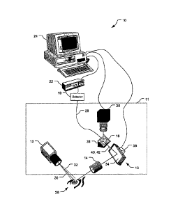
Referring to FIG. 1, a light scattering system 10 includes a light source 12,
a
transfer lens 14, a steered mirror assembly 16, a measurement mirror 18, a CCD
camera
20, a correlator 22, and a computer 24. The combination of the light source
12, transfer
lens 14, mirror assembly 16, measurement mirror 18, and CCD camera 20 forms an
optical unit 11. The optical unit 11 may be moved as a single unit in aligning
the
instrument to a subject's eye 26. The system 10 is configured to send beams of
laser
light into the subject's eye 26. Light scattered from the eye 26 is focused on
the
measurement mirror 18 at a position determined by the steering mirror assembly
16.
Some of the light incident upon the mirror 18 passes through a small hole 38
to an optical
fiber 28 which conducts the light to a photon detector 19. The detector 19 can
output
pulses to the correlator 22 for analysis, correlation can also be done in
software, without
specific hardware correlator or a combination of software and hardware. Other
portions
of the scattered light are directed from the mirror 18 to the CCD camera 20
and images of
the scattered light region are provided to the computer 24. The computer 24
can also
receive correlation functions and intensity measurements of the light received
by the
correlator and process the correlation functions and intensity measurements to
perform
diagnostic tests to determine likelihood of diseases and types of diseases in
the subject,
and to control the redirection of light by the steered mirror assembly 16 to
control the
location in the eye 26 from which light is being measured and provided to the
correlator
22. While not shown, the system 10 includes a chinrest and a forehead rest to
help
position a subject's head such that the subject's eye 26 is positioned to be
illuminated by
the light source 12, with minor adjustments to the light source's position
and/or angle as
appropriate.
13
CA 02647147 2008-09-24
WO 2007/120755
PCT/US2007/009009
The light source 12 may be configured to provide multiple laser beams to the
eye
26. For example the source 12 may be configured to send a laser pencil beam 20
toward
= the eye 26 that will scatter portions of the pencil beam 30. The pencil
beam 30 will
penetrate deep into the eye 26 along a straight line and will be scattered to
varying
degrees by different materials within the eye 26. The laser source 12 may be
further
configured to provide a fan beam, or slit beam, 32 directed at the eye 26. The
fan beam
32 is a very thin, planar beam that will also penetrate deep into the eye 26
and be
scattered by various materials to differing degrees. The fan beam 32 is used
to assist an
operator in aligning the instrument 11 to the subject. During alignment, the
eye
illumination is changed from pencil beam 30 to fan beam 32 and back several
times a
second. During measurement, preferably only the pencil beam 30 is turned on.
The light of the laser beams 30, 32 is preferably of a wavelength that is not
visible
or only slightly visible to the patient such that shining the beams 30, 32
into the patient's
eye 26 will not cause discomfort to the patient, which could result in the
patient moving
undesirably. Preferably, both of the beams 30, 32 have wave lengths between
about 400
nm - 820 nrn.
The transfer lens 14 is arranged with its longitudinal axis perpendicular to
the
pencil beam 30 and the fan beam 32 (i.e., the direction of propagation of the
beam 30,
32). The angle, preferably 90 , between the beams 30, 32 and the axis of the
transfer lens
14 helps to reduce/minimize dimensions of the target region of scattered light
received
from the eye 26. The transfer lens 14 is configured to focus the light
scattered from the
eye 26 onto the measurement mirror 18. The steered mirror assembly 16 includes
the
mirror 34 and a mirror driver motor 36. The mirror 34 is configured, and the
assembly
16 is positioned, such that the mirror 34 receives the focused scattered light
from the
transfer lens 14 and redirects this light in beams 40, 42, corresponding to
the beams 30,
32, to a focused image of the scattering region on the measurement mirror 18.
The mirror
34 is connected to the driver motor 36 that is configured to adjust the angle
of the mirror
34 in two axes in accordance with control signals received from the computer
24. The
motor 36 is configured to drive the mirror 34 to direct the scattered light
from the transfer
lens 14 such that the light is incident upon the mirror 18 at a desired
relative location
14
CA 02647147 2008-09-24
WO 2007/120755
PCT/US2007/009009
(e.g., such that a desired portion of the scattered light passes through a
hole in the mirror
18).
The measurement mirror 18 is configured and disposed to reflect light from the
steered mirror assembly 16 to the CCD camera 20. The mirror 18 reflects
scattered light
from the mirror 34 such that the CCD camera 20 can receive reflected light
from the
beams 40, 42 for imaging scattered light from the eye 26. A hole 38 may be
provided in
the center of the mirror 18. This hole 38 is preferably a pin hole (e.g.,
about 50 p.m in
diameter). The hole allows light from the scattered_beam 40 to pass through
and be
received by an optical fiber 28. The optical fiber 28 transfers indicia of the
portions of
the beam 40 that pass through the pin hole 38 to the detector 19, that
provides electronic
= indicia to the correlator 22.
The detector 19 is connected to the measurement mirror 18 through the fiber
optic
cable 28. The detector 19 is configured to convert the light received from the
cable 28 to
electronic pulses, and to send the pulses to the correlator 22.
The correlator 22 is configured to receive electronic pulses from the detector
19
and is configured to analyze fluctuations in light intensity of the light
received via the pin
hole 38 over time. The correlator 22 is configured to perform auto-correlation
algorithms
using indicia of the received light intensities to determine sizes of protein
aggregates in
the lens of the eye 26. The correlator 22 is further connected to the computer
24 and
configured to provide information to the computer 24 regarding the size of
protein
aggregates in the lens of the eye 26.
The CCD camera 20 is disposed and configured to receive light reflected from
the
measurement mirror 18 from the light beams 40, 42. The camera 20 is configured
to be
focused on the pin hole 38 and to provide an image of the reflected light that
has been
scattered by the eye 26. The camera 20 is configured to process the received
reflected
light to produce images showing a cross-section of the lens of the eye 26 due
to light
scattered from the fan beam 32 and the pencil beam 30. The camera 20 is
further
connected to the computer 24 and configured to provide information to the
computer 24
regarding the images of the eye 26 for display by the computer 24.
CA 02647147 2008-09-24
WO 2007/120755
PCT/US2007/009009
The computer 24 is configured to receive information from the correlator and
the
camera 20 and process this information accordingly to collect desired
information and
perform diagnostic operations. The computer 24 can process indications of
aggregate
types and size from the correlator 22 to determine indications of disease. The
computer
24 can process images of the eye 26 from the camera 20 and provide control
signals to
the assembly 16 to adjust the positioning of the mirror 34 to control which
portion of the
scattered light in the beam 40 is incident upon the pin hole 38.
Referring also to FIG. 2, the computer system 24 includes a processor 82,
memory 84, disk drives 86, a display 88, a keyboard 90, and a mouse 92. The
processor
82 can be a personal computer central processing unit (CPU) such as those made
by
Intel Corporation. The memory 84 includes random access memory (RAM) and read-
only memory (ROM). The disk drives 86 include a hard-disk drive and can
include
floppy-disk drives, a CD-ROM drive, and/or a zip drive. The display 88 is a
cathode-ray
tube (CRT), although other forms of displays are acceptable, e.g., liquid-
crystal displays
(LCD) including TFT displays. The keyboard 90 and mouse 92 provide data input
mechanisms for a user (not shown). The components 82, 84, 86, 88, 90, and 92
are
connected by a bus 94. The computer system 24 can store, e.g:, in the memory
84,
software code containing computer-readable, computer-executable instructions
for
controlling the processor 82 to perform functions described below to image and
analyze
light scattered by the eye 26.
Referring also to FIGS. 3-4, the computer 24 is configured to produce an image
50 of the eye 26 from the scattered light of the beams 30, 32. As indicated in
the image
50, light is scattered with significant intensity at a cornea and appears as a
bright spot 52
in the image 50. As the pencil beam 30 further passes into the eye 26, light
is not
significantly scattered by a vitreous humor region 54 of the eye 26 and thus
appears as a
dark region in the image 50. Moving to the left in the image 50, light is
significantly
scattered by a lens capsule 56 due to Type IV collagens in the lens capsule, a
supra-
nuclear region 58, and a nucleus 60 of the eye 26. The significant scattering
results in the
bright portions shown in the image 50 due to the increased intensity of
scattered light
16
CA 02647147 2008-09-24
WO 2007/120755
PCT/US2007/009009
received by the camera 20. Also, a bright spot 53, the Purlcinje spot, is
caused by light
reflecting from the cornea.
The camera 20 produces about 30 images/second, but one of skill in the art
understands that other frame rates may also be used. Correlation functions are
acquired
in time frames between about one millisecond and one second. Typically, five
correlation functions are obtained at each position in the eye 26 with the
measurement 11
focusing on a given point in the eye. Normal motions of the eye 26 due to,
e.g., pressure
surges due to the heart beat of the subject, as well as other factors,
typically cause the eye
= 26 to move during the time used for obtaining information to produce the
correlation
function. Such motion can reduce the effectiveness of the data produced and
thus the
effectiveness of the measurement taken, and consequently the diagnostic
results. The
system 10, and in particular the computer 24, is preferably configured to help
stabilize the
image 50 by compensating for motions of the eye 26.
When making measurements, preferably only the pencil beam 30 is turned on and
.
the tracking mechanism is active. Referring to FIG. 4, the computer 24 can
=
accommodate movement of the eye 26 due to various causes. For example,
saccadic eye
movements, blinking, pulsation (e.g., due to heart beats), or voluntary
movements of the
eye 26 can be accommodated using the tracking mechanism of the computer
control
signals and the motor 36. The computer 24 can determine the position where the
laser
beam 30 passes between two known regions to determine a reference point for
use in
locating specific portions of the eye 26 and for use in adjusting the mirror
34 in order to
collect data through the pinhole 38 for a desired position of the eye 26. For
example, the
computer 24 can determine the location of an anterior lens capsule interface
61
corresponding to an interface between the lens capsule 56 and the vitreous
humor region
54, a posterior lens capsule interface 63, an air-cornea interface 65, a
cornea-aqueous
region interface 67, a vitreous humor-retina interface, etc. For the interface
61, the
computer 24 can determine the position where the laser beam 30 passes from the
aqueous
humor region 54 into the anterior lens capsule 56 by determining where the
scattered
intensity rises abruptly after the cornea 52 moving from right to left in the
image 50. Any
17
CA 02647147 2008-09-24
WO 2007/120755 PCT/US2007/009009
of the above mentioned interfaces can be used as a reference point for
measurements,
mapping and tracking.
The computer 24 places a marker, e.g., an "X" 55, at the location of the
reference
point, near the anterior lens capsule interface 61, in the captured image 50
to permit
future visual confirmation of proper tracking operation. A pickup point 66
corresponding
to the pinhole 38 remains at the same pixel address in the image 50. A desired
pickup
point 64 in the eye 26 is set in a setup screen to be a specified number of
pixels measured
from the lens capsule 56. Knowing the pixel position of the lens capsule 56,
the desired
pickup point 64, and the actual pickup point 66, the computer 24 can calculate
the present
error between the desired pickup point 64 and the actual pickup point 66 and
move the
mirror 34 to compensate for this difference. This operation is done 30 times a
second
(for example) to maintain the actual pickup point 66 at the desired position
64 in the eye
26. The computer 24 can determine the present position of the lens capsule 56
in this
manner. The computer 24 can determine the distance in pixels from the present
position
=
of the lens capsule 56 to a desired position of the lens capsule 56 in the
image 50. The
determined distance is a horizontal distance (for example) from the present
position of
the eye 26 and its desired position relative to the field of view of the
camera 20 and thus
the image 50. The computer 24 can send control signals to the assembly 16 to
cause the
motor 36 to move the mirror 34 such that the actual horizontal position of the
eye 26 in
the image 50 is the desired horizontal position of the eye 26 in the image 50.
The
computer 24 continues to make these adjustments during measurements of the eye
26.
The computer 24 can further determine the relative vertical distance between
the present
position of the eye 26 and its desired position and send control signals to
the motor 36 to
cause the motor 36 to adjust the mirror 34 to compensate for vertical motion
of the eye
26. The computer 24 can analyze the information obtained over time and
determine what
information should be discarded due to movement of the eye 26 or blinking. The
computer 24 can retain information not tainted by eye movement or blinking (or
for
which movement was sufficiently compensated) and discard information tainted
by eye
movement or blinking (and for which movement was not adequately compensated).
18
CA 02647147 2008-09-24
WO 2007/120755
PCT/US2007/009009
As part of the initial alignment procedure, the computer 24 may be further
configured to superimpose an ellipse 68 on the image 50 with both laser beams
30, 32
turned on. The ellipse 68 is preferably sized and disposed to align with the
pupil 70 of
the eye 26. The ellipse 68 can be sized manually by a user of the computer 24
using, e.g.,
the keyboard 90 or the mouse 92. The user can use the image 50 to select
borders
between the various regions of the lens (cortex 57, supra nucleus 58, nucleus
60) and
have data collected within each region. The user can select to insert or
superimpose the
ellipse 68 and move the image 50 of the eye 26 by moving the optical unit 11
with
respect to the subject. When the optical unit 11 is positioned so that the
ellipse 68
matches the pupil 70 of the eye 26 and the subject is fixating on a target
(not shown), the
laser beam 30 passes through a unique path in the lens of the eye 26 and
measurements
= may be made at a position that is reproducible from one measurement
session to another.
The user can size the ellipse 68, e.g., by selecting the ellipse 68 and
dragging a cursor to
= adjust the size in either axis of the ellipse 68. Using this alignment
procedure, the same
subject can be analyzed before and after various procedures, such as
operations on the
eye 26 or administration of medications, to evaluate the success of the
procedures
performed or medications administered on the subject.
The computer 24 may be further configured to separate the eye image 50 into
regions. As shown in FIG. 3, the computer 24 can analyze the intensity of the
image 50
and separate the image 50 into the cortex 57, the supra-nucleus 58, and the
nucleus 60
regions of the eye 26. The computer 24 can use the segmentation of the eye
image 50 to
control the assembly 16 to determine the position of the measurement region
64. For
example, the computer 24 can specifically choose to measure light scattered
intensity of
the supra-nucleus 58 or nucleus 60 regions. In particular, the computer 24 can
cause
measurements to be taken using the measurement region 64 at, e.g., four
different depths
within the eye 26 relative to the cornea 52.
The system 10 can be used to perform both quasi-elastic light scanning (QLS)
and
other forms of scanning on a single platform/device. An imaging agent can be
introduced
that will bind or attach to specific types of items, e.g., aggregates
indicative of disease,
and will react to light in a way that can be detected distinctively.
Preferably, the imaging
19
=
CA 02647147 2015-08-25
agent is configured to fluoresce in response to light, in which case the
scanning is
referred to as fluorescent ligand scanning (FLS). The imaging agent can be
introduced
into the eye in a variety of ways, e.g., through eye drops, creams, lotions,
salves,
systemically, etc. The light source 12 has the wavelength and polarization
properties
appropriate to the specific imaging agent. For example if the imaging agent is
a
fluorophor, then the wavelength is preferably tuned to the peak of the agent's
absorption
spectrum. The light source 12 can be tuned to the wavelength of light to which
the
imaging agent will react and the resulting image portion that passes through
the pinhole .
38 analyzed by the computer 24 such that the aggregates' presence and quantity
can be
- 10 determined. The imaging agent can take various forms such as a chromophor
(that is
colorimetric, in the visible light spectrum), a fluorophor (e.g., a
fluorescent probe) that
will fluoresce in response to light, or other material that will distinctively
and detectably
react to visible or non-visible (e.g., infrared) light. A distinctive reaction
need not be
unique, but is such that it differs (e.g., in wavelength and/or degree of
reaction) from the
reaction, if any, of materials in the region of interest other than the
imaging agent.
Fluorescing imaging agents preferably fluoresce different wavelengths of light
than
materials in the eye 26 and/or in amounts greater (at the fluorescent
wavelength) than the
materials is the eye 26. Exemplary fluorophors are discussed in U.S. Patent
No.
6,849,249 and include Chrysarnine
or
Chrysarnine derivative compounds such as {(trans, trans), -1-bromo-2,5-bis-(3-
hydroxycaibony1-4-hyrdoxy) styrlbenzene (BSB)}. The system 10 can also use the
same
camera 20 for both the QLS and FLS measurements. The system 10 can perform
optical
sectioning with FLS and the slit beam 32 to assist in mapping the eye 26
(e.g., sectioning
the eye 26). The light scattered from the two beams 30, 32 can be co-
registered on the
image 50 as shown. Further, the computer 24 can use FLS measurements to
confirm
QLS measurements and/or can use QLS measurements to confirm FLS measurements
and diagnostic conclusions.
Thus, the system 10 can be used for diagnostic purposes by contacting an
ocular
tissue of a mammal, e.g., a human subject, with a detectably-labeled compound
which
binds to an amyloid protein or pre-amyloid protein aggregate. The compound
CA 02647147 2015-08-25
preferentially binds to amyloid proteins compared to other 13-pleated sheet
containing
proteins. Preferably, the detectably-labeled compound contains a fluorescent
probe. For
example, the fluorescent probe or fluorophor is a Chrysamine or Chrysamine
derivative
compound such as {(trans, trans), -I -bromo-2,5-bis-(3-hydroxycarbony1-4-
hyrdoxy)styrIbenzene (BSB)}. Chrysamine G and derivatives thereof are known in
the
art (e.g., U.S. Patent Nos. 6,133,259; 6,168,776; 6,114,175). These compounds
bind to
Ai3 peptides, but are not fluorescent. The diagnostic methods utilize a highly
lipophilic
fluorescent amyloid-binding Clu-ysamine G derivative to detect A13 peptides in
the eye.
Bioavailable lipophilic fluorescent probes may also be used. Such fluorophors
and
probes are commercially-available, e.g., from Molecular Probes, Inc. Eugene,
OR. Some
dyes, e.g., X-34 or {(trans, trans), 1-bromo-2,5-bis-(3-hydroxycarbony1-4-
hyrdoxy)styrlbenzene (BSB)} (Styren et al., 2000, J. Histochem. 48:1223-1232;
Link et
al., 2001, Neurobiol. Aging 22:217-226; and Skrovonsky et al., 2000, Proc.
Natl., Acad.
Sci. U.S.A. 97:7609-7614) have been used to analyze brain tissue (but not eye
tissue).
These probes emit light in the blue-green range, thus the level of
fluorescence, which is
diagnostically relevant, exceeds the amount of human lens autofluorescence in
the blue-
green range. Other useful compounds include a detectable methoxy agent such as
Me-
X04 (1,4-bis (4'-hydroxystyr1)-2-methoxybenzene). Other methoxy agents
include, e.g.,
Chrysamine or Chrysamine derivative compound such as {(trans, trans), -1-bromo-
2,5-
bis-(3-hydroxycarbony1-4-hyrdoxy)styrlbenzene (BSB)). Such compounds are
described
in Mathis etal., Curr. Pharm. Des., vol. 10(13):1469-93 (2004); U.S. Patent
Nos.
6,417,178; 6,168,776; 6,133,259; and 6,114,175.
Nonspecific amyloidphilic probes such as thioflavin T,
thioflavin S or Congo red dye may also be used.
The system 10, in particular, the computer 24, can provide photo documentation
of measured results. The computer 24 can provide, for every FLS number
obtained, an
indication of where in the image 50 the light came from that was analyzed for
determining the FLS number. In this manner, the computer 24 can document the
region
from which various FLS indications came from. The FLS number and the
corresponding
region of interest can then be used to determine whether the FLS number
corresponds to
21
CA 02647147 2008-09-24
WO 2007/120755 PCT/US2007/009009
a particular disease or other cause. Indications or FLS numbers indicating
aggregates in
one region of the eye 26 maybe be indicative of disease or other abnormality
while the
same FLS number in a different region of the eye 26 maybe innocuous.
Therefore, the
computer 24 preferably associates measured FLS numbers with corresponding
regions
within the eye 26 from which the measurements were taken to arrive at the FLS
number.
The computer 24 may be further configured to analyze different portions of the
eye 26 to determine distances between intensity peaks in the image 50. For
example, the
intensity peaks can be used to determine the depth of the eye 26, e.g., for
use in selecting
an intra-ocular implant, e.g., the size of an artificial intra-ocular lens
(TOL) for
implantation in the subject's eye 26. Thus, the system 10 can be used to
determine the
appropriate intra-ocular implant to use in a non-invasive manner. The system
10 can also
be used to determine the depth of the anterior chamber, corneal and lens
thicknesses, etc.
Referring to FIG. 5, with further reference to the FIGS 1-3, a process 110 for
measuring and analyzing objects in the subject's eye 26 using the system 10
includes the
stages shown. The process 110 can be used to perform FLS and/or QLS using the
system =
10. The process 110, however, is exemplary only and not limiting. The process
110 may
be modified, e.g., by adding, removing, or rearranging stages.
At stage 112, the laser source 12 shines the laser's beams 30, 32 into the
subject's
eye 26. The beam 32 provides a plane of laser infrared light such that a cross
section of
the eye 26 can be imaged. The fan beam 32 will allow for the cross-section
image 50 to
be formed while the pencil beam 30 provides focused light for analyzing
different regions
of the eye for distinctive characteristics such as aggregates.
At stage 114, the light scattered by the eye 26 from the laser beams 30, 32 is
imaged. The light scattered by the eye 26 is collected at preferably 90
relative to the
incident beam propagation directions. The light scattered by the eye 26 is
focused by the
lens 14 on the measurement mirror 18. The measurement mirror 18 reflects the
scattered
light to the camera 20 that processes the received light to form the cross-
sectional image
50 of the eye 26. The cross-sectional image 50 is a cross-section of the eye
26 with an
overlay of the light scattered due to the beam 30. The cross-sectional image
50 is
preferably of an anterior segment of the eye 26, including the cornea, the
lens, and part of
22
CA 02647147 2008-09-24
WO 2007/120755 PCT/US2007/009009
the nucleus of the eye 26. The image information is provided by the camera 20
to the
computer 24 for display on the computer's monitor 88.
At stage 116, the ellipse 68 is positioned over the image 50 of the eye 26.
The
optical unit 11 can be positioned and the ellipse 68 can be sized manually by
a user of the
instrument 10. For example, the ellipse 68 is sized and the optical unit 11 is
moved so
that the ellipse corresponds with the pupil of the eye 26. The ellipse 68 can
be repeatedly
positioned on the eye 26 such that the process 110 can be repeated at
different times on
the same eye 26 and will allow for consistent measurement of the eye 26 such
that
measurement can confidently be taken for the same region in the eye 26 to
compare
changes in the eye 26 over time.
At state 118, various regions within the eye 26 are identified. This can be
done
manually by the user of the computer 24 manipulating input devices such as the
keyboard .
90 and/or the mouse 92 or automatically by the computer 24. If done
automatically, the
computer 24 analyzes the intensity pattern of the image 50 and identifies
various regions
of the eye 26 given known properties of intensity distributions of eye images.
The
computer 24 identifies the cornea 52 by moving along the direction of
propagation of the
beam 30 and finding a large high-intensity region in the image 50, and
identifies the lens
capsule 56 by moving toward the inner portion of the eye 26 in the image 50
and finding
the next location where the image intensity is significant after a large
region of low
intensity. The computer 24 further sections the image 50 by identifying the
cortex 57, the
supra-nucleus 58 and the nucleus 60 regions by analyzing absolute and/or
relative
intensity levels scattered by the beam 32 along the line 62. The computer 24
stores
indications of distances between the cornea 56 and the various regions within
the eye 26,
e.g., as indications of numbers of pixels between the various objects and
regions of the
eye 26.
At stage 120 the scattered light from the beam 30 is directed to the pin hole
38 in
the measurement mirror 18 to measure desired regions of the eye 26. The
computer 24
sends control signals to the motor 36 to drive and steer the mirror 34 to
direct light
scattered from the beam 30 from a desired region of the eye 26 to the pin hole
38. The
computer 24 determines the desired region of the eye 26 from which
measurements are
23
CA 02647147 2008-09-24
WO 2007/120755 PCT/US2007/009009
desired to be taken. The computer 24 sends the control signals to the motor 36
to steer the
mirror 34 in two axes such that the measurement region 66, corresponding to
the pin hole
38, is positioned at the desired measurement region 64. The computer 24 can
position the
measurement region 64 at a set of desired regions within the eye 26, e.g., a
set of four
regions corresponding to different regions of the eye such as the cortex, two
measurements within the supra nucleus, and one measurement within the nucleus.
Other -
quantities of measurements and/or regions or distributions of measurements
within the
regions maybe used. Further, the computer 24 may position the measurement
region 64
in a particular region or at a particular location to measure characteristics
of the eye 26 at
a particular position within the eye 26 for, e.g., diagnosing particular
abnormalities. For
example, the measurement region 64 can be placed at the supra-nucleus 58 to
investigate
for aggregates corresponding to Alzheimer disease, other neurodegenerative
diseases,
TSEs, etc.. The scattered light received from the measurement region
corresponding to
the pin hole 38 is collected and transmitted through the fiber optic cable 28
to the detector
19 and the detected signal is sent to the correlator 22. The correlator 22
computes
correlation functions to analyze the intensity of received light over time and
provides
indications of this analysis to the computer 24, e.g., for determination of
abnormalities
within the eye 26.
At stage 122, performed during the stage 120, the system 10 accommodates for
motion of the eye 26. The computer 24 analyzes the image 50 to determine the
location
of a specific portion of the eye 26, e.g., the lens capsule relative to a
desired location of
the lens capsule 56 and sends control signals to the motor 36 to adjust the
angle of the
mirror 34 to accommodate for motion of the eye 26. Thus, the system 10 can
provide a
relatively stable image of the eye 26 and can take measurements from a
relatively stable
location within the eye 26 such that measured light intensity accurately
reflects the
existence or non-existence of aggregates and the type of aggregates within the
desired
tested location of the eye 26.
At stage 124, the computer 24 analyzes the measured results from the
correlator
22 for diagnostic purposes. The computer 24 analyzes the data from the
correlator 22 in
conjunction with knowledge of the location of the measured regions 64 within
the eye 26.
24
CA 02647147 2008-09-24
WO 2007/120755
PCT/US2007/009009
Using this information, the computer 24 can determine the existence and type
of
aggregate or other objects within the eye 26 and provide indications, e.g.,
through the
computer's display 88 to a user of the existence, non-existence, and/or type
of object
within the eye 26.
The system 10 has wide applicability for different diagnostic purposes. For
example, the system 10 can be used as described above to determine aggregates
for
diagnosing various types of disease or other types of abnormalities within a
subject.
The system 10 can further be used to determine the depth of a subject's eye
for
use in selecting a size of an intra-ocular implant, e.g., an artificial intra-
ocular lens, to be
inserted into the subject's eye.
Further, the system 10 can also be used to perform FLS and/or QLS without
using
anesthesia. The use of anesthesia on animals inhibits the ability to perform
QLS due to
= dehydration of the eye in non-human animals under anesthesia. The system
10, however,
can perform QLS without the anesthesia, thus improving the quality of
measurements and
diagnostic results from such measurements.
A system similar to the system 10 can be used to perform both QLS and FLS. A
light source other than the laser source 12 can be used. For example, the
light source
may be a broad-spectrum light source that is essentially omnidirectional
(e.g., a light
bulb), and/or that can provide a fan beam, and/or that can provide a pencil
beam. One or
more light sources may be used to provide one type of directionality, or
combinations of
different directionalities. Further, one or more energy sources that provide
energy
outside the light spectrum may be used in combination with an imaging agent
that
responds to energy outside of the light spectrum. For example, an imaging
agent could
be used that responds to microwaves, radio frequency energy, a magnetic field,
etc.
Multiple energy sources that collectively provide, or a single energy source
that provides,
energy both light and min-light energy may also be used in combination with
one or more
imaging agents that respond to the appropriate energy forms. While using these
techniques may not result in the imaging agents fluorescing, these techniques
can be
considered as part of FLS.
25 =
CA 02647147 2008-09-24
WO 2007/120755
PCT/US2007/009009
Referring to FIG. 6, with further reference to the FIGS 1-3, a process 150 for
performing FLS on the subject's eye 26 includes the stages shown. The process
150,
however, is exemplary only and not limiting. The process 150 may be modified,
e.g., by
adding, removing, or rearranging stages. For example, stage 152 may be removed
and
stage 156 modified to eliminate comparing measured intensity with previously-
measured
intensity. Further, while measuring fluorescence in response to light is
discussed below,
the process 150 could be modified to use other forms of energy and/or measure
other
characteristics, as discussed above.
At stage 152, the eye 26 is illuminated and fluorescence measured. The eye 26
is
illuminated with a light source and fluorescence emitted from the eye 26 in
response to
the illumination measured and recorded. The magnitudes of emitted fluorescence
and the
locations of these magnitudes are correlated and recorded.
At stage 154, an imaging agent is introduced into the eye 26. The imaging
agent
is configured to bind to materials/objects of interest that may be present in
the eye 26 and
is configured to fluoresce in response to light from the source. The imaging
agent may
= be introduced in a variety of manners, e.g., through drops applied to the
eye 26,
= intravenously, etc.
At stage 1.56, the eye 26 is illuminated with light from the source and the
fluorescence from the eye 26 measured. The intensity magnitudes and locations
are
correlated and stored, and compared with magnitudes recorded at stage 152,
with
magnitudes measured from similar locations in stages 152 and 156 being
compared. The
comparison includes analyzing differences in the magnitudes and determining
presence
of the material/object of interest, and amount of the material/object if
indeed present in
the eye 26. Conclusions can be determined regarding implications of the
presence and/or
amount of the material/object of interest such as a medical condition of the
subject such
as the existence and/or stage of a disease such as Alzheimer Disease.
FIG. 7 illustrates a scheimpflug illumination and imaging system 160 according
to
some embodiments of the invention, which may include one or more (and
preferably all)
of the following: a light source 162, an optical scanning system 164, a pair
of flat field
lenses 166 & 170, a dichroic beam splitter 172, a pair of mirrors with a slit
174 &176, a
26
=
CA 02647147 2008-09-24
WO 2007/120755
PCT/US2007/009009
pair of detectors 178 & 180, a pair of CCD cameras 182 & 184, an
autocorrelator 186,a
computer and monitor 188, and a ophthalmoscope 190. The a scheimpflug
illumination
and imaging system 160 may be moved as a single unit aligning the system to
the
patient's eye with the ophthalmoscope 190. The system 160 is configured to
send beams
of laser light into a subject's eye 168, in which the light scattered from the
eye 168 is
focused on the minors with a slit on each 174, 176 by the second field lens
170 and the
dichroic beam splitter 172. Some of the light incident upon each mirror 174,
176 may
pass through the slit on each mirror to a QLS and FLS detector 180, 178
respectively.
At least one of the detectors 178, 180, and preferably both can output to the
= autocorrelator 186 for analysis. Other portions of the scattered light may
be directed
from the mirrors 174,176 to the CCD cameras 182, 184 respectively and images
of the
scattered light and fluorescence region may be provided to the computer 188.
The
computer 188 can also receive correlation functions and intensity measurements
of the
. light received by the correlator and process the correlation functions and
intensity
measurements to perform diagnostic tests to determine likelihood of diseases
and types of
diseases in the subject. The computer control system preferably monitors
several and
preferably all aspects of the system through a customized graphical user
interface (GUI).
Image collection software may collect the images and store them in files for
analysis (the files may be analyzed as previously disclosed). The
ophthalmoscope 190
may be a standard ophthalmic head and chin rest for humans. The entire optical
platform
is positioned to the eye 168 through a Joy-stick control (for example). The
range of
motion is preferably sufficient enough to make measurements at any location in
the
anterior segments of both eyes. Custom holders may be adapted to or replace
the head
and chin rest for various animal studies of primates and rodents.
The light source 162 may be configured to provide a polarized laser beam which
is preferably focused through a set of lenses and the optical scanning system
164 to
produce a vertical fan beam of light. One of skill in the art will appreciate
that the optical
scanning system 164 may utilize one of several different methods for producing
a linear
sweeping motion (left and right across the page) of the light emission at the
object plane
of the first flat field lens 166.
27
CA 02647147 2008-09-24
WO 2007/120755
PCT/US2007/009009
The first flat field lens 166, which may contain multiple lens elements, is
preferably tilted at an angle based on the Scheimpflug rule to create a
virtual image plane
that makes a vertical cross sectional plane 169 through the anterior segment
of the
patient's eye 168. The angle of incidence of the illumination is preferably 45
degrees to
the line of sight of the patient. The optical scanning system 164 is used to
sweep the
vertical fan beam of light across the anterior segment of the eye 168. The
angle of
convergence should be fairly steep so that the angle of divergence is
similarly steep. This
exemplary configuration allows not only a sharp focal region within the cross
sectional
plane 169, but also insures that the light exiting from the back of the
natural lens is
similarly divergent and of low energy when it reaches the retina.
The scanning system 164 is preferably used to traverse the beam of light 10mrn
(for example) into the anterior segment of the eye 168 beginning 1-2 mm (for
example)
in front of the cornea. While specific measurement values are given in this
embodiment,
they are exemplary only and not limiting. Single pass scan times of 16-33 msec
(for
example) through the eye can be made with a vertical fan beam of light. The
focused
vertical fan beam of light may be on the order of approximately 501.1m X
lOrrim (width by
length) at the image plane 169. The power requirement is chosen to be eye
safe. Real
time power monitoring can be incorporated to ensure safety.
The second flat field lens 170 may be configured and/or disposed to image the
scattered light at, for example, 45 degrees to the line of sight and 90
degrees with respect
to the illumination as the vertical fan beam of light scanned across the
anterior cross
sectional plane 169 of the eye. The second flat field lens 170, which may
contain
multiple lens elements, may be tilted at an angle based on the Scheimpflug
rule to create
a sharply focused object plane which preferably coincides with the image plane
of the
illumination 169 of the patient's eye 168.
The dichroic beam splitter 172 may be configured and/or disposed to pass the
excitation wavelength of the laser to a front surfaced mirror 174 with a slit
aperture (for
example) in the surface of the mirror. This preferably is the image plane for
the QLS
detection. The angle of incidence of the imaging is preferably 45 degrees to
the line of
sight of the patient. The QLS may be detected at the QLS image plane through a
slit
28
CA 02647147 2008-09-24
WO 2007/120755
PCT/US2007/009009
running horizontally (left and right in the plane of the page) with a width
preferably on
the order of 50qtm X lOmm (W XL) to maximize resolution and efficiency. A
detector
180 (preferably a photomultimplier tube) may be behind the slit where its
signals may be
delivered to an autocorrelator 186 linked to a computer and monitor 188.
As the scattered image of the light fan beam is scanned across the slit, QLS
measurements may be made with the detector 180 and autocorrelator 186. Sample
times
ranging from 50 nsec to 50pec (for example) may be made during the 3-33msec
(for
example) scan. This allows resolution of a few hundred points. The information
may be
read into a file and analyzed by the computer 188. Alignment and summation of
the
cross sectional structures may be made through software algorithms.
The CCD camera 182 may be disposed and/or configured to receive light
reflected from the mirror 174. The CCD camera 182 may be used to disqualify
large eye
movement, adjust for slit movement in the image, and show the cross sectional
excitation
image of the eye 168. The camera 182 may be further connected to the computer
188 and
configured to provide information to the computer 188 regarding the images of
the eye
168 for display by the computer 188. The cross sectional camera may be a
Charged
Couple Device (CCD) or Complementary metal¨oxide¨semiconductor (CMOS) device.
Autocorrelation functions graphically presenting the fast and slow components
of the
light scattering analyses may be made as well as estimates of hydrodynamic
radii (proxy
for molecular size and molecular weight) derived from the slope
determinations.
The QLS measurement is a line scan through the cornea. In other embodiments
two dimensional scans may be made by scanning the slit up and down across the
cross
sectional image or by placing another scanning device between the object and
image
planes or by rastering a single illumination point instead of the light fan
beam.
Fluorescence Ligand Scanning (FLS) is an important second tool for determining
the presence. of amyloid aggregation. As the vertical fan beam of light is
scanned across
the anterior cross section plane of the eye 168, the ligand's flouresced light
may be
imaged at 45 degrees to the line of sight and 90 degrees with respect to the
excitation
illumination by a flat field lens 170. The field lens 170, which may contain
multiple lens
elements, is preferably tilted at an angle based on the Scheimpflug rule to
create an object
29
CA 02647147 2008-09-24
WO 2007/120755
PCT/US2007/009009
plane which coincides with the image plane of the illumination of the
patient's eye 169.
The fluoresced light may be imaged off a dichroic beam splitter 172 that
reflects the
emission wavelength of the ligand to a front surfaced mirror with a slit 176.
This is
preferably the image plane for the FLS detection. The angle of incidence of
the imaging
is preferably 45 degrees to the line of sight of the patient. The FLS may be
detected at
the FLS image plane through a slit running vertically (up and down in the
plane of the
page) with a width preferably on the order of 50 to 200 mX 10mrn (W X L) to
maximize resolution and efficiency. A detector 178 (preferably a
photomultimplier tube)
may be behind the slit where its signals may be delivered to the
autocorrelator 186 linked
to a computer and monitor 188.
As the scattered image of the light fan beam is scanned across the slit in the
mirror 176, FLS measurements may be made with the detector 178. Sample times
ranging from 50 nsec to 50ptsec (for example) may be made during the 3-33msec
(for
example) scan. This allows resolution of a few hundred points. The information
may be
= 15 read into a file and analyzed by the computer 188. Alignment and
summation of the
cross sectional structures may be made through software algorithms.
The CCD camera 184 may be disposed and/or configured to receive light
reflected from the mirror 174. The CCD camera 184 may be used to disqualify
large eye
movement, adjust for slit movement in the image, and show the cross sectional
emission
image of the eye 168. The camera 184 may be further connected to the computer
188 and
configured to provide information to the computer 188 regarding the images of
the eye
168 for display by the computer 188. The cross sectional camera may be a
Charged
Couple Device (CCD) or Complementary metal¨oxide¨semiconductor (CMOS) device.
The FLS measurement is a line scan through the cornea. In other embodiments
two dimensional scans may be made by scanning the slit up and down across the
cross
sectional image or by placing another scanning device between the object and
image
planes or by rastering a single illumination point instead of the light fan
beam.
The cameras 182, 184 focused on the image planes of both the QLS and FLS
image planes may provide cross sectional and fluorescence images respectively.
The
cameras 182, 184 may have frame rates of 30 to 60 fps (for example).
Additionally,
CA 02647147 2008-09-24
WO 2007/120755
PCT/US2007/009009
since the QLS and FLS slits in the mirrors 174, 176 act as fiducials across
the images,
these images provide feedback information of saccadic eye movement and
occlusion
(blinking) to enhance the accuracy and precision of the QLS and FLS
measurements.
Calibration of the system 160 may be made using custom cuvettes filled with
.
custom microspheres. Several concentrations of spheres and different size
spheres may
be utilized. Initial calibration may be with a square cuvette rotated
preferably 45 degrees
to the line of sight. This ensures that the faces of the cuvette are
perpendicular to the
incoming illumination and outgoing scattering. Additionally a second type of
cuvette
= may be made with a tube within a tube. The radii and their positions
preferably
approximate the cornea and the interocular lens. The inner tube may be filled
with
microspheres and the outer tube may be filled with water.
Referring to FIG. 9 with further reference to the FIGS. 8 and 10; alight
scattering
system 230 according to some embodiments of the invention, which may-include
one or
more (and preferably all) of the following: a first laser light source 200, a
first lens 201, a
first dichroic beam splitter 202, a second lens 203, a second laser source
204, a first
microscope objective 205; a mirror 206, a third lens 207, a second dichroic
beam splitter
208, a fourth lens 209, a second microscope objective 210, a light filter 211,
a detector
212, a lens mount 214, a motor 215, a fifth lens 216, a third dichroic beam
splitter 217, a
slit aperture 218, a second detector 219, a second motor 220, a camera 221, a
magnified
view alignment camera 222, a fourth dichoric beam splitter 223, a second light
filter 224,
a narrow angle target 225, a crossed-spot alignment system 226, wide view
alignment
camera 227, and a stereotactic platform 228. The system 230 is configured to
send
beams of laser light into the subject's eye. Light scattered from the eye is
focused onto
the first and second detectors 212, 219.
The first laser light source 200 may be configured to provide a laser beam
that can
be directed to the eye. Preferably, the laser light source beam has a
wavelength of about
780 nm. Light from the laser source 200 may be focused through the set of the
first lens
201, first dichroic beam splitter 202, and second lens 203 to produce a spot
of light that
impinges the eye. The focused spot of light is on the order of 50 to 200 pm
(for example)
31
CA 02647147 2008-09-24
WO 2007/120755
PCT/US2007/009009
in diameter at the eye. The power requirement may be chosen to be eye safe.
Real time
power monitoring may be incorporated to ensure safety.
The second laser light source 204 may be configured to provide a laser beam
that
can be directed to the eye. Preferably, the laser light source beam has a
wavelength of
about 405 nm (for example). In the exemplary embodiment, excitation light from
the
laser source 204 may be used to accomplish FLS measurements. Light from the
laser
source 204 may be focused through the first microscope objective 205, to the
mirror 206.
The microscope objective 205 may be moved out of the optical light path
through the use
of a mechanism 213. The removal of the microscope objective 205 produces a
collimated beam of light instead of a focused spot at the eye. Collimated
light is light
having rays that are parallel and thus includes a plane wavefront.
The mirror 206 may be configured and/or disposed to reflect the light from the
microscope objective 205 through the lens 207, dichroic beam splitters 208,
202 and lens
=
203 to produce a focused spot of light that impinges the eye.
The fourth lens 209 may be configured and/or disposed to focus light reflected
off
of the dichroic beam splitter 208 through the microscope objective 210, and
filter 211 to
the detector 212. Preferably, the detector 212 is photomultiplier tube (PMT)
type
detector with a pinhole over its aperture, however, other types of detectors
may be used.
The aperture of the detector 212 may be the image plane for the FLS detection
of the
system 230. Although not shown, the detector's 212 PMT signals may be
delivered to an
autocorrelator (e.g.186 in FIG. 7) linked to a computer and monitor (e.g. 188
in FIG. 7).
The lens mount 214 may be configured to hold the lens 203 and may be attached
to the motor 215. The focused spot of light originating from the second laser
source 204
may be scanned through the eye at preferably 45 degrees to the line of sight
of the patient
by movement of the motor 215 which may be attached to the lens mount 214. Thus
preferably, the movement of the motor 215 causes the movement of the lens 203
along
the axis of the light beam. The movement of the lens may change the location
of focus
and may result in the movement of the focused spot of light.
Choosing a focused spot to impinge the eye creates a cone of light within the
eye,
maximizing the light intensity at an anatomically desired location for
fluorescence
32
CA 02647147 2008-09-24
WO 2007/120755 PCT/US2007/009009
measurement, while allowing the laser energy to be dispersed over a wider area
of the
retina, which is positioned distal to the lens of the eye. This design allows
more power to
illuminate the region of measurement, while maintaining "eye safe" levels of
illumination
at the retina, which is prone to damage from excessive light exposure.
Specific
calculations for eye-safety are defined within ANSI Z136.1 "Safe use of
Lasers."
As the focused spot is moved, in discrete steps (preferably), across the lens
of the
eye, the ligand's fluorescent emission may be backscattered and imaged back
through the
system through the lens 203, and the dichroic beam splitter 202, to reflect
off the second
dichroic beam splitter 208. The light reflected off of the dichroic beam
splitter 208 as
=
mentioned above may go through the lens 209 and may be imaged to a point by
the
microscope objective 210 through the light filter 211 to the detector 212 with
a pinhole
=
over its aperture.
The signal collected by the detector 212 can be used to perform several
analytical -
techniques to describe the fluorescent behavior of the region of interest such
as
autocorrelation of light intensity over time to perform fluorescent
correlation
spectroscopy and total intensity and/or average intensity over a known
measurement
period can be performed to define gross signal level.
The fifth lens 216 may be directed and/or configured to focus the light
scattered
from the eye at preferably 45 degrees to the line of sight and 90 degrees with
respect to
the path of the illumination laser light beam from the source 200. The fifth
lens 216 may
focus the light onto the third dichroic beam splitter 217.
The third dichroic beam splitter 217 may reflect the light onto a slit
aperture 218.
The slit aperture 218 may be configured to allow light to pass through and be
received by
the second detector 219. This may be the image plane for the QLS detection.
The angle
of incidence of the imaging is preferably 45 degrees to the line of sight of
the patient.
The slit's 218 width is preferably on the order of 50 to 200t.tm X lOmm (W X
L) to
maximize resolution and efficiency. Although not shown, the detector's 219
(APD, or
similar sensitive light detector) signals may be delivered to an
autocorrelator (e.g.186 in
FIG. 7) linked to a computer and monitor (e.g. 188 in FIG. 7).
33
CA 02647147 2008-09-24
WO 2007/120755
PCT/US2007/009009
The scattered image of the light beam may be scanned by translating the slit
aperture 218 and the detector 219 with the motor 220. QLS measurements may be
made
with the detector 219 and an autocorrelator. The sample of a discrete location
/ volume
can be on the order of 30-msec (one frame of video), then the optical system
will scan to
the next anatomical location of the eye for the next measurement, and so-on
through the
anatomical region of interest A preferred method to measure from the lens
capsule to
cortex may include taking measurements in approximately 33-msec "steps"
accounting
for approximately 50 to 200-urn volumes, stepping through the eye. A desirable
feature is
to allow this process to occur without significant eye movement (due to heart
beat or
other eye motion).
There is no limit as to how many times this process may be employed in a
single
QLS measurement session, so a measurement could be as short as a few
millisecond or as
long as 10's of seconds (for example) with repeated "scans" of many "steps"
through the
eye. Sample times ranging from lp.sec to 200usec (for example) may be made
during the
scan. Scan speeds may be varied to capture the different anatomical features
of the lens
and post processed to account for movements due.to a variety of reasons
including heart
beats, the eyes micro-saccades, etc. The information may be read into a file
and analyzed
by the computer. Alignment and summation of the cross sectional structures may
be
made through software algorithms.
The camera, or similar detector 221 may be disposed and/or configured to
receive
scattered light from the eye that travels through the dichroic beam splitter
217, providing
an anatomical reference image. The camera 221 may be used to disqualify large
eye
movement, adjust for slit movement in the image and show the cross sectional
excitation
image. The camera 221 may be a Charged Couple Device (CCD), a Complementary
metal¨oxide¨semiconductor (CMOS), or any other type of appropriate device for
capturing images. Autocorrelation functions graphically presenting the fast
and slow
components of the light scattering analyses may be made as well as estimates
of
hydrodynamic radii (proxy for molecular size and molecular weight) derived
from the
slope determinations. The QLS measurement is a line scan through the cornea.
34
CA 02647147 2008-09-24
WO 2007/120755
PCT/US2007/009009
Additional optical filters may be placed within the optical path to enhance
signal to noise
ratio of the detected signal.
The wide view alignment camera 227 may be configured to help a technician
align the system 230 to a patient's eye. The camera 227 may allow the
technician to
coarsely align the patient.
The magnified view alignment camera 222 may be configured and disposed to
provide a magnified view of the patient's iris as viewed through the
beam*splitter 223 and
a filter 224.
Alignment of the system 230 may be done under Joy-stick control moving the
entire optical system 230 to the left or right eye, up or down. The device can
be manually
aligned to the patient by the operator using a crossed-spot alignment system
226 that
projects two spots that overlay each other on the apex of the cornea centered
on the iris.
Targets can be illuminated with colored light emitting diodes (LEDs) that may
be visible
=
to the eye and to the alignment cameras 222, 227. An infrared (IR) LED
illumination
=
scheme may be included to provide additional illumination for aiming.
A narrow angle target 225 may be configured and/or disposed to be reflected
off
=
of the beam splitter 223 and through the filter 224 to provide a spot target
for the patient
to fixate on with his or her vision. The target may be backlit by a red LED.
The focus of
the target 225 may be adjustable to account for the patient's dioptric
correction. The spot
target subtends approximately 2 degrees. In use, the patient may remove their
glasses
and the target 225 may be adjusted to their nominal average power
prescription. The
magnified view camera 222 may be bore-sighted with the fixation target 225 to
provide a
front view co-axial with the optical axis of the eye.
The stereotactic platform 228 may be a standard ophthalmic head and chin rest
for
humans. The entire optical platform may be positioned to the eye through the
joy-stick
control as described above. The range of motion may be sufficient enough to
make
measurements at any location in the lenses of both eyes. Custom holders may be
designed
to adapt to or replace the head and chin rest for various animal studies of
primates and
rodents.
CA 02647147 2008-09-24
WO 2007/120755
PCT/US2007/009009
As mentioned above, alternative measurement embodiments can be implemented
via removing the microscope objective 205 with the mechanism 213 which can be
actuated manually or with a motor. The removal of the microscope objective 205
from
the optical path of the light from the light source 204 allows the excitation
light from the
source 204 to be emitted as a collimated beam. It is known in the art that
collimation of
lower quality lasers can be accomplished by the addition of appropriate
collimating
optics. In this embodiment format, the collimated pencil beam of light may
transmit
through the eye and impinge as a relatively collimated spot in the retina.
With properly
chosen detector and optics, this arrangement can supply sufficient
illumination without
requiring laser illumination at levels which are not eye-safe.
In this collimated beam configuration, both detectors 212, 219 may be employed
to perform a number of measurements. The collimated pencil beam may be
positioned
across the lens of the eye, and the ligand's fluorescent emission may be
bacicscattered and
imaged back through the system for FLS measurements at the detector 212. The
ligand's
fluorescent emission is also emitted in all directions, and can be imaged at
preferably 45
degrees to the line of sight and 90 degrees with respect to the illumination
beam path by
the lens 216. The light may be imaged off the beam splitter 217 onto the slit
218 with the
detector 219 behind the slit for QLS measurements. The signal collected by
this detector
219 can be used to perform several analytical techniques to describe the
fluorescent
behavior of the region of interest such as; performing QLS at 405-nm similar
to the
measurement done at 780-nm when a 405-nm narrow-pass filter is placed
immediately
prior to the detector, autocorrelation of light intensity over time can be
done to perform
fluorescent correlation spectroscopy, and total intensity and/or average
intensity over a
known measurement period can be performed to define gross signal level. FLS
detection
may also be made in this configuration with an appropriate change in filter
selection.
In other alternative embodiments, the system 230 can be used to detect the
fluorescent decay characteristics of the ligand, allowing an alternate method
to isolate
fluorescence due to ligand from other fluorescent sources, such as lens
autofluorescence.
This can be accomplished inpart, by choosing a 405nm laser source 204 with
fast
switching capability, or by placing a fast switch or shutter (such as a q-
switch) 229 in the
36
CA 02647147 2008-09-24
WO 2007/120755 PCT/US2007/009009
excitation beam path, or in the either detection path (as described above) and
using any of
the fluorescent detection paths described previously.
The optical measurements of the system 230 are critically sensitive to
translational motions of the eye in excess of approximately 150-11m. In the
original
embodiments, a large source of motion artifact was eye motion induced by
heartbeat
associated motion. To avoid these predictable artifacts, a number of methods
can be
=
employed.
Computer algorithms can be utilized that recognize motion artifacts in either
the
position of anatomic structures in the slit-lamp camera 221, by evaluating the
relative
position of measurement volume in relationship to anatomic structures, or by
evaluation
of correlation functions, looking for hallmark characteristics of motion on
this measure.
A second approach to avoiding motion artifact due to heartbeat, may be to
=
synchronize data collection to heart beat. Resting heart rate in humans is
typically 50-85
beats per minute [BPM], but can exceed 120-BPM in cases of pathologic
tachycardia. By
synchronizing measurement to the rest period between beats, this artifact can
be avoided.
Methods to synchronize measurements to the rest periods between heart beats
include:
i. Placing a heart-rate monitor on subjects and using subject
heart rate to
control start and stop of data acquisition, and to calculate the number and
positional
distribution of measurements through an anatomic region of interest. This
could be done
using any number of commercially available heart rate monitor signals or
custom built.
Building a heart-rate monitor into a convenient contact point of the device,
such as forehead or chin rest and using subject heart rate to control start
and stop of data
acquisition, and to calculate the number and positional distribution of
measurements
through an anatomic region of interest. This could be done using any number of
commercially available heart rate monitor signals or custom built, with
appropriate
location of electrodes.
Constructing the system 230 with an incorporated pace-maker, and using
the pacemaker to modulate both heart beat and data collection in an
appropriate manner
to insure clean data collection.
37
CA 02647147 2015-08-25
The scope of the claims should not be limited by the embodiments set forth in
the
examples, but should be given the broadest interpretation consistent with the
description as a
whole. Due to the nature of software, functions described above can be
implemented using
software, hardware, firmware, hardwiring, or combinations- of any of these.
Features
implementing functions may also be physically located at various positions,
including
being distributed such that portions of functions are implemented at different
physical
locations.
Further, while the description above refers to the invention, the description
may
include more than one invention.
38