Note: Descriptions are shown in the official language in which they were submitted.
CA 02647314 2008-09-24
WO 2007/126808 PCT/US2007/007560
PEGYLATED FACTOR VIII
FIELD OF THE INVENTION
[001] The present invention relates to a proteinaceous construct comprising
coagulation factor VIII (FVIII) being bound to at least one soluble polymer,
such as a
poly(alkylene oxide) such as polyethylene glycol. Further the present
invention
relates to methods for prolonging the in vivo-half-life of FVIII in the blood
of a
mammal having a bleeding disorder associated with functional defects or
deficiencies of FV1II.
BACKGROUND OF THE INVENTION
[002] Coagulation factor VIII (FVIII) circulates; in plasma at a very low
concentration
and is bound non-covalently to von Willebrand factor (VWF). During hemostasis,
FVIII is separated from VWF and acts as a cofactor for activated factor IX
(FIXa)-
mediated factor X (FX) activation by enhancing the rate of activation in the
presence
of calcium and phospholipids or cellular membranes.
[003] FVIII is synthesized as a single-chain precursor of approximately 270-
330 kD
with the domain structure A1-A2-B-A3-C1-C2. When purified from plasma, FVIII
is
composed of a heavy chain (A1-A2-B) and a light chain (A3-C1-C2). The
molecular
mass of the light chain is 80 kD whereas, due to proteolysis within the B
domain, the
heavy chain is in the range of 90-220 kD.
[004] FVIII is also synthesized as a recombinant protein for therapeutic use
in
bleeding disorders. Various in vitro assays have been devised to determine the
potential efficacy of recombinant FVIII (rFVIII) as a therapeutic medicine.
These
assays mimic the -in vivo effects of endogenous FVIII. In vitro thrombin
treatment of
FVIII results in a.rapid increase and subsequent decrease in its procoagulant
activity,
as measured by in vitro assay. This activation and inactivation coincides with
CA 02647314 2008-09-24
WO 2007/126808 PCT/US2007/007560
2
specific limited proteolysis both in the heavy and the light chains, which
alter the
availability of different binding epitopes in FVIII, e.g. allowing FVIII to
dissociate from
VWF and bind to a phospholipid surface or altering the binding ability to
certain
monoclonal antibodies.
[005] The lack or dysfunction of FVIII is associated with the most frequent
bleeding
disorder, hemophilia A. The treatment of choice for the management of
hemophilia
A is replacement therapy with plasma derived or rFVIII concentrates. Patients
with
severe haemophilia A with FV11I levels below 1%, are generally on prophylactic
therapy with the aim of keeping FVIIi above 1% between doses. Taking into
account
the average half-lives of the various FVIII products in the circulation, this
can usually
be achieved by giving FVIII two to three times a week.
[006] There are many concentrates on the market for the treatment of
hemophilia A.
One of these concentrates is the recombinant product Advate , which is
produced in
CHO-cells and manufactured by Baxter Healthcare Corporation. No human or
animal plasma proteins or albumin are added in the cell culture process,
purification,
or final formulation of this product.
[007] The aim of many manufacturers of FVIII concentrates and therapeutic
polypeptide drugs is to develop a next generation product with enhanced
pharmacodynamic and pharmacokinetic properties, while maintaining all other
product characteristics.
[008] Therapeutic polypeptide drugs are rapidly degraded by proteolytic
enzymes
and neutralized by antibodies. This reduces their half-life and circulation
time,
thereby limiting their therapeutic effectiveness. The addition of a soluble
polymer or
carbohydrate to a polypeptide has been shown to prevent degradation and
increase
the polypeptides half-life. For instance, PEGylation of polypeptide drugs
protects
them and improves their pharmacodynamic and pharmacokinetic profiles (Harris
JM
CA 02647314 2008-09-24
WO 2007/126808 PCT/US2007/007560
3
et Chess RB, Nat Rev Drug Discov 2003;2:214-21). The PEGylation process
attaches repeating units of polyethylene glycol (PEG) to a polypeptide drug.
PEGylation of molecules can lead to increased resistance of drugs to enzymatic
degradation, increased half-life in vivo, reduced dosing frequency, decreased
immunogenicity, increased physical and thermal stability, increased
solubility,
increased liquid stability, and reduced aggregation.
[009] Thus, the addition of a soluble polymer, such as through PEGylation is
one
approach to improve the properties of a FVIII product. The state of the art is
documented by different patents and patent applications:
[010] US6037452 describes a poly(alkylene oxide)-FVIII or FIX conjugate, where
the protein is covalently bound to a poly(alkylene oxide) through carbonyl-
groups of
said FVIII.
[011] EP1258497B1 describes a method to prepare conjugates of FVIII and a
biocompatible polymer. This patent was supplemented by a publication of R6stin
et
al. (Bioconj Chem 2000;11:387-96). The conjugates comprise a B-domain deleted
recombinant FVIII modified with monomethoxy polyethylene glycol. The conjugate
had reduced FVIII function and the coagulant activity decreased rapidly with
the
degree of modification.
[012] W004075923A3 describes polymer-FVIII molecular conjugate comprising a
plurality of conjugates wherein each conjugate has one to three water soluble
polymers covalently attached to an FVIII molecule. The FVIII molecule is B-
domain-
deleted.
[013] US4970300 describes a modified FVIII, wherein an infusible conjugate
comprising a protein having FVIII activity was covalently linked to a
nonantigenic
ligand.
CA 02647314 2008-09-24
WO 2007/126808 PCT/US2007/007560
4
[014] US6048720 describes conjugates of a polypeptide and a biocompatible
polymer.
[015] W094/15625 describes FVIII bound to polyethylene glycol having a
preferred
molecular weight of no greater than 5,000 Daltons.
[016] There remains a need for an FVIII having an attached soluble polymer to
extend the half-life of the FVIII in vivo, for example, a PEGylated FVIII,
such as full-
length FVIII having PEG greater than 10,000 Daltons conjugated thereto, which
retains functional activity while providing an extended half-life in vivo, as
compared
to non-PEGylated FVIII.
FIGURES
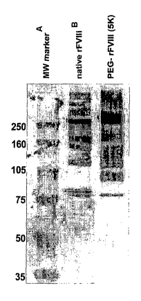
[017] Figure 1 shows the broadening and mass increase of rFVIII after
conjugation
with PEG measured by SDS-PAGE with subsequent immunoblotting.
[018] Figure 2 shows the pharmacokinetics of PEG-rFVIIi conjugate compared to
non-conjugated FVIII in hemophilic mice. Open squares: PEGrFVIII, dose 200 IU
FVIIi/kg. Closed diamonds: native rFVIII, dose 200 IU FVIII/kg.
[019] Figure 3 shows the detailed analysis of PEGylation sites by SDS-PAGE
using
various anti FVIII antibodies.
[020] Figure 4 shows the thrombin-induced activation and inactivation of
native and
PEGylated rFVIII.
[021] Figure 5 shows the bands demonstrating the domains of native and
PEGylated rFVIII.
CA 02647314 2008-09-24
WO 2007/126808 PCT/US2007/007560
[022] Figure 6 shows the extent of PEGylation of various domains of native and
PEGylated rFVIII.
[023] Figure 7 shows the thrombin inactivation rate of native and PEGylated
rFVIII.
DETAILED DESCRIPTION OF THE INVENTION
[024] The invention is a proteinaceous construct comprising an FVIII molecule
having at least a portion of the B domain intact, bound to a water-soluble
polymer
which is a polyalkylene oxide, polyvinyl pyrrolidone, polyvinyl alcohol,
polyoxazoline,
a poly acryloylmorpholine or a carbohydrate, such as polysialic acid (PSA). In
one
embodiment of the invention, the water soluble polymer is a polyethylene
glycol
molecule having a molecular weight of greater than 10,000 Daltons. The
construct
retains the full functional activity of standard therapeutic FVIII products,
and provides
an extended half-life in vivo, as compared to standard therapeutic FVIII
products.
[025] The starting material of the present invention is FVIII, which can be
derived
from human plasma, or produced by recombinant engineering techniques, as
described in patents US4757006; US5733873; US5198349; US5250421;
US5919766; EP 306 968.
(026] Herein, the term "Factor VIII" or "FVIII" refers to any FVIII molecule
which has
at least a portion of the B domain intact, and which exhibits biological
activity that is
associated with native FVIII. In one embodiment of the invention, the FVIII
molecule
is full-length Factor VIII. The FVIII molecule is a protein which is encoded
for by
DNA sequences capable of hybridizing to DNA encoding Factor VIII:C. Such a
protein may contain amino acid deletions at various sites between or within
the
domains A1-A2-B-A3-C1-C2 (US4868112). The FVIII molecule may also be an
CA 02647314 2008-09-24
WO 2007/126808 PCT/US2007/007560
6
analog of native FVIII wherein one or more amino acid residues have been
replaced
by site-directed mutagenesis.
[027] By example, an FVIII molecule can be PEGylated by a variety of chemical
methods (Roberts JM et al., Advan Drug Delivery Rev 2002;54:459-76). For
example, FVIII can be PEGylated by the conjugation of PEG to free SH groups
using
maleimide chemistry or the coupling of PEG hydrazides or PEG amines to
carbohydrate moieties of the FVI II after prior oxidation.
[028] In one embodiment of the invention, FVIII was modified via lysine
residues by
use of polyethylene glycol derivatives containing an active N-
hydroxysuccinimide
ester (NHS) such as Succinimidyl succinate, Succinimidyl glutarate or
Succinimidyl
propionate. These derivatives react with the lysine residues of FVIII under
mild
conditions by forming a stable amide bond. In one embodiment of the invention,
the
chain length of the PEG derivative is 5,000 Da. Other PEG derivatives with
chain
lengths of 500 to 2,000 Da, 2,000 to 5,000 Da, greater than 5,000 up to 10,000
Da or
greater than 10,000 up to 20,000 Da, or greater than 20,000 up to 150,000 Da
can
be used, including linear and branched structures.
[029] Alternative methods for the PEGylation of amino groups are the chemical
conjugation with PEG carbonates by forming urethane bonds, or the reaction
with
aldehydes or ketones by reductive amination forming secondary amide bonds.
[030] In the present invention an FVIII molecule is chemically modified using
PEG
derivatives that are commercially available. These PEG derivatives can have a
linear or branched structures. Examples of PEG-derivatives containing NHS
groups
are listed below.
CA 02647314 2008-09-24
WO 2007/126808 PCT/US2007/007560
7
[031] The following PEG derivatives are examples of those commercially
available
from Nektar Therapeutics (Huntsville, Al; see www.nektar.com/PEG reagent
catalog;
Nektar Advanced PEGylation, price list 2005-2006):
mPEG-Succinimidyi proplonate (mPEG-SPA)
9
mPEG-CH2CFI2-C-0--N
/
0
mPEG-Succinimidyi u-methylbutanoate (mPEG-SMB)
0
1
9
mPEG-CH2CH2~H-C-O-N
CH3 /
0
mPEG-CM-HBA-NHS (CM = carboxymethyl; HBA = Hydroxy butyric acid)
0
9 9
mPEG-CH2C-O-~HCH2C-0-N
CH3
0
Structure of a branched PEG-derivative (Nektar Therapeutics):
Branched PEG N-Hydroxysuccinimide (mPEG2-NHS)
mPEG q
>--C-O-
mPEG
O
CA 02647314 2008-09-24
WO 2007/126808 PCT/US2007/007560
8
[032] This reagent with branched structure is described in more detail by
Kozlowski
et al. (BioDrugs 2001;5:419-29).
[033] Other examples of PEG derivatives are commercially available from NOF
Corporation (Tokyo, Japan; see www.nof.co.jp/english: Catalogue 2005)
General Structure of linear PEG - derivatives (NOF Corp.):
CHgO(CH2CH2O)n X-
O
X = carboxymethyt
O
ii
CH3O(CH2CH2O)n CHZ-C-O-
O
X = carboxypentyl
CH3O(CH2CH2O)n (CH2)5-C-O-N
/
0
x = succinate
CA 02647314 2008-09-24
WO 2007/126808 PCT/US2007/007560
9
O
q R
CH3O(CH2CH2O)_C-CH2CH2-C-O-
mPEG Succinimidyl succinate
O
x = glutarate
0
II II
CH3O(CH2CH20)n C-(CH2)3 C-O-
O mPEG Succinimidyl glutarate
Structures of branched PEG-derivatives (NOF Corp.):
2,3-Bis(methylpolyoxyethylene-oxy)-1-(1,5-dioxo-5-succinimidyloxy,
pentyloxy) propane
H3C-(OCH2 CH2)n O-~HZ O
H3C-(OCH2 CH2)n O-~H ~ 9
CH2 O-C-CH2CH2CH2_C-O-
O
2,3-Bis(methylpolyoxyethylene-oxy)-1-(succinimidyl carboxypentyloxy) propane
H3C-(OCH2 CHZ)n O-~H2
H3C-(OCH2_CH2)ii_O-~H ~
CHj-0-CH2CH2CH2CH2CH2 C-O-
O
[034] These propane derivatives show a glycerol backbone with a 1,2
substitution
pattern. In the present invention branched PEG derivatives based on glycerol
CA 02647314 2008-09-24
WO 2007/126808 PCT/US2007/007560
structures with 1,3 substitution or other branched structures described in
US2003/0143596A1 can also be used.
[035] PEG derivatives with degradable (for example, hydrolysable linkers) as
described by Tsubery et al. (J Biol Chem 2004;279:38118-24) and Shechter et
al.
(W004089280A3) can also be used in the present invention.
[036] Surprisingly, the PEGylated FVIII of this invention exhibits full
functional
activity, combined with an extended FVIII half-life in vivo. In addition the
PEGylated
rFVIII seems to be more resistant against thrombin inactivation. This was
shown by a
variety of in vitro and in vivo methods, and is illustrated by the following
examples.
EXAMPLES
Example 1:
PEGylation of lysine residues in rFVIII with mPEG Succinimidyl succinate
[037] A solution of a rFVIII bulk derived from the Advate manufacturing
process
(3,400 U/rnl) was gel filtrated by use of Econo-Pac 10DG columns (Bio-Rad)
using
mM Hepes buffer, 150 mM NaCl, pH 7.4, containing 0.5% sucrose and 0.1%
Polysorbate 80. Then mPEG Succinimidyl succinate (Abuchowski et al. Cancer
Biochim Biophys 1984;7:175-86) with a chain length of 5,000 Da (PEG-SS 5000)
was added to this solution under gentle stirring (5 mg PEG-SS / mg protein)
and the
pH value was adjusted to 7.4 by drop wise addition of 0.5 M NaOH. Then the
PEGylation was carried out under gentle stirring for 1 hour at room
temperature.
[038] Subsequently the reaction mixture was applied onto an equilibrated ion-
exchange chromatography resin (Fractogel EMD TMAE 650M / Pharmacia XK-10
column, bed height: 15.0 cm) in 20 mM Hepes buffer, 150 mM NaCI, pH 7.4,
containing 0.5% sucrose and 0.1% Polysorbate 80. Thep the column was washed
CA 02647314 2008-09-24
WO 2007/126808 PCT/US2007/007560
11
with 20 CV equilibration buffer to remove excess reagent and the PEGylated
rFVIII
was eluted with elution buffer (20 mM Hepes, 1.0 M NaCI, 0.5% sucrose, 0.1%
Polysorbate 80, pH 7.4). The eluate was concentrated by
ultrafiltration/diafiltration
with a membrane consisting of regenerated cellulose and with a moiecular
weight
cut-off of 30 kD using a buffer system consisting of 20 mM Hepes, 150 mM NaCI,
0.5% sucrose, pH 7.4.
Example 2:
Biochemical characterization of PEGyiated rFVIII in vitro
[039] RFVIII derived from the Advate manufacturing process was PEGylated
according to Example 1 and the PEGylated FVIII product was biochemically
characterized. The functional activity of the PEG-rFVIII was determined by use
of the
FVIII chromogenic assay (Rosen S, Scand J Haematol 1984;33 (Suppl 40):139-45).
The method is based on Ph. Eur. 5th edition (5.05) 2.7.4 Assay of Blood
Coagulation Factor VIII.
[040] A sample, containing factor VIII (FVIII:C) is mixed with thrombin,
activated
factor IX (FIXa), phospholipids and factor X (FX) in a buffer containing
calcium. FVIII
is activated by thrombin and subsequently forms a complex with phospholipids,
FIXa
and calcium ions. This complex activates factor X to factor Xa, which in turn
cleaves
the chromogenic substrate FXa-1 (AcOH*CH30CO-D-CHA-Gly-Arg-pNA). The time
course of para-nitroaniline (pNA) released is measured with a micro plate
reader at
405 nm. The slope of the reaction is proportional to the factor VIII
concentration in
the sample. The FVIII antigen value was measured by use of an ELISA system
commercially available (Cedariane, Homby, Ontario, Canada) with minor
modifications. From these values the ratios FVIII chrornogen/FVIII antigen
were
calculated. The protein content in the preparations was determined by
measuring
the optical density at 280nm. From these data the protein content was
calculated
CA 02647314 2008-09-24
WO 2007/126808 PCT/US2007/007560
12
(Hoyer LW in: Human Protein Data. Installments 1-6; Heberli Ed.; Wiley VCH,
Weinheim, Germany, 1998) and expressed in mg/ml.
Table 1
PEG-rFVIII
Native rFVIII PEG-SS 5K
mg er mg rotein
FVIII:Chr activity 3,430 64
U/ml
FVI11:Ag 4,067 81
U/ml
Ratio 0.84 0.79
FV111:Chr/FV111:A
Recovery of 100 94
biological activit %
[041] The data in Table 1 shows that in the PEGylated rFVIII preparation, the
biological activity (expressed by the ratio FVIII chromogenic activity to
FVIII antigen)
is recovered to more than 90% in comparison to the biological activity of the
native
rFVlli (100%).
Example 3:
Characterization of PEGylated rFVIII by SDS-PAGE and immunoblottinQ techniques
[042] Native rFVIII was characterized by SDS PAGE under reducing conditions by
using a 4-12% polyacrylamide gradient gel obtained from Invitrogen (Carlsbad,
California, USA) according to the instructions of the manufacturer. As
molecular
weight markers (MW) Precision Plus markers (10 kD - 250 kD) obtained from Bio-
Rad (Hercules, CA, USA) were used. Then the proteins were transferred on a
PVDF
membrane obtained from Bio-Rad (Hercules, CA, USA) by electroblotting and
subsequently incubated with a.polyclonal sheep anti human FVIII:C antibody
obtained from Cedarlane (Hornby, Ontario, Canada). The last steps of the
immunostaining procedure were the incubation with an alkaline phosphatase
(ALP)
CA 02647314 2008-09-24
WO 2007/126808 PCT/US2007/007560
13
conjugated anti-sheep antibody obtained from Accurate (Westbury, NY, USA)
followed by the final visualization by use of an ALP substrate kit (Bio-Rad,
Hercules,
CA, USA). The results are summarized in Figure 1. The blot demonstrates the
domain structure of native and PEGylated rFVI11. It is shown that the
PEGylated
rFVIII has broader bands and high molecular masses than the native recombinant
protein.
Example 4:
Pharmacokinetics of PEGylated rFVIII in a FVIII deficient knock out mouse
model
[043] FVIII deficient mice described in detail by Bi et al. (Nat Genet
1995;10:119-21)
were used as a model of severe human hemophilia A. Groups of 5 mice received a
bolus injection (10 mI/kg) via the tail vein with either PEG-rFVII1 (PEG-SS,
5K)
prepared according to Example I or native rFVIII in a dose of 200 IU FVIII/kg
bodyweight. Citrate plasma by heart puncture after anesthesia was prepared
from
the respective groups, 5 minutes, 3, 6, 9 and 24 hours after injection. FVIII
activity
levels were measured in plasma samples. The results of this experiment are
summarized in Figure 2. Mean half life increased from 1.9 hours (for native
rFVIII) to
4.9 hours (for PEGylated rFVlll), area under curve (AUC) increased from 13.0
to
25.2 hours*IU/ml. Half-life calculation was performed with MicroMath
Scientist,
model 1 from pharmacokinetic library (MicroMath, Saint Louis, MO, USA).
Example 5:
Detailed analysis of PEGylation of rFVIII by SDS-PAGE and immunoblotting
techniques
[044] Native and PEGylated rFV111 was digested with I nM thrombin for 60
minutes
at 60 C, which resulted in specific cleavage of the FVIII molecule with well
defined
degradation products. These heavy- and light chain fragments were separated by
SDS-PAGE followed by electroblotting, as described in Example 3. To visualize
the
CA 02647314 2008-09-24
WO 2007/126808 PCT/US2007/007560
14
cleaved fragments, a polyclonal antibody and monoclonal antibodies against the
heavy chain Al and A2 domains, the B domain and the light chain N-terminal A3
domain were applied.
[045] As seen in Figure 3 all domains were PEGylated, albeit to a different
extent.
The B domain was strongly PEGylated. Both the Al and A2 domains of the heavy
chain were partially PEGylated. Various PEGylation-degrees (mono-, di-, tri-
...)
could be observed in the light chain A3-dornain. In agreement with Example 6,
the
PEGylated FVIII seemed to be more resistant to thrombin.
Example 6:
Thrombin-resistancy of PEGylated rFVIII
[046] In vitro thrombin treatment of FVIII results in a rapid increase and
subsequent
decrease in its procoagulant activity. The rate of activation and
inactivation, which
depends on the thrombin concentration and on the integrity of FVIII, was
monitored
by a FIXa cofactor assay, as follows:
[047] FVIII was incubated at 37 C with 0.5 or 1 nM thrombin. Subsamples were
withdrawn at time intervals between 0_5 to 40 minutes and added to a mixture
of
FIXa, FX, PL-vesicles and CaCI2 also containing a specific thrombin inhibitor
to stop
the further thrombin-mediated reactions and incubated for 3 minutes_ A
subsample
was added to a chromogenic substrate, which is selectively cleaved by Fxa and
contained EDTA to stop further Xa activation. After a 15 min incubation, the
reaction
was terminated by acetic acid. The absorbance (A405) values, which are
proportional to the Fxa concentrations, were measured in an ELISA reader and
converted to Fxa concentrations using a purified Fxa reference curve. The
generated Fxa concentrations were plotted against the incubation time with
thrombin.
CA 02647314 2008-09-24
WO 2007/126808 PCT/US2007/007560
[048] Pseudo-first order inactivation rate * of FVIII was determined by
fitting the
declining part of the curves with a single exponential fit.
Table 2
First order inactivation
Rate Relative k'
k' 1/min
hrombin Native FVIII PEG-FVIII PEG/native
0.5 nM 0.14 0.08 0.57
1 nM 0.24 0.14 0.58
[049] As shown in Figure 4 and Table 2, PEGylated rFVIII showed a slower
inactivation rate at both applied thrombin concentrations.
Example 7-
PEGylation of lysine residues in rFVIII with branched 2,3-
Bis(methylpolyoxyethylene-
oxy)-1-(1,5-dioxo-5-succinimidyloxy. pentyloxy) propane
[050] A solution of rFVIII in 20 mM Hepes buffer pH 7.4 containing 150 mM
NaCI,
0.5% sucrose and 0.1% Polysorbate 80 was prepared from bulk material derived
from the Advate manufacturing process containing 489 IU FVIII / ml. A branched
PEG succinimidyl glutarate (PEG-SG) reagent (2,3-Bis(methylpolyoxyethylene-
oxy)-
1-(1,5-dioxo-5-succinimidyloxy, pentyloxy) propane) obtained from NOF
Corporation
(Tokyo, Japan) with a molecular weight of 20 kD was added to 153 ml of this
solution
under gentle stirring (5 mg reagent / mg protein) and the pH. value was
adjusted to
7.4 by drop wise addition of 0.5 M NaOH after 10 minutes. Then the PEGylation
of
rFVlli was performed under gentle stirring for 1 hour at room temperature.
CA 02647314 2008-09-24
WO 2007/126808 PCT/US2007/007560
16
[051] Subsequently the reaction mixture was applied onto an equilibrated ion-
exchange chromatography resin (Fractogel EMD TMAE 650M / Pharmacia XK-50
column, bed height: 14.5 cm) in 20 mM Hepes buffer, 150 mM NaCI, pH 7.4,
containing 0.5% sucrose and 0.1% Polysorbate 80 using a linear flow rate of 1
cm/min. The column was washed with 25 CV equilibration buffer to remove excess
reagent (linear flow rate: 2 cm/min) and the PEGylated rFVIII was eluted with
elution
buffer (20 mM Hepes, 1.0 M NaCI, 0.5% sucrose, 0.1 % Polysorbate 80, pH 7.4)
at a
linear flow rate of 0.5 cm/min. Then the eluate was concentrated by
ultrafiltration/diafiltration with a membrane consisting of regenerated
cellulose and
with a molecular weight cut-off of 30 kD using a buffer system consisting of
20 mM
Hepes, 150 mM NaCI, 0.5% sucrose, pH 7.4.
Example 8:
In-vitro characterization of rFVIIi PEGvlated with branched PEG-SG 20kD
[052] RFVIII derived from the Advate manufacturing process was PEGylated via
lysine residues using a branched PEG-SG reagent according to Example 7 and the
PEGylated rFVIII product was biochemically characterized as described in
Example
2.
Table 3
PEG-rFVIII
Native rFVIII PEG-SG 20K
mg per mg protein)
FVIII:Chr activity 9,950 1,040
U/ml
FVIII:Ag 20,807 1,763
U/ml
Ratio 0.48 0.59
FVIII:Chr/FVIII:A
Recovery of 100 120
biological activity %
CA 02647314 2008-09-24
WO 2007/126808 PCT/US2007/007560
17
[053] The data in Table 3 show that in the PEGylated rFVIII preparation the
biological activity (expressed by the ratio FVIII chromogenic activity to
FVIII antigen)
completely recovered in comparison to the biological activity of the native
rFVIII
(100%).
[054] The PEGylated rFVIII was characterized by SDS-PAGE and immunoblotting
techniques under reducing conditions using a 4-12% polyacrylamide gradient gel
as
described in Example 3. The results are summarized in Figure 5. The blot
demonstrates the domain structure of native and PEGylated rFVIII. It is shown
that
the PEGylated rFVIII has broader bands and high molecular masses than the
native
recombinant protein.
[055] For more detailed analysis of PEGylation of the rFVlii preparation by
SDS-
PAGE and immunoblotting techniques, the native and PEGylated rFVIII was
digested with I nM thrombin for 60 minutes at 60 , which resulted in specific
cleavage of the FVIII molecule with well defined degradation products, as
described
in Example 5. The fragments were separated by SDS-PAGE followed by
electroblotting and visualized by different anti-FVIII antibodies. As seen in
Figure 6,
all domains were PEGylated, albeit to a different extent. The B domain was
strongly
PEGylated. Various PEGylation-degrees (mono-, di-, tri-PEGylation) could be
observed in the light chain A3-domain. The results indicate that the PEGylated
rFVlli seemed to be more resistant to thrombin.
[056] The rate of activation and inactivation by thrombin was monitored by a
FIXa
cofactor assay as described in Example 6. Pseudo-first order inactivation rate
of
FVIII was determined by fitting the declining part of the curves with a single
exponential fit.
CA 02647314 2008-09-24
WO 2007/126808 PCT/US2007/007560
18
Table 4
First order inactivation
Rate Relative k'
k' 1/min
hrombin Native FV111 PEG-FVIII PEG/native
0.5 nM 0.13 0.09 0.67
1 nM 0.21 0.15 0.71
[057] As shown in Figure 7 and Table 4, the PEGylated rFVllt showed a slower
inactivation rate at both applied thrombin concentrations.