Note: Descriptions are shown in the official language in which they were submitted.
CA 02648030 2013-12-20
COMPOSITIONS AND METHODS COMPRISING THE USE OF CELL SURFACE
DISPLAYED HOMING ENDONUCLEASES
SEQUENCE LISTING
This description contains a sequence listing in electronic form in ASCII text
format.
A copy of the sequence listing in electronic form is available from the
Canadian Intellectual
Property Office.
STATEMENT REGARDING FEDERALLY-SPONSORED RESEARCH
This invention was made with United States Government support under grant
number
R21A1064581 awarded by the National Institutes of Health. The United States
Government
has certain rights in the invention.
FIELD OF THE INVENTION
Aspects of the present invention relate generally to novel site-specific DNA
cutting
enzymes, and more particularly to homing endonucleases (HE) (e.g., LAGLIDAG,
HNH, His-
Cys Box, GIY-YIG, I-SspI-type) with novel or altered DNA binding and/or
cutting
specificities, to novel methods of generation, selection and isolation of same
comprising the
use of cell-surface HE display, to novel compositions (e.g., HEs, HE-encoding
nucleic acids,
cells, cell libratires, etc.) and novel uses comprising same including for
example, generation
of targeted double-strand breaks in target viral or cellular genomes, and
specific chromatin
immunoprecipitation.
BACKGROUND
Homing. 'Homing' is a widespread process involving the transfer of an
intervening
sequence (e.g., introns (e.g., group I or group II introns) or inteins) to a
homologous allele that
lacks the sequence, leading to gene conversion and dominant transmission and
inheritance of
the mobile element. Intervening sequences capable of homing are found in all
1
CA 02648030 2008-09-29
WO 2007/123636
PCT/US2007/007637
brances of life (e.g., phage, Eubacteria, Archaea, and eukaryotes), and within
eukaryotes for
example are found within nuclear, mitochondrial and chloroplast genomes.
Homing is
initiated by an endonuclease (homing endonuclease; HE), encoded within the
intervening
sequence or intein, which recognizes a DNA target site and generates a single-
or double-
strand break. HEs are normally expressed in the cytosol and targeted to DNA-
containing
organelles posttranslationally.
Group I and group II introns are distinguished based on their respective
transfer
mechanisms. Transfer of group I introns is completed by cellular mechanisms
that repair the
stand breaks via homologous recombination. Homing of group II introns involves
a more
complex process comprising strand cleavage, reverse splicing to generate a DNA-
RNA
hybrid intermediate, and reverse transcription using the inserted RNA as
template, where the
sequential activities are encoded by within the intron on a single
multifunctional polypeptide
chain. The homing mechanism of inteins is similar to that of group I introns,
but the system
comprises functional fusion (in-frame) of the endonuclease with the intein
host to provide a
polypeptide chain harboring activities of the homing endonuclease, the intein
peptide ligase
and the host protein, and wherein the portions of the intein's surface
participate in DNA
recognition and binding by the endonuclease. In all cases, the homing
endoculease gene is
duplicated into the target site (e.g., non-disruptive sites such as introns
and inteins, etc.).
Homing endonucleases and classes thereof Homing endonucleases are highly
specific DNA cutting enzymes and recognize DNA target sites ranging from about
14 to
about 40 base pairs. While being highly specific to promote precise transfer
of introns or
inteins and avoid genomic toxity, the homing endoculeases must retain
sufficient site
recognition flexibility (sufficient infidelity) to permit lateral transfer in
the face of sequence
variation in diverging targets and host. There are five known families of
homing
endonucleases (LAGLIDAG, HNH, His-Cys Box, GIY-YIG and I-Sspl-type) that
differ in
their conserved nuclease active-site core motifs and catalytic mechanism.
LAGILDADG homing endonucleases (LHE) are the largest family of homing
endonucleases, and are typically encoded within introns (as free-standing
enzymes) or inteins
(as in-frame fusion proteins) of mintochondrial or chloroplast genomes in
single-cell
2
CA 02648030 2008-09-29
WO 2007/123636
PCT/US2007/007637
eukaryotes (e.g., yeast). LAGILDADG homing endonucleases were first defined in
the early
1990s with the discovery that the "homing" property of a mobile intron to
intron-less alleles
of S. Cerevesiae involved the induction of a specific double-strand break in
intronless alleles
of the gene, the break being generated by a nuclease protein encoded by the
mobile intron.
The created double-strand break catalyzes homologous recombination between the
intron
containing and non-containing alleles, resulting in the copying of the intron
into the intron-
less allele. The intron-encoded protein, I-SceI, and related proteins, were
subsequently
designated as "homing" endonucleases. Because of a recognizable motif present
in two
central alpha helixes of I-SceI, this homing endonuclease family, including I-
SceI, became
known as the LAGLIDADG homing endonuclease (LHE) family. LHE proteins are
formed
as homodimers or pseudosymmetric monomers that generally recognize DNA
sequences 18-
24 base-pairs in length (Chevalier & Stoddard, Nucleic Acids Res, 29:3757-
3774, 2001).
Homodimers recognize consensus DNA targets that are constrained to paladromic
or near
palindromic symmetry, whereas monomeric enzymes having two copies of the
consensus
LAGLIDADG motif posses a pair of structurally similar nuclease domains on a
single
polypeptide chain, and are not constrained to symmetric DNA targets.
Generally, the
molecular structures are built around two conserved alpha-helices that contain
the
LAGLIDADG motif, and which forms the center of the interface between enzyme
subunits or
domains as the case may be (Heath, et al., Nat Struct Biol, 4:468-476, 1997).
The final acidic
residues from the central alpha helix helices form part of each domain's
active site that
cleaves one strand of the double-stranded DNA target sequence. The DNA binding
interface
of each domain is made up of a four-stranded antiparallel beta-sheet that is
supported by a
series of framework alpha-helices which form the core of the domain. Unlike
art-recognized
'restriction endonucleases,' which form densely packed and almost completely
saturated
DNA-protein interfaces, the DNA binding interface of LHEs make fewer hydrogen
bonds per
target sequence base pair (Galburt & Stoddard, Biochemistry, 41:13851-13860,
2002). These
structural properties account for the ability of LHEs to withstand moderate
variability in
target sequence recognition (e.g., see Jurica, et al., Mol Cell, 2:469-476,
1998; Chevalier, et
al., J Mol Biol, 329:253-26, 2003; Moure, et al., J Mol Biol, 334:685-695,
2003; and Moure,
3
CA 02648030 2008-09-29
WO 2007/123636
PCT/US2007/007637
et at., Nat Struct Biol, 9:764-77, 2002), a characteristic that has been
essential in maintaining
their genetic mobility and horizontal proliferation (Burt & Koufopanou, Curr
Opin Genet
Dev, 14:609-615, 2004) and which make LHEs ideal substrates for engineering
altered DNA
binding interfaces with novel endonucleolytic specificities (Duan, et al.,
89:555-56, 1997;
Chevalier, et al., Mol Cell, 10:895-905, 2002; Epinat, et at., Nucleic Acids
Res, 31:2952-
2962, 2003 ; Arnould, et al., J Mol Biol, 355:443-458, 2006; and Steuer, et
al., Chembiochem,
5:206-213, 2004). The combination of high target sequence specificity and
adaptable DNA
binding interfaces make LHEs attractive tools for genome engineering
applications which
require the introduction of a double-stranded break at a precise genomic
location (Steuer, et
al., Chembiochem, 5:206-213, 2004; Storici, F., Durham, et al., Proc Nat! Acad
Sci U S A,
100:14994-14999, 2003; Tzfira, et al., Plant Physiol, 33:1011-1023, 2003; and
Miller, et al.,
Mol Cell Biol, 23:3550-3557, 2003). DNA binding by intein-associated LHEs
(e.g., PI-SceI)
involves recruitment of adjacent protein domains (adjacent intein domains).
For example, the
PI-SceI endonuclease intein combination binds a 31 bp site, and the majority
of the energetic
contribution to binding is derived from interactions with the intein peptide
splicing domain;
the endonuclease domain contains the active sites, but exhibits relatively
weak, non-specific
DNA binding.
Despite little primary sequence homology among the LHEs outside of the
LAGLIDADG motif itself, the topologies among the endonuclease domains and the
shape of
their DNA-bound J3-sheets, are remarkably similar, and the structure of the
central core of 13-
sheets is well conserved. These positions correspond to residues that make
contacts to base
pairs in each DNA half-site. Alignments of intein-associated endonuclease
domains indicate
a somewhat more diverged structure of the ft-sheet motifs. In particular
instances, the core
fold of LAGLIDADG enzymes can be tethered to additional functional domains
(e.g.,
NUMODS; nuclear associated modular DNA binding domains) involved in DNA
binding.
Like most nucleases, LHEs require divalent cations for activity. Two metals
(calcium
and copper) fail to support cleavage, two (nickel and zinc) display reduced
cleavage, and
three (magnesium, cobalt and manganese) display full activity under all tested
conditions.
4
CA 02648030 2008-09-29
WO 2007/123636
PCT/US2007/007637
The use of manganese in place of magnesium allows recognition and cleavage of
a broader
repertoire of DNA target sequences than observed with magnesium.
The HNH and His-Cys box homing endonucelases appear to be derived from a
common ancestor built around a consensus nuclease active site architecture
known as a
metal' motif. The HNH homing endonuclease family if generally found in page
introns,= and
possess a long monmeric extended, modular monomeric structure, in which the
relatively
non-specific nuclease domain at the N-terminus is tethered to additional
structural motifs that
confer and restrict DNA binding specificity. Prototypical members (e.g., I-
HmuI) recognize
asymmetric DNA sites of about 24 bp or longer. In contrast, the His-Cys box
homing
to endonucelases are generally encoded in nucleolar introns within rDNA
host genes, have
compact homodimeric structures, recognize shorter symmetric DNA target sites
with higher
overall homing in a manner similar to the LHE systems.
The GIY-YIG endonuclease family members are also encoded within phage introns
and possess modular structures similar to the HNH endocleases. The GIY-YIG
endonuclease
catalytic domain is quite non-specific in its inherent cleavage activity,
again (as for the HNH
family) being restricted to target sites that are dictated by the appended DNA-
binding
modules.
The fifth family, represented by the prototypical enzyme I-SspI found in
Synechocystis, is responsible for the presence and persistence of introns in
cyanobacterial
tRNA genes. I-SspI displays limited homology to known nuclease superfamilies,
and is
currently represented by only a limited number of indentified open reading
frames.
Molecular biology and genome engineering applications. Because of their
relatively
long recognition sequences, homing endonucleases (e.g., LHEs) induce a very
low frequency
of cleavage, even in large vertebrate genomes, and homing endonucleases are
therefore
regarded as having possible utility as rare-cutter endonucleases for use in
molecular biology
and genome engineering applications, particularly those applications which
mimic their well
known natural function of catalyzing homologous recombination via induction of
a DNA
double strand break, such as those related to targeted recombination, gene
repair and gene
conversion.
5
CA 02648030 2008-09-29
WO 2007/123636
PCT/US2007/007637
Engineering and directed evolution of alternative systems. Some efforts have
been
directed to tethering non-specific nuclease domains to sequence-specific DNA
binding
modules such as zinc fingers (resulting in so called zinc finger nucleases, or
ZFNs) for in vivo
use in stimulating homologous recombination (Bilikova et al., 2001, 2003) and
to drive
sequence correction of a disease-causing allele associated with a severe
genetic disorder
(Urnow et al., 2005). However, despite the ease of designing such highly
specific ZFN
reagents, comparison of their properties to those of homing endonucleases
indicates that both
are worthy of development. For example, the nuclease domains of ZFN constructs
appear to
display significant non-specific DNA nicking and cleaving activity in the
engineered
chimeras, and these constructs can generate multiple adjacent phosphate
cleavage events
within a single bound DNA target site, which may enhance non-conservative
break repair
outcomes. By contrast, LHE cleavage is tightly coupled to cognate site
binding, and the
enzyme action, by virtue of tight product binding properties, appears to
strongly enhance the
ratio of homologous recombination relative to undersirable, non-conservative
double-strand
break repair events such as non-homologou end-joining. Additionally, ZFN
chimeras have
the disadvantage that they require expression of two separate chains to
generate double-strand
breaks, and more total coding sequence to generate the acive enzyme. Efforts
have been
made to increase or alter the specificity of type II restriction
endonucleases, but have been
generally unsuccessful. Group II homing endonucleases are promising for
targeted gene
disruptions because they are easily engineered for novel specificities by
altering the cognate
intron sequences (DNA specificity being dictated by base pairing with the RNA
component
of the intron-protein complex, rather than by only the protein contacts to
DNA). However,
these systems are more appropriate for gene disruption by insertion of a
mobile element than
for gene conversion, and require the presence of packaging of significant
amounts of genetic
information, including a large multifunctional reading frame (RT, endonuclease
and
maturase) and the cognate intron sequence for the generation of reactive RNP
for reverse
splicing and gene insertion.
Engineering and directed evolution of homing endonucleases. One strategy in
the art
to alter homing endonuclease specificity for intein-associated enzymes has
been to exchange
6
CA 02648030 2008-09-29
WO 2007/123636
PCT/US2007/007637
entire intein-binding domains or portions thereof. Experiments of this type
have shown, for
example, that the PI-SceI protein splicing domain can be used as a site-
specific DNA-binding
module in chimeric protein constructs (domain swapping between the PI-SceI and
a homolog
from Candida tropicalis (PI-CtrIP) was constructed) (Steuer et al, 2004):
Additionally, several studies have demonstrated that domains from unrelated
free-
standing LAGLIDADG enzymes can be structurally fused to create fully active,
chimeric
homing endonucleases that recognize corresponding chimeric target sites
(Chevalier et al.,
2002, Epinat et al., 2003; Steurer et al., 2004). For example, using
computational redesign,
an artificial highly specific chimeric endonuclease H-DreI was generated by
fusing domains
of homing endonucleases I-DmoI and I-CreI. H-DreI binds a long chimeric DNA
target site
with nanomolar affinity. A related experiment showed that a single-chain
monomeric
endonuclease can be generated from a homodimer predecessor by generating a
fusion of
genes that encoded each subunit connected with an artificial linker (Epinat et
al., 2003).
Specifically, a linker from I-DmoI was used to join two copies of the I-CreI
gene to generate
a pseudo-symmetric single-chain enzyme that cleaves DNA with the same
specificity as
native I-CreI, and was shown to initiate homologous recombination in both
yeast and
mammalian cells.
Moreover, the role and mutability of interfacial residues between LAGLIDADG
helices has been examined by grafting side-chains from the homodimeric I-CreI
into the
corresponding positions in the monomeric I-DmoI enzyme resulting in enzymes
with novel
nicking activities and oligomeric properties (Silva & Belfort, 2004).
Additionally, several methods have been used to alter homing endonuclease
specificity primarily at the level of individual base-pair alterations in the
cognate target site,
and these methods are divided into (i) those select or screen for DNA binding
activity, and
(ii) those that select or screen for cleavage. For example, an adaptation of a
bacterial two-
hybrid strategy was used to select for variants of the intein-encoded PT-See!
endonuclease
(Gimble et al., 2003), and the selected DNA binding specificities ranged from
relaxed
(cleaves WT and mutant targets equally) to being dramatically shifted to
preferring the
7
CA 02648030 2013-12-20
selection targets, but none of the variants displayed the same degree of
specificity as WT PI-
Seel.
A strategy for isolating I-CreI derivative with increased affinities for
altered target
sites has been described (Seligman et al., 2002); Sussman et al., 2004).
Endonuclease mutants
with single amino acid substitutions at positions predicted to make base-
specific DNA
contacts were assayed against DNA target site mutants in an E. coli based
system where
cleavage of target sites results in cell being converted from lack to Lac-,
and where
undesirable activity (cleavage of original WT site) can be suppressed through
a secondary
'negative screen for elimination of an essential reporter (e.g., antibiotic
resistance marker).
Using these methods, enzyme variants with shifted, rather than completely
altered specificity
proteins were obtained (see also Gruen et al., 2002).
Finally, an assay system designed to report on the generation of double-strand
break
induced homologous recombination in eukaryotic cells has been described (Perez
et al 2005;
see also US 2006/0206949 and US2006/0153826 to Arnould et al).
However, such prior art based screening methods whether based on domain
swapping,
domain fusion, enzyme fusion, grafting of side-chains, base-pair alterations
in the cognate
target site (whether based on selecting or screening for DNA binding activity,
or selecting or
screening for cleavage activity) are fundamentally limited or compromised in
their screening
throughput by the fact that they require the generation of combinatorial
endonuclease mutant
libraries and the variant endonucleases must be well tolerated by the host's
genomic DNA;
that is, these prior art methods all require intracellular expression of the
generated homing
endonuclease during the screening or selection, and thereby preclude the
effective expression,
selection and identification of any variant endonuclase specificities
associated with genomic
toxicity (e.g., those that cut in and mediate alteration of essential genomic
positions). An
additional limitation of the prior art is that the intracellular cleavage
system must be
redesigned and generated for each sequence targeted for selection.
Furthermore, while `phage display' methods (Chames, et al., Nucleic Acids
Research
33:e178, pages 1-10), 2005) have been described for selecting variants of a
homodimeric I-
8
CA 02648030 2008-09-29
WO 2007/123636
PCT/US2007/007637
Crel enzyme, this system has several fundamental disadvantages. First, such
phage display
systems have not been demonstrated to provide for display of a single-chain
monomeric I-
CreI enzyme form, most likely because expression of an active single chain
monomeric I-
CreI is either toxic to the host bacteria (e.g., using bacterial hosts, phage
display of a whole
monomeric enzyme would not segregate the active enzyme from the bacterial host
cell DNA,
as bacteria do not have a sequestered protein secretion pathway), or is
disruptive of phage
assembly (presumably, the use of a monomer of the homodimeric form generates
an inactive
fusion protein inside the cell, which would avoid toxicity, and/or was small
enough to allow
for phage assembly). In any case, no full-length single-chain monomeric active
HEs or LHEs
have been surface displayed using phage display, or any other type of display
including cell
surface display. Moreover, additional disadvantages of phage display systems
are that phage
are relatively small (e.g., compared to cells), and are too small to sort by
some methods.
Furthermore, in many instances it may not be possible to phage display enough
molecules to
achieve an adequate signal strength (e.g., depending on the protein, there may
be only a few
molecules per phage), so separation methods are limited to those comprising
matrix/panning
approaches, which substantially limits utility screening throughput.
Pronounced need in the art. There is, therefore, a pronounced need in the art
for
novel site-specific DNA binding and cutting enzymes, and more particularly for
novel
homing endonucleases (HE) with novel DNA binding and cutting specificities,
for novel
methods of generation, selection and isolation of same, for novel compositions
and uses
comprising same, and for novel nucleic acid molecules encoding same. There is
a
pronounced need for novel LHE with novel DNA binding and cutting
specificities, for novel
methods of generation, selection and isolation of same, for novel compositions
and uses
comprising same, and for novel nucleic acid molecules encoding same. There is
a
pronounced need for methods of variant homing endonculease expression,
selection,
screening and identification that are not limited to intracellular expression
of the generated
homing endonuclease during the screening or selection to allow for generation
and
identification of a more diverse set of homing endonuclease binding and
cleavage
specificities.
9
CA 02648030 2014-11-17
,
CA 2648030
SUMMARY
According to particular aspects disclosed herein, DNA target site binding and
cleavage
properties of native homing endonucleases (HE) in solution are recapitulated
on the cell surface
(e.g., as assessed by flow cytometric analysis of both the binding and
cleavage of fluorescently
conjugated double-stranded oligonucleotides (dsOligos)) to provide for novel
cells expressing
one or more cell surface HEs (e.g., expressing one or more HE binding and/or
cleavage
specificities), novel libraries of such cells, and high-throughput methods for
assessing target
site binding, target site cleavage. Additionally, the rapid analysis of HE or
LHE-DNA
interactions on the cell surface with concurrent sorting options provides for
high-throughput
library screening affording rapid identification, analysis and isolation of
novel HEs or LHEs
having novel sequence specificities. Such novel sequence specificities,
obtained by said
methods provide a novel method of introducing a DNA-strand cleavage event in a
target cell.
Particular aspects disclosed herein provide novel methods for the cell surface
display of
functional homing endonucleases (HE) (e.g., LAGLIDAG, HNH, His-Cys Box, GIY-
YIG and
I-SspI-type) or of variants, muteins or derivatives thereof. In particular
aspects, one or more
LAGILDADG homing endonucleases (LHEs), or variants, muteins or derivatives
thereof, are
expressed as membrane-anchored recombinant proteins and thereby functionally
displayed on
the surface of the expressing cells (e.g., expressed on the plasma membrane of
lymphocyte cell
lines by targeting the expression of an LHE-CD80 fusion protein to the
secretory pathway). In
particular embodiments, only a single HE or LHE is expressed and displayed on
a given cell.
In alternate embodiments, a plurality of HEs or LHEs are expressed and
displayed on a given
cell. In particular embodiments, novel cells (e.g., eukaryotic cells,
vertebrate, mammalian or
other metaozoan cells, yeast or other unicellular eukaryotic cells, bacterial
or other prokaryotic
cells etc. expressing such cell surface displayed HEs or LHEs are provided.
Additional aspects provide novel cell-based libraries of such cell surface-
displayed HEs
or LHEs, or of variants, muteins or derivatives thereof. The cells of such
libraries may
CA 02648030 2008-09-29
WO 2007/123636
PCT/US2007/007637
express a single cell surface-displayed HE or LHE or variant, mutein or
derivative thereof, or =
may express a plurality of cell surface-displayed HEs or LHEs, or of variants,
muteins or
derivatives thereof.
Yet additional aspects provide novel methods for assessing HE or LHE target
site
binding (e.g., DNA binding) or variant target site binding, comprising
assessing the target site
binding properties of one or more cell surface displayed functional homing
endonucleases, or
of variants, muteins or derivatives thereof. In particular embodiments, cells
expressing one
or more surface. LHEs are stained with fluorescently conjugated double-
stranded
oligonucleotides (dsOligos) containing respective and/or prospective LHE
target sequences
(or variant target sequences), to provide for analysis of their DNA binding by
flow cytometry.
In certain embodiments, the detected signal is highly or completely sequence
specific and
relatively undetectable or undetectable with dsOligos carrying one or more
base substitutions
(e.g., carrying a single base pair substitution). In particular embodiments,
cell surface
binding of the target sequence or variant target sequence to HE or LHE-
expressing cells is
affected under conditions precluding cleavage of the target sequence or
variant target
sequence.
Further aspects provide novel methods for assessing HE or LHE target site
cleavage
(e.g., DNA binding and cleavage) or variant target site cleavage, comprising
assessing the
target site cleaving properties of one or more cell surface displayed
functional homing
endonucleases, or of variants, muteins or derivatives thereof. In particular
embodiments,
cells expressing one or more surface LHEs are stained with fluorescently
conjugated double-
stranded oligonucleotides (dsOligos) containing respective and/or prospective
appropriately
labeled (e.g., unique fluorophores at opposite termini) LHE target sequences
or variant target
sequences, to provide for analysis of their DNA cleavage (e.g., DNA binding
and cleavage)
by flow cytometry. In certain embodiments, the detected signal is highly or
completely
sequence specific and relatively undetectable or undetectable with dsOligos
carrying one or
more base substitutions (e.g., carrying a single base pair substitution). In
particular
embodiments, the HE or LHE target site cleavage assays comprise cell surface
tethering of
the appropriately labeled target sequence or variant target sequence prior to
cleavage. In
11
CA 02648030 2008-09-29
WO 2007/123636
PCT/US2007/007637
particular embodiments, binding and cleavage assays are uncoupled by affecting
cell surface
binding or tethering of the target sequence or variant target sequence to HE
or LHE-
expressing cells under conditions precluding cleavage, and subsequently and
optionally
adjusting the conditions to support cleavage, and assaying for such cleavage
if present.
Additional aspects comprise novel HE or LHE target nucleic acids comprising
unique
fluorophores at opposite termini, and the use thereof in flow cytometry-based
cleavage
assays.
Additional aspects provide methods, comprising the use of sequence specific
cell
surface displayed HE or LHE interactions with dsOligos under conditions which
prohibit
substrate cleavage to allow for physical isolation of the displaying cells by
multiple cell
separation methods. Particular embodiments provide methods comprising use of
cell-surface
displayed HE or LHE-dsOligo interactions to provide for rapid enrichment
and/or viable
recovery of rare HE LHE expressing cells by FACS and/or MACS. In certain
aspects, such
methods comprise use of both FACS and MACS.
Further aspects provide methods for high-throughput screening of cell-based
libraries
of cell surface-displayed HEs or LHEs, or of variants, muteins or derivatives
thereof to
provide for rapid identification, analysis and isolation of novel HEs or LHEs
with novel
sequence specificities (e.g., target DNA specificities).
Yet further aspects provide a novel method of introducing a DNA-strand
cleavage
event in a cell, comprising: identifying and/or isolating, using at least one
of the above-
described novel compositions or methods, an HE or LHE having an altered target
site
specificity; and introduction of the HE or LHE into a target cell having the
respective DNA
target, wherein target site specific cleavage is, at least in part, afforded.
In particular
embodiments the HE comprises an LHE, and the DNA stand cleavage comprises
sequence
specific double-strand cleavage. In other particular embodiments the LHE is
introduced with
an additional DNA sequence capable of homologously recombining with genomic
DNA
sequences nearby the LHE-induced double strand break.
Yet further aspects provide a novel method of isolating desired genomic DNA
fragments from a cell intact with their endogenously bound regulatory
proteins, comprising:
12
CA 02648030 2016-07-26
CA 2648030
identifying and/or isolating, using at least one of the above-described novel
compositions or
methods, an HE or LHE having an altered target site binding specificity. The
HE or LHE is
then optionally mutated so as to eliminate its enzymatic activity but leave
intact or largely
intact its sequence specific DNA binding activity. The HE or LHE or 'inactive'
HE or LHE is
introduced into a target cell having the respective DNA target, wherein target
site specific
binding is, at least in part, afforded. Art recognized chromatin
immunoprecipitation methods,
and targeted at the HE or LHE or inactive LE or LHE, are then used to isolate
the HE or LHE
or inactive HE or LHE-bound DNA fragments, the high DNA binding specificity of
the
inactive HE or LHE having allowed it to bind to only one or a small number of
DNA sites in
the target genome.
Various embodiments of the claimed invention relate to a method of identifying
a
homing endonuclease with a desired target specificity, comprising: expressing,
using a suitable
recombinant expression system, at least one homing endonuclease (HE) in one or
more cells,
the recombinant expression and the one or more cells suitable to provide for
cell-surface
presentation or display of at least one HE, or fusion, mutein or variant
thereof having the same
or altered target sequence binding, cleavage specificity or both target
sequence binding and
cleavage specificity; contacting the one or more expressing cells with at
least one labeled target
nucleic acid sequence under conditions suitable to allow for target sequence
binding to the at
least one cell-surface HE, fusion, mutein or variant thereof having the same
or altered target
sequence binding, cleavage specificity or both; and selecting, based on a
presence or decrease
of cell-bound label, one or more cells expressing at least one cell surface
HE, fusion, mutein or
variant thereof having a target sequence binding specificity. The method may
further comprise
after contacting to allow for target sequence tethering: adjusting the
conditions to allow for
homing endonuclease-mediated cleavage of the target sequence; and selecting,
based on
detection of cleavage of the tethered labeled target nucleic acid, one or more
cells expressing
the at least one cell surface HE-fusion.
Various aspects of the disclosure relate to a method for introducing a
targeted double-
strand break in a DNA target, the method comprising: identifying a homing
endonuclease with
specificity for the DNA target according to a method as claimed, introducing
the homing
13
CA 02648030 2016-07-26
CA 2648030
endonuclease into a DNA containing subcellular compartment of a cell harboring
the DNA
target, wherein the homing endonuclease introduces a targeted double- strand
break in the DNA
target.
Various aspects of the disclosure relate to a method for chromatin
immunoprecipitation
(CHIP), comprising: obtaining a homing endonuclease selected using at least
one cell, cell
library or method comprising cell-surface presentation or display of at least
one homing
endonuclease (HE), or fusion, mutein or variant thereof having the same or
altered target
sequence binding, cleavage specificity or both, wherein the obtained HE,
fusion, mutein or
variant thereof has a specific, desired DNA target binding specificity within
a target viral or
cellular genome; and introducing into a cell the obtained HE, fusion, mutein
or variant thereof
or an epitope-tagged version thereof to provide for specific homing
endonuclease/DNA
complexes within a target viral or cellular genome. The method may further
comprise
crosslinking of genomic DNA and associated proteins to provide for
crosslinking of the homing
endonuclease to its cognate bound target site; shearing of the crosslinked
genomic DNA; and
immunoprecipitating the homing endonuclease and its bound DNA fragment using
antibodies
to the homing endonuclease or to the epitope tag thereof.
Various embodiments of the claimed invention relate to a method for cell
surface
expression of monomeric homing endonuclease-fusions (HE-fusions) or monomeric
LAGLIDADG homing endonuclease-fusions (LHE-fusions), comprising expressing,
with a
suitable recombinant expression system, at least one HE-CD80 fusion or LHE-
CD80 fusion
comprising a CD80 protein sequence or a portion thereof, in a cell to provide
an HE-CD80
fusion or LHE-CD80 fusion to provide for cell-surface expression thereof
13a
CA 02648030 2014-11-17
CA 2648030
Various embodiments of the claimed invention relate to a cell or library of
cells,
wherein the cell or each cell of the library comprises at least one
recombinant monomeric
homing endonuclease-fusion protein (HE-fusion) expression system suitable to
provide for cell-
surface presentation or display of at least one HE-fusion on the cell, or on
each cell of the
library, and wherein the at least one HE-fusion is functional for cleaving of
a nucleic acid target
sequence.
BRIEF DESCRIPTION OF THE DRAWINGS
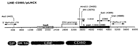
Figures la-id show, according to particular exemplary aspects, vector
schematics and
validation of efficient LHE fusion protein expression in DT40 chicken B-cells.
Figures 2a-2c show, according to particular exemplary aspects, data confirming
that
fluorescently conjugated dsOligos bind cell surface LHEs in a manner which is
sequence
specific and easily resolved by flow cytometry.
Figures 3a and 3b show, according to particular exemplary aspects, that LHEs
expressed on the cell surface reliably discriminate dsOligos containing single-
base pair
differences from their natural target sequences.
Figures 4a and 4b show, according to particular exemplary aspects, that
fluorescent
and/or magnetic strategies facilitate target sequence-specific sorting of
cells expressing surface
LHEs.
Figures 5a-5e show, according to particular exemplary aspects, data confirming
sequence-specific, LHE-mediated cleavage of cell surface-tethered dsOligo
substrates
conjugated with distinct fluorophores at opposite termini.
Figure 6 shows, according to particular exemplary aspects, efficient
enrichment of rare
dsOligo binding cell populations by FACS. Approximately 5 x 10³ IgM+ DT40
cells
expressing 1-Anil (clone B10) were mixed with 5 x 10⁷ of IgM DT40 cells
expressing a non-
13b
CA 02648030 2008-09-29
WO 2007/123636
PCT/US2007/007637
binding mutant I-Anir (for a final ratio of 1:104, or 0.01%) followed by
staining with
dsAni 1 -BT:SAv-PE. For the first round of cell sorting, the instrument
precision was set for
high yield and approximately 105 cells of the top 0.2% PE-positive population
were collected.
This population was grown up for 5-7 days, analyzed by staining with FITC-
conjugated anti-
IgM, and then re-sorted with the instrument precision set for high purity.
Figure 7 shows, according to particular exemplary aspects, a flow diagram
illustrating
exemplary means to generate and use surface displayed HEs (LAGLIDADG
endonucleases)
for identification of new homing endonucleases with novel binding and/or
cleavage
specificities. =
l0
DETAILED DESCRIPTION OF THE INVENTION
DEFINITIONS:
The term "cell" as referred to herein encompasses a living organism capable of
self
replication, and preferably is of size sufficient to allow for separation from
cells with similar
properties by flow cytometry or another suitable separation technology. In
particular cell
embodiments (e.g., eukaryotic cells), cells contain genomic DNA in a
subcellular organelle
(e.g., a nucleus). In other embodiments, genomic DNA is not be contained in a
nucleus (e.g.,
prokaryotic cells). Cells encompassed by the claimed methods include, but are
not limited to
culturable cells capable of cell-surface protein presentation or display, such
as vertebrate or
mammalian or other metazoan cells, yeast or other unicellular eukaryotic
cells, bacterial or
other prokaryotic cells, etc.
The term "homing endonuclease" or "HE" as used herein not only refers to art
recognized HE including but not limited to known LAGLIDAG, HNH, His-Cys Box,
GIY-
YIG, and I-Sspl-type homing endonucleases, but also to functional (sequence
specific
binding and/or cleaving) fusions, muteins or variants thereof. Preferably, the
HEs and
methods of the present invention relate to LAGLIDAG homing endonucleases. In
particular
aspects, the single chain LAGILDADG homing endonucleases_I-AniI (SEQ ID
NO:16), H-
Dre1 (SEQ ID NO:17; (Chain J, E-Drei (gi127065708IpdbI1MOWIJ[27065708]); Chain
G, E-
Drei (gi1270657051pdbt 1 MOWIG [27065705]); Chain D,
E-Drei
14
CA 02648030 2008-09-29
WO 2007/123636
PCT/US2007/007637
(gi1270657021pdbIlMOWID[27065702]); Chain A,
E-Drei
(gi127065699Ipdbl 1 MOWIA[27065699]))), I- DmoI (SEQ ID N018) HEs, I-CreI
(P05725;
SEQ ID NO:20), and fusions, muteins or variants thereof are preferred. Homing
endonucleases are proteins with enzymatic activity able to cleave a double-
stranded DNA
molecule, and having a polynucleotide recognition site of 14-40 bp. In
preferred aspects,
homing endonucleases are of the LAGLIDADG family.
"New homing endonuclease" or "homing endonuclease of altered specificity" is
defined as a homing endonuclease (e.g., LAGILDADG homing endonucleases)
derived from
an initial homing endonuclease presenting a different or altered
binding/recognition and/or
lo cleavage specificity or activity from that of the initial one.
"Altered recognition. and/or cleavage sequence" as used herein refers to a new
or
altered homing endonuclease binding or cleaving a double stranded DNA sequence
with an
altered specificity and/or efficiency (e.g., an altered efficacy of at least 2-
fold, at least 5- fold,
at least 10-fold more than the natural homing endonuclease, preferably at
least 50-fold, more
preferably at least 100-fold. The initial homing endonuclease can be a natural
homing
endonuclease or a modified one (e.g., derived by mutagenesis). In this
context, "natural"
refers to objects found in nature. For example, a homing endonuclease that is
found to be
naturally present in an organism, that can be isolated from a source in nature
and which has
not been intentionally modified by man in the laboratory.
The term "cell-surface presentation or display" of at least one HE, or fusion,
mutein
or variant thereof refers to display or presentation of an expressed HE, or
fusion, mutein or
variant thereof such that it is accessible to contact by one or more target
nucleic acid
molecules and/or specific binding agent (e.g., HE-specific antibodies, antigen
tag-specific
antibodies, etc.). Preferably, such displayed HEs are functional for sequence
specific target
sequence binding and/or cleavage.
The term "recombinant homing endonuclease (HE) expression system" referes to
any
suitable expression system that provide for cell-surface presentation or
display of at least one
HE, or fusion, mutein or variant thereof. Exemplary expression systems include
expression
vectors suitable for respective cell types, and include recombinant expressing
chromosomal
CA 02648030 2013-12-20
sites/sequences (HE sequences inserted (e.g., by homologous recombination or
otherwise)
into a chromosomal site to provide for HE sequence expression). For example,
insertion of a
HE coding sequence within an immunoglobulin light or heavy chain genomic locus
is
encompassed by the present conception.
"Homologous DNA sequences" are those with sufficient identity to another one
to
lead to a homologous recombination, having at least 95% identity, preferably
97%, and more
preferably 99% identity.
"Vector" as used herein refers to a nucleic acid or composite protein/nucleic
acid
assembly which is capable of transporting a nucleic acid into a bacterial or
eukaryotic cell.
Vectors include a number of distinct types. Some types of vectors are capable
of autonomous
replication of nucleic acids to which they are linked. One type of preferred
such vector is a
"plasmid", a double stranded circular nucleic acid capable of extra-
chromosomal replication
in bacteria. Other types of preferred vector are viruses, protein/nucleic acid
assemblages
found in nature which are able to introduce their nucleic acid into
prokaryotic or eukaryotic
cells, and then able to replicate themselves within the cell. Derived from
viruses found in
nature are virus-like particles (VLP's), which are nucleic acid/protein
assemblages which are
able to transfer their nucleic acid, but the nucleic acid no longer includes
sequences required
for self replication within a cell. A number of viral vectors are described in
McVey et al.,
U.S. Pat. No. 5,801,030. Vectors capable of directing the expression of genes
to which they
are operatively linked are referred to as "expression vectors". Large numbers
of suitable
vectors of many types are known to those of skill in the art and are
commercially available.
Vectors typically include a selectable marker gene, such as neomycin
phosphotransferase for
eukaryotic cell culture; TRP1 for S. cerevisiae; and tetracycline, rifampicin
or ampicillin
resistance in E. coli.
The phrases "target site", as used within this application, is defined as
referring to a
distinct DNA sequence to be bound or cleaved by a homing endonuclease.
Additional embodiments, "fusion, mutein or variants", include functional
(e.g., target
sequence-binding and/or cleavage) variants (including conservative amino acid
sequence
variants as described herein, and also non-conservative amino acid sequence
variants),
16
CA 02648030 2008-09-29
WO 2007/123636
PCT/US2007/007637
fragments, muteins, derivatives and fusion proteins thereof. Mutant HEs and
LHEs refers to
amino acid variants of HEs and LHEs that have altertered target sequence
binding and/or
cleavage activity (specificity and/or strength of binding, and/or specificity
and/or cleavage
activity), and includes functional (e.g., target sequence binding but non-
cleaving) variants
(including conservative and non-conservative amino acid sequence variants as
described
herein), fragments, muteins, derivatives and fusion proteins thereof.
Representative, HEs and
LHEs are provided herein.
Biologically Active Variants. Variants of HEs and LHEs have substantial
utility in
various aspects of the present invention. Variants can be naturally or non-
naturally
to occurring. Naturally occurring variants are found in various unicellular
eukaryotic, archael,
and prokaryotic organisms, as well as some bacterial viruses (e.g. phage) ,
and comprise
amino acid sequences which are substantially identical to the exemplary HE and
LHE amino
acid sequences shown herein, and include natural sequence polymorphisms.
Species
homologs of the proteins can be obtained using subgenomic polynucleotides of
the invention,
as described below, to make suitable probes or primers for screening cDNA
expression
libraries from other species of the organism from which the HE or LHE was
originally
isolated, identifying cDNAs which encode homologs of the protein, and
expressing the
cDNAs as is known in the art.
Non-naturally occurring variants which retain substantially the same or
altered
biological activities as naturally occurring protein variants, are also
included here.
Preferably, naturally or non-naturally occurring variants have amino acid
sequences which
are at leagt 85%, 90%, or 95% identical to the exemplary amino acid sequences
shown
hereinin. More preferably, the molecules are at least 98% or 99% identical.
Percent identity
is determined using any method known in the art. A non-limiting example is the
Smith-
Waterman homology search algorithm using an aftine gap search with a gap open
penalty of
12 and a gap extension penalty of 1. The Smith-Waterman homology search
algorithm is
taught in Smith and Waterman, Adv. AppL Math. 2:482-489, 1981.
As used herein, "amino acid residue" refers to an amino acid formed upon
chemical
digestion (hydrolysis) of a polypeptide at its peptide linkages. The amino
acid residues
17
CA 02648030 2013-12-20
described herein are generally in the "L" isomeric form. Residues in the "D"
isomeric form
can be substituted for any L-amino acid residue, as long as the desired
functional property is
retained by the polypeptide. NH2 refers to the free amino group present at the
amino terminus
of a polypeptide. COOH refers to the free carboxy group present at the
carboxyl terminus of
a polypeptide. In keeping with standard polypeptide nomenclature described in
J. Biol.
Chem., 243:3552-59 (1969), abbreviations for amino acid residues are shown in
Table 1:
TABLE 1 ¨ Table of Correspondence
SYMBOL
1-Letter 3-Letter AMINO ACID
Tyr Tyrosine
Gly Glycine
Phe Phenylalanine
Met Methionine
A Ala Alanine
Ser Serine
Ile Isoleucine
Leu Leucine
Thr Threonine
V Val Valine
Pro Praline
Lys Lysine
His Histidine
Gin Glutamine
Glu glutamic acid
Glx Glu and/or Gln
Trp Tryptophan
Arg Arginine
Asp aspartic acid
18
CA 02648030 2013-12-20
SYMBOL
Asn Asparagines
Asx Asn and/or Asp
Cys Cysteine
X Xaa Unknown or other
It should be noted that all amino acid residue sequences represented herein by
a
formula have a left to right orientation in the conventional direction of
amino-terminus to
carboxyl-terminus. In addition, the phrase "amino acid residue" is defined to
include the
amino acids listed in the Table of Correspondence and modified and unusual
amino acids.
Furthermore, it should be noted that a dash at the beginning or end of an
amino acid residue
sequence indicates a peptide bond to a further sequence of one or more amino
acid residues or
to an amino-terminal group such as NH2 or to a carboxyl-terminal group such as
COOH.
Guidance in determining which amino acid residues can be substituted,
inserted, or
deleted without abolishing biological or immunological activity can be found
using computer
programs well known in the art, such as DNASTARTm software. Preferably, amino
acid
changes in the protein variants disclosed herein are conservative amino acid
changes, i.e.,
substitutions of similarly charged or uncharged amino acids. A conservative
amino acid
change involves substitution of one of a family of amino acids which are
related in their side
chains. Naturally occurring amino acids are generally divided into four
families: acidic
(aspartate, glutamate), basic (lysine, arginine, histidine), non-polar
(alanine, valine, leucine,
isoleucine, proline, phenylalanine, methionine, tryptophan), and uncharged
polar (glycine,
asparagine, glutamine, cystine, serine, threonine, tyrosine) amino acids.
Phenylalanine,
tryptophan, and tyrosine are sometimes classified jointly as aromatic amino
acids.
In a peptide or protein, suitable conservative substitutions of amino acids
are known to
those of skill in this art and generally can be made without altering a
biological activity of a
resulting molecule. Those of skill in this art recognize that, in general,
single amino acid
substitutions in non-essential regions of a polypeptide do not substantially
alter biological
activity (see, e.g., Watson et al. Molecular Biology of the Gene, 4th Edition,
1987, The
19
CA 02648030 2008-09-29
WO 2007/123636
PCT/US2007/007637
Benjamin/Cummings Pub. Co., p.224).
Such substitutions may be made in accordance with those set forth in TABLE 2
as
follows:
TABLE 2
Original Conservative
residue substitution
Ala (A) Gly; Ser
Arg (R) Lys
Asn (N) Gin; His
Cys (C) Ser
=
Gin (Q) Asn
Glu (E) = Asp
Gly (G) Ala; Pro
His (H) Asn; Gln
Ile (I) Leu; Val
Leu (L) Ile; Val
Lys (K) Arg; Gln; Glu
Met (M) Leu; Tyr; Ile
Phe (F) -Met; Leu; Tyr
Ser (S) Thr
Thr (T) Ser
Trp (W) Tyr
Tyr (Y) Trp; Phe
I Val (V) Ile; Leu
Other substitutions also are permissible and can be determined empirically or
in
accord with other known conservative (or non-conservative) substitutions.
Variants of the HEs or LHEs disclosed herein also include glycosylated forms,
aggregative conjugates with other molecules, and covalent conjugates with
unrelated
chemical moieties (e.g., pegylated molecules). Covalent variants can be
prepared by linking
functionalities to groups which are found in the amino acid chain or at the N-
or C-terminal
residue, as is known in the art. Variants also include allelic variants,
species variants, and =
CA 02648030 2008-09-29
WO 2007/123636
PCT/US2007/007637
muteins. Truncations or deletions of regions which do not affect functional
activity of the
proteins are also variants.
A subset of mutants, called muteins, is a group of polypeptides in which
neutral
amino acids, such as serines, are substituted for cysteine residues which do
not participate in
disulfide bonds. These mutants may be stable over a broader temperature range
than native
secreted proteins (Mark etal., United States Patent 4,959,314).
Preferably, amino acid changes in the HE or LHE variants are conservative or
non-
conservative amino acid changes, i.e., substitutions of similarly charged or
uncharged amino
acids. A conservative .amino acid change involves substitution of one of a
family of amino
acids which are related in their side chains. Naturally occurring amino acids
are generally
divided into four families: acidic (aspartate; glutamate), basic (lysine,
arginine, histidine),
non-polar (alanine, valine, leucine, isoleucine, proline, phenylalanine,
methionine,
tryptophan), and uncharged polar (glycine, asparagine, glutamine, cystine,
serine, threonine,
tyrosine) amino acids. Phenylalanine, tryptophan, and tyrosine are sometimes
classified
jointly as aromatic amino acids.
It is reasonable to expect, depending upon the location of the replacement,
that an
isolated replacement of a leucine with an isoleucine or valine, an aspartate
with a glutamate, a
threonine with a serine, or a similar replacement of an amino acid with a
structurally related
amino acid will not have a major effect on the biological properties of the
resulting secreted
protein or polypeptide variant. Properties and functions of HE or LHE protein
or polypeptide
variants are of the same type as a protein comprising the amino acid sequence
encoded by the
exemplary sequences shown herin, although the properties and functions of
variants can
differ in degree or specificity (e.g., binding and/or cleavage).
It will be recognized in the art that some amino acid sequences of the HE and
LHE
polypeptides of the invention can be varied without significant effect on the
structure or
function of the protein. If such differences in sequence are contemplated, it
should be
remembered that there are critical areas on the protein which determine
activity. In general,
it is possible to replace residues that form the tertiary structure, provided
that residues
performing a similar function are used. In other instances, the type of
residue may be
21
CA 02648030 2008-09-29
WO 2007/123636
PCT/US2007/007637
completely unimportant if the alteration occurs at a non-critical region of
the protein. The
replacement of amino acids can also change the selectivity of binding to
target nucleic acids.
Thus, the HE or LHE polypeptides of the present invention may include one or
more amino
acid substitutions, deletions or additions, either from natural mutations or
human
manipulation (e.g., mutagenesis).
Of particular interest are substitutions of charged amino acids with another
charged
amino acid and with neutral or negatively charged amino acids. The latter
results in proteins
with reduced positive charge to improve the characteristics of the disclosed
protein. The
prevention of aggregation is highly desirable. Aggregation of proteins not
only results in a
loss of activity but can also be problematic when preparing pharmaceutical
formulations,
because they can be immunogenic (Pinckard et al., Clin. Exp. ImmunoL 2:331-
340, 1967;
Robbins et al., Diabetes 36:838-845, 1987; Cleland et al., Crit. Rev.
Therapeutic Drug
Carrier Systems 10:307-377, 1993).
Amino acids in the HE or LHE polypeptides of the present invention that are
essential
for function can be identified by methods known in the art, such as site-
directed mutagenesis
or alanine-scanning mutagenesis (Cunningham and Wells, Science 244:1081-1085,
1989).
The latter procedure introduces single alanine mutations at every residue in
the molecule.
The resulting mutant molecules are then tested for biological activity such as
binding to a
natural or synthetic binding partner. Sites that are critical for ligand-
receptor binding can
also be determined by structural analysis such as crystallization, nuclear
magnetic resonance
or photoaffinity labeling (Smith et al., J. MoL Biol. 224:899-904, 1992 and de
Vos et al.
Science 255:306-312,1992).
As indicated, changes are preferably of a minor nature, such as conservative
amino
acid substitutions that do not significantly affect the folding or activity of
the protein. Of
course, the number of amino acid substitutions a skilled artisan would make
depends on
many factors, including those described above. Generally speaking, the number
of
substitutions for any given HE or LHE will not be more than 50, 40, 30, 25,
20, 15, 10, 5, 3,
2 or 1. In addition, pegylation of HE or LHE polypeptides and/or muteins is
expected to
22
CA 02648030 2008-09-29
WO 2007/123636
PCT/US2007/007637
provide such improved properties as increased half-life, solubility, and
protease resistance.
Pegylation is well known in the art.
Fusion Proteins. Fusion proteins comprising proteins or polypeptide fragments
of HE
or LHE polypeptide can also be constructed. Fusion proteins are useful for,
inter alio,
generating antibodies against amino acid sequences and for use in various
targeting,
expression and assay systems. For example, fusion proteins can be used to
identify He or
LHE proteins which interact with a target sequence of the invention or which
interfere or
alter HE or LHE biological function. Physical methods, such as protein
affinity
chromatography, or library-based assays for protein-protein interactions, such
as the yeast
two-hybrid or phage display systems, can also be used for this purpose. Such
methods are
well known in the art and can also be used as drug screens. Fusion proteins
comprising a
signal sequence can be used.
A fusion protein comprises two protein segments fused together by means of a
peptide bond. Amino acid sequences for use in fusion proteins of the invention
can be utilize
the exemplarly amino acid sequence shown herein or can be prepared from
biologically
active variants thereof. The first protein segment can include of a full-
length He or LHE.
Other first protein segments can consist of a limited number of contiguous
amino acids.
The second protein segment can be a full-length protein or a polypeptide
fragment.
Proteins commonly used in fusion protein construction include I3-
ga1actosidase,
glucuronidase, green fluorescent protein (GFP), autofluorescent proteins,
including blue
fluorescent protein (BFP), glutathione-S-transferase (GST), luciferase,
horseradish
peroxidase (HRP), and chloramphenicol acetyltransferase (CAT). Additionally,
epitope tags
can be used in fusion protein constructions, including histidine (His) tags,
FLAG tags,
influenza hemagglutinin (HA) tags, Myc tags, VSV-G tags, and thioredoxin (Trx)
tags.
Other fusion constructions can include maltose binding protein (MBP), S-tag,
Lex a DNA
binding domain (DBD) fusions, GAL4 DNA binding domain fusions, and herpes
simplex
virus (HSV) BP16 protein fusions. CD80 fusions are a preferred fusion as
disclosed herein.
These fusions can be made, for example, by covalently linking two protein
segments
or by standard procedures in the art of molecular biology. Recombinant DNA
methods can
23
CA 02648030 2008-09-29
WO 2007/123636
PCT/US2007/007637
be used to prepare fusion proteins, for example, by making a DNA construct
which comprises
a coding region for the exemplary protein sequences shown herein in proper
reading frame
with a nucleotide encoding the second protein segment and expressing the DNA
construct in
a host cell, as is known in the art. Many kits for constructing fusion
proteins are available
from companies that supply research labs with tools for experiments,
including, for example,
Promega Corporation (Madison, WI), Stratagene (La Jolla, CA), Clontech
(Mountain View,
CA), Santa Cruz Biotechnology (Santa Cruz, CA), MBL International Corporation
(MIC;
Watertown, MA), and Quantum Biotechnologies (Montreal, Canada; 1-888-DNA-
KITS).
The term "target" specificity as used herein refers to homing endonclease
target
sequence, and includes HE target sequence binding specificity and/or HE targe
sequence
cleavage specificity.
The term "labeled target nucleic acid sequence" refers to target nucleic acids
labeled
with one more labels suitable for monitoring binding or cleavage events. Such
labels include,
but are not limited to fluorescent labels (PE, Alexa Fluor 647, and other art-
recognized labels
used in FACS or MACS based separations, etc.), eptitope tags, biotin,
streptavidin,
radiolabels, FRET labels, etc. Labeled target nucleic acid sequences include
bifluorescent
double stranded sequences, examples of which are described herein.
The term "selecting" as used herein refers to any method suitable for
separating cells
based on cell-surface presentation or display of HEs. Exemplary methods
include, but are not
limited to magnetic activated cells sorting (MACS), fluorescence activated
cell sorting
(FACS), or combinations thereof (e.g., using labeled target nucleic acids).
The term "tethered target sequence" as used herein refers to binding of one or
more
target sequences to the cell surface by means other than binding to the cell
surface expressed
HE target sequence binding site, to provide for subsequent binding and/or
cleavage by the HE
target sequence binding and/or cleavage site. In particular aspects of the
methods, one end of
the labeled target sequence is tethered to the cell surface, and the other end
of the target
sequence comprises a label which is releasable upon subsequent homing
endonuclease-
mediated cleavage of the tethered target sequence. For example, as described
herein, cells
may first labeled with a biotin-conjugated anti-HA monoclonal antibody (a-HA-
BT)
24
CA 02648030 2008-09-29
WO 2007/123636
PCT/US2007/007637
followed by the addition of pre-formed 647-dsAni 1 -BT:SAv-PE complexes which
contain an
average of three remaining BT-binding sites per SAv tetramer, and this
exemplary staining
protocol serves to tether the 647-dsAni 1 -BT:SAv-PE to the cell surface
independent of any
specific LHE-dsOligo interaction, yet still placing the dsOligo within the
LHE's immediate
environment (FIGURE 5a). Thus, according to particular exemplary aspects,
cleavage events
can be followed using an a-HA-BT tethered dually-fluorescent labeled dsOligos
and the
release of Alexa Fluor 647 following addition of Mg2+ (to provide for cleavage
conditions).
Therefore, the presently disclosed inventive aspects encompass the conception
that where the
tethered double labeled oligos can be cleaved by the surface LHE, the cells
would lose the
fluorescence signal contribution from one label (e.g., Alexa Fluor 647) yet
retain signal from
the other label (e.g., a tightly bound bridging SAv-PE).
PREFERRED EXEMPLARY EMBODIMENTS:
Cells and cell libraries comprising cell-surface presentation or display of at
least one HE:
Particular embodiments of the present invention provide a cell, comprising at
least
one recombinant homing = endonuclease (HE) expression system suitable to
provide for cell-
surface presentation or display of at least one 1-1E, or fusion, mutein or
variant thereof on the
cell. In certain aspects, the cell expresses a single homing endonuclease
(HE), or fusion,
mutein or variant thereof on the cell surface. In additional aspects, the cell
expresses a
plurality of different homing endonuclease (HE), or fusions, muteins or
variants thereof on
the cell surface.
Also provided, is a library of cells, comprising a plurality of cells, wherein
each cell
comprises at least one recombinant homing endonuclease (HE) expression system
suitable to
provide for cell-surface presentation or display of at least one HE, or
fusion, mutein or
variant thereof on the cell, and wherein a plurality of different homing
endonuclease (HE), or
fizions, muteins or variants thereof are represented between and among the
cells of the
library. Preferably, in the inventive cells and libraries thereof, the homing
endonuclease is
functional for at least one of binding of nucleic acid target sequence, and
cleaving of a
nucleic acid target sequence.
CA 02648030 2008-09-29
WO 2007/123636
PCT/US2007/007637
In particular aspects of the cell or library thereof, the homing endonuclease
(HE) is
expressed as a fusion protein suitable to provide for cell-surface
presentation or display of the
at least one HE, or fusion, mutein or variant thereof. In certain embodiments,
the fusion
protein comprises at least one of a signal peptide, an epitope tag, a membrane-
anchoring
moiety or polypeptide, and combinations thereof. In certain embodiments, the
signal peptide
is an immunoglobulin signal peptide, and the membrane anchoring polypeptide
comprises
murine CD80 or a membrane anchoring portion thereof. In "additional
embodiments, the
signal peptide is an immunoglobulin signal peptide, and the membrane anchoring
polypeptide
comprises a mature immunoglobulin light or heavy chain polypeptide or a
membrane-
io anchoring portion thereof.
In particular aspects of the cell or library thereof, the recombinant
expression
comprises expression from at least one recombinant expression vector, or from
at least one
recombinant genomic locus. In particular embodiments, recombinant expression
of the
homing endonuclease (HE), comprises insertion of a HE coding sequence within
an
immunoglobulin light or heavy chain genomic locus. In particular aspects of
the cell or
library thereof, the one or more cells comprise at least one cell selected
from the group
consisting of a eukaryotic cell, a culturable metazoan cell capable of cell-
surface protein
presentation or display, mammalian cell, yeast cell and bacterial cell.
In particular aspects of the cell or library thereof, the homing endonuclease
comprises
at least one selected from the group consisting of LAGLIDAG, HNH, His-Cys Box,
GIY-
YIG, I-SspI-type, and fusions, muteins or variants thereof. Preferably, the
homing
endonuclease comprises or consists of a LAGLIDAG homing endonuclease, or a
fusion,
mutein or variant thereof. In particular embodiments, the homing endonuclease
comprises or
consists of at least one selected from the group consisting of 1-Anil, H-DreI,
I-Sce I, 1-Chu I,
I-Dmo I, I-Cre I, I-Csm I, P1-See I, PI-Tli I, PI-Mtu I, I-Ceu I, I-See II, I-
Sce III, HO, PI-Civ
I, PI-Ctr I, PI-Aae I, PI-Bsu I, PI-Dha I, PI-Dra I, PI-May I, PI-Mch I, PI-
Mfu I, PI-Mfl I, PI-
Mga I, PI-Mgo I, PI-Min I, PI-Mka I, PI-Mle I, PI-Mma I, PI-Msh I, PI-Msm I,
PI-Mth I, P1-
Mm I, PI-Mxe I, PI-Npu I, PI-Pfu I, PI-Rma I, PI-Spb I, PI-Ssp I, PI-Fac I, PI-
Mja I, PI-Pho
I, PI-Tag I, PI-Thy I, PI-Tko I, PI-Tsp I, and fusions, muteins or variants
thereof. In certain
26
CA 02648030 2008-09-29
WO 2007/123636
PCT/US2007/007637
aspects, the homing endonuclease comprises or consists of 1-Anil, or a fusion,
mutein or
variant thereof. In additional aspects, the homing endonuclease comprises or
consists of H-
DreI, or a fusion, mutein or variant thereof.
Methods for identifying a homing endonuclease with a desired target
specificity: .=
Binding. Additional embodiments provide a method for identifying a homing
endonuclease with a desired target specificity, comprising: expressing, using
a suitable
recombinant expression system, at least one homing endonuclease (HE) in one or
more cells,
the recombinant expression and the one or more cells suitable to provide for
cell-surface
to
presentation or display of the at least one HE; contacting the one or more
expressing cells
with at least one labeled target nucleic acid sequence under conditions
suitable to allow for
target sequence binding to the at least one cell-surface HE; and selecting,
based on the
presence of cell-bound label, one or more cells expressing at least one cell
surface HE having
a target sequence binding specificity. In particular aspects of the above
methods, the one or
more cells comprises a library of cells, the library comprising a plurality of
cells, wherein
each cell comprises at least one recombinant homing endonuclease (HE)
expression system
suitable to provide for cell-surface presentation or display of at least one
HE, or fusion,
mutein or variants thereof on the cell, and wherein a plurality of different
homing
endonuclease (HE), or fusions, muteins or variants thereof are represented.
Binding and/or cleavage. Further embodiments provide a method for identifying
a
homing endonuclease with a desired target specificity, comprising: expressing,
using a
suitable recombinant expression system, at least one homing endonuclease (HE)
in one or
more cells, the recombinant expression and the one or more cells suitable to
provide for cell-
surface presentation or display of the at least one HE; contacting the one or
more expressing
cells with at least one labeled target nucleic acid sequence under conditions
suitable to allow
for target sequence binding to the at least one cell-surface HE; adjusting the
conditions to
allow for homing endonuclease-mediated cleavage of the target sequence; and
selecting,
based on a decrease of cell-bound label, one or more cells expressing at least
one cell surface
HE having a target sequence cleaving specificity. In certain aspects of the
above methods,
27
CA 02648030 2008-09-29
WO 2007/123636
PCT/US2007/007637
the one or more cells comprises a library of cells, the library comprising a
plurality of cells,
wherein each cell comprises at least one recombinant homing endonuclease (HE)
expression
system suitable to provide for cell-surface presentation or display of at
least one HE, or
fusion, mutein or variants thereof on the cell, and wherein a plurality of
different homing
endonuclease (HE), or fusions, muteins or variants thereof are represented. In
particular
aspects of the methods, contacting comprises tethering one end of the labeled
target sequence
to the cell surface, and wherein the other end of the target sequence
comprises a label which
is releasable upon subsequent homing endonuclease-mediated cleavage of the
tethered target
sequence. In particular embodiments, the conditions suitable to allow for
target sequence
to binding do not allow for target sequence cleavage by the homing
endonuclease (HE). In
certain embodiments, the conditions comprise concentrations of calcium and/or
copper ions
sufficient to allow for target sequence binding, but lack a concentration of
at least one of
magnesium, cobalt, manganese, nickel and zinc ions sufficient to allow for
target sequence
cleavage. In particular aspects, conditions that allow for homing endonuclease-
mediated
cleavage of the target sequence comprise a concentration of at least one of
magnesium,
cobalt, manganese, nickel and zinc ions sufficient to allow for target
sequence cleavage, and
a concentration of calcium and/or copper ions below a level that significantly
inhibits target
sequence cleavage.
In particular embodiments of the above methods, the homing endonuclease (HE)
is
expressed as a fusion protein suitable to provide for cell-surface
presentation or display of the
at least one HE, or fusion, mutein or variant thereof. In certain aspects of
the methods, the
fusion protein comprises at least one of a signal peptide, an epitope tag, a
membrane-
anchoring moiety or polypeptide, and combinations thereof. In certain aspects,
the Signal
peptide is an immunoglobulin signal peptide, and the membrane anchoring
polypeptide
comprises murine CD80 (e.g., SEQ ID NOS:21, 22) or a membrane anchoring
portion
thereof. In additional aspects, the signal peptide is an immunoglobulin signal
peptide, and
the membrane anchoring polypeptide comprises a mature immunoglobulin light or
heavy
chain polypeptide or a membrane-anchoring portion thereof
28
CA 02648030 2008-09-29
WO 2007/123636
PCT/US2007/007637
In particular embodiments of the methods, the recombinant expression comprises
expression from at least one recombinant expression vector, or from at least
one recombinant
genomic locus. In certain aspects, recombinant expression of the homing
endonuclease (HE),
comprises insertion of a HE coding sequence within an immunoglobulin light or
heavy chain
genomic locus.
In particular embodiments of the methods, the one or more cells comprise at
least one
cell selected from the group consisting of a eukaryotic cell, a culturable
metazoan cell
capable of cell-surface protein presentation or display, mammalian cell, yeast
cell and
bacterial cell. In particular embodiments of the methods, each one of the one
or more cells
expresses a single homing endonuclease (HE) sequence. In additional
embodiments, at least
one of the one or more cells expresses a plurality of different homing
endonuclease (HE)
sequences.
In particular embodiments of the methods, selecting comprises the use of
magnetic
activated cells sorting (MACS), fluorescence activated cell sorting (FACS), or
combinations
thereof.
In certain embodiments, the target sequence comprises a known or putative
homing
endonuclease (HE) binding sequence. In additional embodiments, the target
sequence
comprises a known or putative homing endonuclease (HE) and a known or putative
homing
endonuclease cleavage sequence.
In particular embodiments of the methods, the homing endonuclease comprises at
least one selected from the group consisting of LAGLIDAG, HNH, His-Cys Box,
GIY-YIG,
I-SspI-type, and fusions, muteins or variants thereof. Preferably, the homing
endonuclease
comprises or consists of a LAGLIDAG homing endonuclease, or a fusion, mutein
or variant
thereof. In certain aspects, the homing endonuclease comprises or consists of
at least one
selected from the group consisting of 1-Anil, H-DreI, I-See I, 1-Chu I, I-Dmo
I, I-Cre I, I-Csm
I, P1-See I, PI-Tli I, PI-Mtu I, I-Ceu I, I-See II, I-See III, HO, PI-Civ I,
PI-Ctr I, PI-Aae I, PI-
Bsu I, PI-Dha I, PI-Dra I, PI-May I, PI-Mch I, PI-Mfu I, PI-Mfl I, PI-Mga I,
PI-Mgo I, PI-
Min I, PI-Mka I, PI-Mle I, PI-Mma I, PI-Msh I, PI-Msm I, PI-Mth I, PI-Mtu I,
PI-Mxe I, PI-
Npu I, PI-Pfu I, PI-Rma I, PI-Spb I, PI-Ssp I, PI-Fac I, PI-Mja I, PI-Pho I,
PI-Tag I, PI-Thy I,
29
CA 02648030 2008-09-29
WO 2007/123636
PCT/US2007/007637
PI-Tko I, PI-Tsp I, and fusions, muteins or variants thereof. In particular
aspects, the homing
endonuclease comprises or consists of 1-Anil, or a fusion, mutein or variant
thereof. In
additional aspects, the homing endonuclease comprises or consists of H-DreI,
or a fusion,
mutein or variant thereof.
Methods for obtaining and identifying a variant homing endonuclease with an
altered target
specificity:
Altered target binding specificity. Further embodiments provide a method for
obtaining and identifying a variant homing endonuclease with an altered target
specificity,
comprising: obtaining a nucleic acid sequence encoding an open reading frame
for at least
one initial homing endonuclease (HE); expressing, using a suitable recombinant
expression
system, at least one variant of the nucleic acid sequence in one or more
cells, the recombinant
expression suitable to provide for cell-surface presentation or display of the
at least one HE in
the one or more cells, the at least one variant sequence having been derived
by mutagenesis
from the nucleic acid sequence encoding the initial homing endonuclease (HE);
contacting
the one or more expressing cells with at least one labeled target nucleic acid
sequence under
conditions suitable to allow for target sequence binding to the at least one
cell-surface HE;
and selecting, based on the presence of cell-bound label, one or more cells
expressing at least
one cell surface variant HE having a target sequence binding specificity. In
certain aspects of
the methods, the one or more cells comprises a library of cells, the library
comprising a
plurality of cells, wherein each cell comprises at least one recombinant
homing endonuclease
(HE) expression system suitable to provide for cell-surface presentation or
display of at least
one HE, or fusion, mutein or variants thereof on the cell, and wherein a
plurality of different
homing endonuclease (HE), or fusions, muteins or variants thereof are
represented.
Altered target cleavage specificity. Yet additional embodiments provide a
method for
obtaining and identifying a variant homing endonuclease with an altered target
specificity,
comprising: obtaining a nucleic acid sequence encoding an open reading frame
for at least
one initial homing endonuclease (HE); expressing, using a suitable recombinant
expression
system, at least one variant of the nucleic acid sequence in one or more
cells, the recombinant
CA 02648030 2008-09-29
WO 2007/123636
PCT/US2007/007637
expression suitable to provide for cell-surface presentation or display of the
at least one HE in
the one or more cells, the at least one variant sequence having been derived
by mutagenesis
from the nucleic acid sequence encoding the initial homing endonuclease (HE);
contacting
the one or more expressing cells with at least one labeled target nucleic acid
sequence under
conditions suitable to allow for target sequence binding to the at least one
cell-surface HE;
adjusting the conditions to allow for homing endonuclease-mediated cleavage of
the target
sequence; and selecting, based on a decrease of cell-bound label, one or more
cells expressing
at least one cell surface HE having a target sequence cleaving specificity. In
certain aspects
of the methods, the one or more cells comprises a library of cells, the
library comprising a
plurality of cells, wherein each cell comprises at least one recombinant
homing endonuclease
(HE) expression system suitable to provide for cell-surface presentation or
display of at least
one HE, or fusion, mutein or variants thereof on the cell, and wherein a
plurality of different
homing endonuclease (HE), or fusions, muteins or variants thereof are
represented. In certain
embodiments, contacting comprises tethering one end of the labeled target
sequence to the
cell surface, and wherein the other end of the target sequence comprises a
label which is
releasable upon subsequent homing endonuclease-mediated cleavage of the
tethered target
sequence. In particular embodiments, the conditions suitable to allow for
target sequence
binding do not allow for target sequence cleavage by the homing endonuclease
(HE). In
certain aspects, the conditions comprise concentrations of calcium and/or
copper ions
sufficient to allow for target sequence binding, but lack a concentration of
at least one of
magnesium, cobalt, manganese, nickel and zinc ions sufficient to allow for
target sequence
cleavage. In particular aspects, conditions that allow for homing endonuclease-
mediated
cleavage of the target sequence comprise a concentration of at least one of
magnesium,
cobalt, manganese, nickel and zinc ions sufficient to allow for target
sequence cleavage, and
a concentration of calcium and/or copper ions below a level that significantly
inhibits target
sequence cleavage.
In certain embodiments of the above methods, the homing endonuclease (HE) is
expressed as a fusion protein suitable to provide for cell-surface
presentation or display of the
at least one HE, or fusion, mutein or variant thereof. In particular aspects,
the fusion protein
31
CA 02648030 2008-09-29
WO 2007/123636
PCT/US2007/007637
comprises at least one of a signal peptide, an epitope tag, a membrane-
anchoring moiety or
polypeptide, and combinations thereof. In certain embodiments, the signal
peptide is an
immunoglobulin signal peptide, and the membrane anchoring polypeptide
comprises murine
CD80 (e.g., SEQ ID NOS:21, 22) or a membrane anchoring portion thereof. In
other
embodiments, the signal peptide is an immunoglobulin signal peptide, and the
membrane
anchoring polypeptide comprises a mature immunoglobulin light or heavy chain
polypeptide
or a membrane-anchoring portion thereof.
In particular implementations, the recombinant expression comprises expression
from
at least one recombinant expression vector, or from at least one recombinant
genomic locus.
In certain aspects, recombinant expression of the homing endonuclease (HE)
comprises
insertion of a HE coding sequence within an immunoglobulin light or heavy
chain genomic
locus.
In particular embodiments of the methods, the one or more cells comprise at
least one
cell selected from the group consisting of a eukaryotic cell, a culturable
metazoan cell
capable of cell-surface protein presentation or display, mammalian cell, yeast
cell and
bacterial cell. In certain aspects, each one of the one or more cells
expresses a single homing
endonuclease (HE) sequence. In additional aspects, at least one of the one or
more cells
expresses a plurality of different homing endonuclease (HE) sequences.
In certain implementations, selecting comprises the use of magnetic activated
cells
sorting (MACS), fluorescence activated cell sorting (FACS), or combinations
thereof.
In certain embodiments, the target sequence comprises a known or putative
homing
endonuclease (HE) binding sequence. In additional aspects, the target sequence
comprises a
known or putative homing endonuclease (HE) and a known or putative homing
endonuclease
=
cleavage sequence.
In various aspects, the homing endonuclease comprises at least one selected
from the
group consisting of LAGLIDAG, HNH, His-Cys Box, GIY-YIG, I-Sspi-type, and
fusions,
muteins or variants thereof. Preferably, the homing endonuclease comprises or
consists of a
LAGLIDAG homing endonuclease, or a fusion, mutein or variant thereof. In
certain aspects,
the homing endonuclease comprises or consists of at least one selected from
the group
32
CA 02648030 2008-09-29
WO 2007/123636
PCT/US2007/007637
consisting of 1-Anil, H-DreI, I-Sce I, 1-Chu I, I-Dmo I, I-Cre I, I-Csm I, PI-
Sce. I, PI-Tli I, PI-
Mtu I, I-Ceu I, I-Sce II, I-Sce III, HO, PI-Civ I, PI-Ctr I, PI-Aae I, PI-Bsu
I, PI-Dha I, PI-Dra
I, PI-May I, PI-Mch I, PI-Mfu I, PI-Mfl I, PI-Mga I, PI-Mgo I, PI-Min I, PI-
Mka I, PI-Mle I,
PI-Mma I, PI-Msh I, PI-Msm I, PI-Mth I, PI-Mtu I, PI-Mxe I, PI-Npu I, PI-Pfu
I, PI-Rma I,
PI-Spb I, PI-Ssp I, PI-Fac I, PI-Mja I, PI-Pho I, PI-Tag I, PI-Thy I, PI-'Tko
I, PI-Tsp I, and
fusions, muteins or variants thereof. In particular embodiments, the homing
endonuclease
comprises or consists of 1-Anil, or a fusion, mutein or variant thereof. In
additional
embodiments, the homing endonuclease comprises or consists of H-DreI, or a
fusion, mutein
or variant thereof.
Methods for introducing a targeted double-strand break in the genome of a
virus or of a living
cell:
Particular aspects provide a method for introducing a targeted double-strand
break in
the genome of a virus or of a living cell, comprising: obtaining a homing
endonuclease
selected using at least one cell, cell library or method comprising cell-
surface presentation or
display of at least one homing endonuclease (HE), or fusion, mutein or variant
thereof on the
cell or libraries to provide a homing endonuclease having a specific, desired
DNA target
cleavage specificity within a target viral or cellular genome; and introducing
the homing
endonuclease into a cell harboring the respective target viral or cellular
genome, wherein the
homing endonuclease introduces a targeted double-strand break in the viral or
cellular
genome.
Methods for chromatin immunonrecinitation (CHIP):
Additional aspects provide a method for chromatin inununoprecipitation (CHIP),
comprising: obtaining a homing endonuclease selected using at least one cell,
cell library or
method comprising cell-surface presentation or display of at least one homing
endonuclease
(HE), or fusion, mutein or variant thereof on the cell or libraries to provide
a homing
endonuclease having a specific, desired DNA target cleavage specificity within
a target viral
or cellular genome; and introducing into a cell the homing endonuclease or an
epitope-tagged
33
CA 02648030 2008-09-29
WO 2007/123636
PCT/US2007/007637
version thereof to provide for specific homing endonuclease complexes within a
target viral
or cellular genome. In certain aspects, the methods additionally comprise:
crosslinking of the
genomic DNA and associated proteins to provide for crosslinking of the homing
endonuclease to its cognate bound target site; shearing of the crosslinked
genomic DNA; and
immunoprecipitating the homing endonuclease and its bound DNA fragment using
antibodies
to the homing endonuclease or to the epitope tag thereof.
In particular aspects of the above methods, introducing the homing
endonuclease
comprises introducing the homing endonuclease as a polypeptide linked to one
or more
subcellular localization peptides necessary or sufficient to target the LHE to
an appropriate
organellar compartment. In additional embodiments, introducing the homing
endonuclease
comprises introducing the homing endonuclease in the context of a suitable
expression vector
under the control of appropriate transcriptional regulatory elements. In yet
additional aspects,
introducing the homing endonuclease comprises introducing the homing
endonuclease or a
sequence or vector encoding the homing endonuclease, along with an appropriate
vehicle,
carrier or DNA fragment. In additional implementations, introducing the homing
endonuclease comprises incorporating the homing endonuclease or a sequence
encoding the
homing endonuclease into one or more viral particles. Preferably the virus
does not integrate
into the host cell genome. Preferably, the virus particle is an integrase-
deficient lentiviral
particle, or an HIV- l derived lentiviral particle.
Use of CD80 for cell surface expression of HE fusion proteins:
Particular aspects provide the use of a CD80 nucleic acid or protein sequence,
or a
portion thereof for cell surface expression of homing endonucleases (HEs) or
LAGLIDAG
homing endonuclease (LHEs) (see working Examples described herein).
Use of DT40 cell lines, chicken tumor cell lines or lymphocyte cell lines for
cell surface
expression of HE fusion proteins:
Use of a DT40 cell line specifically, suitable chicken tumor cell line or a
lymphocyte
cell line for cell surface expression of homing endonucleases (HEs) or
LAGLIDAG homing
34
CA 02648030 2008-09-29
WO 2007/123636
PCT/US2007/007637
endonuclease (LHEs) (see working Examples described herein). In particular
embodiments,
B-lymphocyte cell lines are used so as to allow the HEs to be subject to the
endogenous
hypermutation mechanism of B-lymphocytes.
Generation of Homing Endonuclease Variants:
Particular aspects of the present invention provide a method for identifying a
homing
endonuclease (e.g., HE or LHE) specific to a targeted DNA sequence from a
library of
homing endonucleases of various specificities. These may be generated from an
initial
homing endonuclease which is a natural homing endonuclease. Alternatively, the
initial HE
or LAGLIDADG homing endonuclease is not a natural one. In preferred
embodiments, said
LAGLIDADG homing endonucleases are used (e.g., 1-Anil or E-DreI). The methods
comprises placing a library of homing endonuclease variants on the surface of
a cell, and
selection and/or screening of the variants able to bind and/or cleave a
desired target DNA
sequence or part thereof.
In particular aspects, the homing endonucleases are expressed on the surface
of cells
through fusion with one of several surface-bound cell proteins known to those
skilled in the
art. Said protein may be a yeast protein, as described as a general approach
for yeast protein
surface expression (this general method is reviewed in Chao et al, Nature
Protocols, 2006,
1(2):755-768) , murine CD80, as described as a method for expressing antibody
fragments
(Chou et al, Biotechnol Bioeng, 1999, 65:1690-169; also Liao et al, Biotechnol
Bioengin,
2001, 73:313-323), or an immunoglobulin heavy or light chain, as described for
glucoamylase as a means for directing soluble immunoglobulin proteins to a
secretory
pathway in Aspergillus nigrans (Ward et al, Applied and Environmental
Microbiology, 2004,
70(5):2567-2576), but readily modifiable for surface expression of a fusion
protein via fusion
to surface expressed forms of immunoglobulins.
In certain embodiments, the cell based library of surface expressed homing
endonuclease variants is then exposed to a fluorescent labeled oligonucleotide
under
conditions in which binding of the oligonucleotide (e.g., target sequence) ,
and optionally
subjected to a cell sorting protocol based on target sequence binding.
CA 02648030 2008-09-29
WO 2007/123636
PCT/US2007/007637
Generation of the library of homing endonuclease (HE) (e.g., homing
endonuclease)
of different target specificities can be performed by any of various art
recognized methods,
including DNA shuffling, error-prone PCR and expression of the homing
endonuclease in a
cell line in which the gene is susceptible to mutation. Preferably, the
diversity is introduced
by targeted mutagenesis (e.g., cassette mutagenesis, oligonucleotide directed
codon
mutagenesis, targeted random mutagenesis), by random mutagenesis (e.g.,
mutator strains,
Neurospora crassa system (U.S. Pat. No. 6,232,112; W001/70946, error-prone
PCR), by
DNA shuffling, by directed mutation or a combination of these technologies
(See Current
Protocols in Molecular Biology, Chapter 8 "Mutagenesis in cloned DNA", Eds
Ausubel et
al., John Wiley and Sons). The HE variants are preferably prepared by the
targeted
mutagenesis of an initial HE. The diversity is optimally introduced at
positions of the
residues contacting the DNA target or interacting (directly or indirectly)
with the DNA target.
The diversity is preferably introduced in regions interacting with the DNA
target, and more -
preferably introduced at the positions of the interacting amino acids. In
libraries generated by
targeted mutagenesis, amino acid residues (e.g., selected from the standard 20
amino acids)
can be introduced at the chosen variable positions. Preferably, the amino
acids present at the
variable positions are the amino acids well-known to be generally involved in
protein-DNA
interaction. More particularly, these amino acids are generally the
hydrophilic amino acids.
More preferably, the amino acids present at the variable positions comprise D,
E, H, K, N, Q,
R, S, T, Y. Optionally, the amino acids present at the variable positions are
selected from the
group consisting of D, E, H, K, N, Q, R, S, T, Y. Synthetic or modified amino
acids may also
be used.
One preferred way to generate a directed library is the use of degenerated
codons at
the positions where diversity has to be introduced. Several types of
degenerated codons
could be used. A degenerated codon N N K ([ATCG] [ATCG] [TG]) leads to 32
different
codons encoding the 20 amino acids and one stop. A degenerated codon N V K
([ATCG]
[ACG] [TO]) leads to 24 different codons encoding the 15 amino acids and one
stop. A
degenerated codon V V K ([ACG] [ACG] [TG]) leads to 18 different codons
encoding the 12
amino acids (A, D, E, G, H, K, N, P. Q, R, S, T) and no stop. A degenerated
codon R V K
36
CA 02648030 2008-09-29
WO 2007/123636
PCT/US2007/007637
([AG] [ACG] [TG]) leads to 12 different codons encoding the 9 amino acids (A,
D, E, G, K,
N, R, S. T). Preferably, a degenerated codon V V K ([ACG] [ACG] [TO]) leading
to 18
different codons encoding the 12 amino acids (A, D, E, G, H, K, N, P, Q, R, S,
T) is used for
generating the library. Indeed, the V V K degenerated codon does not contain
any stop codon
and comprises all the hydrophilic amino acids.
If a directed library is generated, knowledge on amino acids interacting with
the DNA
target is useful. This knowledge could be provided, for example, by X-ray
cristallography,
Alanine scanning, or cross-linking experiments. The amino acids interacting
with the DNA
target can also be deduced by sequence alignment with a homologous protein.
The custom-made or mutagenized and selected HE is derived from any initial HE.
Optionally, the initial HE is selected so as its natural recognition and
cleavage site is the
closest to the targeted DNA site. Preferably, the initial HE is a homing
endonuclease, as
specified herein. Homing endonucleases fall into 4 families on the basis of
well conserved
amino acid motifs, namely the LAGLIDADG family, the GIY-YIG family, the His-
Cys box
family, and the HNH family (Chevalier et al., 2001, N.A.R, 29, 3757-3774). The
detailed
three-dimensional structures of several homing endonucleases are known, namely
I-Dmo I,
PI-Sce I, PI-Pfu I, I-Cre I, I-Ppo I, and a hybrid homing endonuclease I-Dmo
1/1-Cre I called
E-Dre I (Chevalier et al., 2001, Nat Struct Biol, 8, 312-316; Duan et al.,
1997, Cell, 89, 555-
564; Heath et al., 1997, Nat Struct Biol, 4, 468-476; Hu et al., 2000, J Biol
Chem, 275, 2705-
2712; Ichiyanagi et al., 2000, J Mol Biol, 300, 889-901; Jurica et al., 1998,
Mol Cell, 2, 469-
476; Poland et al., 2000, J Biol Chem, 275, 16408-16413; Silva et al., 1999, 4
Mol Biol, 286,
1123-1136; Chevalier et al., 2002, Molecular Cell, 10, 895-905).
The LAGLIDADG family is the largest family of proteins clustered by their most
general conserved sequence motif: one or two copies of a twelve-residue
sequence: the di-
dodecapeptide, also called LAGLIDADG motif. Homing endonucleases with one
dodecapeptide (D) are around 20 kDa in molecular mass and act as homodimer.
Those with
two copies (DD) range from 25 kDa (230 AA) to 50 kDa (HO, 545 AA) with 70 to
150
residues between each motif and act as monomer. Cleavage is inside the
recognition site,
leaving 4 nt staggered cut with 3'0H overhangs. I-Ceu I, and I-Cre I
illustrate the
37
CA 02648030 2013-12-20
homodimeric homing endonucleases with one Dodecapeptide motif (mono-
dodecapeptide). I-
Dmo I, I-Sce I, PI-Pfu I and PI-Sce I illustrate monomeric homing
endonucleases with two
Dodecapeptide motifs.
The initial LAGLIDADG homing endonuclease can be selected from the group
consisting of: 1-Anil, H-DreI, I-See I, 1-Chu I, I-Dmo I, I-Cre I, I-Csm I, PI-
Sce I, PI-Tli I, PI-
Mtu I, I-Ceu I, I-See II, I-See III, HO, PI-Civ I, PI-Ctr I, PI-Aae I, PI-Bsu
I, PI-Dha I, PI-Dra
I, PI-May I, PI-Mch I, PI-Mfu I, PI-Mfl I, PI-Mga I, PI-Mgo I, PI-Min I, PI-
Mka I, PI-Mle I,
PI-Mma I, PI-Msh I, PI-Msm I, PI-Mth I, PI-Mtu I, PI-Mxe I, PI-Npu I, PI-Pfu
I, PI-Rma I,
PI-Spb I, PI-Ssp I, PI-Fac I, PI-Mja I, PI-Pho I, PI-Tag I, PI-Thy I, PI-Tko
I, and PI-Tsp I;
preferably, I-Sce I, 1-Chu I, I-Dmo I, I-Cre I, I-Csm I, PI-Sce I, PI-Pfu I,
PI-Tli I, PI-Mtu I,
and I-Ceu I. In particular aspects, I-AniI, H-DreI, I-Dmo I, I-Cre I, PI-Sce
I, and PI-Pfu I are
selected. In additional aspects, 1-Anil, H-DreI, I-Cre I are selected. In
further aspects, 1-Anil
and H-DreI are selected.
As reviewed in US 2006/0153826 (see also Stoddard, Quarterly Reviews of
Biophysics, pages 1-47, 2005; Homing endonuclease structure and function;
incorporated by
reference herein in its entirety), the four structures of LAGLIDADG homing
endonucleases,
namely those of I-Dmo I, PI-Sce I, PI-Pfu I, and I-Cre I, reveal the
functional significance of
the LAGIDADG motif, and the nature of the DNA-binding interface. The core
aPPaPPa fold
of the homodimer homing endonuclease is repeated twice in the monomer homing
endonuclease and confers upon the monomer a pseudo-dimeric structure. The
first a-helix of
each domain or subunit contains the defining LAGLIDADG motif. The two
LAGLIDADG
helices of each protein form a tightly packed dimer or domain interface. The
DNA binding
interface is formed by the four . 13-strands of each domain or subunit that
fold into an
antiparallel I3-sheet. A minimal DNA binding moiety could be defined in the
LAGLIDADG
homing endonucleases as a 13-hairpin (2 13-strands connected by a loop or
turn), two such 13-
hairpins being connected into the 4-stranded I3-sheet.
Each domain or subunit interacts with a half recognition site. The external
quarter
recognition site can be defined by its interaction with only one of the 2 13-
hairpins of each
38
CA 02648030 2008-09-29
WO 2007/123636
PCT/US2007/007637
domain or subunit. Therefore, HE variants derived from LAGLIDADG homing
endonuclease can be fragmented in several directed libraries. This fragmented
approach for
the evolution of an initial HE allows the introduction of a greater diversity
(more amino acids
at a position and/or more diversificated positions). In each library, the
diversity is optionally
introduced only in the region involved in the interaction with a half or a
quarter recognition
site, the targeted DNA being modified only for the part interacting with the
region
comprising the introduced diversity. More particularly, if a new half site is
searched for, then
the diversity is optionally introduced in the 4-stranded 13-sheet of one
domain or subunit,
more preferably at the positions of the DNA interacting amino acids in this
structure. If a
new quarter site is searched for, then the diversity is introduced in the
corresponding 13-
hairpin, more preferably at the positions of the DNA interacting amino acids
of this structure.
In particular aspects, a library or set of libraries covers the entire
targeted DNA site.
Hence, if the library or libraries comprise diversity only in the region
interacting with a half-
site, at least two libraries, preferably two, may be used. However, if the
initial HE is a dimer,
one library may suffice with a half-site approach. If the libraries comprise
diversity only in
the region interacting with a quarter site, at least four libraries,
preferably four, are , may be
used. If the initial HE is a dimer, two libraries may suffice with a quarter
site approach.
In particular aspects,. after the selection or screening of the primary
libraries, the
selected elements from the primary libraries are fused or combined in a
subsequent library for
a new cycle of selection. For example, two libraries can be fused by
shuffling. A new cycle
of selection could be then done on the whole targeted DNA site. Optionally,
the new cycle of
selection can be done on a half targeted DNA site if the first libraries are
based on a quarter
site. Subsequently, the results of the selection and/or screening of the half
site are combined
to give a final library which can be screened for the whole targeted DNA site.
Alternatively,
the best elements from each libraries are joined together in order to obtain
an HE able to bind
and cleave the targeted DNA site.
In additional aspects, a library with diversity located only in the region
involved in the =
interaction with a half or a quarter recognition site may be prepared. After
selection or
screening of this library, the selected elements from the library are modified
to introduce
39
CA 02648030 2008-09-29
WO 2007/123636
PCT/US2007/007637
diversity in another region involved in the interaction with recognition site,
leading to a
subsequent library. Libraries are generated until the complete targeted DNA
site is bound
and cleaved by the selected HE. In particular aspects, for a dimeric homing
endonuclease
(such as I-Cre I and I-Ceu I), a library can be generated by introducing
diversity only in the
region interacting with a half-site, a half site corresponding to one monomer
of the initial
homing endonuclease. This library can be used for selection and/or screening
on each half
sites of the target DNA sequence. When positive elements from the library have
been
selected for each half sites, a variant for the first half site and a variant
for the other half site
are brought together for binding and cleaving the whole target DNA sequence.
Alternatively,
the positive variants can be introduced in a single chain HE structure. A
single chain HE may
comprise an enzyme in which the two monomers of an initial dimeric homing
endonuclease
are covalently bound by a linker. If an approach by a quarter site is chosen
from an initial
dimer homing endonuclease, at least two libraries are generated by introducing
diversity only
in the region involved in the interaction with each quarter recognition sites.
After the
selection or screening of the primary libraries, the selected variants from
the primary libraries
are fused in a subsequent library for a new cycle of selection on the half
site. Alternatively,
the best elements from each libraries are joined together to obtain a monomer
able to bind the
half site. Otherwise, a library with diversity only in the region involved in
the interaction
with a quarter recognition site is prepared. Then, after selection or
screening of this library,
the selected elements from the library are modified such as to introduce
diversity in the
region involved in the interaction with the other quarter site, leading to a
subsequent library.
The selection and/or screening of this second library leads to the variant
monomers able to
bind the half site. When positive elements from the library have been selected
for each half
sites, a variant for the first half site and a variant for the other half site
are brought together
for binding and cleaving the target DNA sequence. Alternatively, the positive
variants can be
introduced in a single chain meganuclease structure. Preferably, the custom-
made HE which
recognizes and cleaves a desired polynucleotide target is derived from the
directed evolution
of a homing endonuclease. Where the homing endonuclease is a homodimer, the
approach is
preferably based either on the half recognition site or on the quarter site.
CA 02648030 2008-09-29
WO 2007/123636
PCT/US2007/007637
In a preferred embodiment, the homing endonuclease sequence is integrated into
a
locus (e.g., insertion of a HE coding sequence within an immunoglobulin light
or heavy chain
genomic locus) in a cultured vertebrate B-lymphocyte cell line which causes it
to become
subject to the endogenous hypermutation mechanism present in that cell line,
allowing a
library of homing endonucleases to be created by expansion of the B-cells in
tissue culture.
Selection and Screening:
New homing endonucleases can be identified by their capacity to bind the
target DNA
sequence and/or their ability to cleave it.
In particular embodiments, the method comprises the following steps or
combinations, ordered variants or interations thereof:
one or more selection steps for ability to bind a target DNA sequence;
optionally one or more selection steps for cleavage activity;
optional generation of a new library of homing endonucleases based on the
output of
the above selection steps; and
optional iteration of the one of more above steps or combinations until a
homing
endonuclease with the desired binding and/or cleavage specificity is obtained.
In particular aspects, selection is performed using a DNA region comprising a
double
stranded cleavage site. In particular aspects, the targeted sequences comprise
at least 15
nucleotides, preferably 18 to 40, more preferably 18 to 30 nucleotides. In
case of dimeric
HEs, the targeted DNA polynucleotide can be reduced to at least 8 nucleotides
for binding
only. Preferably, the targeted DNA polynucleotide length is less than 10 kb,
preferably less
than 3 kb, more preferably less than 1 kb. For the DNA binding assay, the
targeted DNA
polynucleotide length is preferably less than 500 bp, more preferably less
than 200 bp.
Any targeted sequence can be used to screen/select a respective HE able to
cleave it.
Optionally, the targeted sequence is chosen such as to present the most
identity with the
original recognition and cleavage site of the initial HE. Therefore, in
particular mutagenesis
approaches, the DNA region in which a double stranded break has to be
introduced is
analyzed to choose at least 1, 2, 3 or 5 sequences of at least 15 nucleotides
length, preferably
41
CA 02648030 2008-09-29
WO 2007/123636
PCT/US2007/007637
18 to 40, more preferably 18 to 30 nucleotides, having at least 25% identity,
preferably 50%
identity and more preferably 75% identity with the original recognition and
cleavage site of
the initial meganuclease. -
The targeted DNA sequence is adapted to the type of HE variant library. If the
library
is based on a half site approach, the targeted DNA sequence used for the
selection/screening
comprises one half original site and one half site of the desired DNA
sequence. If the library
is based on a quarter site approach, the targeted DNA sequence used for the
selection/screening comprises three quarters of the original site and one
quarter site of the
desired DNA sequence
to The HE variants resulting from the selection and/or screening steps
could optionally
be an input for another cycle of diversity introduction. The positive homing
endonuclease
variants selected by the selection and/or screening steps are preferably
validated using an in
vitro and/or ex vivo cleavage assay.
The targeted DNA sequence can be immobilized on a solid support. Said solid
support could be a column, paramagnetic beads or a well of a microplate. For
example, the
polynucleotides comprising the targeted DNA sequence present a ligand (such as
a biotin) at
one end, said ligand allowing the immobilization on a solid support bearing
the target of the
ligand (for example, streptavidin if biotin is used).
In particular aspects, selected HE variants are cloned (e.g. subcloned into an
expression vector). Optionally, the nucleotide sequences encoding the selected
HE variants
are determined, thereby identifying of the HE variants able to bind the
targeted DNA
sequence.
In particular aspects, the selection and screening of homing endonuclease (HE)
variants based on target sequence binding capacity is be made under conditions
that are not
compatible with the HE cleavage activity. For example, as described in more
detail
elsewhere herein, homing endonucleases typically require manganese or
magnesium for
cleavage activity. Therefore, according to particular aspect, binding assays
for HE and LHE
and variants thereof are performed without manganese or magnesium (or with
levels of these
that do not support cleavage). In particular aspects, manganese or magnesium
is replaced by
42
CA 02648030 2008-09-29
WO 2007/123636
PCT/US2007/007637
calcium, preferably calcium at a level that does not preclude subsequently
adjusting the
reaction conditions to promote cleavage (e.g., by subsequently adding
manganese or
magnesium).
Selection Based on Binding Property of Homing endonuclease:
The binding selection assay is based on the enrichment of the homing
endonuclease
variants able to bind the targeted DNA polynucleotide. Therefore, the homing
endonuclease
variants encoded by the library are incubated with an immobilized targeted DNA
polynucleotide so that homing endonuclease variants that bind to the
immobilized targeted
to DNA polynucleotide can be differentially partitioned from those that do
not present any
binding capacity. The homing endonuclease variants which are bound to the
immobilized
targeted DNA polynucleotide are then recovered and amplified for a subsequent
round of
affinity enrichment and amplification. After several rounds of affinity
enrichment and
amplification, the library members that are thus selected can be isolated.
Optionally, the
nucleotide sequences encoding the selected homing endonuclease variants are
determined,
thereby identifying of the homing endonuclease variants able to bind the
targeted DNA
sequence.
Screening Based on Binding Property of Homing endonuclease:
In particular embodiments, homing endonuclease variants are tested for their
binding
capacity, and particular aspects provide a method, comprising: obtaining a
nucleic acid
sequence encoding an open reading frame for at least one initial homing
endonuclease (HE);
expressing, using a suitable recombinant expression system, at least one
variant of the nucleic
acid sequence in one or more cells, the recombinant expression suitable to
provide for cell-
surface presentation or display of the at least one HE in the one or more
cells, the at least one
variant sequence having been derived by mutagenesis from the nucleic acid
sequence
encoding the initial homing endonuclease (HE); contacting the one or more
expressing cells
with at least one labeled target nucleic acid sequence under conditions
suitable to allow for
target sequence binding to the at least one cell-surface HE; and selecting,
based on the
43
CA 02648030 2008-09-29
WO 2007/123636
PCT/US2007/007637
presence of cell-bound label, one or more cells expressing at least one cell
surface variant HE
having a target sequence binding specificity. In particular library screening
embodiments, the
one or more cells comprises a library of cells, the library comprising a
plurality of cells,
wherein each cell comprises at least one recombinant homing endonuclease (HE)
expression
system suitable to provide for cell-surface presentation or display of at
least one HE, or
fusion, mutein or variants thereof on the cell, and wherein a plurality of
different homing
endonuclease (HE), or fusions, muteins or variants thereof are represented.
Selection and/or Screening Based on Cleavage Property of the homing
endonuclease:
In particular embodiments, the selected homing endonuclease variants have to
be
tested for their cleavage capacity. Therefore, said homing endonuclease
variants are
incorporated in a cleavage selection and/or screening experiment, preferably
an in vivo or an
in vitro cleavage assay.
Certain embodiments provide a method for obtaining and identifying a variant
homing endonuclease with an altered target specificity, comprising: obtaining
a nucleic acid
sequence encoding an open reading frame for at least one initial homing
endonuclease (HE);
expressing, using a suitable recombinant expression system, at least one
variant of the nucleic
acid sequence in one or more cells, the recombinant expression suitable to
provide for cell-
surface presentation or display of the at least one HE in the one or more
cells, the at least one
variant sequence having been derived by mutagenesis from the nucleic acid
sequence
encoding the initial homing endonuclease (HO; contacting the one or more
expressing cells
with at least one labeled target nucleic acid sequence under conditions
suitable to allow for
target sequence binding to the at least one cell-surface HE; adjusting the
conditions to allow
for homing endonuclease-mediated cleavage of the target sequence; and
selecting, based on a
decrease of cell-bound label, one or more cells expressing at least one cell
surface HE having
a target sequence cleaving specificity. In certain library screening
implementations, the
method of claim 45, wherein the one or more cells comprises a library of
cells, the library
comprising a plurality of cells, wherein each cell comprises at least one
recombinant homing
endonuclease (HE) expression system suitable to provide for cell-surface
presentation or
44
CA 02648030 2008-09-29
WO 2007/123636
PCT/US2007/007637
display of at least one HE, or fusion, mutein or variants thereof on the cell,
and wherein a
plurality of different homing endonuclease (HE), or fusions, muteins or
variants thereof are
represented.
Selection and screening of homing endonuclease variants based on the cleavage
capacity is performed, at least in part, under conditions compatible with the
cleavage activity.
The homing endonuclease variants used in the selection and/or screening based
on cleavage
capacity may be either the initial library of homing endonuclease variants or
the homing
endonuclease variants selected and/or screened for the binding activity.
If necessary, the selected and/or screened homing endonuclease variants are
subcloned in an appropriate expression vector for the in vitro and in vivo
cleavage assay.
Such subcloning step can be performed in batch or individually. More
particularly, if the
initial homing endonuclease is a dimer, the subcloning step allows the
introduction of the
selected library(ies) in a single chain homing endonuclease structure. If two
libraries have
been selected and/or screened for two half recognition and cleavage sites, the
subcloning step
allows to bring together the two selected libraries in a single chain homing
endonuclease
structure.
HE Delivery:
The HEs or LHEs can be used either as a polypeptide or as a polynucleotide
construct
encoding said polypeptide under the control of appropriate transcription
regulatory elements
including a promoter, for example a tissue specific and/or inducible promoter.
Examples of
inducible promoters are: eukaryotic metallothionine promoter which is induced
by increased
levels of heavy metals, prokaryotic lacZ promoter which is induced in response
to isopropyl-
.beta.-D-thiogalactopyranoside (IPTG) and eukaryotic heat shock promoter which
is induced
by increased temperature. Examples of tissue specific promoters are skeletal
muscle creatine
kinase, prostate-specific antigen (PSA), .alpha.-antitrypsin protease, human
surfactant (SP) A
and B proteins, .beta.-casein and acidic whey protein genes. It is introduced
into somatic cells
of an individual, by any convenient mean well-known to those in the art, alone
or in
CA 02648030 2008-09-29
WO 2007/123636
PCT/US2007/007637
association with either at least an appropriate vehicle or carrier and/or with
the targeting
DNA.
In certain embodiments, the HE (polypeptide) is associated with: liposomes,
polyethyleneimine (PEI); in such a case said association is administered and
therefore
introduced into somatic cells target; membrane trans locating peptides
(Bonetta, 2002, The
Sientist, 16, 38; Ford et al, Gene Ther, 2001, 8, 1-4; Wadia & Dowdy, 2002,
Curr Opin
Biotechnol, 13, 52-56); in such a case, there is a fusion with said peptides.
HEs can also be introduced into somatic tissue(s) from an individual according
to
methods generally known in the art which are appropriate for the particular
homing
endonuclease and cell type.
In additional embodiments, the HE (polynucleotide encoding said homing
endonuclease) and/or the targeting DNA is inserted in a vector. Vectors
comprising targeting
DNA and/or nucleic acid encoding a homing nuclease can be introduced into a
cell by a
variety of methods (e.g., injection, direct uptake, projectile bombardment,
liposomes). HEs
can be stably or transiently expressed into cells using expression vectors.
Techniques of
expression in eukaryotic cells are well known to those in the art. (See
Current Protocols in
Human Genetics: Chapter 12 "Vectors For Gene Therapy" & Chapter 13 "Delivery
Systems
for Gene Therapy"). Optionally, it may be preferable to incorporate a nuclear
localization
signal into the recombinant protein to be sure that it is expressed within the
nucleus.
Preferably, the sequence encoding the homing endonuclease and the targeting
DNA are
inserted in the same vector.
Suitable vectors include, but are not limited to, viral particles, a plasmid,
a RNA
vector or a linear or circular DNA or RNA molecule which may consists of a
chromosomal,
non chromosomal, semisynthetic or synthetic DNA. Preferred vectors are those
capable of
autonomous replication (episomal vector) and/or expression of nucleic acids to
which they
are linked (expression vectors). Large numbers of suitable vectors are known
to those of skill
in the art and commercially available. Viral particles can be derived from a
variety of natural
viruses, including retrovirus, adenovirus, parvovirus (e.g., adenoassociated
viruses),
coronavirus, negative strand RNA viruses such as orthomyxovirus (e.g.,
influenza virus),
46
CA 02648030 2013-12-20
rhabdovirus (e.g., rabies and vesicular stomatitis virus), paramyxovirus (e.g.
measles and
Sendai), positive strand RNA viruses such as picornavirus and alphavirus, and
double
stranded DNA viruses including adenovirus, herpesvirus (e.g., Herpes Simplex
virus types 1
and 2, Epstein-Barr virus, cytomegalovirus), and poxvirus (e.g., vaccinia,
fowlpox and
canarypox). Other viruses include Norwalk virus, togavirus, flavivirus,
reoviruses,
papovavirus, hepadnavirus, and hepatitis virus, for example. Examples of
retroviruses
include: avian leukosis-sarcoma, mammalian C-type, B-type viruses, Dtype
viruses, HTLV-
BLV group, lentivirus, spumavirus (Coffin, J. M., Retroviridae: The viruses
and their
replication, In Fundamental Virology, Third Edition, B. N. Fields, et al.,
Eds., Lippincott-
Raven Publishers, Philadelphia, 1996). Other examples include murine leukemia
viruses,
murine sarcoma viruses, mouse mammary tumor virus, bovine leukemia virus,
feline
leukemia virus, feline sarcoma virus, avian leukemia virus, human T-cell
leukemia virus,
baboon endogenous virus, Gibbon ape leukemia virus, Mason Pfizer monkey virus,
simian
immunodeficiency virus, simian sarcoma virus, Rous sarcoma virus and
lentiviruses. Other
examples of viral particles are described, for example, in McVey et al., U.S.
Pat. No.
5,801,030.
Vectors can also comprise selectable markers (for example, neomycin
phosphotransferase, histidinol dehydrogenase, dihydrofolate reductase,
hygromycin
phosphotransferase, herpes simplex virus thymidine kinase, adenosine
deaminase, glutamine
synthetase, and hypoxanthine-guanine phosphoribosyl transferase for eukaryotic
cell culture;
TRP1 for S. cerevisiae; tetracycline, rifampicin or ampicillin resistance in
E. coil; etc.).
Once in a cell, the homing endonuclease, and if present, the vector comprising
targeting DNA and/or nucleic acid encoding a homing endonuclease are imported
or
translocated by the cell from the cytoplasm to the site of action in the
nucleus or other DNA
containing organelle, such as mitochondria. Preferably, this would be
accomplished by
appending a nuclear or mitochondrial localization sequence, respectively, to
the LHE, of
which many types are know to those of ordinary skill in the art.
47
CA 02648030 2008-09-29
WO 2007/123636
PCT/US2007/007637
It will be appreciated by those skilled in the art having the benefit of this
disclosure
that particular aspects of this invention provide a method and system of
providing cell surface
expression of homing endonucleases (HEs) or LAGLIDAG homing endonuclease
(LHEs) to
provide for novel compositions and methods comprising same. It should be
understood that
the drawings, detailed description and Examples herein are to be regarded in
an illustrative
rather than a restrictive manner, and are not intended to limit the invention
to the particular
forms and examples disclosed. On the contrary, the invention includes any
further
modifications, changes, rearrangements, substitutions, alternatives, design
choices, and
embodiments apparent to those of ordinary skill in the art, without departing
from the spirit
and scope of this invention, as defined by the claimed subject matter. Thus,
it is intended that
the claims be interpreted to embrace all such further modifications, changes,
rearrangements,
substitutions, alternatives, design choices, and embodiments.
Example 1
(The following methods were used in the working Examples herein)
Methods:
Plasmid construction and generation of stable LHE expressing DT40 clones.
Vectors
containing cDNA for both LHEs (1-Anil and H-DreI) were PCR amplified using
following
primers: 1-Anil For SfiI (SEQ ID NO:1) and 1-Anil Rev Sall (SEQ ID NO:2)1H-
DreI For SfiI
(SEQ ID NO:3) and H-DreI Rev Sall (SEQ ID NO:4) and cloned into the pLHCX-phOx
expression vector (Chou, et al., Biotechnol Bioeng, 65:160-169, 1999; Liao, et
al.,
Biotechnol Bioeng, 73:313-323, 2001) by SfiI and Sall digestion to replace
phOx coding
sequence. To place the NeoR gene in frame in the 1-Anil construct, the NeoR
cDNA
including the HSV polyA sequence was amplified using CD8O-NeoR For (SEQ ID
NO:5)
and NeoR Rev ClaI (SEQ ID NO:6), while the existing I-Anil-CD80 expression
construct
(including the 5' signal peptide and HA epitope) was amplified by primers SP
For Hind3
(SEQ ID NO:7)and CD8O-NeoR Rev (SEQ ID NO:8). The entire fusion molecule was
generated by fusion PCR as described previously (Mohler & Blau, Somat Cell Mol
Genet,
48
CA 02648030 2013-12-20
20:153-162, 1994), and subcloned back into the pLHCX plasmid by HindIII and
ClaI
digestion. Mutation of residues K21, T27 for I-AniIm generation was achieved
by site-
directed mutagenesis (Stratagene QuikChange IITm, #200523-5) using 1-Anil K21
T27 SDM
For (SEQ ID NO:9) and 1-Anil K21 T27 SDM Rev (SEQ ID NO:10), and the L223
mutation
arose by PCR error. For transfection of DT40 cells, 30 jig of linearized
plasmid DNA was
electroporated into 107 DT40 cells (IgM-negative where indicated) using a Gene
Pulser
(BioRad) in a final volume of 400 ul of serum-free RPMI media employing the
exponential protocol: 550 V, 25 F, oo resistance with a 4 mm cuvette gap.
After 24h of
culture in drug-free media, cells were plated by limiting dilution in media
containing 2 mg/ml
G418 (Invitrogen, #11811-098) for 10-14 days. Wells containing single G418-
resistant
clones were expanded and screened by flow cytometry for HA surface expression.
Exemplary primers:
1-Anil For SfiI : GGCCCAGCCGGCCATGGGCAGCAGCCATCATCATC (SEQ ID NO:1);
1-Anil Rev Sail: GTCGACATAATTTGAAGGTATTTTTATTTTTTCTG (SEQ ID NO:2);
H-DreI For SfiI : GGCCCAGCCGGCCATGCATAATAATGAGAATGTT (SEQ ID NO:3);
H-DreI Rev Sail: GTCGACCGGGGACGATTTCTTTTTTTCACT (SEQ ID NO:4);
CD8O-NeoR For: CAGACCGTCTTCCTTGGATCGGCCATTGAACAAG (SEQ ID NO:5);
NeoR Rev ClaI : ATCGATGAACAAACGACCCAACACCCGTGCG (SEQ ID NO:6);
SP For Hind3 : AAGCTTATGGAGACAGACACACTCCTGCTATGGG (SEQ ID NO:7);
CD8O-NeoR Rev : CTTGTTCAATGGCCGATCCAAGGAAGACGGTCTG (SEQ ID NO:8);
1-Anil K21 T27 SDM For:
CAGCATCACCAACAAGGGTAAGTACCTACAGTATGAGCTGGGTATCGAG (SEQ ID NO:9);
and
I-AniI K21 T27 SDM Rev:
CTCGATACCCAGCTCATACTGTAGGTACTTACCCTTGTTGGTGATGCTG (SEQ ID NO:10).
Western blotting and glycosylation analysis by PNGase F treatment. Briefly,
7.5x10⁶
cells of the indicated cell lines were washed once in ice-cold PBS containing
0.1% BSA and
lysed for 30 min at 4 C in lysis buffer (25 mM Tris=Cl pH 7.4, 140 mM NaCl,
2mM EDTA,
1% NP4OTM, 0.05% sodium deoxycholate, 0.005% SDS, and protease inhibitors).
The crude
cell lysates were clarified by centrifugation and 50 jig of total protein from
post-nuclear cell
lysates were used for incubation with PNGase F (New England Biolabs, #P0704S)
for 2 hours
49
CA 02648030 2013-12-20
according to manufacturer's guidelines. Samples were analyzed by western
blotting using
anti-HA (Cell Signaling Technology, #2367) and anti-13-actin Ab (Sigma-
Aldrich, #A1978)
followed by HRP-conjugated anti-mouse-IgG (Amersham Biosciences, #NA931V).
Flow cytometry. Standard antibody staining was done in PBS containing 0.2% BSA
using the following antibodies: mouse monoclonal anti-HA (Cell Signaling
Technology,
#2367) followed by PE-conjugated goat anti-mouse IgG1 (Southern Biotech, #1070-
09S);
FITC-conjugated anti-chicken IgM (Bethyl Laboratories Inc., #A30-102F).
Preparation of
dsOligos and subsequent staining was performed as follows: complementary 5'-
biotinylated
and non-biotinylated DNA oligonucleotides (FIGURE 2) were annealed by
incubation at
94 C for 5 minutes and allowed to cool slowly to room temperature, sterilized
by ethanol
precipitation and resuspended to a stock concentration of 1.6 p.M. Cells were
first incubated
at 4 C for 30 minutes in a standard dsOligo blocking and staining buffer
containing 135 mM
NaC1, 5 mM KC1, 10 mM CaC12, 5.6 mM Glucose, 10 mM HEPES, 0.2% BSA and 1
p.g/m1
sonicated salmon sperm DNA, pH 7.4. Concurrent with this incubation, annealed
dsOligos
were complexed with SAv-PE (BD Biosciences, #554061, Mw 300,000) at 1:1 molar
ratio in
the same buffer. The dsOligo-BT:SAv-PE complexes were used to stain the cells
at a final
concentration of 10-50 nM for 30-40 minutes at 4 C. Cells were washed twice
with ice-cold
buffer prior to analysis. Antibody and dsOligo stained cells were analyzed by
flow cytometry
using the Beckton Dickenson FACSCa1iburTM or LSRIITM instruments (BD
Biosciences).
10,000 to 100,000 live cells were acquired per sample and the resulting raw
data were
processed using FlowJoTM software (FlowJo, LLC).
Fluorescence-activated cell sorting (FAGS). Briefly, LHE-expressing clones
were
mixed at the indicated ratios immediately prior to staining. The cells were
stained using the
above protocol with the indicated dsOligo complexes (SAv-Q655 from Invitrogen,
#Q10121MP). The PE- or Q655-positive populations of live-gated doublet-
excluded cells
were sorted using the BD AriaTM cell sorter. Sorted populations were cultured
for 5-7 days
and labeled with either dsOligos or anti-IgM for flow cytometry analysis. In
particular
aspects, the above process was iterated for subsequent rounds of enrichment.
Magnetic cell sorting (MACS). Briefly, cells were mixed at the indicated
ratios
(approximately 5-10 x 107 cells per sample) and labeled for 30 minutes at 4 C
with 100 nM
CA 02648030 2013-12-20
dsAnil in the same buffer used for flow cytometry. After washing, the mixed
population was
incubated with 20-50 tl SAv-coated magnetic beads (Miltenyi Biotec, #130-048-
101) in a
final volume of 0.5-1.0 ml for 20 minutes at 4 C. The samples were washed
twice and
resuspended at a concentration of 2 x 10⁷ cells/mi prior to loading onto the
AutoMACS cell
separator. The "posselds" double column separation program was run and the
positive
fraction was washed and placed immediately in culture. Cells were analyzed by
staining
separately with anti-IgM and dsAnil as described above.
Flow cytometry assay for dsOligo cleavage. Complementary 5'-biotin and 5'-
Alexa
Fluor647TM conjugated (Invitrogen) DNA oligonucleotides were annealed as
described above.
The buffer used for all steps of the cleavage assay contained 10 mM NaC1, 90
mM KC1, 10
mM HEPES, 5.6 mM Glucose, 0.2% BSA, 1 pig/m1 salmon sperm DNA and pH 8.5.
Approximately 1 x 10⁶ cells were first incubated at 4 C with biotinylated
mouse anti-HA Ab
(Abcam, #AB27987-100) at a dilution 1:300 for 30-40 minutes. After washing,
the cells were
stained with 30-50 nM 647-dsOligo-BT:SAv-PE for 30 minutes on ice. For
cleavage, 10 mM
MgC12 was added to the buffer and the reaction was carried out at 42 C for the
designated
time points. The cells were washed in Mg2 -free buffer and analyzed by flow
cytometry.
In-vitro LHE cleavage assay and fluorescence gel imaging. Reaction conditions
were
identical to those described in the flow cytometry cleavage assay except that
30 nM
recombinant 1-Anil was used in place of cells for the in vitro assays. For the
in vitro assay
with bead-complexed oligos, 647-dsOligo-BT:SAv-bead complexes were formed by
incubating 50nM dsOligo with 20u1 SAv-conjugated DynabeadsTM for 30 minutes at
room
temperature. The unbound 647-dsOligo-BT was removed by extensive washing in
cleavage
assay buffer, followed by incubation with 30 nM recombinant 1-Anil for 1 hour
at 42 C.
Oligonucleotide fragments were purified by phenol extraction followed by
ethanol
precipitation. The purified samples were resuspended in FicollTm-based loading
buffer and
resolved by PAGE. The gels were scanned using the TyphoonTm 9410 system (GE
Healthcare) with excitation by the 633 nm laser. Images were acquired with
detector PMT
voltages at both optimal (between 450 and 600 volts) and maximal (between 700
and 850
volts) settings to observe all fluorescent species. Images were processed with
Adobe
PhotoshopTM using linear adjustments and all detectible bands in each lane are
visible.
51
CA 02648030 2013-12-20
Example 2
(Novel expression of homing endonucleases on the plasma membrane surface was
achieved)
Example overview. LHEs are normally expressed in the cytosol and targeted to
DNA-
containing organelles posttranslationally. According to particular aspects of
the present
invention, cell surface display is achieved by cotranslational targeting to
the secretory
pathway and fusion to an appropriate transmembrane domain. Strategies of this
sort have
been previously used to support surface display of antibody fragments (e.g.,
Chou, et al.,
Biotechnol Bioeng, 65:160-169, 1999; and Liao, et al., Biotechnol Bioeng,
73:313-323, 2001),
but prior to the present inventive aspects, DNA target site binding and
cleavage activities of
homing endonucleases (HE) were only known to occur in the context of free-
standing
enzymes in solution and/or intracellularly.
Methods. For this Example, LAGILDADG homing endonuclease (LHE) genes were
inserted between the coding sequences of the N-terminal murine immunoglobulin
signal
peptide (SP) and the transmembrane region of the murine CD80 molecule (FIGURE
1 a). In
Figure 1(a), LHE cDNAs were placed in-frame between a murine immunoglobulin-
derived
N-terminal signal peptide (SP) and the transmembrane spanning region of the
murine CD80
molecule at the C-terminus. G418 resistance was conferred by a NeoR gene
driven by an
independent promoter.
Two different LHE coding sequences were integrated into the CMV promoter-
driven
surface expression constructs: 1-Anil, an endonuclease encoded in the
mitochondrial genome
of Aspergillus nidulans (Bolduc, et al., Genes Dev, 17:2875-2888, 2003); and H-
DreI
(Hybrid-Dmo/CreI, formerly called 'E-DreI'), an engineered endonuclease
containing an N-
52
CA 02648030 2008-09-29
WO 2007/123636
PCT/US2007/007637
terminal domain derived from I-DmoI LHE (Desulfurococcus mobilis) and a C-
terminal
domain derived from I-CreI (Chlamydomonas reinhardtii) (Chevalier, et al., Mol
Cell,
10:895-905, 2002). These constructs included a hemagglutinin (HA) epitope tag
downstream
of the SP to facilitate biochemical and flow cytometric detection.
Results. Transfection of the linearized constructs into DT40 cells resulted in
the
isolation of clonal lines with high levels of 1-Anil and H-Drel surface
expression (FIGURE
1c). FIGURE 1(c) shows Western blot and flow cytometry analysis from clones
expressing
1-Anil (A4 and B3) and H-DreI (C4).
Intracellularly expressed LHEs are not exposed to glycosyltransferase enzymes,
however this is an important consideration when their expression is directed
to the cell
surface. Primary sequence analysis revealed that LHE fusion proteins do
contain potential N-
glycosylation motifs (N-X-S/T where X t P or D). Therefore, to evaluate their
N-
glycosylation status, lysates of LHE-expressing cells were incubated with the
enzyme
peptide-N-glycosidase F (PNGaseF). The N-glycosylation status was estimated by
observing
changes in band mobility during elettrophoresis, which demonstrated that
PNGaseF-treated
LHE fusion proteins migrated faster and with less variability compared with
the untreated
controls (FIGURES lc and id). FIGURE 1(d) shows such data from clone B10
expressing I-
Anil as a fusion with C-terminal NeoR. The status of treatment with PNGase F
is indicated
above the lanes. The corresponding clones were analyzed by flow cytometry for
surface HA
detection.
These results indicate that the membrane-anchored molecules were indeed N-
glycosylated, consistent with their surface expression through the secretory
pathway.
Particular aspects of the present invention comprise application of cell
surface
expressed HE's and LHE's in identification of desired HE and LHE variants from
large
libraries generated by random or targeted mutagenesis. For such aspects, it is
preferable to
have a tight linkage between surface HE or LHE expression and a selection
marker as a
means to enrich for variants that are efficiently expressed. In particular
embodiments, a
strategy involving fusion of a neomycin resistance (NeoR) gene in frame with
the C-terminus
of the CD80 transmembrane domain is and was used (FIGURE 1 b) (Mohler & Blau,
Somat
53
CA 02648030 2008-09-29
WO 2007/123636
PCT/US2007/007637
Cell Mol Genet, 20:153-162, 1994) such that the NeoR activity is positioned on
the cytosolic
face of vesicles and the plasma membrane after expression. FIGURE 1(b)
illustrates how the
SP-HA-LHE-CD80 cassette was placed in-frame with the NeoR gene to allow
coupled
expression from a single promoter. Both constructs include an HA epitope tag
at the N-
terminus of the LHE, and transcription is driven by the CMV promoter.
According to
particular aspects, transfection of LHE-CD8O-NeoR constructs and application
of neomycin
selection allowed the isolation of multiple DT40 clones with stable surface
expression of HA
immunoreactivity from a single promoter (FIGURE Id, showing data from clone
B10
expressing 1-Anil as a fusion with C-terminal NeoR).
Example 3
(Surface expressed LHEs were efficiently labeled with fluorescently conjugated
dsOligos and
detected by flow cytometry)
In this Example, the ability of the inventive cell surface displayed LHEs to
bind annealed
oligonucleotides representing the respective natural target specificities was
confirmed using flow
cytometry.
Methods. H:Es are enzymatically active in the presence of Mg2+ ions, which are
present
in the active site (Chevalier, et al., Nat Struct Biol, 8:312-316, 2001). When
Mg2+ ions are
replaced with Ca2+ ions, LHEs retain their DNA binding properties, while the
cleavage of
target DNA sequence is abolished (Chevalier, et al., Nat Struct Biol, 8:312-
316, 2001;
Chevalier, et at., Biochemistry, 43:14015-14026, 2004). While this metal ion
specificity was
known in the art for free-standing or intracellular enzmes, applicants
conceived that this may
also be true for cell surface displayed HEs and LHEs. Accordingly, a buffer
containing 10
mM Ca2+ was used for cell-surface staining of LHE-expressing clones using
fluorescently
labeled dsOligos. To minimize the effects of variations in dissociation
kinetics of different
LHEs, a single-step staining protocol with pre-formed complexes of
biotinylated dsOligos
(dsOligo-BT, FIGURE 2b) with phycoerythrin-conjugated streptavidin (SAv-PE)
was used.
Since streptavidin contains four high affinity biotin-binding subunits,
complexes (dsOligo-
BT:SAv-PE) were created at a 1:1 molar ratio to maximize the fluorescent
signal per target
54
CA 02648030 2008-09-29
WO 2007/123636
PCT/US2007/007637
sequence. FIGURE 2(b) shows data verifying efficient annealing of the
complementary
oligonucleotides run on a 3% agarose gel, with individual oligos (+S and ¨S)
run as controls.
Results. Staining 1-Anil and H-DreI expressing clones with dsOligos of their
respective natural target sequences generated clearly labeled populations
despite their
apparent N-glycosylation (FIGURE 2c). FIGURE 2(c) shows data from flow
cytometry
analysis of clones stained with fluorescent dsOligos. Staining of 1-Anil and H-
DreI
expressing clones in the presence of 10 mM Calf are shown, with shaded and
open
histograms representing SAv-PE-only controls and dsOligo-BT:SAv-PE stained
cells
respectively. The dsOligos used for each stain are indicated in the upper
right corner of the
histograms.
This analysis indicates that surface expressed LHEs were efficiently labeled
with
fluorescently conjugated dsOligos and detected by flow cytometry, and further
indicates that
glycosylation does not confound surface analysis of these particular LHEs.
To assess the possibility that the inventive expression and detection system
leads to
degenerate DNA substrate recognition, 1-Anil and H-DreI expressing clones were
stained
with dsOligos containing modifications to their respective target sequences.
As expected, no
detectable staining was observed when dsAni 1 or dsDre4 were used to stain non-
corresponding LHE-expressing clones (FIGURE 2c).
To achieve a precise characterization of staining specificity, dsOligos were
designed
bearing single base-pair differences from the known target sequence (dsAni 1-
9A(SEQ ID
NO:11), dsAnil -6A (SEQ ID NO:12), dsDre46T (SEQ ID NO:Y13), dsDre41 T (SEQ ID
NO:14), FIGURE 2a). FIGURE 2(a) illustrates that H-DreI is an engineered
enzyme
composed of domains derived from the I-CreI and I-DmoI LHEs, having a 23-bp
recognition
site (dsDre4, boxed) that is a complex of the natural target sequences bound
by I-CreI (green)
and I-DmoI (purple). The 19-bp 1-Anil recognition site (SEQ ID NO:19) (dsAni
1, boxed)
was placed between stretches of five GC base-pairs designed to enhance the
formation and
stability of the double-stranded complex. Single base-pair changes
(dsDre46T(SEQ ID
NO:13, dsDre41 OT (SEQ ID NO:14), dsAni1-6A (SEQ ID NO:12), and dsAnil-9A (SEQ
ID
NO:11)) are indicated by red boxes and the cleavage sites by red arrows. The
alternative I-
CA 02648030 2013-12-20
=
Anil target sequence (dsAni2) (SEQ ID NO:15), containing two base-pair changes
are shown
in blue boxes. Conjugations with biotin at the 5' termini are depicted, and
Alexa Fluor 647TM
conjugated oligonucleotides for dsAnil and dsAnil'9A were used in the flow
cytometry
cleavage assay.
These substitutions were chosen to interrupt direct contacts within the 1-Anil
and H-
DreI DNA-protein interfaces (Chevalier, et al., Mol Cell, 10:895-905, 2002;
Bolduc, et al.,
Genes Dev, 17:2875-2888, 2003). Remarkably, these single base-pair changes
resulted in
little or no detectable staining above non-specific background levels (FIGURE
3), consistent
with the predicted destabilization of the binding interactions with their
respective LHEs.
FIGURES 3a and 3b show, according to particular exemplary aspects, that LHEs
expressed
on the cell surface reliably discriminate dsOligos containing single-base pair
differences from
their natural target sequences. FIGURE 3(a) and FIGURE 3(b) show data
corresponding to I-
Anil and H-DreI expressing clones, respectively, that were stained with
dsOligo-BT:SAv-PE
complexes containing the natural target sequences (dsAni 1 and dsDre4) or
containing single
base-pair changes (dsAniF6A and dsAnil'9A; dsDre46T and dsDre4I T). Known
target
sequence degeneracy for 1-Anil was thereby also shown herein to be
recapitulated by dsOligo
staining and analysis by flow cytometry. The cells expressing 1-Anil were
efficiently stained
with dsAni2 corresponding to an alternative 1-Anil target sequence known to be
cleaved with
an efficiency that is similar to the natural target sequence.
Conversely, we have generated NeoR-linked clones with mutant I-AniI enzymes
(generally denoted as I-Anir) expressed stably on the cell surface (FIGURE
4b). FIGURES
4a and 4b show, according to particular exemplary aspects, that fluorescent
and/or magnetic
strategies facilitate target sequence-specific sorting of cells expressing
surface LHEs.
FIGURE 4a shows data from three populations of cells expressing different LHEs
(1-Anil, I-
Anir and H-DreI) that were mixed at a 1:100:1 ratio and double stained with
dsAnil-
BT:SAv-PE and dsDre4-BT:SAv-Q655, followed by FACS. The resulting sorted
populations
were cultured for 5-7 days prior to analysis and subsequent rounds of sorting.
In post-sort
analyses, cells stained with dsAni 1 and dsDre4 are shown in red and blue,
respectively.
FIGURE 4(b) shows data from enrichment of low frequency dsOligo binding cells
by MACS.
IgM-negative DT40 cells expressing I-AniIm (top row, third panel) were used as
a background
56
CA 02648030 2013-12-20
population into which IgM-positive B10 cells were added at a frequency of
0.1%. IgM-
positive I-Anir cells were included at 0.5% to control for potential
background dsOligo
binding caused by surface immunoglobulin expression, leading to a total of
0.6% IgM-
positive cells in the input population, the majority of which do not stain
with dsAni 1. This
mixed population was stained and sorted using AutoMACSTm (see Methods under
"Example
1" herein for details). The positive fraction was grown out and analyzed for
IgM expression.
Staining with dsAni I confirmed that the enriched IgM-positive population
primarily
expressed wild-type 1-Anil.
Two I-AniIm clones were used in the experiments of this Example, and were
predicted
to have either core structural changes or designed to have lost specific
contacts at the DNA-
binding interface. Though the structural consequences of these mutations were
not validated,
the failure of the mutant enzymes to bind dsAnil indicates that structural
alterations which do
not inhibit LHE expression have DNA binding consequences that are resolvable
by the
inventive approach. The analysis was further extended to a unique target
sequence variation
against which wild-type 1-Anil is known to maintain its cleavage activity
(dsAni2,
unpublished data, FIGURE 2a). This second 1-Anil target sequence readily
stained clones
expressing 1-Anil, further supporting the correlation of dsOligo-based
interrogation of LHEs
on the cell surface with biochemical cleavage data (FIGURE 3a, bottom panels).
These data therefore indicate, according to particular inventive aspects, that
surface
expressed LHEs reliably discriminate closely related dsOligo sequences in a
manner which
both parallels their reported target sequence cleavage specificities and is
sensitive to
mutations in the DNA binding and core regions of the enzyme.
Example 4
(Cells labeled with dsOligos were subjected to Multi-parameter fluorescence
activated cell
sorting (FACS) for effective enrichment)
In the Example, the inventive labeling method was assessed for utility and
suitability
for sequence dependent physical separation of LHE expressing cells by flow
cytometry.
57
CA 02648030 2008-09-29
WO 2007/123636
PCT/US2007/007637
Methods. Three DT40 clones expressing different LHEs were used: clone B3
expressing 1-Anil; clone C4 expressing H-DreI; and an I-Anir clone carrying a
mutation
proximal to the LAGLIDADG dimerization alpha-helix was utilized as the
background
population. The cells were mixed at a ratio of 1:100:1 for B31-Anir:C4 clones
respectively,
and the mixed population was then stained with dsAnil-BT:SAv-PE and a quantum
dot-
conjugated dsDre4-BT:SAv-Q655
Results.
The dsAni 1-specific and dsDre4-specific populations were isolated
concurrently using FACS and analyzed for their relative target specificities
(FIGURE 4a). A
significant enrichment of both 1-Anil and H-DreI positive populations to 80%
was achieved
after the first round of sorting, and essentially no cross-contamination of
the purified 1-Anil
or H-DreI populations was detected. The capacity of dsOligo-dependent cell
sorting was
further explored by assessing the enrichment of low frequency 1-Anil
expressing cells from a
background of I-Anir expressing cells, for which two iterative rounds of FACS
sorting
enriched an initial 0.01%, population to 33% (FIGURE 6). Figure 6 shows,
according to
=
particular exemplary aspects, efficient enrichment of rare dsOligo binding
cell populations by
FACS. Approximately 5 x 10³ IgM+ DT40 cells expressing 1-Anil (clone B10) were
mixed
with 5 x 10⁷ of IgM" DT40 cells expressing a non-binding mutant I-Anir (for a
final ratio of
1:104, or 0.01%) followed by staining with dsAnil-BT:SAv-PE. For the first
round of cell
sorting, the instrument precision was set for high yield and approximately 105
cells of the top
0.2% PE-positive population were collected. This population was grown up for 5-
7 days,
analyzed by staining with FITC-conjugated anti-IgM, and then re-sorted with
the instrument
precision set for high purity.
These data demonstrate, according to particular inventive aspects, that FACS
sorting
using fluorescently conjugated dsOligos is a highly effective method for the
viable recovery
of LHE expressing cells based on their DNA target specificity, and that rare
clones with
desired specificities may be effectively isolated and enriched from large
background
populations.
Example 5
58
CA 02648030 2008-09-29
WO 2007/123636
PCT/US2007/007637
(Magnetic cell sorting (MACS) was used to rapidly isolate LHE expressing cells
labeled with
biotin-conjugated dsOligos)
According to additionally aspects of the present invention, various methods
can be
used for the enrichment and/or isolation of low-frequency HE or LHE expressing
cells.
In particular embodiments, the utility and suitability of magnetic cell
sorting (MACS)
was assessed and confirmed for isolation of low-frequency LHE expressing cells
(FIGURE
4b). A principle advantage of MACS is its ability to process extremely large
sample sizes in
short time periods (screening rates greater than 105 cells per second were
routinely used in
Applicant's protocols), thereby providing a convenient mechanism to sample
large libraries
of LHE clones. In certain aspects, an IgM-negative background population
expressing high
levels of an I-Anir clone containing a mutated DNA binding interface that was
designed to
eliminate direct contacts with one side of the asymmetric wild-type target
sequence was
employed. Consistent low level staining with dsAni 1 indicateed that low
affinity interactions
with the wild-type target sequence are retained (FIGURE 4b, middle panels).
The IgM-
positive B10 clone expressing wild type 1-Anil was added at a frequency of
0.1%. According
to particular aspects, the use of IgM as a surrogate marker for wild-type 1-
Anil expression
allows for more accurate discrimination of low-percentage populations after
dsOligo
dependent sorting due to a higher signal to noise ratio compared with dsOligo
staining. To
control for potential low affinity interactions of dsOligos with IgM on the
cell surface, IgM-
positive cells expressing I-AniIm were included in the initial sample at a
frequency of
approximately 0.5%. The mixed population was labeled with dsAni 1 -BT in the
presence 10
mM Ca2+, followed by incubation with SAv-coated magnetic beads. Binding and
non-
binding fractions were isolated using a double-column positive selection
protocol on an
AutoMACS cell sorter. Initial experiments indicated that 0.1% starting
populations can be
consistently enriched to by two orders of magnitude after a single round of
MACS with
sample sizes as large as 108 cells, despite residual low affinity interactions
with the bulk of
cells expressing a mutated enzyme. Importantly, the enriched IgM-positive
population was
entirely composed of dsAni 1-binding cells expressing wild-type 1-Anil and not
the IgM-
positive fraction expressing I-Anil' (FIGURE 4b, lower panels). Significantly,
these results
59
CA 02648030 2008-09-29
WO 2007/123636
PCT/US2007/007637
-
additionally establish that high level expression of surface molecules with
the potential for
both spurious (IgM) and specific (I-Anir) low affinity interactions with DNA
substrates do
not compromise the specificity of dsOligo dependent enrichment by MACS.
Example 6
(Cell surface-expressed LHEs were successfully employed for flow cytometry-
based
cleavage assays)
The Example confirms that cell surface-expressed LHEs retained sequence
specific
endonuclease activity, and provides for applications of the inventive subject
matter in cell
sorting based cleavage assays (e.g., flow cytometry-based cleavage assays).
Methods. To evaluate whether surface LHEs retained sequence specific
endonuclease
activity, novel LHE target sequences were designed with two distinct
fluorophores at
opposite termini. In particular exemplary aspects, each oligo was modified at
its 5' terminus
with either Alexa Fluor 647 or biotin during synthesis and were annealed to
obtain dually-
conjugated dsOligos (647-dsOligo-BT, FIGURE 2a) which were mixed with SAv-PE
at a 1:1
molar ratio to obtain a bifluorescent 647-dsAni 1 -BT:SAv-PE staining reagent.
Cells were
first labeled with a biotin-conjugated anti-HA monoclonal antibody (a-HA-BT)
followed by
the addition of pre-formed 647-dsAni 1 -BT:SAv-PE complexes which should
contain an
average of three remaining BT-binding sites per SAv tetramer. This staining
protocol serves
to tether the 647-dsAni 1 -BT:SAv-PE to the cell surface independent of any
specific LHE-
dsOligo interaction, yet still placing the dsOligo within the LHE's immediate
environment
(FIGURE 5a).
Results. FIGURES 5a-5e show, according to particular exemplary aspects, data
confirming sequence-specific, LHE-mediated cleavage of cell surface-tethered
dsOligo
substrates conjugated with distinct fluorophores at opposite termini. FIGURE
5(a) shows a
schematic diagram of an inventive embodiment for assaying surface LHE cleavage
of a-HA-
BT tethered dually-fluorescent labeled dsOligos and the release of Alexa Fluor
647 following
addition of Mg2+ (red dots). Therefore, the presently disclosed inventive
aspects encompass
the conception that if the tethered 647-dsOligo-BT can be cleaved by the
surface LHE, the
CA 02648030 2008-09-29
WO 2007/123636
PCT/US2007/007637
=
cells would lose the fluorescence signal contribution from Alexa Fluor 647 yet
retain signal
from the tightly bound bridging SAv-PE.
Additionally, it was conceived that because both antibody binding and SAv:BT
interactions are independent of divalent cation contribution, a Ca2+ and Mg2+-
free buffer
might be used to stain 1-Anil expressing cells with a-HA-BT followed by 647-
dsAni 1 -
BT:SAv-PE. Prior to the present conception and disclosure, the Calf and Mg2+
specificity on
DNA target site cleavage activities of homing endonucleases (HE) were only
known to occur
in the context of free-standing enzymes in solution and/or intracellularly.
The cells were then
spiked with 10 mM Mg2+ and placed at 42 C in order to restore optimal cleavage
conditions
(Geese, et al., Eur Biochern, 270:1543-1554, 2003) (without Mg2+ for control
samples).
Using bifluorescent dsAni 1 it was possible to readily assay sequence specific
endonuclease
activity by clones expressing wild-type 1-Anil by monitoring changes in the
fluorescence
signals from each fluorophore (FIGURE 5b). FIGURE5(b) shows DT40 and B3 cells
that
were stained with a-HA-BT followed by 647-dsOligo-BT:SAv-PE pre-formed
complexes to
tether the dsOligos to the surface LHE via the HA epitope. Cells with surface
tethered
dsAni 1 or dsAni1-9A substrates were incubated at 42 C for 20 min with (filled
histograms) or
without (open histograms) Mg2+ and analyzed by flow cytometry. Though the
fluorescence
data was collected simultaneously, the fluorescence from Alexa Fluor 647 and
PE are
represented separately in the upper and lower panel sets, respectively, to
demonstrate specific
loss of the untethered fluorophore signal.
Time-course experiments were performed to observe the relative disappearance
of
Alexa Fluor 647 fluorescence, which indicated that the signal progressively
decreased during
the first twenty minutes of incubation. Given the data demonstrating the
strict sequence
specificity of the surface expressed LHE DNA-binding interaction,
bifluorescent dsAni
was used as a stringent control for the specificity of the cleavage reaction.
Consistent with
the clear differences in the binding data for these dsOligos, no relative
fluorescent signal .
changes were observed for dsAni1-9A under optimal cleavage conditions,
confirming that
dsAni1-9A was not cleaved by the surface LHEs. The PE:647 fluorescence ratios
and their
relative changes with each dsOligo species was calculated as an indicator of
the relative
61
CA 02648030 2008-09-29
WO 2007/123636
PCT/US2007/007637
substrate cleavage. This quantification clearly demonstrates a substantial
increase in the
PE:647 ratio only where the bifluorescent dsOligo matched the natural target
sequence for I-
Anil (FIGURE 5c). Figure 5(c) shows quantification of the extent of dsOligo
cleavage by I-
Anil by calculating a ratio of the mean PE to Alexa Fluor 647 fluorescence
intensities. Blue
columns indicate changes in the PE:647 fluorescence ratio for dsAni 1 cleavage
whereas
purple columns show relative ratio shifts for the dsAnir9A substrate.
One possible interpretation of this result is that the sequence-specific
reduction of the
Alexa Fluor 647 signal was due to fluorophore quenching following LHE-binding
and not
necessarily from cleavage and release of the fragment. The presence of the
cleaved fragment
to in the supernatants of cleavage experiments was therefore verified
(FIGURE 5d). FIGURE
5(d), left panel, shows PAGE/fluorescence imaging data from DT40 cells and 1-
Anil
expressing cells (B3) that were stained as described in FIGURE 5(b) and
incubated at 42 C
for 30 min in the presence (+) or absence (-) of Mg2+. FIGURE 5(d), right
panel, shows
PAGE/fluorescence imaging data from 647-dsOligos-BT were bound to SAv-
conjugated
magnetic beads and incubated with recombinant 1-Anil for 1 hour at 42 C. In
both instances,
DNA fragments were purified from supernatants and analyzed by PAGE followed by
fluorescence imaging (see Methods under Example 1 herein).
Significantly, the cells used for the cleavage reactions were analyzed by flow
.cytometry to confirm specific loss of the Alexa Fluor 647 signal (as in
FIGURE 5b). Control
cleavage assays were performed in vitro using recombinant 1-Anil to confirm
that 647-
dsAnil -BT alone or complexed with SAv-coated beads was readily accessible and
efficiently
cleaved by the purified enzyme. In both experiments co-migrating fluorescent
fragments of
smaller molecular weight were identified compared to full-length double-
stranded and
residual single-stranded oligonucleotides. Smaller fragments were not detected
in controls
with dsAni OA or where the cleavage reaction was performed in the absence of
either Mg2+ or
An experiment was additionally performed to confirm that the tethered dsOligos
were
being cleaved by LHEs on the very cells to which they were tethered. This is
an important
validation because cleavage caused by LHEs from adjacent cells might confound
future
62
CA 02648030 2008-09-29
WO 2007/123636
PCT/US2007/007637
attempts at FACS sorting by fluorescent signal loss following dsOligo
cleavage. Using a
mixed population of DT40 cells and 1-Anil expressing (B3) cells at a 10:1
ratio where
contacts between individual 1-Anil expressing cells are decreased, sequence
specific
reduction of Alexa Fluor 647 fluorescence was observed to continue to a
similar extent as in a
pure 1-Anil-positive population (FIGURE 5e). FIGURE 5(e) shows FACS data from
DT40
cells and 1-Anil expressing cells (B3) that were mixed at 10:1 ratio, labeled
as described in
Figure 5(b) and incubated at 42 C for 20 mm with (blue) or without (17) Mg2+
followed by
flow cytometry analysis.
Therefore, the data indicate that individual dsOligos are primarily bound, and
digested
by LHEs autonomously on the cell surface, and that under suitable or optimal
reaction
conditions the surface expressed LHEs are catalytically active and
functionally recapitulate
their highly sequence specific nuclease activity.
Example 7
(Particular aspects provide methods for introducing a DSB into a target cell,
comprising use
of an HE or LHE isolated using at least one of the novel compositions or
methods disclosed
herein)
Particular aspects provide a method for introducing a double strand break in
the
genome of a virus or of a living cell, comprising: isolating an HE or LHE
using at least one of
the novel compositions or methods disclosed herein to provide for a specific,
desired DNA
cleavage specificity within the target viral or cellular genome; and
introducing the cognate
HE or LHE into DNA containing subcellular compartments of a respective living
cell or
population of living cells by any suitable art recognized Method. In
embodiments where the
desired target genome is a virus genome, a fraction of the cell population to
which the
cognate HE or LHE is introduced would present a DNA intermediate of said
target virus
genome. In embodiments where the target genome is the genome of a cell or cell
population
to which the HE or LHE is delivered, each said living cell may comprise an
entire living
organism (e.g., a unicellular organism), or each said cell population may be
all or a subset of
cells of a living organism (e.g., a multicellular organism).
63
CA 02648030 2008-09-29
WO 2007/123636
PCT/US2007/007637
For said application's, the HE or LHE to be introduced would be linked to any
of a
number of forms of subcellular localization peptides necessary or sufficient
to target the HE
or LHE to an appropriate DNA containing cell organellar compartment (e.g., the
cell nucleus,
cell mitochondria, etc), and said targeting peptides could for example be,
respectively,
nuclear localization signals or mitochondrial targeting signals, of which many
forms of each
type of targeting peptide are known to those skilled in the art. Said
subcellular localization
peptide/LHE polypeptide combinations are hereafter referred to as "targeting
HEs" or
"targeting LHEs."
Said introducing the cognate targeting HE or LHE could, for example involve
lo administration alone or in association with an appropriate vehicle or
carrier peptide and/or
with a polynucleotide fragment. Exemplary appropriate vehicles may be selected
from the
group consisting of liposomes, polyethyleneimine, membrane translocating
peptides, and
combinations thereof. In embodiments involving use of membrane translocating
peptides,
such peptides could be appended to the targeting HE or LHE polypeptide through
a peptide
or other chemical bond, or in alternative embodiments, could be a separate
component of the
vehicle.
Alternatively, said targeting HE or LHE polypeptide could be introduced in the
context of a suitable, expression vector; that is, in the form of a
polynucleotide encoding said
targeting HE or LHE polypeptide under the control of appropriate
transcriptional regulatory
elements including a promoter (e.g., a tissue specific and/or inducible
promoter). Such
polynucleotide could be in purified form, or could be in the form of a viral
particle, of which
many forms are known in the art (e.g. retroviral particles including
lentiviral particles,
adenoviral particles, adenoassociated (AAV) viral particles, among many
others).
In particular embodiments, usage would be made of the well known capacity of
HE or
LHE-directed DNA cleavage to induce homologous recombination 0. In such
embodiments,
a polynucleotide fragment would be cointroduced with the targeting HE or LHE
polypeptide
for the purpose of directly (or indirectly, by direction of the production of
new DNA
fragments of identical sequence via templated DNA synthesis) participating in
homologous
recombination with sequences surrounding the HE or LHE cleavage site.
Said
64
CA 02648030 2008-09-29
WO 2007/123636
PCT/US2007/007637
polynucleotide fragment comprises a "site of interest" flanked by flanking
sequences sharing
homologies to sequences on either side of the HE or LHE cleavage site. A "site
of interest"
as referred to herein is any DNA sequence, preferably smaller than 4000 base
pairs, and more
preferably smaller than 2000 base pairs. In particular aspects, the flanking
sequences
comprise at least 50 bp, preferably more than 200 bp, and most preferably more
than 1500 bp
of homology with regions on either side of LHE or HE cleavage site.
Alternatively, a targeting HE or LHE polypeptide is incorporated into viral
particles,
preferably viral particles derived from viruses which do not integrate their
genomes into their
host cell genome, and more preferably lentiviral (e.g. HIV-1) particles
containing an activity-
deficient integrase and/or mutated integrase recognition sites to prevent
viral particle genome
integration (see., e.g., Nightingale et al, Mol. Therapy, 2006, 13(6):1121-
1132). In such
embodiments, incorporation of the HE or LHE into the non-integrating
lentiviral particle may
occur through a fusion of the C-terminus of an accessory protein (e.g. VPR) to
a lentiviral
protease cleavage site fused to the N-terminus of the LHE, as in previously
described fusion
protein approaches (see., e.g., Wu, X et al, J. Virol. 1995; 69(6):3389-98,
Sato A et al,
Microbiol Immunol. 1995;39(12):1015-9).
Such an approach is also described for
incorporation of I-SceI LHE into lentiviral particles in US patent application
20050266565) .
In embodiments in which the HE or LHE polypeptide is incorporated into viral
particles, the viral particle genome optionally includes a polynucleotide
fragment designed to
participate in homologous recombination with sequences surrounding the HE or
LHE
cleavage site. In some embodiments, said viral particles possess a DNA genome
(e.g.
members of the foamy virus family). In such embodiments, said polynucleotide
fragment is a
DNA fragment encoding a "site of interest" flanked by flanking sequences
sharing
homologies to sequences on either side of the HE or LHE cleavage site. A "site
of interest"
as referred to herein is any DNA sequence of a size packageable in said viral
particle,
preferably smaller than 4000 base pairs, and more preferably smaller than 2000
base pairs. In
particular aspects, the flanking sequences comprise at least 50 bp, preferably
more than 200
bp, and most preferably more than 1500 bp of homology with regions on either
side of HE or
LHE cleavage site.
CA 02648030 2008-09-29
WO 2007/123636
PCT/US2007/007637
In other embodiments, said viral particles possess an RNA genome which is
converted to DNA via reverse transcription after viral particle transduction
of target cells
(e.g. lentiviral particles, including HIV-1, HIV-2 and related members of the
lentiviral
family). In such embodiments, said polynucleotide fragment is an RNA fragment,
which after
reverse transcription into a double stranded DNA in the target cell, encodes a
"site of interest"
flanked by flanking sequences sharing homologies to sequences on either side
of the HE or
LHE cleavage site. A "site of interest" as referred to herein is any
nucleotide sequence of a
size packageable in said viral particle, preferably smaller than 4000 base
pairs, and more
preferably smaller than 2000 base pairs. In particular aspects, the flanking
sequences
comprise at least 50 bp, preferably more than 200 bp, and most preferably more
than 1500 bp
of homology with regions on either side of HE or LHE cleavage site.
Example 8
(Particular aspects provide methods for chromatin immunoprecipitation (CHIP))
The chromatin immunoprecipitation (CHIP) method is a widely used method for
isolating genomic DNA fragments bound to various types of DNA binding and
regulatory
proteins (reviewed in Weirunann, Nature Reviews Immunol, 2004, 4(5):381-6),
Elnitski L et
al, Genome Res. 2006 Dec;16(12):1455-64). Briefly, prior art CHIP methods
involve
attempts to isolate DNA sequences which are bound by specific proteins of
interest (e.g.,
endogenous transcription factors or other regulatory proteins) by chemically
`crosslinking' of
the totality of genomic DNA in its intact context with its interacting
proteins, shearing the
DNA/protein complexes to reduce the DNA polymer length and provide DNA
fragments of
tractable size, and precipitating DNA fragments bound to the specific protein
of interest using
antibodies to the protein of interest.
The precise complement of proteins interacting with a given locus under
different
conditions (e.g., cell growth, differentiation stage, tumor stage, etc.) is a
factor of
fundamental importance in understanding the role/regulation of the locus.
However, prior art
CHIP methods are significantly limited in this respect, because a particular
endogenous
66
CA 02648030 2008-09-29
WO 2007/123636
PCT/US2007/007637
regulatory protein may bind at many sites that will be co-immunoprecipitated
using prior art
methods, and thereby precluding specific (e.g., individual/separate) analysis
of the particular
protein interaction at a specific target locus. Therefore, because typical
endogenous
regulatory proteins bind to multiple genomic DNA sites, there is presently no
way to isolate a
specific DNA fragment/regulatory protein complex from among the co-
immunoprecipitated
complexes using conventional CHIP methods and technology.
According to additional aspects of the present invention, the novel methods
disclosed
herein provide for the generation, selection and isolation of highly specific
DNA binding
proteins. Therefore, particular inventive aspects comprise use of the
presently disclosed
methods and compositions for isolating specific genomic loci along with their
bound
regulatory protein components.
Particular embodiments provide methods for isolating a specific genomic
DNA:protein
complex (e.g., specific genomic DNA fragment with bound regulatory proteins)
of a virus or
of a living cell of interest, comprising introduction into said cell an
inactive form of an HE or
LHE, or a cognate epitope-tagged version of said inactive LHE, isolated using
at least one of
the novel compositions or methods disclosed herein to provide for a specific,
desired DNA
binding specificity, and to provide for specific LHE/DNA complexes within the
target viral
or cellular genome. In particular embodiments, the methods further comprise:
crosslinlcing of
the genomes and, associated proteins according to the art-recognized CHIP
chromatin
irnmunoprecipitation techniques to provide for crosslinlcing of the inactive
LHE to its bound
target site; shearing (e.g., by sonication); and immunoprecipitating the
inactive LHE and its
bound DNA fragment using antibodies to the inactive LHE, or to the epitope tag
on the
inactive LHE. Additional embodiments further comprise reversing the
crosslinking process
(as is standard in art-recognized CHIP methods), to provide for dissociation
of the
DNA/protein complexes. Therefore, because the inactive LHE or their cognate
epitope-
tagged versions bind to one or a limited number of genomic target sites,
particular aspects of
the present invention provide improved CHIP methods for identifying components
of specific
genomic DNA:protein complexes of a given specific locus utilizing standard
protein
microanalysis methods, such as mass spectrometry.
67
CA 02648030 2008-09-29
WO 2007/123636
PCT/US2007/007637
Said inactive forms of LHEs are easily constructed by those of ordinary skill
in the art
via mutation of residues critical for LHE endonuclease activity (e.g. see
Chevalier B et al,
Biochemistry. 2004 Nov 9;43(44):14015-26; Chevalier, Nucleic Acids Res. 2001
Sep
15;29(18):3757-74), but which leave intact or largely intact residues required
for sequence
specific DNA binding.
In embodiments where the desired target genome is a virus genome, a fraction
of the
cell population to which the inactive LHE is introduced would present a DNA
intermediate of
said target virus genome. In embodiments where the target genome is the genome
of a cell or
a cell population to which the inactive LHE is delivered, each said living
cell may comprise
to an entire living organism (e.g., a unicellular organism) or may be part
of a cell population,
which is all or a subset of cells of a living organism (e.g., a multicellular
organism).
For said applications, the inactive LHE to be introduced would be linked to
any of a
number of forms of subcellular localization peptides necessary or sufficient
to target the LHE
to an appropriate cell organellar compartment (e.g., the cell nucleus, cell
mitochondria, etc),
and said targeting peptides could for example be, respectively, nuclear
localization signals or
mitochondrial targeting signals, of which many forms of each type of targeting
peptide are
known to those of ordinary skill in the art. Said inactive subcellular
localization peptide/LHE
polypeptide combinations are hereafter referred to as "inactive targeting
LHEs".
Said introducing the cognate inactive targeting LHE could, for example involve
administration alone or in association with an appropriate vehicle or carrier
peptide and/or
with a nucleotide fragment. Exemplary appropriate vehicles may be selected
from the group
consisting of liposomes, polyethyleneimine, membrane translocating peptides,
and
combinations thereof. In embodiments involving use of membrane translocating
peptides,
such peptides could be appended to the inactive targeting LHE polypeptide
through a peptide
bond, or in alternative embodiments, could be a separate component of the
vehicle.
Alternatively, said inactive targeting LHE polypeptide could be introduced in
the
context of a suitable, expression vector; that is, in the form of a
polynucleotide encoding said
inactive targeting LHE polypeptide under the control of appropriate
transcriptional regulatory
elements including a promoter (e.g., a tissue specific and/or inducible
promoter). Such
68
CA 02648030 2008-09-29
WO 2007/123636
PCT/US2007/007637
polynucleotide could be in purified form, or could be in the form of a viral
particle, of which
many forms are known in the art (e.g. retroviral particles including
lentiviral particles,
adenoviral particles, adenoassociated (AAV) viral particles).
Alternatively, an inactive targeting LHE polypeptide is incorporated into
viral
particles, preferably viral particles derived from viruses which do not
integrate their genomes
into their host cell genome, and more preferably lentiviral (e.g. HIV-1)
particles containing
an activity-deficient integrase and/or mutated integrase recognition sites to
prevent viral
particle genome integration (see., e.g., Nightingale et al, Mol. Therapy,
2006, 13(6):1121-
1132). In such embodiments, incorporation of the inactive targeting HE or LHE
into the non-
integrating lentiviral particle occurs through a fusion of the C-terminus of
an accessory
protein (e.g. VPR) to a lentiviral protease cleavage site fused to the N-
terminus of the
intactive targeting HE or LHE, as described (see., e.g., Wu, X et al, J.
Virol. 1995;
69(6):3389-98).
69
CA 02648030 2013-12-20
Various sequences appearing in the sequence listing are reproduced in the
following Table.
SEQUENCE TABLE
<210> 16
<211> 252
<212> PRT
<213> Emericella nidulans
<400> 16
Asp Leu Thr Tyr Ala Tyr Leu Val Gly Leu Phe Glu Gly Asp Gly Tyr
1 5 10 15
Phe Ser Ile Thr Lys Lys Gly Lys Tyr Leu Thr Tyr Glu Leu Gly Ile
20 25 30
Glu Leu Ser Ile Lys Asp Val Gin Leu Ile Tyr Lys Ile Lys Lys Ile
35 40 45
Leu Gly Ile Gly Ile Val Ser Phe Arg Lys Arg Asn Glu Ile Glu Met
50 55 60
Val Ala Leu Arg Ile Arg Asp Lys Asn His Leu Lys Ser Phe Ile Leu
65 70 75 80
Pro Ile Phe Glu Lys Tyr Pro Met Phe Ser Asn Lys Gin Tyr Asp Tyr
85 90 95
Leu Arg Phe Arg Asn Ala Leu Leu Ser Gly Ile Ile Ser Leu Glu Asp
100 105 110
Leu Pro Asp Tyr Thr Arg Ser Asp Glu Pro Leu Asn Ser Ile Glu Ser
115 120 125
Ile Ile Asn Thr Ser Tyr Phe Ser Ala Trp Leu Val Gly Phe Ile Glu
130 135 140
Ala Glu Gly Cys Phe Ser Val Tyr Lys Leu Asn Lys Asp Asp Asp Tyr
145 150 155 160
Leu Ile Ala Ser Phe Asp Ile Ala Gin Arg Asp Gly Asp Ile Leu Ile
165 170 175
Ser Ala Ile Arg Lys Tyr Leu Ser Phe Thr Thr Lys Val Tyr Leu Asp
180 185 190
Lys Thr Asn Cys Ser Lys Leu Lys Val Thr Ser Val Arg Ser Val Glu
195 200 205
Asn Ile Ile Lys Phe Leu Gin Asn Ala Pro Val Lys Leu Leu Gly Asn
210 215 220
Lys Lys Leu Gin Tyr Leu Leu Trp Leu Lys Gin Leu Arg Lys Ile Ser
225 230 235 240
Arg Tyr Ser Glu Lys Ile Lys Ile Pro Ser Asn Tyr
245 250
<210> 17
<211> 194
<212> PRT
<213> Desulfurococcus mobilis
<400> 17
Met His Asn Asn Glu Asn Val Ser Gly Ile Ser Ala Tyr Leu Leu Gly
1 5 10 15
Leu Ile Ile Gly Asp Gly Gly Leu Tyr Lys Leu Lys Tyr Lys Gly Asn
20 25 30
Arg Ser Glu Tyr Arg Val Val Ile Thr Gin Lys Ser Glu Asn Leu Ile
35 40 45
CA 02648030 2013-12-20
Lys Gin His Ile Ala Pro Leu Met Gin Phe Leu Ile Asp Glu Leu Asn
50 55 60
Val Lys Ser Lys Ile Gin Ile Val Lys Gly Asp Thr Arg Tyr Glu Leu
65 70 75 80
Arg Val Ser Ser Lys Lys Leu Tyr Tyr Tyr Phe Ala Asn Met Leu Glu
85 90 95
Arg Ile Arg Leu Phe Asn Met Arg Glu Gin Ile Ala Phe Ile Lys Gly
100 105 110
Leu Tyr Val Ala Glu Gly Asp Lys Thr Leu Lys Arg Leu Arg Ile Trp
115 120 125
Asn Lys Asn Lys Ala Leu Leu Glu Ile Val Ser Arg Trp Leu Asn Asn
130 135 140
Leu Gly Val Arg Asn Thr Ile His Leu Asp Asp His Arg His Gly Val
145 150 155 160
Tyr Val Leu Asn Ile Ser Leu Arg Asp Arg Ile Lys Phe Val His Thr
165 170 175
Ile Leu Ser Ser His Leu Asn Pro Leu Pro Pro Glu Arg Ala Gly Gly
180 185 190
Tyr Thr
<210> 18
<211> 260
<212> PRT
<213> Artificial Sequence
<220>
<223> Desulfurococcus Mobilis/Chlamydomonas Reinhardtii
construct
<400> 18
Met His Asn Asn Glu Asn Val Ser Gly Ile Ser Ala Tyr Leu Leu Gly
1 5 10 15
Leu Ile Trp Gly Asp Gly Gly Leu Tyr Lys Leu Lys Tyr Lys Gly Asn
20 25 30
Arg Ser Glu Tyr Arg Val Val Ile Thr Gin Lys Ser Glu Asn Leu Ile
35 40 45
Lys Gin Phe Ile Ala Pro Arg Met Gin Phe Leu Ile Asp Glu Leu Asn
50 55 60
Val Lys Her Lys Ile Gin Ile Val Lys Gly Asp Thr Arg Tyr Glu Leu
65 70 75 80
Arg Val Her Ser Lys Lys Leu Tyr Tyr Tyr Phe Ala Asn Met Leu Glu
85 90 95
Arg Ile Arg Leu Phe Asn Gly Asn Arg Phe Leu Ala Tyr Leu Ala Gly
100 105 110
Ile Val Asp Gly Asp Gly Ser Ile Ile Ala Gin Ile Lys Pro Asn Gin
115 120 125
Ser Tyr Lys Phe Lys His Gin Leu Ser Leu Thr Phe Gin Val Thr Gin
130 135 140
Lys Thr Gin Arg Arg Trp Phe Leu Asp Lys Leu Val Asp Glu Ile Gly
145 150 155 160
Val Gly Tyr Val Arg Asp Arg Gly Ser Val Ser Asp Tyr Ile Leu Ser
165 170 175
Glu Ile Lys Pro Leu His Asn Phe Leu Thr Gin Leu Gin Pro Phe Leu
180 185 190
71
CA 02648030 2013-12-20
Asn Phe Lys Gin Lys Gin Ala Asn Leu Val Leu Lys Ile Ile Glu Gin
195 200 205
Leu Pro Ser Ala Lys Glu Ser Pro Asp Lys Phe Leu Glu Val Cys Thr
210 215 220
Trp Val Asp Gin Ile Ala Ala Leu Asn Asp Ser Lys Thr Arg Lys Thr
225 230 235 240
Thr Ser Glu Thr Val Arg Ala Val Leu Asp Ser Leu Ser Glu Lys Lys
245 250 255
Lys Ser Ser Pro
260
<210> 19
<211> 19
<212> DNA
<213> Artificial Sequence
<220>
<223> Oligonucleotide - double stranded Anil (plus
strand)
<400> 19
tgaggaggtt tctctgtaa 19
<210> 20
<211> 163
<212> PRT
<213> Chlamydomonas reinhardtii
<400> 20
Met Asn Thr Lys Tyr Asn Lys Glu Phe Leu Leu Tyr Leu Ala Gly Phe
1 5 10 15
Val Asp Gly Asp Gly Ser Ile Ile Ala Gin Ile Lys Pro Asn Gin Ser
20 25 30
Tyr Lys Phe Lys His Gin Leu Her Leu Ala Phe Gin Val Thr Gin Lys
35 40 45
Thr Gin Arg Arg Trp Phe Leu Asp Lys Leu Val Asp Glu Ile Gly Val
50 55 60
Gly Tyr Val Arg Asp Arg Gly Ser Val Ser Asp Tyr Ile Leu Her Glu
65 70 75 80
Ile Lys Pro Leu His Asn Phe Leu Thr Gin Leu Gin Pro Phe Leu Lys
85 90 95
Leu Lys Gin Lys Gln Ala Asn Leu Val Leu Lys Ile Ile Trp Arg Leu
100 105 110
Pro Her Ala Lys Glu Her Pro Asp Lys Phe Leu Glu Val Cys Thr Trp
115 120 125
Val Asp Gin Ile Ala Ala Leu Asn Asp Her Lys Thr Arg Lys Thr Thr
130 135 140
Her Glu Thr Val Arg Ala Val Leu Asp Her Leu Her Glu Lys Lys Lys
145 150 155 160
Her Her Pro
<210> 21
<211> 1701
<212> DNA
72
CA 02648030 2013-12-20
<213> Mus musculus
<400> 21
gagttttata cctcaataga ctcttactag tttctctttt tcaggttgtg aaactcaacc 60
ttcaaagaca ctctgttcca tttctgtgga ctaataggat catctttagc atctgccggg 120
tggatgccat ccaggcttct ttttctacat ctctgtttct cgatttttgt gagcctagga 180
ggtgcctaag ctccattggc tctagattcc tggctttccc catcatgttc tccaaagcat 240
ctgaagctat ggcttgcaat tgtcagttga tgcaggatac accactcctc aagtttccat 300
gtccaaggct cattcttctc tttgtgctgc tgattcgtct ttcacaagtg tcttcagatg 360
ttgatgaaca actgtccaag tcagtgaaag ataaggtatt gctgccttgc cgttacaact 420
ctcctcatga agatgagtct gaagaccgaa tctactggca aaaacatgac aaagtggtgc 480
tgtctgtcat tgctgggaaa ctaaaagtgt ggcccgagta taagaaccgg actttatatg 540
acaacactac ctactctctt atcatcctgg gcctggtcct ttcagaccgg ggcacataca 600
gctgtgtcgt tcaaaagaag gaaagaggaa cgtatgaagt taaacacttg gctttagtaa 660
agttgtccat caaagctgac ttctctaccc ccaacataac tgagtctgga aacccatctg 720
cagacactaa aaggattacc tgctttgctt ccgggggttt cccaaagcct cgcttctctt 780
ggttggaaaa tggaagagaa ttacctggca tcaatacgac aatttcccag gatcctgaat 840
ctgaattgta caccattagt agccaactag atttcaatac gactcgcaac cacaccatta 900
agtgtctcat taaatatgga gatgctcacg tgtcagagga cttcacctgg gaaaaacccc 960
cagaagaccc tcctgatagc aagaacacac ttgtgctctt tggggcagga ttcggcgcag 1020
taataacagt cgtcgtcatc gttgtcatca tcaaatgctt ctgtaagcac agaagctgtt 1080
tcagaagaaa tgaggcaagc agagaaacaa acaacagcct taccttcggg cctgaagaag 1140
cattagctga acagaccgtc ttcctttagt tcttctctgt ccatgtggga tacatggtat 1200
tatgtggctc atgaggtaca atctttcttt cagcaccgtg ctagctgatc tttcggacaa 1260
cttgacacaa gatagagtta actgggaaga gaaagccttg aatgaggatt tctttccatc 1320
aggaagccta cgggcaagtt tgctgggcct ttgattgctt gatgactgaa gtggaaaggc 1380
tgagcccact gtgggtggtg ctagccctgg gcaggggcag gtgaccctgg gtggtataag 1440
aaaaagagct gtcactaaaa ggagaggtgc ctagtcttac tgcaacttga tatgtcatgt 1500
ttggttggtg tctgtgggag gcctgccctt ttctgaagag aagtggtggg agagtggatg 1560
gggtgggggc agaggaaaag tgggggagag ggcctgggag gagaggaggg agggggacgg 1620
ggtgggggtg gggaaaacta tggttgggat gtaaaaacga taataatata aatattaaat 1680
aaaaagagag tattgagcaa a 1701
<210> 22
<211> 306
<212> PRT
<213> Mus musculus
<400> 22
Met Ala Cys Asn Cys Gin Leu Met Gin Asp Thr Pro Leu Leu Lys Phe
1 5 10 15
Pro Cys Pro Arg Leu Ile Leu Leu Phe Val Leu Leu Ile Arg Leu Ser
20 25 30
Gin Val Ser Ser Asp Val Asp Glu Gln Leu Ser Lys Ser Val Lys Asp
35 40 45
Lys Val Leu Leu Pro Cys Arg Tyr Asn Ser Pro His Glu Asp Glu Ser
50 55 60
Glu Asp Arg Ile Tyr Trp Gin Lys His Asp Lys Val Val Leu Ser Val
65 70 75 80
Ile Ala Gly Lys Leu Lys Val Trp Pro Glu Tyr Lys Asn Arg Thr Leu
85 90 95
Tyr Asp Asn Thr Thr Tyr Ser Leu Ile Ile Leu Gly Leu Val Leu Ser
100 105 110
Asp Arg Gly Thr Tyr Ser Cys Val Val Gin Lys Lys Glu Arg Gly Thr
115 120 125
73
CA 02648030 2013-12-20
Tyr Glu Val Lys His Leu Ala Leu Val Lys Leu Ser Ile Lys Ala Asp
130 135 140
Phe Ser Thr Pro Asn Ile Thr Glu Ser Gly Asn Pro Ser Ala Asp Thr
145 150 155 160
Lys Arg Ile Thr Cys Phe Ala Ser Gly Gly Phe Pro Lys Pro Arg Phe
165 170 175
Ser Trp Leu Glu Asn Gly Arg Glu Leu Pro Gly Ile Asn Thr Thr Ile
180 185 190
Ser Gln Asp Pro Glu Ser Glu Leu Tyr Thr Ile Ser Ser Gln Leu Asp
195 200 205
Phe Asn Thr Thr Arg Asn His Thr Ile Lys Cys Leu Ile Lys Tyr Gly
210 215 220
Asp Ala His Val Ser Glu Asp Phe Thr Trp Glu Lys Pro Pro Glu Asp
225 230 235 240
Pro Pro Asp Ser Lys Asn Thr Leu Val Leu Phe Gly Ala Gly Phe Gly
245 250 255
Ala Val Ile Thr Val Val Val Ile Val Val Ile Ile Lys Cys Phe Cys
260 265 270
Lys His Arg Ser Cys Phe Arg Arg Asn Glu Ala Ser Arg Glu Thr Asn
275 280 285
Asn Ser Leu Thr Phe Gly Pro Glu Glu Ala Leu Ala Glu Gln Thr Val
290 295 300
Phe Leu
305
74