Note: Descriptions are shown in the official language in which they were submitted.
CA 02648952 2008-12-30
? ;
=
ENDOLUMINAL DEVICES, EMBOLIC FILTERS, METHODS OF
,
MANUFACTURE AND USE
FIELD OF THE INVENTION:
The present invention relates to seamless endoluminal devices including frame
patterns for filters, their manufacture and use in the filtration and/or
removal of embolic
matter from fluids flowing in tubular body lumens including, but not limited
to: blood flow in
arteries and veins; airflow within the respiratory tract; and the flow of
urine in the urinary
tract. The seamless filter of the present invention may be self-expanding, is
deployable via a
guidewire-based system and has a low profile.
BACKGROUND OF THE INVENTION:
Embolic protection is a concept of growing clinical importance directed at
reducing
the risk of embolic complications associated with interventional (i.e.,
transcatheter) and
surgical procedures. In therapeutic vascular procedures, liberation of embolic
debris (e.g.,
thrombus, clot, atheromatous plaque, etc.-) can obstruct perfusion of the
downstream
vasculature, resulting in cellular ischemia and/or death. The therapeutic
vascular
procedures most commonly associated with adverse embolic complications
include: carotid
angioplasty with or without adjunctive stent placement and revascularization
of degenerated
saphenous vein grafts. Additionally, percutaneous transluminal coronary
angioplasty
(PTCA) with or without adjunctive stent placement, surgical coronary artery by-
pass grafting,
percutaneous renal artery revascularization, and endovascular aortic aneurysm
repair have
also been associated with complications attributable to atheromatous
embolization. Infra-
operative capture and removal of embolic debris, consequently, may improve
patient
outcomes by reducing the incidence of embolic complications.
The treatment of stenoses of the carotid bifurcation provides a good example
of the
emerging role of adjuvant embolic protection. Cerebrovascular stroke is a
principle source
of disability among adults, and is typically associated with stenoses of the
carotid bifurcation.
The current incidence of cerebrovascular stroke in Europe and the United
States is about
200 per 100,000 population per annum (Bamford, Oxfordshire community stroke
project.
Incidence of stroke in Oxfordshire. First year's experience of a community
stroke register.
BMJ 287: 713-717, 1983; Robins, The national survey of stroke: the National
Institute of
Neurological and Communicative Disorders and Stroke. Office of Biometry and
Field
Studies Report. Chapter 4. Incidence. Stroke 12 (SuppL 1): 1-57, 1981).
Approximately
half of the patients suffering ischemic stroke have carotid artery stenoses
(Hankey,
Investigation and imaging strategies in acute stroke and TIAs. Hospital Update
107 - 124,
1
CA 02648952 2008-12-30
, 1992). Controlled studies have shown that the surgical procedure
carotid endarterectomy
(CEA) can reduce the incidence of stroke in patients compared to medical
therapy with
minimal perioperative complications (<6% for symptomatic patients with
stenoses >70%
[NASCET, Beneficial effect of carotid endarterectomy in symptomatic patients
with high
grade stenoses. NEJM 325: 445 - 453, 1991] and <3% for asymptomatic patients
with 60%
stenoses [ACAS, Endarterectomy for asymptomatic carotid artery stenosis. JAMA
273:
1321-1461, 1995]). These results provide convincing evidence of the benefit of
treating
carotid stenoses. Surgery, however, does have several limitations, including:
increased
mortality in patients with significant coronary disease (18%), restriction to
the cervical portion
of the extra-cranial vasculature, a predeliction for cranial palsies (7.6%-
27%), and restenosis
(5%-19%; Yadav, Elective stenting of the extracranial carotid arteries.
Circulation 95: 376-
381, 1997).
Carotid angioplasty and stenting have been advocated as potential alternatives
to
CEA. Percutaneous techniques have the potential to be less traumatic, less
expensive,
viable in the non-cervical extracranial vasculature, and amenable to patients
whom might
otherwise be inoperable (Yadav, Elective stenting of the extracranial carotid
arteries.
Circulation 95: 376-381, 1997). Despite the potential benefits of this
approach, emboli
liberated during trans-catheter carotid intervention can place the patient at
risk of stroke.
Emboli can be generated during initial accessing of the lesion, balloon pre-
dilatation of the
stenosis, and/or during stent deployment. Additionally, prolapse of
atheromatous material
through the interstices of the stent can embolize after the completion of the
procedure.
The fear of dislodging an embolus from an atherosclerotic plaque has tempered
the
application of angioplasty and endovascular stenting to the supraaortic
arteries and,
particularly, to the carotid bifurcation (Theron, New triple coaxial catheter
system for carotid
angioplasty with cerebral protection. AJNR 11: 869-874, 1990). This concern is
warranted
due to the significant morbidity and/or mortality that such an event might
produce. While the
incidence of stroke may be at an acceptable level for the highly skilled
practitioner, it is likely
to increase as the procedure is performed by less experienced clinicians.
Embolic protection devices typically act as an intervening barrier between the
source
of the clot or plaque and the downstream vasculature. In order to address the
issue of distal
embolization, numerous apparatus have been developed and numerous methods of
embolic
protection have been used adjunctively with percutaneous interventional
procedures. These
techniques, although varied, have a number of desirable features including:
intraluminal
delivery, flexibility, trackability, small delivery profile to allow crossing
of stenotic lesions,
dimensional compatibility with conventional interventional implements, ability
to minimize
flow perturbations, thromboresistance, conformability of the barrier to the
entire luminal
2
CA 02648952 2008-12-30
cross-section (even if irregular), and a means of safely removing the embolic
filter and
trapped particulates.
For example, occlusion balloon techniques have been taught by the prior art
and
involve devices in which blood flow to the vasculature distal to the lesion is
blocked by the
inflation of an occlusive balloon positioned downstream to the site of
intervention. Following
therapy, the intraluminal compartment between the lesion site and the
occlusion balloon is
aspirated to evacuate any thrombus or atheromatous debris that may have been
liberated
during the interventional procedure. These techniques are described in Theron,
New triple
coaxial catheter system for carotid angioplasty with cerebral protection. AJNR
11: 869-874,
1990, and Theron, Carotid artery stenosis: Treatment with protected balloon
angioplasty and
stent placement. Radiology 201: 627-636, 1996, and are commercially embodied
in the
PercuSurge Guardwire PIUSTM Temporary Occlusion and Aspiration System
(Medtronic
AVE). The principle drawback of occlusion balloon techniques stem from the
fact that during
actuation distal blood flow is completely inhibited, which can result in
ischemic pain, distal
stasis/thrombosis, and difficulties with fluoroscopic visualization due to
contrast wash-out
through the treated vascular segment.
Another prior system combines a therapeutic catheter (e.g., angioplasty
balloon) and
integral distal embolic filter. By incorporating a porous filter or embolus
barrier at the distal
end of a catheter, such as an angioplasty balloon catheter, particulates
dislodged during an
interventional procedure can be trapped and removed by the same therapeutic
device
responsible for the embolization. One known device includes a collapsible
filter device
positioned distal to a dilating balloon on the end of the balloon catheter.
The filter comprises
a plurality of resilient ribs secured to the circumference of the catheter
that extend axially
toward the dilating balloon. Filter material is secured to and between the
ribs. The filter
2'5 deploys as a filter balloon is inflated to form a cup-shaped trap. The
filter, however, does not
necessarily seal around the interior vessel wall. Thus, particles can pass
between the filter
and the vessel wall. The device also presents a large profile during
positioning and is
difficult to construct.
The prior art has also provided systems that combine a guidewire and an
embolic
filter. The filters are incorporated directly into the distal end of a
guidewire system for
intravascular blood filtration. Given the current trends in both surgical and
interventional
practice, these devices are potentially the most versatile in their potential
applications.
These systems are typified by a filter frame that is attached to a guidewire
that mechanically
supports a porous filter element. The filter frame may include radially
oriented struts, one or
more circular hoops, or a pre-shaped basket configuration that deploys in the
vessel. The
filter element typically includes a polymeric mesh net, which is attached in
whole or in part to
3
CA 02648952 2008-12-30
the filter frame and/or guidewire. In operation, blood flowing through the
vessel is forced
through the mesh filter element thereby capturing embolic material in the
filter.
Early devices of this type include a removable intravascular filter mounted on
a
hollow guidewire for entrapping and retaining emboli. The filter is deployable
by
manipulation of an actuating wire that extends from the filter into and
through the hollow tube
and out the proximal end. During positioning within a vessel, the filter
material is not fully
constrained so that, as the device is positioned through and past a clot, the
filter material can
potentidlly snag clot material creating freely floating emboli, prior to
deployment.
In another prior art system an emboli capture device is mounted on the distal
end of
a guidewire. The filter material is coupled to a distal portion of the
guidewire and is
= expanded across the lumen of a vessel by a fluid activated expandable
member in
communication with a lumen running the length of the guidewire. During
positioning, as the
device is passed through and beyond the clot, filter material may interact
with the clot to
produce emboli. This device may also be difficult to manufacture.
Another prior art device is adapted for deployment in a body vessel for
collecting
floating debris and emboli in a filter that includes a collapsible proximally
tapered frame for
operably supporting the filter between a collapsed insertion profile and an
expanded
deployment profile. The tapered collapsible frame includes a mouth that is
sized to extend
to the walls of the body vessel in the expanded deployed profile to seal the
filter relative to
the body vessel for collecting debris floating in the body vessel.
A further example of an embolic filter system involves a filter material fixed
to cables
or spines of a central guidewire. A movable core or fibers inside the
guidewire can be
utilized to transition the cables or spines from approximately parallel to the
guidewire to
approximately perpendicular to the guidewire. The filter, however, may not
seal around the
interior vessel wall. Thus, particles can pass between the filter and the
entire vessel wall.
This umbrella-type device is shallow when deployed so that, as it is being
closed for
removal, particles have the potential to escape.
Other disadvantages associated with the predicate devices are that the
steerability of
the guidewire may be altered as compared to the conventional guidewires due to
the
presence and size of the filter. The guidewire, for example, may bend, kink,
and/or loop
around in the vessel, making insertion of the filter through a complex
vascular lesion difficult.
Also, delivery of such devices in a low-profile pre-deployment configuration
can be difficult.
Further, some devices include complex and cumbersome actuation mechanisms.
Also,
retrieving such capture devices after they have captured emboli may be
difficult. Further,
when deployed in curved segments, the interaction of the guidewire and/or
tether elements
can deform the filter frame in such a way as to limit apposition to the host
vessel wall,
thereby allowing potential channels for passage of embolic debris. Also, the
filter media of
4
CA 02648952 2008-12-30
the prior art maintains a pore diameter of approximately 80 to 120 microns. It
is desirable to
minimize the pore size without adversely perturbing blood flow or being prone
to clogging.
Current filter designs suffer from numerous disadvantages due to their
construction.
A typical wire filter is formed by manipulating multiple wires together
through welding or
some other form of attachment. After the wire frame is constructed, it is
formed into the
desired shape and a filter element is affixed onto the wire cage. A typical
wire frame
constructed in this manner is subject to a limited range of manipulation after
the wires are
adhered, since the welds or attachment areas are at an increased risk of
failure due to the
physical constraints of the welds themselves. A wire pair is more inclined to
fracture at the
weakest point, typically, a wire frame, composed of numerous wire pairs, will
separate at the
weld before separating in the length of the wire. Additionally, the welding of
metal involves
the application of increased heat to join a wire pair and a risk exists of the
mesh, formed by
the pairs, dripping or otherwise malforming due to the proclivity of metal to
run before
cooling.
A further disadvantage to a typical wire filter is that the filter element is
difficult to
apply to the frame since the filter is normally applied as a sock, tube, or
other such shape.
The typical wire frame is formed by welding and bending into the desired
shape. The filter is
then affixed onto the shaped wire frame by pulling the formed filter over the
shaped wire
frame. An additional problem evident in this construction is that the filter
element could be
abraded by a protrusion formed by a weld in a wire pair. Such an abrasion
could form a
weakness or a tear in the filter and undermine its desired functionality.
Simple and safe blood filtering and guidewire systems that can be temporarily
placed
in the vasculature to prevent distal embolization during endovascular
procedures, and that
can be used to introduce and/or exchange various instruments to a region of
interest without
compromising the position of the filter or guidewire, are required. Existing
guidewire-based
embolic filtering devices are inadequate for these and other purposes. The
present
apparatus, in contrast, provides a novel means of providing these and other
functions, and
has the further benefit of being easier to manufacture than the devices of the
prior art.
SUMMARY OF THE INVENTION:
The present invention relates to seamless implantable devices, filters,
methods of
manufacture, systems for deployment and methods of use.
One aspect of the present invention is to provide a low profile filter formed
from a
single piece of material.
Another aspect of the present invention is to provide a self-expanding filter
that is
seamless.
5
CA 02648952 2008-12-30
=
,
A further aspect of the present invention is to provide an integral self-
expanding filter
frame that is seamless.
A still further object of the present invention is to provide a seamless, low-
profile filter
that minimally perturbs flow.
A further aspect of the present invention to provide a low profile, seamless
filter that
is readily connected to the guidewire of a endoluminal deployment system.
A further aspect of the invention is to provide a filter apparatus, which
maintains
vessel wall apposition and a maximally open mouth when deployed in tortuous
anatomy.
A further aspect of the invention is to provide a filter frame, which can be
rendered
sufficiently radiopaque.
A further aspect of the present invention is to provide filters which have
increased
capture efficiency and are capable of providing drug delivery.
A further aspect according to the present invention includes providing a
seamless
frame having a proximal end, a longitudinal axis, a seamless support member
circumscribing
the axis and distally spaced from the proximal end, and at least one
attachment strut, and
optionally at least one filter strut seamlessly extending from the support
member.
Another aspect of the present invention is to provide a seamless frame having
a
proximal end, a longitudinal axis, a seamless support member circumscribing
the axis and
distally spaced from the proximal end, and at least one attachment strut,
optionally at least
one filter strut seamlessly extending from the support member, and at least
one or more filter
media layers.
Another aspect of the present invention is to provide implantable devices that
may be
configured as detachable devices designed for permanent implantation and/or
subsequent
retrieval and are used for: temporary vascular occluders; exclusion of
bleeding varices or
aneurysmal vascular segments; a stent, or similar means of providing
structural support to
an endoluminal cavity; a thrombectomy/atherectomy instrument; an implantable
prosthetic
vascular conduit wherein the proximal filter frame functions as an anchoring
stent, and the
distal filter is configured into an open-ended, tubular configuration (similar
to a windsock)
allowing endoluminal lining of a vascular segment with a biocompatible liner.
An aspect of the present invention is to provide seamless implantable devices
formed
from a single piece of material.
Another aspect of the present invention is to provide seamless implantable
devices
that have regions of articulation and/or radiopaque markers.
A further aspect of the present invention to provide seamless implantable
devices
that include radiopaque markers.
6
CA 02648952 2008-12-30
=
A still further aspect of the present invention is to provide stents or
similar means of
providing structural support to an endoluminal cavity, and which may include
regions of
articulation and/or radiopaque markers.
A further aspect of the present invention to provide a seamless stent, or
similar
means of providing structural support to an endoluminal cavity.
A still further aspect of the present invention is to provide a delivery
system for the
inventive seamless devices, stents, occluders, filters and its use. These and
other features
and aspects of the invention will become more apparent in view of the
following detailed
description, non-limiting examples, appended claims and drawings.
BRIEF DESCRIPTION OF THE DRAWINGS:
Figures 1A, 1B and 1C illustrate the steps of constructing a first two-
dimensional
frame, whereas Figures 1D and lE illustrate a resulting three-dimensional
shape with a filter
media attached thereto.
Figures 2A, 2B and 2C respectively illustrate an alternate configuration of a
two-
dimensional frame, a resulting three-dimensional shape after annealing and a
depiction of
the frame with a filter media attached.
Figures 3A, 3B and 3C respectively illustrate an alternate configuration of a
two-
dimensional frame, a resulting three-dimensional shape after annealing and a
depiction of
Figures 4A, 4B and 4C respectively illustrate an alternate configuration of a
two-
dimensional frame, a resulting three-dimensional shape after annealing and a
depiction of
the frame with a filter media attached.
Figures 5A, 5B and 5C respectively illustrate an alternate configuration of a
two-
Figures 6A, 6B and 6C respectively illustrate an alternate configuration of a
two-
dimensional frame, a resulting three-dimensional shape after annealing and a
depiction of
the frame with a filter media attached.
30 Figures 7A, 7B and 7C respectively illustrate an alternate
configuration of a two-
dimensional frame, a resulting three-dimensional shape after annealing and a
depiction of
the frame with a filter media attached.
Figure 7D illustrates an annealed frame pattern having articulation segments
in the
attachment struts and Figure 7E illustrates a frame pattern having
longitudinally spaced
7
=
CA 02648952 2008-12-30
Figures 8A, 8B and 8C respectively illustrate an altemate configuration of a
two-
,
dimensional frame, a resulting three-dimensional shape after annealing and a
depiction of
the frame with a filter media attached.
Figures 9A, 9B and 9C respectively illustrate an alternate configuration of a
two-
dimensional frame, a resulting three-dimensional shape after annealing and a
depiction of
the frame with a filter media attached.
Figures 10A, 10B and 10C respectively illustrate an alternate configuration of
a two-
dimensional frame, a resulting three-dimensional shape after annealing and a
depiction of
the frame with an integral filter media.
Figures 11A, 11B and 11C respectively illustrate an alternate configuration of
a two-
dimensional frame, a resulting three-dimensional shape after annealing and a
depiction of
the frame with a filter media attached.
Figures 12A, 12B, 12C, 12D and 12E respectively illustrate alternate apex and
strut
configurations adapted to accept and house radio-opaque markers.
Figure 13 illustrates a three-dimensional frame with an attached filter media
positioned between a guidewire and an atraumatic tip.
Figures 14A and 14B depict the filtering apparatus as deployed within a vessel
having tortuous anatomy.
Figure 15 illustrates an alternate system for assembling an alternate embolic
filter
configuration.
Figure 16 illustrates a system for assembling a filter-in-filter device.
Figure 17 illustrates the filter-in-filter assembled using the system of
Figure 16.
Figures 18A, 18B and 18C respectively illustrate a tooling device, a two-
dimensional
= frame being formed into a three-dimensional configuration, and the
tooling device supporting
the three-dimensional frame for annealing.
Figures 19A, 19 B and 19C respectively illustrate the steps for converting a
conical
filter into "sombrero" shaped filter configuration.
Figure 19D illustrates a three-dimensional frame supporting a "sombrero"
Shaped filter media.
Figures 19 E and 19F depict an alternative filter sack configuration in which
the sack
resembles an asymmetric cone.
Figures 20A, 20B and 20C respectively illustrate a filter-in-filter
configuration with a
pharmacological agent loaded in the space between the filter media, an
alternate filter
configuration with the filter media pre-loaded with the pharmacological agent,
and the elution
of the pharmacological agent in a lumen/vessel of a host.
Figures 21A and 21B respectively illustrate deployment of an occluder device
in a
lumen/vessel of a host and the detachment of the occluder.
8
CA 02648952 2008-12-30
Figures 22A and 22B respectively illustrate the deployment of an obstruction
remover and collection of removed lesion debris in a lumen/vessel of a host.
Figures 23A, 23B and 23C illustrate the use of an anchoring device for
treatment of
a lesion in tortuous vessels associated with renal anatomy.
Figures 24A, 24B , 24C, 24D and 24E respectively illustrate a two dimensional
frame, a three-dimensional resulting shape, an endovascular device formed from
the three-
dimensional frame and an open-ended windsock, the occlusion of a secular
aneurysm in a
host lumen/vessel with the endovascular device and optional use of a stent
lining the device.
Figure 25A illustrates a delivery catheter having a guidewire lumen and
guidewire
supported filter.
Figures 25B, 25C, 25D and 25E respectively illustrate views of alternate
distal
catheter delivery tips.
Figures 25F, 25G and 25H respectively illustrate three-dimensional top views
of
catheter tube having a channel indented in its surface adjacent its distal
end, a sleeve
covering the indented channel and a guidewire located in the sleeve covered-
indented
channel.
Figures 26A, 26B, 26C 26D and 26E respectively illustrate steps followed in
treating
a lesion in a host lumen/vessel.
Figures 27A, 27B and 27C respectively illustrate a view of the distal tip a
delivery
catheter with an alternate auxiliary lumen configuration, a three-dimensional
top view of the
auxiliary lumen configuration and an auxiliary lumen mounted guidewire.
Figure 28 illustrates a configuration of the present invention deployed as an
iMplantable vena cave filter.
Figures 29A and 29B respectively illustrate an alternate two-dimensional
planar
configuration of the present invention, and a three-quarter isometric view of
this configuration
formed into a three-dimensional shape designed for use as an implantable
stent.
Figure 30 is a flat pattern view of a filter frame and integral tether
elements as would
be cut from a tube.
Figure 31 is a flat pattern view of a filter frame and integral tether
elements after
being formed and annealed at a functional size.
Figures 32A, 32B, 32C, 32D and 32E respectively show variations in the tether
geometry, designed to allow the tethers to articulate with respect to one
another and to the
filter frame itself.
9
CA 02648952 2008-12-30
= = DETAILED DESCRIPTION OF THE INVENTION:
As used herein the following terms are defined as followed:
The term "proximal" is defined as the location closest to the catheter hub and
"distal"
is the location most distant from the catheter hub. With respect to the
inventive three--
dimensional uni-body frame, the term "proximal" is the frame end attached to
the guidewire
or the frame side through which debris enters to be collected by an associated
filter.
The term "uni-body" refers to a frame pattern formed from a single piece of
material
and therefore considered "seamless."
Terms such as unitary, integral, one-piece are synonymous with auni-body" and
also
refer to a frame pattern that is formed from a single or common piece of
material.
Filament, wire or ribbon are alternate terms used to describe the
portions/sections of
pattern material that remain after etching a planar precursor frame material
and form the
attachment struts, the support struts, the filter/filter support struts that
extend in the
longitudinal, circumferential, or any other direction necessary to define a
frame pattern.
Figures 1A -1D schematically show the four method steps that are followed to
manufacture a uni-body, self-expanding filter device in accordance with the
present
invention. Figure 1A shows a flat sheet material 110, preferably a shape
memory alloy
material, e.g., a NiTi alloy, Nitinol, or any other suitable bioacceptable
material, such as a
metal, e.g., stainless steel, or bioacceptable polymer. The flat sheet
material 110 is used to
form the "uni-body" frame pattern 115 of Figure 1C, or other frame patterns
described
hereinafter.
A desired pattern is formed on sheet material 110, as in the case of Figure
1B, which
Shows a radially symmetric filter frame pattern having six "pie" shaped wedges
120. The
wedges 120 are removed by etching in a chemical photo-etching process, or any
other
suitable technique, to form a frame defined by filament sized material. The
frame pattern
can also be obtained by using a laser or any other cutting procedure or
process capable of
precisely etching, stamping, or otherwise cutting the flat sheet 110 into the
preferred shape.
Radial sides 125, 130 and arcuate side 135 circumscribe the wedges 120. Slits
145
are formed and center section 150 i removed by any suitable cutting
technique. After the
slits 145 are formed, and wedges 120 and center section 150 removed, flashing
140 is
removed (such as by trimming with fine scissors or diagonal cutters), leaving
the desired
skeletal two-dimensional filter frame/pattem 115, shown in Figure 1C.
Skeletal frame 115 includes attachment struts 155 with proximal ends 165 that
are to
be fixed or attached to proximal connecting member 170 of Figure 1D, adapted
to
cooperate with a guidewire (not shown). Attachment struts 155 extend
seamlessly from
support struts 156 because they are formed from the same precursor material.
Support
struts 156 are seamlessly connected to or seamlessly interconnected with one
another.
CA 02648952 2008-12-30
Seamlessly interconnected support struts 156 define a boundary, perimeter or
cell having
the configuration of a six-pointed "star." When frame 115 is converted into a
three-
dimensional configuration, the seamlessly associated/interconnected struts 156
typically
form a "closed" support member 156A that circumscribes the longitudinal axis
of the three-
dimensional frame, thereby providing a =radial or transverse dimension to the
three-
dimensional frame. The boundary, perimeter, cell or support member 156A can be
any
geometric configuration so long as it provides a radial dimension (transverse
to the
longitudinal axis) for the frame. The support member circumscribes the
longitudinal axis of
the frame and may also be described as being ring-shaped. In addition to
providing the
frame with a radial dimension, as shown in Figure 1D, the support member 156A
is typically
the location for attaching the proximal open end 161 of the filter 160. Thus
the support
member also functions to maintain the proximal end of filter media 160 in the
open operative
configuration. Filter media 160 may be formed from any suitable material, and
also includes
a closed distal end 162.
The planar, two-dimensional frame pattern of Figure 1C is then annealed,
normally
by thermal annealing, into a three-dimensional configuration. The three-
dimensional
annealed frame configuration 175, may be further processed, as described
hereinafter, to
include a filter media resulting in filter 100 which includes the frame 175
and filter media 160,
as in Figures ID and -1 E. For ease of consistency and brevity throughout the
remainder of
the application and without relinquishing equivalents thereof, Nitinol is used
as the filter
frame material in each and every embodiment shown and described hereinafter.
However,
as discussed above, other suitable materials, such as, titanium nickel alloys,
shape memory
materials, biocompatible metals, e.g., stainless steel, or plastic may be used
for the uni-body
filter frame.
Figures 2A ¨ 2C through 11A ¨ 11C depict alternate filter frame patterns that
can be
formed following the procedures described with reference to Figures 1A ¨1E. As
a result,
the various struts are seamlessly interconnected since they are formed from
the same
precursor material.
= Figure 2A illustrates a plan view of an alternate frame pattern 215
having hoop
shaped struts 256 connected to attachment struts 255. Figure 2B illustrates
the three
dimensional filter frame 275 after annealing with proximal ends of the
attachment struts 255
fixed to the proximal connecting member 270 and struts 256 seamlessly
connected to one
another and forming a closed support member 256A. As with Figure 1C, struts
255
seamlessly extend from support member 256A. Figure 2C illustrates a filter 200
attached to
a guidewire 280. The filter 200 includes the three-dimensional frame 275 with
a filter media
260 having a "butterfly" configuration. The configuration of filter media 260
can also be
described as substantially parabolic.
11
CA 02648952 2008-12-30
Figure 3A illustrates an alternate two-dimensional (plan view) frame pattern
315
having attachment struts 355, attachment strut proximal ends 365, filter
struts 385 which
may also support optional filter media 360 of Figure 3D. Filter pattern 315
also includes
support struts 356. Support struts 356 and filter struts 385, which are
seamlessly associated
with one another, cooperate to define cell 357, which is configured in the
shape of a
diamond. Struts 355, 356 and 385 are seamlessly associated with one another
since they
are formed from the same precursor material. Struts 356 define a boundary
around six cells
357 and form a closed support member 356A for maintaining the three-
dimensional filter of
Figure 3C in an open, operative configuration. Figure 3B illustrates three-
dimensional
frame 375 that is obtained after the two dimensional filter frame pattern is
annealed, with the
proximal ends 365 of the attachment struts 355 fixed to proximal connecting
member 370.
In the embodiment of Figure 3B, the filter struts 385 allow the device to be
used as a filter,
without the filter media shown in Figure 3C. While the filter pattern of
Figure 3B shows six
filter struts 385, any number of filter struts or support struts can be used,
including, but not
limited to 4, 5, 7, 8, 9, 10, 11, 12, etc. In addition, although Figure 3A
depicts the filter frame
315 with diamond shaped cells/openings 357, cells 357 can be of any
geometrical shape or
size, so long as the openings are of sufficient size to permit blood flow
and/or filtering.
Figure 3C illustrates the annealed filter frame pattern of Figure 3B with
filter media 360
attached to a guidewire 380.
Figure 4A illustrates a two-dimensional alternate seamless frame pattern 415
having
attachment struts 455, support struts 456, and filter/filter media support
struts 485. The
pattern is seamless because it is forrned from the same precursor material.
Figures 4B and
4C illustrate three-dimensional views of filter frame pattern 475 after
annealing, with the
proximal ends of the attachment struts 455 fixed to the proximal connecting
member 470,
support struts 456 forming support member 456A, and filter media support
struts 485
extending in a distal direction. Figure 4C illustrates the annealed filter
frame pattern 475 of
Figure 4B with filter media 460 attached to a guidewire 480.
Figure 5A illustrates the two-dimensional altemate seamless filter pattern 515
having
attachment struts 555, support struts 556, filter media support members 590,
and filter
support struts 585. The pattern is seamless because it is formed from the same
precursor
material. Figures 5B and 5C illustrate three-dimensional views of the filter
frame pattern
515 of Figure 5A after annealing, frame 575, with proximal ends of the
attachment struts
555 fixed to the connecting member 570, support struts 556 of Figure 5A form
closed
support member 556A, and filter media support struts 585 extend distally away
from the
proximal connector 570. Figure 5C illustrates the annealed filter frame
Pattern 575 of
Figure 5B with filter media 560 attached to a guidewire 580.
12
CA 02648952 2008-12-30
Figure 6A illustrates the two-dimensional altemate seamless filter pattern 615
having
attachment struts 655, support struts 656 and filter support struts 685. The
pattern is
seamless because it is formed from the same precursor material. Figures 6B and
6C
illustrate three-dimensional views of filter frame pattern 615 of Figure 6A
after annealing,
with the proximal ends of the attachment struts 655 fixed to the connecting
member 670,
support struts 656 of Figure 6A forming support member 656A and filter media
support
struts 685. Figure 6C illustrates the annealed filter frame pattern 675 of
Figure 6B with filter
media 660 attached to a guidewire 680.
Figure 7A illustrates a two-dimensional view of a seamless alternate filter
pattern
715 having attachment struts 755 and support member struts 756. The pattern is
seamless
because it is formed from the same precursor material, for supporting the open
end of filter
media 760 of Figure 7C. Figures 7B and 7C illustrate side views of the three-
dimensional
filter frame 775, after annealing, with the proximal ends of the attachment
struts 755 fixed to
the connecting member 770 and support struts 756 of Figure7A forming support
member
756A. Figure 7C illustrates the annealed filter frame pattern of Figure 7B
with filter media
760 attached to a guidewire 780. Figure 7D illustrates the annealed frame
pattern having
articulation segments 790 in the attachment struts 755. Figure 7E illustrates
an alternate
design, wherein there are two longitudinally spaced support members 756A
seamlessly
interconnected to one another by articulation segments 790, described in
greater detail
hereinafter.
Figure 8A illustrates a two-dimensional view of an alternate spirally
configured filter
pattern 815. Figures 8B and 8C illustrate three-dimensional views of the
filter frame pattern
815 of Figure 8A after annealing, frame 875, with a proximal end of the frame
875 fixed to
the connecting member 870. Figure 8C illustrates the annealed filter frame
pattern of
Figure 8B with filter media 860 attached to a guidewire 880. In the filter
frame illustrated in
Figure 8B, one of the turns of the spirally shaped frame, which is not
"closed," forms the
support member that provides radial dimension to the frame.
Figure 9A illustrates a two-dimensional view of an alternate seamless filter
pattern
915 having attachment struts 955 and filter media support struts 985. The
pattern is
seamless because it is formed from the same precursor material. Figures 9B and
9C
illustrate three-dimensional views of the filter frame pattern after
annealing, frame 975, with
the proximal ends of the attachment struts 955 fixed to the proximal
connecting member
=970. In this embodiment the filter media support struts 985 closest to the
proximal connector
also function as the closed support member described herein to provide the
transverse
dimension of the frame and support the proximal end of the filter 960. Figure
9C illustrates
the annealed filter frame pattern 975 of Figure 9B and filter media 960
attached to a
guidewire 980.
13
CA 02648952 2008-12-30
Figure 10A illustrates a two-dimensional view of an alternate seamless filter
pattern
1015 having attachment struts 1055 and a central portion of the planar Nitinol
precursor
material 1010 rendered porous 1090. Figures 10B and 10C illustrate three-
dimensional
views of the filter frame pattern 1015 of Figure 10A after annealing, frame
1075, with the
proximal ends of the attachment struts 1055 fixed to the connecting member
1070, and the
porous precursor material 1090 having pleats 1095. Figure 10C illustrates the
annealed
filter frame pattern 1075 of Figure 10B attached to a guidewire 1080. A
separate filter
media is not necessary in the embodiment illustrated in Figures 10A- 10C
because the
porous precursor portion 1090 serves as the filter media.
Figure 11A illustrates a two-dimensional view of an alternate seamless filter
pattern
1115 having attachment and filter strut 1156 which will also function as the
closed support
member 1156A shown in Figure 11B. Figures 11B and 11C illustrate three-
dimensional
views of the filter frame pattern 1115 of Figure 11A after annealing, frame
1175, with the
proximal end of the closed support member 1156A fixed to the connecting member
1170.
Figure 11C illustrates the annealed filter frame pattern 1175 of Figure 11B
with filter media
1160 attached to a guidewire 1180.
Although the above embodiments show a single support member 156A, 256A, 356A,
456A, 556A, 656A, 756A, 956A, etc., it is clearly within the scope of the
invention to have a
plurality of longitudinally spaced support members, i.e., members that
circumscribe the
longitudinal axis of the frame, that are seamlessly interconnected with one
another via struts
or articulation segments, as in Figure 7E. Similarly other embodiments
described
hereinafter may also include a plurality of seamlessly interconnected support
members
where the mechanism for interconnection includes struts, and/or the
articulation segments
which are defined hereinafter. In addition, when there are more than two
support members
connected to one another, some or all may be interconnected with struts and
some or all
may be interconnected via articulation segments. Thus, there could be a series
of two,
three, four or more members, and in the case with at least three support
members that
circumscribe the pattern's longitudinal axis, both struts and articulation
segments can be
used in an alternating pattern.
Figures 12A, 12B, 12C, 12D and 12E illustrate alternate configurations of
stent strut,
and apex designs which allow for, accept and house ancillary components.
Figure 12A
depicts a housing 1210, which could be machined, stamped, lasered or etched
into the stent
frame. Housing 1210 is then filled with a material 1250 such as gold or
platinum-iridium (to
provide enhanced radio-opacity) or with a therapeutic agent such as a drug (to
provide a
prescribed biological response). Figure 12B depicts housing 1210 located along
the side of
a strut. Figure 12C depicts multiple housings 1210 along a strut. Figure 12D
depicts
multiple housings 1210 located within the strut periphery. Figure 12E depicts
an alternate
14
CA 02648952 2012-01-04
shape (arrowhead) housing 1210 (to be used as a radiopaque marker housing)
located
within the strut periphery. It should be noted that multiple shapes and sizes
of housings
could be configured. The radiopaque markers could be located in any strut or
support
rtiember of the frame of the filter or the stent. Advantages of the
application of radio-opaque
markers in the fashions shown are: 1) stent cross section thickness is not
increased (lending
to reduction in introductory device profiles), 2) allows for precise and
uniform spacing of
= markers, and 3) allows for a multitude of shapes (dots, arrows and other
characters such as
letters or words) to be easily incorporated into the frame. The housings may
also provide a
cavity in which to insert and/or attach onboard electronics or sensors.
Figure 13 illustrates an embolic filter assembly system 1300 that indudes
three
functionally distinct regions. Section 1300A includes a support wire that
terminates at its
distal end in connecting member 1370. The support wire may be the guidewire
1380 used to
deliver a therapeutic device, e.g., a deployment catheter or angioplasty
balloon. Section
1300B is any one of the embolic filter devices described in Figures 1A-1E
through 11A-11C
described herein, or another other device described hereinafter. Section 1300C
may include
an atraumafic tip 1396 or other suitable tip known to those skilled in the art
having a proximal
end fixed/aftached/cooperating with distal connecting member 1370A.
Figure 14A depicts the filter apparatus 1400 as deployed in a vessel 1420 with
tOrtuous anatomy. As shown, such a condition results in a non-linear apparatus
deployment
configuration. In order for filter frame 1410 to maintain sufficient vessel
wall apposition
(which eliminates peri-device channel formations), the tether elements 1430
must be
capable of deforming and/or articulating.
Figure 14B depicts the filter apparatus 1400 as deployed at a different site
within the
same vessel 1420 anatomy of Figure 14A, once again demonstrating the
flexibility required
of the deflecting and articulating tether elements 1430. It is clear in this
depiction that the
guidewire 1440 does not necessarily follow the host vessel centerline. This
phenomenon,
coupled with anatomical variances and the requirement of complete vessel wall
apposition of
the filter frame 1410 makes the inclusion of articulating tether elements 1430
a benefit and
necessity for safe and confident embolic protedion of downstream vasculature.
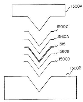
Figure 15 Illustrates an arrangement to attach a filter media to any of the
annealed
filter frames described herein. The frame 1515, is sandwiched between fitter
media portions
1560A and 1560B, which are respectively sandwiched between cushion elements
1500C
and 1500D, which layered assembly is located between heated top plate 1500A
and heated
base plate 1500B. Thus, resulting three-dimensional lamination of Figure 15
has a cross-
sectional view that is substantially conical. Application of heat and
presaire, via heated
platens 1500A and 15008, result in the integral bonding of the filter media
1560A and
1560B, and the interposed frame 1515. The filter frame configuration via the
lamination
CA 02648952 2013-09-05
procedure depicted in Figure 15 results in a filter assembly configuration
resembling a
"butterfly net."
Figure 16 schematically shows an alternate procedure for attaching filter
media to
an annealed filter frame. In Figure 16, an annealed filter frame 1615 is
sandwiched
between adjacent laminae of inner filter media 1660A and outer filter media
1660B. Heat
and pressure are applied via upper and lower punch and die platens 1600A and
1600B.
The application of heat and pressure results in an integral bonding of the
filter media 1660A
and 1660B and interposed frame 1615. During the heating and pressure
lamination
process, a vacuum may be applied in the lower platen 1600B thereby bonding the
filter
media and skeletal filter frame together. The filter media shown in Figure 16
is normally
interposed only within the immediate vicinity of the filter frame 1615.
Additionally, the
application of the vacuum can be used to optimize the filter frame geometry.
The method
shown in Figure 16 can produce a filter frame configuration that resembles a
butterfly net,
such as the one shown in the device of Figures 7A -7C. This method can also be
used to
produce a frame-supported "filter-in-filter," which is shown in further detail
in Figure 17,
described below.
Figure 17 shows an alternate embodiment of the present invention incorporating
a
two stage "filter-in-filter" design. The filter-in-filter design will provide
improved filtration
efficiencies, such as allowing each filter lamina to have a different porosity
by using an inner
filter media 1760A and an outer filter media 1760B. Alternatively, either
filter media 1760A
or 1760B can incorporate an integral Nitinol frame as one of the filter
members.
Alternatively still, both the inner and outer filter media 1760A and 1760B
could be an
integral Nitinol filter frame. Use of an uni-body Nitinol frame, such as those
described
herein, would provide additional structural benefits in the completed filter
frame apparatus.
Embodiments having a filter-in-filter design may be assembled using various
methods
including combinations of methods such as described with reference to Figures
15 and 16.
Figures 18A, 18B, and 18C schematically illustrate an annealing method in
which a
planar, two-dimensional filter frame is converted into a three-dimensional
configuration with
the use of an appropriate fixturing/tooling device, e.g., a mandrel. Mandrel
1800A, shown in
Figure 18A, is used to form the filter frame 1815 of Figure 18B into the
desired shape.
After cutting a flat metal sheet into the desired two dimensional
configuration, such as that
described above, the proximal ends 1865 of attachments struts 1855, i.e., the
endpoints of
the two-dimensional filter frame 1815, are collected at a point along the axis
of radial
symmetry as shown in Figure 18B. As depicted in Figure 18C, the filter frame
1815 is
16
CA 02648952 2013-09-05
,
placed onto the fixturing device, which, in this case, is the mandrel 1800A of
Figure 18A to
impart a defined, three-dimensional configuration, and the frame 1815 of
Figure 18B is
annealed to preserve the desired configuration. After annealing, the three-
dimensional filter
frame 1875 can be elastically deformed into its original two-dimensional shape
where a filter
media can be applied according to any of the methods described and illustrated
herein.
16a
CA 02648952 2012-01-04
Following the attachment of the filter media, the three-dimensional filter
configuration is
readily obtained.
Figures 19A, 19B, 19C and 19D illustrate an altemate filter configuration
using a
sombrero" shaped filter media 1960B with a supporting frame. To form the
sombrero frame
and filter shown in Figure 19D, a conical filter 1960, as shown in Figure 19A,
has its closed
distal end 1962 inverted toward the open proximal end 1961 of the conical
filter 1960A, to
form a convex, hat-like base as shown in Figure 19B. This inversion shortens
the filter
length, but retains the original area of the filter element 1960. Next, the
convexity is
increased until the apex 1963 extends beyond the open end 1961, as shown in
Figure 19C.
The filter 1960 thus has been shortened further, but the effective filter area
still remains
identical to the original conical filter area. The sombrero filter 1960B is
attached to frame
1975, Figure 19D. The frame includes attachment struts that are fixed to a
connecting
member 1970, which in tum is cooperatively associated with a guidewire 1955.
Compared
to conventional conical filter frame designs, the sombrerO filter frame allows
more surface
area per unit length, or, altematively, reduces filter length without
compromising filter surface
area and deflection of the trapped debris away from the vessel centerline. The
desired
sombrero filter frame configuration will also increase the reliable removal of
entrapped
debris.
Figures 19E and 19F depict an alternate filter sack configuration, also
designed to
collect and hold embolic debris away from the vessel centerline. In this case,
an asymmetric
cone shaped filter media sack 1990 is produced and attached to the filter
frame 1960.
Collected emboli will tend to collect at the tip of the sack 1990 and are held
offset in the
vessel, thus allowing relatively unperturbed flow at the vessel centerline.
As shown in Figures 20A, 20B and 20C, a filter in accordance with the present
invention can be used to deliver a pharmaceutical substance, anti-thrombotic
agent, drug,
etc., into the blood flow in a host lumenNessel by deploying the fitter in a
lumenNessel of
interest. In Figure 20A, a filter device such as that described in Figure 17
above, can be
loaded with a pharmacological agent in one or more different areas before
delivery into the
hoet. Thus, the drug can be loaded between layers of the filter media. The
drug 2098 may
be retained within the zone/space/area between the inner filter media 2060A
and outer filter
media 2060B ready to be delivered to the host
Instead of using the filter-in-filter design of Figure 17, any of the other
filter
configurations described herein can be used by imbibing the drug into the
filter media itself.
As shown in Figure 20B, the drug 2098 can be imbibed into the media 2060
itself.
Figure 20C illustrates drug administration in the host by deploying the drug
delivering
system of Figures 20A or 20B in a host lumen/vessel so that the blood flows
through the
filter media to elute the pharmacological agent, e.g., drugs. This method of
localized drug
17
CA 02648952 2008-12-30
=
=
delivery is effective for eluting a pharmacological agent contained either
between adjacent
layers of filter media or imbibed directly into the filter media. Fluid flow
through the filter
device of Figures 20A or 20B, or any other filter configuration described
herein containing
pharmacological agents provides a mechanism of mass transfer to downstream
perfusion
beds. The pharmacological agent could be pre-loaded into the filter or
injected post
deployment perhaps through an extension of the supportiguidewire.
As shown in Figures 21A and 21B, occluding device 2175 can be formed as a
detachable endoluminal filter frame that can be implanted in the host. The
occluding device
2175 thus implanted can either be permanently implanted or retrieved at a
later point in time,
such as is required in vena cava filtering applications. As shown in Figure
21A, blood flow
through the host can be obstructed by the implantation of the filter frame
apparatus 2100.
The filter frame apparatus 2100, used as an indwelling or implantable
occlusion device is
shown in Figure 21A. As shown in Figure 21B, a guidewire or support wire 2180
includes
a distal end 2181 that may be detached from proximal connector 2170 that is
connected to
the occluding device 2175 or filter frame apparatus 2100. The support wire
2180 is used to
position or remove occluding device 2175 or filter frame apparatus 2100 frorn
a lumen in a
host. The guidewire tip 2181 may be of any design for detachment from or
reattachment to
proximal connector 2170. Thus, the guidewire 2180 can have any capture
capability,
including screw threads, magnetic, ball-and-socket, male-female attachment,
bayonet, or
any type of coupling that will allow the guidewire 2180 to detach or reattach
to the proximal
connector 2170 for placement or movement therein.
Figures 22A and 22B illustrate the use of a filter (similar to the filters100
or 700,
respectively shown in Figures 1D or 7C) to remove flow obstructions or to
function as a
thrombectomy device to remove intraluminal thrombus, for example. Figure 22A
shows an
obstruction at the lumen wall in a blood vessel of the host. Though commonly
the lesion will
have formed in a restrictive manner, the lesion is shown in a cross-sectional
area with an
upper and a lower component, that has narrowed the effective diameter of the
lumen. Filter
2200 includes sharpened support members 2285 to enable the filter to be used
as a type of
scraper. The frame 2275 shown herein includes a filter media 2260 as a "catch
bag." In
Figure 22B, the filter 2200 is pulled with sharpened members 2285, effectively
shearing the
obstruction/lesion from the vessel wall of the host. As the lesion is sheared
from the wall,
sheared lesion parts are collected in the catch bag or filter media 2260. In
this manner, the
present filter frame can be used to remove lesions and collect the debris
dislodged into the
blood stream, to lessen the possibility of clotting downstream of the host
vessel. This
approach can likewise be used to capture and remove foreign objects from
bodily
passageways.
18
CA 02648952 2008-12-30
=
Figures 23A, 23B and 23C respectively illustrate the use of the inventive
filter as an
anchoring guidewire to facilitate the retention of a guidewire position in
tortuous vessels of
the renal circulatory system, and in particular for branch lumens offset at
angles of
approximately 900. Using the inventive filter frame as an anchor avoids or
minimizes
damage to the host vessel, and specifically avoids or minimizes damage to the
endothelium
of the host lumen/vessel. Figure 23A shows a lesion 2300A in a branch
lumen/vessel
2300B associated with the renal anatomy of a host. In the non-limiting
embodiment of
Figures 23A¨ 23C, the branch lumen 2300B includes an approximate 90 turn
toward the
existing anatomy shown. As illustrated in Figure 23B a filter frame 2375 is
positioned and
anchored in a renal circulatory vessel 2300B to fix the position of the
support wire 2380. A
slight pressure is imposed on the support wire 2380 and the approximate 90
turn is
extended to more than 90 without dislodging or altering the position of the
guidewire in
relation to the host anatomy as shown in Figure 23B.
As shown in Figure 23C, a therapeutic catheter 2300C can be inserted over the
support wire 2380 of the filter frame to perform the intervention. As a
result, therapy devices
can more easily negotiate a greater than 90 bend as shown in Figures 23B and
23C. Such
therapy devices include, but are not limited to balloons, stents, etc. A
further useful aspect
of this embodiment is that, during its use, a long "exchange length" guidewire
is
unnecessary. Since this device is capable of maintaining it's positioning
after deployment,
the necessity of "rapid exchange" or "monorail" catheters are obviated.
Figures 24A, 24B , 24C, 24D and 24E show a further embodiment of the present
filter frame assembly, which is intended to function as an implantable
endoprosthesis 2476.
As shown in Figures 24A and 24B, the initial seamless filter frame 2475 is
formed from a
loop-type frame 2415 from the same precursor material. In Figure 24C, the
proximal end of
an open-ended "windsock" shaped graft component 2477 is attached to the loop
of the filter
frame 2475 to form an endoprosthesis 2476. In Figure 24D, the loop-type frame
2475 with
the attached open-ended windsock is deployed proximal to an aneurysmal defect,
and the
windsock shaped graft component 2477 extends downstream of the frame,
effectively
excluding the aneurysm 2400A. Thus, frame and the opened ended sock function
as an
implantable prosthetic vascular conduit where the filter frame 2475 functions
as an
anchoring stent, and the open-ended sock functions as a biocompatible liner.
This device,
shown in Figure 24E, may then be optionally lined with a stent 2480. This
embodiment finds
use as a stent and graft combination where the stent element would be deployed
proximal to
the intended therapy site and the graft element would be configured to be
deployed by blood
pressure.
Figures 25A-25H illustrate an exemplary delivery system for deploying the
present
filter frame 2575 or filter 2500 of the present invention. Figure 25A
illustrates a frame 2575
19
CA 02648952 2008-12-30
or frame-filter 2500, such as frame 175 or frame-filter 100 of Figures 1D or
1E, frame 375 or
frame-filter 300 of Figures 3B and 3C, or any of the other frame or frame -
filter assembly
herein described, attached to a support or guidewire 2580 and positioned
within a tubular
delivery sheath 2500A of a delivery catheter. Figures 25B-25D illustrates
front views taken
from sectional plane A-A of Figure 25A, but without the frame 2575 or frame-
filter 2500.
The section A-A1 (Figure 25B) illustrates a dual lumen extrusion catheter
sheath. Section
A-A2 (Figure 25C) illustrates a single lumen extrusion having an additional
covering formed
from a shrink tube. Section A-A3 (Figure 25D) illustrates a second lumen
adhered to the
inner diameter of the tubular delivery sheath 2500A of Figure 25A.
Figure 25E ¨ 25H illustrate the perspective detail of external guidewire 2580
loading
of a catheter lumen. Figure 25E is a front view of the Figure 25G. Figure 25F
illustrates
the catheter having a longitudinally extending indented channel, which, as
seen in Figure
25G is circumscribed by a tubular section 2500C. The guidewire 2580 is
inserted into the
longitudinally extending channel 2500B.between the external wall of the
catheter and the
tubular section 2500C. In use, a filter frame or filter-frame construct is pre-
loaded into the
distal end of the sheath adjacent to an exterior wire guide channel. The
exterior wire guide
is adapted to receive a guidewire in a rapid exchange configuration, however,
unlike the
prior art, the filter frame and guidewire 2580 are completely segregated and
no aperture
exists.
Figures 26A, 26B, 26C, 26D and 26E illustrate a method of using a filter frame
assembly 2600 in accordance with the present invention. In Figure 26A, a
lumen/vessel
2600A of the host has a lesion 2600B. A guidewire 2680 is deployed into the
lumen/vessel
2600A past the target lesion 2600B. Thereafter, guidewire 2680 is back-loaded
into the
delivery system 2600C, such as the one described in Figures 25B-25D, 25F-25G,
or Figure
27B. Then the delivery sheath 2600C is advanced across the target lesion
2600B. The
delivery sheath 2600C is withdrawn, thereby allowing a self-expanding filter
2600 to deploy.
The self-expanding filter 2600 is normally designed to deploy spontaneously
after the
delivery sheath 2600C has been withdrawn in this manner. Thus, as shown in
Figure 26C,
the filter 2600 is deployed downstream of the lesion 2600B. A therapeutic
catheter 2600D,
such as an angioplasty balloon, is routed over the support wire 2680 in Figure
26D to treat
target lesion 2600B. As also shown in Figure 26D, when the therapy is
performed, the filter
2600 functions to capture any emboli dislodged or removed by the therapeutic
catheter
26000. Thereafter, as illustrated in Figure 26E, the filter 2600 is removed
via insertion of a
tubular capturing catheter 2600E over the support wire and retraction of the
filter 2600 into
the capture catheter 2600E is performed. This retraction can be performed by
pulling the
filter 2600 partially back into the capture catheter lumen 2600E, effectively
trapping the
- CA 02648952 2008-12-30
-7
emboli 2600F. In this Manner, the lesion is dissipated through a therapeutic
catheter without
the result of any of the dislodged emboli or debris dislodging into the host.
Figures 27A, 27B and 27C illustrate a lumen 2710 having an auxiliary,
internally
positioned channel 2720 for receiving a guidewire 2730. Figure 27A illustrates
the tip of the
sheath having an internally located, peripherally positioned auxiliary channel
2720 formed by
"pinching" the end of the tube wall as shown in Figure 27B. Figure 27C shows
the
guidewire 2730 inserted through into the slit opening in the side of the
catheter and exiting
the tip.
Figure 28 illustrates the use of the inventive filter 2800, as a vena cava
filter. Since
*10 the inventive filters described herein may be readily detachable, the
filter 2800 can be readily
detached from a deployment guidewire.
Figure 29A illustrates a planar two-dimensional seamless pattern, formed from
metallic material, or any other suitable biocompatible material. Figure 29B
illustrates a
three-dimensional stent member formed from the planar two-dimensional pattern
of Figure
29A, for use as an intraluminal stent. When extremely thin wall sections are
required, such
as in coronary stents, it is appropriate to fabricate the device from a planar
sheet of material.
Planar material can be manufactured thinner than tubing due to the extra
requirements of
concentricity placed upon tubing stock. It should be noted that although only
one design has
been depicted, a wide variety of patterns and cell geometries may be produced
from planar
material. The various cell geometries are defined by the interconnected struts
of the stent. In
Figures 29A and 29B four interconnected struts 2910 define the four sided cell
2920. This
planar material may be metallic or polymeric or a combination thereof, and in
any case, may
also be porous. Once the flat pattern is fabricated, it is formed into a 3-D
shape (in the
depicted instance, an open mesh tube). The formed stent may be either
plastically
deformable (and thus made from a malleable starting material) or may be self-
expanding, in
which case a super-elastic, pseudo-elastic or shape memory material may be
used.
Subsequent processing such as thermal treatment, diametric reduction, de-
burring and
polishing may be incurred, depending upon the specific stent design. It should
be
understood that multiple 3-D stent "units" could be manufactured in such a way
and attached
together to form a much longer device.
Figure 30 depicts a view of a flat pattern of filter frame 3010A and integral
tether
element 3010B geometry as it would be cut from a tube. This tube may be made
of a shape
memory alloy such as Nitinol. Cutting could be accomplished by a variety of
methods
including machining, laser cutting, stamping or etching.
Figure 31 depicts the flat pattern geometry of Figure 30 subsequent to forming
and
annealing at a larger, functional size. Upon annealing, the filter frame 3010A
resiliently
21
CA 02648952 2008-12-30
4
' maintains this larger diametrical profile and the at least one
tether element 3010B extends
seamlessly from it.
Figures 32A-32E depict alternate articulation segments formed as an integral
part of
the tether element thereby forming different tether element geometries, which
allow
articulation of the tether elements 3010B in relation to the filter frame
3010A. Figure 32A
depicts the tether element 3010B with an area of reduced strut width e.g.
reduc,ed cross-
sectional area, to allow increased flexibility. Figure 32B depicts tether
element 3010B with
several individual areas of reduced strut width to allow increased
flexibility. Figure 32C
depicts tether element 3010B with a reduced width and formed "hinging" area to
increase
flexibility. Figure 32D depicts tether element 3010B with a reduced width and
several
formed "hinging" areas to increase flexibility. Figure 32E depicts tether
element 3010B
divided in two for a portion of its length. This division effectively
increases the tether element
flexibility so as to allow articulation. The articulation segment of the
tether element, therefore,
= is configured to enhance the flexibility of the filter apparatus (and
thus, conformance to the
host vessel wall) as well as to minimize inadvertent trauma translated to the
host vessel wall
by movement or manipulation of the guidewire.
An articulation segment of the tether elements or struts is a desirable
feature in that it
allows adequate vessel wall apposition of the filter frame when the filter
device is deployed in
a curved segment of anatomy. In a curved segment, the tether element
articulates and
deflects to adjust for a non-linear deployment situation (See Figure 14).
Thus, the filter
frame itself can maintain an uncompromised and fully deployed condition.
Likewise,
because of its ability to attenuate longitudinal translation, the articulation
segment provides a
means of mitigating trauma of the host vessel wall due to guidewire
manipulation. It should
also be noted that the required deflection and articulation of the tether
elements could be
bought about by metallurgical means rather than, or in combination with,
geometrical means.
For instance, the tether 3010B and frame 3010A elements of Figures 32A-E,
although
= seamless and integral, may be exposed to different thermal processing
parameters (for
example: through the use of fixturing to provide differential heat sink
qualities), thus
rendering the tether 3010B ductile and pliable while the frame 3010A maintains
the stiffness
required for adequate vessel wall apposition.
The articulation segments, though described with respect to the various frame
patterns can be incorporated into any of the endovascular devices described
herein. An
articulation segment is a localized region that provides enhanced longitudinal
flexibility. A
localized region may have a cross-sectional area that is the same as the
remaining part a
strut, but differs in geometry. Alternatively, the localized region could have
the same
geometry but a different cross-sectional area, or both the cross-sectional
area and geometry
22
CA 02648952 2008-12-30
4
of the localized region differ from the remaining part of the strut. An
endovascular stent can
have articulation segments in any of the interconnected struts of Figures 29A
and 29B.
EXAMPLES OF THE PRESENT INVENTION:
EXAMPLE 1: Nitinol Sheet Filter Frame and integral Tethers
A radially-symmetric geometrical pattern comprising interconnected struts
forming
closed polygonal shaped cells was chemically etched from a sheet of Nitinol
(NiTi) to
produce a skeletal filter frame. The etching, preferably photoetching of
Nitinol (Kemac
Technologies, Irwindale, CA) is continued to achieve a desirable material
thickness, to
optimize the moment of inertia of the struts and to polish the surface finish.
This filter frame is then subjected to a thermal treatment to set the phase
transition
=
temperature of the NiTi to approximately 37 C by heating the filter frame to a
temperature of
about 450 C for about 10 minutes in an air convection oven (Carbolite
Corporation,
Sheffield, England ) followed by a rapid quench in ambient temperature water.
The NiTi filter frame was then laminated between two (2) layers of an adhesive-
coated porous polymer. The layers were positioned with the adhesive sides
facing toward
each other, and facing toward the NiTi. The adhesive was used to adhere the
layers of film
together as well as to the NiTi wire framework. A sacrificial porous polymer
cushion material
was used on each side of the device during this lamination procedure to
provide compliance
Of the surface during compression. This compliance allows the earlier
mentioned porous
polymer membrane to conform to the wire shape. The composite sub-assembly
which
included cushion, porou8 polymer/adhesive laminate, NiTi, adhesive/porous
polymer
laminate, and cushion layers was then compressed in a SST fixture and heat
treated at
320 C for 45 minutes in an air convection oven (Grieve Oven, The Grieve
Corporation,
Round Lake, IL).
Once the 'sandwiched' device was removed from the heat source and allowed to
cool, the sacrificial cushion material was peeled away from each side of the
device and the
NiTi wires were disengaged from the fixture. A circular shape of approximately
0.625" in
diameter was trimmed into the porous polymer using a 20-watt carbon dioxide
laser. The
remainder of porous polymer was trimmed from the wire frame by hand and
discarded-.
Following the laser trimming operation (which can also be used to create the
necessary pores in the filter media), the radially-oriented arms (struts) of
the device were
folded up and back on themselves to achieve a hollow, three dimensional, semi-
conical
shape. To maintain the device in this configuration, the NiTi struts were
inserted into a SST
tube. This tube measured approximately 0.05" in length X 0.035" outer diameter
X 0.025"
inner diameter. This tube and indwelling NiTi wires were then crimped to a
0.014" diameter
23
CA 02648952 2012-01-04
guidewire to provide a guidewire based endoluminal embolic protection device.
The device
resembled a three dimensional "whisk" shape with a pleated porous polymer
filter element
attached to it
The resulting pleats are designed to increase filter media surface area over
the
generally conical shapes found in the prior art. This increase in surface area
also allows for
a shorter filter length which enhances deliverability of the device by a)
decreasing friction in
the delivery catheter and b) improving device overall flexibility.
EXAMPLE 2: Nitinol Tube Filter Frameend Integral Tethers.
A 1.3 mm Nitinol tube with a wall thickness of approx 0.1mm (obtained from
Nitinol
Devices and Components, Fremenot, CA) was laser cut (Laserage Technologies
Inc,
Waukegan, IL) to a single, undulating 6 apex ring geometry with integral
tethers. This frame
was then lightly grit blasted at 40 psi with 20 micron silicon carbide media
in a grit blasting
machine made by Comco Inc, Burbank, CA. The ring with integral tethers was
then gently
_ oven (Grieve Oven, The Grieve Corporation, Round Lake, IL) set at 320 C
for approx. one
minute followed by air cooling to room temp.
The NiTi frame was then set atop a filter sack and attached though the
application of
localized heat (the heat causing the FEP coating on the ring to re-melt and
flow onto the
was then inserted into the gold band (from the opposite direction of the
tether lines). The
marker band was then crimped to secure the tethers and guidewire together. A
small
* Trademark
24
CA 02648952 2012-01-04
realize that attachment of the filter to the guidewire could be accomplished
by adhesion,
welding, soldering, brazing, a combination of these, or a number of other
methods.
Upon drying, this embodiment of the endoluminal embolic filter is ready for
testing.
Various illustrative examples of the invention have been described in detail.
In
addition, however, many modifications and changes can be made to these
examples without
departing from the invention as described herein.