Note: Descriptions are shown in the official language in which they were submitted.
CA 02649518 2008-10-10
WO 2007/120775 PCT/US2007/009044
i
SUCTION DOME FOR ATRAUMATICALLY
GRASPING OR MANIPULATING TISSUE
CROSS-REFERENCE TO RELATED APPLICATIONS
[001] This applicatiion relies on and clairns the benefit of the filing date
of U.S.
provisional patent application number 60/791,897, filed 14 April 2006, the
entire
disclosure of which is hereby incorporated herein by reference in its
entirety.
SUMMARY OF THE INVENTION
Field of the Invention
[002] The present invention is generally directed to a surgical device, and
more
particularly to a suction dome for atraumatically grasping and/or manipulating
tissue.
Backjzround of the Invention
[003] Surgical forceps are used for grasping, retracting, and/or dissecting
tissues
during surgical procedures. In essence, forceps act as an extension of a
surgeon's
hands in limited areas of access. Forceps may be used for a variety of
purposes, from
grasping tumors for dissection to moving and manipulating intervening tissues.
[004] Although there are many conf gurations, conventional surgical forceps
are
generally characterized by two opposing fingers, or jaws, which are moved
together in
order to grasp tissue between them. These types of forceps operate by applying
inward compression forces on the tissue until it may be lifted or manipulated
without
slipping. In practice, however, such mechanical forceps exhibit poor tissue
grasping
and holding capabilities. One reason for this is because they require a
certain amount
of friction to exist between the jaws of the forceps and the tissue surface.
The less
friction that is present, the more inward forces must be exerted before a
tissue can be
successfully lifted or manipulated. On the other hand, the more a tissue is
compressed, the more Iikely it is to be injured or to rupture. This is a
particular
concern with fluid-filled cysts, especially where uncertainty exists with
respect to the
wall thickness and resistance to rupture.
-1-
CA 02649518 2008-10-10
WO 2007/120775 PCT/US2007/009044
[0051 In order to obtain a better grasp on tissue with less compression force,
some forceps include either sharp teeth or serrations in the jaws. Examples of
such
teeth include the Richard Wolf 8385.10, 8385.13, and 8383.471 type grasping
forceps.
Although teeth and serrations may help to prevent slipping, they can also
cause trauma
by way of puncturing or lacerating tissue. Such punctures in tissues or tumors
may
increase the risk of patient infection or allow undesirable spreading of
malignant
tumor cells. At the very least, such teeth cause unnecessary damage to target
tissues,
and especially tissues that must be grasped repeatedly or require a great deal
of
manipulation.
[008] Another problem associated with conventional surgical forceps is that of
a
target tissue "bouncing" away from the tip of the forceps as the surgeon
attempts to
grasp the tissue. This occurs frequently with large, smooth, and/or resilient,
or hard
firm tissues (such as glands, organs, cysts and parenchymal tissues). One
factor that
can compound this problem is the limited opening width of the jaws. If a
ta.rget tissue
is larger than the opening in the forceps, and/or if sufficient friction
between the
forceps and tissue is not present, "away"-ward forces might override
compression
forces "normal" to the surface and the tissue will bounce away from the
forceps. In
addition, certain tissue types can also present a challenge to grasp without
causing
injury. For example, ovarian tissue can be difficult to grasp and control
without
tearing and bleeding. Currently, grasping ovarian cysts with conventional
forceps
without rupture is very difficult. Any occurrence of rupture defeats the
purpose of a
cystectomy (making it more difficult to remove). In addition, other tissues,
such as
the spleen, pose serious risks to the patient if ruptared. Thus, not only is
such
"bouncing" an inconvenience to the surgeon, but may consume valuable time
during a
procedure and increase overall health risks to the patient.
[007] Other instruments for engaging and holding tissues have been devised
that
utilize suction to.impose traction on tissues instead of compression. However
some
devices, for example as disclosed in U.S. Patent No. 3,896,810, use suction
structures
composed of rigid materials, such as metal. These structures are not very
flexible or
conformable to different tissue surfaces, and thus are not suited for engaging
-2-
CA 02649518 2008-10-10
WO 2007/120775 PCT/US2007/009044
irregularly shaped, and/or delicate, tissues. Other devices, for example as
disclosed in
U.S. Patent No. 6,641,575, use somewhat flexible suction cups. However, these
still
only physically engage the tissue around the periphery of the cup. Thus,
problems
-occur if the vacuum seal around the periphery is broken (e.g., due to poor
contact with
an irregular tissue surface). In this case, the suction force on tissue is
significantly
weakened arid compromised. Such weakened contact could allow undesirable
fluids
to escape (or enter) the area. This result could increase risk of infection or
spreading
of malignant tumor cells.
[0081 What is needed, therefore, is a device that is able to grasp tissue in a
reliable manner while maintaining tissue integrity. ln addition, what is
needed is a
device that provides improved tissue grasping and manipulating capabilities in
a less
traumatic manner. The suction dome of the present invention is able to meet
these
needs and, at the same time, provides a significant improvement over the prior
art.
These and other advantages of the present invention will become apparent from
the
disclosure herein.
SUMMARY OF THE INVENTION
[0091 In a first aspect, the present invention provides an expandable suction
dome for use at the distal end of a surgical instrument for atraumatically
grasping
and/or manipulating target tissue. The invention also provides a surgical
instrument
including a suction dome comprising: a non-permeable outer membrane defining
an
inner chamber; and a permeable tissue-engaging membrane extending across the
base
of the chamber. In general, the present invention provides a surgical
instrument, such
as a forceps or laparoscope, having a longitudinal suction channel and an
optional
needle channel. The suction dome is translated in a collapsed state through
the
suction channel and is operatively deployed after exiting the distal end of
the
instrument.
[0101 The outer membrane of the suction dome may be coupled to a plurality of
expandable arms and is sufficiently supported to withstand internal vacuum
pressures
and external bodily pressures or artificially produced pressures when
deployed. A
-3-
CA 02649518 2008-10-10
WO 2007/120775 PCT/US2007/009044
primary puxpose of the non-permeable outer membrane is to help maintain a
vacuum-
tight seal between the dome and tissue. The tissue-engaging membrane, on the
other
hand, is sufficiently permeable so as to allow the vacuum to be evenly
distributed over
the entire surface in contact with the tissue. In this way, the suction dome
is able to
atraumatically grasp and manipulate tissue by cupping it with the permeable
membrane. The supporting arms in the dome wall also act as graspers and hold
the
tissue as the wall of the dome is retracted back into the sheath. An optional
needle
may also be inserted through a needle port of the instrument for puncturing,
irrigating,
and/or aspirating tissues with fluids or medications. The target tissue may
include any
tissue, including but not limited to: tumors, cysts, organs, and glands. In
addition,
although a forceps and laparoscope have been mentioned by way of example, it
is to
be understood that any instrument including, but not limited to: endoscopes,
bronchoscopes, and catheters may also be used with the suction dome.
BRIEF DESCRIPTION OF THE DRAWINGS
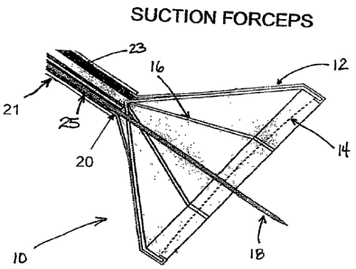
[Oll] Figure 1 is a perspective view of a suction dome showing internal
components and constructed in accordance with the teachings of the present
invention.
[0121 Figure 2A is an isometric view of a surgical instrument comprising a
suction dome, sheath, and handle according to one embodiment.
[013] Figure 2B is a perspective cut-away view of the suction dome with
respect
to the distal end of the surgical instrument shown in Figure 2A.
[0141 Figure 2C is an exemplary cross-sectional illustration of the surgical
instrument taken along the middle portion of the instrument.
[0151 Figure 3 is an exemplary isometric view of the suction and aspiration
ports
at the proximal end of the surgical instrument.
[016] Figure 4Aa is an isometric view of the suction dome in a partially
collapsed state according to another ernbodiment of the present invention.
[0171 Figure 4B is an isometric view of the suction dome in a collapsed state
prior to deployment and/or retraction.
-4-
CA 02649518 2008-10-10
WO 2007/120775 PCT/US2007/009044
[0181 Figure 5A illustrates a general laparoscopic procedure in which the
suction
dome may be used.
[019] Figure 5B illustrates aspiration of a cyst using a needle introduced
through
the suction dome.
[0201 Figures 6A and 6B illustrate removal of a cyst through- an abdominal
cavity
using the suction dome.
[021] Figure 7 is another isometric view of the suction dorne showing the
permeable membrane, and perforations therein, constructed in accordance with
the
teachings of the present invention.
DETAILED DESCRIPTION OF EMBODIMENTS OF THE INVENTION
[0221 Figure 1 shows a representative view of a suction dome according to the
present invention. This figure shows the suction dome (10) at the distal end
of an
instrument having an outer sheath (21) and an inner sheath (20) defining a
suction
channel (23) according to one embodiment of the present invention. In this
figure, the
suction dome (10) is shown in a deployed state and has an inverted umbrella-
lilee
shape defining an internal chamber. The suction dome (] 0) is comprised of a
non-
permeable, outer membrane (12) forming the outer wall; and a semi-permeable
tissue-
engaging membrane (14) extending across the base of the chamber for securely
and
uniformly engaging a surface area of tissue. In this embodiment, the outer
membrane
(12) is integral with, or connected to, the distal portion of the inner sheath
(20).
Preferably, the inner sheath (20) is slidable within the outer sheath (21) for
retraction
and extension of the dome. The suction dome (10) may also include several anns
(16) coupled to the outer rnembrane (12) for support and deployment thereof.
Arms
(16) may be composed of a resilient and/or memory material such that they
expand
automatically as the dome (10) is extended out of the outer sheath (21).
[023] The outer wall (12) and inner sheath (20) of the dome are constructed of
a
non-permeable membrane, which can be made out of any suitable material. It is
desirable to have a non-permeable membrane to maintain sufficient vacuum
between
the suction channel (23) and the base membrane (14). The outer membrane (12)
is
-5-
CA 02649518 2008-10-10
WO 2007/120775 PCT/US2007/009044
also sufficiently. supported (e.g., by arms (16)) so as not to collapse under
negative
vacuum pressure within the dome or external bodily pressures. The outer
membrane
(12) is sufficiently pliable so as to be collapsed when not in use. Suitable
materials
for the outer membrane (12) and/or inner sheath (20) include, but are not
necessarily
limited to: plastic, polyethylene, silicone, rubber, or combinations thereof,
etc. In
addition, it may be desirable for the outer membrane (12) (or a portion
thereof) to be
transparent so as to allow visualization of the tissue underneath or of the
procedure as
it is carried out. It is also desirable for a vacuum-tight seal to be made
between the
suction dome (10) and the tissue to prevent any gas or fluid from entering
and/or
escaping from the interior of the dome. Target tissues may include any type of
bodily
tissue, including, but not limited to organs, glands, cysts, and tumors.
Examples may
include ovarian cysts, gall bladder, ectopic pregnancy, etc.
[0241 The base of the dorne, or tissue-engaging membrane (14) is constructed
of
a semi-permeable membrane. This type of inembrane can be used to allow
negative
air pressure (as applied through the suction channel) to be evenly distributed
over its
surface area and transmitted onto the surface of the tissue in contact. When
suction is
applied, membrane (14) allows negative pressure to pass directly through
perforations,
or holes, therein. In this way, negative pressure is substantially uniformly
applied to
the tissue from the base surface of the dome (10) over the entire tissue
surface area in
contact. The diameter of inembrane (14) should be large enough to apply to an
adequate surface area of the tissue. The membrane (14) is preferably composed
of a
flexible and pliable material that enables the target tissue to be closely
cupped therein
as suction is applied. Thus, the dome is able to more securely engage the
target tissue
and hold it in place with minimal, or no, trauma to the tissue itself.
Moreover, close
contact of inembrane (14) with delicate tissue surfaces such as fluid-filled
cysts may
,
serve as additional reinforcement (as they are penetrated by needles), thereby
reducing
rupture or tearing associated with thin tissues. The permeable membrane (14)
is
sufficiently pliable so as to be collapsed when not in use. Suitable materials
for the
tissue-engaging membrane (14) include, but are not limited to: plastic,
polyethylene,
silicone, rubber, or combinations thereof, etc. It is possible that part or
all of the
-6-
CA 02649518 2008-10-10
WO 2007/120775 PCT/US2007/009044
permeable and non-permeable mernbranes may be composed (in whole, or in part)
of
the same, or different, materials. If the sarne materials are used, the tissue-
engaging
membrane (14) may still be somewhat more flexible than the outer membrane (12)
(e.g., by virtue of the perforations therein). It is also to be understood
that membranes
(12) and (14) may comprise one or more layers of material.
[025] The permeability in membrane (14) may be achieved in any number of
ways, the particular way not being critical to practice of the invention. For
example, it
can be by a plurality of discrete, spaced apart, holes therein or may be
intrinsic to the
material itself (such as with a fabric mesh). In addition, %irregularly shaped
target
tissues, for example, may be more reliably engaged. The size, number and
spacing of
the perforations in the material may also vary depending upon the type of
target tissue
and the necessary amount of suction.
[0261 In operation, the outer membrane (12) and/or inner sheath (20) are
typically operably coupled to arms (16) such that outward extension of the
arms (16)
at the distal end of the instrument causes the suction dome (10) to be
deployed as
shown in Figure 1. The arms (16) may open and close the dome (10) as they are
extended beyond, or retracted within, the outer sheath (21). Arms (16) may be
comprised of a resilient and/or shape-memory material such that they
automatically
expand upon extension from outer sheath (21). In addition, the outer membrane
(12)
~
is integral with, or connected to, the distal end of inner sheath (20) and may
be
extended or retracted e.g., via a proximal retracting mechanism (27, shown in
Figure
2A) operably coupled to the inner sheath (20). In an alternative embodiment,
the arms
(16) may be controlled, e.g., by a longitudinal support wire, or actuation
cable (not
shown) and operably extended or collapsed by movement of a proximal retracting
control mechanism (27, shown in Figure 2). Additionally or alternatively, the
arms
(16) may be composed of a preformed material that automatically extends and
collapses upon exit, or entry, of the outer sheath. As shown in Figure 1. and
Figure 7,
the arms (16) may also include inwardly-folding joints at the distal-most
portions.
Such distal joints on the arms (16) fold inwardly as the dome is retracted,
thereby
-7-
CA 02649518 2008-10-10
WO 2007/120775 PCT/US2007/009044
more firmly grasping and securing tissue therein. Figure 7 also'illustrates
the plurality
of spaced apart perforations, or holes, in the permeable membrane.
[0271 Another advantage of the suction dome according to embodiments of the
present invention is increa.sed maneuverability of tissue. For example, when a
tissue
(such as a cyst) is dissected and freed, the suction dome (10) may be used to
control
movement of the tissue in vertical, horizontal and rotational directions. For
increased
maneuverability, the suction dome may be used in conjunction with a forceps
having
an articulating handle and sheath.
[0281 Also shown in Figure 1 is an optional need] e or cannula (18) which may
be
introduced through a needle channel (25) in the instrument. At the distal end
of the
instrument, the needle (18) may continue to be advanced through the interior
of
suction dome (10) and through perforations in order to penetrate the target
tissue.
Needle (18) can be used for puncturing, irrigating, and/or aspirating tissue.
For
example, if the tissue is a fluid-filled cyst, the needle (18) can be used for
draining the
contents of the cyst. Additionally or alternatively, the needle (18) may be
used for
introducing various fluids or medications to tissues.
[029] Figure 2A illustrates proximal, middle, and distal portions of an
instrument
used with the suction dome according to one embodiment. In the figure, the
middle
portion of the instrurnent comprises a flexible, or rigid, outer sheath (21)
through
which arm control mechanism, suction (23) and needle (25) channels extend
longitudinally. Such outer sheaths for laparoscopy and other surgical tools
are k.nown
in the art, and any suitable size sheath rnay be used according to the present
invention.
t
Typically, suitable diameters for the outer sheath (21) range from 3- 20 num,
and
preferably 3- 10 mm, although other diameters may be used. For example,
diameters
between 10 - 20 mrn may be used for resection of larger tissues such as gall
bladder,
appendix, myomata, etc. The working length of the instrument may be, for
example,
240, 310 or 430 mm, although other lengths may be used. Working lengths of 240
mm are suitable for introduction through an accessory port and lengths of 310
and 430
mm are suitable for introduction through an accessory port or laparoscope. At
the
distal end of the instrument, the suction dome (10) is shown deployed. When a
-8-
CA 02649518 2008-10-10
WO 2007/120775 PCT/US2007/009044
procedure is complete, the dome is retracted back into the sheath, for
exarnple by
releasing a retraction control mechanism (27, discussed below).
[030] AIso at the proximal end of the instrurnent of Figure 2A is a handle
with an
extraction/retraction control mechanism (27) operatively coupled to the
suction dome
(10) via inner sheath (20, shown in Figure 213). In a.n alternative
embodiment, the
retraction control mechanism (27) may be operatively coupled to the suction
dome
(10) via a support wire and/or an actuation cable (not shown), or any other
conventional means, such as those known in the art. The retracting control
mechanism (27) shown in Figure 2A is a conventional scissor-like thumb and
forefinger control; however, it should be understood that the retracting
control
mechanism (27) and/or handle could alternatively comprise other forms, such as
a
spring thumb ring and a friction stop. Additionally, instead of manual
manipulation,
the retraction control mechanism (27) may also be automated.
[0311 Figure 2B illustrates a partial distal view of the suction dome (10)
with
respect to the interior of the instrument. Outer sheath (21) is shown as well
as inner
sheath (20) defining suction channel (23). Figure 2C illustrates an exemplary
cross-
; ,
sectional view ofthe elements. In-this figure, suction channel (23) is defined
by inner
sheath (20) disposed within outer sheath (21). In addition, optional
components may
also include: a support wire, or actuation cable (29), and a needle channel
(25). It is
to be understood that the arrangement of the iriternal components is provided
only by
way of example, and other arrangements may be possible.
[032) The distal, middle and proximal portions of the instrument may be fixed
or, alternatively, rnay be modular so as to improve ease of interchangeability
with
different sized lumens and suction domes. Such interchangeability helps to
reduce
replacement costs as well as cleaning and sterilization times. Current modular
laparoscopes and forceps (including separate handles, mechanical jaw inserts
and
sheath tubes) include, for example: ConMed's DetachaTipT"' System, SpeedLockT"
Laparoscopic Instrumentation, and R.ichard Wolf modular/reusable forceps by
Medical Instruments Corporation. For exarnple, the suction dome of the present
-9-
CA 02649518 2008-10-10
WO 2007/120775 PCT/US2007/009044
invention rnay be used with a conventional modular sheath and handle providing
a
vacuum channel and optionally a needle channel.
[033] At the proximal end of the instrument shown in Figure 3 are entry ports
for
vacuum (24) and needle access (26) for the suction channel (23, shown in
Figure 2B
and 2C) and needle channel (25, shown in Fig. 2B and 2C). The suction entry
port
(24) may be coupled to any conventional source of suction (not shown) to
provide a
sufficient grasping force on t.he target tissue. Such sources include, but are
not limited
to: electro-mechanical pumps or manual pumps. The needle entry port (26) may
be
coupled to a separate conventional irrigation and/or aspiration source (not
shown).
Preferably, the needle access port (26) should be self-sealing to prevent loss
of
vacuum in the suction dome. The needle port may be sized to accept variable
sized
needles (not shown).
[0341 A needle, or cannula, (18, shown in Figure 1) may be used with the
device
of the present invention. The needle or cannula is inserted through the needle
port
(26, shown in Figure 3) and may be coupled to a conventional source of
irrigation
and/or aspiration e.g., to drain contents of the target area, or to introduce
various
fluids or medications. Although any needle (18) suitable for a particular
surgical
procedure may be used, preferably the needle (18) is a long cannula with a
hard
finished beveled tip. Use of a beveled or pointed tip helps to ease
penetration of
tissue and thin membranes and reduces risk of rupture. For exarnple, the
needle (18)
may be a long cannula with a diameter of about 16 - 18 g. In another aspect,
the
needle (18) or cannula may also be composed of a disposable material (such as
plastic) to avoid cross-contamination and to reduce sterilization times.
[035] As illustrated in Figure 4A and 413, the arms (16) may additionally
include
inwardly-folding joints at the distal pbrtions. Such distal joints on the arms
(16) fold
inwardly as the dome (10) is retracted. Figure 4A shows the suction dome (10)
partially collapsed, and Figure 4B shows the suction dome (10) collapsed prior
to
retraction and/or deployment. As the arms (16) come down from all sides, the
tissue
may thereby be grasped with greater power so as to pull it toward, or into,
the
instrument. For example, once finmly grasped by the suction dome (10) using
the
-10-
i
CA 02649518 2008-10-10
WO 2007/120775 PCT/US2007/009044
principles of the present invention, a cyst may be pulled into, or toward, the
instrument along with the suction dome (10) and subsequently pulled out of the
abdomen intact.
[0361 Although a dome-like shape has been a.scribed to the outer membrane (12)
when deployed, it is to be understood that, the shape and size of the suction
dome (10)
rnay vary according to the intended application. Suitable configurations may
also
include frustro-conical, elliptical as well as hemispherical shapes. While the
size and
. ,
diameter of the suction dome rnay vary according to the size and type of
tissue to be
grasped and/or manipulated, suitable diameters include 10 - 100 nvn, and
preferably
around 20 - 30 mm.
EXAMPLE - LAPAROSCOPIC OVARIAN CYSTECTOMY
1037] Laparoscopic ovarian cystectomy is a comrnon surgical procedure. Figures
4A, 4B, 5A, and 5B illustrate such a laparoscopic procedure for draining and
removing an ovarian cyst using the principles of the present invention. As
with
conventional laparoscopic surgery, lateral and/or umbilical incisions made
using e.g.,
(5, 7 or 10 rnm) dilating tip trocars to create entry sites. An endoscope and
a 310 mm
suction forceps are introduced into the abdomen through the entry sites. The
abdomen
is distended with carbon dioxide gas, where pressures in the abdomen should
not
exceed about 20 mm Hg. Under endoscopic observation, the suction forceps
approaches the ovarian cyst. As the forceps draws near the cyst, the suction
dome is
deployed using retraction a control mechanism (not shown). A sufficient amount
of
suction is applied through the suction port to draw the cyst into reliable
contact with
the permeable membrane of the dome.
[0381 Once the cyst has been reliably grasped by the forceps, it is held in
place
while being dissected from the surrounding ovarian tissue using a secondary
laparoscopic device. At this point, the cyst is able to be lifted and freely
moved in any
direction. To prepare the cyst for rernoval from the body, an aspiration
needle is
translated through a needle channel in the forceps, through the interior of
the suction
-11-
CA 02649518 2008-10-10
WO 2007/120775 PCT/US2007/009044
dome, and inserted into the cyst. The cyst is drained through the needle using
conventional aspiration techniques.
[039] Upon complete drainage, the outer membrane of the suction dome is
collapsed (e.g., by closing support arms) around part of the cyst. Suction is
maintained to retain good contact between the membrane and the tissue. The
support
arms in the outer membrane help grasp and secure the deflated cyst. The
suction
forceps is withdrawn through the access port and the cyst pulled out intact
though the
abdomen. Alternatively, the cyst may be placed into an endo-bag for deflation
and
subsequent removal.
[040] While various preferred embodiments have been shown and described, it
will be understood that there is no intent to limit the invention by such
disclosure.
Rather, the disclosure is intended to cover all modifications and altemative
constructions falling within the spirit and scope of the invention as defined
in the
appended claims.
-12-