Note: Descriptions are shown in the official language in which they were submitted.
CA 02650706 2008-10-29
WO 2007/137405 PCT/CA2007/000919
-1 -
TITLE: Methods of Diagnosing and Treating Rheumatoid Arthritis and
Osteoarthritis
FIELD OF THE INVENTION
[0001] The invention relates to a novel cell that is a precursor of a
fibroblast-like synovial cell. The invention also relates to methods of
diagnosing or monitoring rheumatoid arthritis and/or osteoarthritis using gene
expression profiles, protein expression profiles, and/or protein
phosphorylation profiles of different cell types, including the novel
precursor,
CD3+ cells, fibroblast-like synovial cells, and fibrocytes.
BACKGROUND OF THE INVENTION
[0002] Rheumatoid arthritis (RA) is a common, relapsing autoimmune
disease affecting 0.8-1% of the population worldwide (1) (2). RA presents
clinically with joint swelling, deformity, pain, stiffness, and weakness (3).
The
primary sites of tissue damage are joints, but systemic involvement of the
eyes, kidneys, chest and lungs may also occur (4). The rheumatoid synovial
environment is an area of intense immunological activity. The cellular
composition of the affected RA joint is characterized by proliferation of
synovial lining cells, pannus accumulation over articular cartilage and the
infiltration of inflammatory cells, including mononuclear cells and
lymphocytes.
Fibroblast-like synovial (FLS) cells are thought to be responsible for pannus
formation and contribute to bone and cartilage destruction.
[0003] One of the hallmarks of RA is synovial hyperplasia. Two
critical
resident cells types in affected synovial tissue (ST) are: a CD68+/MHCI1+
macrophage-like synoviocyte (MLS) and a CD68-/MHCII- FLS cell (5). The
intimal layer increases from several cells to 15 cells deep, due to increased
FLS cell numbers, through a combination of increased proliferation,
decreased apoptosis and decreased senescence (5). FLS cells synthesize
and secrete many pro-inflammatory mediators ¨ cytokines, chemokines,
growth factors - that are involved in autocrine and paracrine regulation of
inflammation (5) and, therefore, are critical effectors in regulating the
inflammatory response in RA. FLS cells are found in the intima and subintima,
and FLS cells in RA are thought to transform into cells that proliferate in an
CA 02650706 2008-10-29
WO 2007/137405 PCT/CA2007/000919
- 2 -
anchorage-independent manner, lack contact inhibition and secrete cytokines
constitutively. Many growth factors, such as PDGF, bFGF, TGF-8 and activin
are expressed in RA and drive fibroblast proliferation in vitro (6) (7) (8)
(9)
(10).
SUMMARY OF THE INVENTION
[0004] The
present inventors have identified a precursor of a fibroblast-
like synovial cell that comprises a circulating cell that stains positive for
collagen, CD34, CD45, prolyl 4-hydroxylase and CD14. The activation status
of the novel precursor is useful to diagnose or monitor rheumatoid arthritis
in a
subject.
[0005]
Accordingly, the invention includes an isolated precursor of a
fibroblast-like synovial cell, comprising a circulating cell that stains
positive for
collagen, CD34, CD45, prolyl 4-hydroxylase and CD14.
[0006] The
invention also includes a method of diagnosing or
monitoring rheumatoid arthritis in a subject, comprising the steps:
(a) determining the number of isolated precursor cells of the
invention in a sample from the subject; and
(b) comparing the number of isolated precursor cells from the
sample with a control;
wherein a difference in the number of isolated precursor cells in the
sample from the subject as compared to the control is indicative of rheumatoid
arthritis.
[0007]
Another aspect of the invention, is a method of diagnosing or
monitoring rheumatoid arthritis in a subject, comprising the steps:
(a) determining the activation state of the isolated precursor cell of
the invention from a sample from the subject; and
(b) (b) comparing the activation state of the isolated precursor cell
from the sample with a control;
wherein the activation state of the precursor cell is determined by
measuring the phosphorylation levels of signaling molecules, and
wherein a difference in the activation state of the isolated precursor cell
as compared to the control is indicative of rheumatoid arthritis.
CA 02650706 2008-10-29
WO 2007/137405 PCT/CA2007/000919
- 3 -
[0008] The
invention also includes the use of the isolated precursor cell
of the invention to diagnose or monitor rheumatoid arthritis.
[0009] An
additional aspect of the invention is a method of identifying a
substance to treat or prevent rheumatoid arthritis, comprising the steps:
(a) determining the number of isolated precursor cells of the
invention in a sample from a subject treated with a substance;
and
(b) comparing the number of isolated precursor cells from the
sample with a control;
wherein a difference in the number of isolated precursor cells in the
sample from the subject as compared to the control is indicative of a
substance to treat or prevent rheumatoid arthritis.
[0010] A
further aspect of the invention is a method of identifying a
substance to treat or prevent rheumatoid arthritis, comprising the steps:
(a) determining the activation state of the isolated precursor cell of
the invention from a sample from a subject treated with a
substance; and
(b) comparing the activation state of the isolated precursor cell
from the sample with a control;
wherein the activation state of the precursor cell is determined by
measuring the phosphorylation levels of signaling molecules, and
wherein a difference in the activation state of the isolated precursor cell
as compared to the control is indicative of a substance to treat or prevent
rheumatoid arthritis.
[0011] In
addition, the invention includes the use of the isolated
precursor cell of the invention to identify a substance to treat or prevent
rheumatoid arthritis.
[0012] The
inventors have also analyzed the activation status of
circulating CD3+ cells and have determined that increases in phosphorylation
of various signaling molecules correlates with the progression of rheumatoid
arthritis.
CA 02650706 2008-10-29
WO 2007/137405 PCT/CA2007/000919
- 4 -
[0013]
Accordingly, the invention includes a method of diagnosing or
monitoring rheumatoid arthritis in a subject, comprising the steps:
(a) determining the activation state of a CD3+ cell from a sample
from the subject; and
(b) comparing the activation state of the CD3+ cell from the sample
with a control;
wherein, the activation state of the CD3+ cell is determined by
measuring the phosphorylation levels of signaling molecules, and
wherein a difference in the activation state of the CD3+ cell as
compared to the control is indicative of rheumatoid arthritis.
[0014] The
invention also includes the use of a CD3+ cell to diagnose
or monitor rheumatoid arthritis in subject.
[0015]
Another aspect of the invention is a method of identifying a
substance to treat or prevent rheumatoid arthritis comprising the steps:
(a) determining the activation state of a CD3+ cell from a sample
from a subject treated with a substance; and
(b) comparing the activation state of the CD3+ cell from the sample
with a control;
wherein the activation state of the CD3+ cell is determined by
measuring the phosphorylation levels of signaling molecules, and
wherein a difference in the activation state of the CD3+ cell as
compared to the control is indicative of a substance to treat or prevent
rheumatoid arthritis.
[0016] The
invention also includes the use of a CD3+ cell to identify a
substance to treat or prevent rheumatoid arthritis.
[0017] The
inventors have also characterized the gene and protein
expression profiles of synovial tissue fibroblast-like synovial cells, and the
protein phosphorylation profiles of these cells in samples from individuals
with
rheumatoid arthritis or osteoarthritis.
[0018] Accordingly, the invention includes a method of diagnosing or
monitoring rheumatoid arthritis in a subject, comprising the steps:
CA 02650706 2008-10-29
WO 2007/137405 PCT/CA2007/000919
- 5 -
(a) determining the gene expression profile of a synovial tissue
fibroblast-like synovial cell from a sample from the subject; and
(b) comparing the gene expression profile of the synovial tissue
fibroblast-like synovial cell from the sample with a control;
wherein a difference in the gene expression profile of the synovial
tissue fibroblast-like synovial cell as compared to the control is indicative
of
rheumatoid arthritis.
[0019]
Another aspect of the invention is a method of diagnosing or
monitoring osteoarthritis in a subject, comprising the steps:
(a) determining the gene expression profile of a synovial tissue
fibroblast-like synovial cell from a sample from the subject; and
(b) comparing the gene expression profile of the synovial tissue
fibroblast-like synovial cell from the sample with a control;
wherein a difference in the gene expression profile of the synovial
tissue fibroblast-like synovial cell as compared to the control is indicative
of
osteoarthritis.
[0020] An
additional aspect of the invention is a method of diagnosing
or monitoring rheumatoid arthritis in a subject, comprising the steps:
(a) determining the protein expression profile of a synovial tissue
fibroblast-like synovial cell from a sample from the subject; and
(b) comparing the protein expression profile of the synovial tissue
fibroblast-like synovial cell from the sample with a control;
wherein a difference in the protein expression profile of the synovial
tissue fibroblast-like synovial cell as compared to the control is indicative
of
rheumatoid arthritis.
[0021] A
further aspect of the invention is a method of diagnosing or
monitoring osteoarthritis in a subject, comprising the steps:
(a) determining the protein expression profile of a synovial tissue
fibroblast-like synovial cell from a sample from the subject; and
(b) comparing the protein expression profile of the synovial tissue
fibroblast-like synovial cell from the sample with a control;
CA 02650706 2008-10-29
WO 2007/137405 PCT/CA2007/000919
- 6 -
wherein a difference in the protein expression profile of the synovial
tissue fibroblast-like synovial cell as compared to the control is indicative
of
osteoarthritis.
[0022] An
additional aspect of the invention is a method of diagnosing
or monitoring rheumatoid arthritis in a subject, comprising the steps:
(a) determining the protein phosphorylation profile of a synovial
tissue fibroblast-like synovial cell from a sample from the
subject; and
(b) comparing the protein phosphorylation profile of the synovial
tissue fibroblast-like synovial cell from the sample with a
control;
wherein a difference in the protein phosphorylation profile of the
synovial tissue fibroblast-like synovial cell as compared to the control is
indicative of rheumatoid arthritis.
[0023] Another aspect of the invention is a method of diagnosing or
monitoring osteoarthritis in a subject, comprising the steps:
(a) determining the protein phosphorylation profile of a synovial
tissue fibroblast-like synovial cell from a sample from the
subject; and
(b) comparing the protein phosphorylation profile of the synovial
tissue fibroblast-like synovial cell from the sample with a
control;
wherein a difference in the protein phosphorylation profile of the
synovial tissue fibroblast-like synovial cell as compared to the control is
indicative of osteoarthritis.
[0024] The
methods of the invention can also be used to identify
substances to treat or prevent rheumatoid arthritis or osteoarthritis.
[0025] A
further aspect of the invention is a method of diagnosing or
monitoring rheumatoid arthritis in a subject, comprising the steps:
(a) determining the number of circulating fibrocytes in a sample
from the subject; and
CA 02650706 2008-10-29
WO 2007/137405 PCT/CA2007/000919
- 7 -
(b) comparing the number of fibrocytes from the sample with a
control;
wherein a difference in the number of fibrocytes in the sample from the
subject as compared to the control is indicative of rheumatoid arthritis.
[0026] Another
aspect of the invention, is a method of diagnosing or
monitoring rheumatoid arthritis in a subject, comprising the steps:
(a) determining the activation state of a circulating fibrocyte from a
sample from the subject; and
(b) comparing the activation state of the fibrocyte from the sample
with a control;
wherein the activation state of the fibrocyte is determined by measuring
the phosphorylation levels of signaling molecules, and
wherein a difference in the activation state of the fibrocyte as compared
to the control is indicative of rheumatoid arthritis.
[0027] The
invention also includes using circulating fibrocytes to
diagnose or monitor rheumatoid arthritis.
[0028] An
additional aspect of the invention is a method of identifying a
substance to treat or prevent rheumatoid arthritis, comprising the steps:
(a) determining the number of circulating fibrocytes in a sample
from a subject treated with a substance; and
(b) comparing the number of fibrocytes from the sample with a
control;
wherein a difference in the number of fibrocytes in the sample from the
subject as compared to the control is indicative of a substance to treat or
prevent rheumatoid arthritis.
[0029] A
further aspect of the invention is a method of identifying a
substance to treat or prevent rheumatoid arthritis, comprising the steps:
(a) determining the activation state of circulating fibrocytes from a
sample from a subject treated with a substance; and
(b) comparing the activation state of the fibrocyte from the sample
with a control;
CA 02650706 2015-08-13
WO 2007/137405 PCT/CA2007/000919
- 8 -
wherein the activation state of the fibrocyte is determined by measuring
the phosphorylation levels of signaling molecules, and
wherein a difference in the activation state of the fibrocyte as compared
to the control is indicative of a substance to treat or prevent rheumatoid
arthritis.
[0030] In addition, the invention includes the use of circulating
fibrocytes to identify a substance to treat or prevent rheumatoid arthritis.
[0031] Other features and advantages of the present invention will
become apparent from the following detailed description. It should be
understood, however, that the detailed description and the specific examples
while indicating preferred embodiments of the invention are given by way of
illustration only, since various changes and modifications within the spirit
and
scope of the invention will become apparent to those skilled in the art from
this detailed description.
BRIEF DESCRIPTION OF THE DRAWINGS
[0032] The invention will now be described in relation to the
drawings in
which:
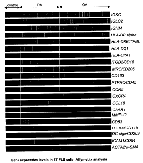
[0033] Figure 1 is heat map representation showing the gene
expression levels of synovial tissue fibroblast-like synovial cells in
controls or
subjects with rheumatoid arthritis or osteoarthritis.
[0034] Figure 2 is a heat map representation showing the phospho-
protein signature profiles of synovial tissue fibroblast-like synovial cells
(ST
FLS) from subjects with rheumatoid arthritis or osteoarthritis. Cell lysates
derived from ST FLS cells from affected joints from 2 OA patients (LHS panel)
and 3 RA patients (RHS panel) were analyzed using customized BD phospho-
protein PowerBlots. Heat map representation: phospho-protein expression
profiles are represented as a "heat-map".
CA 02650706 2015-08-13
WO 2007/137405 PCT/CA2007/000919
- 9 -
Each array map represents 80
phosphospecificities. Similar patterns are indicative of similar phospho-
protein
profiles. This is evident for the 2 representative OA ST FLS cell specimens.
These are distinguishable from the 3 RA ST FLS cell signature profiles.
Interestingly, in regard to the RA specimens, although exhibiting similar
phosphorylation-activation of many signaling effectors and kinases, there are
clusters of distinctive patterns, allowing for stratification of the RA
specimens
into 3 sub-groups, exemplified by the 3 profiles provided.
[0035] Figure 3 shows the protein expression in synovial tissue
fibroblast-like synovial cells derived from individuals with rheumatoid
arthritis
or osteoarthritis.
[0036] Figure 4 shows the immunostaining results of peripheral blood
fibrocytes for various markers. (A) FACS analysis of a population of
peripheral
blood fibrotcyes. (B) Confocal microscopy of a single peripheral blood
fibrocyte. Images were collected using an upright Leica SP2 confocal laser-
scanning microscope (Leica Microsystems Heidelberg GmbH, Mannheim,
Germany), 100X oil immersion lens (1.4 NA) and 4X digital zoom.
[0037] Figure 5 shows the results of an intracellular analysis of
phospho-STAT3 in CD3-, collagen+ peripheral blood fibrocytes.
[0038] Figure 6 shows the results of an intracellular analysis of
phospho-STAT5 in CD3-, collagen+ peripheral blood fibrocytes.
[0039] Figure 7 shows the results of an intracellular analysis of
phospho-ERK in CD3-, collagen+ peripheral blood fibrocytes.
[0040] Figure 8 shows the results of an intracellular analysis of
phospho-p38 MAPK in CD3-, collagen+ peripheral blood fibrocytes.
[0041] Figure 9 shows protein phosphorylation profiles of sub-
populations and primary cells.
[0042] Figure 10 shows the results of an intracellular analysis of
phospho-STAT3 and phospho-p38 in CD3+ cells from healthy individuals,
early rheumatoid arthritis patients and late rheumatoid arthritis patients.
[0043] Figure 11 depicts a signalizing cascade.
CA 02650706 2008-10-29
WO 2007/137405 PCT/CA2007/000919
- 10 -
[0044] Figure 12 shows circulating fibrocytes in CIA. Mice with CIA
exhibit obvious swelling of the paws/joints compared to naïve animals (A-1,
11). Affected joints were scored on a scale of 0-16 and the cumulative disease
score is shown in panel B. Mice with CIA cumulative score of 6/individual paw
score of 2, show obvious swelling and cellular infiltrates in the dermis (C
III,
magnification 100X; C IV, magnification 200X *) compared to control animals
(C I, magnification 100X; C II, magnification 200X). The majority of the
inflammatory infiltrate were neutrophils (PMN) but macrophages (M),
lymphocytes (L) and plasma cells (P) were also observed (C V, 1000X
magnification). Swelling (**) and early inflammatory infiltrates (&) were
observed in the intra-articular spaces (C VI, 200X magnification). PBMC were
collected by cardiac puncture at different stages of disease and FACS
analysis performed to detect the a-SMA/CI fibrocyte population (D). Panel E
describes the a-SMA/CI fibrocyte population on day 30. Large granular cells
were gated by FSC/SSC and double positive cells are shown. Notably, the
number of circulating fibrocytes was higher in mice with higher disease
scores.
[0045] Figure 13 shows evidence of increased p-STAT5 in circulating
fibrocytes from animals with early stages of CIA. PBMC were isolated from
the peripheral blood of control and CIA mice at stage 1-2. The cells were
stained for Collagen I-A1exa647, a-SMA-FITC and p-STAT5-PE and analyzed
by FACS. The Coll+ cells were gated and the percentage of a-SMA/p-STAT5
double positive cells are shown (A, R2 gate and B).
[0046] Figure 14 shows immunohistochemistry of paraffin embedded
joints from mice with collagen induced arthritis stained with a-SMA-FITC
(green) and CD45-PE (red). a-SMA also stains smooth muscle and is clearly
identified surrounding an artery (open arrow). A profound cellular influx of
leukocytes is evident (*) in the CIA joint tissue and the majority of these
cells
do not stain with a-SMA (overlay). a-SMA/CD45 fibrocytes/myofibroblasts
were easily identified in the CIA joints (closed arrow) and were more abundant
than in normal joints.
CA 02650706 2008-10-29
WO 2007/137405 PCT/CA2007/000919
- 11 -
[0047]
Figure 15 shows immunohistochemistry of plastic embedded
joints from mice with collagen induced arthritis stained with a-SMA-FITC
(green) and CD45-PE (red). a-SMA/CD45 double positive
fibrocytes/myofibroblasts were identified in the CIA joints.
DETAILED DESCRIPTION OF THE INVENTION
[0048] The
inventors have discovered a precursor of a fibroblast-like
synovial cell. This cell is a circulating cell and can migrate to affected
joints in
subjects with rheumatoid arthritis, and is able to transform into resident
myofibroblasts and fibroblast-like synovial cells. The precursor cell stains
positive for collagen, CD34, CD45, prolyl 4-hydroxylase and CD14, and can
be isolated from the circulatory system, for example from peripheral blood
mononuclear cells from healthy individuals and individuals with rheumatoid
arthritis.
[0049]
Accordingly, the invention provides an isolated precursor of a
fibroblast-like synovial cell, comprising a circulating cell that stains
positive for
collagen, CD34, CD45, prolyl 4-hydroxylase and CD14.
[0050] The
term "isolated precursor of a fibroblast-like synovial cell" as
used herein refers to the precursor cells of the invention substantially free
of
other cell types. In another embodiment, the cells are also substantially free
of
cellular debris or other cellular material, or culture medium.
[0051] The
invention includes a method of diagnosing or monitoring
rheumatoid arthritis in a subject, comprising the steps:
(a) determining the number of isolated precursor cells of the
invention in a sample from the subject; and
(b) comparing the number of isolated precursor cells from the
sample with a control;
wherein a difference in the number of isolated precursor cells in the
sample from the subject as compared to the control is indicative of rheumatoid
arthritis.
[0052] The
inventors also analyzed the activation state of this
circulating precursor cell in healthy individuals and subjects with early
rheumatoid arthritis and subjects with later stages of disease. The activation
CA 02650706 2008-10-29
WO 2007/137405 PCT/CA2007/000919
- 12 -
state of the precursor cell was determined by analyzing the phosphorylation
levels of signaling molecules, including STAT3, STAT5, ERK and /or p38
MAPK.
[0053]
Accordingly, the invention includes a method of diagnosing or
monitoring rheumatoid arthritis in a subject, comprising the steps:
(a) determining the activation state of the isolated precursor cell of
the invention from a sample from the subject; and
(b) comparing the activation state of the isolated precursor cell from
the sample with a control;
wherein the activation state of the precursor cell is determined by
measuring the phosphorylation levels of signaling molecules, and
wherein a difference in the activation state of the isolated precursor cell
as compared to the control is indicative of rheumatoid arthritis.
[0054] The
phrase "diagnosing or monitoring rheumatoid arthritis" as
used herein refers to a method or process of determining if a subject has or
does not have rheumatoid arthritis, or determining the severity or degree of
rheumatoid arthritis.
[0055] The
term "subject" as used herein refers to any member of the
animal kingdom, preferably a human being.
[0056] The term "sample" as used here refers to any fluid, cell or tissue
sample from an individual which includes the precursor cell of the invention.
In
one embodiment, the sample is from the circulatory system of the individual.
[0057] The
term "control" as used herein refers to a sample from a
subject or a group of subjects who are either known as having rheumatoid
arthritis or not having rheumatoid arthritis, or who are known as having a
particular severity or degree of rheumatoid arthritis or not. A subject known
not to have rheumatoid arthritis is also referred to as a "healthy individual"
herein. For example, the control can be from a healthy individual, a subject
with early stage rheumatoid arthritis or a subject with late stage rheumatoid
arthritis. A person skilled in the art will appreciate that a subject with
early
stage rheumatoid arthritis can be defined to include individuals within the
first
year of onset of symptoms with 3 swollen joints.
CA 02650706 2008-10-29
WO 2007/137405 PCT/CA2007/000919
- 13 -
[0058] The phrase "difference in number of isolated precursor cells
in
the sample from the subject as compared to the control is indicative of
rheumatoid arthritis" refers to the difference in frequency of cells. There
are
generally greater numbers of the isolated precursor cells in samples from
subjects with rheumatoid arthritis as compared to healthy individuals. In
addition, there are greater numbers of the isolated precursor cells in samples
from subjects as the disease progresses. For example, there are greater
numbers of the isolated precursor cell from subjects with late stage
rheumatoid arthritis as compared to subjects with early stage rheumatoid
arthritis. Thus, if the control is a healthy individual, then there are
greater
numbers of the isolated precursor cells in the samples from subjects with
rheumatoid arthritis as compared to the control. If the control is a subject
with
early stage rheumatoid arthritis, then there are greater numbers of the
precursor cells in samples with late stage rheumatoid arthritis as compared to
the control.
[0059] The "activation state of the isolated precursor cell" can be
determined by measuring the activation status of signaling molecules within
the isolated precursor cell. For example, the activation status of signaling
molecules can be determined by measuring the phosphorylation levels of
signaling molecules, such as STAT3, STAT5, ERK and/or p38 MAPK.
[0060] The phrase "difference in the activation state of the isolated
precursor cell as compared to the control is indicative of rheumatoid
arthritis"
refers to a difference in the frequency or levels of phosphorylation of
signaling
molecules in the isolated precursor cell, including STAT3, STAT5, ERK and
p38 MAPK, as compared to the control. There are more frequent and/or
higher levels of phosphorylation of signaling molecules in samples from
subjects with rheumatoid arthritis as compared to healthy individuals. There
are generally more frequent and/or higher levels of phosphorylation of
signaling molecules in samples from subjects as the disease progresses. For
example, there are more frequent and/or higher levels of phosphorylation of
signaling molecules in samples from subjects with late stage rheumatoid
arthritis as compared to subjects with early stage rheumatoid arthritis. Thus,
if
CA 02650706 2008-10-29
WO 2007/137405 PCT/CA2007/000919
- 14 -
the control is a healthy individual, then there are more frequent and/or
higher
levels of phosphorylation of signaling molecules in precursor cells in samples
from subjects with rheumatoid arthritis as compared to the control. If the
control is a subject with early stage rheumatoid arthritis, then there are
more
frequent and/or higher levels of phosphorylation of signaling molecules in
precursor cells in samples from subjects with late stage rheumatoid arthritis
as compared to the control.
[0061] The
invention also includes the use of the isolated precursor cell
of the invention to diagnose or monitor rheumatoid arthritis.
[0062] The
isolated precursor cell of the invention can also be used in
methods of drug discovery or methods to identify substances that can treat or
prevent rheumatoid arthritis. For example, an additional aspect of the
invention is a method of identifying a substance to treat or prevent
rheumatoid
arthritis, comprising the steps:
(a) determining the number of isolated precursor cells of the
invention in a sample from a subject treated with a substance;
and
(b) comparing the number of isolated precursor cells from the
sample with a control;
wherein a difference in the number of isolated precursor cells in the
sample from the subject as compared to the control is indicative of a
substance to treat or prevent rheumatoid arthritis.
[0063] In
another example, the invention includes a method of
identifying a substance to treat or prevent rheumatoid arthritis, comprising
the
steps:
(a) determining the activation state of the isolated precursor cell of
the invention from a sample from a subject treated with a
substance; and
(b) comparing the activation state of the isolated precursor cell
from the sample with a control;
wherein the activation state of the precursor cell is determined by
measuring the phosphorylation levels of signaling molecules, and
CA 02650706 2008-10-29
WO 2007/137405 PCT/CA2007/000919
- 15 -
wherein a difference in the activation state of the isolated precursor cell
as compared to the control is indicative of a substance to treat or prevent
rheumatoid arthritis.
[0064] The
phrase "treat or prevent rheumatoid arthritis" as used herein
refers to a medical aid to counteract the disease itself, the symptoms and/or
the progression of the disease.
[0065]
"Measuring the phosphorylation levels of signaling molecules"
as used herein refers to measuring the frequency and/or intensity of
phosphorylation of signaling molecules, such as STAT3, STAT5, ERK and/or
p38 MAPK.
[0066] A
person skilled in the art will appreciate that the control can be
a sample from a subject not treated with a substance or treated with a
substance that is known not to treat or prevent rheumatoid arthritis. In one
embodiment, reduced numbers of the isolated precursor cell in the sample as
compared to the control is indicative of a substance for the treatment or
prevention of rheumatoid arthritis. In
another embodiment, a reduced
activation state of the isolated precursor cell in the sample as compared to
the
control is indicative of a substance for the treatment or prevention of
rheumatoid arthritis. In addition, the control can be a sample from the same
subject, but before treatment with the substance to be tested or samples from
the subject taken at different points of time during treatment with the
substance to be tested.
[0067]
Substances for the treatment or prevention of rheumatoid
arthritis can also be identified using cells or cell lines. For example,
individual
precursor cells or cell lines derived from the precursor cell of the invention
can
be contacted with a substance and then the activation state of the cells can
be
compared to a control.
[0068] The
inventors have also studied the activation status of
circulating CD3+ cells from rheumatoid arthritis patients at different stages
of
disease. They discovered that there are progressive increases in the
phosphorylation of signaling molecules, such as STAT3 and p38 MAPK, in
CD3+ cells, which correlate with disease progression.
CA 02650706 2008-10-29
WO 2007/137405 PCT/CA2007/000919
- 16 -
[0069]
Accordingly, the invention includes method of diagnosing or
monitoring rheumatoid arthritis in a subject, comprising the steps:
(a) determining the activation state of a CD3+ cell from a sample
from the subject; and
(a) comparing the activation state of the CD3+ cell from the
sample with a control;
wherein, the activation state of the CD3+ cell is determined by
measuring the phosphorylation levels of signaling molecules, such as STAT3
and/or p38 MAPK, and
wherein a difference in the activation state of the CD3+ cell as
compared to the control is indicative of rheumatoid arthritis.
[0070] The
"activation state of the CD3+ cell" can be determined by
measuring the activation status of signaling molecules within CD3+ cell. For
example, the activation status of signaling molecules can be determined by
measuring the phosphorylation levels of signaling molecules, such as STAT3
and/or p38 MAPK.
[0071] The
phrase "difference in the activation state of the CD3+ cell as
compared to the control" refers to a difference in the frequency or levels of
phosphorylation of signaling molecules in the CD3+ cell, including STAT3
and/or p38 MAPK, as compared to the control.
[0072] The
term "sample" as used here refers to any fluid, cell or tissue
sample from an individual which includes a CD3+ cell.
[0073] The
invention also includes the use of a CD3+ cell to diagnose
or monitor rheumatoid arthritis in a subject.
[0074] The findings of the inventors can also be used in methods of
drug discovery. Accordingly, the invention includes method of identifying a
substance to treat or prevent rheumatoid arthritis comprising the steps:
(a) determining the activation state of a CD3+ cell from a sample
from a subject treated with a substance; and
(b) comparing the activation state of the CD3+ cell from the sample
with a control;
CA 02650706 2008-10-29
WO 2007/137405 PCT/CA2007/000919
- 17 -
wherein the activation state of the CD3+ cell is determined by
measuring the phosphorylation levels of signaling molecules, and
wherein a difference in the activation state of the CD3+ cell as
compared to the control is indicative of a substance to treat or prevent
rheumatoid arthritis.
[0075] The
invention also includes the use of a CD3+ cell to identify a
substance to treat or prevent rheumatoid arthritis.
[0076] The
inventors have also analyzed and characterized the gene
expression and protein expression profiles of synovial tissue fibroblast-like
synovial cells from subjects with rheumatoid arthritis and osteoarthritis. The
inventors have discovered that there are different gene expression and
protein expression profiles, and protein phosphorylation profiles in synovial
tissue fibroblast-like synovial cells from subjects with rheumatoid arthritis,
osteoarthritis and healthy individuals.
[0077] The
inventors discovered a number of rheumatoid arthritis
specific genes that can be used to characterize the gene expression profile in
the method of the invention. These genes include transport, apoptosis
regulatory, cell adhesion, cell surface signaling receptors, intracellular
signaling, secreted stimulatory and immunomodulatory genes. In addition, the
inventors discovered a significant differential expression of 154 genes in
fibroblast-like synovial cells in subjects with osteoarthritis or rheumatoid
arthritis. See Tables 1 and 2.
[0078]
Accordingly, the invention includes a method of diagnosing or
monitoring rheumatoid arthritis in a subject, comprising the steps:
(a) determining the gene expression profile of a synovial tissue
fibroblast-like synovial cell from a sample from the subject; and
(b) comparing the gene expression profile of the synovial tissue
fibroblast-like synovial cell from the sample with a control;
wherein a difference in the gene expression profile of the synovial
tissue fibroblast-like synovial cell as compared to the control is indicative
of
rheumatoid arthritis.
CA 02650706 2008-10-29
WO 2007/137405 PCT/CA2007/000919
- 18 -
[0079] In one embodiment, the gene expression profile that
characterizes subjects with rheumatoid arthritis includes enhanced gene
expression of one or more of the genes listed in Table 1.
[0080] In another embodiment, the gene expression profile that
characterizes subjects with rheumatoid arthritis includes enhanced gene
expression of genes encoding immunoglobulin constant regions, CD53,
CD11b, CD18, CD86, CD206, CD163, mannose receptor, DC-SIGN, C3AR1,
Fc-receptors, complement receptors, and/or MHC class II molecules as
compared to the control.
[0081] The invention also includes a method of diagnosing or
monitoring osteoarthritis in a subject, comprising the steps:
(a) determining the gene expression profile of a synovial tissue
fibroblast-like synovial cell from a sample from the subject; and
(b) comparing the gene expression profile of the synovial tissue
fibroblast-like synovial cell from the sample with a control;
wherein a difference in the gene expression profile of the synovial
tissue fibroblast-like synovial cell as compared to the control is indicative
of
osteoarthritis.
[0082] The term "control" as used here refers to a sample from a
subject or group of subjects who are either known as having osteoarthritis or
not, or who are known as having a particular severity or degree of
osteoarthritis or not.
[0083] In one embodiment, the gene expression profile that
characterizes subjects with osteoarthritis includes enhanced gene expression
of one or more of the genes listed in Table 2.
[0084] The term "sample" as used here refers to any fluid, cell or
tissue
sample from an individual which includes a synovial tissue fibroblast-like
synovial cell.
[0085] The term "gene expression profile" as used herein refers to
the
level of RNA expressed from one or more gene in the synovial tissue
fibroblast-like synovial cell from a sample from the subject.
CA 02650706 2008-10-29
WO 2007/137405 PCT/CA2007/000919
- 19 -
[0086] The term "difference in gene expression profile" as used here
refers to an increase or decrease in the measurable expression of RNA of a
particular gene or group of genes as compared to the measurable expression
of RNA of the same gene or group of genes in a second sample. The
comparison can be made between individual samples or populations of
samples. In one embodiment, the differential expression can be compared
using the ratio of the level of expression of the gene as compared with the
expression level of the gene of a control, wherein the ratio is not equal to
1Ø
For example, an RNA is differentially expressed if the ratio of the level of
expression in a first sample as compared with a second sample is greater
than or less than 1Ø For example, a ratio of greater than 1, 1.2, 1.5, 1.7,
2, 3,
3, 5, 10, 15, 20 or more, or a ratio less than 1, 0.8, 0.6, 0.4, 0.2, 0.1,
0.05,
0.001 or less. In another embodiment the differential expression is measured
using p-value. For instance, when using p-value, a gene is identified as being
differentially expressed as between a first and second population when the p-
value is less than 0.1, preferably less than 0.05, more preferably less than
0.01, even more preferably less than 0.005, the most preferably less than
0.001.
[0087] In addition to measuring the gene expression profile, a person
skilled in the art will appreciate that the protein expression profile of a
synovial
tissue fibroblast-like synovial cell can be measured.
[0088] The term "protein expression profile" as used herein refers to
the level of one or more proteins expressed in the synovial tissue fibroblast-
like synovial cell from a sample from the subject. The protein expression
profile can include measurements of the expression of transport, apoptosis
regulatory, cell adhesion, cell surface signaling receptors, intracellular
signaling, secreted stimulatory and immunomodulatory proteins. This includes
measuring the protein expression of immunoglobulin constant regions, CD53,
CD11b, CD18, CD86, CD206, CD163, mannose receptor, DC-SIGN, C3AR1,
Fc-receptors, complement receptors, and/or MHC class II molecules.
[0089] Accordingly, the invention includes a method of diagnosing or
monitoring rheumatoid arthritis in a subject, comprising the steps:
CA 02650706 2008-10-29
WO 2007/137405 PCT/CA2007/000919
- 20 -
(a) determining the protein expression profile of a synovial tissue
fibroblast-like synovial cell from a sample from the subject; and
(b) comparing the protein expression profile of the synovial tissue
fibroblast-like synovial cell from the sample with a control;
wherein a difference in the protein expression profile of the synovial
tissue fibroblast-like synovial cell as compared to the control is indicative
of
rheumatoid arthritis.
[0090] In
one embodiment, the protein expression profile that
characterizes subjects with rheumatoid arthritis includes enhanced protein
expression of one or more proteins encoded by the genes listed in Table 1.
[0091] The
invention also includes a method of diagnosing or
monitoring osteoarthritis in a subject, comprising the steps:
(a) determining the protein expression profile of a synovial tissue
fibroblast-like synovial cell from a sample from the subject; and
(b) comparing the protein expression profile of the synovial tissue
fibroblast-like synovial cell from the sample with a control;
wherein a difference in the protein expression profile of the synovial
tissue fibroblast-like synovial cell as compared to the control is indicative
of
osteoarthritis.
[0092] In one
embodiment, the protein expression profile that
characterizes subjects with osteoarthritis includes enhanced protein
expression of one or more of the proteins encoded by the genes listed in
Table 2.
[0093] The
term "difference in protein expression profile" as used here
refers to an increase or decrease in the measurable expression of a particular
protein or group of proteins as compared to the measurable expression of the
same protein or group of proteins in a second sample. The comparison can
be made between individual samples or populations of samples. In one
embodiment, the differential expression can be compared using the ratio of
the level of expression of the protein as compared with the expression level
of
the protein of a control, wherein the ratio is not equal to 1Ø For example,
a
protein is differentially expressed if the ratio of the level of expression in
a first
CA 02650706 2008-10-29
WO 2007/137405 PCT/CA2007/000919
- 21 -
sample as compared with a second sample is greater than or less than 1Ø
For example, a ratio of greater than 1, 1.2, 1.5, 1.7, 2, 3, 3, 5, 10, 15, 20
or
more, or a ratio less than 1, 0.8, 0.6, 0.4, 0.2, 0.1, 0.05, 0.001 or less. In
another embodiment the differential expression is measured using p-value.
For instance, when using p-value, a protein is identified as being
differentially
expressed as between a first and second population when the p-value is less
than 0.1, preferably less than 0.05, more preferably less than 0.01, even more
preferably less than 0.005, the most preferably less than 0.001.
[0094] In
addition to gene and protein expression profiles, the inventors
also examined protein phosphorylation profiles in synovial tissue fibroblast-
like synovial cells.
[0095]
Accordingly, the invention includes a method of diagnosing or
monitoring rheumatoid arthritis in a subject, comprising the steps:
(a) determining the protein phosphorylation profile of a synovial
tissue fibroblast-like synovial cell from a sample from the
subject; and
(a) comparing the protein phosphorylation profile of the synovial
tissue fibroblast-like synovial cell from the sample with a
control;
wherein a difference in the protein phosphorylation profile of the
synovial tissue fibroblast-like synovial cell as compared to the control is
indicative of rheumatoid arthritis.
[0096] The
term "protein phosphorylation profile" as used herein refers
to the level or frequency of phosphorylation of one or more proteins
expressed in synovial tissue fibroblast-like synovial cells from a sample from
a
subject.
[0097] In
one embodiment, the difference in the protein phosphorylation
profile includes a difference in phosphorylation of AKT, FAK, p38, JNK, cdc-2
and/or PLC-g1 in the synovial tissue fibroblast-like synovial cells in the
subject
as compared to the control.
[0098] The
invention also includes a method of diagnosing or
monitoring osteoarthritis in a subject, comprising the steps:
CA 02650706 2008-10-29
WO 2007/137405 PCT/CA2007/000919
- 22 -
(a) determining the protein phosphorylation profile of a synovial
tissue fibroblast-like synovial cell from a sample from the
subject; and
(b) comparing the protein phosphorylation profile of the synovial
tissue fibroblast-like synovial cell from the sample with a
control;
wherein a difference in the protein phosphorylation profile of the
synovial tissue fibroblast-like synovial cell as compared to the control is
indicative of osteoarthritis.
[0099] The
methods of the invention can also be used to identify
substances to treat or prevent rheumatoid arthritis or osteoarthritis.
[00100] The
inventors also determined that the number of circulating
fibrocytes and the activation status of fibrocytes can be used to diagnose or
monitor rheumatoid arthritis in a subject.
[00101] The term
"sample" as used here refers to any fluid, cell or tissue
sample from an individual which includes circulating fibrocytes. In one
embodiment, the sample is from the circulatory system of the individual.
[00102]
Accordingly, a further aspect of the invention is a method of
diagnosing or monitoring rheumatoid arthritis in a subject, comprising the
steps:
(a) determining the number of circulating fibrocytes in a sample
from the subject; and
(b) comparing the number of fibrocytes from the sample with a
control;
wherein a difference in the number of fibrocytes in the sample from the
subject as compared to the control is indicative of rheumatoid arthritis.
[00103] The
phrase "difference in number of fibrocytes in the sample
from the subject as compared to the control is indicative of rheumatoid
arthritis" refers to the difference in frequency of cells. There are generally
greater numbers of the circulating fibrocytes in samples from subjects with
rheumatoid arthritis as compared to healthy individuals. In addition, there
are
greater numbers of circulating fibrocytes in samples from subjects as the
CA 02650706 2008-10-29
WO 2007/137405 PCT/CA2007/000919
- 23 -
disease progresses. For example, there are greater numbers of circulating
fibrocytes from subjects with late stage rheumatoid arthritis as compared to
subjects with early stage rheumatoid arthritis. Thus, if the control is a
healthy
individual, then there are greater numbers of circulating in the samples from
subjects with rheumatoid arthritis as compared to the control. If the control
is
a subject with early stage rheumatoid arthritis, then there are greater
numbers
of circulating fibrocytes in samples with late stage rheumatoid arthritis as
compared to the control.
[00104]
Another aspect of the invention, is a method of diagnosing or
monitoring rheumatoid arthritis in a subject, comprising the steps:
(a) determining the activation state of a circulating fibrocyte from a
sample from the subject; and
(b) comparing the activation state of the fibrocyte from the sample
with a control;
wherein the activation state of the fibrocyte is determined by measuring
the phosphorylation levels of signaling molecules, and
wherein a difference in the activation state of the fibrocyte as compared
to the control is indicative of rheumatoid arthritis.
[00105] The
"activation state of the fibrocyte" can be determined by
measuring the activation status of signaling molecules within the isolated
precursor cell. For example, the activation status of signaling molecules can
be determined by measuring the phosphorylation levels of signaling
molecules, such as STAT5.
[00106] The
phrase "difference in the activation state of the fibrocyte as
compared to the control is indicative of rheumatoid arthritis" refers to a
difference in the frequency or levels of phosphorylation of signaling
molecules
in the isolated precursor cell, such as STAT5, as compared to the control.
There are more frequent and/or higher levels of phosphorylation of signaling
molecules in samples from subjects with rheumatoid arthritis as compared to
healthy individuals. There are generally more frequent and/or higher levels of
phosphorylation of signaling molecules in samples from subjects as the
disease progresses. For example, there are more frequent and/or higher
CA 02650706 2008-10-29
WO 2007/137405 PCT/CA2007/000919
- 24 -
levels of phosphorylation of signaling molecules in samples from subjects with
late stage rheumatoid arthritis as compared to subjects with early stage
rheumatoid arthritis. Thus, if the control is a healthy individual, then there
are
more frequent and/or higher levels of phosphorylation of signaling molecules
in precursor cells in samples from subjects with rheumatoid arthritis as
compared to the control. If the control is a subject with early stage
rheumatoid
arthritis, then there are more frequent and/or higher levels of
phosphorylation
of signaling molecules in precursor cells in samples from subjects with late
stage rheumatoid arthritis as compared to the control.
[00107] The
invention also includes using circulating fibrocytes to
diagnose or monitor rheumatoid arthritis.
[00108] An
additional aspect of the invention is a method of identifying a
substance to treat or prevent rheumatoid arthritis, comprising the steps:
(a) determining the number of circulating fibrocytes in a sample
from a subject treated with a substance; and
(b) comparing the number of fibrocytes from the sample with a
control;
wherein a difference in the number of fibrocytes in the sample from the
subject as compared to the control is indicative of a substance to treat or
prevent rheumatoid arthritis.
[00109] A
further aspect of the invention is a method of identifying a
substance to treat or prevent rheumatoid arthritis, comprising the steps:
(a) determining the activation state of circulating fibrocytes from a
sample from a subject treated with a substance; and
(b) comparing the activation state of the fibrocyte from the sample
with a control;
wherein the activation state of the fibrocyte is determined by measuring
the phosphorylation levels of signaling molecules, and
wherein a difference in the activation state of the fibrocyte as compared
to the control is indicative of a substance to treat or prevent rheumatoid
arthritis.
CA 02650706 2014-06-27
- 25 -
[00110] In addition, the invention includes the use of circulating
fibrocytes to identify a substance to treat or prevent rheumatoid arthritis.
[00111] The following non-limiting examples are illustrative of the
present invention:
EXAMPLES
Example 1: Characterization of gene and protein expression profiles in
RA ST FLS cells.
[00112] The activated phenotype of distinct cell populations in
affected
RA patients determines disease severity. Suppression of the activated
phenotype of cells in early RA synovitis will subdue the disease process. A
clear understanding of those factors that contribute to the activated
phenotype
is required. The objective of the inventors' studies was to determine the
signature gene and protein expression profiles in target cell populations in
patients diagnosed with RA. The inventors' hypothesized that a signature
pattern of activated factors in distinct cell types would identify potential
therapeutic targets. Over the past few years, the inventors have initiated a
collection of blood, synovial fluid and ST from RA patients, osteoarthritis
(OA)
patients and trauma patients at the time of joint surgery. Sample collection
involves confirmation of the diagnosis of RA/OA using clinical, serologic and
radiologic data and informed consent on all study participants. Clinical
parameters are recorded at the time of sample collection. Synovial samples
are collected by joint aspiration, fine needle biopsy and via arthroscopic or
other surgeries. FLS cells from ST from affected RA joints, OA joints and
tissue from trauma patients (non-RA, non-OA, surgery) were collected using a
negative-selection protocol for cell fractionation, and cultured in DMEM
media.
Initial studies examined gene expression profiles of RNA from freshly
harvested ST, cells that were passaged up to 3X and cells maintained beyond
three passages. Affymetrix microarray analysis (U133 Plus 2.0 microarray
representing over 30,000 genes) was performed. RA FLS cells at 3X passage
exhibit similar gene expression profiles compared to freshly harvested FLS
cells, yet the inventors observed that this characteristic gene expression
profile changed with extended time in culture. All subsequent gene expression
CA 02650706 2008-10-29
WO 2007/137405 PCT/CA2007/000919
- 26 -
analyses were conducted on RNA extracted from ST FLS cells cultured for <3
passages. Gene expression data were initially analyzed using GeneSpring 6.1
software (Silicon Genetics). Results of gene tree data analysis of ST FLS
cells
have revealed distinct and reproducible expression profiles, reflective of the
different patient populations (trauma patients, OA, RA), that is significant
as
determined by one-way ANOVA. Gene tree data analysis sorted the RA-
specific genes into functional groupings: transport, apoptosis regulatory,
cell
adhesion, cell surface signaling receptors, intracellular signaling, secreted
stimulatory, and immunomodulatory genes. A gene tree based comparison of
OA versus RA ST FLS cell expression profiles revealed significant differential
expression of 154 genes (one-way ANOVA, p<0.01), including 17 apoptosis
regulatory, 17 cell adhesion, 39 cell surface receptors, 25 immunomodulatory
and 28 signal transduction genes. To further analyze these data, the Binary
Tree-Structured Vector Quantization (BTSVQ) program was used (11).
BTSVQ is a computational tool that combines partitive k-means clustering and
Self-Organizing Maps (SOM) to analyze and visualize microarray gene
expression data with minimal noise and without pre-conceived bias. These
analyses confirmed distinct signature patterns of gene expression for FLS
cells derived from RA (n=16) and OA (n=20) specimens and further
distinguished healthy control (trauma patients, n=6) gene expression patterns
from RA and OA expression profiles (Figure 1). Differentially regulated genes
in RA FLS cells included cytokines, chemokines and receptors. In agreement
with previous studies (12), the inventors also show a transformed phenotype
in RA FLS cells, with expression of many genes that are typically not found in
fibroblasts. High levels of gene expression for immunoglobulin constant
regions: IGKC, IGLC2 and IGHM were seen, typically expressed in B cells.
Also, gene expression for many receptors found on antigen presenting cells
such as: CD53, CD11b, CD18, CD86, scavenger receptors (CD206, CD163
and the mannose receptor, MRC), DC-SIGN and complement receptor
(C3AR1), were observed. The MRC processes molecules for presentation of
MHCII and binds to both endogenous and exogenous ligands. Increases in
gene expression for Fc- and complement receptor gene expression were
CA 02650706 2014-06-27
- 27 -
observed in RA FLS cells and suggest that these cells trap antigens in the
form of immune complexes on their cell surface for presentation. Gene
expression for HLA-DR, DQ and DP were also upregulated in RA vs. OA or
control FLS cells in the array analyses. HLA molecules are expressed on
CD68+ synoviocytes following treatment with IFN-y (13). Notably, gene
expression for CD68 was not evident in the isolated FLS cells. Upregulation
of HLA molecules would be consistent with the role of synovial fibroblasts in
antigen presentation during an autoimmune disease. The likelihood of
contaminating leukocytes, e.g. macrophage-like synoviocytes (MLS), in the
fibroblast preparations was eliminated since only adherent prolyI-4
hydroxylase (5B5 antibody) positive cells were processed for gene expression
analysis, and there was no significant CD2, CD3 or CD5 gene expression
observed in the analysis. This suggests that RA FLS cells either upregulate
the B cell/macrophage/dendritic cell markers, de-differentiate into this
phenotype, or that immature fibroblast-like cells are recruited and
differentiate
into these cells in affected joints.
[00113] In parallel, protein extracted from the RA, OA and surgical
trauma ST FLS cells was analyzed using the BD BioScience PowerBlotTM Western
immunoarray technology platform. The full array allows 996 proteins to be
interrogated, and the inventors have developed a customized
mini-array that specifically interrogates phospho-proteins. In agreement with
the data for distinct gene expression, distinct protein expression patterns
were
observed in the RA FLS cells compared to OA FLS cells (Figure 2). RA FLS
cells were activated as determined by higher levels of phospho-AKT,
phospho-FAK, phospho-p38, JNK, cdc-2 and PLC-y1 proteins (Figure 3). The
PI3K/AKT pathway plays an important role in balancing apoptosis and survival
(14) and is involved in cell cycle progression, glucose metabolism and
chemotaxis (15). RA fibroblasts show enhanced survival in response to TNF-a
(16) and TGF-R (17) through an AKT-dependent pathway (17). Additionally,
this pathway has also been implicated in TRAIL induced proliferation in RA
fibroblasts (18). Activation of the insulin-like growth factor-1 receptor (IGF-
1R)
CA 02650706 2008-10-29
WO 2007/137405 PCT/CA2007/000919
- 28 -
(19), IL-17 (20) and the IL-18 receptor (21) also effect signal transduction
in
an AKT-dependent manner to induce cytokine/chemokine or adhesion
molecule synthesis in RA fibroblasts. Focal adhesion kinase (FAK) regulates
anti-apoptotic pathways through P1 3K-dependent and -independent pathways.
FAK is a widely expressed cytoplasmic protein tyrosine kinase involved in
integrin mediated signal transduction. B1 integrin signaling through FAK
upregulates ICAM-1 and Fas in RA FLS cells (22). The JNK and p38
pathways are preferentially activated by stress, inflammatory cytokines and
growth factors. TNF can signal through both JNK and p38 to activate AP-1
and NF-kB (23) and IL-1 upregulates MMP13 (24) and collagenase 1 (25) via
the JNK pathway in RA FLS cells. The entry of all cells into mitosis is
regulated by cdc2 and increased cdc2 phosphorylation has been reported in
RA fibroblasts and may be involved in aberrant mitosis (26). Viewed
altogether, the data confirms that multiple genes and signaling pathways are
activated in RA FLS cells contributing to the distinct phenotype of these
cells.
Example 2: Fibrocytes in RA.
[00114] RA is a complex heterogeneous disease. Hyperproliferation of
FLS cells is considered to be a major contributor to pannus formation;
however, little direct evidence supports hyperproliferation of these cells in
vivo. Few mitotic figures are observed, thymidine uptake only occurs in a
percentage of the cells (27) and FLS cells divide very slowly in culture (28).
Decreased senescence in FLS cells may occur and RA FLS cells have been
shown to retain telomerase activity (29), but these cells are not immortalized
in vitro (29) (30). The outgrowth of FLS cells in the ST of affected RA joints
is,
therefore an enigma, and one possible explanation might be that FLS cells
are recruited from the circulation. Many years ago, a circulating population
of
fibroblasts-like cells, was identified (31). More recently, these cells have
been
characterized further, designated as fibrocytes, and implicated in influencing
disease development in tumor biology, scleroderma, asthma and pulmonary
fibrosis (32) (33) (34) (35).
CA 02650706 2008-10-29
WO 2007/137405 PCT/CA2007/000919
- 29 -
[00115] Fibrocytes are unique circulating cells that are relatively
rare in
the circulation, comprising only 0.1-1% of the white blood cells.
Characterization of these cells has been predominantly by FACS staining of
cells cultured for up to two weeks ex vivo and relatively little work has been
performed in vivo. In vitro, fibrocytes have a unique phenotype and express
markers of both stromal cells and hematopoietic cells, including: fibronectin,
collagen, prolyl 4-hydroxylase, CD11a, CD11b, CD13, CD18, CD45RO,
ICAM1, CD80, CD86, CXCR4, CCR7 and CCR5 (32) (36). Fibrocytes
originate from the bone marrow (37) and arise from a CD14+ve pool of cells in
the circulation, but fibrocytes themselves become CD14- (38). At the time of
culture, these cells express CD34 and CD45 (33). When cultured ex vivo,
these cells become adherent and develop a spindle¨shaped morphology (33)
(39) and downregulate CD45 and CD34. Three weeks after culture these
cells express the myofibroblast marker, a-smooth muscle actin (SMA) (33),
and have the ability to contract collagen gels in vitro (38). Ex vivo cultured
fibrocytes differentiated into a-SMA expressing cells when directly co-
cultured
with T cells or upon TGF-p stimulation (38). Fibrocyte differentiation is
inhibited by serum amyloid P (SAP) (36). Recently, in vivo evidence indicates
that the fibrocyte population contributes to the myofibroblast population in a
murine wound healing model (37). Fibrocytes secrete collagen and
fibronectin, thereby contributing to granulation formation, and they are
contractile, enhancing wound contraction and healing. They have also been
shown to present antigen (40) and can secrete chemokines, cytokines and
angiogenic factors (32), suggesting that fibrocytes contribute to both
inflammation and its resolution.
[00116] Increased myofibroblast-like cells in the joint are observed
in RA
and correlate with the degree of inflammatory synovitis. It has been suggested
that RA myofibroblasts originate from the circulating fibrocyte population
(41)
(42). The percentage of a-SMA expressing cells in RA patients varies from 1-
30% (43) and the inventors have observed upregulated a-SMA gene
expression in RA FLS cells (Figure 1). Whether these myofibroblasts were
derived locally, or were recruited from the circulating pool, is unclear.
CA 02650706 2008-10-29
WO 2007/137405 PCT/CA2007/000919
- 30 -
Certainly, circulating endothelial progenitor cells are increased during the
onset of collagen induced arthritis, likely contributing to neoangiogenesis in
affected tissues (Kurosaka et al 2005). Endothelial precursors are recruited
in
RA (44) and increased circulating endothelial precursors are observed in
murine CIA (45) and in patients with RA (46). The inference is that
circulating
endothelial precursors will traffic to affected RA joints to promote the
neoangiogenesis. Given that fibrocytes express CCR3, CCR5 and CCR7, and
the cognate ligands for these receptors are found in synovial fluid, it is
likely
that fibrocytes will also traffic to the inflamed RA joint. Indeed, RA FLS
cells
expressed higher levels of some fibrocyte markers including: CCR5, CXCR4,
CD54, CD18, CD11b and CD45 (Figure 1). Fibrocytes and fibroblasts can
differentiate into a-SMA expressing myofibroblasts upon stimulation with TGF-
b Notably, increased levels of TGF-p are present in RA STS. Myofibroblast
transformation involves activation of adhesion and integrin signaling through
FAK and AKT pathways (47). TGF-p promotes AKT-dependent survival of
mesenchymal cells through p38 MAPK-induced growth factor secretion (48).
Constitutive phosphorylation of FAK is involved in myofibroblast
differentiation
in scleroderma (49). Apparently, many of the pathways the inventors have
identified as potentially activated in the RA FLS cells have been implicated
in
fibrocyte differentiation.
Example 3: Signal taxonomy in defined RA cell populations.
[00117] Changes in intracellular protein levels, subcellular
localization,
or activation state are considered to be reflective of a cell's capabilities
or
functions. Some of these events are relatively transitory ¨ such as some
phosphorylation of proteins in cell signaling cascades. Some of the relevant
cell populations are so rare as to make their isolation for standard
biochemical
analysis nearly impossible. Remodeling of such cell signaling mechanisms
drives disease pathogenesis contributing to immune cell dysregulation despite
intense therapy regimens. Therefore, to understand how signaling networks
are remodeled in RA there is a need to measure complex populations of
CA 02650706 2008-10-29
WO 2007/137405 PCT/CA2007/000919
- 31 -
immune system cells and phenotype them not only for their cell lineage
status, but also for their relative activation state.
[00118] Studies in human
myeloid leukemia have shown that signaling
can be mapped at the individual cell level by flow cytometry and have
demonstrated links between oncogene mutations and patterns of proliferative
signaling in tumors (50). Furthermore, this work suggested that a tumor could
be described by its signal transduction potential and that this status
stratified
patient risk of relapse following chemotherapy. Since RA is a systemic and
chronic inflammatory autoimmune disease that targets synovial joints, disease
pathogenesis is multifactorial and extends beyond T or FLS cell mediated
destruction of cartilage. Production of pro-inflammatory cytokines such as
TNFa and IL-13 by activated monocytes and macrophages contribute to
tissue destruction by activation of chondrocytes and fibroblasts that release
metalloproteinases and collagenases into the synovial cavity. Cartilage loss
and bone erosion are physical manifestations of disease progression.
Additional blood borne compartments, such as B cells, contribute to
production of autoantibodies and rheumatoid factor and have been regarded
as playing important roles. Both T and B cell lymphocyte deregulation, as well
as the involvement of chondrocytes and fibroblasts suggest a dynamic
interaction of cell-to-cell communications contribute to disease pathogenesis
of RA.
[00119] The nature of
the intracellular pathways activated with most
primary immune cell interactions is not well understood in vivo and less clear
in disease states. In many cases it is only understood using derived cell
lines,
in vitro, and at best is often accomplished by lysis of cells and western
immunoblots of total cellular material. Therefore significant information on
population variations that exist is missed and advances in genomics and
proteomic technologies that rely on lysate material do not access the
heterogeneous subsets that exist in the immune system.
[00120] Multiparameter flow
cytometric analysis allows for small
subpopulations to be discerned using cell surface markers - representing
different cellular subsets, differentiation or activation states.
CA 02650706 2008-10-29
WO 2007/137405 PCT/CA2007/000919
- 32 -
[00121] The inventors developed a series of assay systems for flow
cytometric based biochemical analysis at the single cell level for kinase and
phospho-protein profiling. Measurement of up to 12 simultaneous protein or
kinase events per cell for studying signaling events in primary cells are
possible with these systems (51) (52) (53) (54). This allows for unprecedented
study of signaling in autoimmune diseases as immunocytes are particularly
amenable to these techniques.
[00122] Underlying the gross dysfunctionality of autoimmunity are the
signaling systems that drive their actions. The last two decades of research
have uncovered numerous pathways leading from surface receptors to gene
regulation. Many of these pathways, if not all, at one point or another pass
signals through phosphorylation or dephosphorylation events on proteins or
lipids. Integration of signaling events leads to relocalization of proteins
within
cells, such as with translocation of proteins to the nucleus. Until recently,
measurement of such events has been limited to cell lines or bulk lysis
assays. Interpretation of such assays is considered a pale rendition of what
we know is the intricacy of complex population primary cell events. Therefore
a true understanding of the nature of signaling dysfunctions during disease
processes in heterogeneous patient samples or animal model systems has
been beyond reach.
[00123] It can be hypothesized that the activation profiles of
proteins,
such as phospho-proteins that drive proliferation and activation signaling
cascades will differ, in disease states, both from a "normal" profile of a non-
disease presence and from other samples with significantly different
pathology. During disease pathogenesis, cells are accessing different
environmental cues, or ignoring those that might be attempting to block their
replication (or induce their apoptosis). As such, it would be expected that
there would be underlying differences in the activation profiles of certain
phospho-proteins across immune and non-immune cells in RA pathogenesis.
[00124] Therefore, with sufficient understanding of relationships between
cell signaling and immunopathology, activation profiles of phospho-proteins
could indicate the presence of individual aggressive cell subsets within a
CA 02650706 2008-10-29
WO 2007/137405 PCT/CA2007/000919
- 33 -
complex population of cells. Since it is hard to predict which kinases might
be
relevant in different cell subsets, this hypothesis has limited utility in the
absence of a high throughput manner to measure many kinases. However, if
one could measure dozens of kinase activation profiles¨or their target
proteins¨simultaneously¨there is the opportunity to generate invaluable
information about the role of signaling events in rheumatologic disease, test
hypotheses of signaling systems in response to various therapies, as well as
develop diagnostic indicators based on kinase profiles. Finally, it might be
possible to reveal signaling states that are hidden from obvious view if one
were only looking at basal phosphorylation states.
[00125] Knowledge of intracellular signaling differences among
arthritic
immune cells could therefore provide the basis for an improved autoimmune
classification system. Importantly, such a classification system would go
beyond a simple signature, but could be used to infer mechanism associated
with the signature.
[00126] In the present studies the inventors have shown that by
surveying phospho-protein/basal phosphorylation states, the underlying
dysregulated signaling nodes in primary human RA samples can be revealed
and the identification of signaling pathology profiles can be enabled.
Specifically, based on our gene and protein expression data elaborating an
emerging phenotype for the RA FLS, the inventors examined peripheral blood
mononuclear cells (PBMC) for a fibrocyte population with similar properties.
At
the outset, PBMC from healthy individuals were analyzed by multiparameter
flow cytometry to identify the fibrocyte population. The data in Figure 4
indicate that immunostaining for collagen, CD34, CD45, prolyl 4-hydroxylase
and CD14 identifies this precursor fibrocyte population. In subsequent
experiments, multiparametric staining using both surface and intracellular
stains was employed to determine the activation status of the circulating
fibrocytes in the PBMC of healthy individuals, patients with early RA and
later
stage disease. Early RA was defined as patients within the first year of onset
of symptoms with 3 swollen joints. Changes in the signaling status that occurs
during the functional activation of this fibrocyte population might include
CA 02650706 2015-08-13
WO 2007/137405 PCT/CA2007/000919
- 34 -
phosphorylation-activation of STATs, Erk and MAP kinases. Accordingly,
using polychromatic analyses the inventors examined the frequency of
activated fibrocytes, focusing on STAT3, STAT5, Erk and p38 MAPK. CD3-
CD45+collagen+ fibrocytes were stained for phospho-STAT3 (Figure 5),
phosph-STAT5 (Figure 6), phospho-Erk (Figure 7) and phospho-p38 MAPK
(Figure 8) and the frequency of staining determined in each patient
population: healthy individuals (n=20), early RA patients (n=8), late stage
disease RA patients (n=4). These data indicate that the frequency of
phospho-staining for these signaling effectors in the RA patient fibrocytes is
significantly higher (panels B) than that seen in fibrocytes from healthy
individuals.
Example 4: Collagen-induced arthritis model
[00127] Collagen-induced arthritis (CIA) is a widely used model of
rheumatoid arthritis (55) (56). Mice with CIA exhibit obvious swelling of paws
and joints as compared to control animals (Figure 12A). The mice were given
a clinical disease score based on the severity of the disease (Figure 12B).
Figure 12C shows immunohistochemistry staining of the joint.
[00128] The inventors collected PBMC by cardiac puncture at different
stages of disease and analyzed the cells by FACS for positive staining to a-
SMA and collagen. As can be seen in Figures 12D and E, the number of
circulating fibrocytes is higher in mice with higher disease scores.
[00129] The circulating fibrocytes also showed increased p-STAT5 in
the
animals with CIA as compared to the control animals (Figure 13).
[00130] Figures 14 and 15 show the results of immunohistochemistry of
samples from mice with CIA as compared to controls.
[00131] The scope of the claims should not be limited by the preferred
embodiments and examples, but should be given the broadest interpretation
consistent with the description as a whole.
CA 02650706 2015-08-13
- 35 -
Table 1
o
w
-.1
Probe set Public ID Chromosomal
Target description
Unigene ID Gene Title Gene Symbol
ID number number (NCB!) location
ca
--.1
4=.
gb:Y13786.2 /DB_XREF=gi:12053590 /FEA=FLmRNA /CNT=58
fTID=Hs.278679.1 /TIER=Stack /STK=24 /UG=Hs.278679 /LL=8728
ADAM metallopeptidase domain
209765_at Y13786 chr5q32-q33
Hs.289368 ADAM19
/UG_GENE=ADAM19 /DEF=Homo sapiens mRNA for meltrin-betaADAM 19
19 (meltrin beta)
homologue. /PROD=meltrin-betaADAM 19 homologue /FL=gb:AF311317.1
gb:NM 014479.1 /DB XREF=gi:7657318 /GEN=M12.219 /FEA=FLmRNA
/CNT=2f /TID=Hs.145216.0 /TIER=FL+Stack /STK=14 /UG=Hs.145296
206134_at NM- 014479
/LL=27299 /DEF=Homo sapiens disintegrin protease (M12.219), mRNA. chr8p21.2
Hs.145296 ADAM-like, decysin 1 ADAMDEC1
/PROD=disintegrin protease /FL=gb:NM_014479.1
gb:NM 004833.1 /DB XREF=gi:4757733 /GEN=AIM2 /FEA=FLmRNA
/CNT=1/TID=Hs.1051715.0 TTIER=FL /STK=7 /UG=Hs.105115 /LL=9447
n
206513_at NM -004833
/DEF=Homo sapiens absent in melanoma 2 (AIM2), mRNA. /PROD=absent chr1q22
Hs.105115 absent in melanoma 2 AIM2
in melanoma 2 /FL=gb:AF024714.1 gb:NM_004833.1
o
iv
o)
gb:NM 018476.1 /DB XREF=gi:8923715 /GEN=HBEX2 /FEA=FLmRNA
in
/CNT=1f6 fTID=Hs.28319.0 TTIER=FL+Stack /STK=54 /UG=Hs.283719
0
-A
218332_at NM_018476 /LL=55859
/DEF=Homo sapiens uncharacterized hypothalamus protein chrxq21-q23
Hs.334370 brain expressed, X-linked 1 BEX11 o
HBEX2 (HBEX2), mRNA. /PROD=uncharacterized hypothalamus protein
GO o)
HBEX2
0) iv
o
i
gb:NM 004334.1 /DB_XREF=gi:4757873 /GEN=BST1 /FEA=FLmRNA
o
m
/CNT=4/TID=Hs.169998.0 TTIER=FL+Stack /STK=21 /UG=Hs.169998
bone marrow stromal cell antigen 1
BST1
205715_at NM -004334
/LL=683 /DEF=Homo sapiens bone marrow stromal cell antigen 1 (BST1),
chr4p15 Hs.169998 1 H
0
mRNA. /PROD=bone marrow stromal cell antigen 1 precursor /FL=gb:D21
i
iv
gb:NM 000491.2 /DB XREF=gi:11038661 /GEN=C1QB /FEA=FLmRNA
ko
/CNT=15 /TID=Hs.898.0 /TIER=FL+Stack /STK=16 /UG=Hs.8986 /LL=713
complement component 1, q
C1QB
202953_at NM- 000491
/DEF=Homo sapiens complement component 1, q subcomponent, beta
chr1p36.3-p34.1 Hs.8986
subcomponent, beta polypeptide
polypeptide (C1QB), mRNA. /PROD=complement component 1, q
subcompon
gb:U62027.1 /DB_XREF=gi:1511643 /GEN=HNFAGO9 /FEA=FLmRNA
/CNT=43 /TID=Hs.155935.0 /TIER=FL+Stack /STK=19 /UG=Hs.155935
complement component 3a
209906_at U62027 /LL=719 /DEF=Human anaphylatoxin C3a receptor (HNFAG09)
mRNA, chr12p13.31 Hs.155935 C3AR1
receptor 1
complete cds. /PROD=anaphylatoxin C3a receptor /FL=gb:U62027.1
IV
gb:U28488.1
n
gb:AL133706 /DB XREF=gi:6601894 /DB XREF=DKFZp76100410_s1
Endothelial differentiation, n
229824_at AL133706
/CLONE=DKFZp76700410 /F EA=EST /CNf=29 TTID=Hs.4257.0 chr9q22.1-q22.2
Hs.4257 sphingolipid G-protein-coupled C9orf47
/TIER=Stack /STK=28 /UG=Hs.4257 /UG_TITLE=ESTs
receptor, 3 n.)
o
o
.---.1
o
o
o
1-,
Table 1 (continuation)
gb:NM 005408.1 /DB XREF=0:4885586 /GEN=SCYA13 /FEA=FLmRNA
0
206407_s_at NM 005408
- /CNT=1/TID=Hs.113fi.0 TTIER=FL /STK=6 /UG=Hs.11383 /LL=6357
/DEF=Homo sapiens small inducible cytokine subfamily A (Cys-Cys),
chr17q11.2 Hs.414629 chemokine (C-C motif) ligand 13
CCL13 r.)
o
=
---.1
member 13 (SCYA13), mRNA. /PROD=small inducible cytokine subfamily A
1-,
.
(44
gb:AB000221.1 /DB XREF=gi:2289718 /GEN=PARC /FEA=FLmRNA
---.1
4=.
/CNT=50 TTID=Hs.168-30.0 TTIER=FL+Stack /STK=21 /UG=H
chemokine (C-C motif) ligand 18
s.16530
o
209924_at AB000221 chr17q11.2
Hs.16530 (pulmonary and activation- CCL18 un
/LL=6362 /DEF=Homo sapiens mRNA for CC chemokine, complete cds.
regulated)
/PROD=CC chemokine /FL=gb:AB000221.1 gb:NM_002988.1
gb:S69738.1 /DB XREF=gi:545464 /GEN=MCP-1 /FEA=mRNA /CNT=1
/TID=Hs.303649.1 7TIER=ConsEnd /STK=0 /UG=Hs.303649 /LL=6347
216598_s_at S69738
chemokine (C-C motif) ligand 2 CCL2
/DEF=MCP-1=monocyte chemotactic protein (human, aortic endothelial cells,
chr17q11.2-q21.1 Hs.303649
mRNA, 661 nt). /PROD=MCP-1
_
_
gb:NM 002983.1 /DB_XREF=0:4506842 /GEN=SCYA3 /FEA=FLmRNA
chemokine (C-C motif) ligand 3 ///
CCL3 ///
/CNT=7EI/TID=Hs.73817.0 /TIER=FL+Stack /STK=30 /UG=Hs.73817
chemokine (C-C motif) ligand 3-like
205114_s_at NM 002983
1 Hs
.512304 /LL=6348 /DEF=Homo sapiens small inducible cytokine A3 (homologous to
chr17q21..512304
1 /// chemokine (C-C motif) ligand
CCL3L1 /// (-)
CCL3L3
mouse Mip-la) (SCYA3), mRNA. /PROD=small inducible cytokine A3 (hom
3-like 3
o
n)
gb:A1984980 /DB XREF=g1:5812257 /DB XREF=wr88g11.x1
o)
/CLONE=IMAGE:2:94820 /FEA=FLmRNATCNT=29 /TID=Hs.271387.0
in
o
214038_at A1984980 /TIER=Stack
/STK=16 /UG=Hs.271387 /LL=6355 /UG GENE=SCYA8 chr17q11.2 Hs.271387
chemokine (C-C motif) ligand 8 CCL8 -A
/UG_TITLE=small inducible cytokine subfamily A (Cys7Cys), member 8
I o
(monocyte chemotacti
GO o)
o
1
gb:AF063591.1 /DB XREF=gi:12002013 /FEA=FLmRNA /CNT=108
o
m
/TID=Hs.79015.0 fT1E-R=FL+Stack /STK=37 /UG=Hs.79015 /LL=4345
i
209583_s_at AF063591
chr3q12-q13 Hs.79015 CD200 antigen CD200 H
/UG_GENE=M0X2 /DEF=Homo sapiens brain my033 protein mRNA,
o
complete cds. /PROD=brain my033 protein /FL=gb:AF063591.1
i
n.)
lo
gb:AF290886.1 /DB XREF=g1:13383467 /FEA=FLmRNA /CNT=8
MD=Hs.278694.0 /TER=FL /STK=0 /UG=Hs.278694 /LL=30835
207277_at AF290886 chr19p13
Hs.278694 CD209 antigen CD209
/UG GENE=CD209 /DEF=Homo sapiens DC-SIGN mRNA, complete cds.
/PRoD=DC-SIGN /FL=gb:AF290886.1 gb:M98457.1 gb:NM_021155.1
_
gb:NM 000560.1 /DB XREF=gi:10834971 /GEN=CD53 /FEA=FLmRNA
/CNT=17 frID=Hs.822712.0 /TIER=FL+Stack /STK=64 /UG=Hs.82212
203416_at NM -000560
/LL=963 /DEF=Homo sapiens CD53 antigen (CD53), mRNA. /PROD=CD53 chr1p13
Hs.443057 CD53 antigen CD53
antigen /FL=gb:NM_000560.1 gb:M60871.1 gb:M37033.1
IV
n
gb:NM 022842.1 /DB_XREF=gi:12383093 /GEN=FLJ22969
/FEA=FL-mRNA /CNT=97 1T1D=Hs.146170.0 /TIER=FL+Stack /STK=16
218451_at NM_022842 /UG=Hs.146170
/LL=64866 /DEF=Homo sapiens hypothetical protein chr3p21.31 Hs.146170
CUB domain containing protein 1 CDCP1 n
FLJ22969 (FLJ22969), mRNA. /PROD=hypothetical protein FLJ22969
r.)
/FL=gb:NM_022842.1
o
o
---.1
o
o
o
1-,
Table 1 (continuation)
gb:X98568 /DB XREF=gi:1405722 /FEA=DNA /CNT=2 /TID=Hs.179729.1
O
/TIER=ConsEnd ISTK=0 /UG=Hs.179729 /LL=1300 /UG_GENE=COL10A1
collagen, type X, alpha 1(Schmid t.)
217428_s_at X98568 chr6q21-q22
Hs.179729 COL10A1 =
/UG TITLE=collagen, type X, alpha 1 (Schmid metaphyseal
metaphyseal chondrodysplasia)
.-
ci
--.1
chorTdrodysplasia) /DEF=H.sapiens type X collagen gene
1-,
-
low-affinity beta,
4=.
gb:AV756141 /DB XREF=gi:10913989 /DB XREF=AV756141
ci
/CLONE=BMFAKFIO /F=FLmRNA /CNT=E
colony stimulating factor 2
EA 1 /TID=Hs.285401.0
receptor,
colony stimulating factor 2 receptor,
205159_at AV756141 fTIER=Stack
/STK=20 /UG=Hs.285401 /LL=1439 /UG GENE=CSF2RB chr22q13.1 Hs.285401
CSF2RB
/UG_TITLE=colony stimulating factor 2 receptor, beta, l
(granulocyte-macrophage) /// ow-affinity beta, low-affinity (granulocyte-
(granulocyte-macrophage)
macrophage)
gb:NM 001814.1 /DB XREF=gi:4503140 /GEN=CTSC /FEA=FLmRNA
/CNT=27 /TID=1-1s.100-29.0 /TIER=FL+Stack /STK=125 /UG=Hs.10029
201487_at chr11q14.1-q14.3
Hs.128065 cathepsin C CTSC
NM-001814 /LL=1075 /DEF=Homo sapiens cathepsin C (CTSC), mRNA.
/PROD=cathepsin C /FL=gb:NM_001814.1
c)
gb:NM 000609.1 /DB_XREF=gi:10834987 /GEN=SDF1 /FEA=FLmRNA
/CNT=1i-4 /TID=Hs.237356.0 /TIER=FL+Stack /STK=60 /UG=Hs.237356
chemokine (C-X-C motif) ligand CXCL12 o
203666_at NM 000609
- /LL=6387
/DEF=Homo sapiens stromal cell-derived factor 1 (SDF1), mRNA. chr10q11.1
Hs.436042
12 (stromal cell-derived factor 1)
iv
/PROD=stromal cell-derived factor 1 /FL=gb:L36033.1 gb:NM_00060
m
in
0
gb:BC004564.1 /DB XREF=gi:13528734 /F=FLmRNA /CNT=30
i -A
o
/TID=Hs.122559.1 TTIE EAR=FL+Stack /STK=12 /UG=Hs.122559
/DEF=Homo m
223553_s_at BC004564
chr5q35.3 Hs.122559 docking protein 3 DOK3 03
sapiens, Similar to hypothetical protein FLJ22570, clone MGC:10476, mRNA,
CO iv
complete cds. /PROD=Similar to hypothetical protein FLJ22570 /1
o
o
m
i
gb:NM 015507.2 /DB XREF=gi:13124887 /GEN=EGFL6 /FEA=FLmRNA
H
/CNT=1fi fTID=Hs.128744.0 TTIER=FL+Stack /STK=40 /UG=Hs.12844
o
219454_at NM 015507
- /LL=25975
/DEF=Homo sapiens EGF-like-domain, multiple 6 (EGFL6), chrxp22 Hs.12844
EGF-like-domain, multiple 6 EGFL6 i
iv
mRNA. /PROD=epidermal growth factor-like protein 6precursor /FL=gb:NM_
ko
gb:BCO20763.1 /DB_XREF=gi:18088866 /TID=Hs2.433300.2 /CNT=3
/FEA=FLmRNA /TIER=FL /STK=1 /LL=2207 /UG GENE=FCER1G
Fc fragment of IgE, high affinity I,
1554899_s_at BCO20763
chr1q23 Hs.433300 FCER1G
/UG=Hs.433300 /DEF=Homo sapiens, Similar to fragment of IgE, high
receptor for; gamma polypeptide
affinity I, receptor for; gamma polypeptide, clone MGC:22620 IMAGE:470442
gb:NM 021642.1 /DB XREF=gi:11056051 /GEN=FCGR2A _
/FEA=FL-mRNA /CNT=1-42 TTID=Hs.78864.0 TTIER=FL+Stack /STK=8
low affinity
203561_at NM_021642 /UG=Hs.78864
/LL=2212 /DEF=Homo sapiens Fc fragment of IgG, low chr1q23 Hs.352642
Fc fragment of IgG, FCGR2A
Ila, receptor (CD32)
00
affinity Ila, receptor for (CD32) (FCGR2A), mRNA. /PROD=Fc fragment of
n
IgG, low affi
gb:AF154054.1 /DB XREF=gi:10863087 /GEN=DRM /FEA=FLmRNA
n
/CNT=228 TTID=Hs.46-098.0 /TIER=FL+Stack /STK=20 /UG=Hs.40098
gremlin 1, cysteine knot
n.)
218468_s_at AF154054 /LL=26585
/DEF=Homo sapiens DRM (DRM) mRNA, complete cds. chr15q13-q15 Hs.40098
superfamily, homolog (Xenopus GREM1 O
ci
/PROD=DRM /FL=gb:NM_013372.1 gb:AF110137.2 gb:AF045800.1
laevis) .---.1
gb:AF154054.1
ci
ci
ci
1-,
Table 1 (continuation)
gb:X76775 /DB XREF=gi:512468 /FEA=DNA 1 /CNT=1 /TID=Hs.77522.1
0
fTIER=ConsEnd ISTK=0 /UG=Hs.77522 /LL=31-08 /UG GENE=HLA-DMA
major histocompatibility complex, t.)
217478_s_at X76775 chr6p21.3
Hs.351279 HLA-DMA o
/UG TITLE=major histocompatibility complex, class II, Eon alpha
class II, DM alpha o
--.1
/DE=H.sapiens HLA-DMA gene
1-,
(44
gb:NM 002118.1 /DB XREF=gi:4504398 /GEN=HLA-DMB /FEA=FLmRNA
major histocompatibility complex, --.1
4=.
/CNT=91-/TID=Hs.116270 /TIER=FL+Stack /STK=49 /UG=Hs.1162 /LL=3109
class II, DM beta /// major o
203932_at chr6p21.3
Hs.1162 HLA-DMB
NM-0021 18 /DEF=Homo sapiens major histocompatibility complex, class
II, DM beta histocompatibility complex, class II,
(HLA-DMB), mRNA. /PROD=major histocompatibility complex, c
DM beta
gb:M27487.1 /DB XREF=gi:703088 /GEN=HLA-DPA1 /FEA=FLmRNA
/CNT=358 fTID=Hs114.0 fTIER=FL+Stack /STK=139 /UG=Hs.914
major histocompatibility complex,
211991_s_at M27487 chr6p21.3
Hs.914 HLA-DPA1
/DEF=Homo sapiens MHC class II DPw3-alpha-1 chain mRNA, complete
class II, DP alpha 1
cds. /PROD=MHC class II DP3-alpha /FL=gb:M27487.1
gb:BG397856 /DB XREF=gi:13291304 /DB XREF=602438950F1
major histocompatibility complex,
EA
/CLONE=IMAGE:45i4956 /F=mRNA /CNT7.167 /TID=Hs.198253.2
class II, DQ alpha 1 /// major FILA-DQA1 ///
212671_s_at
BG397856 387679 3 Hs.
fTIER=Stack /STK=59 /UG=Hs.198253 /LL=3117 /UG GENE=HLA-DQA1 chr6p21 .
histocompatibility complex, class II, HLA-DQA2 o
/UG_TITLE=major histocompatibility complex, class 11, DQ alpha 1
DQ alpha 2
o
n.)
gb:M60334.1 /DB_XREF=gi:188255 /GEN=HLA-DRA /FEA=FLmRNA
major histocompatibility complex, o)
/CNT=470 fTID=Hs.76807.0 /TIER=FL /STK=0 /UG=Hs.76807 /LL=3122
Hs.409805
class II, DR alpha /// major Lri
208894_at M60334 chr6p21.3
HLA-DRA o
/DEF=Human MHC class II HLA-DR-alpha mRNA, complete cds.
histocompatibility complex, class II, -A
/PROD=cell surface glycoprotein /FL=gb:M60334.1 gb:NM_019111.1
DR alpha I o
CO
o)
gb:NM 002125.1 /DB XREF=gi:4504412 /GEN=HLA-DRB5
CO n.)
/FEA=FLmRNA/CNT=6-2 /TID=Hs.308026.0 /TIER=FL /STK=1o
o
204670_x_at NM_002125 /UG=Hs.308026
/LL=3127 /DEF=Homo sapiens major histocompatibility chr6p21.3 Hs.308026
major histocompatibility complex, HLA-DRB1 1 m
class II, DR beta 1
i
complex, class II, DR beta 5 (HLA-DRB5), mRNA. /PROD=major
H
histocompatibility complex,
o
i
gb:U65585.1 /DB_XREF=gi:5478215 /GEN=HLA-DRB1 /FEA=FLmRNA
n)
ko
/CNT=126 fTID=Hs.180255.0 /TIER=FL /STK=0 /UG=Hs.180255 /LL=3123
major histocompatibility complex,
class II, DR beta 1 II/ major
209312_x_at U65585
/DEF=Homo sapiens MHC class II antigen (HLA-DRB1) mRNA,
H- HLA-DRB1
LA
histocompatibility complex, class II,
DRB1*PBL allele, complete cds. /PROD=MHC class II antigen
DR beta 1
/FL=gb:NM_0021
.
_
gb:NM 021983.2 /DB XREF=gi:11875206 /GEN=HLA-DRB4
/FEA=FL-mRNA /CNT=2-fTID=Hs.293934.0 /TIER=FL /STK=0
208306_x_at NM_021983 /UG=Hs.293934
/LL=3126 /DEF=Homo sapiens major histocompatibility chr6p21.3 Hs.308026
Major histocompatibility complex, HLA-DRB3
class II, DR beta 3
complex, class II, DR beta 4 (HLA-DRB4), mRNA. /PROD=major
IV
histocompatibility complex,
n
gb:AF005487.1 /DB XREF=gi:5915893 /FEA=mRNA /CNT=2
fTID=Hs.167385.0 fTER=ConsEnd /STK=0 /UG=Hs.167385n
major histocompatibility complex,
217362_x_at AF005487 /UG TITLE=Homo
sapiens MHC class II antigen HLA-DRB6 mRNA, partial chr6p21.3 ma HLA-
DRB6
class II, DR beta 6 (pseudogene)
t..)
cds 7DEF=Homo sapiens MHC class II antigen (DRB6) mRNA, HLA-
o
DRB6*0201 allele, sequenc
o
--.1
o
o
o
o
1-,
o
Table 1 (continuation)
gb:AW299531 /DB XREF=gi:6709208 /DB XREF=xs51a03.x1
O
229400_at AVV299531 /CLONE=IMAGE:2773132
/FEA=EST /CNT=717 /TID=Hs.188023.0 chr2q31.1 Hs.123070 homeo box D10
HOXD10 t.)
o
/TIER=Stack /STK=10 /UG=Hs.188023 /UG_TITLE=ESTs
o
---.1
gb:BC001872.1 /DB XREF=gi:12804852 /FEA=FLmRNA /CNT=302
(44
/TID=Hs.302063.0 TTIER=FL+Stack /STK=207 /UG=Hs.302063 /LL=3507
---.1
immunoglobulin heavy constant
-6.
IGHM
209374_s_at BC001872 /UG GENE=IGHM
/DEF=Homo sapiens, clone MGC:1228, mRNA, complete chr14q32.33 Hs.439852
o
MU
cdsT/PROD=Unknown (protein for MGC:1228) /FL=gb:BC002963.1
gb:BC001872
Hs.377975 ///
gb:BG485135 /DB XREF=gi:13417414 /DB XREF=602503756F1
/CLONE=IMAGE:46T EA
7445 /F=mRNA /CNT;101 /TID=Hs.325722.1
Immunoglobulin kappa variable 1-
214669_x_at BG485135 chr2
449606 ///
p Hs12
Hs..494060 /// IGKC
fTIER=ConsEnd /STK=0 /UG=Hs.325722 /LL=28875 /UG_GENE=IGKV3D-
5
Hs.512126 ///
15 /UG_TITLE=immunoglobulin kappa variable 3D-15
Hs 534005
'
gb:BE217880 /DB XREF=gi:8905198 /DB_XREF=hv31a11.x1
/CLONE=IMAGE:31-i5004 /FEA=EST /CNT=52 /TID=Hs.237868.1
n
226218_at 6E217880 chr5p13
Hs.362807 Interleukin 7 receptor IL7R
/TIER=Stack /STK=24 /UG=Hs.237868 /LL=3575 /UG_GENE=IL7R
o
/UG_TITLE=interleukin 7 receptor
iv
rn
in
integrin, alpha M (complement
0
-A
component receptor 3, alpha; also
i o
known as CD11b (p170),
-1=. rn
gb:NM 000632.2 /DB XREF=gi:6006013 /GEN=ITGAM /FEA=FLmRNA
iv
/CNT=46"/TID=Hs.17261.0 fTIER=FL /STK=1 /UG=Hs.172631 /LL=3684
macrophage antigen alpha 0o
I
205786_s_at NM- 000632
/DEF=Homo sapiens integrin, alpha M (complement component receptor 3,
chr16p11.2 Hs.172631 polypeptide) /// integrin,
alpha M ITGAM o
(complement component receptor
co
I
alpha; also known as CD11b (p170), macrophage antigen alpha pol
3, alpha; also known as CD11b
H
(p170), macrophage antigen alpha
o
i
polypeptide)
iv
to
gb:NM 000211.1 /DB_XREF=gi:4557885 /GEN=ITGB2 /FEA=FLmRNA
integrin, beta 2 (antigen CD18
/CNT=116 /TID=Hs.83968.0 /TIER=FL+Stack /STK=80 /UG=Hs.83968
(p95), lymphocyte function- ITGB2
202803_s_at NM -000211
/LL=3689 /DEF=Homo sapiens integrin, beta 2 (antigen CD18 (p95),
chr21q22. 3 Hs.375957
associated antigen 1; macrophage
lymphocyte function-associated antigen 1; macrophage antigen 1 (mac-1)
antigen 1 (mac-1) beta subunit)
gb:AVV574798 /DB XREF=gi:7246337 /DB_XREF=UI-HF-BKO-abh-e-04-0-
Ul.s1 /CLONE=IMAdE:3056335 /FEA=mRNA /CNT=35 /TID=Hs.43616.0
IV
228167_at A1N574798
chr3q27.3 Hs.439354 kelch-like 6 (Drosophila) KLHL6
n
/TIER=Stack /STK=18 /UG=Hs.43616 /UG_TITLE=Homo sapiens mRNA for
FLJ00029 protein, partial cds
n
t,..)
o
o
--.1
o
o
o
o
,-,
o
Table 1 (continuation)
gbA1074145 /DB XREF=gi:3400789 /DB XREF=ov13a06.x1
O
/CLONE=IMAGE:137170 /FEA=FLmRNA7CNT=48 TTID=Hs.107318.0
t.)
kynurenine 3-monooxygenase
o
KM0
205306_x_at A1074145 /TIER=Stack
/STK=10 /UG=Hs.107318 /LL=8564 /UG_GENE=KM0 chr1q42-q44 Hs.409081 o
(kynurenine 3-hydroxylase)
---.1
/UG_T1TLE=kynurenine 3-monooxygenase (kynurenine 3-hydroxylase)
/FL=gb:NM_003679.1 gb:AFO
(44
---.1
_
4=.
gb:A1589086 /DB XREF=gi:4598134 /DB XREF=tf80g10.x1
o
/CLONE=IMAGE:2T05634 /FEA=FLmRNATCNT=274 /T1D=Hs.79356.0
lysosomal associated
LAPTM5
201720_s_at A1589086 iTIER=Stack /STK=22 /UG=Hs.79356 /LL=7805 /UG
GENE=LAPTM5
- chr1p34
Hs.436200 multispanning membrane protein 5
/UG TITLE=Lysosomal-associated multispanning membrane protein-5
/FL=-gb:NM_006762.1 gb:U
gb:NM 004789.1 /DB XREF=gi:4758673 /GEN=LHX2 /FEA=FLmRNA
/CNT=16-/TID=Hs.1569-.0 /TIER=FL /STK=1 /UG=Hs.1569 /LL=9355
206140_at NM -004789
/DEF=Homo sapiens LIM homeobox protein 2 (LHX2), mRNA. /PROD=LIM
chr9q33-q34.1 Hs.1569 LIM homeobox 2 LHX2
homeobox protein 2 /FL=gb:NM_004789.1 gb:AF124735.1
c)
gb:AU147799 /DB_XREF=g1:11009320 /DB XREF=AU147799
/CLONE=MAMMA1001744 /FEA=mRNA /CNT=43 /T1D=Hs.288467.0
213909_at AU147799 chr3q29
Hs.288467 leucine rich repeat containing 15 LRRC15 o
/TIER=Stack /STK=20 /UG=Hs.288467 /UG_TITLE=Homo sapiens cDNA
n.)
FLJ12280 fis, clone MAMMA1001744
m
in
0
-A
gb:AV711904 /DB XREF=g1:10731210 /DB XREF=AV711904
lysozyme (renal amyloidosis) /// o
/CLONE=DCAAIE01/FEA=mRNA /CNT=21 ri I lD=Hs.277431.0
fTIER=Stack chr19q13.4 /// leukocyte immunoglobulin-
like 4=6 m
213975_s_at AV711904
Hs.149924 LYZ /// L1LRB1 _% n.)
/STK=8 /UG=Hs.277431 /UG_TITLE=Homo sapiens cDNA: FLJ23356 fis, chr12q15
receptor, subfamily B (with TM and o
clone HEP14919
ITIM domains), member 1 i o
m
i
H
gb:NM 007287.1 /DB XREF=gi:6042199 /GEN=MME /FEA=FLmRNA
membrane metallo- o
i
/CNT=2.F9 TTID=Hs.1291.2 /TIER=FL+Stack /STK=59 /UG=Hs.1298
endopeptidase (neutral MME n.)
203435_s_at NM -007287
/LL=4311 /DEF=Homo sapiens membrane metallo-endopeptidase (neutral
chr3q25.1-q25.2 Hs.307734
endopeptidase, enkephalinase,
ko
endopeptidase, enkephalinase, CALLA, CD10) (MME), transcript variant Ibis
CALLA, CD10)
gb:NM 002426.1 /DB XREF=gi:4505206 /GEN=MMP12 /FEA=FLmRNA
/CNT=7ITTID=Hs.1695-.0 fTIER=FL+Stack /STK=18 /UG=Hs.1695 /LL=4321
matrix metallopeptidase 12 MMP12
204580_at NM -002426
/DEF=Homo sapiens matrix metalloproteinase 12 (macrophage elastase)
chr11q22.3 Hs.1695
(macrophage elastase)
(MMP12), mRNA. /PROD=matrix metalloproteinase 12 preproprotein
gb:A1375115 /DB_XREF=gi:4175105 /DB XREF=tc09e10.x1
MOB1, Mps One Binder kinase
226844_at A1375115
/CLONE=IMAGE:2063370 /FEA=EST /CNT-=43 /TID=Hs.293849.0 chr9p21.2
Hs.128905 MOBKL2B IV
activator-like 2B (yeast)
n
/TIER=Stack /STK=27 /UG=Hs.293849 /UG_TITLE=ESTs
gb:T64884 /DB XREF=g1:673929 /DB XREF=yd10b06.s1
EA
n
/CLONE=IMAGE766707 /F=mRNA /C-NT=72 /TID=Hs.288581.0
226818_at T64884 chr11q12.1
Hs.62264 macrophage expressed gene 1 MPEG1
iTIER=Stack /STK=8 /UG=Hs.288581 /UG_TITLE=Homo sapiens cDNA
o
FLJ14296 fis, clone PLACE1008455
o
---.1
o
o
o
1-,
Table 1 (continuation)
gb:NM 002438.1 /DB XREF=gi:4505244 /GEN=MRC1 /FEA=FLmRNA
0
/CNT=64-/TID=Hs.7518.0 /TIER=FL+Stack /STK=25 /UG=Hs.75182
mannose receptor, C type 1 /// MRC1 M n.)
204438_at NM- 002438
/LL=4360 /DEF=Homo sapiens mannose receptor, C type 1 (MRC1), mRNA.
chr10p13 Hs.75182
mannose receptor, C type 1-like 1
MRC1L1 =
o
---.1
/PROD=mannose receptor, C type 1 /FL=gb:NM_002438.1 gb:J05550.1
1-,
_
(44
gb:AF354928.1 /DB XREF=gi:15808758 TTID=Hs2.325960.4 /CNT=1
---.1
4=.
/FEA=FLmRNA /TIERILFL /STK=1 /LL=51338 /UG GENE=MS4A4A
membrane-spanning 4-domains, o
1555728_a_at AF354928
chr11q12 Hs.325960 MS4A4A
/UG=Hs.325960 /DEF=Homo sapiens MS4A4A protein mRNA, complete cds,
subfamily A, member 4
alternatively spliced. /PROD=MS4A4A protein /FL=gb:AF354928.1
gb:BC001606.1 /DB XREF=gi:12804408 /FEA=FLmRNA /CNT=31
/TID=Hs.949.0 /TIER=TL /STK=4 /UG=Hs.949 /LL=4688 /UG GENE=NCF2
neutrophil cytosolic factor 2
209949_at BC001606 chr1q25
Hs.949 (65kDa, chronic granulomatous NCF2
/DEF=Homo sapiens, Similar to neutrophil cytosolic factor 2 (65kD, chronic
disease, autosomal 2)
granulomatous disease, autosomal 2), clone MGC:2275, mRNA, co
gb:NM 030769.1 /DB_XREF=gi:13540532 /GEN=C1ORF13
N-acetylneuraminate pyruvate
/FEA=FLmRNA /CNT=1 MD=I-IsAffx.900046.172 TTIER=FL /STK=0
lyase (dihydrodipicolinate n
221210_s_at NM_030769 /DEF=Homo sapiens
hypothetical protein similar to swine acylneuraminate chr1q25 Hs.64896
synthase) Ill N-acetylneuraminate NPL
lyase (C1ORF13), mRNA. /PROD=hypothetical protein similar to
pyruvate lyase (dihydrodipicolinate o
swineacylneuram
synthase) n.)
rn
in
o
gb:NM 016588.1 /DB XREF=gi:7706122 /GEN=L0051299
-A
/FEA=FLmRNA /CNT=1-06 /TID=Hs.103291.0 fTIER
I =FL+Stack /STK=36 o
218625_at NM 016588
_ /UG=Hs.103291 /LL=51299 /DEF=Homo sapiens neuritin
(L0051299), chr6p25.1 Hs.103291 neuritin 1 NRN1 -P=
cn
N.)
1..)
mRNA. /PROD=neuritin /FL=gb:NM_016588.1 gb:BC002683.1
o
gb:AF136631.1
i o
m
i
gb:AB032953.1 /DB XREF=gi:6329762 /GEN=KIAA1127 /FEA=mRNA
H
/CNT=32 fTID=Hs.17560.0 /TIER=ConsEnd /STK=2 /UG=Hs.173560
odz, odd Oz/ten-m homolog 2 o
231867_at AB032953 chr5q34
Hs.173560 ODZ2 i
/LL=57451 /DEF=Homo sapiens mRNA for KIAA1127 protein, partial cds.
(Drosophila) n)
/PROD=KIAA1127 protein
ko
gb:AW575754 /DB_XREF=gi:7247293 /DB XREF=UI-HF-BMO-adw-c-06-
0-Ul.s1 /CLONE=IMAGE:3063154 /FEA=ESf /CNT=60 /TID=Hs.86437.0
phosphoinositide-3-kinase
226459_at A1N575754
chr10q24.1 Hs.374836 PIK3AP1
/TIER=Stack /STK=35 /UG=Hs.86437 /UG TITLE=ESTs, Highly similar to
adaptor protein 1
AF219140 1 gastric cancer-related protein-GCYS-20 (H.sapiens)
gb:NM 005084.1 /DB XREF=gi:4826883 /GEN=PLA2G7 /FEA=FLmRNA
phospholipase A2, group VII
:FiT
/CNT=2ID=Hs.933071.0 iTIER=FL+Stack /STK=8 /UG=Hs.93304
(platelet-activating factor IV
206214_at NM_005084 /LL=7941 /DEF=Homo
sapiens phospholipase A2, group VII (platelet- chr6p21.2-p12 Hs.93304
acetylhydrolase, plasma) /// PLA2G7 n
phospholipase A2, group VII
activating factor acetylhydrolase, plasma) (PLA2G7), mRNA.
(platelet-activating factor
/PROD=phospho
n
acetylhydrolase, plasma)
n.)
o
o
---.1
o
o
o
1-,
Table 1 (continuation)
gb:Y00062.1 /DB XREF=gi:34275 /FEA=mRNA /CNT=145
o
TTID=Hs.170121.1 7TIER=Stack /STK=18 /UG=Hs.170121 /LL=5788
protein tyrosine phosphatase, t.)
212588_at Y00062 /UG_GENE=PTPRC /UG TITLE=protein tyrosine phosphatase,
receptor chr1q31-q32 Hs.444324 PTPRC
receptor type, C
type, C /DEF=Human mRNA for T200 leukocyte common antigen (CD45,
---.1
LC-A).
(44
Cluster Incl. AF034956:Homo sapiens RAD51D mRNA, complete cds
37793_r_at AF034956 chr17q11
Hs.125244 RAD51-like 3 (S. cerevisiae) RAD51L3 ---.1
/cds=(124,993) /gb=AF034956 /gi=2920581 /ug=Hs.125244 /len=1564
4=.
gb:NM 014737.1 /DB XREF=gi:7661963 /GEN=RASSF2 /FEA=FLmRNA
/CNT=12 /TID=Hs.809135.0 TTIER=FL+Stack /STK=30 /UG=Hs.80905
Ras association (RaIGDS/AF-6)
203185_at NM_014737 /LL=9770 /DEF=Homo
sapiens Ras association (RaIGDSAF-6) domain chr2Opter-p12.1 Hs.80905
RASSF2
domain family 2
family 2 (RASSF2), mRNA. /PROD=Ras association (RaIGDSAF-6) domain
famil
gb:A1659418 /DB XREF=gi:4762988 /DB_XREF=tu30a07.x1
/CLONE=IMAGE:n52532 /FEA=mRNA /CNT=77 iTID=Hs.14040.0
225763_at A1659418 chr1q22-q24
Hs.233125 RCSD domain containing 1 RCSD1
/TIER=Stack /STK=23 /UG=Hs.14040 /UG_TITLE=Homo sapiens cDNA:
FLJ21772 fis, clone C0LF7808
c)
gb:NM 005615.1 /DB XREF=gi:5032044 /GEN=RNASE6 /FEA=FLmRNA
ribonuclease, RNase A family, k6
/CNT=5IfTID=Hs.232E0 /TIER=FL+Stack /STK=30 /UG=Hs.23262
213566_at NM -005615
/LL=6039 /DEF=Homo sapiens ribonuclease, RNase A family, k6 (RNASE6),
chr14q11.2 Hs.23262 /// ribonuclease, RNase A
family, RNASE6 o
n.)
k6
mRNA. /PROD=ribonuclease, RNase A family, k6 /FL=gb:NM_005615.1
m
Lri
o
gb:NM 020125.1 /DB_XREF=g1:9910341 /GEN=SBBI42 /FEA=FLmRNA
-A
I
/CNT=3T/TID=Hs.20450.0 /TIER=FL /STK=0 /UG=Hs.20450 /LL=56833
o
219385_at NM_020125 /DEF=Homo
sapiens BCM-like membrane protein precursor (SBBI42), chr1q23.2
Hs.438683 SLAM family member 8 SLAMF8
mRNA. /PROD=BCM-like membrane protein precursor /FL=gb:NM_020125.1
n)
i
o
gb
o
m
I
gb:NM 007256.1 /DB XREF=gi:6005819 /GEN=SLC21A9 /FEA=FLmRNA
H
/CNT=1R) fTID=1-1s.78871.0 /TIER=FL+Stack /STK=25 /UG=Hs.7884
solute carrier organic anion SLCO2B1 o
I
203473_at NM -007256
/LL=11309 /DEF=Homo sapiens solute carrier family 21 (organic anion
chr11q13 Hs.7884
transporter family, member 2B1
n.)
transporter), member 9 (SLC21A9), mRNA. /PROD=solute carrier family
ko
gb:AVV269421 /DB XREF=gi:6656451 /DB XREF=xv42e03.x1
240715_at AVV269421
/CLONE=IMAGE:28T5804 /FEA=EST /CNT=-4 alD=Hs.128093.0 chr12q24.1
Hs.381715 T-box 5 TBX5
TTIER=ConsEnd /STK=4 /UG=Hs.128093 /UG_TITLE=ESTs
gb:NM 004666.1 /DB XREF=gi:4759311 /GEN=VNN1 /FEA=FLmRNA
/CNT=3 1/TID=Hs.121171.0 TTIER=FL+Stack /STK=13 /UG=Hs.12114
205844_at NM- 004666
/LL=8876 /DEF=Homo sapiens vanin 1 (VNN1), mRNA. /PROD=vanin 1
chr6q23-q24 Hs.12114 vanin 1 /// vanin 1 VNN1
/FL=gb:U39664.1 gb:NM_004666.1
IV
n
gb:NM 004665.1 /DB XREF=gi:4759313 /GEN=VNN2 /FEA=FLmRNA
/CNT=2Z/TID=Hs.12111712.0 /TIER=FL+Stack /STK=13 /UG=Hs.121102
n
205922_at NM -004665
vanin 2 /// vanin 2
VNN2
/LL=8875 /DEF=Homo sapiens vanin 2 (VNN2), mRNA. /PROD=vanin 2
i,-
/FL=gb:D89974.1 gb:NM_004665.1
t.)
---.1
1-,
Table 1 (continuation)
gb:A1741188 /DB_XREF=gi:5109476 /DB XREF=wg26a11.x1
o
Zinc finger protein, subfamily 1A,
227346_at A1741188
/CLONE=IMAGE:2366204 /FEA=EST /CNT¨=36 fTID=Hs.121587.0
ZNFN1A1
1 (Ikaros)
/TIER=Stack /STK=12 /UG=Hs.121587 /UG_TITLE=ESTs
(44
Immunoglobulin kappa light chain
gb:BG536224 /DB XREF=gi:13527769 /DB_XREF=602565445F1
VJ region (ID POM010) ///
/CLONE=IMAGE:46-60258 /FEA=DNA /CNT=36 /TID=Hs.123030.0
Hs.469271 /// Immunoglobulin kappa light chain
214836_x_at BG536224
fTIER=ConsEnd /STK=0 /UG=Hs.123030 /UG TITLE=Human kappa- Hs.525895 /// VJ
region (ID P0M022) /// (clone
immunoglobulin germline pseudogene (Chr22.,i) variable region (subgroup V
Hs.534006 TR1.6VL) anti-thyroid peroxidase
kappa II)
monoclonal autoantibody IgK
chain, V region
gb:NM 016184.1 /DB XREF=g1:7705337 /GEN=CLECSF6 /FEA=FLmRNA
/CNT=1/TID=Hs.1155:15.0 /TIER=FL /STK=7 /UG=Hs.115515 /LL=50856
C-type (calcium dependent,
219947_at NM 016184
/DEF=Homo sapiens C-type (calcium dependent, carbohydrate-recognition
chr12p13 Hs.115515 carbohydrate-recognition domain)
CLECSF6
lectin, superfamily member 6
domain) lectin, superfamily member 6 (CLECSF6), mRNA. /PROD=
endothelial differentiation,
sphingohpid G-protein-coupled
EDG3
receptor, 3
0
0
immunoglobulin heavy constant
41.
alpha 1 /// immunoglobulin heavy
IGHA1; IGHA2;
constant alpha 2 (A2m marker) ///
MGC27165
hypothetical protein MGC27165
mast cell-expressed membrane
MCEMP1
protein 1
protein kinase substrate MK2S4
MK2S4
Table 2
o
w
-.1
Probe set Public ID Chromosomal
ID number number (NCB!)
Target description location
Unigene ID Gene Title Gene Symbol
--.1
4=.
o
gb:NM 003812.1 /DB XREF=gi:4501912 /GEN=ADAM23 /FEA=FLmRNA
/CNT=2/TID=Hs.716470 fTIER=FL+Stack /STK=11 /UG=Hs.7164 /LL=8745
206046_at NM- 003812 chr2q33
Hs.432317 ADAM metallopeptidase domain 23 ADAM23
/DEF=Homo sapiens a disintegrin and metalloproteinase domain 23
(ADAM23), mRNA. /PROD=a disintegrin and metalloproteinase domain
gb:NM 000024.2 /DB_XREF=gi:13162366 /GEN=ADRB2 /FEA=FLmRNA
/CNT=3-/TID=Hs.2551.0 fTIER=FL /STK=0 /UG=Hs.2551 /LL=154
adrenergic, beta-2-, receptor,
206170_at NM- 000024
ADRB2
/DEF=Homo sapiens adrenergic, beta-2-, receptor, surface (ADRB2), mRNA.
surface
/PROD=adrenergic, beta-2-, receptor, surface /FL=gb:NM_000024.2 g
n
gb:NM 000685.2 /DB_XREF=gi:6715581 /GEN=AGTR1 /FEA=FLmRNA
/CNT=6.1/TID=Hs.89472.0 fTIER=FL+Stack /STK=18 /UG=Hs.89472
205357_s_at NM 000685
chr3q21-q25 Hs.89472 angiotensin II receptor,
type 1 AGTR1 o
- /LL=185 /DEF=Homo sapiens angiotensin receptor 1 (AGTR1),
transcript iv
m
variant 1, mRNA. /PROD=angiotensin receptor 1 /FL=gb:M93394.1 gb:NM_
o
aldo-keto reductase family 1,
1 o
gb:S68290.1 /DB_XREF=gi:544763 /GEN=chlordecone reductase homolog
m
member C1 (dihydrodiol
/FEA=mRNA /CNT=1 /TID=Hs.306098.1 /TIER=ConsEnd /STK=0
216594_x_at S68290 chr10p15-p14
Hs.201967 dehydrogenase 1; 20-alpha (3- AKR1C1 01 iv
/UG=Hs.306098 /LL=1645 /UG_TITLE=aldo-keto reductase family 1, member
o
alpha)-hydroxysteroid
1
C1 (dihydrodiol dehydrogenase 1; 20-alpha (3-alpha)-hydroxystero
o
dehydrogenase)
co
i
.
1--,
o
gb:U05598.1 /DB XREF=gi:531159 /FEA=FLmRNA /CNT=124
aldo-keto reductase family 1, i
iv
/TID=Hs.201967.0 TTIER=FL+Stack /STK=29 /UG=Hs.201967 /LL=1646
meMber C2 (dihydrodiol
209699_x_at U05598 /UG GENE=AKR1C2
/DEF=Human dihydrodiol dehydrogenase mRNA, chr10p15-p14 Hs.201967
dehydrogenase 2; bile acid binding AKR1C2
complete cds. /PROD=dihydrodiol dehydrogenase /FL=gb:U05598.1
protein; 3-alpha hydroxysteroid
gb:AB031083.1 gb:AB
dehydrogenase, type III)
,
gb:T99215 /DB XREF=gi:748952 /DB_XREF=ye63a06.s1
/CLONE=IMAGE7122386 /FEA=FLmRNA /CNT=174 fTID=Hs.168640.1
ankylosis, progressive homolog
223093_at T99215 chr5p15.1
Hs.156727 ANKH
/TIER=Stack /STK=13 /UG=Hs.168640 /LL=56172 /UG GENE=ANKH
(mouse)
/UG_TITLE=ankylosis, progressive (mouse) homolog /F1=gb:AF274753.1
n
gb:AW138143 /DB XREF=gi:6142543 /DB XREF=UI-H-B11-acy-b-09-0-
1-3
Sorbin and SH3 domain containing
227827_at AW138143 Ul.s1 /CLONE=IMA6E2715976 /FEA=EST ZNT=32
fTID=Hs.71721.0
2
ARGBP2 n
/TIER=Stack /STK=16 /UG=Hs.71721 /UG_TITLE=ESTs
n.)
o
o
---.1
o
o
o
1-,
Table 2 (continuation)
gb:BE876610 /DB_XREF=gi:10325386 /DB XREF=601487767F1
O
/CLONE=IMAGE:3889936 /FEA=EST /CNT=T3 fTID=Hs.172382.1
Betaine-homocysteine n.)
230309_at
BE876610 BHMT2 o
/TIER=Stack /STK=11 /UG=Hs.172382 /LL=23743 /UG GENE=BHMT2
methyltransferase 2 o
/UG_T1TLE=betaine-homocysteine methyltransferase 2-
--.1
gb:NM 001718.2 /DB_XREF=gi:4809281 /GEN=BMP6 /FEA=FLmRNA
(44
--.1
/CNT=18/TID=Hs.285671.0 /TIER=FL /STK=0 /UG=Hs.285671 /LL=654
4=.
206176_at NM 001718 /DEF=Homo sapiens bone
morphogenetic protein 6 (BMP6), mRNA. chr6p24-p23 Hs.285671 bone
morphogenetic protein 6 BMP6 =
_
/PROD=bone morphogenetic protein 6 precursor /FL=gb:M60315.1
gb:NM_001718. _
gb:A1620677 /DB_XREF=g1:4629803 /DB_XREF=tu85e09.x1
241412_at A1620677 /CLONE=IMAGE:2257864
/FEA=EST /CNT=7 /TID=Hs.154191.0 chr4q13-q21 Hs.73105 betacellulin
BTC
iTIER=ConsEnd /STK=0 /UG=Hs.154191 /UG_TITLE=ESTs
gb:BC006115.1 /DB XREF=gi:13543948 /FEA=FLmRNA /CNT=1
chromosome 9 open reading frame
/T1D=HsAffx.900859.6-40 /TIER=FL /STK=0 /DEF=Homo sapiens, Similar to
224458_at BC006115
chr9q31.1 Hs.431270 125 /// chromosome 9 open reading
C9orf125
RIKEN cDNA 2810432L12 gene, clone MGC:12992, mRNA, complete cds.
frame 125
/PROD=Similar to RIKEN cDNA 2810432L12 gene /FL=gb:BC006115.1
c)
gb:AI816793 /DB XREF=gi:5435872 /DB XREF=wj34b11.x1
chromosome 20 open reading o
235182_at AI816793 /CLONE=IMAGE:2,a)4701
/FEA=EST /CNT-=13 /TID=Hs.135100.0 chr20p12.1 Hs.156650 C20orf82
iv
frame 82
TTIER=ConsEnd /STK=4 /UG=Hs.135100 /UG_TITLE=ESTs
o)
in
o
gb:AA418816 /DB XREF=g1:2080617 /DB XREF=zw01a04.s1
I -A
chromosome 6 open reading frame
C6orf117 o
227226_at AA418816 /CLONE=IMAGE:70982
/FEA=EST /CNT=4 /TID=Hs.20953.0 /TIER=Stack chr6q14.3 Hs.425033
117
-P= o)
/STK=24 /UG=Hs.20953 /UG_TITLE=ESTs
Ch
_
iv
gb:S73751.1 /DB XREF=gi:688112 /FEA=FLmRNA /CNT=178
i o
o
/TID=Hs.76688.0 flIER=FL /STK=0 /UG=Hs.76688 /LL=1066
carboxylesterase 1 m
209616_s_at S73751 /UG GENE=CES1 /DEF=Homo
sapiens acyl coenzyme A:cholesterol chr16q13-q22.1 Hs.278997
(monocyte/macrophage serine CES1 i
H
acyliansferase mRNA, complete cds. /PROD=acyl coenzyme A:cholesterol
esterase 1) o
acyltransferase /FL.
iv
lo
gb:M25915.1 /DB_XREF=gi:180619 /FEA=FLmRNA /CNT=470
clusterin (complement lysis
/TID=Hs.75106.0 fTIER=FL /STK=3 /UG=Hs.75106 /LL=1191
inhibitor, SP-40,40, sulfated
208791_at M25915 /UG GENE=CLU
/UG_TITLE=clusterin (complement lysis inhibitor, SP- chr8p21-p12
Hs.436657 glycoprotein 2, testosterone- CLU
40,41), sulfated glycoprotein 2, testosterone-repressed prostate message 2,
repressed prostate message 2,
apolipopr
apolipoprotein J)
gb:NM 000095.1 /DB XREF=gi:4557482 /GEN=COMP /FEA=FLmRNA
/CNT=6 ZTTID=Hs.158470 /TIER=FL+Stack /STK=16 /UG=Hs.1584 /LL=1311
IV
205713_s_at NM_000095 /DEF=Homo sapiens
cartilage oligomeric matrix protein chr19p13.1 Hs.1584 cartilage
oligomeric matrix protein COMP n
(pseudoachondroplasia, epiphyseal dysplasia 1, multiple) (COMP), mRNA.
/PROD=
n
t,..)
o
o
--.1
o
o
o
o
,-,
o
Table 2 (continuation)
gb:NM 004750.1 /DB XREF=gi:4758061 /GEN=CRLF1 /FEA=FLmRNA
O
/CNT=26-/TID=Hs.1149.-48.0 /TIER=FL+Stack /STK=13 /UG=Hs.114948
t.)
206315_at NM -004750
/LL=9244 /DEF=Homo sapiens cytokine receptor-like factor 1 (CRLF1),
chr19p12 Hs.114948 cytokine receptor-like factor 1 CRLF1 =
=
---.1
mRNA. /PROD=cytokine receptor-like factor 1 /FL=gb:AF073515.1 gb:NM
1-,
.
_
r....)
gb:NM 001890.1 /DB XREF=gi:4503084 /GEN=CSN1 /FEA=FLmRNA
---.1
4=.
/CNT=4 7TID=Hs.3155.0-/TIER=FL /STK=0 /UG=Hs.3155 /LL=1446
o
208350_at NM -001890
/DEF=Homo sapiens casein, alpha (CSN1), mRNA. /PROD=casein, alpha
chr4q21.1 Hs.3155 casein alpha s1 CSN1S1
/FL=gb:NM_001890.1 gb:U23157.1
gb:AU154504 /DB XREF=gi:11016025 /DB XREF=AU154504
/CLONE=NT2RP4061328 /FEA=FLmRNA /CITIT=212 /TID=Hs.154654.0
cytochrome P450, family 1,
202435_s_at AU154504 TTIER=Stack /STK=20 /UG=Hs.154654 /LL=1545 /UG
GENE=CYP1B1
- chr2p21
Hs.154654
subfamily B, polypeptide 1
CYP1B1
/UG_TITLE=cytochrome P450, subfamily I (dioxin-inducible), polypeptide 1
(glaucoma 3, p
,
gb:NM 014618.1 /DB XREF=gi:7657008 /GEN=DBCCR1 /FEA=FLmRNA
/CNT=3/TID=Hs.6090:0 /TIER=FL+Stack /STK=8 /UG=Hs.6090 /LL=1620
n
205818_at NM- 014618
/DEF=Homo sapiens deleted in bladder cancer chromosome region chr9q32-q33
Hs.6090 deleted in bladder cancer 1 DBC1
candidate 1 (DBCCR1), mRNA. /PROD=deleted in bladder cancer chromoso
oiv
o)
in
gb:U73945.1 /DB_XREF=gi:1755147 /FEA=FLmRNA /CNT=26
o
/TID=Hs.32949.0 /TIER=FL /STK=0 /UG=Hs.32949 /LL=1672
-A
1
210397_at U73945 chr8p23.2-
p23.1 Hs.32949 defensin, beta 1 DEFB1 o
/UG GENE=DEFB1 /DEF=Human beta-defensin-1 mRNA, complete cds.
o'
/PRoD=beta-defensin-1 /FL=gb:U73945.1 gb:NM_005218.2
^,1 iv
o
gb:NM 004944.1 /DB_XREF=gi:4826697 /GEN=DNASE1L3
o
/FEA=FL-
1
mRNA /CNT=51 TTID=Hs.88646.0 /TIER=FL /STK=0 /UG=Hs.88646
m
i
205554_s_at NM 004944
_ /LL=1776 /DEF=Homo sapiens deoxyribonuclease I-like 3
(DNASE1L3), chr3p21.1-3p14.3 Hs.88646
deoxyribonuclease I-like 3 DNASE1L3 H
mRNA. /PROD=deoxyribonuclease l-like 3 /FL=gb:U56814.1 gb:AF047354.1
o
i
gb:NM
iv
gb:NM 001394.2 /DB XREF=gi:12707552 /GEN=DUSP4 /FEA=FLmRNA
'. /CNT=1C75 fTID=Hs.235.0
/TIER=FL /STK=4 /UG=Hs.2359 /LL=1846
204014_at NM 001394
_ /DEF=Homo sapiens dual specificity phosphatase 4 (DUSP4),
mRNA. chr8p12-p11 Hs.417962 dual specificity
phosphatase 4 DUSP4
/PROD=dual specificity phosphatase 4 /FL=gb:NM_001394.2 gb:BC002671.1
9
gb:M74921.1 /DB XREF=gi:182275 /GEN=ETS /FEA=FLmRNA /CNT=130
TTID=Hs.82002.0 /TTER=FL /STK=6 JUG=Hs.82002 /LL=1910 /DEF=Human
204271_s_at M74921 chr13q22
Hs.82002 endothelin receptor type B EDNRB
endothelin receptor mRNA, complete cds. /PROD=endothelin receptor
/FL=gb:D90402.1 gb:NM_000115.1 gb:M74921.1
IV
n
gb:AF213459.1 /DB XREF=gi:12003434 /GEN=EPHA3 /FEA=FLmRNA
/CNT=20 fTID=Hs.12i642.0 TTIER=FL /STK=0 /UG=Hs.123642 /LL=2042
n
206070_s_at AF213459
/DEF=Homo sapiens ephrin receptor EPHA3 complete form (EPHA3) mRNA,
chr3p11.2 Hs.123642 EPH receptor A3 EPHA3
t.)
complete cds. /PROD=ephrin receptor EPHA3 complete form /FL=gb:NM
o
_
o
---.1
o
o
o
1-,
Table 2 (continuation)
gb:BE466525 /DB XREF=gi:9512223 /DB_XREF=hx94b10.x1
0
/CLONE=IMAGE:31-65451 /FEA=mRNA /CNT=52 /TID=Hs.234773.0
n.)
221884_at BE466525 /TIER=Stack /STK=13
/UG=Hs.234773 /UG_TITLE=Homo sapiens cDNA: chr3q24-q28 Hs.436019
ecotropic viral integration site 1 EVIl o
o
FLJ22281 fis, clone HRC03849, highly similar to S69002 human mRNA for
---.1
AML1-EVI-1_
1-,
_
(44
gb:BE674583 /DB XREF=gi:10035124 /DB_XREF=7e02h07.x1
---.1
Eyes absent homolog 4
4=.
238877_at 6E674583
/CLONE=IMAGE:3211341 /FEA=EST /CNT=7 TTID=Hs.102408.0
EYA4 o
/TIER=ConsEnd /STK=4 /UG=Hs.102408 /UG_TITLE=ESTs
(Drosophila)
_
gb:NM 014745.1 /DB_XREF=gi:7662013 /GEN=KIAA0233 /FEA=FLmRNA
/CNT=12-4 TTID=Hs.79077.0 /TIER=FL+Stack /STK=53 /UG=Hs.79077
family with sequence similarity 38,
202771_at NM 014745
chr16q24.3 Hs.79077 FAM38A
- /LL=9780 /DEF=Homo sapiens KIAA0233 gene product (KIAA0233), mRNA.
member A
/PROD=KIAA0233 gene product /FL=gb:D87071.1 gb:NM_014745.1
gb:A1676059 /DB XREF=gi:4876539 /DB XREF=wc04g08.x1
227475_at A1676059 /CLONE=IMAGE:2i14238
/F EA=EST /CNT-=31 /TID=Hs.163900.0 chr6p25 Hs.297452 forkhead box 01
FOXQ1
/TIER=Stack /STK=23 /UG=Hs.163900 /UG_TITLE=ESTs
0
gb:NM 007197.1 /DB XREF=gi:6005761 /GEN=FZD10 /FEA=FLmRNA
/CNT=2 F/TID=Hs.316674.0 fTIER=FL+Stack /STK=16 /UG=Hs.31664
219764_at NM 007197
-
(Drosophila) FZD10 o
/LL=11211 /DEF=Homo sapiens frizzled (Drosophila) homolog 10 (FZD10),
chr12q24.33 Hs.31664 frizzled homolog 10 (Drosop
F')
m
mRNA. /PROD=frizzled (Drosophila) homolog 10 /FL=gb:AB027464.1 gb:N
Ln
o
gb:AVV340311 /DB_XREF=gi:6836937
0 /DB XREF=hc95f03.x1 -A
227405_s_at AW340311
/CLONE=IMAGE:2907773 /FEA=EST /CNT=5 1
8 TTID=Hs.32659.0 chr10p11.21
Hs.302634 frizzled homolog 8 (Drosophila) FZD8
/TIER=Stack /STK=24 /UG=Hs.32659 /UG_TITLE=ESTs
CO n.)
o
1
gb:NM 000809.1 /DB_XREF=gi:4557604 /GEN=GABRA4 /FEA=FLmRNA
o
co
i
/CNT=8 TTID=Hs.248112.0 /TIER=FL /STK=0 /UG=Hs.248112 /LL=2557
gamma-aminobutyric acid (GABA)
GABRA4
H
208463_at NM 000809
- /DEF=Homo
sapiens gamma-aminobutyric acid (GABA) A receptor, alpha 4 chr4p12
Hs.248112
A receptor, alpha 4
o
I
(GABRA4), mRNA. /PROD=gamma-aminobutyric acid A receptor, alpha
I \ )
lo
gb:NM 024642.1 /DB XREF=gi:13375880 /GEN=FLJ21212
UDP-N-acetyl-alpha-D-
/FEA=FL-mRNA /CNT=7-0 fTID=Hs.47099.0 /TIER=FL /STK=0 /UG=Hs.47099
galactosamine:polypeptide N-
GALNT12
218885_s_at NM- 024642
/LL=79695 /DEF=Homo sapiens hypothetical protein FLJ21212 (FLJ21212),
chr9q22.33 Hs.47099
acetylgalactosaminyltransferase 12
mRNA. /PROD=hypothetical protein FLJ21212 /FL=gb:NM_024642.1
(GaINAc-T12)
gb:AVV006648 /DB XREF=gi:5855426 /DB XREF=wt06e01.x1
230360_at AW006648 /CLONE=IMAGE:25E6680
/FEA=EST /CNT=710 TTID=Hs.30484.0 chr15q21.2 Hs.30484 gliomedin
GLDN
/TIER=Stack /STK=8 /UG=Hs.30484 /UG_TITLE=ESTs
IV
n
gb:R20102 /DB XREF=gi:774736 /DB XREF=yg39h06.r1
/CLONE=IMAGE.735031 /FEA=FLmRNA-/CNT=20 fTID=Hs.288642.0
guanine nucleotide binding protein
n
206355_at R20102 TTIER=ConsEnd /STK=0
/UG=Hs.288642 /LL=2774 /UG_GENE=GNAL chr18p11.22- Hs.136295 (G
protein), alpha activating activity GNAL
/UG TITLE=guanine nucleotide binding protein (G protein), alpha activating
polypeptide, olfactory type n.)
o
activity p11 .21
y polypept
o
---.1
o
o
o
1-,
Table 2 (continuation)
gb:A1703476 /DB_XREF=gi:4991376 /DB XREF=we24f08.x1
0
/CLONE=IMAGE:2342055 /FEA=EST /CNT-=30 /TID=1-1s.250899.1
t.)
227769_at
A1703476 G protein-coupled receptor 27 GPR27 =
/TIER=Stack /STK=17 /UG=Hs.250899 /LL=3281 /UG_GENE=HSBP1
=
---.1
/UG_TITLE=heat shock factor binding protein 1
1-,
'
(44
gb:NM 005756.1 /DB_XREF=0:5031732 /GEN=GPR64 /FEA=FLmRNA
---.1
4=.
/CNT=2Z/TID=Hs.184942.0 fTIER=FL+Stack /STK=9 /UG=Hs.184942
o
206002_at NM- 005756
/LL=10149 /DEF=Homo sapiens G protein-coupled receptor 64 (GPR64),
chrxp22.13 Hs.421137 G protein-coupled receptor 64
GPR64
mRNA. /PROD=G protein-coupled receptor 64 /FL=gb:NM_005756.1
gb:NM 002084.2 /DB XREF=gi:6006000 /GEN=GPX3 /FEA=FLmRNA
/CNT=4a /TID=Hs.1727153.0 /TIER=FL+Stack /STK=256 /UG=Hs.172153
201348_at NM 002084
- /LL=2878 /DEF=Homo sapiens glutathione peroxidase 3
(plasma) (GPX3), chr5q23 Hs.386793 glutathione peroxidase
3 (plasma) GPX3
mRNA. /PROD=plasma glutathione peroxidase 3 precursor /FL=gb:NM_O
gb:NM 017409.1 /DB_XREF=gi:8393550 /GEN=HOXC10 /FEA=FLmRNA
/CNT=6/TID=Hs.44276.0 TTIER=FL+Stack /STK=18 /UG=Hs.44276
218959_at NM_017409 /LL=3226 /DEF=Homo
sapiens homeo box C10 (HOXC10), mRNA. chr12q13.3 Hs.44276 homeo
box C10 HOXC10 (-)
/PROD=homeo box C10 /FL=gb:BC001293.1 gb:NM_017409.1
, gb:AF255675.1
o
gb:D13889.1 /DB_XREF=gi:464181 /GEN=Id-1H /FEA=FLmRNA
n.)o)
/CNT=355 /TID=Hs.75424.1 iTIER=FL /STK=0 /UG=Hs.75424 /LL=3397
in
208937_s_at
D13889 inhibitor of DNA binding 1,
chr20q11
Hs.410900 dominant negative helix-loop-helix 101 o
/DEF=Human mRNA for Id-1H, complete cds. /PROD=Id-1H
-A
/FL=gb:NM_002165.1 gb:BC000613.1 gb:D13889.1
protein i o
41.
o)
CO
n.)
o
i
o
m
I
immunoglobulin heavy locus ///
H
o
I
gb:M87789.1 /DB XREF=gi:185361 /FEA=FLmRNA /CNT=1
immunoglobulin heavy constant
/TID=Hs.300697.0 rrIER=FL /STK=0 /UG=Hs.300697 /LL=3502
gamma 1 (G1m marker) /// IGH@ /// IGHG1 n)
ko
211430_s_at M87789 /UG
immunoglobulin heavy constant GENE=IGHG3 /DEF=Human (hybridoma H210) anti-
hepatitis A 1gG chr14q32.33 Hs.413826 IGHG2 ///
variable region, constant region, complementarity-determining regions
gamma 2 (G2m marker) N IGHG3 /// IGHM
mRNA, complete
immunoglobulin heavy constant
gamma 3 (G3m marker) ///
immunoglobulin heavy constant mu
gb:AV733266 /DB XREF=gi:10850811 /DB XREF=AV733266
IV
/CLONE=cdAAJGOZ/FEA=EST /CNT=270 TTID=Hs.76325.1 /TIER=Stack
Immunoglobulin J polypeptide,
212592_at AV733266 chr4q21
Hs.381568 linker protein for immunoglobulin IGJ
n
/STK=67 /UG=Hs.76325 /LL=10569 /UG_GENE=SLU7 /UG_T1TLE=step II
splicing factor SLU7
alpha and mu polypeptides
n
gb:M87790.1 /DB_XREF=gi:185363 /FEA=FLmRNA /CNT=660
TTID=Hs.181125.0 /TIER=FL+Stack /STK=584 /UG=Hs.181125 /LL=3535
t.)
o
209138_x_at M87790 /UG_GENE=IGL
/DEF=Human (hybridoma H210) anti-hepatitis A chr22q11.2 Hs.458262
lmmunoglobulin lambda joining 3 IGLC2 o
---.1
immunoglobulin lambda chain variable region, constant region,
o
complementarity-de
o
o
1-,
Table 2 (continuation)
gb:AF112345.1 /DB XREF=gi:6650627 /GEN=ITGA10 /FEA=FLmRNA
0
/CNT=13 fT1D=Hs.15R37.0 /TIER=FL /STK=0 /UG=Hs.158237 /LL=8515
t.)
206766_at AF112345 chr1q21
Hs.158237 integrin, alpha 10 ITGA10 =
/DEF=Homo sapiens integrin alpha 10 subunit (ITGA10) mRNA, complete
.---.1
cds. /PROD=integrin alpha 10 subunit /FL=gb:AF112345.1 gb:NM_0036
1-,
(44
gb:AL359052.1 /DB_XREF=gi:8518175 /FEA=mRNA /CNT=13
.---.1
TTID=Hs.311054.0 /TIER=ConsEnd /STK=5 /UG=Hs.311054 /DEF=Homo
lntegrin, beta-like 1 (with EGF-like 4=.
214927_at AL359052
ITGBL1
o
sapiens mRNA full length insert cDNA clone EUROIMAGE 1968422.
repeat domains)
/PROD=ITGBL1, integrin beta-like 1
gb:BF514079 /DB XREF=gi:11599258 /DB XREF=UI-H-BW1-amw-b-08-0-
Ul.s1 /CLONE=IMAaE:3071198 /FEA=EST /ENT=61 /TID=Hs.7934.1
221841_s_at BF514079
chr9q31 Hs 376206 Kruppel-like factor 4 (gut) KLF4
/TIER=Stack /STK=11 /UG=Hs.7934 /LL=9314 /UG_GENE=KLF4
/UG_TITLE=Kruppel-like factor 4 (gut)
_
gb:NM 005574.2 /DB XREF=gi:6633806 /GEN=LMO2 /FEA=FLmRNA
/CNT=9/TID=Hs.184515.0 /TIER=FL+Stack /STK=42 /UG=Hs.184585
LIM domain only 2 (rhombotin-like LMO2
204249_s_at NM 005574
- /LL=4005
/DEF=Homo sapiens LIM domain only 2 (rhombotin-like 1) (LM02), chr11p13
Hs.283063
1)
mRNA. /PROD=LIM domain only 2 /FL=gb:NM_005574.2
n
gb:A1700341 /DB_XREF=gi:4988241 /DB XREF=wd06e10.x1
o
n.)
228653_at A1700341
/CLONE=IMAGE:2327370 /FEA=EST /CNT-=23 TTID=Hs.110406.0 SAM domain
containing 1 L0C389432 cn
/TIER=Stack /STK=14 /UG=Hs.110406 /UG_TITLE=ESTs
in
0
-A
gb:NM 012302.1 /DB XREF=gi:6912463 /GEN=KIAA0786 /FEA=FLmRNA
o
/CNT=87TID=Hs.242127
I 0 TTIER=FL /STK=0 /UG=Hs.24212
/LL=23266 CP cn
206953_s_at NM 012302
- /DEF=Homo
sapiens latrophilin (KIAA0786), mRNA. /PROD=latrophilin chr1p31.1
Hs.24212 latrophilin 2 LPHN2
o
/FL=gb:NM_012302.1 gb:AF104939.1
i o
m
I
gb:AK021919.1 /DB XREF=gi:10433216 /FEA=FLmRNA /CNT=13
H
TTID=Hs.125790.2 TTIER=ConsEnd /STK=0 /UG=Hs.125790 /LL=79442
o
I
231781_s_at AK021919 /UG GENE=LRRC2
/UG TITLE=leucine-rich repeat-containing 2 chr3p21.31 Hs.380055
leucine rich repeat containing 2 LRRC2
iv
/DEF=Homo sapiens cDtsTA FLJ11857 fis, clone HEMBA1006807, moderately
ko
similar to H
gb:AF262032.1 /DB XREF=gi:9964006 /GEN=MAB21L2 /FEA=FLmRNA
/CNT=17 fTID=Hs.25T390.0 fTIER=FL /STK=0 /UG=Hs.251390 /LL=10586
210302_s_at AF262032
chr4q31 Hs.251390 mab-21-like 2 (C. elegans) MAB21L2
/DEF=Homo sapiens MAB21L2 protein (MAB21L2) mRNA, complete cds.
/PROD=MAB21L2 protein /FL=gb:AF262032.1 gb:NM_006439.2
gb:NM 000900.1 /DB XREF=gi:4505178 /GEN=MGP /FEA=FLmRNA
/CNT=2 7 /TID=Hs.2797)09.0 /TIER=FL+Stack /STK=84 /UG=Hs.279009
202291_s_at NM 000900
- /LL=4256
/DEF=Homo sapiens matrix Gla protein (MGP), mRNA. chr12p13.1-p12.3
Hs.365706 matrix Gla protein MGP IV
n
/PROD=matrix Gla protein /FL=gb:NM_000900.1 gb:M58549.1
n
gb:NM 002425.1 /DB_XREF=gi:4505204 /GEN=MMP10 /FEA=FLmRNA
/CNT=21/TID=Hs.2258.0 TTIER=FL /STK=6 /UG=Hs.2258 /LL=4319
matrix metallopeptidase 10 t.)
MMP10
o
205680_at NM 002425
- /DEF=Homo
sapiens matrix metalloproteinase 10 (stromelysin 2) (MMP10), chr11q22.3
Hs.2258
(stromelysin 2)
o
mRNA. /PROD=matrix metalloproteinase 10 preproprotein /FL=gb:BC002
.---.1
o
o
o
1-,
Table 2 (continuation)
gb:NM 002430.1 /DB XREF=gi:4505222 /GEN=MN1 /FEA=FLmRNA
0
/CNT=43-/TID=Hs.268515.0 /TIER=FL+Stack /STK=15 /UG=Hs.268515
meningioma (disrupted in balanced t=-)
o
205330_at NM 002430
chr22q121
Hs268515 M N1
- /LL=4330 /DEF=Homo sapiens meningioma (disrupted in
balanced ..
translocation) 1
o
translocation) 1 (MN1), mRNA. /PROD=meningioma 1 /FL=gb:NM_002430.1
---.1
1-,
(....)
gb:BF476502 /DB_XREF=gi:11547329 /DB XREF=naa27a03.x1
-
EA
--.1
-6.
/CLONE=IMAGE:3255844 /F=EST /CNT=T
, 6 /TID=Hs.154145.3 o
213924_at
BF476502 Metallophosphoesterase 1 MPPE1
/TIER=Stack /STK=9 /UG=Hs.154145 /LL=65258 /UG_GENE=FLJ11585
/UG_TITLE=hypothetical protein FLJ11585
gb:AL096842.1 /DB_XREF=gi:5524930 /FEA=mRNA /CNT=243
fTID=Hs.7946.0 /TIER=Stack /STK=99 /UG=Hs.7946 /LL=57509
212096_s_at AL096842
chr8p22 Hs.7946 mitochondrial tumor suppressor 1
MTUS1
/UG GENE=KIAA1288 /UG TITLE=KIAA1288 protein /DEF=Homo sapiens
mRNA; cDNA DKFZp586D1-519 (from clone DKFZp586D1519).
gb:NM 030571.1 /DB XREF=gi:13386479 /GEN=MGC10924 _
/FEA=FCmRNA /CNT=61 iTID=Hs.9788.0 /TIER=FL+Stack /STK=152
217800_s_at NM 030571
_ /UG=Hs.9788 /LL=80762 /DEF=Homo sapiens hypothetical
protein chr5q31.3 Hs.9788 Nedd4 family interacting protein 1
NDFIP1
(-)
MGC10924 similar to Nedd4 VWV-binding protein 5 (MGC10924), mRNA.
/PROD=hypothetical
o
gb:AF278532.1 /DB_XREF=gi:11120047 /FEA=FLmRNA /CNT=97
iv
alD=Hs.102541.0 /TIER=FL+Stack /STK=49 /UG=Hs.102541 /LL=59277
o)
in
223315_at AF278532 /UG GENE=NTN4 /DEF=Homo
sapiens beta-netrin mRNA, complete cds. chr12q22-q23 Hs.102541 netrin
4 NTN4 o
/PR5D=beta-netrin /FL=gb:AF119916.1 gb:AF297711.1 gb:NM_021229.1
-A
I
0
gb:AF27
C31 cn
,
gb:AV723308 /DB XREF=gi:10826596 /DB XREF=AV723308
_%. iv
236088_at AV723308 /CLONE=HTBBFC0
/FEA=EST /CNT=11 frib=Hs.171136.0 chr1p13.3 Hs.111224 netrin G1
NTNG1 1 o
o
TTIER=ConsEnd /STK=2 /UG=Hs.171136 /UG_TITLE=ESTs
co
i
H
gb:AL049923.1 /DB_XREF=gi:4884169 /FEA=mRNA /CNT=140
o
i
/TID=Hs.109694.0 /TIER=Stack /STK=25 /UG=Hs.109694 /LL=57601
212582_at AL049923 chr12q14
Hs109694 oxysterol binding protein-like 8
OSBPL8 iv
/UG GENE=KIAA1451 /UG TITLE=KIAA1451 protein /DEF=Homo sapiens
ko
mRNA; cDNA DKFZp564E2- .
282 (from clone DKFZp564E2282).
IV
n
n
t..,
o
o
--.1
o
o
o
o
,-,
o
Table 2 (continuation)
o
w
protocadherin alpha 9 ///
PCDHA9 /// ---.1
protocadherin alpha subfamily C, 2
PCDHAC2 ///
(44
/// protocadherin alpha subfamily C,
PCDHAC1 /// ---.1
1 N protocadherin alpha 13 ///
PCDHA13 /// .6.
o
protocadherin alpha 12 ///
PCDHAl2 /// tit
gb:A1268404 /DB XREF=gi:3887571 /DB XREF=qm05e10.x1
protocadherin alpha 11 /// PCDHA11 M
/CLONE=IMAGE:1i80970 /FEA=FLmRNA7CNT=73 fTID=Hs.167399.0
protocadherin alpha 10 ///
PCDHA10 ///
223435_s_at A1268404 chr5q31
Hs.247734 protocadherin alpha 8 /// PCDHA8 ///
/TIER=Stack /STK=13 /UG=Hs.167399 /LL=56143 /UG GENE=PCDHA5
/UG_T1TLE=protocadherin alpha 5 /FL=gb:NM_018908"."1 gb:AF152313.1
protocadherin alpha 7 /// PCDHA7 ///
protocadherin alpha 6 ///
PCDHA6 ///
protocadherin alpha 5 ///
PCDHA5 ///
protocadherin alpha 4 ///
PCDHA4 ///
protocadherin alpha 3 ///
PCDHA3 ///
protocadherin alpha 2 ///
PCDHA2 ///
protocadherin alpha 1
PCDHA1
(-)
o
n.)
gb:BQ894022 /DB XREF=gi:22286036
1558680_s_a
cn
in
/DB XREF=AGENC-OURT_8122690 /CLONE=IMAGE:6180918
o -A
BQ894022 fT1DHs2.383511.1 /CNT=7
/FEA=mRNA fTIER=ConsEnd /STK=0 chr2q32.1 Hs.416061
phosphodiesterase 1A, PDE1A 1 o
t
/UG=Hs.383511 /UG TITLE=Homo sapiens HSPDE1A mRNA for
(31 cn
calmodulin-dependent
calmodulin-dependent
phosphodiesterase, partial cds, N-terminal
gb:NM 015869.1 /DB XREF=gi:7705548 /GEN=PPARG /FEA=FLmRNA
1 o
o
/CNT=4 TTID=Hs.100727t.1 fTIER=FL /STK=0 /UG=Hs.100724 /LL=5468
peroxi co
some proliferative activated
i
PPARG
208510_s_at NM 015869
- /DEF=Homo
sapiens peroxisome proliferative activated receptor, gamma chr3p25
Hs.387667
receptor, gamma
H
(PPARG), mRNA. /PROD=peroxisome proliferative activated receptorg
0i
_
n.)
gb:NM 005807.1 /DB XREF=gi:5031924 /GEN=PRG4 /FEA=FLmRNA
ko
/CNT=2/TID=Hs.2187-61.0 /TIER=FL /STK=0 /UG=Hs.218791 /LL=10216
206007_at NM 005807
- /DEF=Homo
sapiens proteoglycan 4, (megakaryocyte stimulating factor, chr1q25-q31
Hs.432458 proteoglycan 4 PRG4
articular superficial zone protein) (PRG4), mRNA. /PROD=megakary
gb:NM 006080.1 /DB XREF=gi:5174672 /GEN=SEMA3A /FEA=FLmRNA
/CNT=1 I/TID=Hs.241470 fTIER=FL /STK=0 /UG=Hs.2414 /LL=10371
sema domain, immunoglobulin
206805_at NM 006080
- /DEF=Homo
sapiens sema domain, immunoglobulin domain (Ig), short basic chr7p12.1
Hs.252451 domain (Ig), short basic domain, SEMA3A
domain, secreted, (semaphorin) 3A (SEMA3A), mRNA. /PROD=sema dom
secreted, (semaphorin) 3A
IV
n
gb:NM 012431.1 /DB_XREF=gi:6912649 /GEN=SEMA3E /FEA=FLmRNA
/CNT=7 TT1D=Hs.212414.0 /T1ER=FL /STK=0 /UG=Hs.212414 /LL=9723
sema domain, immunoglobulin
206941_x_at NM 012431
- /DEF=Homo
sapiens sema domain, immunoglobulin domain (Ig), short basic chr7q21.11
Hs.528721 domain (1g), short basic domain, SEMA3E n
(semaphorin) 3E
domain, secreted, (semaphorin) 3E (SEMA3E), mRNA. /PROD=sema d
secreted, t=-)
o
o
---.1
o
o
o
1-,
Table 2 (continuation)
gb:AL036088 /DB XREF=gi:5405713 /DB XREF=DKFZp564J0223_s1
0
sema domain, transmembrane
/CLONE=DKFZp5aJ0223 /FEA=mRNA /Ct,7T=49 TTID=Hs.191098.0
t.)
226492_at AL036088 chr15q21.1
Hs.191098 domain (TM), and cytoplasmic SEMA6D
o
/TIER=Stack /STK=19 /UG=Hs.191098 /LL=57618 /UG_GENE=KIAA1479
O
domain, (semaphorin) 6D
---.1
/UG_TITLE=KIAA1479 protein
1-,
(44
gb:AVV003584 /DB XREF=gi:5850500 /DB XREF=wq98h04.x1
EA
---.1
4=.
/CLONE=IMAGE:24i0119 /FEA=FLmRNA /CNT=207 /TID=Hs.31386.0
o
223121_s_at AVV003584
/TIER=Stack /STK=26 /UG=Hs.31386 /LL=6423 /UG chr4q31.3
Hs.31386 secreted frizzled-related protein 2 SFRP2
/UG_TITLE=secreted frizzled-related protein 2 /FL=glIGENE=SFRP2 AF311912.1
gb:NM 005413.1 /DB XREF=gi:4885596 /GEN=SIX3 /FEA=FLmRNA
/CNT=1,TiTID=Hs.227n7.0 TTIER=FL /STK=1 /UG=Hs.227277 /LL=6496
sine oculis homeobox homolog 3 SIX3
206634_at NM 005413
- /DEF=Homo sapiens sine oculis homeobox (Drosophila) homolog
3 (SIX3), chr2p16-p21 Hs.227277
(Drosophila)
mRNA. /PROD=sine oculis homeobox (Drosophila) homolog 3 /FL=gb:N
gb:AL136944.1 /DB_XREF=gi:12053382 /GEN=DKFZp586J0624
/FEA=FLmRNA /CNT=341 /TID=Hs.5944.0 /TIER=FL+Stack /STK=188
solute carrier family 40 (iron-
223044_at AL136944 /UG=Hs.5944 /LL=30061
/DEF=Homo sapiens mRNA; cDNA chr2q32 Hs.409875 SLC40A1
regulated transporter), member 1
n
DKFZp586J0624 (from clone DKFZp586J0624); complete cds.
/PROD=hypothetical protein /FL=gb:
o
gb:AB040875.1 /DB XREF=gi:13516845 /GEN=hxCT /FEA=FLmRNA
n.)
/CNT=45 /TID=Hs.6612.1 TTIER=FL+Stack /STK=33 /UG=Hs.6682
solute carrier family 7, (cationic o)
in
209921_at AB040875 /LL=23657 /DEF=Homo
sapiens hxCT mRNA for cystineglutamate chr4q28-q32 Hs.6682 amino
acid transporter, y+ system) SLC7A11 0
-A
exchanger, complete cds. /PROD=cystineglutamate exchanger
member 11o
1
/FL=gb:AB040875.1
CJ1 cn
03
iv
gb:AJ249900.1 /DB XREF=gi:10432430 /GEN=smoc1 /FEA=FLmRNA
o
i
/CNT=89 /TID=Hs.14T44.0 /TIER=Stack /STK=33 /UG=Hs.14144 /LL=64093
SPARC related modular calcium o
222784_at AJ249900 chr14q24.2
Hs.14144 SMOC1 co
/DEF=Homo sapiens mRNA for secreted modular calcium-binding protein
binding 1 i
(smoc1 gene). /PROD=secreted modular calcium-binding protein /
H
o
i
gb:AA005105 /DB XREF=gi:1448894 /DB XREF=zh96f09.s1
serine palmitoyltransferase, long n.)
227752_at AA005105
/CLONE=IMAGE:42i161 /FEA=EST /CNT=8 /TID=Hs.18441.0 /TIER=Stack Hs.425023
chain base subunit 2-like SPTLC2L ko
/STK=11 /UG=Hs.18441 /UG_TITLE=ESTs
(aminotransferase 2)
gb:BF967657 /DB XREF=gi:12334872 /DB XREF=602287358T1
/CLONE=IMAGE:434495 /FEA=FLmRNA /C-NT=403 /TID=Hs.90005.0
203000_at BF967657 MER=Stack /STK=18
/UG=Hs.90005 /LL=11075 /UG GENE=SCGN10 chr8q21.13 Hs.90005 stathmin-
like 2 STMN2
/UG_TITLE=superiorcervical ganglia, neural specific 1-0
/FL=gb:NM_007029.1 gb:D50375.
gb:AF063606.1 /DB XREF=gi:12002041 /FEA=FLmRNA /CNT=1
IV
/TID=Hs.17481.0 /TIE-R=FL /STK=0 /UG=Hs.17481 /DEF=Homo sapiens
transcription elongation factor A n
211276_at
AF063606 TCEAL2
brain my048 protein mRNA, complete cds. /PROD=brain my048 protein
(SII)-like 2
/FL=gb:AF063606.1
n
t,..)
o
o
--.1
o
o
o
o
,-,
o
Table 2 (continuation)
gb:NM 003256.1 /DB XREF=gi:4507514 /GEN=TIMP4 /FEA=FLmRNA
0
206243 at NM 003256
/CNT=28-/TID=Hs.1907i7.0 /TIER=FL+Stack /STK=16 /UG=Hs.190787
t.)
¨ /LL=7079
/DEF=Homo sapiens tissue inhibitor of metalloproteinase 4 chr3p25
Hs.190787 TIMP metallopeptidase inhibitor 4 TIMP4
---1
(TIMP4), mRNA. /PROD=tissue inhibitor of metalloproteinase 4precurso
1--,
(44
gb:BC002660.1 /DB XREF=gi:12803650 /FEA=FLmRNA /CNT=110
---1
fT1D=Hs.170453.0 fTIER=FL+Stack /STK=19 /UG=Hs.170453 /LL=7111
4=.
o
203661_s_at BC002660 /UG GENE=TMOD
/DEF=Homo sapiens, tropomodulin, clone MGC:3643, chr9q22.3 Hs.374849
tropomodulin 1 TMOD1 :.#ÚmRfiA, complete cds. /PROD=tropomodulin
/FL=gb:NM_003275.1
gb:M77016.1 gb:
gb:NM 005118.1 /DB XREF=gi:4827031 /GEN=TNFSF15 /FEA=FLmRNA
221085 at NM 005118
/CNT=2 ITID=Hs.2413u.0 fTIER=FL /STK=0 /UG=Hs.241382 /LL=9966
tumor necrosis factor (ligand) TNFSF15
_
¨ /DEF=Homo
sapiens tumor necrosis factor (ligand) superfamily, member 15 chr9q32
Hs.241382
superfamily, member 15
(TNFSF15), mRNA. /PROD=tumor necrosis factor (ligand) super!
gb:AA149745 /DB XREF=gi:1720818 /DB XREF=zo02h04.s1
/CLONE=1MAGE:56-6551 /FEA=FLmRNA /Cik1T=143 fTID=Hs.12372.0
202341_s_at AA149745 /TIER=Stack /STK=18
/UG=Hs.12372 /LL=23321 /UG GENE=KIAA0517 chr4q31.3 Hs.435734
tripartite motif-containing 2 TRIM2 o
/UG_TITLE=tripartite motif protein TRIM2 /FL=gb:AFn0018.1
gb:NM 015271.1
o
gb:AA-904430 /DB XREF=gi:3039553 /DB XREF=ok07f12.s1
n.)
o)
/CLONE=IMAGE:157)7151 /FEA=EST /CNT;5 fTID=Hs.122049.0
in
242162_at AA904430 fTIER=ConsEnd /STK=1
/UG=Hs.122049 /UG TITLE=ESTs, Weakly similar chr2q36.3 Hs.424594 WD
repeat domain 69 VVDR69 0
-A
to T2D4 HUMAN TRANSCRIPTION INITIATION FACTOR TFIID 100 KDA
i o
SUBUNIT (H.sapiens)_
01 o)
_
gb:NM 003412.1 /DB XREF=gi:4507970 /GEN=ZIC1 /FEA=FLmRNA
206373 at
NM o
1
/CNT=16-fTID=Hs.411571.0 fTIER=FL /STK=7 /UG=Hs.41154 /LL=7545
Zic family member 1 (odd-paired o
ZIC1
co
_
¨003412
/DEF=Homo sapiens Zic family member 1 (odd-paired Drosophila homolog)
homolog, Drosophila) 1
(ZIC1), mRNA. /PROD=Zic family member 1 (odd-paired Drosophilahomo
H
0
i
gb:BC000487.1 /DB XREF=gi:12653432 /FEA=FLmRNA /CNT=3 -
n.)
/TID=Hs.296380.1 /TIER=FL /STK=0 /UG=Hs.296380 /LL=22932
zona pellucida glycoprotein 3 ko
210910_s_at BC000487 /UG_GENE=POMZP3
/DEF=Homo sapiens, Similar to POM (P0M121 rat chr7q11.23 Hs.296380
(sperm receptor) /// POM (P0M121 ZP3 /// POMZP3
homolog) and ZP3 fusion protein, clone MGC:8359, mRNA, complete cds.
homolog, rat) and ZP3 fusion
/PROD=Similar _
collomin
COLM
IV
n
n
t..,
o
o
--.1
o
o
o
o
,-,
o
CA 02650706 2008-10-29
WO 2007/137405 PCT/CA2007/000919
- 55 -
FULL CITATIONS FOR REFERENCES REFERRED TO IN THE
SPECIFICATION
1. Sayah A, English JC, 3rd. Rheumatoid arthritis: a review of the
cutaneous manifestations. J Am Acad Dermatol 2005;53(2):191-209.
2. Gabriel SE. The epidemiology of rheumatoid arthritis. Rheum Dis Clin
North Am 2001;27(2):269-81.
3. Lee DM, Weinblatt ME. Rheumatoid arthritis. Lancet
2001;358(9285):903-11.
4. Ryan S. Rheumatology. Sharing care in an outpatient clinic. Nurs
Stand 1995;10(6):23-5.
5. Mor A, Abramson SB, Pillinger MH. The fibroblast-like synovial cell in
rheumatoid arthritis: a key player in inflammation and joint destruction. Clin
Immunol 2005; 115(2):118-28.
6. Melnyk VO, Shipley GD, Sternfeld MD, Sherman L, Rosenbaum JT.
Synoviocytes synthesize, bind, and respond to basic fibroblast growth factor.
Arthritis Rheum 1990;33(4):493-500.
7. Manabe N, Oda H, Nakamura K, Kuga Y, Uchida S, Kawaguchi H.
Involvement of fibroblast growth factor-2 in joint destruction of rheumatoid
arthritis patients. Rheumatology (Oxford) 1999;38(8):714-20.
8. Allen JB, Manthey CL, Hand AR, Ohura K, Ellingsworth L, Wahl SM.
Rapid onset synovial inflammation and hyperplasia induced by transforming
growth factor beta. J Exp Med 1990;171(1):231-47.
9. Ota F, Maeshima A, Yamashita S, lkeuchi H, Kaneko Y, Kuroiwa T, et
al. Activin A induces cell proliferation of fibroblast-like synoviocytes in
rheumatoid arthritis. Arthritis Rheum 2003;48(9):2442-9.
10. Gribi R, Tanaka T, Harper-Summers R, Yu J. Expression of activin A in
inflammatory arthropathies. Mol Cell Endocrinol 2001;180(1-2):163-7.
CA 02650706 2008-10-29
WO 2007/137405 PCT/CA2007/000919
- 56 -
11 . Sultan M, Wigle DA, Cumbaa CA, Maziarz M, Glasgow J, Tsao MS, et
al. Binary tree-structured vector quantization approach to clustering and
visualizing microarray data. Bioinformatics 2002;18 Suppl 1:S111-9.
12. Kasperkovitz PV, Timmer TC, Smeets TJ, Verbeet NL, Tak PP, van
Baarsen LG, et al. Fibroblast-like synoviocytes derived from patients with
rheumatoid arthritis show the imprint of synovial tissue heterogeneity:
evidence of a link between an increased myofibroblast-like phenotype and
high-inflammation synovitis. Arthritis Rheum 2005;52(2):430-41.
13. Spurrell DR, Oldford SA, Frost T, Larsen B, Codner D, Edgecombe A,
et al. Discordant expression of HLA class II-associated co-chaperones and
HLA-DRB alleles in cultured fibroblast-like synoviocytes. Hum Immunol
2004;65(12):1516-29.
14. Franke TF, Kaplan DR, Cantley LC. PI3K: downstream AKTion blocks
apoptosis. Cell 1997;88(4):435-7.
15. Cantley LC. The phosphoinositide 3-kinase pathway. Science
2002;296(5573):1655-7.
16. Zhang HG, Wang Y, Xie JF, Liang X, Liu D, Yang P, et al. Regulation
of tumor necrosis factor alpha-mediated apoptosis of rheumatoid arthritis
synovial fibroblasts by the protein kinase Akt. Arthritis Rheum
2001;44(7): 1555-67.
17. Kim G, Jun JB, Elkon KB. Necessary role of phosphatidylinositol 3-
kinase in transforming growth factor beta-mediated activation of Akt in normal
and rheumatoid arthritis synovial fibroblasts. Arthritis Rheum
2002;46(6):1504-11.
18. Morel J, Audo R, Hahne M, Combe B. Tumor necrosis factor-related
apoptosis-inducing ligand (TRAIL) induces rheumatoid arthritis synovial
fibroblast proliferation through mitogen-activated protein kinases and
phosphatidylinositol 3-kinase/Akt. J Biol Chem 2005;280(16):15709-18.
19. Pritchard J, Tsui S, Horst N, Cruikshank WW, Smith TJ. Synovial
fibroblasts from patients with rheumatoid arthritis, like fibroblasts from
Graves'
CA 02650706 2008-10-29
WO 2007/137405 PCT/CA2007/000919
- 57 -
disease, express high levels of IL-16 when treated with Igs against insulin-
like
growth factor-1 receptor. J Immunol 2004;173(5):3564-9.
20. Hwang SY, Kim JY, Kim KW, Park MK, Moon Y, Kim WU, et al. IL-17
induces production of IL-6 and IL-8 in rheumatoid arthritis synovial
fibroblasts
via NF-kappaB- and P13-kinase/Akt-dependent pathways. Arthritis Res Ther
2004;6(2):R120-8.
21. Morel JC, Park CC, Zhu K, Kumar P, Ruth JH, Koch AE. Signal
transduction pathways involved in rheumatoid arthritis synovial fibroblast
interleukin-18-induced vascular cell adhesion molecule-1 expression. J Biol
Chem 2002;277(38):34679-91.
22. Nakayamada S, Okada Y, Saito K, Tamura M, Tanaka Y. Beta1
integrin/focal adhesion kinase-mediated signaling induces intercellular
adhesion molecule 1 and receptor activator of nuclear factor kappaB ligand on
osteoblasts and osteoclast maturation. J Biol Chem 2003;278(46):45368-74.
23. Scott BB, Zaratin PF, Gilmartin AG, Hansbury MJ, Colombo A,
Belpasso C, et al. TNF-alpha modulates angiopoietin-1 expression in
rheumatoid synovial fibroblasts via the NF-kappa B signalling pathway.
Biochem Biophys Res Commun 2005;328(2):409-14.
24. Migita K, Miyashita T, Maeda Y, Aoyagi T, Kawabe Y, Nakamura M, et
al. FK506 suppresses the stimulation of matrix metalloproteinase 13 synthesis
by interleukin-1beta in rheumatoid synovial fibroblasts. Immunol Lett
2005;98(2):194-9.
25. Hammaker DR, Boyle DL, Chabaud-Riou M, Firestein GS. Regulation
of c-Jun N-terminal kinase by MEKK-2 and mitogen-activated protein kinase
kinase kinases in rheumatoid arthritis. J Immunol 2004;172(3):1612-8.
26. Kawasaki H, Komai K, Nakamura M, Yamamoto E, Ouyang Z,
Nakashima T, et al. Human wee1 kinase is directly transactivated by and
increased in association with c-Fos/AP-1: rheumatoid synovial cells
overexpressing these genes go into aberrant mitosis. Oncogene
2003,22(44):6839-44.
CA 02650706 2008-10-29
WO 2007/137405 PCT/CA2007/000919
- 58 -
27. Nykanen P, Helve T, Kankaanpaa U, Larsen A. Characterization of the
DNA-synthesizing cells in rheumatoid synovial tissue. Scand J Rheumatol
1978;7(2):118-22.
28. Jacobs RA, Perrett D, Axon JM, Herbert KE, Scott DL. Rheumatoid
synovial cell proliferation, transformation and fibronectin secretion in
culture.
Clin Exp Rheumatol 1995;13(6):717-23.
29. Tsumuki H, Hasunuma T, Kobata T, Kato T, Uchida A, Nishioka K.
Basic FGF-induced activation of telomerase in rheumatoid synoviocytes.
Rheumatol Int 2000;19(4):123-8.
30. Yudoh
K, Matsuno H, Nezuka T, Kimura T. Different mechanisms of
synovial hyperplasia in rheumatoid arthritis and pigmented villonodular
synovitis: the role of telomerase activity in synovial proliferation.
Arthritis
Rheum 1999;42(4):669-77.
31. Dunphy J. The fibroblast-a unique ally for the surgeon. The New
England Journal of Medicine 1963;268:1367-77.
32. Quan TE, Cowper S, Wu SP, Bockenstedt LK, Bucala R. Circulating
fibrocytes: collagen-secreting cells of the peripheral blood. Int J Biochem
Cell
Biol 2004;36(4):598-606.
33. Phillips RJ, Burdick MD, Hong K, Lutz MA, Murray LA, Xue YY, et al.
Circulating fibrocytes traffic to the lungs in response to CXCL12 and mediate
fibrosis. J Clin Invest 2004;114(3):438-46.
34. Barth PJ, Ebrahimsade S, Ramaswamy A, Moll R. CD34+ fibrocytes in
invasive ductal carcinoma, ductal carcinoma in situ, and benign breast
lesions. Virchows Arch 2002;440(3):298-303.
35. Schmidt
M, Sun G, Stacey MA, Mori L, Mattoli S. Identification of
circulating fibrocytes as precursors of bronchial myofibroblasts in asthma. J
Immunol 2003;171(1):380-9.
36.
Pilling D, Buckley CD, Salmon M, Gomer RH. Inhibition of fibrocyte
differentiation by serum amyloid P. J Immunol 2003;171(10):5537-46.
CA 02650706 2008-10-29
WO 2007/137405 PCT/CA2007/000919
- 59 -
37. Mori L, Bellini A, Stacey MA, Schmidt M, Mattoli S. Fibrocytes
contribute to the myofibroblast population in wounded skin and originate from
the bone marrow. Exp Cell Res 2005;304(1):81-90.
38. Abe R, Donnelly SC, Peng T, Bucala R, Metz CN. Peripheral blood
fibrocytes: differentiation pathway and migration to wound sites. J Immunol
2001;166(12):7556-62.
39. Bucala R, Spiegel LA, Chesney J, Hogan M, Cerami A. Circulating
fibrocytes define a new leukocyte subpopulation that mediates tissue repair.
Mol Med 1994;1(1):71-81.
40. Chesney J,
Bacher M, Bender A, Bucala R. The peripheral blood
fibrocyte is a potent antigen-presenting cell capable of priming naive T cells
in
situ. Proc Natl Acad Sci U S A 1997;94(12):6307-12.
41. Reines BP.
Is rheumatoid arthritis premature osteoarthritis with fetal-
like healing? Autoimmun Rev 2004;3(4):305-11.
42. Buckley CD.
Why does chronic inflammatory joint disease persist? Clin
Med 2003;3(4):361-6.
43. Mattey DL, Dawes PT, Nixon NB, Slater H. Transforming growth factor
beta 1 and interleukin 4 induced alpha smooth muscle actin expression and
myofibroblast-like differentiation in human synovial fibroblasts in vitro:
modulation by basic fibroblast growth factor. Ann Rheum Dis 1997;56(7):426-
31.
44. Ruger B, Giurea A, Wanivenhaus AH, Zehetgruber H, Hollemann D,
Yanagida G, et al. Endothelial precursor cells in the synovial tissue of
patients
with rheumatoid arthritis and osteoarthritis. Arthritis Rheum 2004;50(7):2157-
66.
45. Kurosaka D, Yasuda J, Yoshida K, Yasuda C, Toyokawa Y, Yokoyama
T, et al. Kinetics of circulating endothelial progenitor cells in mice with
type II
collagen arthritis. Blood Cells Mol Dis 2005.
CA 02650706 2008-10-29
WO 2007/137405 PCT/CA2007/000919
- 60 -
46. Grisar J, Aletaha D, Steiner CW, Kapral T, Steiner S, Seidinger D, et
al. Depletion of endothelial progenitor cells in the peripheral blood of
patients
with rheumatoid arthritis. Circulation 2005;111(2):204-11.
47. Vittal R, Horowitz JC, Moore BB, Zhang H, Martinez FJ, Toews GB, et
al. Modulation of prosurvival signaling in fibroblasts by a protein kinase
inhibitor protects against fibrotic tissue injury. Am J Pathol 2005;166(2):367-
75.
48. Horowitz JC, Lee DY, Waghray M, Keshamouni VG, Thomas PE,
Zhang H, et al. Activation of the pro-survival phosphatidylinositol 3-
kinase/AKT pathway by transforming growth factor-beta1 in mesenchymal
cells is mediated by p38 MAPK-dependent induction of an autocrine growth
factor. J Biol Chem 2004;279(2):1359-67.
49. Mimura Y, lhn H, Jinnin M, Asano Y, Yamane K, Tamaki K.
Constitutive phosphorylation of focal adhesion kinase is involved in the
myofibroblast differentiation of scleroderma fibroblasts. J Invest Dermatol
2005; 124(5):886-92.
50. Irish JM, Hovland R, Krutzik PO, Perez OD, Bruserud 0, Gjertsen BT,
et al. Single cell profiling of potentiated phospho-protein networks in cancer
cells. Cell 2004;118(2):217-28.
51. Perez OD, Nolan GP. Simultaneous measurement of multiple active
kinase states using polychromatic flow cytometry. Nat Biotechnol
2002;20(2): 155-62.
52. Perez OD, Mitchell D, Jager GC, South S, Murriel C, McBride J, et al.
Leukocyte functional antigen 1 lowers T cell activation thresholds and
signaling through cytohesin-1 and Jun-activating binding protein 1. Nat
Immunol 2003;4(11):1083-92.
53. Perez OD, Mitchell D, Jager GC, Nolan GP. LFA-1 signaling through
p44/42 is coupled to perforin degranulation in CD56+CD8+ natural killer cells.
Blood 2004; 104(4):1083-93.
CA 02650706 2008-10-29
WO 2007/137405 PCT/CA2007/000919
- 61 -
54. Sachs K, Perez 0, Pe'er D, Lauffenburger DA, Nolan GP. Causal
protein-signaling networks derived from multiparameter single-cell data.
Science 2005;308(5721):523-9.
55. Kannan K, Ortmann, RA, Kimpel D. Animal models of rheumatoid
arthritis and their relevance to human disease. Pathophysiology 2005;12:167-
181.
56. Holmdahl R, Bockermann R, Backlund J, Yamada H. The molecular
pathogenesis of collagen-induced arthritis in mice - a model for rheumatoid
arthritis. Ageing Research Reviews 2002;1:135-147.