Note: Descriptions are shown in the official language in which they were submitted.
CA 02651499 2014-02-05
METHODS AND COMPOSITIONS FOR CCR-5 GENE INACTIVATION
STATEMENT OF RIGHTS TO INVENTIONS
MADE UNDER FEDERALLY SPONSORED RESEARCH
[0002] Not applicable.
TECHNICAL FIELD
[0003] The present disclosure is in the fields of polypeptide and
genome
engineering and homologous recombination.
BACKGROUND
[0004] Various methods and compositions for targeted cleavage of
genomic
DNA have been described. Such targeted cleavage events can be used, for
example,
to induce targeted mutagenesis, induce targeted deletions of cellular DNA
sequences,
and facilitate targeted recombination at a predetermined chromosomal locus.
See, for
example, United States Patent Publications 20030232410; 20050208489;
20050026157; 20050064474; 20060188987; and International Patent Publication
WO 07/014275.
[0005] CCR5, a 7-transmembrane chemokine receptor, is the major co-
receptor for HIV-1 entry into CD4 T cells (Samson et al. (1996) Nature 382:722-
725;
Deng et al. (1996) Nature 381:661-666; Alkhatib (1996) Science 272:1955-1958).
Since the discovery of the HIV-1 resistance conferring homozygous 432 deletion
in
the CCR5 gene, CCR5 has been intensively studied as a prime target for HIV
therapy. Although small molecules have been shown to induce receptor
internalization or block CCR5-HIV interaction (Fatkenheuer et al. (2005) Nat.
Med.
11:1170-1172), these small molecule approaches have resulted in the
development of
resistance via selection for escape mutants which interestingly continue to
use CCR5
for viral entry (Kuhmann et al. (2004)J. Virol. 78:2790-2807). Similarly,
intrabody,
antisense and RNAi-based approaches have to date only partially blocked CCR5
expression.
1
CA 02651499 2014-02-05
[0006] Thus, there remains a need for compositions that completely
knock-
out CCR5 for phenotypic penetrance and long-term resistance to HIV infection.
SUMMARY
[0006a] Certain exemplary embodiments provide a protein comprising an
engineered zinc finger protein DNA-binding domain that binds to a target site
in a
human CCR5 gene, wherein the DNA-binding domain comprises four zinc finger
recognition regions designated and ordered Fl to F4 comprising any one of the
following combinations of (i) to (iv) recognition helix regions: (i) Fl:
DRSNLSR
(SEQ ID NO:2); F2: TSANLSR (SEQ ID NO:3) or ISSNLNS (SEQ ID NO:5) or
VSSNLTS (SEQ ID NO:6) or TSGNLTR (SEQ ID NO:8); F3: RSDNLAR (SEQ ID
NO:4); and F4: TSANLSR (SEQ ID NO:3) or NRDNLSR (SEQ ID NO:7) or
TSGNLTR (SEQ ID NO:8); (ii) Fl: RSDNLSV (SEQ ID NO:10) or RSDNLSN
(SEQ ID NO:14) or RSDNLGV (SEQ ID NO:16) or RSDHLSE (SEQ ID NO:18);
F2: QNANRIT (SEQ ID NO:11) or QRVNLIV (SEQ ID NO:15) or QKINLQV
(SEQ ID NO:17); F3: RSDVLSE (SEQ ID NO:12); and F4: QRNHRTT (SEQ ID
NO:13); (iii) Fl: RSAHLSE (SEQ ID NO:20); F2: RSANLSE (SEQ ID NO:21);
F3: RSANLSV (SEQ ID NO: 22); and F4: DRANLSR (SEQ ID NO:23);
(iv) Fl: RSDSLSK (SEQ ID NO:25); F2: DNSNRIK (SEQ ID NO:26);
F3: RSAVLSE (SEQ ID NO:27); and F4: TNSNRIT (SEQ ID NO:28).
[0007] Disclosed herein are compositions and methods for partial or
complete inactivation of a target gene. Also disclosed are methods of making
and
using these compositions (reagents), for example to inactivate a gene in a
cell for
therapeutic purposes and/or to produce cell lines in which a target gene is
inactivated.
[0008] In one aspect, provided herein are zinc finger nucleases (ZFNs) that
have target sites in the human CCR-5 gene. In some embodiments, cleavage
within
the CCR-5 gene with these nucleases results in permanent disruption (e.g.,
mutation)
of the CCR5 gene. In certain embodiments, the zinc finger domain(s) is(are)
engineered to bind to a target site upstream of the naturally occurring CCR5
432
mutation. The zinc finger proteins may include 1, 2, 3, 4, 5, 6 or more zinc
fingers,
2
CA 02651499 2014-02-05
each zinc finger having a recognition helix that binds to a target subsite in
the target
gene. In certain embodiments, the target gene is CCR-5 and the zinc finger
proteins
comprise 4 fingers (designated Fl, F2, F3 and F4 and ordered Fl to F4 from N-
terminus to C-terminus) and comprise the amino acid sequence of the
recognition
regions shown in Table 1.
[0009] Thus, in certain aspects, provided herein is a protein
comprising an
engineered zinc finger protein DNA-binding domain, wherein the DNA-binding
domain comprises four zinc finger recognition regions ordered Fl to F4 from N-
terminus to C-terminus, and wherein F1, F3, and F4 comprise the following
amino
acid sequences: Fl: DRSNLSR (SEQ ID NO:2); F3: RSDNLAR (SEQ ID NO:4); and
F4: TSGNLTR (SEQ ID NO:8). In certain embodiments, F2 comprises the amino
acid sequence ISSNLNS (SEQ ID NO:5). Alternatively, F2 comprises the amino
acid
sequence VSSNLTS (SEQ ID NO:6).
[0010] Any of the proteins described herein may further comprise a
cleavage
domain and/or a cleavage half-domain (e.g., a wild-type or engineered Fokl
cleavage
2a
CA 02651499 2014-02-05
half-domain). Thus, in any of the ZFNs described herein, the nuclease domain
may
comprise a wild-type nuclease domain or nuclease half-domain (e.g., a Fold
cleavage
half domain). In other embodiments, the ZFNs comprise engineered nuclease
domains or half-domains, for example engineered Fold cleavage half domains
that
form obligate heterodimers.
10011] In another aspect, the disclosure provides a polynucleotide
encoding
any of the proteins described herein. Any of the polynucleotides described
herein
may also comprise sequences (donor or patch sequences) for targeted insertion
into
the target gene (e.g., CCR-5).
(0012) In yet another aspect, a gene delivery vector comprising any
of the
polynucleotides described herein is provided. In certain embodiments, the
vector is
an adenovirus vector (e.g., an Ad5(35 vector). Thus, also provided herein are
adenovirus (Ad) vectors comprising a sequence encoding it least one zinc
finger
nuclease (Z'FN) and/or a donor sequence for targeted integration into a target
gene. In
certain embodiments, the Ad vector is a chimeric Ad vector, for example an
Ad5/35
vector. In additional embodiments, the target gene is the human CCR-5 gene.
The
vectors described herein may comprise donor sequences. In certain embodiments,
a
single vector comprises sequences encoding one or more ZFNs and the donor
sequence(s). In other embodiments, the donor sequence(s) are contained in a
first
vector and the ZFN-encoding sequences are present in a second vector.
[0013) The ZFN-sequences of the vectors (e.g., Ad vectors) described
herein
will typically encode a fusion of a zinc finger protein (ZIT) and a cleavage
domain or
cleavage half-domain (i.e., a nuclease domain). The zinc finger protein
portion of the
ZFN is engineered to bind to a target site in the target gene. Zinc finger
proteins may
include 1, 2, 3, 4, 5, 6 or more zinc fingers, each zinc finger having a
recognition
helix that binds to a target subsite in the target gene. In certain
embodiments, the
target gene is CCR-5 and the zinc finger proteins comprise 4 fingers
(designated Fl,
F2, F3 and F4) and comprise the amino acid sequence of the recognition regions
shown in Table 1.
[0014] In any of the polynucleotides or proteins described herein,
the cleavage
domain may comprise at least one cleavage domain or at least one cleavage half-
domain. In certain embodiments, the cleavage domain or cleavage half-domain is
a
3
CA 02651499 2008-11-06
WO 2007/139982
PCT/US2007/012588
wild-type cleavage domain (e.g., a Fold wild-type cleavage half-domain). In
other
embodiments, the cleavage domain or cleavage half-domain is engineered.
[0015] In yet another aspect, the disclosure provides an isolated
cell
comprising any of the proteins, polynucleotides and/or vectors described
herein. In
certain embodiments, the cell is selected from the group consisting of a
hematopoietic
stem cell, a T-cell (e.g., CD4+ T-cell), a macrophage, a dendritic cell and an
antigen-
presenting cell. In another aspect, cells comprising one or more Ad vectors as
described herein (Ad-ZFN, Ad-ZFN-donor and/or Ad-donor vectors) are also
described. Cells include, for example, peripheral Blood Mononuclear Cells
(PBMCs),
macrophages, mesenchymal stem cells, human embryonic stem cells (hES cells),
hematopoietic stem cell (e.g., CD34+ cells), T-cells (e.g., CD4+ cells),
dendritic cells
or antigen-presenting cells; or a cell line such as K562 (chronic myelogenous
leukemia), HEK293 (embryonic kidney), PM-1(CD4+ T-cell), THP-1 (monocytic
leukemia) or GHOST(osteosarcoma).
[0016] In another aspect, described herein are methods of inactivating a
target
gene in a cell by introducing one or more proteins, polynucleotides and/or
vectors into
the cell as described herein. In any of the methods described herein the ZFNs
may
induce targeted mutagenesis, targeted deletions of cellular DNA sequences,
and/or
facilitate targeted recombination at a predetermined chromosomal locus. Thus,
in
certain embodiments, the ZFNs delete one or more nucleotides of the target
gene. In
other embodiments, a genomic sequence in the target gene is replaced, for
example
using an Ad-ZFN as described herein and a "donor" sequence that is inserted
into the
gene following targeted cleavage with the ZFN. The donor sequence may be
present
in the Ad-ZFN vector, present in a separate Ad vector or, alternatively, may
be
introduced into the cell using a different nucleic acid delivery mechanism. In
certain
embodiments, the target gene is a CCR-5 gene.
[0017] In another aspect, methods of using the zinc finger proteins
and fusions
thereof for mutating the CCR-5 gene and/or inactivating CCR-5 function in a
cell or
cell line are provided. Thus, a method for inactivating a CCR-5 gene in a
human cell
is provided, the method comprising administering to the cell any of the
proteins or
polynucleotides described herein.
[0018] In yet another aspect, the disclosure provides a method for
treating or
preventing HIV infection in a subject, the method comprising: (a) introducing,
into a
cell, a first nucleic acid encoding a first polypeptide, wherein the first
polypeptide
4
CA 02651499 2008-11-06
WO 2007/139982
PCT/US2007/012588
comprises: (i) a zinc finger DNA-binding domain that is engineered to bind to
a first
target site in the CCR5 gene; and (ii) a cleavage domain; under conditions
such that
the polypeptide is expressed in the cell, whereby the polypeptide binds to the
target
site and cleaves the CCR5 gene; and (b) introducing the cell into the subject.
In
certain embodiments, the cell is selected from the group consisting of a
hematopoietic
stem cell, a T-cell, a macrophage, a dendritic cell and an antigen-presenting
cell. The
nucleic acid may comprise any of the polyn' ucleotides described herein. In
any of the
methods, the first nucleic acid may further encode a second polypeptide,
wherein the
second polypeptide comprises: (i) a zinc finger DNA-binding domain that is
engineered to bind to a second target site in the CCR5 gene; and (ii) a
cleavage
domain; such that the second polypeptide is expressed in the cell, whereby the
first
and second polypeptides bind to their respective target sites and cleave the
CCR5
gene. Similarly, any of these methods may further comprise the step of
introducing
into the cell a second nucleic acid, wherein the second nucleic acid contains
two
regions of homology to the CCR-5 gene, flanking a sequence that is non-
homologous
to the CCR-5 gene.
[0019] In any of the methods and compositions described herein, the
cell can
be, for example, a hematopoietic stem cell (e.g., a CD34+ cell), a T-cell
(e.g., a CD4+
cell), a macrophage, a dendritic cell or an antigen-presenting cell; or a-cell
line such
as K562 (chronic myelogenous leukemia), HEIC293 (embryonic kidney), PM-1(CD4+
T-cell), THP-1 (monocytic leukemia) or GHOST(osteosarcoma).
[0020] Furthermore, any of the methods described herein can be
practiced in
vitro, in vivo and/or ex vivo. In certain embodiments, the methods are
practiced ex
vivo, for example to modify PBMCs, e.g., T-cells, to make them resistant to
HD/
infection via disruption of CCR-5.
BRIEF DESCRIPTION OF THE DRAWINGS
[0021] Figure 1 shows schematic diagrams of the Ad5/35 vectors in
which
sequences encoding El are deleted and replaced with a transgene expression
cassette
(e.g., encoding GFP, ZFNs and/or donor sequences).
[0022] Figure 2 shows the amino acid sequence of the wild-type Fokl
cleavage half-domain (SEQ ID NO:33). Positions at which the sequence can be
altered (486, 490, 499 and 538) to form engineered cleavage half-domains are
bolded
and underlined.
5
CA 02651499 2008-11-06
WO 2007/139982
PCT/US2007/012588
[0023] Figure 3 shows the amino acid sequence of an exemplary
engineered
cleavage half-domain (SEQ ID NO:34) that forms a heterodimer with the
engineered
cleavage half-domain shown in FIG. 4. Positions at which the sequence was
altered
as compared to wild-type (corresponding to amino acid residues 486 and 499)
are
underlined.
[0024] Figure 4 shows the amino acid sequence of another exemplary
engineered cleavage half-domain (SEQ ID NO:35) that can be used in the ZFNs
described herein. Positions at which the sequence was altered as compared to
wild-
type (corresponding to amino acid residues 490 and 538) are underlined.
[0025] Figure 5 shows the nucleotide sequence of portion of a CCR-5 gene
(SEQ ID NO:36) used to make a donor (patch) sequence having CCR-5 homology
arms. See also Example 1.
[0026] Figure 6 shows the nucleotide sequence of a 47 bp "patch"
sequence
(SEQ ID NO:37) used for insertion into the CCR-5 gene. See also Example 1.
[0027] Figure 7 shows the nucleotide sequence of the donor sequence (SEQ
ID NO:38) used for targeted insertion into the CCR-5 gene. The 5' CCR-5
homology
arm corresponds to nucleotides 1-471; the "patch" sequence for targeted
insertion into
CCR-5 is underlined and corresponds to nucleotides 472-518; and the 3' CCR-5
homology arm corresponds to nucleotides 519-1928. See also Example 1.
[0028] Figure 8 depicts sequences of a portion of the CCR-5 gene in cells
transduced with Ad5/35-ZFN. Cell type is shown in Column 3. Missing bases as
compared to wild-type CCR-5 sequence are denoted with a period.
[0029] Figure 9 depicts sequence analysis of a portion of the CCR-5
gene in
cells transduced with an Ad5/35-ZFN. Cell type is indicated in Column 3. The
modified genomes shown in this figure had various small insertions (underlined
bases) in the CCR-5 gene and, in one case, a deletion, indicated by a period.
[0030] Figure 10 depicts sequence analysis of a portion of the CCR-5
gene in
cells transduced with an Ad5/35-ZFN. The modified genomes shown in this Figure
had various longer insertions (underlined bases) in the CCR-5 gene.
[0031] Figure 11, panels A and B, depict the time course of percentage of
CCR-5 modified T-cells in T-cells transduced with Ad5/35 ZFN215 (Panel A) and
Ad5/35 ZFN 224 (Panel B), following challenge with wild-type HIV or mock
infection.
6
CA 02651499 2008-11-06
WO 2007/139982
PCT/US2007/012588
[0032] Figure 12 is a schematic depicting the target sites in the
CCR5 gene
for CCR5-ZFNs pairs 215 and 224.
[0033] Figure 13 shows levels of target gene disruption in GHOST-CCR5
cells transduced with an Ad5/35 vector encoding the indicated ZFNs targeting
either
CCR5 or 1L2-RT. See Example 14. Lower migrating products (indicated by arrows)
are a direct measure of ZFN-mediated gene disruption. "NTD" indicates non-
transduced cells.
[0034] Figure 14, panels A and B, are graphs depicting flow cytometry
measurements of CCR5 surface expression (Fig. 14A) or GFP expression (Fig.
14B)
of GHOST-CCR5 cells transduced with an Ad5/35 vector encoding ZFN 215 or ZFN
224. Fig. 14A depicts decreased CCR5 surface expression as measured by flow
cytometry in GHOST-CCR5 cells transduced with the indicated vector. NTD refers
to non-transduced; IL2R refers to cells containing IL2Ry-targeted ZFNs; 215
and 224
refer to cells containing ZFN pair 215 or 224, respectively. "MFr indicates
mean
fluorescence intensity. Fig. 14B shows protection from challenge with HIV-1BAL
as
measured by flow cytometry 48 hours after HIV challenge of CCR5-ZFN215 and
CCCR-ZFN224 modified cells compared to IL-2ry ZFN and control GHOST-CCR
cells. GFP fluorescence indicates HIV entry and is plotted as an average
percent
infected relative to positive control. Bar graphs represent averages of
triplicates.
[0035] Figure 15 shows the level of ZFN-disrupted CCR alleles, determined
by Cel-1 assay, at days 3, 10,21, 31, 42 and 52 post-HIV-1 challenge with R5-
tropic
HIV-1BAL or after mock HIV infection. Cells with disrupted CCR5 alleles
remained
at stable levels in mock infected cultures, but were enriched in the presence
of HIV-1.
[0036] Figure 16 shows sequences of CCR5 alleles in ZFN-treated PM1
cells
at day 52 post-HIV challenge.
[0037] Figure 17 shows levels of ZFN-disrupted CCR5 alleles in
primary
CD4 T cells from an anonymous healthy donor transduced with an Ad5/35 vector
expressing CCR5-ZFN215, CCR5-ZFN224, or GFP; at MOIs of 30 or 100, as
determined by SurveyorTm nuclease assay. Bands corresponding to disrupted CCR5
alleles are indicated by arrows. The percentage of disrupted CCR5 alleles is
indicated
below each lane.
[0038] Figure 18 depicts the population doubling rate for CD4 T cells
transduced with Ad5/35 vectors whose genomes encoded either CCR5-ZFNs or GFP
(control cells). Cells were transduced with the Ad5/35 vectors on day 0. The
line
7
CA 02651499 2008-11-06
WO 2007/139982
PCT/US2007/012588
connecting points shown by triangles depicts doubling rates of non-transduced
cells;
the line connecting points shown by squares depicts doubling rates of Ad5/35
CCR5
ZFN 224-transduced cells; and the line connecting points shown by diamonds
depicts
doubling rates of Ad5/35 GFP transduced cells.
[0039] Figure 19 depicts enrichment of ZFN-disrupted CCR5 alleles in ZFN
215-transduced CD4 + T cells over time following in vitro challenge with CCR5-
tropic
HIV-15i, compared to mock infected cultures. CCR5 disruption was measured
using
the Surveyor nuclease (Cel-l) assay. The line joining squares depicts HIV
infected
cells and the line joining the triangles depicts mock infected cells. An ¨10%
starting
level of ZFN-disrupted CCR5 alleles was obtained by mixing Ad5/35 transduced
CD4
T cells with unmodified CD4 T cells (1:3).
[0040] Figure 20 is a graph depicting average intranuclear P53BP1
immunostaining foci in primary CD4 + T Cells, determined 24 hours after
transduction
with Ad5/35 vectors expressing CCR5 ZFN pairs 215 or 224. Intranuclear foci
were
counted from a minimum of 100 nuclei per condition using VolocityTm software.
Results obtained from positive control cells treated with etoposide and
negative
control cells (non-transduced) are also shown.
[0041] Figure 21 is a graph depicting in vivo CCR5 disruption
frequencies,
measured using the Surveyor nuclease (Cel-l) assay, in CD4 cells isolated on
day 40
from the spleens of control (mock infected) or HIV-infected mice. Results for
each
group were averaged and analyzed using an unpaired T-test.
DETAILED DESCRIPTION
[0042] Disclosed herein are zinc finger nuclease (ZFNs) targeting
the human
CCR5 gene (CCR5-ZFNs). These ZFNs efficiently generate a double strand break
(DSB), for example at a predetermined site in the CCR5 coding region. The site
can
be, for example, upstream of the CCR56.32 mutation. Transient expression of
the
ZFNs described herein promotes highly efficient and permanent disruption of
the
CCR5 gene in human cells, including primary human CD4 T lymphocytes, confers
robust protection against HIV-1 infection and provides a powerful selective
advantage
to these cells both in vitro and in vivo.
100431 In particular, transient delivery of CCR5-ZFNs results in the
permanent disruption of the human CCR5 gene with efficiencies surpassing 50%
in
8
CA 02651499 2008-11-06
WO 2007/139982
PCT/US2007/012588
primary human CD4 T cells. CCR5-ZFN action is highly specific and well
tolerated,
as revealed by (i) examination of the stability, growth and engraftment
characteristics
of the ZFN-modified sub-population even in the absence of selection, (ii)
direct
= staining for intranuclear DSB-induced 53BP1 foci, and (iii) testing for
cleavage at the
most similar putative off-target genomic sites. Moreover, in the presence of a
selective pressure in the form of active 11IV-1 infection, ZFN-modification
confers a
profound survival advantage during CCR5-tropic (but not CXCR4-tropic) HIV-1
challenge assays in vitro to levels comparable to those obtained with
homozygous
CCR5A32 cells.
[0044] CCR5-ZFN-mediated genome editing as described herein may be
employed to generate a CCR5 null genotype in primary human cells. Moreover, as
expected for a genetically determined trait, the ZFN-modified cells
demonstrated
stable and heritable resistance to HIV-1 infection both in vitro and in vivo.
[0045] Small molecule, intrabody, and anti-sense or RNAi-based
approaches
to HIV treatment via CCR5 disruption incompletely repress or block CCR5 at the
mRNA or protein level. See, Levine et al. (2006) Proc. Nat'l Acad. Sci. USA
103:17372-17377; Trkola et al. (2002) Proc. Nat'l Acad. Sci. USA 99:395-400).
Thus, unlike other approaches, the CCR5-ZFNs described herein generate a true
CCR5 null cell, which, like the naturally selected CCR5A32, is permanently and
completely CCR5 negative, preferentially survives HIV-1 infection, and gives
rise to
daughter cells that are equally resilient to HIV-1 infection. Permanent
genetic
modification by CCR5-ZFNs blocks viral entry without the requirement for the
integration of any foreign DNA into the genome, as transient ZFN gene delivery
and
expression is sufficient to eliminate CCR5 expression.
[0046] Also disclosed herein are adenovirus (Ad) vectors comprising ZFNs
and/or donor sequences and cells comprising these Ad vectors. These Ad vectors
are
useful in methods for targeted cleavage of cellular chromatin and for targeted
alteration of a cellular nucleotide sequence, e.g., by targeted cleavage
followed by
non-homologous end joining or by targeted cleavage followed by homologous
recombination between an exogenous polynucleotide (comprising one or more
regions of homology with the cellular nucleotide sequence) and a genomic
sequence.
9
CA 02651499 2008-11-06
WO 2007/139982 PCT/US2007/012588
[0047] General
[0048] Practice of the methods, as well as preparation and use of
the
compositions disclosed herein employ, unless otherwise indicated, conventional
techniques in molecular biology, biochemistry, chromatin strudture and
analysis,
computational chemistry, cell culture, recombinant DNA and related fields as
are
within the skill of the art. These techniques are fully explained in the
literature. See,
for example, Sambrook et al. MOLECULAR CLONING: A LABORATORY MANUAL,
Second edition, Cold Spring Harbor Laboratory Press, 1989 and Third edition,
2001;
Ausubel et al., CURRENT PROTOCOLS IN MOLECULAR BIOLOGY, John Wiley & Sons,
New York, 1987 and periodic updates; the series METHODS IN ENZYMOLOGY,
Academic Press, San Diego; Wolffe, CHROMATIN STRUCTURE AND FUNCTION, Third
edition, Academic Press, San Diego, 1998; METHODS IN ENZYMOLOGY, Vol. 304,
"Chromatin" (P.M. Wassarman and A. P. Wolfe, eds.), Academic Press, San Diego,
1999; and METHODS IN MOLECULAR BIOLOGY, Vol. 119, "Chromatin Protocols"
(P.B. Becker, ed.) Humana Press, Totowa, 1999.
Definitions
[0049] The terms "nucleic acid," "polynucleotide," and
"oligonucleotide" are used
interchangeably and refer to a deoxyribonucleotide or ribonucleotide polymer,
in linear or
circular conformation, and in either single- or double-stranded form. For the
purposes of
the present disclosure, these terms are not to be construed as limiting with
respect to the
length of a polymer. The terms can encompass known analogues of natural
nucleotides, as
well as nucleotides that are modified in the base, sugar and/or phosphate
moieties (e.g.,
phosphorothioate backbones). In general, an analogue of a particular
nucleotide has the
same base-pairing specificity; i.e., an analogue of A will base-pair with T.
[0050] The terms "polypeptide," "peptide" and "protein" are used
interchangeably
to refer to a polymer of amino acid residues. The term also applies to amino
acid polymers
in which one or more amino acids are chemical analogues or modified
derivatives of a
corresponding naturally-occurring amino acids.
[0051] "Binding" refers to a sequence-specific, non-covalent interaction
between macromolecules (e.g., between a protein and a nucleic acid). Not all
components of a binding interaction need be sequence-specific (e.g., contacts
with
phosphate residues in a DNA backbone), as long as the interaction as a whole
is
sequence-specific. Such interactions are generally characterized by a
dissociation
CA 02651499 2008-11-06
WO 2007/139982 PCT/US2007/012588
constant (K,i) of 10-61\44 or lower. "Affinity" refers to the strength of
binding:
increased binding affinity being correlated with a lower IQ.
[0052] A "binding protein" is a protein that is able to bind non-
covalently to
another molecule. A binding protein can bind to, for example, a DNA molecule
(a DNA-
binding protein), an RNA molecule (an RNA-binding protein) and/or a protein
molecule (a
protein-binding protein). In the case of a protein-binding protein, it can
bind to itself (to
form homodimers, homotrimers, etc.) and/or it can bind to one or more
molecules of a
different protein or proteins. A binding protein can have more than one type
of binding
activity. For example, zinc finger proteins have DNA-binding, RNA-binding and
protein-
binding activity.
[0053] A "zinc finger DNA binding protein" (or binding domain) is a
protein, or a
domain within a larger protein, that binds DNA in a sequence-specific manner
through one
or more zinc fingers, which are regions of amino acid sequence within the
binding domain
whose structure is stabilized through coordination of a zinc ion. The term
zinc finger
DNA binding protein is often abbreviated as zinc finger protein or ZFP.
[0054] Zinc finger binding domains can be "engineered" to bind to a
predetermined nucleotide sequence. Non-limiting examples of methods for
engineering zinc finger proteins are design and selection. A designed zinc
finger
protein is a protein not occurring in nature whose design/composition results
principally from rational criteria. Rational criteria for design include
application of
substitution rules and computerized algorithms for processing information in a
database storing information of existing ZFP designs and binding data. See,
for
example, US Patents 6,140,081; 6,453,242; and 6,534,261; see also WO 98/53058;
WO 98/53059; WO 98/53060; WO 02/016536 and WO 03/016496.
[0055] A "selected" zinc finger protein is a protein not found in nature
whose
production results primarily from an empirical process such as phage display,
interaction
trap or hybrid selection. See e.g., US 5,789,538; US 5,925,523; US 6,007,988;
. US 6,013,453; US 6,200,759; WO 95/19431; WO 96/06166; WO 98/53057;
WO 98/54311; WO 00/27878; WO 01/60970 WO 01/88197 and WO 02/099084.
[0056] The term "sequence" refers to a nucleotide sequence of any length,
which can be DNA or RNA; can be linear, circular or branched and can be either
single-stranded or double stranded. The term "donor sequence" refers to a
nucleotide
sequence that is inserted into a genome. A donor sequence can be of any
length, for
example between 2 and 10,000 nucleotides in length (or any integer value
11
CA 02651499 2008-11-06
WO 2007/139982
PCT/US2007/012588
therebetween or thereabove), preferably between about 100 and 1,000
nucleotides in
length (or any integer therebetween), more preferably between about 200 and
500
nucleotides in length.
[0057] A "homologous, non-identical sequence" refers to a first
sequence
which shares a degree of sequence identity with a second sequence, but whose
sequence is not identical to that of the second sequence. For example, a
polynucleotide comprising the wild-type sequence of a mutant gene is
homologous
and non-identical to the sequence of the mutant gene. In certain embodiments,
the
degree of homology between the two sequences is sufficient to allow homologous
recombination therebetween, utilizing normal cellular mechanisms. Two
homologous
non-identical sequences can be any length and their degree of non-homology can
be
as small as a single nucleotide (e.g., for correction of a genomic point
mutation by
targeted homologous recombination) or as large as 10 or more lcilobases (e.g.,
for
insertion of a gene at a predetermined ectopic site in a chromosome). Two
polynucleotides comprising the homologous non-identical sequences need not be
the
same length. For example, an exogenous polynucleotide (i.e., donor
polynucleotide)
of between 20 and 10,000 nucleotides or nucleotide pairs can be used.
[0058] Techniques for determining nucleic acid and amino acid
sequence
identity are known in the art. Typically, such techniques include determining
the
nucleotide sequence of the mRNA for a gene and/or determining the amino acid
sequence encoded thereby, and comparing these sequences to a second nucleotide
or
amino acid sequence. Genomic sequences can also be determined and compared in
this fashion. In general, identity refers to an exact nucleotide-to-nucleotide
or amino
acid-to-amino acid correspondence of two polynucleotides or polypeptide
sequences,
respectively. Two or more sequences (polynucleotide or amino acid) can be
compared by determining their percent identity. The percent identity of two
sequences, whether nucleic acid or amino acid sequences, is the number of
exact
matches between two aligned sequences divided by the length of the shorter
sequences and multiplied by 100. An approximate alignment for nucleic acid
sequences is provided by the local homology algorithm of Smith and Waterman,
Advances in Applied Mathematics 2:482-489 (1981). This algorithm can be
applied
to amino acid sequences by using the scoring matrix developed by Dayhoff,
Atlas of
Protein Sequences and Structure, M.O. Dayhoff ed., 5 suppl. 3:353-358,
National
Biomedical Research Foundation, Washington, D.C., USA, and normalized by
12
CA 02651499 2008-11-06
WO 2007/139982
PCT/US2007/012588
Gribskov, Nucl. Acids Res. 14(6):6745-6763 (1986). An exemplary implementation
of this algorithm to determine percent identity of a sequence is provided by
the
Genetics Computer Group (Madison, WI) in the "BestFit" utility application.
The
default parameters for this method are described in the Wisconsin Sequence
Analysis
Package Program Manual, Version 8 (1995) (available from Genetics Computer
Group, Madison, WI). A preferred method of establishing percent identity in
the
context of the present disclosure is to use the MPSRCH package of programs
copyrighted by the University of Edinburgh, developed by John F. Collins and
Shane
S. Sturrok, and distributed by IntelliGenetics, Inc. (Mountain View, CA). From
this
suite of packages the Smith-Waterman algorithm can be employed where default
parameters are used for the scoring table (for example, gap open penalty of
12, gap
extension penalty of one, and a gap of six). From the data generated the
"Match"
value reflects sequence identity. Other suitable programs for calculating the
percent
identity or similarity between sequences are generally known in the art, for
example,
another alignment program is BLAST, used with default parameters. For example,
BLASTN and BLASTP can be used using the following default parameters: genetic
code = standard; filter = none; strand = both; cutoff = 60; expect = 10;
Matrix =
BLOSUM62; Descriptions =50 sequences; sort by = HIGH SCORE; Databases =
non-redundant, GenBank + EMBL + DDBJ + PDB + GenBank CDS translations +
Swiss protein + Spupdate + PLR. Details of these programs can be found at the
following intemet address: http://www.ncbi.nlm.gov/cgi-bin/BLAST. With respect
to
sequences described herein, the range of desired degrees of sequence identity
is
approximately 80% to 100% and any integer value therebetween. Typically the
percent identities between sequences are at least 70-75%, preferably 80-82%,
more
preferably 85-90%, even more preferably 92%, still more preferably 95%, and
most
preferably 98% sequence identity.
[0059] Alternatively, the degree of sequence similarity between
polynucleotides can be determined by hybridization of polynucleotides under
conditions that allow formation of stable duplexes between homologous regions,
followed by digestion with single-stranded-specific nuclease(s), and size
determination of the digested fragments. Two nucleic acid, or two polypeptide
sequences are substantially homologous to each other when the sequences
exhibit at
least about 70%-75%, preferably 80%-82%, more preferably 85%-90%, even more
preferably 92%, still more preferably 95%, and most preferably 98% sequence
13
CA 02651499 2008-11-06
WO 2007/139982
PCT/US2007/012588
identity over a defined length of the molecules, as determined using the
methods
above. As used herein, substantially homologous also refers to sequences
showing
complete identity to a specified DNA or polypeptide sequence. DNA sequences
that
are substantially homologous can be identified in a Southern hybridization
experiment
under, for example, stringent conditions, as defined for that particular
system.
Defining appropriate hybridization conditions is within the skill of the art.
See, e.g.,
Sambrook et al., supra; Nucleic Acid Hybridization: A Practical Approach,
editors
B.D. Hames and S.J. Higgins, (1985) Oxford; Washington, DC; IRL Press).
[0060] Selective hybridization of two nucleic acid fragments can be
determined as follows. The degree of sequence identity between two nucleic
acid
molecules affects the efficiency and strength of hybridization events between
such
molecules. A partially identical nucleic acid sequence will at least partially
inhibit the
hybridization of a completely identical sequence to a target molecule.
Inhibition of
hybridization of the completely identical sequence can be assessed using
hybridization assays that are well known in the art (e.g., Southern (DNA)
blot,
Northern (RNA) blot, solution hybridization, or the like, see Sambrook, et
al.,
Molecular Cloning: A Laboratory Manual, Second Edition, (1989) Cold Spring
Harbor, N.Y.). Such assays can be conducted using varying degrees of
selectivity, for
example, using conditions varying from low to high stringency. If conditions
of low
stringency are employed, the absence of non-specific binding can be assessed
using a
secondary probe that lacks even a partial degree of sequence identity (for
example, a
probe having less than about 30% sequence identity with the target molecule),
such
that, in the absence of non-specific binding events, the secondary probe will
not
hybridize to the target.
[0061] When utilizing a hybridization-based detection system, a nucleic
acid
probe is chosen that is complementary to a reference nucleic acid sequence,
and then
by selection of appropriate conditions the probe and the reference sequence
selectively hybridize, or bind, to each other to form a duplex molecule. A
nucleic
acid molecule that is capable of hybridizing selectively to a reference
sequence under
moderately stringent hybridization conditions typically hybridizes under
conditions
that allow detection of a target nucleic acid sequence of at least about 10-14
nucleotides in length having at least approximately 70% sequence identity with
the
sequence of the selected nucleic acid probe. Stringent hybridization
conditions
typically allow detection of target nucleic acid sequences of at least about
10-14
14
CA 02651499 2008-11-06
WO 2007/139982
PCT/US2007/012588
nucleotides in length having a sequence identity of greater than about 90-95%
with
the sequence of the selected nucleic acid probe. Hybridization conditions
useful for
probe/reference sequence hybridization, where the probe and reference sequence
have
a specific degree of sequence identity, can be determined as is known in the
art (see,
for example, Nucleic Acid Hybridization: A Practical Approach, editors B.D.
Hames
and S.J. Higgins, (1985) Oxford; Washington, DC; IRL Press).
[0062] Conditions for hybridization are well-known to those of skill
in the art.
Hybridization stringency refers to the degree to which hybridization
conditions
disfavor the formation of hybrids containing mismatched nucleotides, with
higher
stringency correlated with a lower tolerance for mismatched hybrids. Factors
that
affect the stringency of hybridization are well-known to those of skill in the
art and
include, but are not limited to, temperature, pH, ionic strength, and
concentration of
organic solvents such as, for example, formamide and dimethylsulfoxide. As is
known to those of skill in the art, hybridization stringency is increased by
higher
temperatures, lower ionic strength and lower solvent concentrations.
[0063] With respect to stringency conditions for hybridization, it
is well
known in the art that numerous equivalent conditions can be employed to
establish a
particular stringency by varying, for example, the following factors: the
length and
nature of the sequences, base composition of the various sequences,
concentrations of
salts and other hybridization solution components, the presence or absence of
blocking agents in the hybridization solutions (e.g., dextran sulfate, and
polyethylene
glycol), hybridization reaction temperature and time parameters, as well as,
varying
wash conditions. The selection of a particular set of hybridization conditions
is
selected following standard methods in the art (see, for example, Sambrook, et
al.,
Molecular Cloning: A Laboratory Manual, Second Edition, (1989) Cold Spring
Harbor, N.Y.).
[0064] "Recombination" refers to a process of exchange of genetic
information between two polynucleotides. For the purposes of this disclosure,
"homologous recombination (HR)" refers to the specialized form of such
exchange
that takes place, for example, during repair of double-strand breaks in cells.
This
process requires nucleotide sequence homology, uses a "donor" molecule to
template
repair of a "target" molecule (i.e., the one that experienced the double-
strand break),
and is variously known as "non-crossover gene conversion" or "short tract gene
conversion," because it leads to the transfer of genetic information from the
donor to
CA 02651499 2014-02-05
the target. Without wishing to be bound by any particular theory, such
transfer can
involve mismatch correction of heteroduplex DNA that forms between the broken
target and the donor, and/or "synthesis-dependent strand annealing," in which
the
donor is used to resynthesize genetic information that will become part of the
target,
and/or related processes. Such specialized BR often results in an alteration
of the
sequence of the target molecule such that part or all of the sequence of the
donor
polynucleotide is incorporated into the target polynucleotide.
[0065] "Cleavage" refers to the breakage of the covalent backbone of
a DNA
molecule. Cleavage can be initiated by a variety of methods including, but not
limited
to, enzymatic or chemical hydrolysis of a phosphodieiter bond. Both single-
stranded
cleavage and double-stranded cleavage are possible, and double-stranded
cleavage
can occur as a result Of two distinct single-stranded cleavage events. DNA
Cleavage
can result in the production of either blunt ends or staggered ends. In
certain
embodiments, fusion polypeptides are used for targeted double-stranded DNA
cleavage.
[0066] An "cleavage half-domain" is a polypeptide sequence which, in
conjunction with a second polypeptide (either identical or different) forms a
complex
having cleavage activity (preferably double-strand cleavage activity). The
terms "first
and second cleavage half-domains;" "-t= and cleavage half-domains" and "right
and
left cleavage half-domains" are used interchangeably to refer to pairs of
cleavage half-
domains that dimerize.
[00671 An "engineered cleavage half-domain" is a cleavage half-domain
that
has been modified so as te form obligate heterodimers with another cleavage
half-
domain (e.g., another engineered cleavage half-domain). See, also, U.S. Patent
25. Publication Nos. 20050064474 and 20060188987.
[0068j "Chromatin" is the nucleoprotein structure comprising the
cellular
genome. Cellular chromatin comprises nucleic acid, primarily DNA, and protein,
including histones and non-histone chromosomal proteins. The majority of
eukaryotic cellular chromatin exists in the form of nucleosomes, wherein a
nucleosome core comprises approximately 150 base pairs of DNA associated with
an
octamer comprising two each of histones H2A, H2B, H3 and H4; and linker DNA
(of
variable length depending on the organism) extends between nucleosome cores. A
16
CA 02651499 2008-11-06
WO 2007/139982
PCT/US2007/012588
molecule of histone H1 is generally associated with the linker DNA. For the
purposes
of the present disclosure, the term "chromatin" is meant to encompass all
types of
cellular nucleoprotein, both prokaryotic and eukaryotic. Cellular chromatin
includes
= both chromosomal and episomal chromatin.
[0069] A "chromosome," is a chromatin complex comprising all or a portion
of the genome of a cell. The genome of a cell is often characterized by its
karyotype,
which is the collection of all the chromosomes that comprise the genome of the
cell.
The genome of a cell can comprise one or more chromosomes.
[0070] An "episome" is a replicating nucleic acid, nucleoprotein
complex or
other structure comprising a nucleic acid that is not part of the chromosomal
karyotype of a cell. Examples of episomes include plasmids and certain viral
genomes.
[0071] An "accessible region" is a site in cellular chromatin in
which a target
site present in the nucleic acid can be bound by an exogenous molecule which
recognizes the target site. Without wishing to be bound by any particular
theory, it is
believed that an accessible region is one that is not packaged into a
nucleosomal
structure. The distinct structure of an accessible region can often be
detected by its
sensitivity to chemical and enzymatic probes, for example, nucleases.
[0072] A "target site" or "target sequence" is a nucleic acid
sequence that
defines a portion of a nucleic acid to which a binding molecule will bind,
provided
sufficient conditions for binding exist. For example, the sequence 5'-GAATTC-
3' is
a target site for the Eco RI restriction endonuclease.
[0073] An "exogenous" molecule is a molecule that is not normally
present in
a cell, but can be introduced into a cell by one or more genetic, biochemical
or other
methods. "Normal presence in the cell" is determined with respect to the
particular
developmental stage and environmental conditions of the cell. Thus, for
example, a
molecule that is present only during embryonic development of muscle is an
exogenous molecule with respect to an adult muscle cell. Similarly, a molecule
induced by heat shock is an exogenous molecule with respect to a non-heat-
shocked
cell. An exogenous molecule can comprise, for example, a functioning version
of a
malfunctioning endogenous molecule or a malfunctioning version of a normally-
functioning endogenous molecule.
[0074] An exogenous molecule can be, among other things, a small
molecule,
such as is generated by a combinatorial chemistry process, or a macromolecule
such
17
CA 02651499 2008-11-06
WO 2007/139982
PCT/US2007/012588
as a protein, nucleic acid, carbohydrate, lipid, glycoprotein, lipoprotein,
polysaccharide, any modified derivative of the above molecules, or any complex
comprising one or more of the above molecules. Nucleic acids include DNA and
RNA, can be single- or double-stranded; can be linear, branched or circular;
and can
be of any length. Nucleic acids include those capable of forming duplexes, as
well as
triplex-forming nucleic acids. See, for example, U.S. Patent Nos. 5,176,996
and
5,422,251. Proteins include, but are not limited to, DNA-binding proteins,
transcription factors, chromatin remodeling factors, methylated DNA binding
proteins, polymerases, methylases, demethylases, acetylases, deacetylases,
lcinases,
phosphatases, integrases, recombinases, ligases, topoisomerases, gyrases and
helicases.
[0075] An exogenous molecule can be the same type of molecule as an
endogenous molecule, e.g., an exogenous protein or nucleic acid. For example,
an
exogenous nucleic acid can comprise an infecting viral genome, a plasmid or
episome
introduced into a cell, or a chromosome that is not normally present in the
cell.
Methods for the introduction of exogenous molecules into cells are known to
those of
skill in the art and include, but are not limited to, lipid-mediated transfer
(i.e.,
liposomes, including neutral and cationic lipids), electroporation, direct
injection, cell
fusion, particle bombardment, calcium phosphate co-precipitation, DEAE-dextran-
mediated transfer and viral vector-mediated transfer.
[0076] By contrast, an "endogenous" molecule is one that is normally
present
in a particular cell at a particular developmental stage under particular
environmental
conditions. For example, an endogenous nucleic acid can comprise a chromosome,
the genome of a mitochondrion, chloroplast or other organelle, or a naturally-
occurring episomal nucleic acid. Additional endogenous molecules can include
proteins, for example, transcription factors and enzymes.
[0077] A "fusion" molecule is a molecule in which two or more
subunit
molecules are linked, preferably covalently. The subunit molecules can be the
same
chemical type of molecule, or can be different chemical types of molecules.
Examples of the first type of fusion molecule include, but are not limited to,
fusion
proteins (for example, a fusion between a ZFP DNA-binding domain and a
cleavage
domain) and fusion nucleic acids (for example, a nucleic acid encoding the
fusion
protein described supra). Examples of the second type of fusion molecule
include,
18
CA 02651499 2008-11-06
WO 2007/139982
PCT/US2007/012588
but are not limited to, a fusion between a triplex-forming nucleic acid and a
polypeptide, and a fusion between a minor groove binder and a nucleic acid.
[0078] Expression of a fusion protein in a cell can result from
delivery of the
fusion protein to the cell or by delivery of a polynucleotide encoding the
fusion
protein to a cell, wherein the polynucleotide is transcribed, and the
transcript is
translated, to generate the fusion protein. Trans-splicing, polypeptide
cleavage and
polypeptide ligation can also be involved in expression of a protein in a
cell. Methods
for polynucleotide and polypeptide delivery to cells are presented elsewhere
in this
disclosure.
[0079] A "gene," for the purposes of the present disclosure, includes a DNA
region encoding a gene product (see infra), as well as all DNA regions which
regulate
the production of the gene product, whether or not such regulatory sequences
are
adjacent to coding and/or transcribed sequences. Accordingly, a gene includes,
but is
not necessarily limited to, promoter sequences, terminators, translational
regulatory
sequences such as ribosome binding sites and internal ribosome entry sites,
enhancers,
silencers, insulators, boundary elements, replication origins, matrix
attachment sites
and locus control regions.
[0080] "Gene expression" refers to the conversion of the information,
contained in a gene, into a gene product. A gene product can be the direct
transcriptional product of a gene (e.g., mRNA, tRNA, rRNA, antisense RNA,
ribozyme, structural RNA or any other type of RNA) or a protein produced by
translation of a mRNA. Gene products also include RNAs which are modified, by
processes such as capping, polyadenylation, methylation, and editing, and
proteins
modified by, for example, methylation, acetylation, phosphorylation,
ubiquitination,
ADP-ribosylation, myristilation, and glycosylation.
[0081] "Modulation" of gene expression refers to a change in the
activity of a
gene. Modulation of expression can include, but is not limited to, gene
activation and
gene repression.
[0082] "Eucaryotic" cells include, but are not limited to, fungal
cells (such as
yeast), plant cells, animal cells, mammalian cells and human cells (e.g., T-
cells).
[0083] A "region of interest" is any region of cellular chromatin,
such as, for
example, a gene or a non-coding sequence within or adjacent to a gene, in
which it is
desirable to bind an exogenous molecule. Binding can be for the purposes of
targeted
DNA cleavage and/or targeted recombination. A region of interest can be
present in a
19
CA 02651499 2014-02-05
chromosome, an episome, an organellar genome (e.g., mitochondrial,
chloroplast), or
an infecting viral genome, for example. A region of interest can be within the
coding
region of a gene, within transcribed non-coding regions such as, for example,
leader
sequences, trailer sequences or introns, or within non-transcribed regions,
either
upstream or downstream of the coding region. A region of interest can be as
small as
a single nucleotide pair or up to 2,000 nucleotide pairs in length, or any
integral value
of nucleotide pairs.
[0084] The terms "operative linkage" and "operatively, linked" (or
"operably
linked") are used interchangeably with reference to ajwctaposition of two or
more
components (such as sequence elements), in which the components are arranged
such
that both components function normally and allow the possibility that at least
one of
the components can mediate a function that is exerted upon at least one of the
other
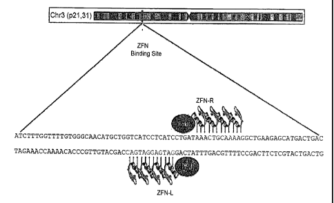
components. By way of illustration, a transcriptional regulatory sequence,
such as a
promoter, is operatively linked to a coding sequence if the transcriptional
regulatory
sequence controls the level of transcription of the coding sequence in
response to the
presence or absence of one or more transcriptional regulatory factors. A
transcriptional regulatory sequence is generally operatively linked in cis
with a coding
sequence, but need not be directly adjacent to it. For example, an enhancer is
a
transcriptional regulatory sequence that is operatively linked to a coding
sequence,
even though they are not contiguous.
[0085] With respect to fusion polypeptides, the term "operatively
linked" can
refer to the fact that each of the components performs the same function in
linkage to
the other component as it would if it were not so linked. For example, with
respect to
a fusion polypeptide in which a ZFP DNA-binding domain is fused to a cleavage
domain, the ZIT DNA-binding domain and the cleavage domain are in operative
linkage if, in the fusion polypeptide, the ZFP DNA-binding domain portion is
able to
bind its target site and/or its binding site, while the cleavage domain is
able to cleave
DNA in the vicinity of the target site.
[0086] A "functional fragment" of a protein, polypeptide or nucleic
acid is a
protein, polypeptide or nucleic acid whose sequence is not identical to the
full-length
protein, polypeptide or nucleic acid, yet retains the same function as the
full-length
protein, polypeptide or nucleic acid. A functional fragment can possess more,
fewer,
or the same number of residues as the corresponding native molecule, and/or
can
contain one or more amino acid or nucleotide substitutions. Methods for
CA 02651499 2014-02-05
determining the function of a nucleic acid (e.g., coding function, ability to
hybridize
to another nucleic acid) are well-known in the art. Similarly, methods for
determining
protein function are well-known. For example, the DNA-binding function of a
polypeptide can be determined, for example, by filter-binding, electrophoretic
mobility-shift, or immunopre,cipitation assays. DNA cleavage can be assayed by
gel
electrophoresis. See Ausubel et at., supra. The ability of a protein to
interact with
another protein can be determined, for example, by co-immunoprecipitation, two-
hybrid assays or complementation, both genetic and biochemical. See, for
example,
Fields et a/. (1989) Nature 340:245-246; U.S. Patent No. 5;585,245 and PCT WO
98/44350.
Zinc Finger Nucleases
[00871 Described herein are zinc finger nucleases (ZFNs) that can be
used for
gene inactivation, for example inactivation of the CCR5 gene. ZFNs comprise a
zinc
finger protein (ZFP) and a nuclease (cleavage) domain.
A. Zinc Finger Proteins
[0088) Zinc finger binding domains can be engineered to bind to a
sequence
of choice. See, for example, Beerli etal. (2002) Nature BiotechnoL 20:135-141;
Pabo
et al (2001) Ann. Rev. Biochern. 70:313-340; 'salmi et at. (2001) Nature
BiotechnoL
19:656-660; Segal et al. (2001) Curr. Opin. Biotechnol. 12:632-637; Chao et aL
(2000) Curr. Opin. Struct. BioL 10:411-416. An engineered zinc finger binding
domain can have a novel binding specificity, compared to a naturally-occurring
zinc
finger protein. Engineering methods include, but are not limited to, rational
design
and various types of selection. Rational design includes, for example, using
databases
comprising triplet (or quadruplet) nucleotide sequences and individual zinc
finger
amino acid sequences, in which each triplet or quadruplet nucleotide sequence
is
associated with one or more amino acid sequences of zinc fingers which bind
the
particular triplet or quadruplet sequence. See, for example, co-owned U.S.
Patents
6,453,242 and 6,534,261.
100891 Exemplary selection methods, including phage display and two-
hybrid
systems, are disclosed in US Patents 5,789;538; 5,925,523; 6,007,988;
6,013,453;
6,410,248; 6,140,466; 6,200,759; and 6,242,568; as well as WO 98/37186;
WO 98/53057; WO 00/27878; WO 01/88197 and GB 2,338,237.
21
CA 02651499 2014-02-05
[0090] Enhancement of binding specificity for zinc finger binding
domains
has been described, for example, in co-owned WO 02/077227.
[00911 Selection of target sites; ZFPs and methods for design and
construction
of fusion proteins (and polynucleotides encoding same) are known to those of
skill in
the art and described in detail in related to U.S. Publication Nos.
20030232410;
20050208489; 2005064474; 20050026157; 20060188987; International Publication
WO 07/014275.
[0092] In certain embodiments, the zinc finger nucleases of the Ad-ZFN
vectors described herein bind in a CCR-5 gene. Table 1 describes a number of
zinc
finger binding domains that have been engineered to bind to nucleotide
sequences in
the human CCR-5 gene. Each row describes a separate zinc finger DNA-binding
domain. The DNA target sequence for each domaiiis shown in the first column
(DNA target sites indicated in uppercase letters; non-contacted nucleotides
indicated
in lowercase), and the second through fifth columns show the amino acid
sequence of
the recognition region (amino acids -1 through +6, with respect to the start
of the
helix) of each of the zinc fingers (F1 through F4) in the protein. Also
provided in the
first column is an identification number for each protein.
22
CA 02651499 2008-11-06
WO 2007/139982
PCT/US2007/012588
Table 1: Zinc linger nucleases targeted to the human CCR-5 gene
r162 designs
Target sequence F1 F2 F3 F4
DRSNLSR (SEQ TSANLSR (SEQ RSDNLAR (SEQ TSANLSR (SEQ
GATGAGGATGAC ID NO:2) ID NO:3) ID NO:4) ID NO:3)
(SEQ ID NO:1) 7296
DRSNLSR (SEQ ISSNLNS (SEQ RSDNLAR (SEQ TSANLSR (SEQ
GATGAGGATGAC ID NO:2) ID NO:5) ID NO:4) ID NO:3)
(SEQ ID NO:1) 8181
DRSNLSR (SEQ VSSNLTS (SEQ RSDNLAR (SEQ TSANLSR (SEQ
GATGAGGATGAC ID NO:2) ID NO:6) ID NO:4) ID NO:3)
(SEQ ID NO:1) 8182
DRSNLSR (SEQ ISSNLNS (SEQ RSDNLAR (SEQ NRDNLSR
GATGAGGATGAC ID NO:2) ID NO:5) ID NO:4) (SEQ ID
NO:7)
(SEQ ID NO:1) 8262
DRSNLSR (SEQ ISSNLNS (SEQ RSDNLAR (SEQ TSGNLTR (SEQ
GATGAGGATGAC ID NO:2) ID NO:5) ID NO:4) ID NO:8)
(SEQ ID NO:1) 8266
DRSNLSR (SEQ VSSNLTS (SEQ RSDNLAR (SEQ TSGNLTR (SEQ
GATGAGGATGAC ID NO:2) ID NO:6) ID NO:4) ID NO:8)
(SEQ ID NO:1) 8267
DRSNLSR (SEQ TSGNLTR (SEQ RSDNLAR (SEQ TSGNLTR (SEQ
GATGAGGATGAC ID NO:2) ID NO:8) ID NO:4) ID NO:8)
(SEQ ID NO:1) 7741
168 designs
Target sequence F1 F2 F3 F4
RSDNLSV (SEQ QNANRIT (SEQ RSDVLSE (SEQ QRNHRTT (SEQ
AAACTGCAAAAG ID NO:10) ID NO:11) ID NO:12) ID NO:13)
(SEQ ID NO:9) 7745
RSDNLSN (SEQ QNANRIT (SEQ RSDVLSE (SEQ QRNHRTT (SEQ
AAACTGCAAAAG ID NO:14) ID NO:1 1) ID NO:12) ID NO:13)
(SEQ ID NO:9) 8165
RSDNLSV (SEQ QRVNLIV (SEQ RSDVLSE (SEQ QRNHRTT (SEQ
AAACTGCAAAAG ID NO:10) ID NO:15) ID NO:12) ID NO:13)
(SEQ ID NO:9) 8191
RSDNLGV (SEQ QKINLQV (SEQ RSDVLSE (SEQ QRNHRTT (SEQ
AAACTGCAAAAG ID NO:16) ID NO:17) ID NO:12) ID NO:13)
(SEQ ID NO:9) 8196
RSDNLSV (SEQ QKINLQV (SEQ RSDVLSE (SEQ QRNHRTT (SEQ
AAACTGCAAAAG ID NO:10) ID NO:17) ID NO:12) ID NO:13)
(SEQ ID NO:9) 8196z
AAACTGCAAAAG RSDNLGV (SEQ QKINLQV (SEQ RSDVLSE (SEQ QRNHRTT (SEQ
(SEQ ID NO:9) ID NO:16) ID NO:17) ID NO:12)
ID NO:13)
8196zg
RSDHLSE (SEQ QNANRIT (SEQ RSDVLSE (SEQ QRNHRTT (SEQ
AAACTGCAAAAG ID NO:18) ID NO:11) ID NO:12) ID NO:13)
(SEQ ID NO:9) 7568
23
CA 02651499 2014-02-05
r627 designs
Target sequence F1 F2 F3 F4
RSAHLSE (SEQ RSANLSE (SEQ RSANLSV (SEQ DRANLSR (SEQ ID
GACAAGCAGCGG ID NO:20) ID N021) ID NO: 22) N0:23)
(SEQ ID NO:19) 7524
=633 designs
Target sequence Ft F2 F3 F4
RSDSLSK (SEQ DNSNRIK (SEQ RSAVLSE (SEQ TNSNRIT (SEQ ID
CATCTGcTACTCG ID NO:25) ID N026) ID NO27) N0:28)
(SEQ ID NO:24) 8040
[0093] As
described below, in certain embodiments, a four-finger binding
domain as shown in Table 1 is fused to a cleavage half-domain, such as, for
example,
the cleavage domain of a Type us restriction endonuclease such as Fokl. A pair
of
such zinc finger/nuclease half-domain fusions are used for targeted cleavage,
as
disclosed, for example, in U.S. Patent Publication No. 20050064474. For
example,
ZFN-215 denotes the pair of fusion proteins containing the zinc finger binding
domains designated 8267 (which recognizes the target sequence shown in SEQ ID
NO: 1 and comprises the 4 recognition helices depicted in SEQ ID NOs 2, 6, 4
and 8)
and 8196z (which recognizes the target sequence shown in SEQ ID NO:9 and
comprises the 4 recognition helices depicted in SEQ ID NOs:10, 17, 12 and 13).
ZFN-201 denotes the pair of fusion proteins containing the zinc finger binding
domains designated 8266 (which recognizes the target sequence shown in SEQ ID
NO:1 and comprises the 4 recognition helices depicted in SEQ ID NOs:2, 2, 4
and 8)
and 8196z (which recognizes the target sequence shown in SEQ ID NO:9 and
comprises the 4 recognition helices depicted in SEQ ID NOs:10, 17, 12 and 13).
[00941 For targeted cleavage, the near edges of the binding sites can
separated
by 5 or more nucleotide pairs, and each of the fusion proteins can bind to an
opposite
strand of the DNA target. Hence, any one of the proteins identified as an
"r162
design" in Table 1 (indicating that it binds to the reverse strand and that
the
downstream edge of its binding site is at nucleotide 162) can be paired with
any of the
proteins identified as a "168 design" (indicating that it binds to the strand
opposite
that bound by the r162 designs and that the upstream edge of its binding site
is at
nucleotide 168). For example, protein 8267 can be paired with protein 8196 or
with
24
CA 02651499 2008-11-06
WO 2007/139982
PCT/US2007/012588
protein 8196z or with any of the other 168 designs; and protein 8266 can be
paired
with either of proteins 8196 or 8196z or with any other of the 168 designs.
All
pairwise combinations of the r162 and 168 designs can be used for targeted
cleavage
and mutagenesis of a CCR-5 gene. Similarly, the 7524 protein (or any other
r627
design) can be used in conjunction with the 8040 protein (or any other 633
design) to
obtain targeted cleavage and mutagenesis of a CCR-5 gene.
[0095] The CCR5-ZFNs described herein can be targeted to any
sequence in
the CCR5 genome. For example, CCR5 genomic sequences (including allelic
variants such as CCR5-A32) are well known in the art and individuals
homozygous
for the CCR5-A32 (see, e.g., Liu et al. (1996) Cell 367-377), are resistant to
HIV-1
infection.
B. Cleavage Domains
[0096] The ZFNs also comprise a nuclease (cleavage domain, cleavage
half-
domain). The cleavage domain portion of the fusion proteins disclosed herein
can be
obtained from any endonuclease or exonuclease. Exemplary endonucleases from
which a cleavage domain can be derived include, but are not limited to,
restriction
endonucleases and homing endonucleases. See, for example, 2002-2003 Catalogue,
New England Biolabs, Beverly, MA; and Belfort et al. (1997) Nucleic Acids Res.
25:3379-3388. Additional enzymes which cleave DNA are known (e.g., Si
Nuclease;
mung bean nuclease; pancreatic DNase I; micrococcal nuclease; yeast HO
endonuclease; see also Linn et al. (eds.) Nucleases, Cold Spring Harbor
Laboratory
Press,1993). One or more of these enzymes (or functional fragments thereof)
can be
used as a source of cleavage domains and cleavage half-domains.
[0097j Similarly, a cleavage half-domain can be derived from any nuclease
or
portion thereof, as set forth above, that requires dimerization for cleavage
activity. In
general, two fusion proteins are required for cleavage if the fusion proteins
comprise
cleavage half-domains. Alternatively, a single protein comprising two cleavage
half-
domains can be used. The two cleavage half-domains can be derived from the
same
endonuclease (or functional fragments thereof), or each cleavage half-domain
can be
derived from a different endonuclease (or functional fragments thereof). In
addition,
the target sites for the two fusion proteins are preferably disposed, with
respect to
each other, such that binding of the two fusion proteins to their respective
target sites
places the cleavage half-domains in a spatial orientation to each other that
allows the
CA 02651499 2008-11-06
WO 2007/139982
PCT/US2007/012588
cleavage half-domains to form a functional cleavage domain, e.g., by
dimerizing.
Thus, in certain embodiments, the near edges of the target sites are separated
by 5-8
nucleotides or by 15-18 nucleotides. However any integral number of
nucleotides or
nucleotide pairs can intervene between two target sites (e.g., from 2 to 50
nucleotide
pairs or more). In general, the site of cleavage lies between the target
sites.
[0098] Restriction endonucleases (restriction enzymes) are present
in many
species and are capable of sequence-specific binding to DNA (at a recognition
site),
and cleaving DNA at or near the site of binding. Certain restriction enzymes
(e.g.,
Type HS) cleave DNA at sites removed from the recognition site and have
separable
binding and cleavage domains. For example, the Type IIS enzyme Fok I catalyzes
double-stranded cleavage of DNA, at 9 nucleotides from its recognition site on
one
strand and 13 nucleotides from its recognition site on the other. See, for
example, US
Patents 5,356,802; 5,436,150 and 5,487,994; as well as Li etal. (1992) Proc.
Natl.
Acad. Sci. USA 89:4275-4279; Li etal. (1993) Proc. Natl. Acad. Sci. USA
90:2764-
2768; Kim etal. (1994a) Proc. Natl. Acad. Sci. USA 91:883-887; Kim etal.
(1994b)
J. Biol. Chem. 269:31,978-31,982. Thus, in one embodiment, fusion proteins
comprise the cleavage domain (or cleavage half-domain) from at least one Type
IIS
restriction enzyme and one or more zinc finger binding domains, which may or
may
not be engineered.
[0099] An exemplary Type US restriction enzyme, whose cleavage domain is
separable from the binding domain, is Fok I. This particular enzyme is active
as a
dimer. Bitinaite etal. (1998) Proc. Natl. Acad. Sci. USA 95: 10,570-10,575.
Accordingly, for the purposes of the present disclosure, the portion of the
Fok I
enzyme used in the disclosed fusion proteins is considered a cleavage half-
domain.
Thus, for targeted double-stranded cleavage and/or targeted replacement of
cellular
sequences using zinc finger-Fok I fusions, two fusion proteins, each
comprising a
Fokl cleavage half-domain, can be used to reconstitute a catalytically active
cleavage
domain. Alternatively, a single polypeptide molecule containing a zinc finger
binding
domain and two Fok I cleavage half-domains can also be used. Parameters for
targeted cleavage and targeted sequence alteration using zinc finger-Fok I
fusions are
provided elsewhere in this disclosure.
26
CA 02651499 2014-02-05
[01001 A cleavage domain or cleavage half-domain can be any portion
of a
protein that retains cleavage activity, or that retains the ability to
multimerize (e.g.,
dimerize) to form a functional cleavage domain.
[01011 Exemplary Type HS restriction enzymes are described in
International
Publication WO 07/014275, incorporated herein in its entirety. Additional
restriction
enzymes also contain separable binding and cleavage domains, and these are
contemplated by the present disclosure. See, for example, Roberts et at (2003)
Nucleic Acids Res. 31:418-420.
101021 In certain embodiments, the cleavage domain comprises one or
more
engineered cleavage half-domain (also referred to as dimerization domain
mutants)
that minimize or prevent homodimerization, as described, for example, in U.S.
Patent
Publication Nos. 20050064474 and 20060188987. Amino acid residues at positions
446, 447, 479, 483, 484, 486, 487, 490, 491, 496, 498, 499, 500, 531, 534,
537, and
538 of Fokl are all targets for influencing dimerization of the Fold cleavage
half-
domains.
[0103] Exemplary engineered cleavage half-domains of Fok I that form
obligate heterodimers include a Pair in which a first cleavage half-domain
includes
mutations at amino acid residues at positions 490 and 538 ofFok I and a second
cleavage half-domain includes mutations at amino acid residues 486 and 499.
See
Figures 2,3 and 4.
[01041 Thus, in one embodiment, as shown in Figures 3 and 4, the
mutation at
490 replaces Glu (E) with Lys (K); the mutation at 538 replaces Iso (I) with
Lys (K);
the mutation at 486 replaced Gin (Q) with Glu (E); and the mutation at
position 499
replaces Iso (I) with Lys (K). Specifically, the engineered cleavage half-
domains
- decribed herein were prepared by mutating positions 490 (E--+K) and 538 (I--
,K) in
one cleavage half-domain to produce an engineered cleavage half-domain
designated
"E490K:I538K" and by mutating positions 486 (Q-*E) and 499 (I-4.,) in another
cleavage half-domain to produce an engineered cleavage half-domain designated
"Q486E1499L". The engineered cleavage half-domains described herein are
obligate
heterodimer mutants in which aberrant cleavage is minimized or abolished.
27
CA 02651499 2014-02-05
101051 Engineered cleavage half-domains described herein can be
prepared
using any suitable method, for example, by site-directed mutagenesis of wild-
type
cleavage half-domains (Fok I) as described in U.S. Patent Publication No.
20050064474 (Example 5).
C. Additional Methods for Targeted Cleavage in CCR5
(01061 Any nuclease having a target site in a CCR5 gene can be used
in the
methods disclosed herein. For example, homing endonucleases and meganucleases
have very long recognition sequences, some of which are likely to be present,
on a
statistical basis, once in a human-sized genome. Any such nuclease having a
unique
target site in a CCIt5 gene can be used instead of, or in addition to, a zina
finger
nuclease, for targeted cleavage in a CCR5 gene.
[01071 Exemplary homing endonucleases include I-SceI,I-Ceul,PI-Pspl,
PI-
Sce,I-SceIV I-TevII and I-
TevIII. Their recognition sequences are known. See also U.S. Patent No.
5,420,032;
U.S. Patent No. 6,833,252; Belfort et aL (1997) Nucleic Acids Res. 25:3379-
3388;
Dujon etal. (1989) Gene 82:115-118; Perler etal. (1994) Nucleic Acids Res. 22,
1125-1127; Jasin (1996) Trends Genet. 12:224-228; Gimble et a/. (1996) J. MoL
Biol. 26.3:163-180; Argast et al. (1998)1 IfoL BioL 280:345-353 and the New
England Biolabs catalogue.
[01081 Although the cleavage specificity of most homing endonucleases
is not
absolute with respect to their recognition sites, the sites are of sufficient
length that a
single cleavage event per mammalian-sized genome can be obtained by expressing
a
homing endonuclease in a cell containing a single copy of its recognition
site. It has
also been reported that the specificity of homing endonucleases and
meganucleases
can be engineered to bind non-natural target sites. See, for example,
Chevalier et at.
(2002) Molec. Cell10:895-905; Epinat et at. (2003) Nucleic Acids Res. 31:2952-
2962; Ashworth et al. (2006) Nature 441:656-659; Paques et aL (2007) Current
Gene Therapy 7:49-66.
Delivery '
[0109] The ZFNs described herein may be delivered to a target cell by
any
suitable means. Methods of delivering proteins comprising zinc fingers are
described,
28
CA 02651499 2014-02-05
for example, in U:S. Patent Nos. 6,453,242; 6,503,717; 6,534,261; 6,599,692;
6,607,882; 6,689,558; 6,824,978; 6,933,113; 6,979,539; 7,013,219; and
7,163,824.
[01101 ZFNs as described herein may also be delivered using vectors
containing sequences encoding one or more ZFNs. Any vector systems may be used
including, but not limited to, plasmid vectors, retroviral vectors, lentiviral
vectors,
adenovirus vectors, poxvirus vectors; herpesvirus vectors and adeno-associated
virus
vectors, etc. See, also, U.S. Patent Nos. 6,534,261; 6,607,882; 6,824,978;
6,933,113;
6,979,539; 7,013,219; and 7,163,824.
[0111] In certain embodiments, the vector is an adenovirus vector.
Thus,
described herein are adenovirus (Ad) vectors for introducing heterologous
sequences
(e.g., zinc finger nucleases (ZFNs)) into cells.
[0112] Non-limiting examples of Ad vectors that can be used in the
present
application include recombinant (such as El-deleted), conditionally
replication
competent (such as oncolytic) and/or replication competent Ad vectors derived
from
human or non-human serotypes (e.g., Ad5, Adl 1, Ad35, or porcine adenovirus-
3);
and/or chimeric Ad vectors (such as Ad5/35) or tropism-altered Ad vectors with
engineered fiber (e.g., knob or shaft) proteins (such as peptide insertions
within the HI
loop. of the knob protein). Also useful are "gutless" Ad vectors, e.g., an Ad
vector in
which all adenovirus genes have been removed, to reduce immunogenicity and to
increase the size of the DNA payload. This allows, for example, simultaneous
delivery of sequences encoding ZFNs and a donor sequence. Such gutless vectors
are
especially useful when the donor sequences include large transgenes to be
integrated
via targeted integration.
[0113] Replication-deficient recombinant adenoviral vectors (Ad) can
be
produced at high titer, and they readily infect a number of different cell
types. Most
adenovirus vectors are engineered such that a transgene replaces the Ad Ela,
and/or E3 genes; subsequently the replication defective vector is propagated
in cells
that provide one or more of the deleted gene functions in trans. For example,
human
293 cells supply El function. Ad vectors can transduce multiple types of
tissues in
vivo, including non-dividing, differentiated cells such as those found in
liver, kidney
and muscle. Conventional Ad vectors have a large carrying capacity. An example
of
the use of an Ad vector in a clinical trial involved polynucleodde therapy for
29
CA 02651499 2008-11-06
WO 2007/139982
PCT/US2007/012588
antitumor immunization with intramuscular injection (Sterman et al., Hum. Gene
Ther. 7:1083-1089(1998)).
[0114]
Additional examples of the use of adenovirus vectors for gene transfer
in clinical trials include Rosenecker et al., Infection 24:1 5-10 (1996);
Welsh et al.,
Hum. Gene Ther. 2:205-18 (1995); Alvarez et al., Hum. Gene Ther. 5:597-613
(1997); Topf et al., Gene Ther. 5:507-513 (1998).
[0115] In
certain embodiments, the Ad vector is a chimeric adenovirus vector,
containing sequences from two or more different adenovirus genomes. For
example,
the Ad vector can be an Ad5/35 vector. Ad5/35 is created by replacing one or
more
of the fiber protein genes (knob, shaft, tail, penton) of Ad5 with the
corresponding
fiber protein gene from a B group adenovirus such as, for example, Ad35. The
Ad5/35 vector and characteristics of this vector are described, for example,
in Ni et al.
(2005) "Evaluation of biodistribution and safety of adenovirus vectors
containing
group B fibers after intravenous injection into baboons," Hum Gene Ther 16:664-
677;
Nilsson et al. (2004) "Functionally distinct subpopulations of cord blood
CD34+ cells
are transduced by adenoviral vectors with serotype 5 or 35 tropism," Mol Ther
9:377-
388; Nilsson et al. (2004) "Development of an adenoviral vector system with
adenovirus serotype 35 tropism; efficient transient gene transfer into primary
malignant hematopoietic cells," J Gene Med 6:631-641; Schroers et al. (2004)
"Gene
transfer into human T lymphocytes and natural killer cells by Ad5/F35 chimeric
adenoviral vectors," Exp Hematol 32:536-546; Seshidhar et al. (2003)
"Development
of adenovirus serotype 35 as a gene transfer vector," Virology 311:384-393;
Shayalchmetov et al. (2000) "Efficient gene transfer into human CD34(+) cells
by a
retargeted adenovirus vector," J Virol 74:2567-2583; and Soya et al. (2004),
"A
tumor-targeted and conditionally replicating oncolytic adenovirus vector
expressing
TRAIL for treatment of liver metastases," Mol Ther 9:496-509.
[0116] As
noted above, ZFNs and polynucleotides encoding these ZFNs may
be delivered to any target cell. Generally, for inactivating a gene CCR-5, the
cell is
an immune cell, for example, a lymphocyte (B-cells, T-cells such as T helper
(TH) and
T cytotoxic cells (Tc), null cells such as natural killer (NK) cells); a
mononuclear cell
(monocytes, marcophages); a granulocytic cell (granulocytes, neutrophils,
eosinophils, basophils); a mast cell; and/or a dendritic cell (Langerhans
cells,
interstitial dendritic cells, interdigitating dendritic cells, circulating
dendritic cells).
Macrophages, B lymphocytes and dendritic cells are exemplary antigen-
presenting
CA 02651499 2008-11-06
WO 2007/139982
PCT/US2007/012588
cells involved in TH cell activation. In certain embodiments, the target cell
is a TH
cell, characterized by expression of CD4 on the surface. The target cell may
also be a
hematopoietic stem cell, which may give rise to any immune cell.
Applications
[0117] The disclosed methods and compositions can be used to cleave
DNA at
a region of interest in cellular chromatin (e.g., at a desired or
predetermined site in a
genome, for example, in a gene, either mutant or wild-type); to replace a
genomic
sequence (e.g., a region of interest in cellular chromatin, see, also, Example
5 below)
with a homologous non-identical sequence (i.e., targeted recombination); to
delete a
genomic sequence by cleaving DNA at one or more sites in the genome, which
cleavage sites are then joined by non-homologous end joining (NHEJ); to screen
for
cellular factors that facilitate homologous recombination; to replace a wild-
type
sequence with a mutant sequence; and/or to convert one allele to a different
allele.
Such methods also allow for generation and/or modification of cells lines (for
therapeutic and non-therapeutic uses), treatment of infections (viral or
bacterial) in a
host (e.g., by blocking expression of viral or bacterial receptors, thereby
preventing
infection and/or spread in a host organism); to treat genetic diseases.
[0118] Thus, the compositions and methods described herein can be
used for
gene modification, gene correction, and gene disruption. Non-limiting examples
of
gene modification includes homology directed repair (HDR)-based targeted
integration; HDR-based gene correction; HDR-based gene modification; HDR-based
gene disruption; NHEJ-based gene disruption and/or combinations of HDR, NHEJ,
and/or single strand annealing (SSA). Single-Strand Annealing (SSA) refers to
the
repair of a double strand break between two repeated sequences that occur in
the same
orientation by resection of the DSB by 5'-3' exonucleases to expose the 2
complementary regions. The single-strands encoding the 2 direct repeats then
anneal
to each other, and the annealed intermediate can be processed such that the
single-
stranded tails (the portion of the single-stranded DNA that is not annealed to
any
sequence) are be digested away, the gaps filled in by DNA Polymerase, and the
DNA
ends rejoined. This results in the deletion of sequences located between the
direct
repeats.
31
CA 02651499 2008-11-06
WO 2007/139982
PCT/US2007/012588
[01191 The compositions (e.g., Ad-ZFN vectors) and methods described
herein can also be used in the treatment of various genetic diseases and/or
infectious
diseases.
[01201 The compositions and methods can also be applied to stem cell
based
therapies, including but not limited to:
[01211 (a) Correction of somatic cell mutations by short patch gene
conversion or targeted integration for monogenic gene therapy
[01221 (b) Disruption of dominant negative alleles
[01231 (c) Disruption of genes required for the entry or productive
infection of
pathogens into cells
[0124] (d) Enhanced tissue engineering, for example; by:
(i) Modifying gene activity to promote the differentiation or formation
of functional tissues; and/or
(ii) Disrupting gene activity to promote the differentiation or formation
of functional tissues
101251 (e) Blocking or inducing differentiation, for example, by:
(i) Disrupting genes that block differentiation to promote stem cells to
differentiate down a specific lineage pathway
(ii) Targeted insertion of a gene or siRNA expression 'cassette that can
stimulate stem cell differentiation.
(iii) Targeted insertion of a gene or siRNA expression cassette that can
block stem cell differentiation and allow better expansion and maintenance of
pluripotency
(iv) Targeted insertion of a reporter gene in frame with an endogenous
gene that is a marker of pluripotency or differentiation state that would
allow an easy
marker to score differentiation state of stem cells and how changes in media,
cytoldnes, growth conditions, expression of genes, expression of siRNA
molecules,
exposure to antibodies to cell surface markers, or drugs alter this state.
[0126] (f) Somatic cell nuclear transfer, for example, a patient's
own somatic
cells can be isolated, the intended target gene modified in the appropriate
manner, cell
clones generated (and quality controlled to ensure genome safety), and the
nuclei
from these cells isolated and transferred into unfertilized eggs to generate
patient-
specific hES cells that could be directly injected or differentiated before
engrafting
into the patient, thereby reducing or eliminating tissue rejection.
32
CA 02651499 2014-02-05
101271 (g) Universal steincells by knocking out MHC receptors ¨ This
approach would be used to generate cells of diminished or altogether abolished
immunological identity. Cell types for this procedure include but are not
limited to,
T-cells, 13 cells, hematopoietic stem cells, and embryonic stem cells.
Therefore, these
stem cells or their derivatives (differentiated cell types or tissues) could
be potentially
engrafted into any person regardless of their origin or histocompatibility.
101281 The compositions and methods can also be used for somatic cell
therapy (e.g., autologous cell therapy and/or universal T-cell by knocking out
MHC
receptors, see section (g) above), thereby allowing production of stocks of T-
cells that
have been modified to enhance their biological properties. Such cells can be
infused
into a variety of patients independent of the donor source of the T-cells and
their
histocompatibility to the recipient.
[01291 In addition, the use of Ad vectors as described herein to
deliver ZFNs
enhances rates of NHEJ. Without being bound by one theory, it appears that
this
effect is due to the inhibitory effect the E4 proteins (E4 ORF6 (E4 34k), E4
ORF3)
may have on DSB repair.
25
EXAMPLES
Example 1: Vector Construction
A. Construction of Ad5/35-GFP and Ad5/35-ZFN Vectors
[0132] Chimeric Ad5/35 vectors (Nilsson et al. (2004) Mol Ther 9:377-
388;
Nilsson et al. (2004) J Gene Med 6:631-641) were constructed as shown
schematically in Figure 1. Briefly, a sequence encoding. an expression
cassette for
GFP (top line), an expression cassette for a pair of engineered ZFNs (ZFN1 and
ZFN2) which target the endogenous CCR5 locus (middle line), or a homologous
33
CA 02651499 2014-02-05
donor sequence for targeting a 47-bp patch sequence in the CCR5 locus (bottom
line)
was inserted in place of the El genes using the AdEasy bacterial recombination
system (Stratagene).
[0133] The Ad-2I5 and Ad-201 vector constructs each comprise
sequences
encoding two ZFNs that cleave the endogenous CCR5 gene at sequences encoding
Leu55. Both ZFNs are expressed under the control of a single promoter via a 2A
fusion. As shown in Table 1 above, the ZFN215 pair encoded by the Ad-215
construct comprise zinc finger binding domains 8267and 8196z from the r162 and
168 designs, respectively; while the ZFN201 pair encoded by the Ad-201 vector
comprises zinc finger binding domains 8266 and 8196z from the r162 and 168
designs, respectively. The ZFNs encoded by the Ad-215 and Ad-201 vectors are
fused to a wild-type Fold cleavage half-domain. See Figure 2.
[0134] The ZFN224 pair encoded by the Ad-224 construct comprise zinc
finger binding domains 8267and 8196z from the r162 and 168 designs,
respectively.
Thus, The Ad-224 construct has the same zinc finger proteins as Ad-215.
However,
the ZFNs encoded by the Ad-224 construct contain mutant FokI cleavage-half
domain. In particular, the 8267 zinc finger domain is fused to a Fokl cleavage
half-
domain containing the Q486E and I499L mutations (Figure 3), and 8196z zinc
finger
domain is fused to a Fokl cleavage half-domain containing the E490K and 153 8K
mutations (Figure 4).
B. Donor Vector for Targeted Insertion
[01351 To make a donor vector (Ad5/35 P on, Fig 1 bottom line), an
1881-bp
fragment of the human genome corresponding to the CCR5 locus was PCR amplified
and cloned into the PCR4-TOPO vector (Invitrogen). The sequence of the
fragment is
shown in Figure 5 (SEQ ID NO:36).
[0136] In order to generate a cloning site at which to insert a
"patch"
sequence, 2 nucleotides of the sequence shown in Figure 5 were changed to
generate a
Xbal recognition site. Specifically, the nucleotide sequence "atectgataa" (SEQ
ID
NO:39) (nucleotides 470-479 in the donor fragment) was changed to
"atcla_gataa"
(SEQ ID NO:40) (the 2 bases changed are underlined) via the QuicicChange Site-
Directed Mutagenesis Kit (Stratagene).
34
CA 02651499 2008-11-06
WO 2007/139982
PCT/US2007/012588
[0137] The resulting DNA sequence was then digested with Xbal, and a
47bp
"patch" sequence, shown in Figure 6 (SEQ ID NO:37), was inserted at the Xbal
site.
[0138] The resulting sequence, shown in Figure 7 (SEQ ID NO:38), was
inserted into the Ad5/35 vector as described above for the GFP and ZFN
expression
cassettes. With respect to the sequence shown in Figure 7, the 5'homology arm
correspond to nucleotides 1-471; the "patch" sequence for targeted insertion
into
CCR-5 is underlined and corresponds to nucleotides 472-518; and the 3'
homology
arm corresponds to nucleotides 519-1928.
Example 2: Transduction of hES Cells with Ad5/35-GFP vectors
[0139] Chimeric Ad5/35-GFP vectors as described in Example 1 were
introduced into human embryonic stem (hES) cells as follows.
[0140] Infections of hES cells were performed in 500 Al volumes
using
400,000 cells and 25 1, 5 Al or 0.5/21 of the Ad5/35-GFP vector (MOI of 8200,
1640
and 164 respectively). After 4 hours, the cells were washed and plated onto
fresh
murine embryonic fibroblast (MEF) feeder cells. Fluorescence microscopy of
living
cells, obtained approximately 20 hours post-infection, showed fluorescence in
stem
cell colonies that had been infected with 5 and 25 IA of virus, and no
fluorescence in
the feeder cells. FACS analysis for GFP fluorescence was performed ¨22 hours
post-
infection. The results are shown in Table 2.
Table 2
Infection % of fluorescent T-cells
Mock infection 0.74%
Ad5/35-GFP 0.5 l 39.4%
Ad5/35-GFP 512,1 91%
Ad5/35-GFP 25 1 95%
[0141] These results indicate that Ad5/35 vectors are capable of
infecting
human embryonic stem cells at high efficiency.
Example 3: Modification of the CCR-5 Gene using Ad5/35-ZFNs
[0142] CD4 T-cells and PBMCs were obtained from AllCells. Cells
were
cultured in RPMI + 10% FBS + 1% L-Glutamine (30 mg/mL) + IL-2 (lng/mL,
CA 02651499 2008-11-06
WO 2007/139982
PCT/US2007/012588
Sigma) and activated with anti. CD3-CD28 beads according to manufacturer's
protocol (Dynal). Cells were seeded at 3E5 celllmL in 1mL volume in a 24 well
plate.
[0143] Adenoviral vectors as described in Example 1 (Ad5/35 GFP,
Ad5/35
215 or Ad5/35 224) were added two days later at an MOI of 10, 30, or 100 (MOI
calculated based on infectious titer).
[0144] Cells were harvested 2 days after exposure to virus and gene
modification efficiency was determined using a Cel-1 assay, performed as
described
in International Patent Publication WO 07/014275. See, also, Oleykowslci et
al.
(1998) Nucleic Acids res. 26:4597-4602; Qui et al. (2004) BioTechniques 36:702-
707; Yeung et al. (2005) BioTechniques 38:749-758.
[0145] Results are shown in Table 3.
Table 3
% CCR-5 Alleles Modified
PBMCs CD4+ T-
cells
Control not detectable not
detectable
Ad5/35-ZFN215 at MOI 10 6.1 6.7
Ad5/35-ZFN215 at MOI 30 14.0 16.5
Ad5/35-ZFN215 at MOI 100 31.2 32.3
Ad5/35-ZFN224 at MOI 10 3.6 1.8
Ad5/35-ZFN224 at MOI 30 7.4 9.0
Ad5/35-ZFN224 at MOI 100 15.0 14.9
Ad5/35-GFP at MO! 10 not detectable not
detectable
Ad5/35-GFP at MO! 30 not detectable not
detectable
[0146] These results indicate that gene modification was observed after
infection of cells with Ad5/35 vectors encoding both the ZFN215 nuclease pair
(comprising wild type Fokl cleavage half-domains) and the ZFN 224 nuclease
pair
(comprising the mutant Fokl cleavage half-domains described in Example 1A). In
addition, the results show that gene modification levels increased in a dose-
dependent
manner.
[0147] To
determine the persistence of ZFN-induced gene modification,
infected cells were kept in culture for another 8 days (same culture medium as
36
CA 02651499 2008-11-06
WO 2007/139982
PCT/US2007/012588
before). Cells were counted and diluted with fresh medium every 2 days. Cells
were
then harvested (10 days after viral transduction) and the Cel-1 assay was
repeated. In
addition, a fraction of the CD4+T-ce1l population was re-activated with anti
CD3/
CD28 beads (Dynal) on day 7. Results are shown in Table 4.
Table 4
% Cells Infected
Re-activated CD4 Non-activated CD4
Control not detectable not detectable
Ad5/35-ZFN215 at MO! 30 10.1 10.9
Ad5/35-ZFN215 at MOI 100 29.0 28.2
Ad5/35-ZFN224 at MO! 30 6.7 6.4
Ad5/35-ZFN224 at MOI 100 16.1 16.6
Ad5/35-GFP at MOI 100 not detectable not detectable
[01481 These
results show that the degree of gene modification was
maintained as the CD4 + T-cells expanded. Furthermore, reactivation had no
apparent
effect on the growth of the modified cells as compared to unmodified cells.
Example 4: Modification of the CCR-5 Gene using Ad5/35-ZFNs in
CD34+ Cells
101491 CD4
+ T-cells and CD34+ cells were obtained from AllCells. On day 0,
CD4 + T-cells were cultured in RPMI + 10% FBS + 1% L-Glutamine (30 mg/mL) +
I1-2 (lng/mL, Sigma) and activated with anti CD3-CD28 beads according to the
manufacturer's protocol (Dynal). CD34+ cells were cultured in serum free
medium
(Stemspan H3000, Stem Cell Technologies) and supplemented with cytolcines
(Stemspan CC100, Stem Cell Technologies). Cells were seeded at 6E5 cell/mL in
1
mL volume in 24 well plates. Adenoviral vectors (Ad5/35 GFP or Ad5/35 224)
were
added the next day (day 1) at different MOIs (MOI calculated based on
infectious
titer).
[0150]
Cells were harvested on day 4 and gene modification efficiencies were
determined using a Cel-1 assay.
37
CA 02651499 2008-11-06
WO 2007/139982
PCT/US2007/012588
[0151] For CD4+ T-cells, Ad5/35 224 induced CCR-5 gene modification
with
an efficiency of 18.0%, 34.5% and 48.4% at MOIs of 25, 50 and 100,
respectively.
101521 Similarly, for CD34+ cells, Ad5/35 224 induced CCR5 gene
modification at efficiencies of 10.9% and 11.1%, at MOIs of 10 and 50,
respectively.
Example 5: Targeted Insertion of an Exogenous Sequence into the CCR-
5 Gene Using Ad5/35-ZFNs
[0153] On day 0, CD4+ T-cells were cultured in RPMI + 10% FBS + 1% L-
Glutamine (30 mg/mL) + m-2 (ing/mL, Sigma) and activated with anti CD3-CD28
beads according to the manufacturer's protocol (Dynal). Cells were seeded at
3E5
, cells/mL in 1 mL volume in a 24 well plate. Two days later (day 2), cells
were co-
transduced with different combinations of Ad5/35 224 and Ad5/35 P on donor
(Figures 1 and 7). Ad5/35 224 was added at MOIs of 0, 25, 50, and 100, and
Ad5/35
P on donor was added at MOIs of 0, 100, and 300 (MOI calculated based on
infectious titer). Cells were harvested 2 days after infection (day 4) and the
targeted
integration efficiency was determined by RFLP assay, as follows.
[0154] Genomic DNA was isolated from transduced cells and PCR
amplified
with primers outside the region of donor homology. The amplified fragment was
then
incubated with the restriction enzyme Bg11, whose recognition site is
contained within
the P on donor (Patch) sequence. The frequency of targeted integration of the
Patch
sequence was calculated by determining the ratio of cleaved to un-cleaved
products.
The frequency of targeted integration of the Patch sequence was 3.1% when
cells
were co-transduced with Ad5/35 224 at an MOI of 50 and Ad5/35 P on at an MOI
of
300.
Example 6: Non-Homologous End Joining Induced Using Ad5/35-ZFPs
[0155] To determine the types of ZFN-mediated mutations generated by
targeted cleavage followed by non-homologous end joining (NHEJ), genomic CCR-5
sequence of modified cells were sequenced and analyzed. Briefly, PBMCs and
CD4+
T-cells, transduced with Ad5/35 ZFN 215 in the same manner as described in
Example 3, were harvested and genomic DNA was extracted from these cells. The
CCR5 locus was then PCR amplified, Topo cloned into the PCR4-TOPO vector
(Invitrogen), and bacterial clones were sequence analyzed and compared to the
wild-
type CCR5 locus.
38
CA 02651499 2008-11-06
WO 2007/139982
PCT/US2007/012588
[0156] Mutations induced by targeted ZFN cleavage included both
deletions
and insertions, and the size of such changes varied over a wide range. Both
deletions
and insertions as short as a single nucleotide pair were observed, as were
insertions of
up to almost 100 nucleotide pairs. The sequences of some exemplary mutations
induced by targeted, ZFN-mediated cleavage are shown in Figures 8-10.
[0157] Figure 8 shows the sequences of a number of deletions
identified
during the analysis. Missing base pairs are denoted with periods.
[0158] An insertion of 5 base pairs occurred at a high frequency in
ZFN-
treated cells. This 5 base-pair insertion is a duplication of the 5 base-pair
sequence
between the ZFN-binding sites, converting the sequence
GTCATCCTCATCCTGATAAACTGCAAAAG (SEQ ID NO:41) to
GTCATCCTCATCCTGATCTGATAAACTGCAAAAG (SEQ NO:42), with the
inserted sequence underlined.
[0159] Another frequently-observed mutation was a four base-pair
insertion
between the two ZFN binding sites, converting the sequence
GTCATCCTCATCCTGATAAACTGCAAAAG (SEQ ID NO:41) to
GTCATCCTCATCCTTCTAGATAAACTGCAAAAG (SEQ ID NO:43), with the
inserted sequence underlined.
[0160] Figure 9 shows the nucleotide sequences of additional
mutations. In
one case, a combination of a one-nucleotide deletion and a five-nucleotide
insertion
was observed.
[0161] Figure 10 shows the sequences of a number of longer
insertions
resulting from targeted ZFN cleavage.
[0162] A summary of the sequence modifications to the CCR-5 locus is
shown below in Table 5.
39
CA 02651499 2008-11-06
WO 2007/139982
PCT/US2007/012588
Table 5
TYPE OF MUTATION NUMBER IN EACH CELL TOTALS
TYPE
PBMCs CD4+ T-cells
Deletion 10 8 18
bp 11 14 25
Insertions 4 bp 8 2 10
Other 3 3 6
long 1 3 4
Wild-type 10 14 24
Totals 43 44 87
Example 7: Cell Viability Post Ad5/35-ZFN Transduction
5 [0163] Cell viability post-transduction with Ad5/35-ZFN constructs
was also
assessed. Briefly, CD4+ T-cells and PBMCs were obtained from AllCells. On day
0,
cells were cultured in RPM! + 10%.FBS + 1% L-Glutarnine (30 mg/mL) + IL-2
(lng/mL, Sigma) and activated with anti CD3-CD28 beads according to the
manufacturer's protocol (Dynal). Cells were seeded at 3E5 cell/mL in 1 mL
volume
in 24 well plates. Adenoviral vectors (Ad5/35 GFP, Ad5/35 215 or Ad5/35 224)
were
added two days later (day 2) at MO! of 10, 30, and 100 (MOI calculated based
on
infectious titer). Cell counts and cell viability were measured at days 4, 6,
8, 10 and
12 using the VIACOUNT protocol provided with the GUAVA analytic flow
cytometer, following the manufacturer's instructions (Guava Technologies).
[0164] Ad5/35 ZFNs were generally well tolerated, with at least 75% of the
cells (up to 90% at the lower MO!) being viable at all time points. Thus,
minimal
toxicity was observed.
Example 8: Cell Growth Post Ad5/35-ZFN Transduction
[0165] Cell growth (doubling) post-transduction with Ad5/35-ZFN constructs
was also assessed.
101661 On day 0, CD4+ T-cells and PBMCs were cultured in RPM! + 10%
FBS + 1% L-Glutamine (30 mg/mL) + IL-2 (lng/mL, Sigma). Cells were activated
CA 02651499 2008-11-06
WO 2007/139982
PCT/US2007/012588
on days 0 and 6 with anti CD3-CD28 beads according to the manufacturer's
protocol
(Dynal). Cells were initially seeded at 3E5 cell/mL in 1 mL volume in 24 well
plates.
Adenoviral vectors (Ad5/35 GFP, Ad5/35 215 or Ad5/35 224) were added on day 2
at
MOIs of 10, 30, or 100 (MOI calculated based on infectious titer). Cell counts
and
cell viability were measured using the VIACOUNT protocol provided with the
GUAVA analytic flow cytometer as described in Example 7.
[0167] Cell growth or doubling was minimally affected by the
adenovirus
(except for Ad5/35 215 at a MOI of 100). Overall, at least 8 doublings (i.e.,
>100-
fold expansion) were achieved over a 14 day period in both CD4+ T-cells and
PBMCs.
Example 9: Measuring Persistence of Adenovirus Genome in CD4+ T-
cells
[0168] CD4+ T-cells were cultured in RPM! + 10% FBS + 1% L-Glutamine
(30 mg/mL) + IL-2 (lng/mL, Sigma) and activated with anti CD3-CD28 beads
according to the manufacturer's protocol (Dynal). Cells were seeded at 6E5
cell/mL
in 1 mL volume in a 24 well plate. Adenoviral vectors (Ad5/35 215 or Ad5/35
224)
were added the next day at MOI of 10, 30 and 100 (MOI calculated based on
infectious titer). Cells were harvested on days 4 and 14. DNA was extracted
with a
Masterpure kit (Epicenter Biotechnologies). Persistence of adenoviral genomes
was
quantified by presence of Ad genomic DNA as measured by TaqMan PCR (Applied
Biosystem). Primer/probes were designed to target and detect the E4 region of
the
adenoviral genome. Detection limit of the TaqMan PCR protocol is ¨104
adenoviral
genome per cell.
[0169] Overall, between 2 days and 12 days post-transduction, the level of
adenoviral genomes per cell decreased by 100-1000 fold. Less than 10-2 genome
per
cell was detected at the highest MOI (100) by day 12 post transduction.
Example 10: Measuring Persistence of Protein Expression in CDel+ T-
cells
[0170] On day 0, CD4+ T-cells were cultured and activated as
described in
Example 9. Adenoviral vectors (Ad5/35 GFP) were added the day after activation
(day 1) at MOIs of 50, 100, 250 and 500 (MOI calculated based on infectious
titer).
41
CA 02651499 2008-11-06
WO 2007/139982
PCT/US2007/012588
Percent GFP-positive cells and mean fluorescent intensity (MFI) was determined
by
GUAVA analytical flow cytometry every 2-3 days.
[0171] Significant GFP expression was observed initially (day 3) at
all MOIs.
GFP fluorescence persisted through day 13, from 80-100% of cells GFP positive
on
day 3 to 30-60% of cells GFP positive on day 13. However, MFI decreased
significantly over the same period (by almost 100 fold), suggesting that even
though
cells on day 13 were scored as GFP positive by the flow cytometer, they
contained
significantly less GFP protein. In addition, it is well known that GFP has a
relatively
long half-life (>24 hrs).
=10
Example 11: Measuring ZFN mRNA in CD4f T-cells Transduced with
Ad5/35-ZFN vectors
[0172] On day 0, CD4+ T-cells were cultured and activated as
described in
Example 9. Adenoviral vectors (Ad5/35 215 or Ad5/35 224) were added the next
day
(day 1) at MOIs of 30 and 100 (MOI calculated based on infectious titer).
Cells were
harvested on days 3 and 9 (i.e., 2 and 8 days post-transduction), and RNA was
extracted with a High Pure RNA isolation kit (Roche). ZFN mRNA was quantified
by RT-TaqMan PCR (Applied Biosystems). The primer/probe set was designed and
optimized to anneal to sequences encoding the Fok I cleavage half-domain.
[0173] Significant amounts of ZFN mRNA were detected (1 x 10⁵¨ 1 x 10⁶
copies per cell depending on MOI) 2 days post-transduction. However, by 8 days
post-transduction, ZFN mRNA levels in all but one sample (Ad5/35 215 at MOI
of100) were below the detection limit of the assay. Approximately 100
copies/cell
were detected in the Ad5/35 215 MO! 100 sample; representing a thousand-fold
decrease in mRNA levels between 2 and 8 days post-transduction.
Example 12: Disruption of the CCR-5 Gene in Primary Human CD4+ T-
Cells Using Ad5/35-ZFN vectors
[01741 Primary human CD4+ T-cells (obtained from donors at Univ. of
Pennsylvania) were mock transduced or transduced with either the Ad5/35 GFP,
Ad5/35 215 or Ad5/35 224.
[0175] On day 1, the T-cells were pelleted and resuspended to a
concentration
of 1 x 10⁶/m1 in Xvivo 15 (BioWhittaker, Walkersville, MD) supplemented with
10%
fetal calf serum and 1% L-Glutamine. One ml of cells was exposed to anti-
42
CA 02651499 2008-11-06
WO 2007/139982
PCT/US2007/012588
CD3/CD28 beads (prepared as described by Levine et al. (1997)J. Immunol.
159:5921-5930). The next day (day 2) 1 x 10⁶ cells were transduced with the
Ad5/35
GFP, Ad5/35 215 or Ad5/35 224 vectors at an MOI of 30 or 100. The following
day
the volume of medium was doubled with Xvivol5 containing 10% fetal calf serum,
1% L-Glutarnine, 0.9% N-acetylcysteine, and 300 IU/ml human recombinant IL-2.
For each condition, cells were counted every 2 days on a Coulter Counter, a
test
sample was pelleted for analysis, and the remaining culture was seeded and fed
to 1 x
106cells/ml. On day 6, beads were removed using a magnet, and the cells were
cultured with the Xvivol5/FCS/L-Glut/NAC/IL2 medium described above. On day 8,
a fraction of cells from each sample was pelletal for analysis by Cel-1 and
the
remaining cells in each sample (at a concentration of 1 x 10⁶ cells/m1) were
infected
with the CCR5 tropic linf-1 strain US1 at an MOI of 0.1 (see Example 13).
[0176] Every two days cells were counted, pelleted, a small amount
was
collected for analysis by Cel-1, and the remaining cells were seeded and fed
with the
same Xvivol5/FCS/L-glut/NAC/ IL2 containing medium.
[0177] On
Day 13, re-stimulation of CD4+ T-cell cultures was performed with
a mix of irradiated (3000 rad) allogeneic PBMCs and irradiated (10,000rad)
K562
cells expressing CD32 plus OKT3 (anti-CD3) and anti-CD28.
[0178]
Genomic DNA was harvested 13 days post-transduction and the CCR5
disruption efficiency was measured by the Cel-1 assay. Results are shown in
Table 6.
Table 6
Control GFP
Ad/ZFN 215 Ad/ZFN 224
MO! 30
100 30 100 30 100
Percentage of alleles
modified 0 0
0 54 44 44 30
Example 13: .111V Challenge
[0179] In addition, the primary human CD4+ T-cells transduced with Ad5/35
vectors as described in Example 12 were diluted 1:3 with untransduced cells
and
either mock-infected or infected (MO! of 0.1) with a replication competent HIV
strain, US!. These cells were then passaged to allow multiple rounds of HIV
infection and replication. A small amount of cells were isolated on each of
days 0, 5,
43
CA 02651499 2008-11-06
WO 2007/139982
PCT/US2007/012588
11, and 17 post-infection from both the mock- and HIV-infected cultures to
monitor
the percentage of cells containing disrupted CCR5 genes. Genomic DNA was
isolated from each cell sample and the presence of mutant CCR5 alleles was
determined using a Cel-1 assay.
[0180] Results are shown in Figure 11 panels A (Ad5/35 215 transduced
cells)
and B (Ad5/35 224 transduced cells) and indicate that, in cells that had been
transfected with Ad ZFN vectors, the number of cells containing sequence
alterations
in their CCR-5 gene increased after HIV infection (ZFN HIV) but did not
increase in
cells that had not been infected with HIV (ZFN mock).
Example 14: ZFN disruption of CCR5 in GHOST-CCR5 cells
A. Determination of ZFN-induced mutations in GHOST-CCR5 cells
[0181] GHOST-CCR5 cells, a reporter cell line for HIV-1 infection
containing
multiple (-4) copies of an autologous CCR5 expression cassette and an
inducible GFP
marker gene under the control of the HIV-2 LTR (Momer et al. (1999)J. Vim!.
73:2343-2349), were obtained from the NM AIDS Research and Reference Reagent
=
Program and transduced with an Adenovirus (Ad5/35) vector (Schroers et al.
(2004)
Experimental Hematology 32:536-546) encoding the CCR5-ZFN pairs 215 (see above
text related to Table 1) and 224 (containing the ZFNs denoted in Table 1 as
8196z and
8266). Binding sites for both of these of the ZFN pairs are the same and are
shown in
Figure 12.
[0182] Induction of ZFN-mediated mutations at the target site was
determined
using an assay based upon the Surveyor Tm nuclease (Transgenomic), also known
as
Cel-1, a mismatch sensitive enzyme that cleaves DNA at the site of ZFN-induced
mutations. Briefly, genomic DNA was extracted from modified and control cells
using the MasturePuren4 DNA purification kit (Epicentre Biotechnologies) and
supplemented with 5 uCi a-P32 dATP and 5 uCi a-P32 dCTP for radioactive PCR.
[0183] Radioactive PCR (50 reactions) was performed (AccuPrimeTm
PCR
kit (Invitrogen)) on 100 ng of the genomic DNA extracted from modified and
control
cells. Briefly, a 292-bp fragment of the CCR5 locus encompassing the CCR5-ZFN
= target site was amplified for 30 cycles (95 - 30 sec., 60 C - 30 sec.,
and 68 C - 30
sec.) using the primers C5_Cel_160_F1: AAGATGGATTATCAAGTGTCAGTCC
44
CA 02651499 2008-11-06
WO 2007/139982
PCT/US2007/012588
(SEQ ID NO:29); and C5_Cel_160_R1: CAAAGTCCCACTGGGCG (SEQ ID
NO:30).
[0184] The PCR product was spun through a G-50 column (GE
Healthcare)
and 1 Al of the purified product was mixed with 1 Al of 10X annealing buffer
(1X
annealing buffer - 10 m1\4 Tris, 100 mM NaC1) and water to a final volume of
10 1.
The DNA was denatured and re-annealed in a PCR block using a program that
allows
heteroduplexes to form (95 C - 10 min; 95 C to 85 C at -2 C/s; and 85 C to 25
C at -
0.1 C/s). After re-annealing, 1 1 of the Surveyor' nuclease (Transgenomics), 1
Al
10X AccuPrimem PCR buffer II, and water were added to a total volume of 20 1.
The reaction was incubated at 42 C for 20 min to digest heteroduplexes, and
the
cleaved products were resolved on a non-denaturing 10% TBE polyacrylamide gel
(Bio-Rad). The gel was dried and analyzed using a Phosphorimager . The level
of
ZFN-induced target gene disruption was determined by obtaining the ratio of
the
uncleaved parental fragment to the two faster-migrating cleaved products. The
proportion of ZFN-disrupted CCR5 alleles in the original sample was calculated
using
the formula: (14(Parental fraction)) x 100. The assay is sensitive to single
nucleotide changes and has a detection limit of¨l% ZFN-modified alleles.
[0185] The results, shown in Figure 13, demonstrated that CCR5-ZFNs
as
described herein are highly efficient (50-80%) in mutating CCR5 in GHOST-CCR5
cells (lanes 3 and 4). Non-transduced control cells (lane 1) and cells
transduced with
an Ad5/35 vector encoding IL-2Ry-specific ZFNs (Urnov et aL (2005) Nature
435:646-651; lane 2) did not exhibit any detectable CCR5 modification,
indicating the
results were CCR-5-ZFN specific.
B. HIV Challenge
[0186] In addition, the transduced cell populations were maintained
in culture
and one week later were infected with HIV-1BAL, a prototype CCR5-tropic HIV-1
isolate. Challenge viruses were obtained from the NIH AIDS Research and
Reference
Reagent Program and propagated in CD8-depleted PBMC to generate working
stocks.
[0187] Immediately prior to 11IV-1 infection, CCR5 surface expression was
analyzed and shown to be reduced by >10-fold in the pools of CCR5-ZFN
transduced
cells compared to control cells treated with IL2Ry-ZFNs (Fig. 14A).
CA 02651499 2008-11-06
WO 2007/139982
PCT/US2007/012588
[0188] Results of the HIV-1 BAL challenge demonstrated a substantial
decrease
in HIV-1 infection in CCR5-ZFN treated samples after one week, as measured by
loss
of HIV LTR-driven GFP induction 48 hours after infection (Fig. 14B). Genetic
modification at the intended target site within CCR5 was confirmed through
sequencing of genomic DNA from the CCR5-ZFN treated GHOST-CCR5 cells.
[0189] In addition, single cell derived clones isolated from the CCR5-
ZFN-
transduced GHOST-CCR5 cells were expanded over a period of several weeks. The
CCR5 transgene was genotyped and a clone possessing only disrupted CCR5
alleles
was tested and shown to be resistant to HIV infection by HIV-1BAL.
Introduction of
a CCR-5 transgene into these cells restored infectability by HIV,
demonstrating that
resistance to HIV-1 infection was mediated exclusively by a defect in viral
entry via
ZFN-mediated CCR5 disruption.
[0190] These results show that the CCR5-ZFNs efficiently cleave their
DNA
target site in the CCR5 gene, and confirm that a high proportion of ZFN-
induced
mutations prevent CCR5 cell-surface expression, resulting in complete
resistance to
CCR5-tropic HIV-1 infection.
Example 15: CCR5-ZFN modification confers a survival advantage
[0191] The following experiments were conducted to evaluate if ZFN-
mediated disruption of CCR5 would confer the long-term resistance to HIV-1
expected from a permanent genetic change.
A. CCR-5 Disruption after Long-Term Culture
[0192] PM1 cells, a CD4+ T-cell line with levels of CCR5 expression
similar
to primary CD4+ T cells, were electroporated with a CCR5-ZFN expression
plasmid
encoding the ZFN201 pair to yield an endogenous CCR5 disruption level of 2.4%
of
the alleles.
[0193] This ZFN-treated cell population was then infected with HIV-1
gm, or
mock infected on day 7, cells were expanded in continuous culture for 70 days,
and
the proportion of ZFN-modified alleles measured by DNA analysis pre-infection
and
on days 3, 10,21, 31, 42 arid 52 after infection.
101941 As shown in Figure 15, by day 52 of infection, the HIV-1
infected
PM1 culture underwent a ¨30-fold enrichment for ZFN-modified CCR5 alleles
(-73%). In contrast, the mock infected population showed stable persistence of
the
46
CA 02651499 2008-11-06
WO 2007/139982
PCT/US2007/012588
ZFN-disrupted CCR5 alleles (-2.3%), indicating no adverse consequences in
growth
rates for cells carrying a ZFN-modified allele in the absence of selective
pressure.
PM1 cells electroporated with control (non-CCR5-targeted) ZFN expression
plasmids
were susceptible to HIV-1 infection and showed no evidence of CCR5 disruption.
[0195] These results demonstrate that HIV-I infection provides a powerful
selective advantage for CCR5-ZFN modified cells and that the selective
advantage is
maintained long-term in culture.
B. ZFN-mediated mutations
[0196] The molecular identity of the ZFN-mediated mutations in the CCR5
gene in the PM1 cells was also determined by PCR-amplification and sequencing
of
the targeted region of CCR5 at day 52 post-infection.
[0197] Numerous molecularly distinct short deletions and insertions
in 78% of
sequence reads (63 out of 81 sequences) were observed (Figure 16), indicating
that
persistence of modified CCR5 alleles in the presence of HIV did not result
from a
single rare event.
[0198] All of the mutations mapped at or near the ZFN recognition
sites,
suggesting the permanent modifications of the CCR5 gene sequence resulted from
ZFN cleavage and subsequent repair via NHEJ. While a broad range of different
deletion and insertion mutations were observed, a specific 5-bp insertion (a
duplication of the sequence between the ZFN binding sites which results in
introduction of two stop codons immediately downstream of the isoleucine codon
at
position 56) represented >30% of all modified sequences (Figure 16).
C. Superinfection
[0199] Superinfection experiments were also conducted to confirm
that
CCR5-ZFN modified PM1 cells remained susceptible to CXCR4-tropic HIV-I and
maintained a selective advantage when re-infected with CCR5-tropic virus.
[0200] Briefly, PM1 cells were mock transfected, or transfected with
plasmids
encoding the CCR5-ZFN 201 pair or a control ZFN pair (GR) as described above.
These cell populations were challenged with HIV-1BAL and on day 59 post-
infection a
portion of each sample was mixed with parental, non-transfected PM1 cells and
re-
infected with either CXCR4-tropic HIV-1BK132 or CCR5-tropic HIV-1BAL. These re-
infected cultures were followed over time and analyzed for gene disruption
frequency
47
CA 02651499 2008-11-06
WO 2007/139982
PCT/US2007/012588
on day 21 post-reinfection (day 80 post-initial infection). Cells infected
with HIV-
1BAL re-enriched for ZFN-modified cells (64%) following dilution with the PM1
cells,
whereas in cell populations that were mock infected or infected with the CXCR4-
tropic HIV-1 BK132, little or no selective advantage was observed for CCR5
disrupted
cells.
102011 GHOST-CXCR4 cells were also challenged with supernatants (5
.1)
from cultures of HIV-1 challenged CCR5-ZFN transfected PM1 cells removed at
early (day 3) and late (day 56) time points. These cultures showed no CXCR4-
dependent infection. The same supernatants applied to GHOST-CCR5 cells
remained
infectious, although to a lesser degree, with the exception of the CCR5-ZFN
transfected sample suggesting that the >30-fold enrichment for CCR5 null PM1
cells
had resulted in greatly reduced viral infectivity by day 56 of the culture.
Thus, viral
evolution toward CXCR4 co-receptor usage was not detected in supernatants
collected at early and late timepoints from CCR5-ZFN treated and HIV-1
infected
cultures.
102021 In addition, V3 loop sequences were obtained from supernatants
of
HIV-1 challenged PM-1 cells transfected with plasmids expressing either CCR5-
ZFNs or a GFP control to determine the effects of ZFN generated CCR5 null cell
enrichment on viral tropism over time. 150 proviral HIV DNA sequences were
isolated from longitudinal culture of HIV-1BAL infected CCR5 ZFN-treated PM-1
cells; of these, 88 were isolated on day 3, and 62 were isolated on day 52
after
infection. As a control, 78 HIV DNA sequences were isolated from the HIV
infected
= GFP-treated PM-1 cells; 45 at day 3 and 33 on day 52. The sequences were
evaluated
for changes in tropism by matching the R5, R5X4, or X4 consensus V3 loop
sequences disclosed by Hung et al. (1999)J. Virol. 73:8216-8226. All V3 loop
sequences from the GFP and CCR5-ZFN treated at both day 3 and day 52 samples
most closely matched the CCR5 consensus sequence, suggesting no rapid
evolution
toward switching co-receptor usage; consistent with the above data showing
infectivity in only the CCR5-GHOST reporter cell line.
102031 These results demonstrate that transient expression of CCR5-ZFNs
establishes stable and selective resistance to CCR5-tropic HIV-1, similar to
that
observed in individuals carrying the naturally occurring CCR5A32 mutation.
48
CA 02651499 2008-11-06
WO 2007/139982 PCT/US2007/012588
Example 16: In vitro selection of CCR5-ZFN modified primary CD4 T
cells A. Disruption of CCR5 with ZFNs
[0204] To determine the efficacy of CCR5-ZFNs in primary human
cells,
CD4 + T cells from healthy donors with a wild-type CCR5 gene were transduced
with
Ad5/35 vectors encoding either CCR5-ZFNs 215 or CCR5-ZFNs 224 to provide
transient, high efficiency ZFN delivery. Multiplicity of infection (MOI)-
dependent
levels of ZFN-mediated CCR5 disruption (reaching 40-60% of the CCR5 alleles)
were observed in multiple experiments using cells isolated from different
donors. An
example is shown in Figure 17.
[0205] As shown in Figure 18, the population-doubling rate of the modified
primary CD4 T cells was indistinguishable from that of non-transduced cells,
with the
proportion of CCR5-modified alleles remaining stable for at least one month
during in _
vitro culture.
B. HIV Challenge
[0206] The resistance of bulk ZFN-modified CD4 T cells to HIV
infection in
vitro was also evaluated.
102071 Individuals carrying the naturally occurring CCR56.32
mutation have
been shown to be protected from HIV infection and progression. See, for
example,
Samson etal. (1996) Nature 382:722-725 (1996); Huang (1996) Nat Med. 2:1240-
1243 (1996); Berger et al. (1999) Annu. Rev. Immunol 17:657-700. In a control
experiment, CD4+ T cells from a donor homozygous for the CCR56.32 allele were
mixed with CD4 + T cells from a CCR5 wild type donor at the indicated ratios,
and
challenged with HIV-1BAL. Following challenge, an ¨2-fold enrichment for
CCR5.6,32 CD4 T cells, compared to the parallel mock-infected samples, was
observed.
[0208] Infection of a bulk CCR5-ZFN transduced CD4 + T cell
population with
CCR5-tropic HIV-lusi also resulted in a two-fold enrichment of gene-edited
cells
containing ZFN-disrupted CCR5 alleles (measured using the Surveyor nuclease
(Cel-1) assay as described above) over 17 days of culture, while mock-infected
control populations maintained a stable level of ZFN-disrupted CCR5 alleles
(Figure
19). In parallel experiments, CCR5-ZFN transduced cells challenged with HIV-
lusi
produced significantly lower levels of soluble p24 than controls, consistent
with the
49
CA 02651499 2008-11-06
WO 2007/139982
PCT/US2007/012588
frequency of CCR5 disruption in the population. CD4 T cells transduced with an
Ad5/35 GFP control vector showed no detectable disruption of their CCR5 gene.
[0209] Thus, CD4 + T cells made CCR5 null via ZFN transduction were
selected with similar efficiency to CD4 T cells homozygous for the naturally
occurring CCR5.632 allele during HIV-1 infection.
Example 17: Specificity of CCR5-ZFNs in primary CD4 T cells
A. Double-stranded breaks
[0210] To quantify the number of double-stranded breaks (DSBs)
generated
post-ZFN expression, we conducted intranuclear staining for genome-wide DSBs
via
immunodetection of P53BP1 foci as an unbiased measure of ZFN action throughout
the nucleus. P53BP1 is recruited to the sites of DSBs early in the repair
response and
is required for NHEJ (Schultz et al. (2000) J Cell Bio/. 151:1381-1390).
Briefly, 24
hours post-transduction of CD4+ T cells with Ad5/35 vectors expressing CCR5
targeted ZFNs, the number of 53BP1 immunoreactive foci per nucleus of the CD4
T
cells was determined.
[0211] Intranuclear staining for P53BP1 was performed using fixation
with
methanol or paraformaldehyde followed by nuclear permeabilization with 0.5%
Triton. Affinity purified rabbit anti-P53BP1 (Bethel Laboratories) and
secondary
Alexa FluorTM 488 F(ab')2 goat anti-rabbit IgG (H-FL) antibody was from
Invitrogen.
Antibodies were used at 2 to 5 Rg/m1 final concentration. Epifluorescence
microscopy was performed using a Zeiss Axioplan-II (Thomwood NY) with a Zeiss
63x Plan Apo objective having a numerical aperture of 1.4.
[0212] Images were acquired and analyzed using Improvision VolocityTm
software package (Lexington MA) acquisition and classification modules.
Analysis
of discrete regions of P53BP1 fluorescence was performed by adjusting exposure
time
and thresholds to minimize autofluorescence and by intensity gating to include
the top
40% of fluorescence. Individual regions identified were then enumerated and
measured. Only green fluorescent regions that colocalized with DAPI
fluorescence
were included in final analyses.
[0213] Results are shown in Figure 20. There was no significant
difference in
the mean number of intranuclear P53BP1 foci when comparing non-transduced and
ZFN 224 transduced CD4 T cells. In contrast, etoposide-treated positive
control cells
CA 02651499 2008-11-06
WO 2007/139982
PCT/US2007/012588
(p = 0.004) or cells transduced with an Ad5/35 vector expressing ZFN 215
(p=0.003)
showed a statistically significant elevation in P53BP1 intranuclear foci when
compared to non-transduced cells. In addition, no significant difference in
the mean
perimeter of p53BP1 foci was observed among all conditions. DNA analysis
confirmed equivalent degrees of cleavage at the intended target site in the
CCR5 gene
by both ZFN 215 and ZFN 224.
B. Determination of the consensus ZFN binding site
[0214] To confirm the specificity of ZFN 224 action, the consensus
ZFN
binding sites were determined and found to match the unique intended target
sequence
in CCR5. Binding site preferences for each of the 2 zinc finger proteins
comprising
ZFN-224 were assayed using a site selection method as follows: (1) first, an
HA-
tagged version of the ZFP of interest was expressed via the TnT quick coupled
transcription-translation system (Promega), and incubated with a pool of
partially
randomized DNA sequences in the presence of biotinylated anti-HA Fab fragments
(Roche) and poly dIdC competitor DNA (Sigma); (2) the protein ¨ along with any
productively bound DNA sequences ¨ was captured on streptavidin coated
magnetic
beads (Dynal); (3) the magnetic beads were placed in Roche PCR master mix
containing the appropriate primers and the bound DNA was then released and PCR
amplified. This amplified pool of DNA was then used as the starting DNA pool
for
subsequent rounds of ZFP binding, enrichment and amplification. Cycles
comprising
steps (1)¨(3) were repeated for a total of four rounds of selection. Finally,
DNA
fragments amplified after the final round were cloned and sequenced. The
randomized region of each DNA sequence was aligned to determine the consensus
binding site sequence for the zinc finger DNA binding domain. The consensus
binding sites determined by this method agreed with the binding sites
specified in
Table 1.
[0215] Target sequence preferences for the two CCR5 ZFNs of the ZFN
224
pair (8196z and 8266, Table 1), determined as described above, were used to
guide a
genome wide bioinformatic prediction of the top 15 potential off-target sites
in the
human genome. This bioinformatic analysis searched for and ranked the
potential
off-target sites as follows:
[0216] All potential DNA binding sites for the two members of the
ZFN224
pair (8196z and 8266, Table 1) were identified in the human genome, allowing
for up
51
CA 02651499 2008-11-06
WO 2007/139982
PCT/US2007/012588
to two base pair mismatches from the consensus sequences of each target site
determined as described above.
[0217] All possible cleavage locations were identified using the
complete list
of binding sites (identified as described in the previous paragraph) that
allowed any
two ZFNs (including homodimerization and heterodimerization events) to bind in
the
appropriate configuration for nuclease activity (i.e. ZFNs binding on opposite
sides of
the DNA with either a 5 or 6 bp spacing between them).
[0218] The resulting list of potential cleavage sites was then ranked
to give
priority to those sites with the highest similarity to the consensus for each
ZFN as
defined by the site selection method described above. Briefly, the site
selection data
was used to create a probability for the recognition of all four nucleotides
(A, C, G or
T) in each of the 12 positions of the binding site for each ZFN. Each putative
ZFN
binding site was scored as the product of these twelve (12) probabilities.
(Note that to
eliminate a score or probability of zero every position had a single count
added for
each nucleotide (A, C, G or T) prior to normalization to ensure no entry in
the
probability table was zero). Similarly, the score for a given off-target
cleavage site
(requiring two such ZFN sites to be occupied) was calculated as the product of
the
two scores given to each of the two ZFN binding sites comprising the putative
cleavage site. Of the 15 sites identified, 7 fall within annotated genes and 2
of these
fall within exonic sequence. These seven genes share the following
characteristics; (i)
their mutation or disruption has not been connected with any known pathology;
and
(ii) with the exception of CCR2, they have no described function in CD4 T-
cells.
[0219] Surveyor Tm nuclease assays revealed no detectable ZFN
activity (1%
limit of detection) at any of these sites with the exception of CCR2 (the
closest
relative of the CCR5 gene in the human genome). We observed 4.1% modification
of
CCR2 alleles in the population under conditions that revealed 35.6% ZFN-
modified
CCR5 alleles. However, loss of CCR2 in CD4 T cells should be well tolerated
since
CCR2-/- mice display numerous mild phenotypes predominantly associated with
delayed macrophage trafficking and recruitment (Peters et al. (2000)1 Immunol.
165:7072-7077). Mutant alleles of CCR2 have been correlated with delayed
progression to AIDS in HIV infected individuals, although no influence on the
incidence of HIV-1 infection was observed (Smith et at. (1997) Nat. Med.
3:1052-
1053). Thus, parallel mutation of CCR2 is unlikely to be deleterious and may
increase protection of modified CD4 T cells to HIV infection.
52
CA 02651499 2008-11-06
WO 2007/139982
PCT/US2007/012588
102201 The combination of ZFP consensus binding site-directed
analysis of
the most similar off-target sites in the genome with the unbiased intranuclear
staining
for genome-wide DSB generation indicates that ZFN 224 is a highly specific
engineered nuclease with measurable activity only at the CCR5 gene and, to a
¨10-
fold lesser extent, at the CCR5 homologue CCR2.
Example 18: In vivo selection of CCR5-ZFN modified primary CD4 T
cells
[0221) A NOG/SCID mouse model of HIV infection was used to test
adoptive
transfer and protection from }Iry infection of the ZFN-modified CD4 T cells in
vivo.
See Schultz etal. (2007) Nat. Rev. Immunol. 7:118.
[0222] Primary CD4 T cells were transduced with the Ad5/35 vectors
and
expanded in culture using anti-CD3/anti-CD28 coated magnetic beads in the
presence
of IL-2. NOG/SCID mice (7-9 weeks old) were randomly assigned to 2 treatment
groups (n=8 mice per group) with equal mix of males and females in each group.
These mice were maintained in a defined flora animal facility. Both groups
received
an IP injection of 100p.1 of PBS containing 7.5 million CCR5-ZFN ex-vivo
expanded
primary human CD4 T cells and 1 million resting, autologous PBMCs to promote
engraftment in combination. In addition, the mock treated animals received 1
million
non-infected PHA-activated autologous PBMCs, whereas the infected group of
animals received 1 million CCR5-tropic HIV-1us1 infected PHA-activated PBMCs.
[0223] To assess engraftment, peripheral blood sampling was
performed three
and four weeks after adoptive transfer and analyzed for engraftment by flow
cytometry for human CD45, CD4 and CD8. After 4.5 weeks, mice were sacrificed
and splenic CD4 T lymphocytes were purified using Miltenyi MACS separation
kit.
Only samples with greater than 75% purity were used for the final analysis. To
determine CCR5 disruption frequency, a modified Surveyor Tm nuclease assay was
employed by utilizing a nested PCR approach to fully remove contaminating
mouse
genomic DNA. The DNA from purified splenic CD4 cells was amplified first using
50 pmols of outside primers (R5-det-out-F1: CTGCCTCATAAGGTTGCCCTAAG
(SEQ ID NO:31); C5_HDR_R: CCAGCAATAGATGATCCAACTCAAATTCC
(SEQ ID NO:32)) for 25 cycles (95 C - 30 sec., 58 C - 30 sec., and 68 C - 3
min.),
53
CA 02651499 2008-11-06
WO 2007/139982
PCT/US2007/012588
the resulting material was gel purified, and the SurveyorTM nuclease assay was
performed on the purified product as per the manufacturers' recommendations.
[0224] After a month of HIV infection in vivo, mice were sacrificed
and
genomic DNA from human CD4 T lymphocytes purified from the spleen was used for
analysis of ZFN-mediated CCR5 disruption, using the Surveyor Tm nuclease assay
described above. Samples from 2 mice (one HIV-infected and one mock-infected)
were excluded from analysis due to inadequate CD4 cell purification.
[0225] All groups showed equal engraftment, although the HIV
infected
groups exhibited a reduced CD4 to CD8 T cell ratio, consistent with HIV-
induced
CD4 T cell depletion.
[0226] Further, Figure 21 shows that an approximately 3-fold
enrichment for
ZFN-disrupted CCR5 alleles was observed in the HIV infected group (27.5%
average
CCR5- disruption), compared to animals receiving an identical starting
population of
ZFN-treated CD4 T cells in the absence of 11W infection (mock group, 8.5%
average
CCR5 disruption, p=0.008).
[0227] These data demonstrate (i) a selective advantage for ZFN-
transduced
primary human CD4 + T cells in the presence of HIV-1 in vivo, and (ii) normal
engraftment and growth of these same ZFN-transduced cells even in the absence
of
this selective pressure. These data indicate that the transient delivery of
engineered
ZFNs succeeded in reproducing the CCR56,32 null genotype (and resulting
phenotypes).
[0228] Thus, ZFNs as described herein cleave specifically in the
CCR5 gene,
and cause permanent disruption of greater than 50% of the CCR5 alleles in a
bulk
population of primary human CD4 + T-cells. In addition, the genetic disruption
of
CCR5 by ZFNs provides robust, stable, and heritable protection against HIV-1
infection in vitro and in vivo. ZFN-modified CD4 T-cells engraft and
proliferate
normally upon stimulation.
54