Note: Descriptions are shown in the official language in which they were submitted.
CA 02651567 2008-11-05
WO 2007/134050 PCT/US2007/068469
BINDING POLYPEPTIDES WITH OPTIMIZED SCAFFOLDS
FIELD OF THE INVENTION
The invention relates to variant isolated heavy chain variable domains (VH)
with
increased folding stability, and libraries comprising a plurality of such
molecules. The
invention also relates to methods and compositions useful for identifying
novel binding
polypeptides that can be used therapeutically or as reagents.
BACKGROUND
Phage display technology has provided a powerful tool for generating and
selecting
novel proteins that bind to a ligand, such as an antigen. Using the techniques
of phage
display allows the generation of large libraries of protein variants that can
be rapidly sorted
for those sequences that bind to a target antigen with high affinity. Nucleic
acids encoding
variant polypeptides are fused to a nucleic acid sequence encoding a viral
coat protein, such
as the gene III protein or the gene VIII protein. Monovalent phage display
systems where the
nucleic acid sequence encoding the protein or polypeptide is fused to a
nucleic acid sequence
encoding a portion of the gene III protein have been developed. (Bass, S.,
Proteins, 8:309
(1990); Lowman and Wells, Methods: A Companion to Methods in Enzymology, 3:205
(1991)). In a monovalent phage display system, the gene fusion is expressed at
low levels
and wild type gene III proteins are also expressed so that infectivity of the
particles is
retained. Methods of generating peptide libraries and screening those
libraries have been
disclosed in many patents (e.g. U.S. Patent No. 5,723,286, U.S. Patent No.
5,432, 018, U.S.
Patent No. 5,580,717, U.S. Patent No. 5,427,908 and U.S. Patent No.
5,498,530).
The demonstration of expression of peptides on the surface of filamentous
phage and
the expression of functional antibody fragments in the periplasm of E. coli
was important in
the development of antibody phage display libraries. (Smith et al., Science
(1985), 228:1315;
Skerra and Pluckthun, Science (1988), 240:1038). Libraries of antibodies or
antigen binding
polypeptides have been prepared in a number of ways including by altering a
single gene by
inserting random DNA sequences or by cloning a family of related genes.
Methods for
displaying antibodies or antigen binding fragments using phage display have
been described
in U.S. Patent Nos. 5,750,373, 5,733,743, 5,837,242, 5,969,108, 6,172,197,
5,580,717, and
1
CA 02651567 2008-11-05
WO 2007/134050 PCT/US2007/068469
5,658,727. The library is then screened for expression of antibodies or
antigen binding
proteins with desired characteristics.
Phage display technology has several advantages over conventional hybridoma
and
recombinant methods for preparing antibodies with the desired characteristics.
This
technology allows the development of large libraries of antibodies with
diverse sequences in
less time and without the use of animals. Preparation of hybridomas or
preparation of
humanized antibodies can easily require several months of preparation. In
addition, since no
immunization is required, phage antibody libraries can be generated for
antigens which are
toxic or have low antigenicity (Hoogenboom, Immunotechniques (1988), 4:1-20).
Phage
antibody libraries can also be used to generate and identify novel human
antibodies.
Phage display libraries have been used to generate human antibodies from
immunized
and non-immunized humans, germ line sequences, or naive B cell Ig repertories
(Barbas &
Burton, Trends Biotech (1996), 14:230; Griffiths et al., EMBO J. (1994),
13:3245; Vaughan
et al., Nat. Biotech. (1996), 14:309; Winter EP 0368 684 B1). Naive, or
nonimmune, antigen
binding libraries have been generated using a variety of lymphoidal tissues.
Some of these
libraries are commercially available, such as those developed by Cambridge
Antibody
Technology and Morphosys (Vaughan et al., Nature Biotech 14:309 (1996);
Knappik et al., J.
Mol. Biol. 296:57 (1999)). However, many of these libraries have limited
diversity.
The ability to identify and isolate high affinity antibodies from a phage
display library
is important in isolating novel human antibodies for therapeutic use.
Isolation of high affinity
antibodies from a library is traditionally thought to be dependent, at least
in part, on the size
of the library, the efficiency of production in bacterial cells and the
diversity of the library
(see, e.g., Knappik et al., J. Mol. Biol. (1999), 296:57). The size of the
library is decreased
by inefficiency of production due to improper folding of the antibody or
antigen binding
protein and the presence of stop codons. Expression in bacterial cells can be
inhibited if the
antibody or antigen binding domain is not properly folded. Expression can be
improved by
mutating residues in turns at the surface of the variable/constant interface,
or at selected CDR
residues. (Deng et al., J. Biol. Chem. (1994), 269:9533, Ulrich et al., PNAS
(1995),
92:11907-11911; Forsberg et al., J. Biol. Chem. (1997), 272 :12430). The
sequence of the
framework region is also a factor in providing for proper folding when
antibody phage
libraries are produced in bacterial cells.
Antibodies have become very useful as therapeutic agents for a wide variety of
conditions. For example, humanized antibodies to HER-2, a tumor antigen, are
useful in the
2
CA 02651567 2008-11-05
WO 2007/134050 PCT/US2007/068469
diagnosis and treatment of cancer. Other antibodies, such as anti-INF-y
antibody, are useful
in treating inflammatory conditions such as Crohn's disease. Antibodies,
however, are large,
multichain proteins, which may pose difficulties in targeting molecules in
obstructed
locations and in production of the antibodies in host cells. Different
antibody fragments (i.e.,
Fab', F(ab)2, scFV) have been explored; most suffer the same drawbacks as full-
length
antibodies, but to different degrees. Recently, isolated antibody variable
domains (i.e., VL,
VH) have been studied.
Isolated VH or VL domains are the smallest functional antigen-binding
fragments of
an antibody. They are small, and thus can be used to target antigens in
obstructed locations
like tumors. Drug- or radioisotope-conjugated VH or VL can be more safely used
in
treatment because isolated VH or VL should be rapidly cleared from the system,
thus
minimizing contact time with the drug or radioisotope. Furthermore, isolated
VH or VL can
theoretically be highly expressed in bacterial cells, thus permitting
increased yields and less
need for costly and time-consuming mammalian cell expression. Development of
VH or VL-
based therapeutics have been hampered thus far by a tendency to aggregate in
solution,
believed to be due to the exposure to the solvent of a large hydrophobic patch
that would
normally associate with the other antibody chain (VH typically associates with
VL in the
context of a full-length antibody molecule).
Studies of single-chain antibodies lacking light chain that were discovered to
naturally
circulate in camel serum showed that a heavy chain is capable of recognizing
and specifically
binding antigen despite possessing only three of the six antigen recognition
sites typically
found in an antigen binding fragment having both light and heavy chains
(Hamers-Casterman
et al., Nature (1993) 363:446-8). The VHH domains (heavy chain variable domain
of the HC
antibody) of those camelid antibodies are highly soluble and expressed in
large quantities in
bacterial hosts. When first cloned, VHH solubility was attributed to four
highly conserved
mutations at the former interface with VL: Val37Tyr or Phe, Gly44Glu or Gln,
Leu45Arg or
Cys, and Trp47Gly or Ser, Leu, or Phe (Muyldermans et al., Protein Eng. (1994)
7:1129-35).
When such mutations were introduced in human VH domains in a process known as
camelisation, the modified domains aggregated less, but expression of the
domains was
significantly impaired (Davies et al., Biotechnology (1995) 13: 475-479). The
discovery of
llama VHH sequences not including the camelid conserved mutations has since
further
weakened support for the role of those mutations in domain solubilization and
expression
(Harmsen et al., Mol. Immunol. (2000) 37: 579-90; Tanha et al., J. Immunol.
Methods (2002)
3
CA 02651567 2008-11-05
WO 2007/134050 PCT/US2007/068469
263:97-109; Vranken et al., Biochemistry (2002) 41:8570-79). Studies of
camelid VHH also
showed that their CDR-H3 was on average longer than that of human
counterparts, possibly
folding back onto and protecting residues from the hydrophobic interface with
VL from
solvent exposure (Desmyter et al., Nat. Struct. Biol. (1996) 3:803-811;
Desmyter et al., J.
Biol. Chem. (2002) 277:23645-50). Lengthening of CDR-H3 in camelised and human
VH
domains improved solubility and expression of those domains (Tanha et al., J.
Biol. Chem.
(2001) 276:24774-80; Ewert et al., J. Mol. Biol. (2003) 325:531-553).
Other approaches have also been attempted to improve human VH properties.
Modification of the glycine at position 44 to lysine in a murine VH was
reported to prevent
non-specific binding and aggregation of those proteins without further
camelisation at the
former VL interface (Reiter et al., J. Mol. Biol. (1999) 290:685-98).
Separately, improved
solubility and decreased aggregation were observed in a human VH in which the
histidine at
position 35 was modified to glycine. (Jespers et al., J. Mol. Biol. (2004)
337: 893-903). The
crystal structure of that domain showed that the side-chain of framework
residue Trp47 fits
into a cavity created by the removal of the side chain at position 35, in
sharp contrast to the
glycine at position 47 in the camel VHH. Id. Furthermore, no length
modifications were
made to CDR-H3 in that molecule, and it is unclear what effect lengthening CDR-
H3 might
have had in the context of the His35Gly mutation. Heat-selection studies have
been
performed to identify residues that may be involved in temperature stability
(see
W02004/101790). No systematic analysis of VH modifications has yet been
undertaken to
understand the principles driving the conformational stability of the human VH
domain, and
in particular which residues support its proper folding.
VH domains appear to be ideal scaffolds for the development of synthetic phage-
displayed libraries. Because of their small size and single domain nature,
properly folded VH
domains are likely to be highly expressed and secreted in bacterial hosts, and
therefore, to be
better displayed on phage than Fab or scFv. Moreover, VH domains have only
three CDRs
and are thus more straightforward to engineer for high specificity and
affinity against a target
of choice. However, as described above, the general principles and specific
residues involved
in proper folding of a human VH domain have not yet been ascertained. There
remains a
need to improve the human VH domain such that it is optimized for use in phage
display
libraries, where it must permit modification within the CDRs while still
allowing proper
folding, high levels of expression, and low aggregation. The invention
described herein
meets this need and provides other benefits.
4
CA 02651567 2008-11-05
WO 2007/134050 PCT/US2007/068469
SUMMARY OF THE INVENTION
The present invention provides isolated antibody variable domains with
enhanced
folding stability which can serve as scaffolds for antibody construction and
selection, and
also provides methods of producing such antibodies. The invention is based on
the surprising
result that isolated heavy chain antibody variable domains can be greatly
enhanced in
stability by framework region modifications that decrease the hydrophobicity
of the region of
the heavy chain antibody variable domain that would typically interact with an
antibody light
chain variable domain. Certain such isolated heavy chain antibody variable
domains also
allow nonbiased diversification at one or more of the heavy chain
complementarity
determining regions (CDRs). The polypeptides and methods of the invention are
useful in
the isolation of high affinity binding molecules to target antigens, and the
resulting well-
folded antibody variable domains can readily be adapted to large scale
production.
An isolated antibody variable domain is provided by the invention, wherein the
antibody variable domain comprises one or more amino acid alterations as
compared to the
naturally-occurring antibody variable domains, and wherein the one or more
amino acid
alterations increase the stability of the isolated antibody variable domain.
In one
embodiment, the antibody variable domain is a heavy chain antibody variable
domain. In one
aspect, the antibody variable domain is of the VH3 subgroup. In another
aspect, the
increased stability of the antibody variable domain is measured by a decrease
in aggregation
of the antibody variable domain. In another aspect, the increased stability of
the antibody
variable domain is measured by an increase in T. of the antibody variable
domain. In
another aspect, the increased stability of the antibody variable domain is
measured by an
increased yield in a chromatography assay. In another embodiment, the one or
more amino
acid alterations increase the hydrophilicity of a portion of the antibody
variable domain
responsible for interacting with a light chain variable domain. In one aspect,
the VH domain
prior to mutation has the sequence of SEQ ID NO: 1. In another aspect, the VH
domain prior
to mutation has the sequence of SEQ ID NO: 2.
In one embodiment, an isolated heavy chain antibody variable domain is
provided
wherein the heavy chain antibody variable domain comprises one or more amino
acid
alterations as compared to the naturally-occurring heavy chain antibody
variable domain, and
wherein the one or more amino acid alterations increase the stability of the
isolated heavy
chain antibody variable domain, and wherein the one or more amino acid
alterations are
selected from alterations at amino acid positions 35, 37, 45, 47, and 93-102.
In one aspect,
5
CA 02651567 2008-11-05
WO 2007/134050 PCT/US2007/068469
amino acid position 35 is alanine, amino acid position 45 is valine, amino
acid position 47 is
methionine, amino acid position 93 is threonine, amino acid position 94 is
serine, amino acid
position 95 is lysine, amino acid position 96 is lysine, amino acid position
97 is lysine, amino
acid position 98 is serine, amino acid position 99 is serine, amino acid
position 100 is proline,
and amino acid position 100a is isoleucine. In another aspect, the isolated
heavy chain
antibody variable domain has an amino acid sequence comprising SEQ ID NOs: 28
and 54.
In another aspect, amino acid position 35 is glycine, amino acid position 45
is tyrosine, amino
acid position 93 is arginine, amino acid position 94 is threonine, amino acid
position 95 is
phenylalanine, amino acid position 96 is threonine, amino acid position 97 is
threonine,
amino acid position 98 is asparagine, amino acid position 99 is serine, amino
acid position
100 is lysine, and amino acid position 100a is lysine. In another aspect, the
isolated heavy
chain antibody variable domain has an amino acid sequence comprising SEQ ID
NOs: 26 and
52. In another aspect, amino acid position 35 is serine, amino acid position
37 is alanine,
amino acid position 45 is methionine, amino acid position 47 is serine, amino
acid position
93 is valine, amino acid position 94 is threonine, amino acid position 95 is
glycine, amino
acid position 96 is asparagine, amino acid position 97 is arginine, amino acid
position 98 is
threonine, amino acid position 99 is leucine, amino acid position 100 is
lysine, and amino
acid position 100a is lysine. In another aspect, the isolated heavy chain
antibody variable
domain has an amino acid sequence comprising SEQ ID NOs: 31 and 57. In another
aspect,
amino acid position 35 is serine, amino acid position 45 is arginine, amino
acid position 47 is
glutamic acid, amino acid position 93 is isoleucine, amino acid position 95 is
lysine, amino
acid position 96 is leucine, amino acid position 97 is threonine, amino acid
position 98 is
asparagine, amino acid position 99 is arginine, amino acid position 100 is
serine, and amino
acid position 100a is arginine. In another aspect, the isolated heavy chain
antibody variable
domain has an amino acid sequence comprising SEQ ID NOs: 39 and 65. In one
aspect, the
VH domain prior to mutation has the sequence of SEQ ID NO: 1. In another
aspect, the VH
domain prior to mutation has the sequence of SEQ ID NO: 2.
In another aspect, the amino acid at amino acid position 35 is a small amino
acid. In
another aspect, the small amino acid is selected from glycine, alanine, and
serine. In another
aspect, the amino acid at amino acid position 37 is a hydrophobic amino acid.
In another
aspect, the hydrophobic amino acid is selected from tryptophan, phenylalanine,
and tyrosine.
In another aspect, the amino acid at amino acid position 45 is a hydrophobic
amino acid. In
another aspect, the hydrophobic amino acid is selected from tryptophan,
phenylalanine, and
6
CA 02651567 2008-11-05
WO 2007/134050 PCT/US2007/068469
tyrosine. In another aspect, amino acid position 35 is selected from glycine
and alanine and
amino acid position 47 is selected from tryptophan and methionine. In another
aspect, amino
acid position 35 is serine, and amino acid position 47 is selected from
phenylalanine and
glutamic acid. In one aspect, the VH domain prior to mutation has the sequence
of SEQ ID
NO: 1. In another aspect, the VH domain prior to mutation has the sequence of
SEQ ID NO:
2.
In another embodiment, an isolated heavy chain antibody variable domain is
provided
wherein the heavy chain antibody variable domain comprises one or more amino
acid
alterations selected from alterations at amino acid positions 35, 37, 39, 44,
45, 47, 50, 91, 93-
100b, 103, and 105 as compared to the naturally-occurring heavy chain antibody
variable
domain, wherein the one or more amino acid alterations increase the stability
of the isolated
heavy chain antibody variable domain. In one aspect, amino acid position 35 is
glycine,
amino acid position 39 is arginine, amino acid position 45 is glutamic acid,
amino acid
position 50 is serine, amino acid position 93 is arginine, amino acid position
94 is serine,
amino acid position 95 is leucine, amino acid position 96 is threonine, amino
acid position 97
is threonine, amino acid position 99 is serine, amino acid position 100 is
lysine, amino acid
position 100a is threonine, and amino acid position 103 is arginine. In
another aspect, the
isolated heavy chain antibody variable domain has an amino acid sequence
comprising SEQ
ID NOs: 139 and 215. In another aspect, the amino acid at any of amino acid
positions 39,
45, and 50 is a hydrophilic amino acid. In another aspect, each of the amino
acids at amino
acid positions 39, 45, and 50 are hydrophilic amino acids. In another aspect,
amino acid
position 39 is arginine, amino acid position 45 is glutamic acid, and amino
acid position 50 is
serine. In another aspect, each of the amino acids at amino acid positions 39,
45, and 50 are
hydrophilic amino acids. In another aspect, amino acid position 39 is
arginine, amino acid
position 45 is glutamic acid, and amino acid position 50 is serine. In one
aspect, the VH
domain prior to mutation has the sequence of SEQ ID NO: 1. In another aspect,
the VH
domain prior to mutation has the sequence of SEQ ID NO: 2.
An isolated heavy chain antibody variable domain is provided wherein the heavy
chain antibody variable domain comprises one or more amino acid alterations as
compared to
the naturally-occurring antibody variable domain, wherein amino acid positions
37, 44, and
91 are wild-type, and wherein the one or more amino acid alterations increase
the stability of
the isolated heavy chain antibody variable domain. In one aspect, the isolated
heavy chain
antibody variable domain is tolerant to substitution at each amino acid
position in CDR-H3.
7
CA 02651567 2008-11-05
WO 2007/134050 PCT/US2007/068469
In another aspect, the isolated heavy chain antibody variable domain has an
amino acid
sequence comprising SEQ ID NO: 26. In another aspect, the isolated heavy chain
antibody
variable domain has an amino acid sequence comprising SEQ ID NO: 139. In
another aspect,
the VH domain prior to mutation has the sequence of SEQ ID NO: 1. In another
aspect, the
VH domain prior to mutation has the sequence of SEQ ID NO: 2.
An isolated heavy chain antibody variable domain is provided, wherein the
heavy
chain antibody variable domain comprises one or more amino acid alterations at
amino acid
positions 35, 37, 39, 44, 45, 47, 50, and 91 as compared to the naturally-
occurring heavy
chain antibody variable domain, and wherein the one or more amino acid
alterations increase
the stability of the isolated heavy chain antibody variable domain. In one
aspect, the amino
acid at amino acid position 35 is selected from glycine, alanine, serine, and
glutamic acid; the
amino acid at amino acid position 39 is glutamic acid; and the amino acid at
amino acid
position 50 is selected from glycine and arginine, and wherein the amino acids
at amino acid
positions 37, 44, 47, and 91 are wild-type. In another aspect, the amino acid
at amino acid
position 35 is glycine, the amino acid at amino acid position 37 is a
hydrophobic amino acid;
the amino acid at amino acid position 39 is arginine; the amino acid at amino
acid position 44
is a small amino acid; the amino acid at amino acid position 45 is glutamic
acid; the amino
acid at amino acid position 47 is selected from leucine, valine, and alanine;
the amino acid at
amino acid position 50 is serine; and the amino acid at amino acid position 91
is a
hydrophobic amino acid. In one aspect, the VH domain prior to mutation has the
sequence of
SEQ ID NO: 1. In another aspect, the VH domain prior to mutation has the
sequence of SEQ
ID NO: 2.
An isolated heavy chain antibody variable domain is provided, wherein the
amino
acid at amino acid position 35 is glycine; wherein the amino acid at amino
acid position 39 is
arginine; wherein the amino acid at amino acid position 45 is glutamic acid;
wherein the
amino acid at amino acid position 47 is leucine; and wherein the amino acid at
amino acid
position 50 is arginine. In one aspect, the VH domain prior to mutation has
the sequence of
SEQ ID NO: 1. In another aspect, the VH domain prior to mutation has the
sequence of SEQ
ID NO: 2.
An isolated heavy chain antibody variable domain is provided, wherein the
isolated
heavy chain antibody variable domain comprises one or more amino acid
alterations as
compared to the naturally-occurring heavy chain antibody variable domain,
wherein the one
or more amino acid alterations increase the stability of the isolated heavy
chain antibody
8
CA 02651567 2008-11-05
WO 2007/134050 PCT/US2007/068469
variable domain, and wherein the heavy chain antibody variable domain has an
amino acid
sequence comprising SEQ ID NO: 26. In one aspect, the VH domain prior to
mutation has
the sequence of SEQ ID NO: 1. In another aspect, the VH domain prior to
mutation has the
sequence of SEQ ID NO: 2.
An isolated heavy chain antibody variable domain is provided, wherein the
heavy
chain antibody variable domain comprises one or more amino acid alterations as
compared to
the naturally-occurring heavy chain antibody variable domain, wherein the one
or more
amino acid alterations increase the stability of the isolated heavy chain
antibody variable
domain, and wherein the heavy chain antibody variable domain has an amino acid
sequence
comprising SEQ ID NO: 139. In one aspect, the heavy chain antibody variable
domain
further comprises an alteration at amino acid position 35. In another such
aspect, the amino
acid at amino acid position 35 is selected from glycine, serine and aspartic
acid. In another
aspect, the heavy chain antibody variable domain further comprises an
alteration at amino
acid position 39. In another such aspect, the amino acid at amino acid
position 39 is aspartic
acid. In another aspect, the heavy chain antibody variable domain further
comprises an
alteration at amino acid position 47. In another such aspect, the amino acid
at amino acid
position 47 is selected from alanine, glutamic acid, leucine, threonine, and
valine. In another
aspect, the heavy chain antibody variable domain further comprises an
alteration at amino
acid position 47 and another amino acid position. In another such aspect, the
amino acid at
amino acid position 47 is glutamic acid and the amino acid at amino acid
position 35 is
serine. In one aspect, the VH domain prior to mutation has the sequence of SEQ
ID NO: 1.
In another aspect, the VH domain prior to mutation has the sequence of SEQ ID
NO: 2.
An isolated heavy chain antibody variable domain is provided, wherein the
framework regions of the antibody variable domain comprise two amino acid
alterations as
compared to the naturally-occurring antibody variable domain, and wherein the
two amino
acid alterations increase the stability of the antibody variable domain. In
one embodiment,
the heavy chain antibody variable domain comprises a leucine at amino acid
position 47 and
a threonine at amino acid position 37. In another embodiment, the heavy chain
antibody
variable domain comprises a leucine at amino acid position 47 and an amino
acid at amino
acid position 39 selected from serine, threonine, lysine, histidine,
glutamine, aspartic acid,
and glutamic acid. In another embodiment, the heavy chain antibody variable
domain
comprises a leucine at amino acid position 47 and an amino acid at amino acid
position 45
selected from serine, threonine, and histidine. In another embodiment, the
heavy chain
9
CA 02651567 2008-11-05
WO 2007/134050 PCT/US2007/068469
antibody variable domain comprises a leucine at amino acid position 47 and an
amino acid at
amino acid position 103 selected from serine and threonine. In another
embodiment, the
heavy chain antibody variable domain comprises a glycine at amino acid
position 35, an
arginine at amino acid position 39, a glutamic acid at amino acid position 45,
a leucine at
amino acid position 47, and a serine at amino acid position 50. In one aspect,
the heavy chain
antibody variable domain further comprises a serine at amino acid position 37.
In one aspect,
the VH domain prior to mutation has the sequence of SEQ ID NO: 1. In another
aspect, the
VH domain prior to mutation has the sequence of SEQ ID NO: 2.
An isolated heavy chain antibody variable domain is provided, wherein the
framework regions of the antibody variable domain comprise three amino acid
alterations as
compared to the naturally-occurring antibody variable domain, and wherein the
three amino
acid alterations increase the stability of the antibody variable domain. In
one embodiment,
the heavy chain antibody variable domain comprises three mutations selected
from V37S,
W47L, S50R, W 103 S, and W 103R. In another embodiment, the heavy chain
antibody
variable domain comprises a leucine at amino acid position 47 and two
mutations selected
from V37S, S50R, and W103S. In another embodiment, the heavy chain antibody
variable
domain comprises a leucine at amino acid position 47 and two mutations
selected from V37S,
S50R, and W 103R. In one aspect, the VH domain prior to mutation has the
sequence of SEQ
ID NO: 1. In another aspect, the VH domain prior to mutation has the sequence
of SEQ ID
NO: 2.
An isolated heavy chain antibody variable domain is provided, wherein the
framework regions of the antibody variable domain comprise four amino acid
alterations as
compared to the naturally-occurring antibody variable domain, and wherein the
four amino
acid alterations increase the stability of the antibody variable domain. In
one embodiment,
the heavy chain antibody variable domain comprises a serine at amino acid
position 37, a
leucine at amino acid position 47, an arginine at amino acid position 50, and
an amino acid at
amino acid position 103 selected from serine and arginine. In another
embodiment, the heavy
chain antibody variable domain comprises a serine at amino acid position 37, a
leucine at
amino acid position 47, an arginine at amino acid position 50, and an arginine
at amino acid
position 103. In another embodiment, the heavy chain antibody variable domain
comprises a
serine at amino acid position 37, a leucine at amino acid position 47, an
arginine at amino
acid position 50, and a serine at amino acid position 103. In one aspect, the
VH domain prior
CA 02651567 2008-11-05
WO 2007/134050 PCT/US2007/068469
to mutation has the sequence of SEQ ID NO: 1. In another aspect, the VH domain
prior to
mutation has the sequence of SEQ ID NO: 2.
In another embodiment, the invention provides an isolated heavy chain antibody
variable domain comprising mutations at amino acid positions 35, 39, and 45,
and further
comprising one or more amino acid mutations at amino acid positions selected
from 37, 47,
50, and 103. In one aspect, the mutations at amino acid positions 35, 39, and
45 are H35G,
Q39R, and L45E. In another aspect, the one or more amino acid mutations at
amino acid
positions selected from 37, 47, 50, and 103 are selected from V37S, W47L,
S50R, W103R,
and W 103 S. In another aspect, the VH domain prior to mutation has the
sequence of SEQ ID
NO: 1. In another aspect, the VH domain prior to mutation has the sequence of
SEQ ID NO:
2.
In another embodiment, the invention provides an isolated heavy chain antibody
variable domain comprising mutations at amino acid positions 35, 39, and 45,
and 50, and
further comprising one or more amino acid mutations at amino acid positions
selected from
37, 47, and 103. In one aspect, the mutations at amino acid positions 35, 39,
45, and 50 are
H35G, Q39R, L45E, and R50S. In another aspect, the one or more amino acid
mutations at
amino acid positions selected from 37, 47, and 103 are selected from V37S,
W47L, W103R,
and W 103 S. In another aspect, the VH domain prior to mutation has the
sequence of SEQ ID
NO: 1. In another aspect, the VH domain prior to mutation has the sequence of
SEQ ID NO:
2.
In another embodiment, a polynucleotide encoding any of the foregoing antibody
variable domains is provided. In another embodiment, a replicable expression
vector
comprising such a polynucleotide is provided. In another embodiment, a host
cell comprising
such a replicable expression vector is provided. In another embodiment, a
library of such
replicable expression vectors is provided. In another embodiment, a plurality
of any of the
foregoing antibody variable domains is provided. In one aspect, each antibody
variable
domain of the plurality of antibody variable domains comprises one or more
variant amino
acids in at least one complementarity determining region (CDR). In one such
aspect, the at
least one complementarity determining region is selected from CDR-HI, CDR-H2,
and CDR-
H3.
In another embodiment, a composition comprising any of the foregoing antibody
variable domains is provided. In one aspect, the composition further comprises
a suitable
diluent. In another aspect, the composition further comprises one or more
additional
11
CA 02651567 2008-11-05
WO 2007/134050 PCT/US2007/068469
therapeutic agents. In another such aspect, the one or more additional
therapeutic agents
comprise at least one chemotherapeutic agent. In another embodiment, a kit is
provided,
comprising any of the foregoing antibody variable domains. In one aspect, the
kit further
comprises one or more additional therapeutic agents. In another aspect, the
kit further
comprises instructions for use.
In another embodiment, a method of generating a plurality of isolated heavy
chain
antibody variable domains is provided, comprising altering one or more
framework regions
of the heavy chain antibody variable domain as compared to the naturally-
occurring heavy
chain antibody variable domain, wherein the one or more amino acid alterations
increases the
stability of the heavy chain antibody variable domain. In one aspect, the one
or more amino
acid alterations are amino acid alterations described herein.
In another embodiment, any of the above-described isolated heavy chain
antibody
variable domains may be modular binding units in bispecific or multi-specific
antibodies.
In another embodiment, a method of increasing the stability of an isolated
heavy chain
antibody variable domain is provided, comprising altering one or more
framework amino
acids of the antibody variable domain as compared to the naturally-occurring
antibody
variable domain, wherein the one or more framework amino acid alterations
increases the
stability of the isolated heavy chain antibody variable domain. In one aspect,
the one or more
amino acid alterations are amino acid alterations described herein.
BRIEF DESCRIPTION OF THE FIGURES
Figure 1A depicts the nucleotide (SEQ ID NOs. 269 and 270) and amino acid (SED
ID NO: 1) sequences of the 4D5 heavy chain variable domain (VH), with the
Protein A-
binding sequences and CDR-H1, CDR-H2, and CDR-H3 indicated. Figure 1B depicts
the
nucleotide (SEQ ID NOs. 271 and 272) and amino acid sequences (SEQ ID NO: 2)
of the
4D5 heavy chain variable domain used to construct the Lib2_3 mutants described
in Example
4, which differs from the sequence in Figure 1A at four amino acids
underlined).
Figure 2 schematically illustrates the arrangement of genetic elements and the
human
4D5 VH domain coding sequence in plasmid pPAB43431-7.
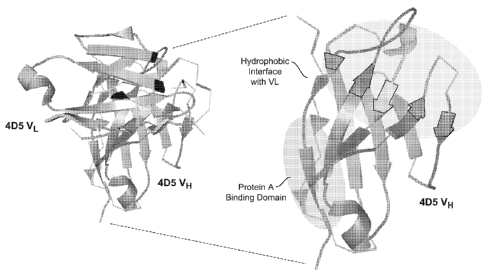
Figure 3 depicts the crystallographic structure of the wild-type VL and VH
domains
from the 4D5 monoclonal antibody (left image). The enlarged VH domain (right
image)
shows the different regions of the 4D5 VH domain that interact with Protein A
or VL.
12
CA 02651567 2008-11-05
WO 2007/134050 PCT/US2007/068469
Figures 4A and 4B show the wild-type 4D5 VH domain amino acid sequence and
each of the 25 unique amino acid sequences obtained from Library 1 selectants,
as described
in Example 1. Each of the Library 1 sequences was identical to the wild-type
sequence at all
positions not otherwise indicated. The boxed residues indicate groupings of
sequences based
on the residue at position 35 (glycine, alanine, or serine).
Figure 5 shows a bar graph of the purification yields for each of Library 1 VH
domain
selectants Libl_17, Libl_62, Libl_87, Libl_90, Libl_45, and Libl_66 in
comparison with
the wild-type 4D5 VH domain, as described in Example 1D(1).
Figures 6A-6D show traces from gel filtration/light scattering analyses of the
wild-
type 4D5 VH domain and each of Library 1 VH domain selectants Libl_17,
Lib1_62,
Lib187, Lib1_90, Lib1_45, and Lib1_66, as described in Example 1D(2).
Figure 7 shows melting curves over a 25-85 C range for the wild-type 4D5 VH
domain ("WT") and for each of Library 1 VH domain selectants Libl_17, Libl_62,
Lib1_87,
Lib190, Lib1_45, and Lib1_66, as described in Example 1D(3). The light line
indicates the
refolding transition, where the temperature was decreased from 85 C to 25 C.
The heavy
line depicts the unfolding transition, where the temperature was increased
from 25 C to 95
C. The reversibility of the phenomenon was assessed by placing the protein
sample at 85 C,
followed by cooling down the protein sample from 85 C to 25 C and then
heating it again to
95 C.
Figure 8 shows a graph depicting the results of the Protein A ELISA assay
described
in Example 1E.
Figures 9A-9D show the wild-type 4D5 VH domain amino acid sequence and each of
the 74 unique amino acid sequences obtained from Library 2 selectants, as
described in
Example 2. Each of the Library 2 sequences was identical to the wild-type
sequence at all
positions not otherwise indicated.
Figures 10A and lOB depict the results from experiments assessing the ability
of
Library 2 selectants to bind to Protein A, as described in Example 2. Figure
10A shows a bar
graph of the purification yields obtained using column chromatography with
Protein A-
conjugated resin for the wild-type 4D5 VH domain, Libl_62, and eleven Library
2 clones of
interest. Figure 10B shows the results of a Protein A ELISA for wild-type 4D5
VH domain,
Lib1_62, and eleven Library 2 clones of interest.
Figure 11 shows traces from gel filtration/light scattering analyses of the
wild-type
4D5 VH domain and the Lib2_3 VH domain, as described in Example 2.
13
CA 02651567 2008-11-05
WO 2007/134050 PCT/US2007/068469
Figure 12 shows melting curves over a 25-85 C range for the wild-type 4D5 VH
domain ("WT") and for the Lib2_3 VH domain, as described in Example 2. The
light line
indicates the refolding transition, where the temperature was decreased from
85 C to 25 C.
The heavy line depicts the unfolding transition, where the temperature was
increased from 25
C to 95 C. The reversibility of the phenomenon was assessed by placing the
protein sample
at 85 C, followed by cooling down the protein sample from 85 C to 25 C and
then heating
it again to 95 C.
Figure 13 shows two tables corresponding to the randomized residues from
Library 2
that were wild-type (V37, G44, W47, and Y91) or mutagenic (H35G, Q39R, L45E,
and
R50S) in the Lib2_3 VH domain. The tables list the number of times that a
particular one of
the twenty amino acids appeared in the sequences obtained from Libraries 3 and
4, as
described in Example 3. Light shading denotes that the amino acid was
prevalent at the
indicated position, while a darker shading denotes that the amino acid had a
low incidence at
the indicated position. "TH" indicates transformed Shannon entropy.
Figure 14 shows a bar graph depicting the wild-type/alanine ratio at each of
the VH
domain CDR-H3 positions alanine scanned in Library 5, as described in Example
5.
Figures 15A-C show traces from gel filtration/light scattering analyses of the
amber
Lib2_3 mutant and each of Lib2 3.4D5H3.G35S, Lib2 3.4D5H3.R39D,
Lib2 3.4D5H3.W47A, Lib2 3.4D5H3.W47E, Lib2 3.4D5H3.W47L, Lib2 3.4D5H3.W47T,
Lib2_3.4D5H3.W47V, and Lib2_3.4D5H3.W47E, as described in Example 4.
Figures 16A and 16B show melting curves over a 25-85 C range for WT 4D5, the
Lib2_3 amber mutant, Lib2 3.4D5H3.W47A, Lib2 3.4D5H3.W47E, Lib2 3.4D5H3.W47L,
Lib2 3.4D5H3.W47T, Lib2 3.4D5H3.W47V, Lib2 3.4D5H3.W47E, as described in
Example 4. The dotted line indicates the refolding transition, where the
temperature was
decreased from 85 C to 25 C. The solid line depicts the unfolding
transition, where the
temperature was increased from 25 C to 95 C. The reversibility of the
phenomenon was
assessed by placing the protein sample at 85 C, followed by cooling down the
protein sample
from 85 C to 25 C and then heating it again to 95 C.
Figures 17A-D show traces from gel filtration/light scattering analyses of
each of
Lib2 3.4D5H3.W47L/V37S, Lib2 3.4D5H3.W47L/V37T, Lib2 3.4D5H3.W47L/R39S,
Lib2 3.4D5H3.W47L/R39T, Lib2 3.4D5H3.W47L/R39K, Lib2 3.4D5H3.W47L/R39H,
Lib2_3.4D5H3.W47L/R39Q, and Lib2_3.4D5H3.W47L/R39D, Lib2_3.4D5H3.W47L/R39E
Lib2 3.4D5H3.W47L/E45S Lib2 3.4D5H3.W47L/E45T Lib2 3.4D5H3.W47L/E45H,
14
CA 02651567 2008-11-05
WO 2007/134050 PCT/US2007/068469
Lib2 3.4D5H3.W47L/W103S, Lib2 3.4D5H3.W47L/W103T, and
Lib2_3.4D5H3.W47L/W47L, as described in Example 4.
Figure 18 shows melting curves over a 25-85 C range for
Lib2_3.4D5H3.W47L/V37S, as described in Example 4. The dotted line indicates
the
refolding transition, where the temperature was decreased from 85 C to 25 C.
The solid line
depicts the unfolding transition, where the temperature was increased from 25
C to 95 C.
The reversibility of the phenomenon was assessed by placing the protein sample
at 85 C,
followed by cooling down the protein sample from 85 C to 25 C and then
heating it again to
95 C.
Figure 19 shows the results of a Protein A ELISA for wild-type 4D5 VH domain,
the
4D5 Fab, Libl_62, Libl_90, Lib2_3, Lib2_3 with a wild-type 4D5H3 domain, and
Lib2 3.4D5H3.T57E.
Figures 20A and 20B show crystal structures of various VH and VHH domains, as
described in Example 6. Figure 20A shows the structure of the Herceptin VH
domain (left
panel), as described in Cho et al. (Nature. (2003) Feb 13;421(6924):756-60),
and the structure
of VH-Bla. The VH-Bla structure has a resolution of 1.7A, R( ,Yst) of 16.4%,
R(fYee) of
20.4%, and a root mean square deviation (calculated with framework Calpha
atoms of the
1N8Z VH domain for molecular replacement) of 0.65 (based on 108/120
residues). Figure
20B shows detail views of the region surrounding residue 35 of the crystal
structures obtained
for a camelid anti-human chorionic gonadotropin VHH domain (Bond et al., J.
Mol. Biol.
332: 643-655 (2003)) (upper left panel), a HEL-binding VH domain (VH-He14)
(Jespers et
al., J. Mol. Biol. 337: 893-903 (2004)) (upper right panel), the Herceptin VH
domain (bottom
left panel) and VH-B 1a (bottom right panel).
Figure 21 shows traces from gel filtration/light scattering analyses of two
different
concentrations of VH domain B1a, as described in Example 7a.
Figures 22A and 22B show traces from gel filtration/light scattering analyses
of
different oligomeric states of B1a, as described in Example 7a.
Figure 23 shows the results from reducing and non-reducing SDS-polyacrylamide
gel
electrophoresis analyses of different oligomeric states of B 1a, as described
in Example 7a.
Figures 24A-B show a table providing protein yield, extinction coefficient,
molecular
weight, peak area, retention time, melting temperature and refolding
percentage data for
many VH domains described herein (see, e.g., Example 7B and Example 8).
CA 02651567 2008-11-05
WO 2007/134050 PCT/US2007/068469
Figures 25A-25F show traces from gel filtration/light scattering analyses of
mutant
Bla VH domains, as described in Example 7b.
Figures 26A-26H shows graphs of the percentage of folding observed upon
increase
(solid line) and decrease (broken line) of temperature for certain VH domains
described
herein, as described in Example 7b.
Figures 27A-27D show melting curves over a 25-85 C range for the Bla VH
domain
and several Bla mutant VH domains, as described in Example 7b. The dotted line
indicates
the refolding transition, where the temperature was decreased from 85 C to 25
C. The solid
line depicts the unfolding transition, where the temperature was increased
from 25 C to 95
C. The reversibility of the phenomenon was assessed by placing the protein
sample at 85 C,
followed by cooling down the protein sample from 85 C to 25 C and then
heating it again to
95 C.
Figures 28A-28C show traces from gel filtration/light scattering analyses of
mutant
VH domains, as described in Example 8.
Figures 29A-29C show graphs of the percentage of folding observed upon
increase
(top, solid line) and decrease (bottom, broken line) of temperature for
certain VH domains
described herein, as described in Example 8.
Figures 30A-30C show show melting curves over a 25-85 C range for certain Bla
mutant VH domains, as described in Example 8. The dotted line indicates the
refolding
transition, where the temperature was decreased from 85 C to 25 C. The solid
line depicts
the unfolding transition, where the temperature was increased from 25 C to 95
C. The
reversibility of the phenomenon was assessed by placing the protein sample at
85 C,
followed by cooling down the protein sample from 85 C to 25 C and then
heating it again to
95 C.
DISCLOSURE OF THE INVENTION
A. Definitions
The term "affinity purification" means the purification of a molecule based on
a
specific attraction or binding of the molecule to a chemical or binding
partner to form a
combination or complex which allows the molecule to be separated from
impurities while
remaining bound or attracted to the partner moiety.
The term "antibody" is used in the broadest sense and specifically covers
single
monoclonal antibodies (including agonist and antagonist antibodies), antibody
compositions
16
CA 02651567 2008-11-05
WO 2007/134050 PCT/US2007/068469
with polyepitopic specificity, affinity matured antibodies, humanized
antibodies, chimeric
antibodies, single chain antigen binding molecules such as monobodies, as well
as antigen
binding fragments or polypeptides (e.g., Fab, F(ab')2, scFv and Fv), so long
as they exhibit
the desired biological activity.
As used herein, "antibody variable domain" refers to the portions of the light
and
heavy chains of antibody molecules that include amino acid sequences of
Complementary
Determining Regions (CDRs; ie., CDR1, CDR2, and CDR3), and Framework Regions
(FRs;
i.e. FR1, FR2, FR3, and FR4). A FR includes those amino acid positions in an
antibody
variable domain other than CDR positions as defined herein. VH refers to the
variable
domain of the heavy chain of an antibody. VL refers to the variable domain of
the light chain
of an antibody. VHH refers to the heavy chain variable domain of a monobody.
According to
the methods used in this invention, the amino acid positions assigned to CDRs
and FRs are
defined according to Kabat (Sequences of Proteins of Immunological Interest
(National
Institutes of Health, Bethesda, Md., 1987 and 1991)). Amino acid numbering of
antibodies
or antigen binding fragment thereof is also according to that of Kabat et al.
cited supra.
As used herein "CDR" refers to a contiguous sequence of amino acids that form
a
loop in an antigen binding pocket or groove. The amino acid sequences included
in a CDR
loop are selected based on structure or amino acid sequence. In an embodiment,
the loop
amino acids of a CDR are determined by inspection of the three-dimensional
structure of an
antibody, antibody heavy chain, or antibody light chain. The three-dimensional
structure
may be analyzed for solvent accessible amino acid positions as such positions
are likely to
form a loop in an antibody variable domain. The three dimensional structure of
the antibody
variable domain may be derived from a crystal structure or protein modeling.
In another
embodiment, the loop boundaries of the CDR are determined according to Chothia
(Chothia
and Lesk, 1987, J. Mol. Biol., 196:901-917). One to three amino acid residues
may
optionally be added to the C-terminal and N-terminal ends of the Chothia CDRs.
In some
embodiments, the amino acid positions of CDR1 comprise, consist essentially of
or consist of
amino acid positions 24 to 34, the amino acid positions of CDR2 comprise,
consist essentially
of or consist of amino acid positions 51 to 56 and the CDR3 positions
comprise, consist
essentially of or consist of amino acid positions 96 to 101 of an antibody
heavy chain variable
domain.
"Antibody fragments" comprise only a portion of an intact antibody, generally
including an antigen binding site of the intact antibody and thus retaining
the ability to bind
17
CA 02651567 2008-11-05
WO 2007/134050 PCT/US2007/068469
antigen. Nonlimiting examples of antibody fragments encompassed by the present
definition
include: (i) the Fab fragment, having VL, CL, VH and CH1 domains having one
interchain
disulfide bond between the heavy and light chain; (ii) the Fab' fragment,
which is a Fab
fragment having one or more cysteine residues at the C-terminus of the CH1
domain; (iii) the
Fd fragment having VH and CH1 domains; (iv) the Fd' fragment having VH and CH1
domains and one or more cysteine residues at the C-terminus of the CH1 domain;
(v) the Fv
fragment having the VL and VH domains of a single arm of an antibody; (vi) the
dAb
fragment which consists of a VH domain; (vii) hingeless antibodies including
at least VL,
VH, CL, CH1 domains and lacking hinge region; (viii) F(ab')2 fragments, a
bivalent fragment
including two Fab' fragments linked by a disulfide bridge at the hinge region;
(ix) single
chain antibody molecules (e.g. single chain Fv; scFv); (x) "diabodies" with
two antigen
binding sites, comprising a heavy chain variable domain (VH) connected to a
light chain
variable domain (VL) in the same polypeptide chain; (xi) single arm antigen
binding
molecules comprising a light chain, a heavy chain and a N- terminally
truncated heavy chain
constant region sufficient to form a Fc region capable of increasing the half
life of the single
arm antigen binding domain; and (xii) "linear antibodies" comprising a pair of
tandem Fd
segments (VH-CH I -VH-CH 1) which, together with complementary light chain
polypeptides,
form a pair of antigen binding regions.
The term "monobody" as used herein, refers to an antigen binding molecule with
at
least one heavy chain variable domain and no light chain variable domain. A
monobody can
bind to an antigen in the absence of light chains and typically has three CDR
regions
designated CDRH1, CDRH2 and CDRH3. A heavy chain IgG monobody has two heavy
chain antigen binding molecules connected by a disulfide bond. The heavy chain
variable
domain comprises one or more CDR regions, e.g., a CDRH3 region.
A "Vh" or "VH" or "VH domain" refers to a variable domain of an antibody heavy
chain. A "VL" or "VL" or "VL domain" refers to a variable domain of an
antibody light
chain. A "VHH" or a "VhH" refers to a variable domain of a heavy chain
antibody that
occurs in the form of a monobody. A "camelid monobody" or "camelid VHH" refers
to a
monobody or antigen binding portion thereof obtained from a source animal of
the camelid
family, including animals having feet with two toes and leathery soles.
Animals in the
camelid family include, but are not limited to, camels, llamas, and alpacas.
The term "monoclonal antibody" as used herein refers to an antibody obtained
from a
population of substantially homogeneous antibodies, i.e., the individual
antibodies
18
CA 02651567 2008-11-05
WO 2007/134050 PCT/US2007/068469
comprising the population are essentially identical except for variants that
may arise during
production of the antibody.
The monoclonal antibodies herein specifically include "chimeric" antibodies in
which
a portion of the heavy and/or light chain is identical with or homologous to
corresponding
sequences in antibodies derived from a particular species or belonging to a
particular
antibody class or subclass, while the remainder of the chain(s) is identical
with or
homologous to corresponding sequences in antibodies derived from another
species or
belonging to another antibody class or subclass, as well as fragments of such
antibodies, so
long as they exhibit the desired biological activity (U.S. Patent No.
4,816,567; and Morrison
et al., Proc. Natl. Acad. Sci. USA 81:6851-6855 (1984)).
"Humanized" forms of non-human (e.g., murine) antibodies are chimeric
antibodies
that contain minimal sequence derived from non-human immunoglobulin. For the
most part,
humanized antibodies are human immunoglobulins (recipient antibody) in which
residues
from a hypervariable region (HVR) of the recipient are replaced by residues
from a
hypervariable region (HVR) of a non-human species (donor antibody) such as
mouse, rat,
rabbit or nonhuman primate having the desired specificity, affinity, and
capacity. In some
instances, framework region (FR) residues of the human immunoglobulin are
replaced by
corresponding non-human residues to improve antigen binding affinity.
Furthermore,
humanized antibodies may comprise residues that are not found in the recipient
antibody or
the donor antibody. These modifications may be made to improve antibody
affinity or
functional activity. In general, the humanized antibody will comprise
substantially all of at
least one, and typically two, variable domains, in which all or substantially
all of the
hypervariable regions correspond to those of a non-human immunoglobulin and
all or
substantially all of the FRs are those of a human immunoglobulin sequence.
Humanized
antibodies can also be produced as antigen binding fragments as described
herein. The
humanized antibody optionally will also comprise at least a portion of an
immunoglobulin
constant region (Fc), typically that of or derived from a human
immunoglobulin. For further
details, see Jones et al., Nature 321:522-525 (1986); Riechmann et al., Nature
332:323-329
(1988); and Presta, Curr. Op. Struct. Biol. 2:593-596 (1992). See also the
following review
articles and references cited therein: Vaswani and Hamilton, Ann. Allergy,
Asthma &
Immunol. 1:105-115 (1998); Harris, Biochem. Soc. Transactions 23:1035-1038
(1995); Hurle
and Gross, Curr. Op. Biotech 5:428-433 (1994).
19
CA 02651567 2008-11-05
WO 2007/134050 PCT/US2007/068469
A "human antibody" is one which possesses an amino acid sequence which
corresponds to that of an antibody produced by a human and/or has been made
using any of
the techniques for making human antibodies as disclosed herein. This
definition of a human
antibody specifically excludes a humanized antibody comprising non-human
antigen binding
residues.
As used herein, "highly diverse position" refers to a position of an amino
acid located
in the variable regions of an antibody light or heavy chain that has a number
of different
amino acid represented at the position when the amino acid sequences of known
and/or
naturally occurring antibodies or antigen binding fragment or polypeptides are
compared.
The highly diverse positions are typically found in the CDR regions. In one
aspect, the
ability to determine highly diverse positions in known and/or naturally
occurring antibodies is
facilitated by the data provided by Kabat, Sequences of Proteins of
Immunological Interest
(National Institutes of Health, Bethesda, MD, 1987 and 1991). An Internet-
based database
located at http://www.bioinf.org.uk/abs/simkab.html provides an extensive
collection and
alignment of human light and heavy chain sequences and facilitates
determination of highly
diverse positions in these sequences. According to the invention, an amino
acid position is
highly diverse if it has preferably from about 2 to about 11, preferably from
about 4 to about
9, and preferably from about 5 to about 7 different possible amino acid
residue variations at
that position. In some embodiments, an amino acid position is highly diverse
if it has
preferably at least about 2, preferably at least about 4, preferably at least
about 6, and
preferably at least about 8 different possible amino acid residue variations
at that position.
As used herein, "library" refers to a plurality of antibody, antibody fragment
sequences, or antibody variable domains (for example, polypeptides of the
invention), or the
nucleic acids that encode these sequences, the sequences being different in
the combination
of variant amino acids that are introduced into these sequences according to
the methods of
the invention.
A "scaffold", as used herein, refers to a polypeptide or portion thereof that
maintains a
stable structure or structural element when a heterologous polypeptide is
inserted into the
polypeptide. The scaffold provides for maintenance of a structural and/or
functional feature
of the polypeptide after the heterologous polypeptide has been inserted. In
one embodiment,
a scaffold comprises one or more FR regions of an antibody variable domain,
and maintains a
stable structure when a heterologous CDR is inserted into the scaffold.
CA 02651567 2008-11-05
WO 2007/134050 PCT/US2007/068469
A "source antibody", as used herein, refers to an antibody or antigen binding
polypeptide whose antigen binding determinant sequence serves as the template
sequence
upon which diversification according to the criteria described herein is
performed. A source
antibody variable domain can include an antibody, antibody variable domain,
antigen binding
fragment or polypeptide thereof, a monobody, VHH, a monobody or antibody
variable
domain obtained from a naive or synthetic library, camelid antibodies,
naturally occurring
antibody or monobody, synthetic antibody, or recombinant antibody, humanized
antibody or
monobody, germline derived antibody or monobody, chimeric antibody or
monobody, and
affinity matured antibody or monobody. In one embodiment, the polypeptide is
an antibody
variable domain that is a member of the Vh3 subgroup.
As used herein, "solvent accessible position" refers to a position of an amino
acid
residue in the variable region of a heavy and/or light chain of a source
antibody or antigen
binding polypeptide that is determined, based on structure, ensemble of
structures and/or
modeled structure of the antibody or antigen binding polypeptide, as
potentially available for
solvent access and/or contact with a molecule, such as an antibody-specific
antigen. These
positions are typically found in the CDRs, but can also be found in FR and on
the exterior
surface of the protein. The solvent accessible positions of an antibody or
antigen binding
polypeptide, as defined herein, can be determined using any of a number of
algorithms
known in the art. In certain embodiments, solvent accessible positions are
determined using
coordinates from a 3-dimensional model of an antibody or antigen binding
polypeptide, e.g.,
using a computer program such as the Insightll program (Accelrys, San Diego,
CA). Solvent
accessible positions can also be determined using algorithms known in the art
(e.g., Lee and
Richards, J. Mol. Biol. 55, 379 (1971) and Connolly, J. Appl. Cryst. 16, 548
(1983)).
Determination of solvent accessible positions can be performed using software
suitable for
protein modeling and 3-dimensional structural information obtained from an
antibody.
Software that can be utilized for these purposes includes SYBYL Biopolymer
Module
software (Tripos Associates). Generally, where an algorithm (program) requires
a user input
size parameter, the "size" of a probe which is used in the calculation is set
at about 1.4
Angstrom or smaller in radius. In addition, determination of solvent
accessible regions and
area methods using software for personal computers has been described by
Pacios ((1994)
"ARVOMOL/CONTOUR: molecular surface areas and volumes on Personal Computers."
Comput. Chem. 18(4): 377-386; and (1995). "Variations of Surface Areas and
Volumes in
Distinct Molecular Surfaces of Biomolecules." J. Mol. Model. 1: 46-53.)
21
CA 02651567 2008-11-05
WO 2007/134050 PCT/US2007/068469
The phrase "structural amino acid position" as used herein refers to an amino
acid of a
polypeptide that contributes to the stability of the structure of the
polypeptide such that the
polypeptide retains at least one biological function such as specifically
binding to a molecule
e.g., an antigen or a target molecule. Structural amino acid positions are
identified as amino
acid positions less tolerant to amino acid substitutions without affecting the
structural
stability of the polypeptide. Amino acid positions less tolerant to amino acid
substitutions can
be identified using a method such as alanine scanning mutagenesis or shotgun
scanning as
described in WO 01/44463 and analyzing the effect of loss of the wild type
amino acid on
structural stability.
The term "stability" as used herein refers to the ability of a molecule to
maintain a
folded state under physiological conditions such that it retains at least one
of its normal
functional activities, for example, binding to an antigen or to a molecule
like Protein A. The
stability of the molecule can be determined using standard methods. For
example, the
stability of a molecule can be determined by measuring the thermal melt
("T,Y,") temperature.
The T. is the temperature in degrees Celsius at which 1/2 of the molecules
become unfolded.
Typically, the higher the T,Y,, the more stable the molecule.
The phrase "randomly generated population" as used herein refers to a
population of
polypeptides wherein one or more amino acid positions in a domain has a
variant amino acid
encoded by a random codon set which allows for substitution of a1120 naturally
occurring
amino acids at that position. For example, in one embodiment, a randomly
generated
population of polypeptides having randomized VH or portions thereof includes a
variant
amino acid at each position in the VH that is encoded by a random codon set. A
random
codon set includes but is not limited to codon sets designated NNS and NNK.
"Cell", "cell
line", and "cell culture" are used interchangeably herein and such
designations include all
progeny of a cell or cell line. Thus, for example, terms like "transformants"
and
"transformed cells" include the primary subject cell and cultures derived
therefrom without
regard for the number of transfers. It is also understood that all progeny may
not be precisely
identical in DNA content, due to deliberate or inadvertent mutations. Mutant
progeny that
have the same function or biological activity as screened for in the
originally transformed cell
are included. Where distinct designations are intended, it will be clear from
the context.
"Control sequences" when referring to expression means DNA sequences necessary
for the expression of an operably linked coding sequence in a particular host
organism. The
control sequences that are suitable for prokaryotes, for example, include a
promoter,
22
CA 02651567 2008-11-05
WO 2007/134050 PCT/US2007/068469
optionally an operator sequence, a ribosome binding site, and possibly, other
as yet poorly
understood sequences. Eukaryotic cells are known to utilize promoters,
polyadenylation
signals, and enhancers.
The term "coat protein" means a protein, at least a portion of which is
present on the
surface of the virus particle. From a functional perspective, a coat protein
is any protein,
which associates with a virus particle during the viral assembly process in a
host cell, and
remains associated with the assembled virus until it infects another host
cell. The coat
protein may be the major coat protein or may be a minor coat protein. A
"major" coat protein
is generally a coat protein which is present in the viral coat at preferably
at least about 5,
more preferably at least about 7, even more preferably at least about 10
copies of the protein
or more. A maj or coat protein may be present in tens, hundreds or even
thousands of copies
per virion. An example of a major coat protein is the p8 protein of
filamentous phage.
As used herein, "codon set" refers to a set of different nucleotide triplet
sequences
used to encode desired variant amino acids. A set of oligonucleotides can be
synthesized, for
example, by solid phase synthesis, containing sequences that represent all
possible
combinations of nucleotide triplets provided by the codon set and that will
encode the desired
group of amino acids. A standard form of codon designation is that of the IUB
code, which is
known in the art and described herein. A "non-random codon set", as used
herein, thus refers
to a codon set that encodes select amino acids that fulfill partially,
preferably completely, the
criteria for amino acid selection as described herein. Synthesis of
oligonucleotides with
selected nucleotide "degeneracy" at certain positions is well known in that
art, for example
the TRIM approach (Knappek et al.; J. Mol. Biol. (1999), 296:57-86); Garrard &
Henner,
Gene (1993), 128:103). Such sets of nucleotides having certain codon sets can
be
synthesized using commercial nucleic acid synthesizers (available from, for
example,
Applied Biosystems, Foster City, CA), or can be obtained commercially (for
example, from
Life Technologies, Rockville, MD). Therefore, a set of oligonucleotides
synthesized having
a particular codon set will typically include a plurality of oligonucleotides
with different
sequences, the differences established by the codon set within the overall
sequence.
Oligonucleotides, as used according to the invention, have sequences that
allow for
hybridization to a variable domain nucleic acid template and also can, but
does not
necessarily, include restriction enzyme sites useful for, for example, cloning
purposes.
A "fusion protein" and a "fusion polypeptide" refer to a polypeptide having
two
portions covalently linked together, where each of the portions is a
polypeptide having a
23
CA 02651567 2008-11-05
WO 2007/134050 PCT/US2007/068469
different property. The property may be a biological property, such as
activity in vitro or in
vivo. The property may also be a simple chemical or physical property, such as
binding to a
target molecule, catalysis of a reaction, etc. The two portions may be linked
directly by a
single peptide bond or through a peptide linker containing one or more amino
acid residues.
Generally, the two portions and the linker will be in reading frame with each
other.
"Heterologous DNA" is any DNA that is introduced into a host cell. The DNA may
be derived from a variety of sources including genomic DNA, cDNA, synthetic
DNA and
fusions or combinations of these. The DNA may include DNA from the same cell
or cell
type as the host or recipient cell or DNA from a different cell type, for
example, from a
mammal or plant. The DNA may, optionally, include marker or selection genes,
for example,
antibiotic resistance genes, temperature resistance genes, etc.
"Ligation" is the process of forming phosphodiester bonds between two nucleic
acid
fragments. For ligation of the two fragments, the ends of the fragments must
be compatible
with each other. In some cases, the ends will be directly compatible after
endonuclease
digestion. However, it may be necessary first to convert the staggered ends
commonly
produced after endonuclease digestion to blunt ends to make them compatible
for ligation.
For blunting the ends, the DNA is treated in a suitable buffer for at least 15
minutes at 15 C
with about 10 units of the Klenow fragment of DNA polymerase I or T4 DNA
polymerase in
the presence of the four deoxyribonucleotide triphosphates. The DNA is then
purified by
phenol-chloroform extraction and ethanol precipitation or by silica
purification. The DNA
fragments that are to be ligated together are put in solution in about
equimolar amounts. The
solution will also contain ATP, ligase buffer, and a ligase such as T4 DNA
ligase at about 10
units per 0.5 g of DNA. If the DNA is to be ligated into a vector, the vector
is first
linearized by digestion with the appropriate restriction endonuclease(s). The
linearized
fragment is then treated with bacterial alkaline phosphatase or calf
intestinal phosphatase to
prevent self-ligation during the ligation step.
A "mutation" is a deletion, insertion, or substitution of a nucleotide(s)
relative to a
reference nucleotide sequence, such as a wild type sequence.
As used herein, "natural" or "naturally occurring" polypeptides or
polynucleotides
refers to a polypeptide or a polynucleotide having a sequence of a polypeptide
or a
polynucleotide identified from a nonsynthetic source. For example, when the
polypeptide is
an antibody or antibody fragment, the nonsynthetic source can be a
differentiated antigen-
specific B cell obtained ex vivo, or its corresponding hybridoma cell line, or
from the serum
24
CA 02651567 2008-11-05
WO 2007/134050 PCT/US2007/068469
of an animal. Such antibodies can include antibodies generated in any type of
immune
response, either natural or otherwise induced. Natural antibodies include the
amino acid
sequences, and the nucleotide sequences that constitute or encode these
antibodies, for
example, as identified in the Kabat database. As used herein, natural
antibodies are different
than "synthetic antibodies", synthetic antibodies referring to antibody
sequences that have
been changed, for example, by the replacement, deletion, or addition, of an
amino acid, or
more than one amino acid, at a certain position with a different amino acid,
the different
amino acid providing an antibody sequence different from the source antibody
sequence.
"Operably linked" when referring to nucleic acids means that the nucleic acids
are
placed in a functional relationship with another nucleic acid sequence. For
example, DNA
for a presequence or secretory leader is operably linked to DNA for a
polypeptide if it is
expressed as a preprotein that participates in the secretion of the
polypeptide; a promoter or
enhancer is operably linked to a coding sequence if it affects the
transcription of the
sequence; or a ribosome binding site is operably linked to a coding sequence
if it is
positioned so as to facilitate translation. Generally, "operably linked" means
that the DNA
sequences being linked are contiguous and, in the case of a secretory leader,
contingent and
in reading frame. However, enhancers do not have to be contiguous. Linking is
accomplished by ligation at convenient restriction sites. If such sites do not
exist, the
synthetic oligonucleotide adapters or linkers are used in accord with
conventional practice.
"Phage display" is a technique by which variant polypeptides are displayed as
fusion
proteins to at least a portion of a coat protein on the surface of phage,
e.g., filamentous phage,
particles. A utility of phage display lies in the fact that large libraries of
randomized protein
variants can be rapidly and efficiently sorted for those sequences that bind
to a target
molecule with high affinity. Display of peptide and protein libraries on phage
has been used
for screening millions of polypeptides for ones with specific binding
properties. Polyvalent
phage display methods have been used for displaying small random peptides and
small
proteins through fusions to either gene III or gene VIII of filamentous phage.
Wells and
Lowman, Curr. Opin. Struct. Biol., 3:355-362 (1992), and references cited
therein. In
monovalent phage display, a protein or peptide library is fused to a gene III
or a portion
thereof, and expressed at low levels in the presence of wild type gene III
protein so that phage
particles display one copy or none of the fusion proteins. Avidity effects are
reduced relative
to polyvalent phage so that sorting is on the basis of intrinsic ligand
affinity, and phagemid
CA 02651567 2008-11-05
WO 2007/134050 PCT/US2007/068469
vectors are used, which simplify DNA manipulations. Lowman and Wells, Methods:
A
companion to Methods in Enzymology, 3:205-0216 (1991).
A "phagemid" is a plasmid vector having a bacterial origin of replication,
e.g.,
Co1E1, and a copy of an intergenic region of a bacteriophage. The phagemid may
be used on
any known bacteriophage, including filamentous bacteriophage and lambdoid
bacteriophage.
The plasmid will also generally contain a selectable marker for antibiotic
resistance.
Segments of DNA cloned into these vectors can be propagated as plasmids. When
cells
harboring these vectors are provided with all genes necessary for the
production of phage
particles, the mode of replication of the plasmid changes to rolling circle
replication to
generate copies of one strand of the plasmid DNA and package phage particles.
The
phagemid may form infectious or non-infectious phage particles. This term
includes
phagemids, which contain a phage coat protein gene or fragment thereof linked
to a
heterologous polypeptide gene as a gene fusion such that the heterologous
polypeptide is
displayed on the surface of the phage particle.
The term "phage vector" means a double stranded replicative form of a
bacteriophage
containing a heterologous gene and capable of replication. The phage vector
has a phage
origin of replication allowing phage replication and phage particle formation.
The phage can
be a filamentous bacteriophage, such as an M13, fl, fd, Pf3 phage or a
derivative thereof, or a
lambdoid phage, such as lambda, 21, phi80, phi8l, 82, 424, 434, etc., or a
derivative thereof.
"Oligonucleotides" are short-length, single- or double-stranded
polydeoxynucleotides
that are prepared by known methods such as chemical synthesis (e.g. phosphotri
ester,
phosphite, or phosphoramidite chemistry, using solid-phase techniques such as
described in
EP 266,032 published 4 May 1988, or via deoxynucloside H-phosphonate
intermediates as
described by Froeshler et al., Nucl. Acids, Res., 14:5399-5407 (1986)).
Further methods
include the polymerase chain reaction defined below and other autoprimer
methods and
oligonucleotide syntheses on solid supports. All of these methods are
described in Engels et
al., Agnew. Chem. Int. Ed. Engl., 28:716-734 (1989). These methods are used if
the entire
nucleic acid sequence of the gene is known, or the sequence of the nucleic
acid
complementary to the coding strand is available. Alternatively, if the target
amino acid
sequence is known, one may infer potential nucleic acid sequences using known
and
preferred coding residues for each amino acid residue. The oligonucleotides
can be purified
on polyacrylamide gels or molecular sizing columns or by precipitation.
26
CA 02651567 2008-11-05
WO 2007/134050 PCT/US2007/068469
DNA is "purified" when the DNA is separated from non-nucleic acid impurities.
The
impurities may be polar, non-polar, ionic, etc.
A "transcription regulatory element" will contain one or more of the following
components: an enhancer element, a promoter, an operator sequence, a repressor
gene, and a
transcription termination sequence. These components are well known in the
art, e.g., U.S.
Patent No. 5,667,780.
A"transformant" is a cell that has taken up and maintained DNA as evidenced by
the
expression of a phenotype associated with the DNA (e.g., antibiotic resistance
conferred by a
protein encoded by the DNA).
"Transformation" means a process whereby a cell takes up DNA and becomes a
"transformant". The DNA uptake may be permanent or transient.
A "variant" or "mutant" of a starting or reference polypeptide (for e.g., a
source
antibody or its variable domain(s)), such as a fusion protein (polypeptide) or
a heterologous
polypeptide (heterologous to a phage), is a polypeptide that 1) has an amino
acid sequence
different from that of the starting or reference polypeptide and 2) was
derived from the
starting or reference polypeptide through either natural or artificial
(manmade) mutagenesis.
Such variants include, for example, deletions from, and/or insertions into
and/or substitutions
of, residues within the amino acid sequence of the polypeptide of interest.
For example, a
fusion polypeptide of the invention generated using an oligonucleotide
comprising a
nonrandom codon set that encodes a sequence with a variant amino acid (with
respect to the
amino acid found at the corresponding position in a source antibody/antigen
binding fragment
or polypeptide) would be a variant polypeptide with respect to a source
antibody or antigen
binding fragment or polypeptide. Thus, a variant VH refers to a VH comprising
a variant
sequence with respect to a starting or reference polypeptide sequence (such as
that of a source
antibody or antigen binding fragment or polypeptide). A variant amino acid, in
this context,
refers to an amino acid different from the amino acid at the corresponding
position in a
starting or reference polypeptide sequence (such as that of a source antibody
or antigen
binding fragment or polypeptide). Any combination of deletion, insertion, and
substitution
may be made to arrive at the final variant or mutant construct, provided that
the final
construct possesses the desired functional characteristics. The amino acid
changes also may
alter post-translational processes of the polypeptide, such as changing the
number or position
of glycosylation sites. Methods for generating amino acid sequence variants of
polypeptides
are described in U.S. Patent No. 5,534,615, expressly incorporated herein by
reference.
27
CA 02651567 2008-11-05
WO 2007/134050 PCT/US2007/068469
A "wild type" or "reference" sequence or the sequence of a "wild type" or
"reference"
protein/polypeptide, such as a coat protein, a CDR, or a variable domain of a
source antibody,
is the reference sequence from which variant polypeptides are derived through
the
introduction of mutations. In general, the "wild type" sequence for a given
protein is the
sequence that is most common in nature. Similarly, a "wild type" gene sequence
is the
sequence for that gene which is most commonly found in nature. Mutations may
be
introduced into a "wild type" gene (and thus the protein it encodes) either
through natural
processes or through man induced means. The products of such processes are
"variant" or
"mutant" forms of the original "wild type" protein or gene.
As used herein "Vh3" refers to a subgroup of antibody variable domains. The
sequences of known antibody variable domains have been analyzed for sequence
identity and
divided into groups. Antibody heavy chain variable domains in subgroup III are
known to
have a Protein A binding site.
A "plurality" or "population" of a substance, such as a polypeptide or
polynucleotide
of the invention, as used herein, generally refers to a collection of two or
more types or kinds
of the substance. There are two or more types or kinds of a substance if two
or more of the
substances differ from each other with respect to a particular characteristic,
such as the
variant amino acid found at a particular amino acid position. In a nonlimiting
example, there
is a plurality or population of polynucleotides of the invention if there are
two or more
polynucleotides of the invention that are substantially the same, preferably
identical, in
sequence except for one or more variant amino acids at particular CDR amino
acid positions.
B. Modes of the Invention
A diverse library of isolated antibody variable domains is useful to identify
novel
antigen binding molecules having high affinity. Generating a library with
antibody variable
domains that are not only highly diverse, but are also structurally stable
permits the isolation
of high affinity binding antibody variable domains from the library that can
more readily be
produced in cell culture on a large scale. The present invention is based on
the showing that
the folding stability of an isolated heavy chain antibody variable domain can
be enhanced by
enhancing the hydrophilicity of those portions of the heavy chain antibody
variable domain
that typically interact with the light chain antibody variable domain when in
the context of an
intact antibody. In one aspect, VH residues that typically interact with the
VL domain
include amino acid positions 37, 39, 44, 45, 47, 91, and 103. In certain
embodiments, one or
28
CA 02651567 2008-11-05
WO 2007/134050 PCT/US2007/068469
more of the VH residues that typically interact with the VL domain are
increased in
hydrophilicity while one or more other such residues are maintained or
decreased in
hydrophilicity. It will be understood that by increasing the hydrophobicity of
one or more
residues that typically interact with the VL domain, the hydrophilicity of one
or more other
such residues, or the overall hydrophilicity of the portion of the VH domain
that interacts
with a VL domain may be increased. In certain embodiments, such modifications
improve
stability of the overall isolated heavy chain antibody variable domain while
still permitting
full and unbiased diversification at one or more of the three heavy chain
complementarity
determining regions.
It will be appreciated by one of ordinary skill in the art that yield,
aggregation
tendency, and thermal stability, while indicators of the overall folding
stability of the protein,
may be separately useful. Thus, as a nonlimiting example, a mutant VH domain
with
improved yield and thermal stability but also increased aggregation tendency
relative to a
wild-type VH domain may still be useful for applications in which increased
aggregation is
not problematic. Similarly, in another nonlimiting example, a mutant VH domain
with
dereased yield but decreased aggregation tendency and increased thermal
stability relative to
a wild-type VH domain may still be useful for applications in which large
quantities of
protein are not required, or where it is feasible to perform multiple rounds
of protein
isolation.
In one embodiment, modifications of the amino acid at position 37 of the
isolated VH
domain are provided. In one aspect, the amino acid at position 37 is a
hydrophobic amino
acid. In one such aspect, the amino acid at position 37 is selected from
tryptophan,
phenylalanine, and tyrosine. In another embodiment, modifications of the amino
acid at
position 39 of the isolated VH domain is provided. In one aspect, the amino
acid at position
39 is a hydrophilic amino acid. In one aspect, the amino acid at position 39
is selected from
arginine and aspartic acid. In another embodiment, modifications of the amino
acid at
position 45 of the isolated VH domain are provided. In one aspect, the amino
acid at position
45 is a hydrophobic amino acid. In one such aspect, the amino acid at position
45 is selected
from tryptophan, phenylalanine, and tyrosine. In another aspect, the amino
acid at position
45 is a hydrophilic amino acid. In one such aspect, the amino acid at position
45 is glutamic
acid. In another embodiment, modifications of the amino acid at position 47 of
the isolated
VH domain are provided. In one aspect, the amino acid at position 47 is
selected from
alanine, glutamic acid, leucine, threonine, and valine. In another embodiment,
an isolated
29
CA 02651567 2008-11-05
WO 2007/134050 PCT/US2007/068469
VH domain comprises two or more modifications at amino acid positions 37, 39,
44, 45, 47,
91, and/or 103. In another embodiment, an isolated VH domain comprises three
or more
modifications at amino acid positions 37, 47, 50, and/or 103. In another
embodiment, an
isolated VH domain comprises four or more modifications at amino acid
positions 37, 47, 50,
and 103. In another embodiment, the above mutations are made in the context of
SEQ ID
NO: 1. In another embodiment, the above mutations are made in the context of
SEQ ID NO:
2.
The invention also provides further modifications that may be made within the
framework regions of the isolated heavy chain variable domain to further
increase the folding
stability of the polypeptide. It was known that the stability of an isolated
heavy chain
antibody variable domain was enhanced when the histidine at amino acid
position 35 was
modified to glycine (Jespers et al., J. Mol. Biol. (2004) 337: 893-903).
Applicants herein also
identify other structural modifications that improve isolated heavy chain
antibody binding
domain stability.
In one aspect, modifications of the histidine at amino acid position 35 of the
isolated
VH domain to an amino acid other than glycine are provided. In one such
aspect, the
histidine at amino acid position 35 is modified to a serine. In another such
aspect, the
histidine at amino acid position 35 is modified to an alanine. In another such
aspect, the
histidine at amino acid position 35 is modified to an aspartic acid. In
another aspect, the
histidine at amino acid position 35 is modified to glycine, and one or more
additional
mutations are made in VH such that the isolated VH domain has increased
folding stability
relative to a VH domain with a single mutation comprising H35G.
In another aspect, modifications of the amino acid at position 50 of the
isolated VH
domain are provided. In one such aspect, the amino acid at position 50 is
modified to a
hydrophilic amino acid. In another such aspect, the amino acid at position 50
is modified to a
serine. In another such aspect, the amino acid at position 50 is modified to a
glycine. In
another such aspect, the amino acid at position 50 is modified to an arginine.
In another
embodiment, an isolated VH domain comprises modifications at both amino acid
positions 35
and 50.
In another embodiment, an isolated VH domain comprises two or more
modifications
at amino acid positions 35, 37, 39, 44, 45, 47, 50, 91, and/or 103. In one
example, the
invention provides a novel combination of modifications at amino acid
positions 35 and 47 of
an isolated VH domain. In one aspect, the amino acid at position 35 is serine,
and the amino
CA 02651567 2008-11-05
WO 2007/134050 PCT/US2007/068469
acid at position 47 is selected from phenylalanine and glutamic acid. In
another aspect, the
amino acid at position 35 is glycine and the amino acid at position 47 is
methionine. In
another aspect, the amino acid at position 35 is alanine and the amino acid at
position 47 is
selected from tryptophan and methionine.
In another embodiment, an isolated VH domain comprises three or more
modifications at amino acid positions 37, 47, 50, and 103. In another
embodiment, an
isolated VH domain comprises
The polypeptides of the invention find uses in research and medicine. The
polypeptides described herein are isolated VH domains with enhanced folding
stability
relative to wild-type VH domains, which can be specific for one or more target
antigens.
Such VH domains can be used, for example, as diagnostic reagents for the
presence of the
one or more target antigens. It may be preferred to use the VH domains of the
invention over
a wild-type VH domain specific for the one or more target antigens because the
increased
folding stability of the VH domains of the invention may permit them to retain
activity for
longer periods of time and under harsher conditions than a wild-type VH domain
might,
thereby making them desirable reagents for use in, e.g., diagnostic kits. For
the same reason,
the VH domains of the invention may be preferred for the construction of,
e.g., affinity
chromatography columns for the purification of the one or more target
antigens. Increased
folding stability of the VH domains of the invention should increase their
ability to withstand
denaturation over wild-type VH domains, and thus permit more stringent
purification and
selection conditions than a wild-type VH domain might allow. Enhanced folding
stability
also improves the yield of a protein when prepared, e.g., from cellular
culture, due to less
presence of misfolded or unfolded species that would typically be degraded by
cellular
proteases.
The polypeptides of the invention also find uses in medicine. Isolated VH
domains
may themselves serve as therapeutics, binding to one or more target antigens
in vivo, or may
be fused to one or more therapeutic molecules and serve a targeting function.
In either case,
enhanced stability of the VH domain/fusion protein should enhance its
efficacy, potentially
decrease the amount of the VH domain/fusion protein needed to be administered
to achieve a
given therapeutic outcome, thereby potentially decreasing nonspecific
interactions with non-
target antigens.
In another embodiment, the present invention provides methods of significantly
increasing the folding stability of an isolated heavy chain antibody binding
domain without
31
CA 02651567 2008-11-05
WO 2007/134050 PCT/US2007/068469
compromising the ability of the domain to be diversified for one or more
specific target
antigens. The invention also provides isolated heavy chain antibody binding
domains
particularly well suited as VH domain scaffolds for display and selection of
VH domains
specific for one or more target antigens.
In another embodiment, both FR and CDR amino acid positions in the VH domain
are
modified such that the VH domain has increased folding stability relative to a
wild-type VH
domain. The modified CDR amino acid positions may be in CDRH1, CDRH2, and/or
CDRH3, and mixtures thereof. In one aspect, the VH domain is an isolated VH
domain. In
another aspect, the VH domain is associated with a VL domain. In such an
aspect, the VL
domain may also include modifications at one or more amino acid positions,
e.g., at CDRL1,
CDRL2, CDRL3, and/or VL FR residues.
CDR amino acid positions can each be mutated using a non-random codon set
encoding the commonly occurring amino acids at each amino acid position. In
some
embodiments, when a solvent accessible and highly diverse amino acid position
in a CDR
region is to be mutated, a codon set is selected that encodes preferably at
least about 50%,
preferably at least about 60%, preferably at least about 70%, preferably at
least about 80%,
preferably at least about 90%, preferably all the target amino acids (as
defined above) for that
position. In some embodiments, when a solvent accessible and highly diverse
amino acid
position in a CDR region is to be mutated, a codon set is selected that
encodes preferably
from about 50% to about 100%, preferably from about 60% to about 95%,
preferably from at
least about 70% to about 90%, preferably from about 75% to about 90% of all
the target
amino acids (as defined above) for that position.
In another aspect of the invention, the residues of one or more CDR regions of
a
polypeptide of the invention are those of naturally occurring antibodies or
antigen-binding
fragments thereof, or can be those from known antibodies or antigen-binding
fragments
thereof that bind to a particular antigen whether naturally occurring or
synthetic. In some
embodiments, the CDR regions may be randomized at each amino acid position. It
will be
understood by those of skill in the art that antigen binding molecules of the
invention may
require further optimization of antigen binding affinity using standard
methods. In one
embodiment, one or more CDR region amino acid sequences are taken from a
camelid
antibody amino acid sequence. In another embodiment, one or more CDR region
amino acid
sequences are taken from the closest human germline sequence corresponding to
a camelid
antibody amino acid sequence.
32
CA 02651567 2008-11-05
WO 2007/134050 PCT/US2007/068469
The diversity of the library or population of the antibody variable domains is
designed
to maximize diversity while optimizing of the structure of the antibody
variable domain to
provide for increased ability to isolate high affinity antibodies having
improved folding
stability relative to a wild-type VH domain. The number of positions mutated
in the antibody
variable domain is minimized or specifically targeted. In some cases, the
variant amino acids
at each position are designed to include the commonly occurring amino acids at
each
position, while preferably (where possible) excluding uncommonly occurring
amino acids.
In other cases, structural amino acid positions are identified and diversity
is minimized at
those positions to ensure a well folded polypeptide. In certain embodiments, a
single antibody
or antigen binding polypeptide including at least one CDR is used as the
source polypeptide.
The invention provides methods of generating VH domains having improved
folding
stability relative to a wild-type VH domain while still permitting
diversification at one or
more CDR amino acid positions such that one or more VH domains with improved
folding
stability with specificity for a particular target antigen can be identified.
The invention also
provides methods for designing a VH domain having improved folding stability
relative to a
wild-type VH domain while still permitting diversification at one or more CDR
amino acid
positions. The invention also provides methods of increasing the stability of
an isolated
heavy chain antibody variable domain, comprising increasing the hydrophilicity
of one or
more amino acids of the heavy chain antibody variable domain known to interact
with the VL
domain.
In one aspect, the VH domain can be modified at one or more amino acid
positions
known to interact with VL. In one such aspect, the hydrophilicity of the
portion of the VH
domain known to interact with the VL is increased. In another such aspect, the
hydrophobicity of the portion of the VH domain known to interact with the VL
is decreased.
In one such aspect, the one or more amino acid positions in the VH domain
known to interact
with the VL are selected from amino acid positions 37, 39, 44, 45, 47, 91, and
103.
It is surprising that a library of antibody variable domains with high
affinity antigen
binders having diversity in sequences and size while also having increased
folding stability
can be generated using a single source polypeptide as a template and targeting
diversity to
particular positions using particular amino acid substitutions.
1. Generating Diversity in Isolated VH
High quality polypeptide libraries of antibody variable domains may be
generated by
33
CA 02651567 2008-11-05
WO 2007/134050 PCT/US2007/068469
diversifying one or more heavy chain antibody variable domain (VH) framework
amino acid
positions, and optionally one or more CDRs, of a source antibody or antibody
fragment. The
polypeptide libraries comprise a plurality of variant polypeptides having at
least one amino
acid modification at a VH framework residue that increases the folding
stability of the VH.
In certain embodiments, the framework and/or CDR modifications are designed to
provide
for amino acid sequence diversity at certain positions while maximizing
structural stability of
the VH domain.
The diversity of the library or population of the heavy chain antibody
variable
domains is designed to maximize diversity while enhancing structural stability
of the heavy
chain antibody variable domain to provide for increased ability to isolate VH
having high
affinity for one or more target antigens. The number of positions mutated in
the heavy chain
antibody variable domain framework region is minimized or specifically
targeted. In some
embodiments, structural amino acid positions are identified and diversity is
minimized at
those positions to ensure a well-folded polypeptide. Preferably, a single
antibody or antigen
binding polypeptide including at least one CDR is used as the source
polypeptide.
The source polypeptide may be any antibody, antibody fragment, or antibody
variable
domain whether naturally occurring or synthetic. A polypeptide or source
antibody variable
domain can include an antibody, antibody variable domain, antigen binding
fragment or
polypeptide thereof, a monobody, VHH, a monobody or antibody variable domain
obtained
from a naive or synthetic library, camelid antibodies, naturally occurring
antibody or
monobody, synthetic antibody or monobody, recombinant antibody or monobody,
humanized
antibody or monobody, germline derived antibody or monobody, chimeric antibody
or
monobody, and affinity matured antibody or monobody. In one embodiment, the
polypeptide
is an antibody variable domain that is a member of the Vh3 subgroup.
Source antibody variable domains include, but are not limited to, antibody
variable
domains previously used to generate phage display libraries, such as VHH-RIG,
VHH-VLK,
VHH-LLR, and VHH-RLV (Bond et al., 2003, J. Mol. Biol., 332:643-655), and
humanized
antibodies or antibody fragments, such as mAbs 4D5, 2C4, and A4.6. 1. Table A
shows the
amino acid sequence of CDR3 in the source VHH-RIG, VHH-VLK, VHH-LLR, and VHH-
RLV scaffolds. In an embodiment, the library is generated using the heavy
chain variable
domain (VHH) of a monobody as a source antibody. The small size and simplicity
make
monobodies attractive scaffolds for peptidomimetic and small molecule design,
as reagents
for high throughput protein analysis, or as potential therapeutic agents. The
diversified VHH
34
CA 02651567 2008-11-05
WO 2007/134050 PCT/US2007/068469
domains are useful, inter alia, in the design of enzyme inhibitors, novel
antigen binding
molecules, modular binding units in bispecific or intracellular antibodies, as
binding reagents
in protein arrays, and as scaffolds for presenting constrained peptide
libraries.
Table A
VHH SEQ CDRH3 Position
Scaffold ID 96 97 98 99 100 100a 100b 100c 100d 100e 100f 100g 100h 100i 100j
100k 1001
NO:
RIG 3 R I G R S V F N L R R E S W V T W
LLR 4 L L R R G V N A T P N W F G L V G
VLK 5 V L K R R G S S V A I F T R V Q S
RLV 6 R L V N G L S G L V S W E M P L A
One criterion for generating diversity in the polypeptide library is selecting
regions of
the VH domain that normally interact with a VL domain ("VL-interacting"
residues). Such
regions typically have significant hydrophobic character, and in the absence
of a VL domain,
lead to aggregation and decreased stability of the isolated VH domain. One way
of
determining whether a given amino acid position is part of a VL-interacting
region on a VH
domain is to examine the three dimensional structure of the antibody variable
domain, for
example, for VL-interacting positions. If such information is available, amino
acid positions
that are in proximity to the antigen can also be determined. Three dimensional
structure
information of antibody variable domains are available for many antibodies or
can be
prepared using available molecular modeling programs. VL- interacting amino
acid positions
can be found in FR and/or at the edge of CDRs, and typically are exposed at
the exterior of
the protein (see, e.g., Figure 3). Preferably, appropriate amino acid
positions are identified
using coordinates from a 3-dimensional model of an antibody, using a computer
program
such as the Insightll program (Accelrys, San Diego, CA). Such amino acid
positions can also
be determined using algorithms known in the art (e.g., Lee and Richards, J.
Mol. Biol. 55,
379 (1971) and Connolly, J. Appl. Cryst. 16, 548 (1983)). Determination of VL-
interacting
positions can be performed using software suitable for protein modeling and 3-
dimensional
structural information obtained from an antibody. Software that can be
utilized for these
purposes includes SYBYL Biopolymer Module software (Tripos Associates).
Generally,
where an algorithm (program) requires a user input size parameter, the "size"
of a probe
which is used in the calculation is set at about 1.4 Angstrom or smaller in
radius. In addition,
determination of solvent accessible regions and area methods using software
for personal
computers has been described by Pacios ((1994) "ARVOMOL/CONTOUR: molecular
surface areas and volumes on Personal Computers", Comput. Chem. 18(4): 377-
386; and
"Variations of Surface Areas and Volumes in Distinct Molecular Surfaces of
Biomolecules."
CA 02651567 2008-11-05
WO 2007/134050 PCT/US2007/068469
J. Mol. Model. (1995), 1: 46-53). The location of amino acid positions
involved in VL
interaction may vary in different antibody variable domains, but typically
involve at least one
or a portion of an FR and occasionally at least one portion of a CDR.
In some instances, selection of VL-interacting residues is further refined by
choosing
VL-interacting residues that collectively form a minimum contiguous patch when
the
reference polypeptide or source antibody is in its 3-D folded structure. A
compact
(minimum) contiguous patch may comprise a portion of the FR and only a subset
of the full
range of CDRs, for example, CDRH1/H2/H3. VL-interacting residues that do not
contribute
to formation of such a patch may optionally be excluded from diversification.
Refinement of
selection by this criterion permits the practitioner to minimize, as desired,
the number of
residues to be diversified. This selection criterion may also be used, where
desired, to choose
residues to be diversified that may not necessarily be deemed to be VL-
interacting. For
example, a residue that is not deemed VL-interacting, but forms a contiguous
patch in the 3-
D folded structure with other residues that are deemed VL-interacting may be
selected for
diversification. Selection of such residues would be evident to one skilled in
the art, and its
appropriateness can also be determined empirically and according to the needs
and desires of
the skilled practitioner.
VH framework region and CDR diversity may be limited at structural amino acid
positions. A structural amino acid position refers to an amino acid position
in a VH
framework region or CDR that contributes to the stability of the structure of
the polypeptide
such that the polypeptide retains at least one biological function such as
specifically binding
to a molecule such as an antigen. In certain embodiments, such a polypeptide
specifically
binds to a target molecule that binds to folded polypeptide and does not bind
to unfolded
polypeptide, such as Protein A. Structural amino acid positions of a VH
framework region or
CDR are identified as amino acid positions less tolerant to amino acid
substitutions without
negatively affecting the structural stability of the polypeptide. Typically,
CDR regions do not
contain structural amino acid positions, but upon modification of one or more
FR amino acid
positions, one or more CDR amino acid positions may become a structural amino
acid
position.
Amino acid positions less tolerant to amino acid substitutions can be
identified using
a method such as alanine scanning mutagenesis or shotgun scanning as described
in WO
01/44463 and analyzing the effect of loss of the wild type amino acid on
structural stability at
positions in the VH framework region or CDR. An amino acid position is
important to
36
CA 02651567 2008-11-05
WO 2007/134050 PCT/US2007/068469
maintaining the structure of the polypeptide if a wild type amino acid is
replaced with a
scanning amino acid in an amino acid position in a VH framework region and the
resulting
variant exhibits poor binding to a target molecule that binds to folded
polypeptide. A
structural amino acid position is a position in which the ratio of sequences
with the wild type
amino acid at a position to sequences with the scanning amino acid at that
position is at least
about 3 to 1, 5 tol, 8 tol, or about 10 to 1 or greater.
Alternatively, structural amino acid positions and nonstructural amino acid
positions
in a VH framework region or CDR can be determined by calculating the Shannon
entropy at
each selected VL-interacting position. Antibody variable domains with each
selected amino
acid position (whether a CDR or FR position) are randomized and selected for
stability by
binding to a molecule that binds properly folded antibody variable domains,
such as protein
A. Binders are isolated and sequenced and the sequences are compared to a
database of
antibody variable domain sequences from an appropriate species (e.g., human
and/or mouse).
The per residue variation in the randomized population can be estimated using
the Shannon
entropy calculation, with a value close to about 0 indicating that the amino
acid in that
position is conserved and values close to about 4.23 representing an amino
acid position that
is tolerant to substitution with a1120 amino acids. A structural amino acid
position is
identified as a position that has a Shannon entropy value of about 2 or less.
In a further embodiment, structural amino acid positions can be determined
based on
weighted hydrophobicity for example, according to the method of Kyte and
Doolittle.
Structural amino acid positions and nonstructural amino acid positions in a VH
framework
region or CDR can be determined by calculating the weighted hydrophobicity at
each
selected VL-interacting position. Antibody variable domains with each selected
amino acid
position (whether a CDR or FR position) are randomized and selected for
stability by binding
to a molecule that binds properly folded antibody variable domains, such as
protein A.
Binders are isolated and sequenced. The weighted hydrophobicity at each
position is
calculated and those positions that have a weighted hydrophobicity of greater
than the
average hydrophobicity for any amino acid are selected as structural amino
acid positions.
The weighted hydrophobicity is in one embodiment greater than -0.5, and in
another
embodiment greater than 0 or 1.
Once the structural amino acid positions are identified, diversity is
minimized or
limited at those positions in order to provide a library with a diverse VH
framework region
while minimizing structural perturbations. The number of amino acids that are
substituted at
37
CA 02651567 2008-11-05
WO 2007/134050 PCT/US2007/068469
a structural amino acid position is no more than about 1 to 7, about 1 to 4 or
about 1 to 2
amino acids. In some embodiments, a variant amino acid at a structural amino
acid position
is encoded by one or more nonrandom codon sets. The nonrandom codon sets
encode
multiple amino acids for a particular position, for example, about 1 to 7,
about 1 to 4 amino
acids or about 1 to 2 amino acids.
In one embodiment, the amino acids that are substituted at structural
positions are
those that are found at that position in a randomly generated VH framework
region
population at a frequency at least one standard deviation above the average
frequency for any
amino acid at the position. In one embodiment, the frequency is at least 60%
or greater than
the average frequency for any amino acid at that position, more preferably the
frequency is at
least one standard deviation (as determined using standard statistical
methods) greater than
the average frequency for any amino acid at that position. In another
embodiment, the set of
amino acids selected for substitution at the structural amino acid positions
comprise, consist
essentially of, or consist of the 6 amino acids that occur most commonly at
that positions as
determined by calculating the fractional occurrence of each amino acid at that
positions using
standard methods. In some embodiments, the structural amino acids are
preferably a
hydrophobic amino acid or a cysteine as these amino acid positions are more
likely to be
buried and point into the core.
A variant VH framework region is typically positioned between the VH CDRs. The
randomized VH framework regions may contain one or more non-structural amino
acid
positions that have a variant amino acid. Non-structural amino acid positions
may vary in
sequence and length. The non-structural amino acid positions can be
substituted randomly
with any of the naturally occurring amino acids or with selected amino acids.
In some
embodiments, one or more non-structural positions can have a variant amino
acid encoded by
a random codon set or a nonrandom codon. The nonrandom codon set preferably
encodes at
least a subset of the commonly occurring amino acids at those positions while
minimizing
nontarget sequences such as cysteine and stop codons. Examples of nonrandom
codon sets
include but are not limited to DVK, XYZ, and NVT. Examples of random codon
sets include
but are not limited to NNS and NNK.
In another embodiment, VH diversity is generated using the codon set NNS. NNS
and NNK encode the same amino acid group. However, there can be individual
preferences
for one codon set or the other, depending on the various factors known in the
art, such as
efficiency of coupling in oligonucleotide synthesis chemistry.
38
CA 02651567 2008-11-05
WO 2007/134050 PCT/US2007/068469
In some embodiments, the practitioner of methods of the invention may wish to
modify the amount/proportions of individual nucleotides (G, A, T, C) for a
codon set, such as
the N nucleotide in a codon set such as in NNS. This is illustratively
represented as XYZ
codons. This can be achieved by, for example, doping different amounts of the
nucleotides
within a codon set instead of using a straight, equal proportion of the
nucleotides for the N in
the codon set. Such modifications can be useful for various purposes depending
on the
circumstances and desire of the practitioner. For example, such modifications
can be made to
more closely reflect the amino acid bias as seen in a natural diversity
profile, such as the
profile of the VH domain.
In some embodiments, non-structural amino acid position regions can also vary
in
length. For example, FR3 of naturally occurring heavy chains can have lengths
ranging from
29 amino acids up to 41 amino acids depending on whether the CDRs are defined
according
to Kabat or Chothia. The contiguous loop of nonstructural amino acids can vary
from about
1 to 20 amino acids, more preferably 6 to 15 amino acids and more preferably
about 6 to 10
amino acids.
When the polypeptide is an antibody heavy chain variable domain, diversity at
other
selected framework region residues aside from the structural amino acids may
also be limited
in order to preserve structural stability of the polypeptide. The diversity in
framework
regions can also be limited at those positions that form the light chain
interface. In some
embodiments, the positions that form the light chain interface are diversified
with residues
encoding hydrophilic amino acids. The amino acid positions that are found at
the light chain
interface in the VHH of camelid monobodies include amino acid position 37,
amino acid
position 45, amino acid position 47 and amino acid position 91. Heavy chain
interface
residues are those residues that are found on the heavy chain but have at
least one side chain
atom that is within 6 angstroms of the light chain. The amino acid positions
in the heavy
chain that are found at the light chain interface in human heavy chain
variable domains
include positions 37, 39, 44, 45, 47 , 91, and 103.
Once the libraries with diversified VH framework regions are prepared they can
be
selected and/or screened for binding to one or more target antigens. In
addition, the libraries
may be selected for improved binding affinity to particular target antigen.
The target
antigens may be any type of antigenic molecule but preferably are a
therapeutic target
molecule including, but not limited to, interferons, VEGF, Her-2, cytokines,
and growth
factors. In certain embodiments, the target antigen may be one or more of the
following:
39
CA 02651567 2008-11-05
WO 2007/134050 PCT/US2007/068469
growth hormone, bovine growth hormone, insulin like growth factors, human
growth
hormone including n-methionyl human growth hormone, parathyroid hormone,
thyroxine,
insulin, proinsulin, amylin, relaxin, prorelaxin, glycoprotein hormones such
as follicle
stimulating hormone(FSH), leutinizing hormone (LH), hematopoietic growth
factor,
fibroblast growth factor, prolactin, placental lactogen, tumor necrosis
factors, mullerian
inhibiting substance, hepatocyte growth factor, mouse gonadotropin -associated
polypeptide,
inhibin, activin, vascular endothelial growth factors, integrin, nerve growth
factors such as
NGF-beta, insulin- like growth factor- I and II, erythropoietin,
osteoinductive factors,
interferons, colony stimulating factors, interleukins, bone morphogenetic
proteins,
LIF,SCF,FLT-3 ligand and kit-ligand, or receptors for any of the foregoing.
Another aspect of the invention includes compositions of the polypeptides,
fusion
proteins or libraries of the invention. Compositions comprise a polypeptide, a
fusion protein,
or a population of polypeptides or fusion proteins in combination with a
physiologically
acceptable carrier.
2. Variant VHs
As discussed above, randomized VHs can generate polypeptide libraries that
bind to a
variety of target molecules, including antigens. These randomized VHs can be
incorporated
into other antibody molecules or used to form a single chain mini-antibody
with an antigen
binding domain comprising a heavy chain variable domain but lacking a light
chain. Within
the VH, amino acid positions that are primarily structural have limited
diversity and other
amino acids that do not contribute significantly to structural stability may
be varied both in
length and sequence diversity.
Polypeptides comprising a VH domain described herein are also provided by the
invention. Polypeptides comprising a VH domain include, but are not limited
to, a camelid
monobody, VHH, camelized antibodies, antibody or monobody variable domain
obtained
from a naive or synthetic library, naturally occurring antibody or monobody,
recombinant
antibody or monobody, humanized antibody or monobody, germline derived
antibody or
monobody, chimeric antibody or monobody, and affinity matured antibody or
monobody. It
will be appreciated by those of ordinary skill in the art that amino acid
modifications that
enhance folding stability of an isolated VH domain may be more or less
effective for that
purpose when the VH domain is part of a larger molecule, e.g., an antibody or
a fusion
protein. When the intent is for the VH domain to be used in the context of a
larger molecule,
CA 02651567 2008-11-05
WO 2007/134050 PCT/US2007/068469
e.g., a fusion protein, then randomization of one or more nonstructural amino
acid positions
suspected or known to be VL-interacting may be performed in the context of the
larger
molecule rather than in the VH domain alone.
A number of different combinations of structural amino acid positions and
nonstructural amino acid positions can be designed in a VH template. In some
variations of
the aforementioned embodiments, and as described in the examples herein, non-
structural
amino acid positions can also vary in length.
3. Diversity in CDR regions
The library or population of the heavy chain antibody variable domains is
designed to
maximize diversity while also maximizing structural stability of the heavy
chain antibody
variable domain to provide for increased ability to isolate high affinity
binders. The number
of positions mutated in the heavy chain antibody variable domain framework
region is
minimized or specifically targeted. In some embodiments, structural amino acid
positions are
identified and diversity is minimized at those positions to ensure a well-
folded polypeptide.
The positions mutated or changed include positions in FR and/or one or more of
the CDR
regions and combinations thereof.
The source polypeptide may be any antibody, antibody fragment, or antibody
variable
domain whether naturally occurring or synthetic. A polypeptide or source
antibody variable
domain can include an antibody, antibody variable domain, antigen binding
fragment or
polypeptide thereof, a monobody, VHH, a monobody or antibody variable domain
obtained
from a naive or synthetic library, camelid antibodies, naturally occurring
antibody or
monobody, synthetic antibody or monobody, recombinant antibody or monobody,
humanized
antibody or monobody, germline derived antibody or monobody, chimeric antibody
or
monobody, and affinity matured antibody or monobody. In one embodiment, the
polypeptide
is a heavy chain antibody variable domain that is a member of the Vh3
subgroup.
Source antibody variable domains include, but are not limited to, antibody
variable
domains previously used to generate phage display libraries, such as VHH-RIG,
VHH-VLK,
VHH-LLR, and VHH-RLV (Bond et al., 2003, J. Mol. Biol., 332:643-655), and
humanized
antibodies or antibody fragments, such as mAbs 4D5, 2C4, and A4.6. 1. In one
embodiment,
the library is generated using the heavy chain variable domain (VHH) of a
monobody. The
small size and simplicity make monobodies attractive scaffolds for
peptidomimetic and small
molecule design, as reagents for high throughput protein analysis, or as
potential therapeutic
41
CA 02651567 2008-11-05
WO 2007/134050 PCT/US2007/068469
agents. The diversified VHH domains are useful, inter alia, in the design of
enzyme
inhibitors, novel antigen binding molecules, modular binding units in
bispecific or
intracellular antibodies, as binding reagents in protein arrays, and as
scaffolds for presenting
constrained peptide libraries.
One criterion for generating diversity in the polypeptide library is selecting
amino
acid positions that (1) interact with a VL domain and/or (2) interact with a
target antigen.
Three dimensional structure information of antibody variable domains are
available for many
antibodies or can be prepared using available molecular modeling programs. VL-
interacting
accessible amino acid positions can be found in FR and CDRs. In certain
embodiments, VL-
interacting positions are determined using coordinates from a 3-dimensional
model of an
antibody, using a computer program such as the Insightll program (Accelrys,
San Diego,
CA). VL-interacting amino acid positions can also be determined using
algorithms known in
the art (e.g., Lee and Richards, J. Mol. Biol. 55, 379 (1971) and Connolly, J.
Appl. Cryst. 16,
548 (1983)). Determination of such VL-interacting positions can be performed
using
software suitable for protein modeling and 3-dimensional structural
information obtained
from an antibody. Software that can be utilized for these purposes includes
SYBYL
Biopolymer Module software (Tripos Associates). Generally, where an algorithm
(program)
requires a user input size parameter, the "size" of a probe which is used in
the calculation is
set at about 1.4 Angstrom or smaller in radius. In addition, determination of
VL-interacting
regions and area methods using software for personal computers has been
described by
Pacios ((1994) "ARVOMOL/CONTOUR: molecular surface areas and volumes on
Personal
Computers", Comput. Chem. 18(4): 377-386; and "Variations of Surface Areas and
Volumes
in Distinct Molecular Surfaces of Biomolecules." J. Mol. Model. (1995), 1: 46-
53). The
location of VH amino acid positions involved in a VL-interaction may vary in
different
antibody variable domains, but typically involve at least one or a portion of
a FR
andoccasionally a portion of a CDR region.
In some instances, selection of VL-interacting residues is further refined by
choosing
VL-interacting residues that collectively form a minimum contiguous patch when
the
reference polypeptide or source antibody is in its 3-D folded structure. A
compact
(minimum) contiguous patch may comprise a portion of the FR and only a subset
of the full
range of CDRs, for example, CDRH1/H2/H3. VL-interacting residues that do not
contribute
to formation of such a patch may optionally be excluded from diversification.
Refinement of
selection by this criterion permits the practitioner to minimize, as desired,
the number of
42
CA 02651567 2008-11-05
WO 2007/134050 PCT/US2007/068469
residues to be diversified. This selection criterion may also be used, where
desired, to choose
residues to be diversified that may not necessarily be deemed VL-interacting.
For example, a
residue that is not deemed VL-interacting, but that forms a contiguous patch
in the 3-D folded
structure with other residues that are deemed VL-interacting may be selected
for
diversification. Selection of such residues would be evident to one skilled in
the art, and its
appropriateness can also be determined empirically and according to the needs
and desires of
the skilled practitioner.
CDR diversity may be limited at structural amino acid positions. A structural
amino
acid position refers to an amino acid position in a CDR of a polypeptide that
contributes to
the stability of the structure of the polypeptide such that the polypeptide
retains at least one
biological function such as specifically binding to a molecule such as an
antigen, or
specifically binds to a target molecule that binds to folded polypeptide and
does not bind to
unfolded polypeptide, such as Protein A. Structural amino acid positions of a
CDR are
identified as amino acid positions less tolerant to amino acid substitutions
without affecting
the structural stability of the polypeptide, as described above.
Amino acid positions less tolerant to amino acid substitutions can be
identified using
a method such as alanine scanning mutagenesis or shotgun scanning as described
in WO
01/44463 and analyzing the effect of loss of the wild type amino acid on
structural stability at
positions in the CDR. An amino acid position is important to maintaining the
structure of the
polypeptide if a wild type amino acid is replaced with a scanning amino acid
in an amino acid
position in a CDR and the resulting variant exhibits poor binding to a target
molecule that
binds to folded polypeptide. A structural amino acid position is a position in
which the ratio
of sequences with the wild type amino acid at a position to sequences with the
scanning
amino acid at that position is at least about 3 to 1, 5 tol, 8 tol, or about
10 to 1 or greater.
Alternatively, structural amino acid positions and nonstructural amino acid
positions
in a VH framework region or CDR can be determined by calculating the Shannon
entropy at
each selected VL-interacting position. Antibody variable domains with each
selected amino
acid position (whether a CDR or FR position) are randomized and selected for
stability by
binding to a molecule that binds properly folded antibody variable domains,
such as protein
A. Binders are isolated and sequenced and the sequences are compared to a
database of
antibody variable domain sequences from an appropriate species (e.g., human
and/or mouse).
The per residue variation in the randomized population can be estimated using
the Shannon
entropy calculation, with a value close to about 0 indicating that the amino
acid in that
43
CA 02651567 2008-11-05
WO 2007/134050 PCT/US2007/068469
position is conserved and values close to about 4.23 representing an amino
acid position that
is tolerant to substitution with a1120 amino acids. A structural amino acid
position is
identified as a position that has a Shannon entropy value of about 2 or less.
In a further embodiment, structural amino acid positions can be determined
based on
weighted hydrophobicity for example, according to the method of Kyte and
Doolittle.
Structural amino acid positions and nonstructural amino acid positions in a VH
framework
region or CDR can be determined by calculating the weighted hydrophobicity at
each
selected VL-interacting position. Antibody variable domains with each selected
amino acid
position (whether a CDR or FR position) are randomized and selected for
stability by binding
to a molecule that binds properly folded antibody variable domains, such as
protein A.
Binders are isolated and sequenced. The weighted hydrophobicity at each
position is
calculated and those positions that have a weighted hydrophobicity of greater
than the
average hydrophobicity for any amino acid are selected as structural amino
acid positions.
The weighted hydrophobicity is in one embodiment greater than -0.5, and in
another
embodiment greater than 0 or 1.
In some embodiments, structural amino acid positions in a CDRH1 are selected
or
located near the N and C terminus of the CDRH1 allowing for a central portion
that can be
varied. The structural amino acid positions are selected as the boundaries for
a CDRH1 loop
of contiguous amino acids that can be varied randomly, if desired. The variant
CDRH1
regions can have a N terminal flanking region in which some or all of the
amino acid
positions have limited diversity, a central portion comprising at least one or
more non-
structural amino acid position that can be varied in length and sequence, and
C- terminal
flanking sequence in which some or all amino acid positions have limited
diversity.
Initially, a CDRH1 region can include amino acid positions as defined by
Chothia
including amino acid positions 26 to 32. Additional amino acid positions can
also be
randomized on either side of the amino acid positions in CDRH1 as defined by
Chothia,
typically 1 to 3 amino acids at the N and/ or C terminal end. The N terminal
flanking region,
central portion, and C-terminal flanking region is determined by selecting the
length of
CDRH1, randomizing each position and identifying the structural amino acid
positions at the
N and C-terminal ends of the CDR to set the boundaries of the CDR. The length
of the N and
C terminal flanking sequences should be long enough to include at least one
structural amino
acid position in each flanking sequence. In some embodiments, the length of
the N-terminal
flanking region is at least about from 1 to 4 contiguous amino acids, the
central portion of one
44
CA 02651567 2008-11-05
WO 2007/134050 PCT/US2007/068469
or more non-structural positions can vary from about 1 to 20 contiguous amino
acids, and the
C-terminal portion is at least about from 1 to 6 contiguous amino acids. The
central portion
of contiguous amino acids can comprise, consist essentially of or consist of
about 9 to about
15 amino acids and more preferably about 9 to 12 amino acids.
In some embodiments, structural amino acid positions in a CDRH2 are located
near
the N terminus of the CDRH2 allowing for a portion of CDRH2 adjacent to the N
terminal
that can be varied. The variant CDRH2 regions can have a N terminal flanking
region in
which some or all of the amino acid positions have limited diversity, and a
portion
comprising at least one or more non-structural amino acid position that can be
varied in
length and sequence.
Initially, a CDRH2 region can include amino acid positions as defined by
Chothia
including amino acid positions 53 to 55. Additional amino acid positions can
be randomized
on either side of the amino acid positions in CDRH2 as defined by Chothia,
typically 1 to 3
amino acids on the N and/or C terminus. The length of the N terminal flanking
region, and
randomized central portion is determined by selecting the length of CDRH2,
randomizing
each position and identifying the structural amino acid positions at the N
terminal ends of the
CDR. The length of the N terminal flanking sequence should be long enough to
include at
least one structural amino acid position. In some embodiments, the length of
the N-terminal
flanking region is at least about from 1 to 4 contiguous amino acids, and the
randomized
portion of one or more non-structural positions can vary from about 1 to 20
contiguous amino
acids. The central portion of contiguous amino acids can comprise, consist
essentially of or
consist of about 5 to about 15 amino acids and more preferably about 5 to 12
amino acids.
In some embodiments, structural amino acid positions in a CDRH3 are located
near
the N and C terminus of the CDRH3 allowing for a central portion that can be
varied. The
variant CDRH3 regions can have a N terminal flanking region in which some or
all of the
amino acid positions have limited diversity, a central portion comprising at
least one or more
non-structural amino acid position that can be varied in length and sequence,
and C- terminal
flanking sequence in which some or all amino acid positions have limited
diversity.
The length of the N terminal flanking region, central portion, and C-terminal
flanking
region is determined by selecting the length of CDRH3, randomizing each
position and
identifying the structural amino acid positions at the N and C-terminal ends
of the CDRH3.
The length of the N and C terminal flanking sequences should be long enough to
include at
least one structural amino acid position in each flanking sequence. In some
embodiments, the
CA 02651567 2008-11-05
WO 2007/134050 PCT/US2007/068469
length of the N-terminal flanking region is at least about from 1 to 4
contiguous amino acids,
the central portion of one or more non-structural positions can vary from
about 1 to 20
contiguous amino acids, and the C-terminal portion is at least about from 1 to
6 contiguous
amino acids.
In one embodiment, the CDRH3 is about 17 amino acids long and a library
comprising a variant CDRH3 is generated. The variant CDRH3 comprises, consists
essentially of, at least one structural amino acid position selected from at
least one or two N
terminal amino acids and at least one of the last six C terminal amino acids.
The central
portion comprises 11 amino acids that can be randomized if desired.
In one embodiment, the CDRH3 is an amino acid loop corresponding to amino acid
positions 96 to 101 in the heavy chain of a monobody. The structural amino
acids positions
comprise, consist essentially of or consist of the two N terminal amino acid
positions
corresponding to amino acid positions 96, and 97, respectively. Table B shows
the positions
of the insertion of a randomized loop of amino acids into CDRH3. (SEQ ID NO:
249)
Table B
C G A G X X X X X X X X X X X X X X X X X D
92 96 97 98 99 100 a b c d e f g h i j k 1 101
The amino acids that are substituted at structural positions can be those that
are found
at that position in a randomly generated CDR population at a frequency at
least one standard
deviation above the average frequency for any amino acid at the position. In
one
embodiment, the frequency is at least 60% or greater than the average
frequency for any
amino acid at that position, more preferably the frequency is at least one
standard deviation
(as determined using standard statistical methods) greater than the average
frequency for any
amino acid at that position. In another embodiment, the set of amino acids
selected for
substitution at the structural amino acid positions comprise, consist
essentially of, or consist
of the 6 amino acids that occur most commonly at that position as determined
by calculating
the fractional occurrence of each amino acid at that position using standard
methods. In some
embodiments, the structural amino acids are preferably a hydrophobic amino
acid or a
cysteine as these amino acid positions are more likely to be buried and point
into the core.
The variant CDR is typically positioned between at amino acid positions that
are
typical boundaries for CDR regions in naturally occurring antibody variable
domains and
46
CA 02651567 2008-11-05
WO 2007/134050 PCT/US2007/068469
may be inserted within a CDR in a source variable domain. Typically, when the
variant CDR
is inserted into a source or wild type antibody variable domain, the variant
CDR replaces all
or a part of the source or wild type CDR. The location of insertion of the CDR
can be
determined by comparing the location of CDRs in naturally occurring antibody
variable
domains. Depending on the site of insertion the numbering can change.
The randomized CDR may also contain one or more non-structural amino acid
positions that have a variant amino acid. Non-structural amino acid positions
may vary in
sequence and length. In some embodiments, one or more non-structural amino
acid positions
are located in between the N terminal and C terminal flanking regions. The non-
structural
amino acid positions can be substituted randomly with any of the naturally
occurring amino
acids or with selected amino acids. In some embodiments, one or more non-
structural
positions can have a variant amino acid encoded by a random codon set or a
nonrandom
codon. The nonrandom codon set preferably encodes at least a subset of the
commonly
occurring amino acids at those positions while minimizing nontarget sequences
such as
cysteine and stop codons. Examples of nonrandom codon sets include but are not
limited to
DVK, XYZ, and NVT. Examples of random codon sets include but are not limited
to NNS
and NNK.
In another embodiment, CDR diversity is generated using the codon set NNS. NNS
and NNK encode the same amino acid group. However, there can be individual
preferences
for one codon set or the other, depending on the various factors known in the
art, such as
efficiency of coupling in oligonucleotide synthesis chemistry.
In some embodiments, the practitioner of methods of the invention may wish to
modify the amount/proportions of individual nucleotides (G, A, T, C) for a
codon set, such as
the N nucleotide in a codon set such as in NNS. This is illustratively
represented as XYZ
codons. This can be achieved by, for example, doping different amounts of the
nucleotides
within a codon set instead of using a straight, equal proportion of the
nucleotides for the N in
the codon set. Such modifications can be useful for various purposes depending
on the
circumstances and desire of the practitioner. For example, such modifications
can be made to
more closely reflect the amino acid bias as seen in a natural diversity
profile, such as the
profile of CDR.
Once the libraries with diversified CDR regions are prepared they can be
selected
and/or screened for binding one or more target antigens. In addition, the
libraries may be
selected for improved binding affinity to particular target antigen. The
target antigens may
47
CA 02651567 2008-11-05
WO 2007/134050 PCT/US2007/068469
include any type of antigenic molecule. In certain embodiments, the target
antigens include
therapeutic target molecules, including, but not limited to, interferons,
VEGF, Her-2,
cytokines, and growth factors. In certain embodiments, the target antigen may
be one or
more of the following: growth hormone, bovine growth hormone, insulin like
growth factors,
human growth hormone including n-methionyl human growth hormone, hepatocyte
growth
factor, parathyroid hormone, thyroxine, insulin, proinsulin, amylin, relaxin,
prorelaxin,
glycoprotein hormones such as follicle stimulating hormone (FSH), leutinizing
hormone
(LH), hemapoietic growth factor, fibroblast growth factor, prolactin,
placental lactogen,
tumor necrosis factors, mullerian inhibiting substance, mouse gonadotropin -
associated
polypeptide, inhibin, activin, vascular endothelial growth factors, integrin,
nerve growth
factors such as NGF-beta, insulin-like growth factor- I and II,
erythropoietin, osteoinductive
factors, interferons, colony stimulating factors, interleukins, bone
morphogenetic proteins,
LIF, SCF, FLT-3 ligand and kit-ligand, and receptors for any of the foregoing.
Antibody variable domains with targeted diversity in one or more FRs can be
combined with targeted diversity in one or more CDRs as well. A combination of
regions
may be diversified in order to provide for high affinity antigen binding
molecules or to
improve the affinity of a known antibody such as a humanized antibody.
4. Polypeptide Variant Construction
In some embodiments, amino acid sequence modification(s) of the polypeptides
described herein are contemplated, e.g., to increase the folding stability of
the polypeptides.
Amino acid sequence variants of the antibody are prepared by introducing
appropriate
nucleotide changes into the nucleic acid encoding a polypeptide of the
invention, or by
peptide synthesis. Such modifications include, for example, deletions from,
and/or insertions
into and/or substitutions of, residues within the amino acid sequences of the
polypeptide of
the invention (e.g., an isolated VH domain). Any combination of deletion,
insertion, and
substitution can be made to arrive at the final construct, provided that the
final construct
possesses the desired characteristics. The amino acid alterations may be
introduced in the
subject polypeptide amino acid sequence at the time that sequence is made.
A useful method for identification of certain residues or regions of an
antibody,
antibody fragment, or VH domain that are preferred locations for mutagenesis
is called
"alanine scanning mutagenesis" as described by Cunningham and Wells (1989)
Science,
244:1081-1085. In that methodology, a residue or group of target residues are
identified
48
CA 02651567 2008-11-05
WO 2007/134050 PCT/US2007/068469
(e.g., charged residues such as arg, asp, his, lys, and glu) and replaced by a
neutral or
negatively charged amino acid (e.g., alanine or polyalanine) to affect the
interaction of the
amino acids with antigen. Those amino acid locations demonstrating functional
sensitivity to
the substitutions then are refined by introducing further or other variants
at, or for, the sites of
substitution. Thus, while the site for introducing an amino acid sequence
variation is
predetermined, the nature of the mutation per se need not be predetermined.
For example, to
analyze the performance of a mutation at a given site, ala scanning or random
mutagenesis is
conducted at the target codon or region and the expressed immunoglobulins are
screened for
the desired activity.
Amino acid sequence insertions include amino- and/or carboxyl-terminal fusions
ranging in length from one residue to polypeptides containing a hundred or
more residues, as
well as intrasequence insertions of single or multiple amino acid residues.
Examples of
terminal insertions include an antibody with an N-terminal methionyl residue
or the antibody
fused to a cytotoxic polypeptide. Other insertional variants of the antibody
molecule include
the fusion to the N- or C-terminus of the antibody to an enzyme (e.g. for
ADEPT) or a
polypeptide which increases the serum half-life of the antibody.
Another type of variant is an amino acid substitution variant. These variants
have at
least one amino acid residue in the antibody molecule replaced by a different
residue. The
sites of greatest interest for substitutional mutagenesis include the
hypervariable regions, but
FR alterations are also contemplated as described herein. Conservative
substitutions are
shown in Table C under the heading of "preferred substitutions". If such
substitutions result
in a change in biological activity, then more substantial changes, denominated
"exemplary
substitutions" in Table C, or as further described below in reference to amino
acid classes,
may be introduced and the products screened.
49
CA 02651567 2008-11-05
WO 2007/134050 PCT/US2007/068469
TABLE C
Original Exemplary Preferred
Residue Substitutions Substitutions
Ala (A) Val; Leu; Ile Val
Arg (R) Lys; Gln; Asn Lys
Asn (N) Gln; His; Asp, Lys; Arg Gln
Asp (D) Glu; Asn Glu
Cys (C) Ser; Ala Ser
Gln (Q) Asn; Glu Asn
Glu (E) Asp; Gln Asp
Gly (G) Ala Ala
His (H) Asn; Gln; Lys; Arg Arg
Ile (I) Leu; Val; Met; Ala; Leu
Phe; Norleucine
Leu (L) Norleucine; Ile; Val; Ile
Met; Ala; Phe
Lys (K) Arg; Gln; Asn Arg
Met (M) Leu; Phe; Ile Leu
Phe (F) Trp; Leu; Val; Ile; Ala; Tyr Tyr
Pro (P) Ala Ala
Ser(S) Thr Thr
Thr (T) Val; Ser Ser
Trp (W) Tyr; Phe Tyr
Tyr (Y) Trp; Phe; Thr; Ser Phe
Val (V) Ile; Leu; Met; Phe; Leu
Ala; Norleucine
Substantial modifications in the biological properties of the antibody,
antibody
fragment, or VH domain are accomplished by selecting substitutions that differ
significantly
in their effect on maintaining (a) the structure of the polypeptide backbone
in the area of the
substitution, for example, as a sheet or helical conformation, (b) the charge
or hydrophobicity
of the molecule at the target site, or (c) the bulk of the side chain. Amino
acids may be
grouped according to similarities in the properties of their side chains (in
A. L. Lehninger, in
Biochemistry, second ed., pp. 73-75, Worth Publishers, New York (1975)):
(1) non-polar: Ala (A), Val (V), Leu (L), Ile (I), Pro (P), Phe (F), Trp (W),
Met (M)
(2) uncharged polar: Gly (G), Ser (S), Thr (T), Cys (C), Tyr (Y), Asn (N), Gln
(Q)
CA 02651567 2008-11-05
WO 2007/134050 PCT/US2007/068469
(3) acidic: Asp (D), Glu (E)
(4) basic: Lys (K), Arg (R), His(H)
Alternatively, naturally occurring residues may be divided into groups based
on
common side-chain properties:
(1) hydrophobic: Norleucine, Met, Ala, Val, Leu, Ile;
(2) neutral hydrophilic: Cys, Ser, Thr, Asn, Gln;
(3) acidic: Asp, Glu;
(4) basic: His, Lys, Arg;
(5) residues that influence chain orientation: Gly, Pro;
(6) aromatic: Trp, Tyr, Phe.
Non-conservative substitutions will entail exchanging a member of one of these
classes for another class. Such substituted residues also may be introduced
into the
conservative substitution sites or, into the remaining (non-conserved) sites.
One type of substitutional variant involves substituting one or more CDR
residues of a source
antibody (e.g. a humanized or human antibody) for one or more CDR residues of
a
polypeptide of the invention. Generally, the resulting variant(s) selected for
further
development will have modified (e.g., improved) biological properties relative
to the parent
polypeptide from which they are generated. A convenient way for generating
such
substitutional variants involves affinity maturation using phage display.
Briefly, several
amino acid positions (e.g. 6-7 sites) are mutated to generate all possible
amino acid
substitutions at each site. The antibodies thus generated are displayed from
filamentous
phage particles as fusions to at least part of a phage coat protein (e.g., the
gene III product of
M13) packaged within each particle. The phage-displayed variants are then
screened for their
biological activity (e.g. binding affinity and/or folding stability) as herein
disclosed. In order
to identify candidate sites for modification, scanning mutagenesis (e.g.,
alanine scanning) can
be performed to identify amino acid positions contributing significantly to
antigen binding
and/or folding stability. Alternatively, or additionally, it may be beneficial
to analyze a
crystal structure of the antigen-antibody complex to identify contact points
between the
antibody, antibody fragment, or VH domain and the antigen. Such contact
residues and
neighboring residues are candidates for substitution according to techniques
known in the art,
including those elaborated herein. Once such variants are generated, the panel
of variants is
subjected to screening using techniques known in the art, including those
described herein,
51
CA 02651567 2008-11-05
WO 2007/134050 PCT/US2007/068469
and antibodies, antibody fragments, or VH domains with superior properties in
one or more
relevant assays may be selected for further development.
5. Polynucleotides, Vectors, Host Cells, and Recombinant Methods
a. Oligonucleotides and Recombinant Methods
Nucleic acid molecules encoding amino acid sequence variants of the antibody,
antibody fragment, or VH domain are prepared by a variety of methods known in
the art.
These methods include, but are not limited to, isolation from a natural source
(in the case of
naturally occurring amino acid sequence variants) or preparation by
oligonucleotide-mediated
(or site-directed) mutagenesis, PCR mutagenesis, and cassette mutagenesis of
an earlier
prepared variant or a non-variant version of the antibody, antibody fragment,
or VH domain.
For example, libraries can be created by targeting VL accessible amino acid
positions in VH,
and optionally in one or more CDRs, for amino acid substitution with variant
amino acids
using the Kunkel method. See, for e.g., Kunkel et al., Methods Enzymol.
(1987), 154:367-
382 and the examples herein. Generation of randomized sequences is also
described below in
the Examples.
The sequence of oligonucleotides includes one or more of the designed codon
sets for
a particular position in a CDR or FR region of a polypeptide of the invention.
A codon set is
a set of different nucleotide triplet sequences used to encode desired variant
amino acids.
Codon sets can be represented using symbols to designate particular
nucleotides or equimolar
mixtures of nucleotides as shown in below according to the IUB code.
IUB CODES
G Guanine
A Adenine
T Thymine
C Cytosine
R (A or G)
Y (C or T)
M (A or C)
K (G or T)
S (C or G)
W (A or T)
H (A or C or T)
B (C or G or T)
52
CA 02651567 2008-11-05
WO 2007/134050 PCT/US2007/068469
V (A or C or G)
D (A or G or T)
N (AorCorGorT)
For example, in the codon set DVK, D can be nucleotides A or G or T; V can be
A or
G or C; and K can be G or T. This codon set can present 18 different codons
and can encode
amino acids Ala, Trp, Tyr, Lys, Thr, Asn, Lys, Ser, Arg, Asp, Glu, Gly, and
Cys.
Oligonucleotide or primer sets can be synthesized using standard methods. A
set of
oligonucleotides can be synthesized, for example, by solid phase synthesis,
containing
sequences that represent all possible combinations of nucleotide triplets
provided by the
codon set and that will encode the desired group of amino acids. Synthesis of
oligonucleotides with selected nucleotide "degeneracy" at certain positions is
well known in
that art. Such sets of nucleotides having certain codon sets can be
synthesized using
commercial nucleic acid synthesizers (available from, for example, Applied
Biosystems,
Foster City, CA), or can be obtained commercially (for example, from Life
Technologies,
Rockville, MD). Therefore, a set of oligonucleotides synthesized having a
particular codon
set will typically include a plurality of oligonucleotides with different
sequences, the
differences established by the codon set within the overall sequence.
Oligonucleotides, as
used according to the invention, have sequences that allow for hybridization
to a variable
domain nucleic acid template and also can include restriction enzyme sites for
cloning
purposes.
In one method, nucleic acid sequences encoding variant amino acids can be
created by
oligonucleotide-mediated mutagenesis. This technique is well known in the art
as described
by Zoller et al, 1987, Nucleic Acids Res. 10:6487-6504. Briefly, nucleic acid
sequences
encoding variant amino acids are created by hybridizing an oligonucleotide set
encoding the
desired codon sets to a DNA template, where the template is the single-
stranded form of the
plasmid containing a variable region nucleic acid template sequence. After
hybridization,
DNA polymerase is used to synthesize an entire second complementary strand of
the
template that will thus incorporate the oligonucleotide primer, and will
contain the codon sets
as provided by the oligonucleotide set.
Generally, oligonucleotides of at least 25 nucleotides in length are used. An
optimal
oligonucleotide will have 12 to 15 nucleotides that are completely
complementary to the
template on either side of the nucleotide(s) coding for the mutation(s). This
ensures that the
oligonucleotide will hybridize properly to the single-stranded DNA template
molecule. The
53
CA 02651567 2008-11-05
WO 2007/134050 PCT/US2007/068469
oligonucleotides are readily synthesized using techniques known in the art
such as that
described by Crea et al., Proc. Nat'l. Acad. Sci. USA, 75:5765 (1978).
The DNA template is generated by those vectors that are either derived from
bacteriophage M13 vectors (the commercially available M13mp18 and M13mp19
vectors are
suitable), or those vectors that contain a single-stranded phage origin of
replication as
described by Viera et al., Meth. Enzymol., 153:3 (1987). Thus, the DNA that is
to be mutated
can be inserted into one of these vectors in order to generate single-stranded
template.
Production of the single-stranded template is described in sections 4.21-4.41
of Sambrook et
al., above.
To alter the native DNA sequence, the oligonucleotide is hybridized to the
single
stranded template under suitable hybridization conditions. A DNA polymerizing
enzyme,
usually T7 DNA polymerase or the Klenow fragment of DNA polymerase I, is then
added to
synthesize the complementary strand of the template using the oligonucleotide
as a primer for
synthesis. A heteroduplex molecule is thus formed such that one strand of DNA
encodes the
mutated form of gene 1, and the other strand (the original template) encodes
the native,
unaltered sequence of gene 1. This heteroduplex molecule is then transformed
into a suitable
host cell, usually a prokaryote such as E. coli JM101. After growing the
cells, they are plated
onto agarose plates and screened using the oligonucleotide primer
radiolabelled with a 32-
Phosphate to identify the bacterial colonies that contain the mutated DNA.
The method described immediately above may be modified such that a homoduplex
molecule is created wherein both strands of the plasmid contain the
mutation(s). The
modifications are as follows: The single stranded oligonucleotide is annealed
to the single-
stranded template as described above. A mixture of three deoxyribonucleotides,
deoxyriboadenosine (dATP), deoxyriboguanosine (dGTP), and deoxyribothymidine
(dTT), is
combined with a modified thiodeoxyribocytosine called dCTP-(aS) (which can be
obtained
from Amersham). This mixture is added to the template-oligonucleotide complex.
Upon
addition of DNA polymerase to this mixture, a strand of DNA identical to the
template except
for the mutated bases is generated. In addition, this new strand of DNA will
contain dCTP-
(aS) instead of dCTP, which serves to protect it from restriction endonuclease
digestion.
After the template strand of the double-stranded heteroduplex is nicked with
an appropriate
restriction enzyme, the template strand can be digested with ExoIII nuclease
or another
appropriate nuclease past the region that contains the site(s) to be
mutagenized. The reaction
is then stopped to leave a molecule that is only partially single-stranded. A
complete double-
54
CA 02651567 2008-11-05
WO 2007/134050 PCT/US2007/068469
stranded DNA homoduplex is then formed using DNA polymerase in the presence of
all four
deoxyribonucleotide triphosphates, ATP, and DNA ligase. This homoduplex
molecule can
then be transformed into a suitable host cell.
As indicated previously the sequence of the oligonucleotide set is of
sufficient length
to hybridize to the template nucleic acid and may also, but does not
necessarily, contain
restriction sites. The DNA template can be generated by those vectors that are
either derived
from bacteriophage M13 vectors or vectors that contain a single-stranded phage
origin of
replication as described by Viera et al. ((1987) Meth. Enzymol., 153:3). Thus,
the DNA that
is to be mutated must be inserted into one of these vectors in order to
generate single-stranded
template. Production of the single-stranded template is described in sections
4.21-4.41 of
Sambrook et al., supra.
According to another method, a library can be generated by providing upstream
and
downstream oligonucleotide sets, each set having a plurality of
oligonucleotides with
different sequences, the different sequences established by the codon sets
provided within the
sequence of the oligonucleotides. The upstream and downstream oligonucleotide
sets, along
with a variable domain template nucleic acid sequence, can be used in a
polymerase chain
reaction to generate a "library" of PCR products. The PCR products can be
referred to as
"nucleic acid cassettes", as they can be fused with other related or unrelated
nucleic acid
sequences, for example, viral coat proteins and dimerization domains, using
established
molecular biology techniques.
Oligonucleotide sets can be used in a polymerase chain reaction using a
variable
domain nucleic acid template sequence as the template to create nucleic acid
cassettes. The
variable domain nucleic acid template sequence can be any portion of the heavy
immunoglobulin chains containing the target nucleic acid sequences (ie.,
nucleic acid
sequences encoding amino acids targeted for substitution). The variable region
nucleic acid
template sequence is a portion of a double stranded DNA molecule having a
first nucleic acid
strand and complementary second nucleic acid strand. The variable domain
nucleic acid
template sequence contains at least a portion of a variable domain and has at
least one CDR.
In some cases, the variable domain nucleic acid template sequence contains
more than one
CDR. An upstream portion and a downstream portion of the variable domain
nucleic acid
template sequence can be targeted for hybridization with members of an
upstream
oligonucleotide set and a downstream oligonucleotide set.
CA 02651567 2008-11-05
WO 2007/134050 PCT/US2007/068469
A first oligonucleotide of the upstream primer set can hybridize to the first
nucleic
acid strand and a second oligonucleotide of the downstream primer set can
hybridize to the
second nucleic acid strand. The oligonucleotide primers can include one or
more codon sets
and be designed to hybridize to a portion of the variable region nucleic acid
template
sequence. Use of these oligonucleotides can introduce two or more codon sets
into the PCR
product (ie., the nucleic acid cassette) following PCR. The oligonucleotide
primer that
hybridizes to regions of the nucleic acid sequence encoding the antibody
variable domain
includes portions that encode CDR residues that are targeted for amino acid
substitution.
The upstream and downstream oligonucleotide sets can also be synthesized to
include
restriction sites within the oligonucleotide sequence. These restriction sites
can facilitate the
insertion of the nucleic acid cassettes [i.e., PCR reaction products] into an
expression vector
having additional antibody sequence. In one embodiment, the restriction sites
are designed to
facilitate the cloning of the nucleic acid cassettes without introducing
extraneous nucleic acid
sequences or removing original CDR or framework nucleic acid sequences.
Nucleic acid cassettes can be cloned into any suitable vector for expression
of a
portion or the entire light or heavy chain sequence containing the targeted
amino acid
substitutions generated via the PCR reaction. According to methods detailed in
the invention,
the nucleic acid cassette is cloned into a vector allowing production of a
portion or the entire
light or heavy chain sequence fused to all or a portion of a viral coat
protein (i.e., creating a
fusion protein) and displayed on the surface of a particle or cell. While
several types of
vectors are available and may be used to practice this invention, phagemid
vectors are the
preferred vectors for use herein, as they may be constructed with relative
ease, and can be
readily amplified. Phagemid vectors generally contain a variety of components
including
promoters, signal sequences, phenotypic selection genes, origin of replication
sites, and other
necessary components as are known to those of ordinary skill in the art.
When a particular variant amino acid combination is to be expressed, the
nucleic acid
cassette contains a sequence that is able to encode all or a portion of the
heavy or light chain
variable domain, and is able to encode the variant amino acid combinations.
For production
of antibodies containing these variant amino acids or combinations of variant
amino acids, as
in a library, the nucleic acid cassettes can be inserted into an expression
vector containing
additional antibody sequence, for example all or portions of the variable or
constant domains
of the light and heavy chain variable regions. These additional antibody
sequences can also
56
CA 02651567 2008-11-05
WO 2007/134050 PCT/US2007/068469
be fused to other nucleic acids sequences, such as sequences that encode viral
coat proteins
and therefore allow production of a fusion protein.
Methods for conducting alanine scanning mutagenesis are known to those of
skill in
the art and are described in WO 01/44463 and Morrison and Weiss, Cur. Opin.
Chem. Bio.,
5:302-307 (2001). Alanine scanning mutagenesis is a site directed mutagenesis
method of
replacing amino acid residues in a polypeptide with alanine to scan the
polypeptide for
residues involved in an interaction of interest. Standard site-directed
mutagenesis techniques
are utilized to systematically substitute individual positions in a protein
with an alanine
residue. Combinatorial alanine scanning allows multiple alanine substitutions
to be assessed
in a protein. Amino acid residues are allowed to vary only as the wild type or
as an alanine.
Utilizing oligonucleotide-mediated mutagenesis or cassette mutagenesis,
binomial
substitutions of alanine or seven wild type amino acids may be generated. For
these seven
amino acids, namely aspartic acid, glutamic acid, glycine, proline, serine,
threonine, and
valine, altering a single nucleotide can result in a codon for alanine.
Libraries with alanine
substitutions in multiple positions are generated by cassette mutagenesis or
degenerate
oligonucleotides with mutations in multiple positions. Shotgun scanning
utilizes successive
rounds of binding selection to enrich residues contributing binding energy to
the receptor-
ligand interaction.
b. Vectors
One aspect of the invention includes a replicable expression vector comprising
a
nucleic acid sequence encoding a gene fusion, wherein the gene fusion encodes
a fusion
protein comprising an antibody variable domain, or an antibody variable domain
and a
constant domain, fused to all or a portion of a viral coat protein. Also
included is a library of
diverse replicable expression vectors comprising a plurality of gene fusions
encoding a
plurality of different fusion proteins including a plurality of the antibody
variable domains
generated with diverse sequences as described above. The vectors can include a
variety of
components and are preferably constructed to allow for movement of antibody
variable
domain between different vectors and /or to provide for display of the fusion
proteins in
different formats.
Examples of vectors include phage vectors. The phage vector has a phage origin
of
replication allowing phage replication and phage particle formation. The phage
is in certain
embodiments a filamentous bacteriophage, such as an M13, fl, fd, Pf3 phage or
a derivative
57
CA 02651567 2008-11-05
WO 2007/134050 PCT/US2007/068469
thereof, or a lambdoid phage, such as lambda, 21, phi80, phi8l, 82, 424, 434,
etc., or a
derivative thereof.
Examples of viral coat proteins include infectivity protein PIII, major coat
protein
PVIII, p3, Soc, Hoc, gpD (of bacteriophage lambda), minor bacteriophage coat
protein 6
(pVI) (filamentous phage; J. Immunol. Methods, 1999, 231(1-2):39-51), variants
of the M13
bacteriophage major coat protein (P8) (Protein Sci 2000 Apr; 9(4):647-54). The
fusion
protein can be displayed on the surface of a phage and suitable phage systems
include
M13K07 helper phage, M13R408, M13-VCS, and Phi X 174, pJuFo phage system (J.
Virol.
2001 Aug; 75(15):7107-13), hyperphage (Nat Biotechnol. 2001 Jan; 19(1):75-8).
The
preferred helper phage is M13K07, and the preferred coat protein is the M13
Phage gene III
coat protein. The preferred host is E. coli, and protease deficient strains of
E. coli. Vectors,
such as the fthl vector (Nucleic Acids Res. 2001 May 15;29(10):E50-0) can be
useful for the
expression of the fusion protein.
The expression vector also can have a secretory signal sequence fused to the
DNA
encoding each subunit of the antibody or fragment thereof. This sequence is
typically located
immediately 5' to the gene encoding the fusion protein, and will thus be
transcribed at the
amino terminus of the fusion protein. However, in certain cases, the signal
sequence has been
demonstrated to be located at positions other than 5' to the gene encoding the
protein to be
secreted. This sequence targets the protein to which it is attached across the
inner membrane
of the bacterial cell. The DNA encoding the signal sequence may be obtained as
a restriction
endonuclease fragment from any gene encoding a protein that has a signal
sequence. Suitable
prokaryotic signal sequences may be obtained from genes encoding, for example,
LamB or
OmpF (Wong et al., Gene, 68:1931 (1983), MalE, PhoA and other genes. A
preferred
prokaryotic signal sequence for practicing this invention is the E. coli heat-
stable enterotoxin
II (STII) signal sequence as described by Chang et al., Gene 55:189 (1987),
and malE.
The vector also typically includes a promoter to drive expression of the
fusion protein.
Promoters most commonly used in prokaryotic vectors include the lac Z promoter
system, the
alkaline phosphatase pho A promoter, the bacteriophage y-PL promoter (a
temperature
sensitive promoter), the tac promoter (a hybrid trp-lac promoter that is
regulated by the lac
repressor), the tryptophan promoter, and the bacteriophage T7 promoter. For
general
descriptions of promoters, see section 17 of Sambrook et al. supra. While
these are the most
commonly used promoters, other suitable microbial promoters may be used as
well.
58
CA 02651567 2008-11-05
WO 2007/134050 PCT/US2007/068469
The vector can also include other nucleic acid sequences, for example,
sequences
encoding gD tags, c-Myc epitopes, poly-histidine tags, fluorescence proteins
(e.g., GFP), or
beta-galactosidase protein which can be useful for detection or purification
of the fusion
protein expressed on the surface of the phage or cell. Nucleic acid sequences
encoding, for
example, a gD tag, also provide for positive or negative selection of cells or
virus expressing
the fusion protein. In some embodiments, the gD tag is preferably fused to an
antibody
variable domain which is not fused to the viral coat protein. Nucleic acid
sequences
encoding, for example, a polyhistidine tag, are useful for identifying fusion
proteins including
antibody variable domains that bind to a specific antigen using
immunohistochemistry. Tags
useful for detection of antigen binding can be fused to either an antibody
variable domain not
fused to a viral coat protein or an antibody variable domain fused to a viral
coat protein.
Another useful component of the vectors used to practice this invention are
phenotypic selection genes. Typical phenotypic selection genes are those
encoding proteins
that confer antibiotic resistance upon the host cell. By way of illustration,
the ampicillin
resistance gene (ampr), and the tetracycline resistance gene (tetr) are
readily employed for
this purpose.
The vector can also include nucleic acid sequences containing unique
restriction sites
and suppressible stop codons. The unique restriction sites are useful for
moving antibody
variable domains between different vectors and expression systems. The
suppressible stop
codons are useful to control the level of expression of the fusion protein and
to facilitate
purification of soluble antibody fragments. For example, an amber stop codon
can be read as
Gln in a supE host to enable phage display, while in a non-supE host it is
read as a stop codon
to produce soluble antibody fragments without fusion to phage coat proteins.
These synthetic
sequences can be fused to one or more antibody variable domains in the vector.
It is preferable to use vector systems that allow the nucleic acid encoding an
antibody
sequence of interest, for example a VH having variant amino acids, to be
easily removed
from the vector system and placed into another vector system. For example,
appropriate
restriction sites can be engineered in a vector system to facilitate the
removal of the nucleic
acid sequence encoding an antibody or antibody variable domain having variant
amino acids.
The restriction sequences are usually chosen to be unique in the vectors to
facilitate efficient
excision and ligation into new vectors. Antibodies or antibody variable
domains can then be
expressed from vectors without extraneous fusion sequences, such as viral coat
proteins or
other sequence tags.
59
CA 02651567 2008-11-05
WO 2007/134050 PCT/US2007/068469
Between nucleic acid encoding an antibody variable domain (gene 1) and the
viral
coat protein (gene 2), DNA encoding a termination codon may be inserted, such
termination
codons including UAG (amber), UAA (ocher) and UGA (opel). (Microbiology, Davis
et al.,
Harper & Row, New York, 1980, pp. 237, 245-47 and 374). The termination codon
expressed in a wild type host cell results in the synthesis of the gene 1
protein product
without the gene 2 Protein Attached. However, growth in a suppressor host cell
results in the
synthesis of detectable quantities of fused protein. Such suppressor host
cells are well known
and described, such as E. coli suppressor strain (Bullock et al.,
BioTechniques 5:376-379
(1987)). Any acceptable method may be used to place such a termination codon
into the
mRNA encoding the fusion polypeptide.
The suppressible codon may be inserted between the first gene encoding an
antibody
variable domain, and a second gene encoding at least a portion of a phage coat
protein.
Alternatively, the suppressible termination codon may be inserted adjacent to
the fusion site
by replacing the last amino acid triplet in the antibody variable domain or
the first amino acid
in the phage coat protein. When the plasmid containing the suppressible codon
is grown in a
suppressor host cell, it results in the detectable production of a fusion
polypeptide containing
the polypeptide and the coat protein. When the plasmid is grown in a non-
suppressor host
cell, the antibody variable domain is synthesized substantially without fusion
to the phage
coat protein due to termination at the inserted suppressible triplet UAG, UAA,
or UGA. In
the non-suppressor cell the antibody variable domain is synthesized and
secreted from the
host cell due to the absence of the fused phage coat protein which otherwise
anchored it to
the host membrane.
In some embodiments, the VH FR and/or CDR being diversified (randomized) may
have a stop codon engineered in the template sequence (referred to herein as a
"stop
template"). This feature provides for detection and selection of successfully
diversified
sequences based on successful repair of the stop codon(s) in the template
sequence due to
incorporation of the oligonucleotide(s) comprising the sequence(s) for the
variant amino
acids of interest. This feature is further illustrated in the Examples herein.
The light and/or heavy antibody variable domains can also be fused to an
additional
peptide sequence, the additional peptide sequence allowing the interaction of
one or more
fusion polypeptides on the surface of the viral particle or cell. These
peptide sequences are
herein referred to as "dimerization sequences", "dimerization peptides" or
"dimerization
domains". Suitable dimerization domains include those of proteins having
amphipathic alpha
CA 02651567 2008-11-05
WO 2007/134050 PCT/US2007/068469
helices in which hydrophobic residues are regularly spaced and allow the
formation of a
dimer by interaction of the hydrophobic residues of each protein; such
proteins and portions
of proteins include, for example, leucine zipper regions. The dimerization
regions can be
located between the antibody variable domain and the viral coat protein.
In some cases the vector encodes a single antibody-phage polypeptide in a
single
chain form containing, for example, the heavy chain variable region fused to a
coat protein.
In these cases the vector is considered to be "monocistronic", expressing one
transcript under
the control of a certain promoter. A vector may utilize an alkaline
phosphatase (AP) or Tac
promoter to drive expression of a monocistronic sequence encoding VL and VH
domains,
with a linker peptide between the VL and VH domains. This cistronic sequence
is connected
at the 5' end to an E. coli malE or heat-stable enterotoxin II (STII) signal
sequence and at its
3' end to all or a portion of a viral coat protein. In some embodiments, the
vector may further
comprise a sequence encoding a dimerization domain (such as a leucine zipper)
at its 3' end,
between the second variable domain sequence and the viral coat protein
sequence. Fusion
polypeptides comprising the dimerization domain are capable of dimerizing to
form a
complex of two scFv polypeptides (referred to herein as "(ScFv)2-pIII)").
In other cases, e.g., the variable regions of the heavy and light chains can
be
expressed as separate polypeptides, the vector thus being "bicistronic",
allowing the
expression of separate transcripts. In these vectors, a suitable promoter,
such as the Ptac or
PhoA promoter, can be used to drive expression of a bicistronic message. A
first cistron,
encoding, for example, a light chain variable domain, is connected at the 5'
end to a E. coli
malE or heat-stable enterotoxin II (STII) signal sequence and at the 3' end to
a nucleic acid
sequence encoding a gD tag. A second cistron, encoding, for example, a heavy
chain
variable domain, is connected at its 5' end to an E. coli malE or heat-stable
enterotoxin II
(STII) signal sequence and at the 3' end to all or a portion of a viral coat
protein.
c. Introduction of Vectors into Host Cells
Vectors constructed as described in accordance with the invention are
introduced into
a host cell for amplification and/or expression. Vectors can be introduced
into host cells using
standard transformation methods including electroporation, calcium phosphate
precipitation
and the like. If the vector is an infectious particle such as a virus, the
vector itself provides
for entry into the host cell. Transfection of host cells containing a
replicable expression
vector which encodes the gene fusion and production of phage particles
according to standard
61
CA 02651567 2008-11-05
WO 2007/134050 PCT/US2007/068469
procedures provides phage particles in which the fusion protein is displayed
on the surface of
the phage particle.
Replicable expression vectors are introduced into host cells using a variety
of
methods. In one embodiment, vectors can be introduced into cells using
electroporation as
described in WO/00106717. Cells are grown in culture in standard culture
broth, optionally
for about 6-48 hours (or to OD600 = 0.6 - 0.8) at about 37 C, and then the
broth is centrifuged
and the supernatant removed (e.g. decanted). Initial purification is, e.g., by
resuspending the
cell pellet in a buffer solution (e.g. 1.0 mM HEPES pH 7.4) followed by
recentrifugation and
removal of supernatant. The resulting cell pellet is resuspended in dilute
glycerol (e.g. 5-20%
v/v) and again recentrifuged to form a cell pellet and the supernatant
removed. The final cell
concentration is obtained by resuspending the cell pellet in water or dilute
glycerol to the
desired concentration.
A particularly preferred recipient cell is the electroporation competent E.
coli strain of
the present invention, which is E. coli strain SS320 (Sidhu et al., Methods
Enzymol. (2000),
328:333-363). Strain SS320 was prepared by mating MC1061 cells with XL1-BLUE
cells
under conditions sufficient to transfer the fertility episome (F' plasmid) or
XL1-BLUE into
the MC1061 cells. Strain SS320 has been deposited with the American Type
Culture
Collection (ATCC), 10801 University Boulevard, Manassas, Virginia USA, on June
18, 1998
and assigned Deposit Accession No. 98795. Any F' episome which enables phage
replication
in the strain may be used in the invention. Suitable episomes are available
from strains
deposited with ATCC or are commercially available (CJ236, CSH18, DHF', JM101,
JM103,
JM105, JM107, JM109, JM110), KS1000, XL1-BLUE, 71-18 and others).
The use of higher DNA concentrations during electroporation (about lOX)
increases
the transformation efficiency and increases the amount of DNA transformed into
the host
cells. The use of high cell concentrations also increases the efficiency
(about l OX). The
larger amount of transferred DNA produces larger libraries having greater
diversity and
representing a greater number of unique members of a combinatorial library.
Transformed
cells are generally selected by growth on antibiotic containing medium.
d. Display of Fusion Polypeptides
Fusion polypeptides comprising an antibody variable domain can be displayed on
the
surface of a cell or virus in a variety of formats. These formats include, but
are not limited to,
single chain Fv fragment (scFv), F(ab) fragment, variable domain of a monobody
and
multivalent forms of these fragments. The multivalent forms can be a dimer of
ScFv, Fab, or
62
CA 02651567 2008-11-05
WO 2007/134050 PCT/US2007/068469
F(ab)', herein referred to as (ScFv)z, F(ab)2 and F(ab)'2, respectively. The
multivalent forms
of display are preferred in part because they have more than one antigen
binding site which
generally results in the identification of lower affinity clones and also
allows for more
efficient sorting of rare clones during the selection process.
Methods for displaying fusion polypeptides comprising antibody fragments, on
the
surface of bacteriophage, are well known in the art, for example as described
in patent
publication number WO 92/01047 and herein. Other patent publications WO
92/2079 1; WO
93/06213; WO 93/11236 and WO 93/19172, describe related methods and are all
herein
incorporated by reference. Other publications have shown the identification of
antibodies
with artificially rearranged V gene repertoires against a variety of antigens
displayed on the
surface of phage (for example, Hoogenboom & Winter, 1992, J. Mol. Biol., 227:
381-388;
and as disclosed in WO 93/06213 and WO 93/11236).
When a vector is constructed for display in a scFv format, it includes nucleic
acid
sequences encoding an antibody variable light chain domain and an antibody
variable heavy
chain variable domain. Typically, the nucleic acid sequence encoding an
antibody variable
heavy chain domain is fused to a viral coat protein. One or both of the
antibody variable
domains can have variant amino acids in at least one CDR or FR. The nucleic
acid sequence
encoding the antibody variable light chain is connected to the antibody
variable heavy chain
domain by a nucleic acid sequence encoding a peptide linker. The peptide
linker typically
contains about 5 to 15 amino acids. Optionally, other sequences encoding, for
example, tags
useful for purification or detection can be fused at the 3' end of either the
nucleic acid
sequence encoding the antibody variable light chain or antibody variable heavy
chain domain
or both.
When a vector is constructed for F(ab) display, it includes nucleic acid
sequences
encoding antibody variable domains and antibody constant domains. A nucleic
acid
encoding a variable light chain domain is fused to a nucleic acid sequence
encoding a light
chain constant domain. A nucleic acid sequence encoding an antibody heavy
chain variable
domain is fused to a nucleic acid sequence encoding a heavy chain constant CH1
domain.
Typically, the nucleic acid sequence encoding the heavy chain variable and
constant domains
are fused to a nucleic acid sequence encoding all or part of a viral coat
protein. One or both
of the antibody variable light or heavy chain domains can have variant amino
acids in at least
one CDR and/or FR. The heavy chain variable and constant domains are in one
embodiment
expressed as a fusion with at least a portion of a viral coat and the light
chain variable and
63
CA 02651567 2008-11-05
WO 2007/134050 PCT/US2007/068469
constant domains are expressed separately from the heavy chain viral coat
fusion protein. The
heavy and light chains associate with one another, which may be by covalent or
non-covalent
bonds. Optionally, other sequences encoding, for example, polypeptide tags
useful for
purification or detection, can be fused at the 3' end of either the nucleic
acid sequence
encoding the antibody light chain constant domain or antibody heavy chain
constant domain
or both.
In one embodiment, a bivalent moiety, for example, a F(ab)2 dimer or F(ab)'2
dimer,
is used for displaying antibody fragments with the variant amino acid
substitutions on the
surface of a particle. It has been found that F(ab)'2 dimers have the same
affinity as F(ab)
dimers in a solution phase antigen binding assay but the off rate for F(ab)'2
are reduced
because of a higher avidity in an assay with immobilized antigen. Therefore
the bivalent
format (for example, F(ab)'2) is a particularly useful format since it can
allow the
identification of lower affinity clones and also allows more efficient sorting
of rare clones
during the selection process.
6. Fusion Polypeptides
Fusion polypeptide constructs can be prepared for generating fusion
polypeptides that
bind with significant affinity to potential ligands. In particular, fusion
polypeptides
comprising an isolated VH with one or more amino acid alterations that
increase the stability
of the polypeptide and a heterologous polypeptide sequence (e.g., that of at
least a portion of
a viral polypeptide) are generated, individually and as a plurality of unique
individual
polypeptides that are candidate binders to targets of interest. Compositions
(such as libraries)
comprising such polypeptides find use in a variety of applications, in
particular as large and
diverse pools of candidate immunoglobulin polypeptides (in particular,
antibodies and
antibody fragments) that bind to targets of interest.
In some embodiments, a fusion protein comprises an isolated VH, or a VH and a
constant domain, fused to all or a portion of a viral coat protein. Examples
of viral coat
proteins include infectivity protein PIII, major coat protein PVIII, p3, Soc,
Hoc, gpD (of
bacteriophage lambda), minor bacteriophage coat protein 6(pVI) (filamentous
phage; J
Immunol. Methods. 1999 Dec 10;231(1-2):39-51), variants of the M13
bacteriophage major
coat protein (P8) (Protein Sci. 2000 Apr; 9(4):647-54). The fusion protein can
be displayed
on the surface of a phage and suitable phage systems include M13K07 helper
phage,
M13R408, M13-VCS, and Phi X 174, pJuFo phage system (J Virol. 2001 Aug;
75(15):7107-
64
CA 02651567 2008-11-05
WO 2007/134050 PCT/US2007/068469
13.v), hyperphage (Nat Biotechnol. 2001 Jan; 19(1):75-8). In one embodiment,
the helper
phage is M13K07, and the coat protein is the M13 Phage gene III coat protein.
Tags useful for detection of antigen binding can also be fused to either an
antibody
variable domain not fused to a viral coat protein or an antibody variable
domain fused to a
viral coat protein. Additional peptides that can be fused to antibody variable
domains include
gD tags, c-Myc epitopes, poly-histidine tags, fluorescence proteins (e.g.,
GFP), or (3-
galactosidase protein which can be useful for detection or purification of the
fusion protein
expressed on the surface of the phage or cell.
In certain embodiments, the stability and/or half-life of a VH domain of the
invention
is modulated by fusing or otherwise associating one or more additional
molecules to the VH
domain. Isolated VH domains are relatively small molecules, and the addition
of one or more
fusion partners (either active partners, such as, but not limited to, one or
more additional VH
or VL domains, an enzyme, or another binding partner, or nonfunctional
partners, such as,
but not limited to, albumin) increases the size of the protein and may
decrease its rate of
clearance in vivo. Another approach known in the art is to increase the size
of a protein by
increasing the amount of posttranslational modification that the protein
undergoes. As
nonlimiting examples, additional glycosylation sites can be added within the
protein, or the
protein can be PEGylated, as is known in the art. Another approach to
increasing circulating
half-life of VH domains is to associate them with another VH or VL domain that
binds serum
albumin (see, e.g., EP1517921B).
These VH domain constructs may also comprise a dimerizable sequence that when
present as a dimerization domain in a fusion polypeptide provides for
increased tendency for
heavy chains to dimerize to form dimers of Fab or Fab' antibody
fragments/portions. These
dimerization sequences may be in addition to any heavy chain hinge sequence
that may be
present in the fusion polypeptide. Dimerization domains in fusion phage
polypeptides bring
two sets of fusion polypeptides (LC/HC-phage protein/fragment (such as pIII))
together, thus
allowing formation of suitable linkages (such as interheavy chain disulfide
bridges) between
the two sets of fusion polypeptide. Vector constructs containing such
dimerization sequences
can be used to achieve divalent display of antibody variable domains, for
example the
diversified fusion proteins described herein, on phage. In one embodiment, the
intrinsic
affinity of each monomeric antibody fragment (fusion polypeptide) is not
significantly altered
by fusion to the dimerization sequence. In another embodiment, dimerization
results in
divalent phage display which provides increased avidity of phage binding, with
significant
CA 02651567 2008-11-05
WO 2007/134050 PCT/US2007/068469
decrease in off-rate, which can be determined by methods known in the art and
as described
herein. Dimerization sequence-containing vectors of the invention may or may
not also
include an amber stop codon 5' of the dimerization sequence. Dimerization
sequences are
known in the art, and include, for example, the GCN4 zipper sequence
(GRMKQLEDKVEELLSKNYHLENEVARLKKLVGERG) (SEQ ID NO: 250).
It is contemplated that the isolated VH domains described herein or obtained
using the
methodologies described herein may be employed as isolated VH domains, or may
be
combined with one or more other VH domains to form an antibody- or antibody
fragment-
like structure. Methods of incorporating one or more VH domains into an
antibody-like or
antibody fragment-like structure are well known in the art, and such antibody-
like or
antibody-fragment-like structures may contain one or more framework regions,
constant
regions, or other portions of one or more native or synthetic antibodies
sufficient to maintain
the one or more VH domains in a spatial orientation in which they are capable
of binding to a
target. In certain embodiments, a molecule comprising two or more isolated VH
domains is
specific for a single target. In certain embodiments, a molecule comprising
two or more
isolated VH domains is specific for more than one target. In certain
embodiments, a
molecule comprising two or more isolated VH domains is bispecific.
It is further contemplated that the isolated VH domains described herein may
be
associated with another molecule while retaining their binding properties. In
a nonlimiting
example, one or more isolated VH domains of the invention may be associated
with an
antibody, an scFv, a heavy chain of an antibody, a light chain of an antibody,
a Fab fragment
of an antibody, or an F(ab)2 fragment of an antibody. Such association may be
covalent (i.e.,
by direct fusion or by indirect fusion via one or more linking molecules) or
noncovalent (i.e.,
by disulfide bond, charge-charge interaction, biotin-streptavidin linkage, or
other noncovalent
association known in the art).
7. Antibodies
The libraries described herein may be used to isolate antibodies, antibody
fragments,
monobodies, or antibody variable domains specific for an antigen of choice.
Monobodies are
antigen binding molecules that lack light chains. Although their antigen
combining site is
found only in a heavy chain variable domain, the affinities for antigens have
been found to be
similar to those of classical antibodies (Ferrat et al., Biochem J., 366:415
(2002)). Because
monobodies bind their targets with high affinity and specificity, monobodies
may used as
modules in the design of traditional antibodies. A traditional antibody may be
constructed by
66
CA 02651567 2008-11-05
WO 2007/134050 PCT/US2007/068469
converting a high affinity heavy chain antibody or monobody to a Fab or IgG
and pairing the
converted heavy chain antibody or monobody with an appropriate light chain.
The
monobodies may also be utilized to form novel antigen binding molecules or
mini-antibodies
without the need for any light chain. These novel mini-antibodies or antigen
binding
molecules are similar to other single chain type antibodies, but the antigen
binding domain is
a heavy chain variable domain.
Antibody variable domains specific for a target antigen can be combined with
each
other or with constant regions to form an antigen binding antibody fragment or
full length
antibody. These antibodies can be used in purification, diagnostic and in
therapeutic
applications. It will be understood that in certain embodiments described
herein, variant
isolated heavy chain antibody variable domains have modifications that enhance
the stability
of the isolated heavy chain antibody variable domain in the absence of a light
chain, and
which may concomitantly decrease the ability of the isolated heavy chain
antibody variable
domain to associate with a light chain variable domain. Thus, in certain
embodiments where
a VH domain of the invention is combined into a single molecule with a VL
domain,
recombinant methods may be used to overcome such a decrease in binding
affinity between
the VH domain of the invention and a VL domain. Such methods are well known to
those of
ordinary skill in the art and include, e.g., genetically or chemically fusing
the VH domain to
the VL domain.
8. Uses and Methods
The invention provides novel methods for diversifying heavy chain antibody
variable
domain sequences such that their stability is enhanced, and also provides
libraries comprising
a multiplicity, generally a great multiplicity, of diversified heavy chain
antibody variable
domain sequences with enhanced folding stability. Such libraries are useful
for, for example,
screening for synthetic antibody or antigen binding polypeptides with
desirable activities
such as binding affinities and avidities. Such libraries provide a
tremendously useful
resource for identifying immunoglobulin polypeptide sequences that are capable
of
interacting with any of a wide variety of target molecules. For example,
libraries comprising
diversified immunoglobulin polypeptides of the invention expressed as phage
displays are
particularly useful for, and provide a high throughput for, efficient and
automatable systems
of screening for antigen binding molecules of interest. In some embodiments,
the diversified
antibody variable domains are provided in a monobody that binds to antigen in
the absence of
light chains. The population of variant VH, optionally in combination with one
or more
67
CA 02651567 2008-11-05
WO 2007/134050 PCT/US2007/068469
variant CDRs, can then be utilized in libraries to identify novel antigen
binding molecules
with desired stability.
Also provided are methods for designing VH regions that can be used to
generate a
plurality of stable VH regions. The invention provides methods for generating
and isolating
novel antibodies or antigen binding fragments or antibody variable domains
with high folding
stability that preferably have a high affinity for a selected antigen. A
plurality of different
antibodies or antibody variable domains are prepared by mutating
(diversifying) one or more
selected amino acid positions in a source heavy chain variable domain to
generate a diverse
library of antigen binding variable domains with variant amino acids at those
positions. The
diversity in the isolated heavy chain variable domains is designed so that
highly diverse
libraries are obtained with increased folding stability. In one aspect, the
amino acid positions
selected for variation are one or more amino acid positions that interact with
the VL, for
example as determined by analyzing the structure of a source antibody and/or
natural
immunoglobulin polypeptides. In another aspect, the amino acid positions
selected for
variation include one or more amino acid positions that interact with the VL
and further
include one or more amino acid positions in one or more CDRs. In another
aspect, the amino
acid positions are those positions in a VH region that are structural, and for
which diversity is
limited while the remaining positions can be randomized to generate a library
that is highly
diverse and well folded.
Variable domain fusion proteins expressing the variant amino acids can be
expressed
on the surface of a phage or a cell and then screened for the ability of
members of the group
of fusion proteins to specifically bind a target molecule, such as a target
protein, which is
typically an antigen of interest or is a molecule that binds to folded
polypeptide and does not
bind to unfolded polypeptide or both. Target proteins may include protein L or
Protein A
which specifically binds to antibody or antibody fragments and can be used to
enrich for
library members that display correctly folded antibody fragments (fusion
polypeptides). In
another embodiment, a target molecule is a molecule that specifically binds to
folded
polypeptide and does not bind to unfolded polypeptide and does not bind at an
antigen
binding site. For example, the Protein A binding site of Vh3 antibody variable
domains are
found on the opposite B sheet from the antigen binding site. Another example
of a target
molecule includes an antibody or antigen binding fragment or polypeptide that
does not bind
to the antigen binding site and binds to folded polypeptide and does not bind
to unfolded
polypeptide, such as an antibody to the Protein A binding site. Target
proteins can also
68
CA 02651567 2008-11-05
WO 2007/134050 PCT/US2007/068469
include specific antigens, such as receptors, and may be isolated from natural
sources or
prepared by recombinant methods by procedures known in the art.
Screening for the ability of a fusion polypeptide to bind a target molecule
can also be
performed in solution phase. For example, a target molecule can be attached
with a
detectable moiety, such as biotin. Phage that binds to the target molecule in
solution can be
separated from unbound phage by a molecule that binds to the detectable
moiety, such as
streptavidin-coated beads where biotin is the detectable moiety. Affinity of
binders (fusion
polypeptide that binds to target) can be determined based on concentration of
the target
molecule used, using formulas and based on criteria known in the art.
Target antigens can include a number of molecules of therapeutic interest.
Included
among cytokines and growth factors are growth hormone, bovine growth hormone,
insulin
like growth factors, human growth hormone including n-methionyl human growth
hormone,
parathyroid hormone, thyroxine, insulin, proinsulin, amylin, relaxin,
prorelaxin, glycoprotein
hormones such as follicle stimulating hormone(FSH), leutinizing hormone (LH),
hematopoietic growth factor, fibroblast growth factor, prolactin, placental
lactogen, tumor
necrosis factors, mullerian inhibiting substance, mouse gonadotropin -
associated polypeptide,
inhibin, activin, vascular endothelial growth factors, integrin, nerve growth
factors such as
NGF-beta, insulin- like growth factor- I and II, erythropoietin,
osteoinductive factors,
interferons, colony stimulating factors, interleukins, bone morphogenetic
proteins,
LIF,SCF,FLT-3 ligand and kit-ligand.
The purified target protein may be attached to a suitable matrix such as
agarose beads,
acrylamide beads, glass beads, cellulose, various acrylic copolymers,
hydroxyalkyl
methacrylate gels, polyacrylic and polymethacrylic copolymers, nylon, neutral
and ionic
carriers, and the like. Attachment of the target protein to the matrix may be
accomplished by
methods described in Methods in Enzymology, 44 (1976), or by other means known
in the
art.
After attachment of the target protein to the matrix, the immobilized target
is
contacted with the library expressing the fusion polypeptides under conditions
suitable for
binding of at least a portion of the phage particles with the immobilized
target. Normally, the
conditions, including pH, ionic strength, temperature and the like will mimic
physiological
conditions. Bound particles ("binders") to the immobilized target are
separated from those
particles that do not bind to the target by washing. Wash conditions can be
adjusted to result
in removal of all but the higher affinity binders. Binders may be dissociated
from the
69
CA 02651567 2008-11-05
WO 2007/134050 PCT/US2007/068469
immobilized target by a variety of methods. These methods include competitive
dissociation
using the wild-type ligand, altering pH and/or ionic strength, and methods
known in the art.
Selection of binders typically involves elution from an affinity matrix with a
ligand. Elution
with increasing concentrations of ligand should elute displayed binding
molecules of
increasing affinity.
The binders can be isolated and then reamplified or expressed in a host cell
and
subjected to another round of selection for binding of target molecules. Any
number of
rounds of selection or sorting can be utilized. One of the selection or
sorting procedures can
involve isolating binders that bind to protein L or an antibody to a
polypeptide tag such as
antibody to the gD protein or polyhistidine tag. Another selection or sorting
procedure can
involve multiple rounds of sorting for stability, such as binding to a target
molecule that
specifically binds to folded polypeptide and does not bind to unfolded
polypeptide followed
by selecting or sorting the stable binders for binding to an antigen (such as
VEGF).
In some cases, suitable host cells are infected with the binders and helper
phage, and
the host cells are cultured under conditions suitable for amplification of the
phagemid
particles. The phagemid particles are then collected and the selection process
is repeated one
or more times until binders having the desired affinity for the target
molecule are selected. In
certain embodiments, at least two rounds of selection are conducted.
After binders are identified by binding to the target antigen, the nucleic
acid can be
extracted. Extracted DNA can then be used directly to transform E. coli host
cells or
alternatively, the encoding sequences can be amplified, for example using PCR
with suitable
primers, and then inserted into a vector for expression.
A preferred strategy to isolate high affinity binders is to bind a population
of phage to
an affinity matrix which contains a low amount of ligand. Phage displaying
high affinity
polypeptide is preferentially bound and low affinity polypeptide is washed
away. The high
affinity polypeptide is then recovered by elution with the ligand or by other
procedures which
elute the phage from the affinity matrix.
In certain embodiments, the process of screening is carried out by automated
systems
to allow for high-throughput screening of library candidates.
In some cases the novel VH sequences described herein can be combined with
other
sequences generated by introducing variant amino acids via codon sets into
CDRs in the
heavy and/or light chains, for example through a 2-step process. An example of
a 2-step
process comprises first determining binders (generally lower affinity binders)
within one or
CA 02651567 2008-11-05
WO 2007/134050 PCT/US2007/068469
more libraries generated by randomizing VH FRs, and optionally one or more
CDRs, wherein
the VH FR is randomized and each library is different or, where the same
domain is
randomized, it is randomized to generate different sequences. VH framework
region and/or
CDR diversity from binders from a heavy chain library can then be combined
with CDR
diversity from binders from a light chain library (e.g. by ligating different
CDR sequences
together). The pool can then be further sorted against target to identify
binders possessing
increased affinity. Novel antibody sequences can be identified that display
higher binding
affinity to one or more target antigens.
In some embodiments, libraries comprising polypeptides of the invention are
subjected to a plurality of sorting rounds, wherein each sorting round
comprises contacting
the binders obtained from the previous round with a target molecule distinct
from the target
molecule(s) of the previous round(s). Preferably, but not necessarily, the
target molecules are
homologous in sequence, for example members of a family of related but
distinct
polypeptides, including, but not limited to, cytokines (for example, alpha
interferon
subtypes).
Another aspect of the invention involves a method of designing an isolated VH
region
that is well folded and stable for phage display. The method involves
generating a library
comprising polypeptides with variant VH regions, selecting the members of the
library that
bind to a target molecule that binds to folded polypeptide and does not bind
to unfolded
polypeptide, analyzing the members of the library to identify structural amino
acid positions
in the isolated VH region, identifying at least one amino acid that can be
substituted at the
structural amino acid position, wherein the amino acid identified is one that
occurs
significantly more frequently than random (one standard deviation or greater
than the
frequency of any amino acid at that position) in polypeptides selected for
stability, and
designing an isolated VH region that has at least one or the identified amino
acids in the
structural amino acid position.
It is contemplated that the sequence diversity of libraries created by
introduction of
variant amino acids in VH by any of the embodiments described herein can be
increased by
combining these VH variations with variations in other regions of the
antibody, specifically
in CDRs of either the light and/or heavy chain variable sequences. It is
contemplated that the
nucleic acid sequences that encode members of this set can be further
diversified by
introduction of other variant amino acids in the CDRs of either the light or
heavy chain
sequences, via codon sets. Thus, for example, in one embodiment, an isolated
VH sequence
71
CA 02651567 2008-11-05
WO 2007/134050 PCT/US2007/068469
described herein that has a variation at one or more FR amino acid positions
and that binds a
target antigen can be combined with diversified CDRH1, CDRH2, or CDRH3
sequences, or
any combination of diversified CDRs.
Another aspect of the invention involves a method of generating a population
of
variant VH polypeptides comprising identifying VH amino acid positions
involved in
interfacing with VL; and replacing the amino acid in at least one such amino
acid position
with at least one alternate amino acid to generate a population of
polypeptides that have
different amino acid sequences in VH. In one such aspect, an amino acid
position in the VH
polypeptide is replaced with the most commonly occurring amino acids at that
position in a
population of polypeptides with randomized VH.
The method may further comprise generating a plurality of such isolated VH
that
further have a variant CDR-H1. The method may further comprise generating a
plurality of
such isolated VH with a variant CDR2. The method may further comprise
generating a
plurality of such isolated VH with a variant CDR3.
Another aspect of the invention is a method of generating a scaffold heavy
chain
antibody variable domain with increased folding stability relative to a wild-
type heavy chain
antibody variable domain. The method involves generating a library of antibody
variable
domains randomized at each amino acid position in the VH. The library is
sorted against a
target molecule that binds to folded polypeptide and does not bind to unfolded
polypeptide,
e.g., in one embodiment, Protein A. The library is further sorted using one or
more
methodologies to assess folding stability. Multiple rounds of amplification
and selection may
take place. In certain embodiments, at least three rounds of amplification and
selection are
conducted. At the fourth or fifth rounds, the sequence of each of the four
most dominant
clones is identified. The identity of the structural amino acid positions in
any particular clone
may be confirmed using, for example, combinatorial alanine scanning
mutagenesis. A VH
scaffold with increased folding stability relative to a wild-type VH
polypeptide is then
prepared by limiting the diversity at the identified structural amino acid
positions and
modifying one or more nonstructural amino acid positions identified in the
screening and
selection process to enhance the folding stability of the isolated VH domain.
A protein of the present invention (e.g., a VH domain, or an antibody,
antibody
fragment, or fusion protein comprising such VH domain) may also be used in,
for example, in
vitro, ex vivo and in vivo therapeutic methods. A protein of the invention can
be used as an
antagonist to partially or fully block the specific antigen activity in vitro,
ex vivo and/or in
72
CA 02651567 2008-11-05
WO 2007/134050 PCT/US2007/068469
vivo. Moreover, at least some of the proteins of the invention can neutralize
antigen activity
from other species. Accordingly, the proteins of the invention can be used to
inhibit a
specific antigen activity, e.g., in a cell culture containing the antigen, in
human subjects or in
other mammalian subjects having the antigen with which a protein of the
invention cross-
reacts (e.g. chimpanzee, baboon, marmoset, cynomolgus and rhesus, pig or
mouse). In one
embodiment, the protein of the invention can be used for inhibiting antigen
activities by
contacting a protein of the invention with the antigen such that antigen
activity is inhibited.
In certain embodiments, the antigen is a human protein molecule.
In one embodiment, a protein of the invention (e.g., a VH domain of the
invention, or
an antibody, antibody fragment, or fusion protein comprising such VH domain),
can be used
in a method for inhibiting an antigen in a subject suffering from a disorder
in which the
antigen activity is detrimental, comprising administering to the subject a
protein of the
invention such that the antigen activity in the subject is inhibited. In
certain embodiments,
the antigen is a human protein molecule and the subject is a human subject.
Alternatively,
the subject can be a mammal expressing the antigen with which a protein of the
invention
binds. Still further the subject can be a mammal into which the antigen has
been introduced
(e.g., by administration of the antigen or by expression of an antigen
transgene). A protein of
the invention can be administered to a human subject for therapeutic purposes.
Moreover, a
protein of the invention can be administered to a non-human mammal expressing
an antigen
with which the protein of the invention cross-reacts (e.g., a primate, pig or
mouse) for
veterinary purposes or as an animal model of human disease. Regarding the
latter, such
animal models may be useful for evaluating the therapeutic efficacy of
proteins of the
invention (e.g., testing of dosages and time courses of administration).
In one aspect, a protein of the invention (e.g., a VH domain of the invention
or an
antibody, antibody fragment, or fusion protein comprising such VH domain) with
blocking
activity against one or more target antigens is specific for a ligand antigen,
and inhibits the
antigen activity by blocking or interfering with the ligand-receptor
interaction involving the
ligand antigen, thereby inhibiting the corresponding signal pathway and other
molecular or
cellular events. In another aspect, a protein of the invention may be specific
for one or more
receptors, and interfere with receptor activation while not necessarily
preventing ligand
binding. In certain embodiments, proteins of the invention may exclusively
bind to ligand-
receptor complexes. A protein of the invention can also act as an agonist of a
particular
73
CA 02651567 2008-11-05
WO 2007/134050 PCT/US2007/068469
antigen receptor, thereby potentiating, enhancing or activating either all or
partial activities of
the ligand-mediated receptor activation.
In certain embodiments, a fusion protein comprising a VH domain of the
invention
conjugated with a cytotoxic agent is administered to the patient. In one
aspect, such a fusion
protein and/or antigen to which it is bound is/are internalized by a cell,
resulting in increased
therapeutic efficacy of the fusion protein in killing the target cell to which
it binds. In
another aspect, the cytotoxic agent targets or interferes with nucleic acid in
the target cell.
Examples of such cytotoxic agents include many chemotherapeutic agents well
known in the
art (including, but not limited to, a maytansinoid or a calicheamicin), a
radioactive isotope, or
a ribonuclease or a DNA endonuclease.
Antibodies of the invention can be used either alone or in combination with
other
compositions in a therapy. For instance, an antibody of the invention may be
co-administered
with another antibody, chemotherapeutic agent(s) (including cocktails of
chemotherapeutic
agents), other cytotoxic agent(s), anti-angiogenic agent(s), cytokines, and/or
growth
inhibitory agent(s). Such combined therapies noted above include combined
administration
(where the two or more agents are included in the same or separate
formulations), and
separate administration, in which case, administration of the antibody of the
invention can
occur prior to, and/or following, administration of the adjunct therapy or
therapies.
The protein of the invention (e.g., a VH domain of the invention, or an
antibody,
antibody fragment, or fusion protein comprising such VH domain) (and adjunct
therapeutic
agent) is/are administered by any suitable means, including parenteral,
subcutaneous,
intraperitoneal, intrapulmonary, and intranasal, and, if desired for local
treatment,
intralesional administration. Parenteral infusions include intramuscular,
intravenous,
intraarterial, intraperitoneal, or subcutaneous administration. In addition,
the protein of the
invention may be suitably administered by pulse infusion, particularly with
declining doses of
the protein. Dosing can be by any suitable route, for example by injections,
such as
intravenous or subcutaneous injections, depending in part on whether the
administration is
brief or chronic.
A composition of a protein of the invention (e.g., a VH domain of the
invention, or an
antibody, antibody fragment, or fusion protein comprising such VH domain) will
be
formulated, dosed, and administered in a fashion consistent with good medical
practice.
Factors for consideration in this context include the particular disorder
being treated, the
particular mammal being treated, the clinical condition of the individual
patient, the cause of
74
CA 02651567 2008-11-05
WO 2007/134050 PCT/US2007/068469
the disorder, the site of delivery of the agent, the method of administration,
the scheduling of
administration, and other factors known to medical practitioners. The protein
of the
invention need not be, but can be optionally formulated with one or more
agents currently
used to prevent or treat the disorder in question. The effective amount of
such other agents
depends on the amount of protein of the invention present in the formulation,
the type of
disorder or treatment, and other factors discussed above. These are generally
used in the
same dosages and with administration routes as used hereinbefore or about from
1 to 99% of
the heretofore employed dosages.
For the prevention or treatment of disease, the appropriate dosage of an
protein of the
invention (e.g., a VH domain of the invention or an antibody or an antibody,
antibody
fragment, or fusion protein comprising such VH domain) (when used alone or in
combination
with other agents such as chemotherapeutic agents) will depend on the type of
disease to be
treated, the type of protein, the severity and course of the disease, whether
the protein is
administered for preventive or therapeutic purposes, previous therapy, the
patient's clinical
history and response to the protein, and the discretion of the attending
physician. The protein
of the invention is suitably administered to the patient at one time or over a
series of
treatments. Depending on the type and severity of the disease, about 1 g/kg
to 15 mg/kg
(e.g. 0.1 mg/kg-lOmg/kg) of antibody is an initial candidate dosage for
administration to the
patient, whether, for example, by one or more separate administrations, or by
continuous
infusion. One typical daily dosage might range from about 1 g/kg to 100 mg/kg
or more,
depending on the factors mentioned above. For repeated administrations over
several days or
longer, depending on the condition, the treatment is sustained until a desired
suppression of
disease symptoms occurs. One exemplary dosage of a protein of the invention
would be in
the range from about 0.05 mg/kg to about 10 mg/kg. Thus, one or more doses of
about 0.5
mg/kg, 2.0 mg/kg, 4.0 mg/kg or 10 mg/kg (or any combination thereof) may be
administered
to the patient. Such doses may be administered intermittently, e.g. every week
or every three
weeks (e.g. such that the patient receives from about two to about twenty,
e.g. about six doses
of a protein of the invention). An initial higher loading dose, followed by
one or more lower
doses may be administered. An exemplary dosing regimen comprises administering
an initial
loading dose of about 4 mg/kg, followed by a weekly maintenance dose of about
2 mg/kg of a
protein of the invention. However, other dosage regimens may be useful. The
progress of
this therapy is easily monitored by conventional techniques and assays.
CA 02651567 2008-11-05
WO 2007/134050 PCT/US2007/068469
In another embodiment, an article of manufacture containing materials useful
for the
treatment, prevention and/or diagnosis of one or more disorders is provided,
comprising a
container and a label or package insert on or associated with the container.
Suitable containers
include, for example, bottles, vials, syringes, etc. The containers may be
formed from a variety of
materials such as glass or plastic. The container holds a composition which is
by itself or when
combined with another composition effective for treating, preventing and/or
diagnosing the
condition and may have a sterile access port (for example the container may be
an intravenous
solution bag or a vial having a stopper pierceable by a hypodermic injection
needle). At least one
active agent in the composition is a protein of the invention (e.g., a VH
domain, or an antibody,
antibody fragment, or fusion protein comprising such VH domain). The label or
package insert
indicates that the composition is used for treating the condition of choice,
such as cancer.
Moreover, the article of manufacture may comprise (a) a first container with a
composition
contained therein, wherein the composition comprises a protein of the
invention; and (b) a second
container with a composition contained therein, wherein the composition
comprises a further
cytotoxic agent. The article of manufacture in this embodiment of the
invention may further
comprise a package insert indicating that the first and second protein
compositions can be used to
treat a particular condition, for example cancer. Alternatively, or
additionally, the article of
manufacture may further comprise a second (or third) container comprising a
pharmaceutically-
acceptable buffer, such as bacteriostatic water for injection (BWFI),
phosphate-buffered saline,
Ringer's solution and dextrose solution. It may further include other
materials desirable from
a commercial and user standpoint, including other buffers, diluents, filters,
needles, and syringes.
All publications (including patents and patent applications) cited herein are
hereby
incorporated in their entirety by reference.
Having generally described the invention, the same will be more readily
understood
by reference to the following examples, which are provided by way of
illustration and are not
intended as limiting.
EXAMPLES
Example 1. Construction, Sorting, and Analysis of Phage-displayed VH library
1.
A. Preparation of parental phagemid construct
The VH domain of human antibody 4D5 (Herceptin ) was selected as the parent
scaffold for library construction. The amino acid sequence of the 4D5 VH
domain used for
the following experiments appears in Figure lA (SEQ ID NO: 3). The 4D5 VH
domain is a
76
CA 02651567 2008-11-05
WO 2007/134050 PCT/US2007/068469
member of the VH3 family and binds to Protein A. A phagemid was constructed by
insertion
of a nucleic acid sequence encoding the open reading frame of the 4D5 VH
domain into a
phagemid construct using standard molecular biology techniques. The resulting
construct,
pPAB43431-7, encoded a 4D5 VH domain fusion construct under the control of the
IPTG-
inducible Ptaq promoter. From the N-terminus to the C-terminus, the 4D5 VH
domain fusion
protein comprised: a maltose-binding protein signal peptide, the 4D5 VH
domain, a Gly/Ser-
rich linker peptide, and P3C, as shown in Figure 2.
B. Construction of Library 1
The relative importance of the length of CDR-H3 and the presence of the main
camelid residues (amino acid positions 37, 45, and 47) as well as previously
identified
residue 35 were investigated as potential contributors to isolated VH folding
and stability. A
human VH domain phage-displayed library was constructed using the pPAB43431-7
construct using a previously described methodology (Sidhu et al., Meth.
Enzymol. 328: 333-
363 (2000)). Within the construct, VH amino acid positions 35, 37, 45, and 47
were
replaced by degenerate codons, and 7 to 17 degenerate codons were also
permitted between
amino acid positions 92 and 103 (within CDR-H3).
Prior to library construction, phagemid pPAB43431-7 was modified using the
Kunkel
mutagenesis method by introducing TAA stop codons at locations where the
phagemid was
to be mutated. For Library 1, two stop-codon-encoding oligonucleotides were
used: A1:
ACT GCC GTC TAT TAT TGT TAA TAA TAA TGG GGT CAA GGA ACA CTA (SEQ
ID NO: 247) and A3: GAC ACC TAT ATA CAC TGG TAA CGT CAG GCC CCG GGT
AAG GGC TAA GAA TGG GTT GCA AGG ATT (SEQ ID NO: 248). The resulting "Stop
Template" version of pPAB43431-7 was used as the template in a second round of
Kunkel
mutagenesis with degenerate oligonucleotides designed to simultaneously (a)
repair the stop
codons and (b) introduce the desired mutations. The oligonucleotides used for
the
mutagenesis reaction were:
Oligo 1-1. ATT AAA GAC ACC TAT ATA NNS TGG NNS CGT CAG GCC CCG GGT
AAG GGC NNS GAA NNS GTT GCA AGG ATT TAT CTT (SEQ ID NO: 7)
Oligo 1-2. ACT GCC GTC TAT TAT TGT NNS NNS NNS NNS NNS NNS NNS TGG
GGT CAA GGA ACA CTA (SEQ ID NO: 8)
Oligo 1-3. ACT GCC GTC TAT TAT TGT NNS NNS NNS NNS NNS NNS NNS NNS
TGG GGT CAA GGA ACA CTA (SEQ ID NO: 9)
Oligo 1-4. ACT GCC GTC TAT TAT TGT NSS NNS NNS NNS NNS NNS NNS NNS NNS
TGG GGT CAA GGA ACA CTA (SEQ ID NO: 10)
77
CA 02651567 2008-11-05
WO 2007/134050 PCT/US2007/068469
Oligo 1-5. ACT GCC GTC TAT TAT TGT NNS NNS NNS NNS NNS NNS NNS NNS
NNS NNS TGG GGT CAA GGA ACA CTA (SEQ ID NO: 11)
Oligo 1-6. ACT GCC GTC TAT TAT TGT NNS NNS NNS NNS NNS NNS NNS NNS
NNS NNS NNS TGG GGT CAA GGA ACA CTA (SEQ ID NO: 12)
Oligo 1-7. ACT GCC GTC TAT TAT TGT AGC NNS NNS NNS NNS NNS NNS NNS
NNS NNS NNS NNS TGG GGT CAA GGA ACA CTA (SEQ ID NO: 13)
Oligo 1-8. ACT GCC GTC TAT TAT TGT NNS NNS NNS NNS NNS NNS NNS NNS
NNS NNS NNS NNS NNS TGG GGT CAA GGA ACA CTA (SEQ ID NO: 14)
Oligo 1-9. ACT GCC GTC TAT TAT TGT NNS NNS NNS NNS NNS NNS NNS NNS
NNS NNS NNS NNS NNS NNS TGG GGT CAA GGA ACA CTA (SEQ ID NO: 15)
Oligo 1-10. ACT GCC GTC TAT TAT TGT NNS NNS NNS NNS NNS NNS NNS NNS
NNS NNS NNS NNS NNS NNS NNS TGG GGT CAA GGA ACA CTA (SEQ ID NO: 16)
Oligo 1-11. ACT GCC GTC TAT TAT TGT NNS NNS NNS NNS NNS NNS NNS NNS
NNS NNS NNS NNS NNS NNS NNS NNS TGG GGT CAA GGA ACA CTA (SEQ ID
NO: 17)
Oligo 1-12. ACT GCC GTC TAT TAT TGT NNS NNS NNS NNS NNS NNS NNS NNS
NNS NNS NNS NNS NNS NNS NNS NNS NNS TGG GGT CAA GGA ACA CTA (SEQ
ID NO: 18).
The first mutagenic oligonucleotide (Oligo 1-1) included randomization at VH
amino acid
positions 35, 37, 45, and 47. The remaining oligonucleotides (Oligo 1-2
through Oligo 1-12)
were permutations of the same desired sequence, in which between 7 and 17
randomized
codons were included between VH amino acid positions 92 and 103 (CDR-H3). In
each
case, residues were hard-randomized using the NNS mixed codon set (where N
corresponds
to G, C, A, or T and S corresponds to G or C), as indicated in the
oligonucleotide sequences
above. The mutagenesis reactions were performed with all twelve of the
mutagenic
oligonucleotides as described previously (Sidhu et al., Meth. Enzymol. 328:
333-363 (2000)),
with the exception that no uridine was used, and the helper phage used was
K07M13.
The mutagenesis reactions were electroporated into E. coli strain SS320, and
phage
production was initiated by the addition of M13-K07 helper phage. After
overnight growth
at 37 C, phage was harvested by precipitation with polyethylene glycol
(PEG)/NaC1 and
resuspended in PBT buffer (phosphate-buffered saline (PBS) including 0.5% BSA
and 0.1%
Tween 20). The diversity of Library 1 was 2x10¹⁰ unique members.
78
CA 02651567 2008-11-05
WO 2007/134050 PCT/US2007/068469
C. Sorting (affinity selection) of VH Library 1
VH Library 1 was sorted by several rounds of stringent Protein A binding
selection to
identify phage expressing properly folded VH domains. Correctly folded VH
domains were
expected to retain the ability to bind Protein A (see Figure 3). Ninety-six
well plates (Nunc
Maxisorp) were coated overnight at 4 C with 100 L Protein A (10 g/ml) per
well and
blocked for one hour with 200 L/well of PBS containing 0.5% BSA at room
temperature.
Phage solution from Library 1 was added to the coated immunoplates (100 L per
well of
10i2 pfu/mL solution). Following a two hour incubation at room temperature to
permit phage
binding, the plates were washed ten times with PBST buffer (PBS containing
0.05% Tween
20).
Bound phage was eluted from each well with 100 L 0.1 M HC1 for five minutes
and
the eluants from each well were neutralized with 15 L 1.0 M Tris base pH
11Ø The eluted
phage were further amplified in E. coli XL1-blue cells with the addition of
M13-K07 helper
phage (New England Biolabs). The amplified phage were used for further rounds
of
selection. The amplified phage libraries were cycled through four additional
rounds of
affinity plate selection against Protein A.
After the fifth round of Protein A selection, the amplified Library 1 VH
domains were
sorted based on their abilities to bind to an anti-pentahistidine tag (SEQ ID
NO: 273)
antibody (Qiagen). E. coli CJ236 cells (100 L) were incubated with 10 L of
the phage
library pool from the fifth round of Protein A sorting for 20 minutes at 37 C
with agitation.
The infection mixture was spread on a large carbenicillin Petri dish and
incubated overnight
at 37 C. The bacterial layer was resuspended in about 15 mL of 2YT buffer
containing
carbenicillin and chloramphenicol at the surface of the petri dish. The
solution was removed
from the dish and 30 L of a 1011 pfu/mL solution of M13-K07 helper phage was
added,
followed by incubation at 37 C for one hour with agitation. One milliliter of
the
bacteria/phage mixture was transferred to about 250 mL 2YT buffer containing
carbenicillin
and kanamycin, and incubated overnight at 37 C with agitation. DNA was
purified and a
small-scale Kunkel mutagenesis was performed as described above to introduce a
hexahistidine tag (SEQ ID NO: 274) and amber stop codon into the library. The
mutagenic
oligonucleotide used was:
TCCTCGAGTGGCGGTGGCCACCATCACCATCACCATTAGTCTGGTTCCGGTGATT
TT (SEQ ID NO: 19). The products of the mutagenesis reaction were
electroporated into E.
coli XL-1 blue cells, and a library was constructed as above. A selection was
performed
79
CA 02651567 2008-11-05
WO 2007/134050 PCT/US2007/068469
against anti-pentahistidine tag (SEQ ID NO: 273) antibody (Qiagen) (100
L/well of a 5
g/mL solution). After the hexahistidine (SEQ ID NO: 274) selection and
amplification, one
final round of Protein A sorting was performed under the same conditions
described above.
D. Sequencing and VH domain analysis
Individual clones from the seventh round of selection for Library 1 were grown
overnight in a 96 well format at 37 C in 400 L of 2YT broth supplemented
with
carbenicillin and M13-K07 helper phage. Culture supernatants containing phage
particles
were used as templates for PCR reactions to amplify the DNA fragment encoding
the VH
domain. PCR primers were designed to add M13F and M13R universal sequencing
primers
at either end of the amplified fragment, thus allowing the M13F and M13R
primers to be
used in sequencing reactions. The forward PCR primer sequence was
TGTAAAACGACGGCCAGTCACACAGGAAACAGCCAG (SEQ ID NO: 20) and the
reverse PCR primer sequence was
CAGGAAACAGCTATGACCGTAATCAGTAGCGACAGA (SEQ ID NO: 21). Amplified
DNA fragments were sequenced using big-dye terminator sequencing reactions
using
standard methodologies. The sequencing reactions were analyzed on an ABI Prism
3700 96-
capillary DNA analyzer (PE Biosystems, Foster City, CA). All reactions were
performed in a
96-well format.
Of the 100 clones that were sequenced, 57 readable sequences were obtained. Of
those 57 sequences, 25 were unique and are set forth in Figures 4A and 4B. No
consensus
sequence was observable in CDR-H3. Moreover, there was no clear preference in
CDR-H3
length among the selected VH domains. Several general trends were observed in
the
sequence results regarding the residues along the former VH-VL interface.
First, there was a
clear preference for small residues at position 35, such as glycine, alanine,
and serine.
Second, positions 37 and 45 were predominantly hydrophobic (i.e., tryptophan,
phenylalanine, and tyrosine). Third, position 47 appeared to depend on the
residue at
position 35. For example, when a glycine or alanine was found at position 35,
position 47
was occupied by a bulky hydrophobic residue such as tryptophan or methionine.
In contrast,
when position 35 was a serine, position 47 was occupied by glutamate or
phenylalanine.
Protein A selection of phage-displayed VH domains served as a useful tool to
select
for proteins that are potentially well expressed in E. coli because the
process of displaying a
protein on the surface of phage particles is similar to the process for
expression of a protein in
E. coli. Thus, if a VH domain was sufficiently stable to be expressed on
phage, it would
CA 02651567 2008-11-05
WO 2007/134050 PCT/US2007/068469
likely be well expressed in E. coli. However, further characterization of the
VH domain
selectants from Library 1 was necessary to clearly establish the VH domain as
correctly
folded and truly stable. Sixteen of the twenty-five identified unique
sequences were selected
for further analysis based on their frequency among the 100 examined clones
and their
sequences. A three-step screening strategy was used for each protein to (a)
measure the
Protein A binding ability of protein expressed in E. coli; (b) examine the
tendency to
aggregate; and (c) assess thermal stability.
1. VH domain expression
Each of the sixteen selected VH domains were expressed in E. coli as a soluble
protein and the resulting cell lysates were analyzed by chromatography on
columns
containing Protein A-coupled resin. Properly folded VH domains should bind
more tightly to
Protein A than non-correctly folded domains. Consequently, the yield of a
particular VH
domain that specifically bound to Protein A should be indicative of the degree
to which that
domain was correctly folded.
To allow the purification of soluble VH domains in non-suppressor bacterial
strains,
the phagemids were modified by the introduction of an amber stop codon just
before the P3C
open reading frame. Individual VH domains were expressed in E. coli BL21 cells
(Stratagene, La Jolla, CA) in 500 mL shake flask culture by induction with 0.4
mM IPTG for
three hours. Frozen cell pellets were resolubilized in 100 mL 25 mM Tris, 25
mM NaC1, 5
mM EDTA pH 7.1. After homogenization with a cell homogenizer (Ultra-Turrax T8,
IKA
Labortechnik, Staufen, Germany), the cells were lysed in an M-110F
Microfluidizer
Processor (Microfluidics, MA). The cell lysate was centrifuged for 30 minutes
at 8,000 RPM
at 4 C. The supernatant was filtered through a 20 m filter and loaded onto a
2 mL Protein
A-sepharose column for gravity-driven chromatography. After washing the column
with at
least 20 mL of 10 mM Tris, 1 mM EDTA, pH 8.0, each VH domain was eluted with
0.1 M
glycine pH 3Ø Four 2.5 mL fractions were collected, and the eluants were
neutralized with
0.5 mL 1 M Tris pH 8Ø Protein concentrations were determined using amino
acid
composition analysis, a Bradford assay, or absorbance at 280 nm using
extinction coefficients
calculated based on the amino acid sequence of the particular VH domain.
The wild-type 4D5 VH domain was Protein A-purified at a yield of approximately
2
mg/L. Six clones were identified that had a yield at least 4-fold higher than
the wild-type
4D5 VH domain, as shown in Figure 5 and Table 2. Only those six clones were
further
characterized.
81
CA 02651567 2008-11-05
WO 2007/134050 PCT/US2007/068469
2. Analysis of VH domain oligomeric state
Isolated VH domains with minimal tendency to aggregate are preferred both for
library construction and for therapeutic use. Aggregation may interfere with
the ability of the
domain to interact with its target antigen and may be an indicator of improper
folding. The
oligomeric state of the six clones with the highest yields in the Protein A
chromatography
assay was determined by gel filtration chromatography and light scattering
analysis.
Molar mass determination was performed by light scattering using an Agilent
1100
series HPLC system (Agilent, Palo Alto, CA) in line with a Wyatt MiniDawn
Multiangle
Light Scattering detector (Wyatt Technology, Santa Barbera, CA). Concentration
measurements were made using an online Wyatt OPTILA DSP interferometric
refractometer
(Wyatt Technology, Santa Barbera, CA). Astra software (Wyatt Technology) was
used for
light scattering data acquisition and processing. The temperature of the light
scattering unit
was maintained at 25 C and the temperature of the refractometer was kept at
35 C. The
column and all external connections were maintained at room temperature. A
value of 0.185
mL/g was assumed for the dn/dc ratio of the protein. The signal from monomeric
BSA
normalized the detector responses.
VH domain samples (100 L of an approximately 1 mg/mL solution) were loaded
onto a Superdex 75 HR 10/30 column (Amersham Biosciences) at a flow rate of
0.5 mL/min.
The mobile phase was filtered PBS pH 7.2 containing 0.5 M NaC1. Protein
concentrations
were determined using amino acid composition analysis, a Bradford assay, or
absorbance at
280 nm using extinction coefficients calculated based on the sequence of the
VH domain.
The results are shown in Figures 6A-6D and Table 2. The wild-type VH domain
was
retained on the column for an extended period and did not elute as expected
based on its
molecular weight. It eluted from the column in several peaks, and about 50% of
the wild-
type VH domain protein was aggregated, as estimated by light scattering
analysis (see Figure
6A and Table 2). Four of the six variant VH domains (clones Libl_17, Libl_62,
Lib1_87,
and Libl_90) were essentially monomeric as determined by light scattering, and
had similar
retention times on the column to that of the wild-type 4D5 VH domain. All of
the isolated
VH domains had a recovery of close to 100%.
3. Analysis of VH domain thermal stability
The thermal stability of the six VH domains was assessed by measuring the
melting
temperature of each protein. The T,Y, reflects the stability of folding, as
does the melting
curve. Thermal stabilities of the purified VH domain proteins were measured
using a Jasco
82
CA 02651567 2008-11-05
WO 2007/134050 PCT/US2007/068469
spectrometer model J-810 (Jasco, Easton, MD). Purified VH domains were diluted
to 10 M
in PBS. Unfolding of the proteins was monitored at 207 nm over a range of
temperatures
from 25 C to 85 C at 5 degree intervals. Melting temperatures were
determined for both the
unfolding and refolding transitions.
All six VH domain variants had T. greater than the wild-type 4D5 T. (Figure 7
and
Table 2). A Fab version of 4D5 served as a positive control, and had a T. of
80 C and
irreversible folding, as expected. Only three of the six Library 1 VH domains
had fully
reversible melting curves: Libl_62, Libl_87, and Libl_90 (see Figure 7).
Libl_62 had a T.
of 73 C, the highest among all of the variants, and significantly higher than
the wild-type
4D5 VH domain T.
Table 2: Properties of certain library selectants
Clone Yield Calculated Apparent Aggregate Tm Reversible
(mg/L) Mw Mw (%) ( C) folding?
(Dalton) (Dalton)
4D5 (WT) 2 14386 14386 ND* 55 No
Libl17 10 13701 14210 13 70 No
Libl45 13 13990 15640 40 75 No
Lib162 14 13984 14630 15 75 Yes
Lib1 66 6 13726 24400 No 73 No
monomer
Lib187 8 13718 14180 2 65 Yes
Libl90 7 13969 14540 8 67 Yes
Lib2_3 17 13805 15190 12 75 Yes
Lib23.41351-13 11 14124 14450 5 80 Yes
Lib2 3.T57E 3 13833 14090 5 73 Yes
*ND: not determined
E. ELISA binding assays
Nunc 96-well Maxisorp immunoplates were coated overnight at 4 C with 10 g/mL
of each VH domain protein. The wells were blocked with BSA for one hour at
room
temperature. Three-fold serial dilutions of horseradish peroxidase (HRP)
conjugated Protein
A (Zymed laboratories, South San Francisco, CA) were added to the coated and
blocked
immunoplates and incubated for two hours to permit Protein A binding to
immobilized VH
domains. The plates were washed eight times with PBS containing 0.05% Tween
20.
Binding was visualized by the addition of the HRP substrate 3,3'-5,5'-
tetramethylbenzidine/H202 peroxidase (TMB) (Kirkegaard & Perry Laboratories
Inc.,
Gaithersburg, MD, USA) for five minutes. The reaction was stopped with 1.0 M
H3PO4, and
the plates were read spectrophotometrically at 450 nm using the Multiskan
Ascent microtiter
83
CA 02651567 2008-11-05
WO 2007/134050 PCT/US2007/068469
plate reader (Thermo Labsystems, Vantaa, Finland). The results are shown in
Figure 8. Fab
4D5 bound best to Protein A, but Lib1 62 and Lib1 90 both bound Protein A
almost as well
and better than the binding observed between the wild-type 4D5 VH domain and
Protein A.
Example 2. Construction, sorting, and analysis of phage-displayed VH domain
library
2.
Of the six clones from Library 1 analyzed in depth, VH domain Lib162 had the
most
useful combination of characteristics for library construction purposes.
Libl_62 was
essentially monomeric in solution, expressed well in bacteria, and had a high
T,Y,, with a fully
reversible melting curve. Furthermore, it had a high yield in Protein A
chromatography
assays. These results suggested that the Libl_62 protein was correctly folded
and did not
aggregate significantly. Notably, Libl_62 had only two framework amino acid
differences
from the wild-type 4D5 VH domain framework amino acid sequence: a glycine at
position 37
and a tyrosine at position 55. Modifications were made to the Libl_62 sequence
to ascertain
whether the conformational stability of the Libl_62 VH domain could be further
enhanced.
Construction of the second library involved randomizing residues located in
the
central VL-contacting interface of the VH domain, specifically those predicted
to have 20 A2
of their surface normally buried by the VL domain. Those residues included
Q39, G44, R50,
Y91, W103, and Q105. CDR-H3 was also randomized at certain positions between
92
through 104, but without length variation. Additionally, the residues that had
been
randomized in Library 1(positions 35, 37, 45, and 47) were again randomized.
Given that
the Lib162 VH domain was already stable, only soft-randomization was employed
at each
of the randomized positions. A soft-randomization strategy maintained a bias
against the
wild-type sequence while introducing a 50% mutation rate at each selected
position. Using
soft-randomization, mutations would be present in the selectants only if they
were critical for
domain stabilization.
The method for library construction was identical to that for Library 1(see
Example
1B), and used the same stop template as that used in the construction of
Library 1. The
oligonucleotides used for the Library 2 mutagenesis reaction were:
Oligo 2-1. ATT AAA GAC ACC TAT ATA 667 TGG 687 CGT 756 GCC CCG GGT AAG
667 857 GAA 866 GTT GCA 566 ATT TAT CCT ACG AAT GGT (SEQ ID NO: 74)
Oligo 2-2. GAG GAC ACT GCC GTC TAT 858 TGC 565 576 888 575 576 558 877 556
555 678 866 GGT 755 GGA ACA CTA GTC ACC GTC (SEQ ID NO: 75)
84
CA 02651567 2008-11-05
WO 2007/134050 PCT/US2007/068469
The numerical positions in the sequences for Oligo 2-1 and 2-2 indicate that
certain
nucleotide positions were 70% of the time occupied by the indicated base and
10% of the
time occupied by each one of the other three bases. Where such soft
randomization was
included at a particular base, the presence of the soft randomization is
indicated by the
presence of a number at that base position. The number "5" indicates that the
base adenine is
present 70% of the time at that position, while the bases guanine, cytosine,
and thymine are
each present 10% of the time. Similarly, the number "6" refers to guanine, "7"
to cytosine,
and "8" to thymine, where in each case, each of the other three bases is
present only 10% of
the time. The first mutagenic oligonucleotide set based on Oligo 2-1 included
soft
randomization at VH amino acid positions 35, 37, 39, 44, 45, 47, and 50. The
second
mutagenic oligonucleotide set based on Oligo 2-2 included soft randomization
at VH amino
acid positions 91, 93-103, and 105.
Library 2 was sorted through seven rounds of affinity plate selection against
Protein
A to enrich for library members that were likely to be properly folded. The
methodology
used was identical to that used for Library 1(see Example 1C). Further, the
stringency of the
selection was increased in two ways. First, the phage solution was heated at
50 C prior to
panning. Second, the number of washes was increased to 15. After selection,
100 clones
were sequenced, using the same methodology and primers as described in Example
1D.
Seventy-seven readable sequences were obtained, of which 74 were unique
(Figures 9A-9D).
More than 95% of the unique sequences had a glycine at position 35, identical
to the parent
sequence Libl_62. Forty-four of the seventy-four unique sequences were
selected for further
analysis based on the frequency of their occurrence in the seventy-seven
readable sequences
and their amino acid sequences. Those forty-four proteins were further
characterized by the
same screening strategy used to characterize Library 1(see Examples 1D and
1E). Nine of
the clones had an equal or higher yield to that of the Lib 1_62 VH domain in
protein A
chromatography according to Example 1D(a) (Figure l0A). Clone Lib2_3 had a
variable
yield of up to 17 mg/L, which was about 1.7 times greater than that of
Libl_62. As a
qualitative measure of the interaction of each VH domain with Protein A, ELISA
assays were
performed according to the methodology described in Example lE. As shown in
Figure lOB,
Lib2_2, Lib2_19, and Lib2_94 bound less well to Protein A than the other eight
Library 2
clones, which were similar to Libl_62 in terms of Protein A binding. Due to
its significantly
higher yield and specific binding to Protein A, clone Lib2_3 was selected for
further analysis.
CA 02651567 2008-11-05
WO 2007/134050 PCT/US2007/068469
The purified Lib2_3 VH domain was subjected to size-exclusion chromatography
and
light scattering analysis as described in Example 1D(b) and thermal stability
analysis as
described in Example 1D(c). The LibA2_3 VH domain was essentially monomeric
(Figure
11, as compared to LibAl62 curve in Figure 6B). The determined melting curve
was fully
reversible and indicated a Tm of about 73 C, similar to that of LibAl_62
(compare Figure 12
to Figure 7 and Table 2).
The sequences of the Lib 162 and Lib2_3 VH domains differ at three positions.
In
Lib1_62, position 39 was glutamine, position 45 was tyrosine, and position 50
is arginine. In
Lib2_3, position 39 was arginine, position 45 was glutamic acid, and position
50 was serine.
In both sequences, position 35 remained glycine while position 47 remained
tryptophan.
Positions 39, 45, and 50 are located in the region of VH known to interface
with VL, and
according to the crystal structure of 4D5, protrude into the VL layer. The
increase in folding
stability between Lib 162 and Lib2_3 (as evidenced by substantially increased
yield in the
Protein A chromatography assay) observed upon replacement of positions 39, 45,
and 50 with
hydrophilic residues suggested that increasing the hydrophilic character of
the VH-VL
interface region improved the stability of the isolated VH domain.
Example 3. Lead Candidate Framework Shotgun-Scanning Analyses.
While Library 2 was constructed to allow soft randomization at positions 35,
37, 39,
44, 45, 47, 50, and 91 (as well as CDR-H3), the Lib2_3 VH domain sequence
contained
modified residues only at positions 35, 39, 45, and 50 and had wild-type
residues at positions
37, 44, 47, and 91. Two further libraries were constructed using Lib2_3 as a
starting scaffold
to observe any general trends in sequence conservation among correctly folded
domains.
a. Construction of Library 3
Library 3 was constructed to keep constant the VH-VL interface positions in
Lib2_3
that were identical to the wild-type 4D5 VH sequence (positions V37, G44, W47,
and Y91)
while hard-randomizing those interface positions that had varied from the wild-
type 4D5
sequence (positions G35, R39, E45, and S50). The method for library
construction was
identical to that for Library 1(see Example 1B), and used the same stop
template as that used
in the construction of Library 1. The oligonucleotides used for the Library 3
mutagenesis
reaction were:
86
CA 02651567 2008-11-05
WO 2007/134050 PCT/US2007/068469
Oligo 3-1. ATT AAA GAC ACC TAT ATA NNK TGG GTC CGT NNK GCC CCG GGT
AAG GGC NNK GAA TGG GTT GCA NNK ATT TAT CCT ACG AAT GGT (SEQ ID
NO: 228)
Oligo 3-2. ACT GCC GTC TAT TAT TGT AGA TCG CTT ACA ACA GAT TCC AAG
ACA GCT CGA GGT CAA GGA ACA CTA GTC (SEQ ID NO: 229)
Hard-randomizations were performed using the NNK mixed codon set (where N
corresponds
to G, C, A, or T and K corresponds to G or T), as indicated in the
oligonucleotide sequences
above.
Library 3 was cycled through two rounds of affinity plate selection against
Protein A
to enrich for properly folded library members. The methodology used was
similar to that
used for Library 1(see Example 1C), but without an additional selection for
binding to an
anti-hexahistidine (SEQ ID NO: 274) antibody. After selection, 200 clones were
selected for
sequencing, using the same methodology and primers as described in Example 1D.
The
unique sequences were aligned and the occurrence of each amino acid at each
randomized
position was tabulated. The totals were normalized by dividing them by the
number of times
each amino acid was encoded by the redundant NNK codon. The normalized
percentages at
each randomized position are shown in Figure 13.
When positions V37, G44, W47, and Y91 were kept constant, position 35 was
biased
towards a small aliphatic residue such as glycine or alanine. Serine and
glutamine were also
well tolerated. Serine at position 35 had also been observed in Library 1(see
Figures 4A and
4B). Thus, when tryptophan was present at position 47, a small residue at
position 35
appeared to be important for proper folding of the VH domain. Position 39 was
largely
random with a slight preference for glutamate, and position 45 was fully
random. Position 50
had a preference for glycine and arginine. Glutamine is a neutral hydrophilic
residue, and
arginine is a charged polar residue, both of which may serve to further
increase the
hydrophilicity of the VH-VL interface region of the VH domain.
b. Construction of Library 4
Library 4 was constructed to hard-randomize the VH-VL interface positions in
Lib2_3 that were identical to the wild-type 4D5 VH sequence (positions V37,
G44, W47, and
Y91) while keeping constant those interface positions that had varied from the
wild-type 4D5
sequence (positions G35, R39, E45, and S50). Position 105 was also randomized,
as in
Library 2. The method for library construction was identical to that for
Library 1(see
Example 1B). The oligonucleotides used for the Library 4 mutagenesis reaction
were:
87
CA 02651567 2008-11-05
WO 2007/134050 PCT/US2007/068469
Oligo 4-1. ATT AAA GAC ACC TAT ATA GGA TGG NNK CGT CGG GCC CCG GGT
AAG NNK GAG GAA NNK GTT GCA AGT ATT TAT CCT ACG AAT GGT (SEQ ID
NO: 230)
Oligo 4-2. GAG GAC ACT GCC GTC TAT NNK TGT AGA TCG CTT ACA ACA GAT
TCC AAG ACA GCT CGA GGT NNK GGA ACA CTA GTC ACC GTC (SEQ ID NO:
231)
Hard-randomizations were performed using the NNK mixed codon set (where N
corresponds
to G, C, A, or T and K corresponds to G or T), as indicated in the
oligonucleotide sequences
above.
Library 4 was cycled through two rounds of affinity plate selection against
Protein A
to enrich for properly folded library members. The methodology used was
similar to that
used for Library 1(see Example 1C), but without an additional selection for
binding to an
anti-hexahistidine (SEQ ID NO: 274) antibody. After selection, 200 clones were
selected for
sequencing, using the same methodology and primers as described in Example 1D.
The
unique sequences were aligned and the occurrence of each amino acid at each
randomized
position was tabulated. The totals were normalized by dividing them by the
number of times
each amino acid was encoded by the redundant NNK codon. The normalized
percentages at
each randomized position are shown in Figure 13.
When positions G35, R39, E45, and S50 were kept constant, hydrophobic residues
were clearly preferred at positions 37 and 91. Small residues such as alanine
were preferred
at position 44. Position 47 was random, but small aliphatic residues like
leucine, valine, and
alanine were better tolerated than tryptophan at that position. In fact, a
charged residue such
as glutamate occurred at the same frequency as tryptophan at that position.
Notably,
glutamate also appeared at this position with some frequency in Library 1(see
Figures 4A
and 4B).
Example 4. Further Analysis of VH Domain Position 35/47 Mutants.
The results from Libraries 3 and 4 illustrated that a small residue like
alanine, glycine,
or serine is necessary at position 35 of the isolated VH domain when a large,
bulky
hydrophobic residue like tryptophan is present at position 47. One rationale
for the pairing of
a glycine at position 35 with the wild-type tryptophan at position 47 was
provided by Jespers
et al., where a crystal structure of such a mutant VH domain showed that the
side-chain of the
tryptophan fit into a cavity created by the glycine at position 35 (Jespers et
al., J. Mol. Biol.
337: 893-903 (2004)). The present data also showed that glycine was not
tolerated at position
47, unlike the camelid molecules, in accord with previous findings where a
glycine
88
CA 02651567 2008-11-05
WO 2007/134050 PCT/US2007/068469
substitution at position 47 reduced the tendency of the camelized domains to
aggregate but
the modified domains were still poorly expressed and less thermodynamically
stable than
their wild-type counterparts (Davies et al., Biotechnology (1995) 13: 475-
479). However, the
data from Libraries 3 and 4 also surprisingly indicated that other amino acids
aside from
tryptophan were well-tolerated at position 47 when position 35 was a glycine,
and may even
have been better tolerated than tryptophan itself. Furthermore, the data
showed that position
35 did not have to be a glycine if the residue at position 47 was modified.
For example, a
combination of S35/E47 had been conserved in a significant number of sequences
from
Library 1, and the statistical analysis of Libraries 3 and 4 confirmed the
bias for those
residues at positions 35 and 47.
To investigate which other combinations of amino acids at positions 35 and 47
might
support a stable VH domain scaffold, nine Lib2_3 variants were constructed,
expressed,
purified, and characterized, as described above. These variants included G35S,
R39D, R39E,
W47L, W47V, W47A, W47T, W47E, and G35S/W47E. For all of the mutants, the wild-
type
4D5 CDR-H3 was used, and the framework regions were modified at four positions
(71A,
73T, 78A, and 93A) (see Figure 1B). All variants were analyzed for proper
protein folding
by gel filtration and circular dichroism, as described above. The results are
shown in Table 3
and Figures 15A-C and 16A-B.
All Lib2_3 W47 mutants eluted from the gel filtration columns more rapidly
than
Lib2_3.4D5H3 (30 minutes versus 40 minutes), and were approximately 90%
monomer
(Figures 15A-C). Each W47 mutant also displayed a T. greater than 70 C. The
Lib2_3.4D5H3.W47L and Lib2_3.4D5H3.W47V mutants displayed Ts close to 80 C,
slightly greater than that of Lib2_3.4D5H3. These results demonstrated that a
tryptophan at
position 47 was not necessary for maintaining the integrity of VH domain
folding. Replacing
the tryptophan with a smaller branched residue such as leucine or valine
decreased
aggregation of the VH domain while maintaining or even improving the thermal
stability of
the molecule.
89
CA 02651567 2008-11-05
WO 2007/134050 PCT/US2007/068469
Table 3: Properties of Lib2_3 Mutants
Mutant Yield Calculated Apparent Aggregate T. Reversible
(mg/L) MW (D) MW (D) % ( C) folding?
Lib23 WT 7 13043 14690 ND 75 Yes
4D5H3
G35S 7 13073 16660 36 ND* ND
R39D 5 13002 13260 12 ND ND
W47A 2 12928 12450 14 75 Yes
W47E 3 12986 13240 8 75 Yes
W47L 6 12942 13590 9 80 Yes
W47T 6 12958 13430 10 75 Yes
W47V 7 12956 14210 12 80 Yes
G35S/W47E 5 13016 14360 8 ND ND
* ND: not determined. Only those molecules having apparently improved
characteristics by
gel filtration analysis were further analyzed.
A further set of modified VH domains based on the Lib2_3 framework was made to
investigate whether a combination of the W47L mutation and another VL-
interface residue
mutation previously observed to have tolerated amino acid substitution
(positions 37, 39, 45,
or 103) might enhance the stability of the VH domain. Lib 2_3.4D5H3.W47L and
fourteen
derived variants were constructed, expressed, purified, and characterized, as
described above.
These variants included W47L/V37S, W47L/V37T, W47L/R39S, W47L/R39T,
W47L/R39K, W47L/R39H, W47L/R39Q, W47L/R39D, W47L/R39E, W47L/E45S,
W47L/E45T, W47L/E45H, W47L/W103S, and W47L/W103T. For all of the mutants, the
wild-type 4D5 CDR-H3 was used, and the framework regions were modified at four
positions
(71A, 73T, 78A, and 93A) (see Figure 1B). All variants were analyzed for
proper protein
folding by gel filtration and circular dichroism, as described above. The
results are shown in
Table 4 and Figures 17A-D and 18.
Only one clone, Lib2_3.4D5H3.W47L/V37S, showed an improved behavior in the gel
filtration assay, eluting as approximately 97% monomeric at approximately 30
minutes.
However, its yield was lower than that of earlier mutants (about 4 mg/L).
CA 02651567 2008-11-05
WO 2007/134050 PCT/US2007/068469
Table 4: Properties of Lib2_3 Double Mutants
Clone Yield Calculated Apparent Aggregate Tm Reversible
(mg/L) MW (D) MW (D) % ( C) folding?
W47L 7 12942 12800 10 ND ND
W47L/V37S 3 12958 12910 3 73 Yes
W47L/V37T 5 12972 13340 11 ND ND
W47L/R39S 8 12901 13110 10 ND ND
W47L/R39T 6 12915 13410 16 ND ND
W47L/R39K 9 12942 12640 10 ND ND
W47L/R39H 8 12901 14680/15950 15 ND ND
W47L/R39Q 7 12867 13450 10 ND ND
W47L/R39D 5 12929 12910 12 ND ND
W47L/R39E 2 12967 12780 12 ND ND
W47L/E45S 8 12927 17400 17 ND ND
W47L/E45T 6 12925 14620 30 ND ND
W47L/E45H 7 12976 17730/18910 14 ND ND
W47L/W103S 6 12871 12690 8 ND ND
W47L/W 103T 4 12885 12560 12 ND ND
* ND: not determined. Only those molecules having apparently improved
characteristics by
gel filtration analysis were further analyzed.
Example 5. Contributions of CDR-H3 to VH Domain Stability in Certain
Selectants
a. Alanine Scanning Analysis of CDR-H3 in Selectants from Library 1 and
Library 2.
An ideal VH domain scaffold for constructing synthetic phage-displayed CH
libraries
should tolerate amino acid substitution in its CDRs to generate diversity
while maintaining
the overall stability of the domain through its fixed framework residues. The
data from
Library 1 showed a clear pattern of conservation in the region of the VH
domain that
interfaces with VL. However, no consensus sequences were observed in CDR-H3 -
containing
loop of the VH domains, suggesting that that region was not involved in
stabilizing the
folding of most VH domains in the library. To confirm this analysis, an
alanine shotgun-
scanning combinatorial mutagenesis strategy was used to assess the
contribution of each
CDR-H3 loop residue to the folding of Lib1_62 and the ten best-expressed
domains from
Library 2 (Lib2_3, Lib2_4, Lib2_15, Lib2_19, Lib2_48, Lib2_56, Lib2_61,
Lib2_87,
Lib2_89, and Lib2_94).
Each of the amino acids in the CDR-H3 containing loop were alanine-scanned
using
phage-displayed libraries that preferentially allowed the side-chains of the
randomized
residues to vary between wild-type and alanine in equimolar proportions.
Library
construction was performed according to the procedure described in Example 1B.
The stop-
91
CA 02651567 2008-11-05
WO 2007/134050 PCT/US2007/068469
codon-containing oligonucleotides used were A22 (used for all clones except
Lib2_2, Lib2_4
and Lib2_94): ACT GCC GTC TAT TAT TGC TAA TAA TAA GGA ACA CTA GTC
ACC GTC (SEQ ID NO: 232); oligonucleotide A24 (used for Lib2_4):-ACT GCC GTC
TAT
AAA TGC TAA TAA TAA GGA ACA CTA GTC ACC GTC (SEQ ID NO: 233); and
oligonucleotide B 15 (used for Lib2_94): ACT GCC GTC TAT TTT TGT TAA TAA TAA
GGA ACA CTA GTC ACC GTC (SEQ ID NO: 234). The mutagenic oligonucleotides were
as follows:
Oligo 5-1. ACT GCC GTC TAT TAT TGC SST RCT KYT RCT RCT RMC KCT RMA
RMA GST KSG GST SMG GGA ACA CTA GTC ACC GTC (SEQ ID NO: 235)
Oligo 5-2. ACT GCC GTC TAT AAC TGC RCT RCT SYG RCT KCT KCT KYT RMA
RYT KCT KSG GST GMT GGA ACA CTA GTC ACC GTC (SEQ ID NO: 236)
Oligo 5-3. ACT GCC GTC TAT TAT TGC SST KCT SYG RCT RCT GMT KCT RMA
RCT GST SST GST SMG GGA ACA CTA GTC ACC GTC (SEQ ID NO: 237)
Oligo 5-4. ACT GCC GTC TAT AAA TGC SST RCT KYT SCG RYG RMC KCT RMA
RMC GST KSG GST RMA GGA ACA CTA GTC ACC GTC (SEQ ID NO: 238)
Oligo 5-5. ACT GCC GTC TAT TAT TGC SMG RCT KMT RCT RCT RMA KCT RMA
SST GST KCT GST SYG GGA ACA CTA GTC ACC GTC (SEQ ID NO: 239)
Oligo 5-6. ACT GCC GTC TAT TAT TGC SST RCT KYT RMC RCT RMC SYG GMA
GST RCT KSG GST SCG GGA ACA CTA GTC ACC GTC (SEQ ID NO: 240)
Oligo 5-7. ACT GCC GTC TAT TAT TGC KCT RCT KYT SMG GST RMC RCT RMA
RMA GYT KCT GST RMA GGA ACA CTA GTC ACC GTC (SEQ ID NO: 241)
Oligo 5-8. ACT GCC GTC TAT TAT TGC GST RCT KYT KCT KCT RMC KYT RMA
RMA GST SST GST GMA GGA ACA CTA GTC ACC GTC (SEQ ID NO: 242)
Oligo 5-9. ACT GCC GTC TAT TAT TGC RCT RCT KYT GST RCT SMG KMT RMA
RMA GST SST GST SYG GGA ACA CTA GTC ACC GTC (SEQ ID NO: 243)
Oligo 5-10. ACT GCC GTC TAT TAT TGC GST RYG GYT KCT SCG RMA GST SCG
RYT KCT KSG GST SMG GGA ACA CTA GTC ACC GTC (SEQ ID NO: 244)
Oligo 5-11. ACT GCC GTC TAT TAT TGC KCT RCT KMT RMC RCT RMA SCG
RMA GMA RCT SST GST RCT GGA ACA CTA GTC ACC GTC (SEQ ID NO: 245)
Oligo 5-12. ACT GCC GTC TAT TTT TGC SST GST KYT KCT RCT GMT KCT RMA
SST GYT SST GST SST GGA ACA CTA GTC ACC GTC (SEQ ID NO: 246)
92
CA 02651567 2008-11-05
WO 2007/134050 PCT/US2007/068469
In each case, randomizations were performed using degenerate codons (where S
corresponds
to G or C; K corresponds to G or T; R corresponds to A or G; M corresponds to
A or C; and
Y corresponds to C or T), as indicated in the oligonucleotide sequences above.
Library 5 was cycled through two rounds of affinity plate selection against
Protein A
to enrich for potentially highly stable variants. The methodology used was
identical to that
used for Library 1(see Example 1C), but without an additional selection for
binding to an
anti-hexahistidine (SEQ ID NO: 274) antibody. After selection, 100 clones from
each library
were selected for sequencing, using the same methodology and primers as
described in
Example 1D. The wild-type/alanine ratio at each varied position was determined
(Figure 14),
and those ratios were used to assess the contribution of each side-chain to
the overall VH
domain conformational stability.
CDR-H3 residues that are critical for the proper domain fold were not expected
to
tolerate alanine substitution, and therefore the wild-type residues should be
strongly
conserved at any such positions. Thus, residues presenting wild-type/alanine
ratios greater
than one represented residues that were important for VH domain stability.
Residues
presenting wild-type/alanine ratios less than one were tolerant to
substitution. The CDR-H3
residues of the Lib 162 and Lib2_3 VH domains had ratios close to 1 at all
positions (see
Figure 14). Therefore, both clones were tolerant to alanine substitution in
CDR-H3, and
would serve as appropriate scaffolds for phage-displayed VH libraries in that
they had highly
stable domain folding but also a flexible CDR-H3 region to support diversity.
On the
contrary, clone Lib2_87 exhibited several positions intolerant to alanine
substitutions (e.g.,
positions 95, 99, 100a, 100c, and 101) (see Figure 14). Consequently, no
diversity could be
introduced in its CDR-H3 without disrupting the overall domain stability.
b. Selected mutational analysis.
To confirm the alanine shotgun-scanning results, and to ensure that the CDR-H3
was
not itself involved in Protein A binding, two Lib2_3 mutants were constructed.
In the first
mutant, the Lib2_3 CDR-H3 region was replaced by the wild-type 4D5 CDR-H3. In
the
second mutant, Protein A binding of Lib2_3 was intentionally disrupted by
replacing the
threonine at position 57 with glutamate, resulting in a VH domain that should
not bind
normally to Protein A but which should still fold normally (Randen et al.,
Eur. J. Immunol.
23: 2682-86 (1993)). Both mutants were expressed and purified by Protein A
chromatography as described in Example 1.
93
CA 02651567 2008-11-05
WO 2007/134050 PCT/US2007/068469
Lib2_3.4D5H3, in which the Lib2_3 CDR-H3 was replaced by the wild-type 4D5
CDR-H3 exhibited a high purification yield of about 11 mg/L, similar to, but
lower than, the
parent Lib2_3 (up to 17 mg/L). Gel filtration/light scattering analysis showed
that the variant
was monomeric (Table 2). The Lib2_3.4D5H3 T. was close to 80 C, and its
melting curve
was fully reversible (Table 2). The results demonstrate that CDR-H3 was not
significantly
involved in the structural stability of the Lib2_3 VH domain.
Lib2_3.T57E, in which the threonine at position 57 was altered to glutamate,
exhibited a low purification yield (around 2.5 mg/L) in the Protein A
chromatography assay.
A Protein A ELISA assay confirmed that binding to Protein A was effectively
disrupted in
that mutant VH domain (Figure 19). Lib2_3.T57E was monomeric in the gel
filtration/light
scattering assay, and its T. and melting curve were similar to that of Lib2_3
(Table 2),
indicating that the Lib2_3.T57E VH domain was correctly folded. Thus, the
Lib2_3 CDR-
H3 domain was not significantly involved in Protein A binding.
EXAMPLE 6. CRYSTALLOGRAPHIC ANALYSIS OF VH-Bla
Further experiments were undertaken to better understand the molecular basis
for the
high stability of the Lib2_3.4D5H3 VH domain mutant. A version of Lib2_3.4D5H3
was
constructed lacking the histidine tag and having a modified linker region
between the VH
domain and the phage coat protein 3 open reading frames. The histidine tag
tail was first
removed and the linker modified by Kunkel mutagenesis using oligonucleotide El
(GTC
ACC GTC TCC TCG GAC AAA ACT CAC ACA TGC GGC CGG CCC TCT GGT TCC
GGT GAT TTT (SEQ ID NO: 251)), using the procedures described above. An amber
stop
was introduced using Kunkel mutagenesis with oligonucleotide G1 (CTA GTC ACC
GTC
TCC TCG TAG GAC AAA ACT CAC ACA TGC (SEQ ID NO: 252)), following the
procedures described above. The resulting molecule was named VH-Bla.
A crystallographic analysis of the VH-Bla protein was performed. Large scale
preparation of VH-Bla domain was performed as described in Example 1(D)(1)
above.
Following Protein A purification, 10 mg of the domain were loaded on a
SuperdexTM
HiLoadTM 16/60 column (Amersham Bioscience) with 20 mM Tris pH 7.5, 0.5 M NaC1
as
mobile phase at a flow rate of 0.5 ml/min. The VH domain was then concentrated
to 10
mg/ml. Sitting-drop experiments were performed by using the vapor-diffusion
method using
2 l drops consisting of a 1:1 ratio of protein solution and reservoir
solution (1.1 M sodium
malonate pH 7.0, 0.1 M Hepes pH 7.0, 0.5% v/v Jeffamine ED-2001 pH7.0).
Crystals grew
94
CA 02651567 2008-11-05
WO 2007/134050 PCT/US2007/068469
after 1 week at 19 C. The resulting crystals were visibly not single and were
broken down
into smaller entities. Resulting crystals were directly flash frozen in liquid
nitrogen. A data
set was collected at the Stanford Synchrotron Radiation Laboratory (Stanford
University).
The data were processed by using the programs Denzo and Scalepack (Z.
Otwinowski
and W. Minor, Methods in Enzymology, Volume 276: Macromolecular
Crystallography, part
A, p.307-326, 1997,C.W. Carter, Jr. & R. M. Sweet, Eds., Academic Press (New
York)]. The
structures were solved by molecular replacement using the program Phaser
(McCoy et al.
Acta Crystallogr D Biol Crystallo r. 2005 Apr; 61(Pt 4):458-64) and the
coordinates of a
solved Herceptin molecule (PDB entry 1N8Z). The structure was refined using
the program
REFMAC (Murshudov et al. Acta Crystallogr D Biol Crystallogr. 1997 May 1;
53(Pt 3):240-
55). The model was manually adjusted using the program Coot (Emsley et al.
Acta
Crystallogr D Biol Crystallor. 2004 Dec; 60(Pt 12 Pt 1):2126-32). VH-Bla
crystallized in
space group P1 with unit cell dimension of a=50.9A, b=54.1A, c=54.2 A, a=110 ,
(3=95.6
and y=119 . The structure consists of 4 molecules per asymmetric unit._The
resolution of
the crystal structure was 1.7A. R( ,yst) was 16.4% and R(free) was 20.4%, with
a root mean
square deviation (calculated with framework Calpha atoms of the 1N8Z VH domain
for
molecular replacement) of 0.65 (based on 108 of 120 residues). The structure
is shown in
Figure 20A, right panel.
In contrast with the 4D5 VH domain structure (Figure 20A, left panel), the
CDRH3
loop region in VH-B la (Figure 20A, right panel) was shifted to be in closer
proximity to the
bulk of the molecule. The remainder of the VH-B 1 a structure was similar to
that of the
Herceptin VH domain (Figure 20A) (Cho et al. Nature. 2003 Feb 13;421(6924):756-
60), as
indicated by the small rmsd of 0.63A.
A previous study using a modified VH domain had shown that the sidechain of a
tryptophan at position 47 interacted with the cavity formed by replacement of
a serine at
position 35 with a glycine (Jespers et al., J. Mol. Biol. 337: 893-903 (2004))
(Figure 20B,
upper right panel), resulting in a more stable VH domain. A closer examination
of the VH-
B 1 a structure surprisingly revealed a reorientation of the side chains of
Trp95 and Trp 103
from their positions in the Herceptin VH domain structure. Both of those
tyrosine sidechains
were flipped into a cavity formed following the replacement of His35 by a
glycine (Figure
20B, compare bottom right panel to bottom left panel). The sidechain of Trp47,
however, did
not notably change orientation between the 4D5 VH domain structure and the VH-
B1a
CA 02651567 2008-11-05
WO 2007/134050 PCT/US2007/068469
structure (Figure 20B, compare bottom left panel to bottom right panel),
unlike the structure
of VH-He14 (Figure 20B, compare upper and lower right panels).
One possible explanation for these data is that the Lib2_3.4D5H3 VH domain
mutant
has enhanced stability relative to the wild-type 4D5 VH domain because the
sidechains of
Trp95 and Trp 103 fit into the cavity created by the presence of a glycine at
position 35. This
interaction may limit the flexibility of the CDRH3 loop, and may lead to
stabilization of the
structure by, e.g., minimizing unfolding or preventing aberrant folding that
would normally
lead to aggregation and/or degradation. The above data show that while the
sidechain of
Trp47 may interact with the G1y35 cavity in certain circumstances (Jespers, J.
Mol. Biol. 337:
893-903 (2004)), other proximal tryptophans may preferentially interact with
the G1y35
cavity even in the presence of Trp47.
EXAMPLE 7. FURTHER ANALYSIS OF THE Bla VARIANT
a. Oligomeric State Equilibrium Analysis
The oligomeric state of the Bla variant was assessed by gel filtration, using
the light
scattering procedure described in Example 1(D)(2), above. As shown in Figure
21, the B1a
variant eluted as a series of three distinct peaks: largely monomer, but with
some dimer and
trimer peaks also visible. Generalized aggregation was not observed in the Bla
variant,
unlike wild-type VH domain, LibA2_45, LibA2_66, and LibA3_87. Further
experiments
were performed to ascertain whether a dynamic equilibrium existed between the
monomer,
dimer, and trimer forms of the B1a variant, or whether each form was a stable
entity.
The Bla variant was expressed in E. coli and purified using Protein A as
described
above (see Example 1D(1)). Two different concentrations of the purified Bla
protein (1
mg/mL and 5 mg/mL) were then passed through a sizing column as described above
(see
Example 1D(1)). Identical elution profiles and similar oligomeric state ratios
were obtained
for both concentrations (see Table 5), demonstrating that the observed Bla
protein
multimerization was concentration-independent at least up through 5 mg/mL. The
peaks
corresponding to the monomer, dimer, and trimer forms from the 5 mg/mL sample
run were
collected individually and re-injected on the same gel filtration column
approximately 3
hours after the initial run. As shown in Figure 22A, the ratios of monomer,
dimer, and trimer
remained constant in this second sizing column run relative to the ratios
observed in the first
sizing column run. The monomer, dimer, and trimer fractions were stored at 4 C
for one
week, and then were run again on the sizing column. As shown in Figure 22B,
the results
96
CA 02651567 2008-11-05
WO 2007/134050 PCT/US2007/068469
were similar to those observed in the initial run, indicating that the
monomer, dimer, and
trimers of B 1 a are fairly stable.
Table 5: Recovery Times and Yields of Monomer, Dimer, Trimer Forms of Bla
Bla (1 mg/mL)
Multimer State Time (min) Area%
Trimer 22 3
Dimer 25 9
Monomer 45 88
Bla (5 mg/mL)
Multimer State Time Area%
Trimer 22 4
Dimer 25 11
Monomer 44 85
Reinjected monomer from Bla (3 hours)
Multimer State Time Area%
Trimer 22 0
Dimer 25 0
Other 40 1
Monomer 45 99
Reinjected monomer from Bla (1 week)
Multimer State Time Area%
Trimer 20 1
Dimer 24 1
Other 43 2
Monomer 45 96
To further characterize the stable B 1 a protein dimer and trimer formation,
samples
were analyzed on both denaturing and non-denaturing SDS-polyacrylamide gels
(see Figure
23). In each gel, the first and second lanes represent the protein pool after
Protein A
purification at 5 mg/mL or 1 mg/mL, and the other three lanes in each gel show
the re-
injected monomer, dimer, and trimer forms of the protein. Because both gels
showed all
samples migrating at approximately 13 kDa, the size of the monomeric form, it
was apparent
that the formation of the dimer and trimer forms was not dependent on
disulfide bond
formation (compare left and right panels in Figure 23).
Thus, the monomer, dimer, and trimer forms of the Bla protein were separable,
stable, and apparently not due to disulfide bond formation. A possible
explanation for
multimerization of these proteins is that they may result from a strand swap
mechanism, as
has been observed previously in certain camelid VH domains (Spinelli et al.,
2004, FEBS
Lett. 564(1-2): 35-40).
97
CA 02651567 2008-11-05
WO 2007/134050 PCT/US2007/068469
b. Construction of VH-Bla Variants With Point Mutations in the Former
Light Chain Interface
Having found that the B1a protein was largely free from aggregation and stable
in
solution, a series of Bla mutants containing point mutations in the former
light chain
interface were constructed in order to determine the individual contribution
of each residue in
VH domain Bla that differed from the wild-type 4D5 sequence. A series of
mutant Bla VH
domains were prepared in which each of the substituted amino acids was mutated
back to the
wild-type counterpart in three different position 47 backgrounds: tryptophan,
leucine, or
threonine. Twelve mutant B1a VH domains were constructed using Kunkel
mutagenesis as
described above: B1a(G35H/W47); B1a(G35H/W47L); B1a(G35H/W47T);
B1a(Q39R/W47); B1a(Q39R/W47L); B1a(Q39R/W47T); B1a(E45L/W47);
B1a(E45L/W47L); B1a(E45L/W47T); B1a(S50R/W47); B1a(S50R/W47L); and
Bla(S50R/W47T). The oligonucleotides used in the mutagenesis were as follows:
G34 (G35H mutation) ATT AAA GAC ACC TAT ATA CAC TGG GTC CGT CGG GCC
(SEQ ID NO: 253)
G35 (L47W mutation) GGT AAG GGC GAG GAA TGG GTT GCA AGT ATT TAT CCT
(SEQ ID NO: 254)
G36 (L47T mutation) GGT AAG GGC GAG GAA ACC GTT GCA AGT ATT TAT CCT
(SEQ ID NO: 255)
G37 (R39Q / L47W mutations) TAT ATA GGA TGG GTC CGT CAG GCC CCG GGT
AAG GGC GAG GAA TGG GTT GCA AGT ATT TAT CCT (SEQ ID NO: 256)
G38 (R39Q mutation) TAT ATA GGA TGG GTC CGT CAG GCC CCG GGT AAG GGC
GAG (SEQ ID NO: 257)
G39 (R39Q / L47T mutations) TAT ATA GGA TGG GTC CGT CAG GCC CCG GGT
AAG GGC GAG GAA ACC GTT GCA AGT ATT TAT CCT (SEQ ID NO: 258)
G40 (E45L / L47W mutations) CGG GCC CCG GGT AAG GGC CTG GAA TGG GTT
GCA AGT ATT TAT CCT (SEQ ID NO: 259)
G41 (E45L mutation) CGG GCC CCG GGT AAG GGC CTG GAA CTG GTT GCA AGT
ATT (SEQ ID NO: 260)
G42 (E45L / L47T mutations)
CGG GCC CCG GGT AAG GGC CTG GAA ACC GTT GCA AGT ATT TAT CCT (SEQ
ID NO: 261)
G43 (L47W / S50R mutations)
CCG GGT AAG GGC GAG GAA TGG GTT GCA CGT ATT TAT CCT ACG AAT GGT
(SEQ ID NO: 262)
98
CA 02651567 2008-11-05
WO 2007/134050 PCT/US2007/068469
G44 (S50R mutation) GGC GAG GAA CTG GTT GCA CGT ATT TAT CCT ACG AAT
GGT (SEQ ID NO: 263)
G45 (L47T / S50R mutations) CCG GGT AAG GGC GAG GAA ACC GTT GCA CGT
ATT TAT CCT ACG AAT GGT (SEQ ID NO: 264)
Each mutant was subsequently expressed in 500 mL shake flasks of E. coli BL21
and purified
by Protein A chromatography, as described previously. Each clone was analyzed
using three
different criteria: the purification yield after protein A purification (using
the protocols
described in Example 1(D)(1); data from the results are shown in Figures 24A-
24B), the
oligomeric state as determined by gel filtration analysis (using the protocols
described in
Example 1(D)(2); the results are shown in Figures 25A-25F)), and the thermal
stability and
folding percentage, as determined by circular dichroism (using the protocols
described in
Example 1(D)(3); the results are shown graphically in Figures 26A-26H and 27A-
27D, and in
tabular form in Figures 24A and 24B). Figures 24-27 contain graphs and data
that are
referred to in the following descriptions of the Bla VH domain and Bla mutant
VH domains.
Each of the mutated B1a proteins were expressed in E. coli as a soluble
protein and
the resulting cell lysates were purified by chromatography on columns
containing Protein A-
coupled resin using the procedures described in Example 1(D)(1). The wild-type
Bla protein
was Protein A-purified at a yield of up to 7 mg/mL. This protein was 88%
monomeric and
eluted from the S75 chromatography column after 45 minutes, indicating that it
was largely
retained on the column. When the glycine at position 35 of the B1a VH domain
was mutated
back to histidine, the protein could be purified at higher yield (up to 11
mg/L of culture),
while the elution time on the gel filtration column remained unchanged.
However, the G35H
B 1a mutant had a clear tendency to aggregate based on its gel filtration
column profile (only
57% of the domain had the apparent Mw of a monomer). This showed that a
glycine at
position 35 was potentially important, possibly to accommodate a bulky residue
such as
tryptophan at position 47. That tryptophan is physically close to position 35,
and although
the crystal structure of the B 1a VH domain suggests that the tryptophan at
position 47 does
not fit deeply into the cavity created by the removal of the histidine side
chain (unlike the
deep fit observed in the case of He14 (Jespers et al., supra), even the slight
association
between the cleft at position 35 and W47 seems to stabilize the protein. If
W47 were solvent-
exposed in both the Bla VH domain and in the Bla(G35H) VH domain, it would
explain the
fact that the retention time for the two different proteins is the same. An
interaction between
99
CA 02651567 2008-11-05
WO 2007/134050 PCT/US2007/068469
H35 and W47 is apparently detrimental for the conformational stability of the
domain,
hypothetically inducing (3-sheet deformation. The circular dichroism profiles
of the B1a
protein (having a glycine at position 35) and B la (H35) were similar, with
both proteins
having high melting temperature (80 C) and still being refoldable after
thermodenaturation.
The histidine substitution thus did not apparently affect the thermal
stability of the domain,
although it did affect the propensity of the domain to aggregate. Thus it is
apparent that
aggregation and thermal stability seem to be influenced by different residues
and are not
necessarily inter-dependent.
Clone B1a(W47L) had a greatly reduced retention time on the column (31
minutes)
and a slightly higher monomeric content (91%) as compared to Bla. This lowered
retention
time may be attributed to the replacement of the bulky solvent-exposed
tryptophan at position
47 with leucine. When the glycine at position 35 was mutated back to histidine
in the W47L
background, the yield of the mutant Bla protein increased (up to 10 mg/L
culture as
compared to 6 mg/L for the parental clone), while the retention time remained
constant. The
monomeric content decreased to 79%, slightly less than that observed upon G35H
mutation
in the W47 background. That suggests that the aggregation caused by the
presence of a
histidine side chain at position 35 is somehow reduced when a smaller,
aliphatic side chain
such as leucine is present at position 47, even though the monomeric ratio
still drops quite
significantly. The thermal stability was not affected by the presence of a
histidine at position
35 in the L47 context, similar to the findings with the G35H mutant in the W47
context.
Clone B1a(W47T) had a similar chromatographic profile to B1a(W47L), a
threonine
at position 47 apparently also being able to decrease the `stickiness' of the
isolated VH
domain on the gel filtration matrix. When a histidine was introduced at
position 35 in the
context of W47T, the chromatographic profiles were similar to that of the
G35/W47T mutant.
However, the thermal stability of the domain was affected by the presence of
the threonine at
position 47, either with a Glycine or Histidine at position 35. The melting
temperature
dropped from 82 C to 75 C when L47 was replaced by a threonine in a G35
background,
and even more dramatically from 77 C to 65 C in a H35 context. Yet, the
domain was still
able to refold reversibly after thermodenaturation.
Thus, replacement of a histidine with glycine at position 35 was notably
beneficial in
preventing aggregation and maintaining the monomeric form of the B1a VH domain
in
solution, particularly when there was also a tryptophan residue at position
47. However,
replacement of a histidine with a glycine at position 35 had no beneficial
effect on the
100
CA 02651567 2008-11-05
WO 2007/134050 PCT/US2007/068469
thermal stability of the domain or its retention time. Moreover, removal of a
bulky sidechain
at position 47 greatly reduced the retention time of the molecule and had an
effect on its
propensity to aggregate.
Introduction of an arginine at position 39 in the Bla VH domain had no
beneficial
effect on either the protein yield or the retention time of the molecule.
Indeed, mutating this
position back to its original amino acid, a glutamine, did not affect the
protein yield or
retention time of the domain in any of the three position 47 backgrounds
analyzed in the
study. Nevertheless, introduction of an arginine at position 39 significantly
reduced the
aggregation tendency of the VH domain, especially in the W47 framework context
(increasing the monomer percentage from 79% to 88% with W47, from 85% to 91%
with
L47, and from 88% to 90% with T47). The presence of an arginine residue at
position 39
also enhanced the thermal stability of the domain in all backgrounds (an
observed decrease in
melting temperature from 75 C to 80 C with W47, from 75 C to 82 C with
L47, and from
70 C to 75 C with T47). The refoldability of the domain was not affected in
any of the
mutants made. Finally, as already discussed in the context of the G35H and G35
studies, the
presence of a threonine residue at position 47 affects the melting temperature
of the VH
domain.
Introduction of a leucine residue in place of a glutamate residue at position
45 slightly
increased the protein yield when a tryptophan or leucine was present at
position 47. More
importantly, the retention time of the VH domain was considerably reduced in
the E45L/W47
mutant as compared to the E45/W47 molecule - from 75 minutes to 45 minutes.
The
retention time was also reduced to a lesser extent in the presence of leucine
at position 47
(from 37 minutes to 33 minutes). The retention time of Bla(E45L/W47T) was
similar to that
of B la (W47T), suggesting that perhaps the presence of a hydrophilic residue
such as
threonine at position 47 can compensate for the absence of hydrophilic residue
like glutamate
at position 45. The presence of a glutamate residue at position 45 also
apparently reduced the
aggregation tendency of the VH domain (increasing the monomer percentage from
80% to
88% in the W47 context, from 87% to 91% in the L47 context, and from 79% to
90% in the
T47 context). However, glutamate was slightly unfavorable for the thermal
stability of the
domain in the presence of tryptophan or leucine at position 47 (an observed
decrease in
melting temperature from 85 C to 80 C with W47 and from 85 C to 82 C with
L47). The
refoldability of the domain was not affected in any case. Finally, as observed
previously with
glycine or histidine at position 35 and with arginine or glutamate at position
39, the presence
101
CA 02651567 2008-11-05
WO 2007/134050 PCT/US2007/068469
of a threonine at position 47 affects the melting temperature of the VH domain
(from 85 C in
clones Bla(E45L/W47) or Bla(E45L/W47L) to 75 C in clone Bla(E45L/W47T).
The serine at position 50 of the B 1 a VH domain was disadvantageous in many
aspects
(retention time, aggregation tendency, and protein yield). Substitution of the
serine at that
position with an arginine residue dramatically reduced the retention time of
the domain when
a tryptophan was present at position 47 (from 45 minutes to 30 minutes). This
same S50R
substitution decreased retention time to a lesser extent when a leucine was
present at position
47 (from 31 minutes to 29 minutes). The same beneficial effect of the S50R Bla
mutation
was observed for aggregation (increase in monomer percentage from 88% to 92%
with W47,
from 91% to 96% with L47, and from 90% to 96% with T47). The protein yield was
also
improved in all of the position 47 contexts studied (increase in yield from 7
mg/L to 9 mg/L
for W47, from 6 mg/L to 7 mg/L with L47, and from 6 mg/L to 8 mg/L with T47).
The
melting temperature was the only parameter negatively affected by an S50R
mutation in the
B1a VH domain, and only in the context of W47 and W47L (decrease in melting
temperature
from 80 C to 75 C with W47 and from 82 C to 75 C with L47). In the R50
background,
threonine has no negative effect on the melting temperature. Structurally,
positions 35, 47,
and 50 are in very close contact. Serine is a hydrophilic residue but is not
charged at
physiological/neutral pH. Arginine, though, is positively charged at
physiological pH. While
not being bound by any particular theory, it is possible that a positive
charge at position 50
may interact favorably with neighboring residues in the structure, such as
positions 35 and
47, and/or that a positive charge at position 50 stabilizes the domain through
the formation of
a salt bridge with a negatively charged residue such as a glutamic acid at
position 45.
EXAMPLE 8. EFFECTS OF COMBINING SEVERAL MUTANTS ON STABILITY
AND FOLDING
The preceding mutagenesis studies highlighted the importance of several
residues in
the VH domain for stability and proper folding of that domain. To assess
whether
combinations of modifications at such residues might further enhance the
stability/folding of
the domain, a number of VH domains including multiple mutations were
constructed. Eight
mutant B1a VH domains were constructed using Kunkel mutagenesis on a VH domain
already containing a W47L mutation as described above: Bla (W47L/W103R);
B1a(W47L/V37S/S50R); B1a(W47L/V37S/W103S); B1a(W47L/V37S/W103R);
B1a(W47L/S50R/W103S); B1a(W47L/S50R/W103R); B1a(W47L/V37S/S50R/W103S); and
102
CA 02651567 2008-11-05
WO 2007/134050 PCT/US2007/068469
Bla(W47L/V37S/S50R/W103R). The oligonucleotides used in the mutagenesis were
as
follows:
G46 (V37S mutation) GAC ACC TAT ATA GGA TGG TCT CGT CGG GCC CCG GGT
(SEQ ID NO: 265)
G47 (S50R mutation) GAG GAA CTG GTT GCA CGT ATT TAT CCT ACG AAT GGT
(SEQ ID NO: 266)
G48 (W 103 S mutation) TTC TAT GCT ATG GAC TAC TCT GGT CAA GGA ACA CTA
GTC (SEQ ID NO: 267)
G49 (W103R mutation) TTC TAT GCT ATG GAC TAC CGT GGT CAA GGA ACA CTA
GTC (SEQ ID NO: 268)
Each mutant was subsequently expressed in 500 mL shake flasks of E. coli BL21
and purified
by Protein A chromatography, as described previously. Each clone was analyzed
using three
different criteria: the purification yield after protein A purification (using
the protocols
described in Example 1(D)(1); data from the results are shown in Figures 24A
and 24B), the
oligomeric state as determined by light scattering analysis (using the
protocols described in
Example 1(D)(2); the results are shown in Figures 28A-28C and Figures 24A and
24B)), and
the thermal stability and folding percentage, as determined by circular
dichroism (using the
protocols described in Example 1(D)(3); the results are shown graphically in
Figures 29A-
29C and 30A-30C, and in tabular form in Figures 24A and 24B). Figures 24A,
24B, and 28-
contain graphs and data that are referred to in the following descriptions of
the Bla VH
25 domain and B1a mutant VH domains.
Each of the mutated B 1a proteins were expressed in E. coli as a soluble
protein and
the resulting cell lysates were purified by chromatography on columns
containing Protein A-
coupled resin using the procedures described in Example 1(D)(1). The Bla(W47L)
protein
was Protein A-purified at a yield of up to 6 mg/mL (see Example 7). This
protein was 91.5%
30 monomeric, had a T. of 82 C and eluted from the S75 chromatography column
after 32
minutes, indicating that it was retained on the column. As shown in Example 7,
Clone
Bla(W47L/V37S) had increased monomeric content (about 97%) over Bla(W47L), but
significantly decreased thermal stability (T,Y, of 72 C versus the B1a(W47L)
T. of 82 C)
(see Figures 24A and 24B). Also shown in Example 7, Clone Bla(W47L/S50R) had a
greater yield and greater monomeric percentage (about 96%) than the W47L
clone, but a
decreased T. (77 C, higher than that observed for the W47L/V37S mutant) (see
Figures 24A
and 24B). Those two mutations were thus combined, and the triple mutant was
characterized.
103
CA 02651567 2008-11-05
WO 2007/134050 PCT/US2007/068469
Clone Bla(W47L/V37S/S50R) displayed a better yield than the W47L/V37S mutant,
but
lesser than either the W47L mutant or the W47L/S50R mutant. This triple mutant
also had a
high (97%) monomeric content, similar to the W47L/V37S mutant, but higher than
either the
W47L or W47L/S50R mutants. However, the triple mutant had a significantly
lower T.
than any of these other mutants (66 C), demonstrating that neither S50R nor
V37S can
compensate for their separate detrimental effects on the thermal stability of
the protein.
The effect of mutations at position 103 (expected to increase hydrophilicity
of the
former VL interface) was also examined in the context of the W47L mutation.
When the
tryptophan at position 103 of the B 1a VH domain was mutated to arginine, the
protein could
be purified at higher yield (up to 7 mg/L of culture). However, the W47L/W
103R mutant
had a clear tendency to aggregate based on its gel filtration column profile
(only 56% of the
domain had the apparent Mw of a monomer). This showed that an arginine at
position 103
was potentially promoting self-aggregation of the VH domain. The circular
dichroism
profiles of the Bla(W47L) protein and Bla(W47L/W103R) show that the W103R
mutation
slightly increased the thermal stability of the domain. The W47L/W103S clone
(described in
Example 7) had a lesser yield than the W47L/W 103R clone, but a much higher
monomer
percentage. W 103 S does not appear to affect the monomeric content of the
protein or its
thermal stability but removes a bulky hydrophobic residue from the former VL
interface and
reduces the propensity of the domain to interact with the gel filtration
matrix, as shown by a
reduction in the retention time on a gel filtration column.
Clone Bla(W47L/V37S/W103R) had a lower yield and T. than either the W47L or
the W47L/W103R mutants, but did have an improved monomer percentage (69%) over
B la(W47L/W 103R). When the tryptophan at position 103 was replaced by serine
rather than
arginine (Bla(W47L/V37S/W103S)), the yield improved over the W103R mutant, but
was
still less than the yields obtained for the W47L or W47L/W 103R mutants, or
for the
W47L/W103S mutant. Even less aggregation was observed in the W47L/V37S/W103S
mutant than in the W47L/W 103 S mutant, but the T. was significantly lower
than that of the
W47L/W103R mutant. Clone Bla(W47L/S50R/W103S) had a lower yield, but a higher
percentage of monomer content than either the W47L or the W47L/S50R mutants,
and a
lower T. than the W47L mutant. Clone Bla(W47L/S50R/W103R) had the same yield
as the
W47L mutant, but a lower yield than the W47L/S50R mutant, a higher percentage
of
monomer content than either of the other two mutants (and significantly higher
than the
W47L/S50R mutant), and a slightly lower T. than either of the other two
mutants. The
104
CA 02651567 2008-11-05
WO 2007/134050 PCT/US2007/068469
inclusion of mutations at position 103 in the context of W47L and either V37S
or S50R thus
generally tended to decrease aggregation but at the expense of thermal
stability.
The combined effects of mutations at all four positions (47, 37, 50, and 103)
were
assessed. The clone Bla(W47L/V37S/S50R/W103S) had a similar or better yield
than either
the V37S or S50R triple mutant containing W103S, and a higher monomer
percentage (97%)
than either triple mutant, but a significantly lower T. than either triple
mutant (66 C). The
clone Bla(W37L/V37S/S50R/W103R) had a better yield than the W47L/V37S/W103R
triple
mutant but a lesser yield than the W47L/S50R/W 103R triple mutant. The monomer
percentage was identical to that of the S50R triple mutant, but greater than
that of the V37S
triple mutant. The T,Y,, however, was less than either of the triple mutants.
The yield of each of the above-described mutants was reduced compared to the
parental clone Bla(W47L). The best combination appears to be
Bla(W47L/S50R/W103R).
However, other mutants have showed the individual contribution of W103R to the
aggregation of the domain. Therefore even though S50R seems to compensate for
the
negative effect of W103R, it may be more productive for synthetic library
construction to use
B 1 a(W47L/S50R/W 103 S).
105