Note: Descriptions are shown in the official language in which they were submitted.
CA 02652022 2014-04-23
FLEXIBLE VASCULAR OCCLUDING DEVICE
Field of the invention
[02] The invention relates generally to an implantable device that could be
used in the vasculature
to treat common vascular malformations. More particularly, it relates to a
flexible,
biocompatible device that can be introduced into the vasculature of a patient
to embolize and
occlude aneurysms, particularly cerebral aneurysms.
Background of the Invention
[03] Walls of the vasculature, particularly arterial walls, may develop
pathological dilatation
called an aneurysm. Aneurysms are commonly observed as a ballooning-out of the
wall of an
artery. This is a result of the vessel wall being weakened by disease, injury
or a congenital
abnormality. Aneurysms have thin, weak walls and have a tendency to rupture
and are often
caused or made worse by high blood pressure. Aneurysms could be found in
different parts
of the body; the most common being abdominal aortic aneurysms (AAA) and the
brain or
cerebral aneurysms. The mere presence of an aneurysm is not always life-
threatening, but
they can have serious heath consequences such as a stroke if one should
rupture in the brain.
Additionally, as is known, a ruptured aneurysm can also result in death.
[04] The most common type of cerebral aneurysm is called a saccular
aneurysm, which is
commonly found at the bifurcation of a vessel. The locus of bifurcation, the
bottom of the V
in the Y, could be weakened by hemodynamic forces of the blood flow. On a
histological
level, aneurysms are caused by damage to cells in
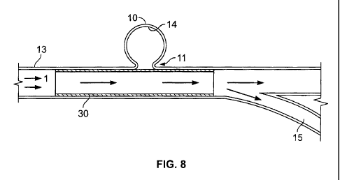
1
CA 02652022 2008-11-12
WO 2007/139699
PCT/US2007/011668
=
the arterial wall. Damage is believed to be caused by shear stresses due to
blood
flow. Shear stress generates heat that breaks down the cells. Such
hernodynamic
stresses at the vessel wall, possibly in conjunction with intrinsic
abnormalities of
the vessel wall, have been considered to be the underlying cause for the
origin,
growth and rupture of these saccular aneurysms of the cerebral arteries
(Lieber
and Gounis, The Physics of Endoluminal stenting in the Treatment of
Cerebrovascular Aneurysms, Neurol Res 2002: 24: S32-S42). In histological
studies, damaged intimal cells are elongated compared to round healthy cells.
Shear stress can vary greatly at different phases of the cardiac cycle,
locations in
the arterial wall and among different individuals as a function of geometry of
the
artery and the viscosity, density and velocity of the blood. Once an aneurysm
is
formed, fluctuations in blood flow within the aneurysm are of critical
importance
because they can induce vibrations of the aneurysm .wall that contribute to
= progression and eventual rupture. For a more detailed description of the
above
concepts see, for example, Steiger, Pathophysiology of Development and
Rupture.
of Cerebral Aneurysms, Acta Neurochir Suppl 1990: 48: 1-57; Fergueson,
Physical Factors in the Initiation, Growth and Rupture of Human Intracranial
Saccular Aneurysms, J Neurosurg 1972: 37: 666-677.
[05] Aneurysms are generally treated by excluding the weakened part of the
vessel
from the arterial circulation. For treating a cerebral aneurysm, such
reinforcement
- is done in many ways: (i) surgical clipping, where a metal clip is
secured around
the base of the aneurysm; (ii) packing the aneurysm with microcoils, which are
small, flexible wire coils; (iii) using embolic materials to "fill" an
aneurysm; (iv)
using detachable balloons or coils to occlude the parent vessel that supplies
the
aneurysm; and (v) endovascular stenting. For a general discussion and review
of
these different methods see Qureshi, Endovascular Treatment of Cerebrovascular
Diseases and Intracranial Neoplasms, Lancet. 2004 Mar 6;363 (9411):804-13;
Brilstra et al. Treatment of Intracranial Aneurysms by Embolization with
Coils: A
Systematic Review, Stroke 1999; 30: 470-476.
2
=
CA 02652022 2008-11-12
WO 2007/139699 PCT/US2007/011668
[06] As minimally invasive interventional techniques gain more prominence,
micro-
catheter based approaches for treating neurovascular aneurysms are becoming
more prevalent_ Micro-catheters, whether flow-directed or wire-directed, are
used
for dispensing embolic materials, microcoils or other structures (e.g.,
stents) for
embolization of the aneurysm. A microcoil can be passed through a micro-
catheter and deployed in an aneurysm using mechanical or chemical detachment
mechanisms, or be deployed into the parent vessel to permanently occlude it
and
thus block flow into the aneurysm. Alternatively, a stent could be tracked
through
the neurovasculature to the desired location. Article by Pereira, History of
Endovascular Aneurysms Occlusion in Management of Cerebral Aneurysms; Eds:
Le Roux et al., 2004, pp: 11-26 provides an excellent background on the
history
of aneurysm detection and treatment alternatives.
[07] As noted in many of the articles mentioned above, and based on the
origin,
formation and rupture of the cerebral aneurysm, it is obvious that the goal of
aneurysmal therapy is to reduce the risk of rupture of the aneurysm and thus
the
consequences of sub-arachnoid hemorrhage. It should also be noted that while
=preventing blood from flowing into the aneurysm is highly desirable, so that
the
weakened wall of the aneurysm doesn't rupture, it may also be vital that blood
flow to the surrounding structures is not limited by the method used to
obstruct
blood flow to the aneurysm. Conventional stents developed for treating other
vascular abnormalities in = the body are ill suited for embolizing cerebral
=
aneurysms. This could lead to all the usual complications when high oxygen
consumers, such as brain tissue, are deprived of the needed blood flow.
[08] There are many shortcomings with the existing approaches for treating
neurovascular aneurysms_ The vessels of the neurovasculature are the most
tortuous in the body; certainly more tortuous than the vessels of the coronary
circulation. Hence, it is a challenge for the surgeon to navigate the
neurovasculature using stiff coronary stents that are sometimes used in the
neurovasculature for treating aneurysms. The bending force of a prosthesis
indicates the maneuverability of the prosthesis through the vasculature; a
lower
= 3
CA 02652022 2008-11-12
WO 2007/139699 PCT/US2007/011668
bending force would imply that the prosthesis is more easily navigated through
the vasculature compared to one with a higher bending force. Bending force for
a
typical coronary stent is 0.05 lb-in (force to bend 0.5 inches cantilever to
90
degree). Hence, it will be useful to have neural prosthesis that is more
.flexible
than existing stents.
[09] Existing stent structures, whether used in coronary vessels or in the
neurovasculature (rnicrocoils) are usually straight, often laser cut from a
straight
tubing or braiding with stiff metallic materials. However, most of the blood
vessels are curved. Hence, current stent structures and microcoils impart
significant stress on the vessel walls as they try to straighten a curved
vessel wall.
For a weakened vessel wall, particularly where there is a propensity for an
aneurysm formation, this could have disastrous consequences.
[10] As noted earlier, the hemodynamic stress placed on the blood vessels,
particularly
at the point of bifurcation, leads to weakening of the vessel walls. The most
significant source of such stress is the sudden change in direction of the
blood
flow. Hence, if one were to minimize the sudden change in direction of blood
flow, particularly at the location of vessel weakness, it would be beneficial.
[11] , Existing approaches to occluding aneurysms could lead to another set of
problems. Methods that merely occlude the aneurysm by packing or filling it
with
embolic material (coils or liquid polymers) do not address the fundamental
flow
abnormalities that contribute to the formation of aneurysm.
[12] A stent structure could be expanded after being placed intraluminally on
a balloon
catheter. Alternatively, self-expanding steins could be inserted in a
compressed
state and expanded upon deployment. For balloon expandable stents, the stent
is
mounted on a balloon at the distal end of a catheter,= the catheter is
advanced .to
the desired location and the balloon is inflated to expand: the stent into a
permanent expanded condition. The balloon is then deflated and the catheter
withdrawn leaving the expanded stent to maintain vessel patency. Because of
the
potentially lethal consequences of dissecting or rupturing an intracerebral
vessel,
4
CA 02652022 2008-11-12
WO 2007/139699 PCT/US2007/011668
the.use of balloon expandable stents in the brain is fraught with problems.
Proper
deployment of a balloon expandable stent requires slight over expanding of the
balloon mounted stent to embed the stent in the vessel wall and the margin of
error is small. Balloon expandable stents are also poorly suited to adapt to
the
natural tapering of cerebral vessels which taper proximally to distally. If a
stent is
placed from a parent vessel into a smaller branch vessel the change in
diameter
between the vessels makes it difficult to safely deploy a balloon expandable
stent.
A self-expanding stent, where the compressed or collapsed stent is held by an
outer restraining sheath over the compressed stent to maintain the compressed
state until deployment. At the time of deployment, the restraining outer
sheath is
retracted to uncover the compressed stent, which then expands to keep the
vessel
open. Additionally, the catheters employed for delivering such prosthesis are
micro-catheters with outer diameter of 0.65 mm to 1.3 mm compared to the
larger
catheters that are used for delivering the large coronary stents to the
coronaries.
[13] US Patent No. 6,669,719 (Wallace et al.) describes a stent and a stent
catheter for
intra-cranial use. A rolled sheet stent is releasably mounted on the distal
tip of a
catheter. Upon the rolled sheet being positioned at the aneurysm, the stent is
released. This results in imi-nediate and complete isolation of an aneurysm
and
surrounding side branches of the circulatory system and redirecting blood flow
away from the aneurysm. A significant drawback of such a system is that the
surrounding side branches, along with the target aneurysm, are deprived of the
needed blood flow after the stent has been deployed.
[14] US Patent No. 6,605,110 (Harrison) describes a self-expanding stent for
delivery
through a tortuous anatomy or for conforming the stent to a curved vessel.
This
patent describes a stent structure with radially expandable cylindrical
elements
arranged in parallel to each other and interspersed between these elements and
connecting two adjacent cylindrical elements are struts that are bendable.
While
this structure could provide the necessary flexibility and bendability of the
stent
for certain applications, it is expensive and complex to manufacture.
=
CA 02652022 2008-11-12
WO 2007/139699 PCT/US2007/011668
[15] US Patent No. 6,572,646 (Boylan) discloses a stent made up of a super-
elastic
alloy, such as Ni-Ti alloy (Nitinol), with a low temperature phase that
induces a
first shape to the stent and a high temperature phase that induces a second
shape
to the stent with a bend along the length. US Patent No. 6,689,162 (Thompson)
discloses a braided prosthesis that uses strands of metal, for providing
strength,
and compliant textile strands. US Patent No. 6,656,218 (Denardo et al.)
describes
an intravascular flow modifier that allows microcoil introduction.
Summary of the Invention
[161 An aspect of the present invention provides a highly flexible implantable
occluding device that can easily navigate the tortuous vessels of the
neurovasculature. Additionally, occluding device can easily conform to the
shape
of the tortuous vessels of the vasculature. Furthermore, the occluding device
can
direct the blood flow within a vessel away from an aneurysm; additionally such
an occluding device allows adequate blood flow to be provided to adjacent
= structures such that those structures, whether they are branch vessels or
oxygen
demanding tissues, are not deprived of the necessary blood flow.
[17] The occluding device is also capable of altering blood flow to the
aneurysm, yet
maintaining the desired blood flow to the surrounding tissue and within the
vessel. In this instance, some blood is still allowed to reach the aneurysm,
but not
enough to create a laminar flow within the aneurysm that would cause injury to
its
thinned walls. Instead, the flow would be intermittent, thereby providing
sufficient time for blood clotting or filler material curing within the
aneurysm.
[18] The occluding device is flexible enough to closely approximate the native
vasculature and conform to the natural tortuous path of the native blood
vessels.
One of the significant attributes of the occluding device according to the
present
invention is its ability to flex and bend, thereby assuming the shape of a
vasculature within the brain. These characteristics are for a neurovascular
occluding device than compared to a coronary stent, as the vasculature in the
brain is smaller and more tortuous.
=
6
CA 02652022 2008-11-12
WO 2007/139699
PCT/US2007/011668
[19] In general terms, aspects of the present invention relate to methods and
devices
for treating aneurysms. In particular, a method of treating an aneurysm with a
neck comprises deploying a vascular occluding device in the lumen of a vessel
at
the location of the aneurysm, whereby the blood flow is redirected away from
the
neck of the aneurysm. The induced stagnation of the blood in the lumen of the
aneurysm would create embolization in the aneurysm. The occluding device
spans the width of the stem of the aneurysm such that it obstructs or
minimizes
the blood flow to the aneurysm. The occluding device is very flexible in both
its
material and its arrangement. As a result, the occluding device can be easily
navigated through the tortuous blood vessels, particularly those in the brain.
Because the occluding device is flexible, very little force is required to
deflect the
occluding device to navigate through the vessels of the neurovasculature,
which is
of significance to the operating surgeon.
[201 A feature of the occluding device, apart from its flexibility, is that
the occluding
device may have an asymmetrical braid pattern with a higher concentration of
braid strands or a different size of braid strands on the surface facing the
neck of
the aneurysm compared to the surface radially opposite to it. In one
embodiment,
the surface facing the aneurysm is almost impermeable and the diametrically
opposed surface is highly permeable. Such a construction would direct blood
flow away from the aneurysm, but maintain blood flow to the side branches of
the
main vessel in which the occluding device is deployed.
=
[21] In another embodiment, the occluding device has an asymmetrical braid
count
along the longitudinal axis of the occluding device. This provides the
occluding
device with a natural tendency to curve, and hence conform to the curved blood
.
vessel. This reduces the stress exerted by the oacluding device on the vessel
wall
and thereby minimizing the chances of aneurysm rupture. Additionally, because
the occluding device is naturally curved, this eliminates the need for the tip
of the
micro-catheter to be curved. Now, when the curved occluding device is loaded
on
to the tip of the 'micro-catheter, the tip takes the curved shape of the
occluding
device. The occluding device could be pre-mounted inside the micro-catheter
and
7
CA 02652022 2008-11-12
WO 2007/139699
PCT/US2007/011668
can be delivered using a plunger, which will push the occluding device out of
the
micro-catheter when desired. The occluding device could be placed inside the
micro-catheter in a compressed state. Upon exiting the micro-catheter, it
could
expand to the size of the available lumen and maintain patency of the lumen
and
allow blood flow through the lumen. The occluding device could have a lattice
structure and the size of the openings in the lattice could vary along the
length of
the occluding device. The size of the lattice openings can be controlled by
the
braid count used to construct the lattice.
[22] According to one aspect of the invention, the occluding device can be
used to
remodel an aneurysm within the vessel by, for example, neck reconstruction or
balloon remodeling. The occluding device can be used to form a barrier that
retains occlusion material within the aneurysm so that introduced material
will not
escape from within the aneurysm due to the lattice density of the occluding
device
in the area of the aneurysm.
=
[23] In another aspect of the invention, a device for occluding an aneurysm is
disclosed. The device is a tubular with a plurality of perforations
distributed on
the wall of the member. The device is placed = at the base of the aneurysm
covering the neck of the aneurysm such that the normal flow to the body of the
aneurysm is disrupted and thereby generating thrombus and ultimately occlusion
of the aneurysm.
=
[24] In yet another aspect of this invention, the device is a braided tubular
member.
= The braided strands are ribbons with rectangular cross section, wires
with a
circular cross section or polymeric strands.
[25] In another embodiment, a device with a braided structure is made in order
to
conform to a curved vessel in the body, where the density of the braid
provides
enough rigidity and radial strength. Additionally, the device can be
compressed
using a force less than 10 grarns. This enables the device to be compliant
with the
artery as the arterial wall is pulsating. Also, the device is capable of
bending
upon applying a force of less than 5 grainkm.
8
CA 02652022 2015-01-26
[26]
Other aspects of the invention include methods corresponding to the devices
and systems
described herein.
[26a] According to an aspect of the invention there is provided a system
comprising: a delivery
device; a self-expanding device, disposed within the delivery device,
comprising, in a cross-
section, a first layer having strands and a second layer having strands, the
strands of the first
layer and the strands of the second layer being helically wound in a lattice
structure arranged
as a flexible tubular body; wherein the first layer of the self-expanding
device comprises a
first cross-sectional diameter, wherein each the strand of the first layer is
circumferentially
spaced from an adjacent the strand of the first layer device by an arc angle
calculated by
dividing 360 degrees by the number of strands in the first layer, wherein,
with respect to the
delivery device, each the strand of the first layer comprises a strand cross-
sectional
dimension being about equal to or less than (a circumference of an inner
surface of the
delivery device)/(a total number of strands in the first layer); and wherein
the second layer of
the device comprises a second cross-sectional diameter different than the
first cross-sectional
diameter, wherein each the strand of the second layer is circumferentially
spaced from an
adjacent the strand of the second layer by the arc angle, wherein each the
strand of the second
layer comprises the strand cross-sectional dimension; wherein the self-
expanding device is
configured to have a surface coverage that is less than or equal to 40% when
expanded out of
the delivery device.
[26b1 According to another aspect of the invention there is provided a system
comprising: a
delivery device; a self-expanding device, disposed within the delivery device,
comprising, in
a cross-section, a first layer having strands and a second layer having
strands, the strands of
the first layer and the strands of the second layer being helically wound in a
lattice structure
arranged as a flexible tubular body; wherein the first layer of the device
comprises a first
cross-sectional diameter, wherein each the strand of the first layer is
circumferentially spaced
from an adjacent the strand of the first layer by a first arc angle calculated
by dividing 360
degrees by the first number of strands in the first layer, wherein, with
respect to the delivery
device, each the strand of the first layer comprises a strand cross-sectional
dimension being
about equal to or less than (a circumference of an inner surface of the
delivery device)/(half a
9
CA 02652022 2015-01-26
total number of strands in the device); and wherein the first layer of the
device comprises a
second cross-sectional diameter different than the first cross-sectional
diameter, wherein each
the strand of the second layer is circumferentially spaced from an adjacent
the strand of the
second layer by a second arc angle calculated by dividing 360 degrees by the
second number
of strands in the second layer, wherein each the strand of the second layer
comprises the
strand cross-sectional dimension; wherein the self-expanding device is
configured to have a
surface coverage that is less than or equal to 40% when expanded out of the
delivery device.
Brief Description of the Drawings
[27] The invention has other advantages and features which will be more
readily apparent from
the following detailed description of the invention and the appended claims,
when taken in
conjunction with the accompanying drawings, in which:
[28] FIG. 1 is an illustration of an aneurysm, branch vessels and blood
flow to the aneurysm.
[29] FIGS. 2A and 2B illustrate one embodiment of an occluding device to
treat aneurysms.
[30] FIG. 3 is an illustration of the embodiment shown in FIG. 2 in a
compressed state inside a
m icro-catheter.
[31] FIG. 4A is another embodiment of an occluding device for treating
aneurysms.
[32] FIGS. 4B and 4C illustrate cross sections of portions of ribbons that
can be used to form the
occluding device of FIG. 4A.
[33] FIG. 5 shows the occluding device in a compressed state inside a micro-
catheter being
advanced out of the micro-catheter using a plunger.
[34] FIG. 6 shows the compressed occluding device shown in FIG. 5 deployed
outside the micro-
catheter and is in an expanded state.
9a
CA 02652022 2015-01-26
[35] FIG. 7 shows the deployed occluding device inside the lumen of a
vessel spanning the neck
of the aneurysm, a bifurcation and branch vessels.
[36] FIG. 8 is a schematic showing the occluding device located in the
lumen of a vessel and the
change in the direction of the blood flow.
9b
CA 02652022 2008-11-12
WO 2007/139699
PCT/US2007/011668
[37] FIG. 9 shows the effect of a bending force on a conventional stent
compared to
the occluding device of the present invention.
[38] FIG. 10 demonstrates the flexibility of the current invention, compared
to a
traditional stent, by the extent of the deformation for an applied force.
[39] FIG. 11 shows the non-uniform density of the braid that provides the
desired
curved occluding device.
[40] FIG. 12 illustrates the difference in lattice density or porosity due
to the non-
uniform density of the braiding of the occluding device.
[41] FIG. 13 shows the varying lattice density occluding device covering the
neck of
an aneurysm.
[42] FIGS. 14 and 15 show an embodiment of the vascular occluding device where
the
lattice density is asymmetrical about the longitudinal axis near the aneurysm
neck.
[43] FIG. 16 illustrates a bifurcated occluding device according to an
embodiment of
the present invention in which two occluding devices of lesser densities are
combined to form a single bifurcated device.
[44] FIG. 17 illustrates an example of a mesh pattern of a lattice in an
occluding
device.
[45] FIG. 18 illustrates an example of a braiding element of a lattice in an
occluding
device.
[46] FIG. 19 illustrates an example of another braiding element of a lattice
in an
occluding device.
[47] FIG. 20 illustrates a braiding element of an occluding device fitted into
a vessel
diameter.
[48] FIG. 21 is a cross sectional view of an example of a protective coil.
CA 02652022 2014-04-23
[49] FIG. 22 illustrates an example of detennining ribbon dimensions of an
occluding device in a
protective coil or a delivery device.
[50] FIG. 23 illustrates another example of determining ribbon dimensions
of an occluding device
in a protective coil or a delivery device.
[51] FIG. 24 illustrates an example of determining a ribbon width based on
a number of ribbons.
[52] FIG. 25 illustrates a relationship between the PPI of the occluding
device in a vessel versus
the PPI of the occluding device in a free-standing state.
[53] FIG. 26 illustrates an example of a maximum ribbon size that fits in a
protective coil.
[54] FIG. 27 is a graph showing the opening sizes of braiding elements in
the occluding device as
a function of the PPI of the lattice structure.
[55] FIG. 28 illustrates the in-vessel PPI as a function of the braided PPI
of a 32 ribbon occluding
device.
[56] FIG. 29 illustrates the percent coverage as a function of the braided
PPI for a 32 ribbon
occluding device.
[57] FIG. 30 illustrates the opening sizes of braiding elements in the
occluding device as a
function of the braided PPI of the lattice structure for a 32 ribbon occluding
device.
Detailed Description Of The Preferred Embodiments
[58] The devices shown in the accompanying drawings are intended for
treating aneurysms. They
are generally deployed, using micro-catheters, at the location of a cerebral
aneurysm that is
intended to be treated. One such system is disclosed in copending U.S. Patent
Application
titled "System and Method for Delivering and Deploying an Occluding Device
Within a
Vessel", issued April 3, 2012 as U.S. Patent No. 8,147,534 B2. The embodiments
of the
11
CA 02652022 2014-04-23
endovascular occluding device according to aspects of the present invention is
useful for
treating cerebral aneurysms that are commonly treated using surgical clips,
microcoils or
other embolic devices.
[59] FIG. 1 illustrates a typical cerebral aneurysm 10 in the brain. A neck
11 of the aneurysm 10
can typically define an opening of between about 2 to 25 mm. As is understood,
the neck 11
connects the vessel 13 to the lumen 12 of the aneurysm 10. As can be seen in
FIG. 1, the
blood flow 1 within the vessel 13 is channeled through the lumen 12 and into
the aneurysm.
In response to the constant blood flow into the aneurysm, the wall 14 of lumen
12 continues
to distend and presents a significant risk of rupturing. When the blood within
the aneurysm
causes pressure against the wall 14 that exceeds the wall strength, the
aneurysm ruptures.
The present invention could prevent such ruptures. Also shown in FIG. I are
the bifurcation
and the side branches 16.
[60] FIG. 2 illustrates one embodiment of a vascular occluding device 20 in
accordance with an
aspect of the present invention. In the illustrated embodiment, the occluding
device 20 has a
substantially tubular structure 22 defined by an outer surface 21, an inner
surface 24 and a
thin wall that extends between the surfaces 21, 24. A plurality of openings 23
extend between
the surfaces 21, 24 and allow for fluid flow from the interior of the
occluding device 20 to the
wall of the vessel. Occluding device 20 is radially compressible and
longitudinally
adjustable.
[61] FIG. 3 shows a micro-catheter 25 and the occluding device 20 inside
the microcatheter 25 in
a compressed state prior to being released within the vasculature of the
patient.
[62] FIG. 4 illustrates another embodiment of the occluding device 30
having two or more strands
of material(s) 31, 32 wound in a helical fashion. The braiding of such
material in this fashion
results in a lattice structure 33. As can be understood, the dimension of the
lattice 33 and the
formed interstices 34 is
12
CA 02652022 2008-11-12
WO 2007/139699 PCT/US2007/011668
=
. determined, at least in part, by the thickness of the strand materials., the
number of
strands and the number of helices per unit length of the occluding device 30.
For-
example, the interstices 34 and/or the dimension of the lattice 33 may be
determined by the number of strands of material(s) 31, 32 wound in helical
= fashion. In one example, any number of braiding ribbons up to 16 braiding
=
ribbons may be used (e.g., 5, 8, 10, 13, 15 or 16 braiding ribbons). In
another
example, 16-32 braiding, ribbons may be used (e.g., 20, 23, 25, 27, 30, or 32
braiding ribbons). In another example greater than 32 braiding ribbons may be
used such as, for example, 35, 40, 48, 50, 55, 60, 80, 100, or greater
braiding
ribbons. Nevertheless, other values are possible.
[63] Hence, strands of material, such as ribbons, may intersect to form a
braid pattern.
The intersection of the strand material may be formed in either a radial or
axial
direction on a surface of a forming device such as a braiding mandrel. When
the
intersection of the strand material is along an axial path, for example, the
intersecting material may be at a fixed or variable frequency. As one example
of
strand material intersecting at a fixed frequency, the.intersecting strand
material
may be along any 1.0 inch axial path on the surface of the forming device
(e.g., a
braiding mandrel) to indicate the pick count. When the intersection of the
strand
material is along a radial path or circumferential path, the spacing of the
strand
material may be uniformly or variably distributed. In one example of the
strand
material along a radial or circumferential path in which the spacing is
uniformly
distributed, the spacing along the radial direction may be determined based on
the
following formula:
Eq. (1): (71) * (forming device diameter)/ (# ribbons/2)
[64] FIG 18 illustrates an example of braiding elements or cells in the radial
and PPI
(picks per inch) directions. Any single element of the braid (i.e., braid
element)
may be combined to form a mesh pattern as illustrated in FIG. 17 on a surface
of a
= forming device (e.g., braiding mandrel). The braid = is capable of
impeding or
disrupting the flow of fluid (e.g., blood) in a vessel (e.g., blood vessel).
The braid
13
CA 02652022 2008-11-12
WO 2007/139699 PCT/US2007/011668
or lattice pattern, density, shape, etc. when the occluding device is deployed
in the
vessel, may at least partially determine the flow within the vessel. Each of
the
parameters of the braid or lattice may also be controlled by a user to control
flow.
[65] Parameters for determining the flow through an occluding device
containing a
lattice pattern, density, shape, etc. include surface coverage of the
occluding
device and cell size of the braid or lattice pattern. Each of these parameters
may
further characterize the geometry of the braid or lattice. Surface coverage
may be
determined as (surface area)/(total surface area), where the surface area is
the
surface area of the frame or solid element and the total surface area is of
the entire
element (i.e., frame and opening).
[66] Cell size may be determined as the maximum length defining a cell
opening.
Braiding patterns that increase surface coverage while decreasing cell size
may
have an increased effect on disrupting or impeding the flow through the braid
or
lattice. Each of the parameters= of surface coverage and cell size may further
be
enhanced by varying the width of the strand material (e.g., the ribbons),
increasing the number of strands of strand material defining the braid, and/or
= increasing the PPI (i.e., Picks Per Inch).
=
[67] The braiding or lattice pattern as described may be further defined by
various
parameters including, for example, the number of strands (e.g., ribbons), the
width of each ribbon/strand, the braiding PPI, and/or the diameter of the
forming
device (e.g., mandrel diameter), to name a-few. Based on the lattice
parameters, a
leg length and a ribbon angle may be determined. The leg length may define the
length of an aspect of the braiding element. For example, if the braiding
element
is diamond shaped as illustrated in FIG. 17, the length of one side of the
diamond
shaped braiding element is the "leg length:" A ribbon angle may define the
angle
created by two intersecting aspects of the braiding element. In the example =
illustrated in FIG. 17, the ribbon angle is the angle formed between two
adjacent =
sides of the diamond shaped braiding element. Radial spacing of braid elements
in
a lattice pattern can define the width of a braiding element in radial
direction.
14
CA 02652022 2008-11-12
WO 2007/139699 PCT/US2007/011668
FIG. 18 illustrates an example of a radial spacing, leg length and ribbon
angle of a
braid element.
[68] Radial spacing of the lattice may be determined as set forth in Equation
1 as
follows:
Eq. (1): Radial Spacing = (it) * (forming device diameter)/(# ribbons/2)
[69] The braiding element may be fitted into a vessel based on the radial
spacing or the
diameter of the vessel. The radial spacing of the lattice may be adjusted
based on
the diameter of the vessel. For example, if the diameter of the vessel is
small, the =
radial spacing may be adjusted to a smaller dimension while the leg length of
the
braid elements may be maintained. Also in this example, the ribbon angle may
also be adjusted to achieve the adjusted radial spacing. Adjusting the ribbon
angle
may also alter the spacing of the braid element in the PPI direction.
[70] FIG.. 19 illustrates an example of determining a radial spacing and
ribbon angle of
a lattice structure in an occluding device. In this example, a lattice or
braid
contains sixteen interlacing ribbons, with each ribbon being 0.004 inches wide
and braided on a forming device such as a mandrel with a diameter of 4.25 mm
and 65 PPI. Thus, in this -example, the number of braiding elements is
sixteen, the
ribbon width is 0.004 inches, the spacing in the PPI direction is 1/65 =
0.01538
= inches and the diameter of the forming device (e.g., mandrel diameter) is
4.25
mm. Hence, the radial spacing may be calculated as: Radial spacing = (n) *
(forming device diameter)/(# ribbons/2) = (3.14) * (0.425/2.54) / (16/2) =
0.0657
inches. FIG. 19 illustrates an example of a braiding element with a radial
spacing
of 0.0657 inches. In addition, the leg length of the example is 0.0337 inches,
the.
ribbon angle is 153.65 degrees, and the spacing of the braiding element in the
PPI
direction, based on the ribbon angle and leg length is 0.0154 inches.
= [71] FIG. 20 illustrates the example of FIG. 19 after the braiding
element is fitted into
an appropriate vessel diameter. In this example, the radial spacing is
adjusted to a
smaller length to accommodate a smaller vessel diameter. The leg length
remains
=
CA 02652022 2008-11-12
WO 2007/139699
PCT/US2007/011668
constant at 0.0337 inches so the ribbon angle changes based on changes in the
radial spacing. In this example, the radial spacing is adjusted to 0.06184
inches
and the ribbon angle is adjusted to 132.79 degrees. Also, the spacing of the
braid
element in the PPI direction is also changed. In this example, the spacing of
the
braid element in the PPI direction increases from 0.0154 inches to 0.0270
inches. =
[72] Table 1 illustrates additional examples of lattice or braid patterns of
varying PPI,
ribbon width (RW), or number of ribbons. 'In addition, each of the braid
patterns
in Table 1 may produce patterns with the same percent coverage within a
vessel.
TABLE 1
# ribbons 16 32 64
Braid diameter (mm) 4.25 4.25 4.25
Braid diameter (in) 0.16732 0.16732 0.16732
PPI 65.00 130.00 260.00
RW (mils) 4.0000 2.0000 = 1.0000
Node Spacing (ppi) 0.01538 = 0.00769 0.00385
Node Spacing (radial) 0.06571 0.03285 0.01643
Ribbon Angle (ppi) 153.65 = 153.65 153.62
Leg Length (in) 0.03374 0.01687 0.00844
Vessel diameter (mm) 4 4 4
In-vessel device Node spacing 0.06184 0.03092 0.01546
In-vessel device Ribbon Angle 132.79 132.79 132.70
= (PPi)
In-vessel device Node spacing 0.02702 0.01351 0.00677
(1)Pi)
In-vessel device PPI 37.01 74.04 147.72
In-vessel' device braided closed - 0.00024814 0.00006203 0.00001551
area (in2)
In-vessel device Braided Open 0.00058741 0.00014680 0.00003681
Area (in2)
In-vessel device coverage 29.7% 29.7% 29.64%
In-vessel device total area. (in2) 0.00083555 0.00020883
0.00005232
In-vessel device cell size (mm) 1.317 0.658 0.329
[73] The occluding device may be placed into a protective coil to enhance
placement
of the occluding device in a vessel. Also, the occluding device may be housed
in a
delivery device, such as a catheter, for placement within a vessel. The
occluding
16
CA 02652022 2008-11-12
WO 2007/139699 PCT/US2007/011668
device.may be created at a size or dimension based on the size of the
protective.
coil, delivery device, or catheter housing the occluding device. For example,
the
number of strands or ribbons in the lattice structure of the occluding device
that fit
into a corresponding protective coil, delivery device, or catheter may be
determined such that the.occluding device is effectively stored or housed
prior to
deployment in a vessel. In one example, the strands of the occluding device
may
Overlap in a 2-layer structure including an inner layer and an outer layer,
the outer
layer contacting the protective coil.
1741 In one example, a housing such as a protective coil, delivery device or
catheter
that houses the occluding device may have a constant size or diameter and the
characteristics of the occluding device may be determined to fit the housing.
For
example, a ribbon size or width may be determined based on the desired size of
the housing. In this way, the size (or diameter) of the housing (e.g.,
protective
coil, delivery device or catheter) may be constant for a variety of occluding
devices that may vary in size or number of ribbons.
[75] FIG. 21 illustrates an example of a cross sectional view of a protective
coil. In this
example, a number of strands or ribbons in a lattice structure of an occluding
device is determined for a protective coil. The protective coil illustrated in
FIG.
21 has. a circular crosS sectional area with a diameter. A strand or ribbon of
a
predetermined thickness or size is placed within the protective coil such that
the
outer surface of the strand/ribbon contact the inner surface of the protective
coil.
The inner surface of the strand/ribbon creates a concave surface within the
protective coil A second strand/ribbon is placed within the protective coil
such
that the outer surface of the second strand/ribbon contacts an inner
circumference
in contact with the the concave surface of the strand/ribbon previously placed
in
the protective coil. The angle from a center point of the circular protective
coil
from one edge of the second strand/ribbon to an opposite edge of the second
strand/ribbon is determined (i.e., the "arc-angle"). Based on these
measurements,
the number of strands or ribbons of the predetermined size or thickness may be
=
17
CA 02652022 2008-11-12
WO 2007/139699
PCT/US2007/011668
determined as follows: (Arc-angle) * (# ribbons/2) < = 360 degrees (i.e., #
ribbons
< = 720 degrees/angle).
[76] In the example illustrated in FIG. 21, an occluding device is constructed
using a
0.001 by 0.004 inch ribbon. The arc-angle of the ribbon element at the center
of
the protective coil between a first line drawn from the center point of the
protective coil to one edge' of an inner layer ribbon and a second line drawn
from
the center point of the protective coil to the opposite edge of the inner
layer
ribbon is 34.14 degrees. Thus, the calculated number of ribbons is less than
or
equal to 720 degrees/34.14 degrees = 20 ribbons.
[77f TABLE 2 illustrates additional examples of different designs for loading
a lattice
structure of an occluding device in a protective coil.
TABLE 2
# ribbons 16 32 _ 64
Protective Coil ID (in) 0.017 0.017 0.017
Ribbon Width (in) 0.004 0.002 0.001
Ribbon Thickness (in) 0.001 0.001 0.001
Inner Circle Angle 36.98 17.83 8.84
Max # Ribbons fitting in inner circle 9.73 20.19 40.72
# ribbons in inner circle 8 16 32
=
[78] FIG. 22 illustrates another example 6f determining ribbon dimensions for
an
occluding device in a protective coil or a delivery device. In this example,
an
occluding device with a lattice or braid structure based on a thickness of a
ribbon.
As FIG. 22 illustrates, the diameter of the protective .coil or delivery
device 2301
is 0.0170 inches. A first ribbon 2302 is fitted within the outer surface of
the
= protective coil or delivery device 2301. A second ribbon 2303 is placed
in contact
with an inner circumference of the protective coil or delivery device 2301
where
the inner circumference is a circumference that is tangential to the inner
surface of
.the first ribbon 2302. The second ribbon 2303 is placed within the inner
circumference such that lateral ends of the second ribbon 2303 are in contact
with
18
=
CA 02652022 2008-11-12
WO 2007/139699 PCT/US2007/011668
=
= the inner circumference of the protective coil or delivery device 2301.
The arc-
angle between a first line extending from the center point of the protective
coil or
delivery device 2301 to one lateral end of the second ribbon 2303 and a second
line extending from the center point of the protective coil or delivery device
2301
to the other lateral end of the second ribbon 2303 is calculated as
illustrated in
FIG. 22.
[79] In this example, the maximum dimensions of the first and second ribbons
2302, =
2303 are determined based on the calculated arc-angle formed. For example, to
allow eight ribbons in the inner circumference of the protective coil or
delivery
device 2301, the arc-angle may be calculated as (360 degrees)/8 = 45 degrees
as
FIG. 22 illustrates. Based on a 45 degree angle, the maximum ribbon width may
be determined as 0.00476 inches to allow eight ribbons of a thickness of 0.001
inches to fit within the inner circumference of the protective coil or
delivery
device 2301.
[8,01 In another example, a narrower ribbon width is used to compensate for
material
tolerance variations and curvature. Based on extensive research and
experimentation by the applicants, it was discovered that a tolerance range
applied
to the ribbon widths of about 20% can compensate for such material tolerance
variations. FIG. 23 illustrates an example of a 20% tolerance range or cushion
applied to ribbon widths of an occluding device.
[81] In this example, 20% additional ribbons are desired in the occluding
device (i.e.,
1.20 * 8 = 9.6 ribbons). The maximum width of the ribbons ma be determined
based on the desired number of 9.6 ribbons by calculating the angle as
described
above. Specifically, the arc-angle may be calculated as (360 degrees)/9.6 =
37.7
degrees. Based on this calculation, the maximum width of the ribbons may be
determined as 0.00405 inches as illustrated in FIG. 23. Thus, in this example,
a
20% cushion is applied to permit 9.6 ribbons in the protective coil or
delivery
device at a maximum width of 0.00405 inches.
19
CA 02652022 2008-11-12
WO 2007/139699
PCT/US2007/011668
[821 Table 3 provides additional examples of ribbon widths for various ribbon
thicknesses. In the examples. provided in Table 3, the ribbon thicknesses
range
from 0.0007 inches to 0.0015 inches.
TABLE 3
Ribbon Thickness (in) Calculated max width (in 20%
cushion width (in)
0.0005 0.00543 00.000463
0.0006 0.00530 0.00452
0.0007 0.00516 0.00440
0.0008 0.00503 0.00428
0.0009 0.00490 0.00417
0.0010 0.00476 0.00405
0.0011 0.00463 0.00393
0.0012 0.00450 0.00382
0.0013 0.00436 0.00370
0.0014= 0.00422 0.00358
0.0015 = 0.00409 0.00346
1831 In another example, an occluding device containing 32 ribbons is
described. FIG.
24 illustrates an example of determining the ribbon width of a 32-ribbon
occluding device based on the number of ribbons that can fit in the protective
coil
or delivery device 2501. In this example, the protective coil or delivery
device
2501 has a diameter of 0.017 inches and the maximum ribbon width that can fit
in
the inner circumference of the protective coil or delivery device 2501
provides an
arc-angle of (360 degrees)/(32/2) ---- 22.5 degrees as illustrated in FIG. 24.
Hence,
to fit 16 ribbons along the inner circumference of the protective coil 2501,
the
width of the = ribbons is determined to be 0.00266 inches, with a thickness of
0.00080 inches as illustrated in FIG. 24. Similarly a 20% cushion may be
applied
to the ribbon widts to provide for narrower ribbon widths to compensate for
material tolerance variations. In this example, the modified ribbon widths may
be
determined based on the new arc-angle requirement 01(360 degrees)/19.2 = 18.75
degrees. Table 4 provides maxiumum ribbon widths for a 32-ribbon occluding
device.
CA 02652022 2008-11-12
WO 2007/139699 PCT/US2007/011668
TABLE 4
Ribbon Thickness (in) Calculated max width (in)
20% cushion width (in) _
0.0005 0.00288 0.00242
0.0006 0.00281 0.00235
0.0007 0.00273 0.00229
0.0008 = 0.00266 0.00223
0.0009 0.00258. 0.00216
0.0010 0.00251 0.00210
[84] Alternatively, a larger number of ribbons may be included in the
occluding
device. For example, the strands or ribbons may be increased to greater than
32,
such as 40, 44, 48, 50, 56, 60, 64, 70, 76, 80, 90, 100, or more. For any
desired
number of ribbons, a ribbon width may be determined based on a calculated
angle
or a ribbon thickness as described. In addition, a cushion may be applied to
=the
ribbon width as described.
= [85] In another example, oversized occluding devices may be used relative
to the
vessel. For example, a larger occluding device relative to the size of the
vessel
lumen may result in enhanced anchoring of the occluding device within the
lumen
of the vessel. FIG. 25 illustrates a relationship between the PPI .of the
occluding
device in place in the vessel ("in-vessel PPI") versus the PPI of the
occluding
device in the free-standing state ("braided PPI"). The graph in FIG. 25
demonstrates that for each design, the PPI of the occluding device in place in
the
vessel approaches a maximum value as the pick count of the occluding device in
the free-standing state increases. For example, for the 4mm vessel design, as
the
PPI of the free-standing occluding device is increased, the PPI of the
occluding
device in the vessel increases until the in-vessel PPI reaches about 45. When
the
in-vessel PPI reaches about 45, further increases in the braided PPI result in
only
minimal further increases in the in-vessel PPI. Also illustrated in FIG. 25,
different vessel designs (e.g., 3 mm vessel design or 5 mm vessel design)
result in
a similar behavior in which the in-vessel PPI approaches a maximum value for
high braided pick counts.
21
CA 02652022 2008-11-12
WO 2007/139699 PCT/US2007/011668
=
[86] Similarly, FIG. 28 illustrates the in-vessel PPI as a function of the
braided PPI of
a 32 ribbon occluding device. In the examples illustrated in FIG. 28, the PPI
of
the occluding device in a vessel ("in-vessel PPI") approaches a maximum value
as
the PPI of the occluding device in a free-standing state ("braided PPI"). FIG.
28
also illustrates alternate vessel designs. As can be seen in the examples of
vessel
. designs of FIG. 28, for each of the vessel designs, the in-vessel PPI
approaches a
maximum value asymptotically as the braided PPI increases.
(871 Similarly, the coverage of the occluding device may be based on ribbon
width or
braided PPI. FIG. 26 illustrates an example in which the ribbon is 0.00467
inches
wide and 0.001 inches and is the maximum ribbon size that fits in the
protective
coil. As FIG. 26 illustrates, the coverage approaches a maximum value of
approximately 65 ¨ 100 PPI range. In this example, the percentage of coverage
asymptotically approaches approximately 40% for a 0.001" X 0.00467" ribbon
and 34% for a 0.001" X 0.004" ribbon.
[88] FIG. 29 illustrates the percent coverage as a function of the .braided
PPI for a 32
ribbon occluding device. As FIG. 29 demonstrates, the %coverage approaches a
maximum value as the braided PPI in increases. For example, for an occluding
device containing. 0.0008 X 0.00266 inch ribbons, the % coverage approaches a
maximum value of about 43% as the braided PPI increases above about 150.
Also, for an occluding device containing 0.0008 X 0.0020 inch ribbons, the %
coverage approaches a maximum value of about 35% as the braided PPI increases
above about 150.
= [89] FIG. 27 is a graph showing the opening sizes of braiding elements in
the
occluding device as a function of the PPI of the lattice structure. As the PPI
= increases, the opening sizes or spaces through which flow of fluid (e.g.,
blood)
decreases. As the PPI of the lattice structure reaches about 100, the opening
sizes
of the braiding elements when in place in a vessel asymptotically approaches a
minimum value. In the examples illustrated in FIG. 27, for a ribbon size of
0.001
X 0.004 inches, the opening sizes of the braiding elements in the lattice
structure
=
22
CA 02652022 2008-11-12
WO 2007/139699
PCT/US2007/011668
=
of an occluding device in a vessel approaches 1280 microns or less. Similarly,
for
=
=
a ribbon size of 0.001 X 0.00467 inches, the opening sizes of the braiding
.elements in the lattice structure of an occluding device in a vessel
approaches
about 1220.
[90] FIG. 30 illustrates the opening sizes of braiding elements in the
occluding device
as a function of the braided PPI of the lattice structure for a 32 ribbon
occluding
device. As FIG. 30 demonstrates, the opening size of braiding elements
approaches a minimum value as the braided PPI in increases. For example, for
an
occluding device containing 0.00.08 X 0.00266 inch ribbons, the opening size
approaches a minimum value of about less than 600 microns as the braided PPI
increases above about 150. Also, for an occluding device containing 0.0008 X
0.0020 inch ribbons, the opening sizes approaches a minimum value of about 640
as the braided PPI increases above about 150.
[91] The occluding device 30 is radially compressible and radially expandable
without
the need for supplemental radially expanding force, such as an inflatable
balloon.
The occluding device 30 is constructed by winding the two strands (31, 32 in
opposite directions. Alternatively, greater than 2 strandS may be wound in
various directions. For example, 8, 10, 12, 14, 22, 28, 30, 32, 36, 40, 44,
48, 52,
58, 64, 70, 86, 90, 110, 116, 120, 128, 136, 150, or greater strands may be
wound
in various directions. In an embodiment, the strands 31, 32 are in the shape
of
rectangular ribbon (See Figure 4C). The.ribbons can be formed of known
flexible
materials including shape memory materials, such as Nitinol, platinum and
stainless steel.
[92] The ribbon used as the braiding material for the strands 31, 32 can
include a
rectangular cross section 35 (Figure 4C). As shown in Figures 4C and 7, the
surface 36 that engages an inner surface of the vessel has a longer dimension
(width) when compared to the wall 38 that extends between the surfaCes 36, 37
(thickness). A ribbon with rectangular cross section has a higher recovery
(expansive) force for the same wall thickness when compared to a wire with a
23
=
CA 02652022 2008-11-12
WO 2007/139699 PCT/US2007/011668
=
circular (round) cross section. Additionally, a flat ribbon allows for more
compact compression of the occluding device 20 and causes less trauma to the
vascular wall when deployed because it distributes the radial expansion forces
over a greater surface area. Similarly, flat ribbons form a more flexible
device for
a given lattice density because their surface area (width) is greater for a
given
thickness in comparison to round wire devices.
[931 While the illustrated embodiment discloses a ribbon having a rectangular
cross
section in which the length is greater than its thickness, the ribbon for an
alternative embodiment of the diSclosed occluding devices may include a square
cross section. In another alternative embodiment, a first portion of the
ribbon
may include a first form of rectangular cross section and a second portion 39
of
the ribbon (Figure 4B) may include a round, elliptical, oval or alternative
form of
rectangular cross section. For example, end sections of the ribbons may have
substantially circular or oval cross section and the middle section of the
ribbons
could have a rectangular cross section.
[94] = In an alternative embodiment as described above, the occluding device
30 can be
formed by winding more than two strands of ribbon. In an embodiment, the
occluding device 30 could include as many as sixteen. strands of ribbon. In =
another embodiment, the occluding device 30 can include as many as 32 strands
of ribbon, as many as 48 strands of ribbon, as many as 60 strands of ribbon,
as
many as 80 strands of ribbon, as many as 100 strands of ribbon, as many as 150
strands of ribbon or greater than 150 strands of ribbon, for example. By using
standard techniques employed in making radially expanding stents, one can
create
an occluding device 30 with interstices 34 that are larger than the thickness
of the
ribbon or diameter of the wire. The ribbons can have different widths. In such
an
embodiment, the different ribbon(s) can have different width(s) to provide
structure support to the occluding device 30 and the vessel wall. The ribbons
according to the disclosed embodiments can also be formed of different
materials.
For example, one or more of the ribbons can be formed of a biocompatible metal
24
CA 02652022 2008-11-12
WO 2007/139699 PCT/US2007/011668
material, such as those disclosed herein, and one or more of the ribbons can
be
formed of a biocompatible polymer.
[95] FIG. 5 shows the intravascular occluding device 30 in a radially
compressed state
located inside the micro-catheter 25. In one embodiment, the occluding device
30,
could be physically attached to the catheter tip. This could be accomplished
by
constraining the occluding device 30 in the.distal segment of the micro-
catheter.
The micro-catheter 25 is slowly advanced over a guidevvire (not shown) by a
plunger 50 and when the tip of the micro-catheter 25 reaches the aneurysm, the
occluding device is released from-the tip. The occluding device 30 expands to
the
size of the vessel and the surface of the occluding device 30 is now apposed
to the
vessel wall 15 as shown in FIG. 6. Instruments and methods for delivering and
deploying the occluding device 30 are disclosed in the above-referenced
copending application.
[96] With reference to FIG. 7, the occluding device 30 is deployed inside the
lumen of
a cerebral vessel 13 with an aneurysm 10. During its deployment, the proximal
end 43 of the occluding device 30 is securely positioned against the lumen
wall of
the vessel 13 before the bifurcation 15 and the distal end 45 of the occluding
device 30 is securely positioned against the lumen wall of the vessel 13
beyond
the neck 11 of aneurysm 10. After the occluding device 30 is properly
positioned
at the desired location within the vessel 13 (for example, see FIG. 7), flow
inside
the lumen of aneurysm 10 is significantly minimized while the axial flow
within
the vessel 13 is not significantly compromised, in part due to the minimal
thickness of the walls 38.
[97] The flow into the aneurysm 10 will be controlled by the lattice density
of the
ribbons and the resulting surface coverage. Areas having greater lattice
densities
=
will have reduced radial (lateral) flow. Conversely, areas of lesser lattice
densities will allow significant radial flow through the occluding device 30.
As
discussed below, the occluding device 30 can have longitudinally extending
(lateral) areas of different densities. In each of these areas, their
circumferential
25=
CA 02652022 2008-11-12
WO 2007/139699 PCT/US2007/011668
densities can be constant or vary. This provides different levels of flow
through
adjacent lateral areas. The location within a vessel of the areas with greater
densities can be identified radiographically so that the relative position of
the
occluding device 30 to the aneurysm 10 and any vascular branches 15, 16 can be
determined. The occluding device 30 can also include radiopaque markers.
[98] The reduction of blood flow within the aneurysm 10 results in a reduction
in force
against the wall 14 and a corresponding reduction in the risk of vascular
rupturing. When the force and volume of blood entering the aneurysm 10 is
reduced by the occluding device, the laminar flow into the aneurysm 10 is
stoited
and the blood within the aneurysm begins to stagnate. Stagnation of blood, as
opposed to continuous flow through the lumen 12 of the aneurysm 10, results in
thrombosis in the aneurysm 10. This also protects the aneurysm from rupturing.
Additionally, due to the density of the portion of the occluding device 30 at
the
bifurcation 15, the openings (interstices) 34 in the occluding device 30 allow
blood flow to continue to the bifurcation 15 and the side branches 16 of the
vessel. If the bifurcation 15 is downstream of the aneurysm, as shown in FIG.
8,
the presence of the occluding device 30 still channels the blood away from the
aneurysm 10 and into the bifurcation 15.
[99] The occluding devices described herein have flexibility to conform to the
curvature of the vasculature. This is in contrast to coronary stents that
cause the
vasculature to conform essentially to their shape. The ability to conform to
the
shape of the vasculature is more significant for neurovascular occluding
devices
= than coronary stents, as the vasculature in the brain is smaller and more
tortuous.
Tables- 5 and 6 demonstrate these characteristics of the claimed neurovascular
occluding device. To demonstrate that the disclosed occluding devices exhibit
very desirable bending characteristics, the following experiment was
performed.
The occluding device made by the inventors was set on a support surface 90 as
shown in FIG. 9. About 0.5 inches of the occluding device 30 was left
unsupported. Then, a measured amount of force was applied to the unsupported
tip until the occluding device was deflected by 90 degrees from the starting
point.
26
CA 02652022 2008-11-12
WO 2007/139699
PCT/US2007/011668
A similar length of a commercially available coronary stent was subjected to
the
=
same bending moment. The results are shown in Table 5. Similar to the reduced
compressive force, the occluding device of -the present invention required an
order
of magnitude lower bending moment (0.005 lb-in compared to 0.05 lb-in for a
coronary. stent). =
=
Table 5: Bending Force Required to Bend a 0.5 " Cantilever Made by the
Occlusion Device
Coronary stent commercially available stent 0.05 lb-in
Neurovascular Occluding Device (30) 0.005 lb-in
[100] The occluding devices according to the present invention also provides
enhanced
=
compressibility (i.e., for a given force how much compression could be
achieved
or to achieve a desired compression how much force should be exerted) compared
to coronary stents. An intravascular device that is not highly compressible is
=
going to exert more force on the vessel wall compared to a highly compressible
device. This is of significant clinical impact in the cerebral vasculature as
it is
detrimental to have an intravascular device that has low compressibility.
Table 6: Compressive Force Required to Compress the Occluding device to 50% of
the
Original Diameter (see FIG. 10)
Coronary stem (commercially available 0.21b
Neurovascular Occluding device (30) 0.021b
=
27
CA 02652022 2008-11-12
WO 2007/139699
PCT/US2007/011668
[101.1 FIGS. 11-13 show an embodiment of the occluding device 60 in which the
lattice
structure 63 of the occluding device 60 is non-uniform across the length of
the
occluding device 60. In the mid-section 65 of the occluding device 60, which
is
the section likely to be deployed at the neck of the aneurysm, the lattice
density
63a is intentionally increased to a value significantly higher than the
lattice.
density elsewhere in the occluding device 60. For example, as seen in FIG. 11,
lattice density 63A is significantly higher than the lattice density 63 in
adjacent
section 64. At one extreme, the lattice density (porosity provided by the
interstices) could be zero, i.e., the occluding device 60 is completely
impermeable. In another embodiment, the lattice density 63A in mid-section 65
could be about 50%, while the lattice density in the other sections 64 of the
occluding device is about 25%. FIG. 12 shows such an occluding device 60 in a
curved configuration and FIG. 13 shows this occluding device 60 deployed in
the
lumen of a vessel. FIG. 13 also illustrates the part of the occluding device
60 with
= increased lattice density 63A positioned along the neck of aneurysm 10.
As with
= any of the disclosed occluding.devices, the lattice density of at least
one portion
of occluding device 60 can be between about 20% and about 80%. The lattice
density of these embodiments could be between about 25% and about 50%.
[1021 Another embodiment of the occluding device 300 is shown in FIGS. 14 and
15.
In this embodiment, the occluding device 300 is deployed in lumen of a vessel
with an aneurysm. The occluding device 300 includes a surface 310 that faces
the
lumen of the aneurysm. This surface 310 has a significantly higher lattice
density
(smaller and/or fewer interstices) compared to the diametrically opposite
surface
= 320. Due to the higher lattice density of surface 310, less blood flows
into the
lumen of the aneurysm. However, there is no negative impact on the blood flow
to the side branches as the lattice density of the surface 320 facing the side
branches is not reduced.
[1.03] Any of the occluding devices disclosed herein can be used with a second
occluding device to create a bifurcated occluding device 400 as shown in
Figure
16. This device could be created in vivo. In forming the occluding device 400,
a
=28
=
CA 02652022 2008-11-12
WO 2007/139699 PCT/US2007/011668
portion of a first occluding device 410 having a low density can be combined
with
a portion Of a second occluding device 410 that also has a low density. The
occluding devices 410, 420 can be any of those discussed herein. After these
portions of the two occluding devices 410, 420 are combined in an interwoven
fashion to form an interwoven region 425, the remaining portions 414, 424 can
branch off in different directions, thereby extending along two braches of the
bifurcation. Areas outside of the interwoven region 425 can have greater
lattice
density for treating an aneurysm or lesser lattice density for allowing flow
to
branches 15, 16 of the vessel.
[104] The density of the lattice for each of the disclosed occluding devices
can be about
=
20% to about 80% of the surface area of its occluding device. In an
embodiment,
the lattice density can be about 20% to about 50% of the surface area of its
occluding device. In yet another embodiment, the lattice density can be about
20% to about 305 of the surface area of its occluding device.
[105] A typical occluding device having sixteen strand braids with 0.005 inch
wide
ribbon, 30 picks per inch (PPI) (number of crosses/points of contact per
inch), and
0.09 inch outer diameter has approximately 30% of lattice density (surface
= covered by the ribbon). In the embodiments disclosed herein, the ribbon
can be
about 0.001 inch thick with a width of between about 0.002 inch to about 0.005
inch. In an embodiment, the ribbon has a thickness of about 0.004 inch. For a
16-strands ribbon that is about 0.001 inch thick and about 0.004 inch wide,
the
coverage for 50 PPI, 40 PPI, and 30 PPI .will have 40%, 32% and 24%
approximate surface coverage, respectively. For a 16-strands ribbon that is
about
0.001 inch thick and about 0.005 inch wide, the coverage for 50 PPI, 40 PPI,
and
30 PPI =will be about 50%, 40% and 30% approximate surface coverage,
respectively.
[106] In choosing a size for the ribbon, one must consider that, when the
ribbons are
bundled up, will they traverse through a micro-catheter. For example, sixteen
strands of a 0.006 inch wide ribbon may not pass through a micro-catheter
having
=
29
CA 02652022 2008-11-12
WO 2007/139699 PCT/US2007/011668
=
an internal diameter of 0.027 inch or less. However, as the width of ribbons
become smaller, the recovery strength may decrease proportionally.
[1071 While other strand geometry may be used, these other geometries, such as
round,
will limit the device due to their thickness dimension. For example, a round
wire
with a 0.002 inch diameter will occupy up to 0.008 inch in cross sectional
space
within the vessel. This space can impact and disrupt the blood flow through
the
=
vessel. The flow in the vessel can be disrupted with this change in diameter.
[1081 Although the detailed description contains many specifics, these should
not be
construed as limiting the scope of the invention but merely as illustrating
different
examples and aspects of the invention. It should be appreciated that the scope
of
the invention includes other embodiments not discussed in detail above.
Various
other modifications, changes and variations which will be apparent to those
skilled in the art may be Made in the arrangement, operation and details of
the
method and apparatus of the present invention disclosed herein without
departing
from the spirit and scope of the invention as defined in the appended claims.
= Therefore, the scope of the invention should be determined by the
appended
claims and their legal equivalents. Furthermore, no element, component or
= method step is intended to be dedicated to the public regardless of
whether the
element, component or method step is explicitly recited in the claims.
[1091 In the claims, reference to an element in the singular is not intended
to mean "one
and only.one" unless explicitly stated, but rather is meant to mean "one or
more."
In addition, it is not necessary for a device or method to address every
problem
that is solvable by different embodiments of the invention in order to be
encompassed by the claims.