Note: Descriptions are shown in the official language in which they were submitted.
CA 02652450 2012-05-14
MODIFIED BACTERIOCINS AND METHODS FOR '1'11EIR USE
FIELD OF THE DISCLOSURE
This disclosure relates to modified forms of naturally occurring high
molecular weight (hmw) bacteriocins, such as the R-type pyocins of Pseudomonas
aeruginosa. The bacteriocins are modified at the ends of their tail fibers in
a region
responsible for binding specificity and affinity to their cognate binding
partners, or receptors,
such as those on the surface of bacteria. Methods for the use of the modified
bacteriocins,
such as to bind receptors, including virulence or fitness factors, on the
surfaces of bacteria,
are also described.
BACKGROUND OF THE DISCLOSURE
Currently far more global attention is focused on threats from viral pathogens
than from bacterial diseases. However, omnipresent antibiotic-resistant
bacteria continue-to
wreak havoc on patient care and cost containment in hospitals and other
medical care
facilities. At the same time, there is a retreat from antibiotic development
in favor of drugs
for chronic diseases and life style improvements: In the last twenty years
only two new
classes of antibiotics (oxazolidinones and lipopeptides) have been introduced
into the U.S.
market (Wenzel, 2004).
In the United States alone, there are over 2 million cases of hospital
acquired
bacterial infections every year. Of these, approximately 90,000 people will
die. The most
alarming statistic is that over 70% of these bacterial culprits are resistant
to at least one
antibacterial drug (Bad Bugs, No Drugs, 2004). This number continues to
increase at an
alarming rate. The annual cost to the U.S. economy of these antibiotic-
resistant nosocomial
1
CA 02652450 2008-11-14
WO 2007/134303
PCT/US2007/068908
infections exceeds $5 billion. The reality of this threatening global
situation will force a new
approach to the development and use of antibacterial agents (Talbot et al.,
2006). Where
extensive use (and abuse) of antibiotics in human and animal medicine
flourished, so has the
emergence of antibiotic-resistant bacterial pathogens to the point that many
antibiotics that
were once "wonder drugs" are now clinically ineffective (Microbial Threats to
Health, 2003).
As one example, Pseudomonas aeruginosa is a ubiquitous pathogen for plants
and animals that is exhibiting a rapidly rising incidence of resistance to
multiple antibiotic
drugs (Microbial Threats to Health, 2003; Bad Bugs, No Drugs, 2004). P.
aeruginosa is an
aerobic, motile, gram-negative, rod. P. aeruginosa normally inhabits soil,
water, and
vegetation. Although it seldom causes disease in healthy people, it is an
opportunistic
pathogen which accounts for about 10% of all nosocomial infections (National
Nosocomial
Infection Survey report-Data Summary from October 1986-April 1996). P.
aeruginosa is the
most common pathogen affecting Cystic Fibrosis (CF) patients with 61% of the
specimens
culturing positive (Govan, J. R. W. and V. Deretic, 1996, Microbiol. Reviews,
60(3):530-
574) as well as one of the two most common pathogens observed in intensive
care units
(Jarvis, W. R. et al., 1992, J. Antimicrob. Chemother., 29(a supp.):19-24).
Mortality from some P. aeruginosa infections can be as high as 50%.
Presently, P. aeruginosa infection can still be effectively controlled by
antibiotics,
particularly by using a combination of drugs. However, resistance to several
of the common
antibiotics has been shown and is particularly problematic in intensive care
units (Archibald,
L. et al., 1997, Clin. Infectious Dis., 24(2):211-215; Fish, D. N., et al.,
1995,
Pharmacotherapy, 15(3):279-291). Additionally, P. aeruginosa has already
demonstrated
mechanisms for acquiring plasmids containing multiple antibiotic resistance
genes (Jakoby,
G. A. (1986), The bacteria, Vol. X, The biology of Pseudomonas, pp. 265-294,
J. R. Sokach
(ed.) Academic Press, London) and at present there are no approved vaccines
for
Pseudomonas infection.
Like many other bacterial species, strain variability in P. aeruginosa is
quite
significant. Variability has been shown to occur by a number of different
mechanisms, these
include, but are not limited to, the integration of prophages into a bacterial
genome (Zierdt,
C. H. and P. J. Schmidt, 1964, J. Bacteriol. 87:1003-1010), the addition of
the cytotoxin gene
from bacteriophages (Hayashi, T., et al., 1994, FEMS Microbiol. Lett. 122:239-
244) and via
transposons (Sinclair, M. I. and B. W. Holloway, 1982, J. Bacteriol. 151:569-
579). Through
this type of diversity, new pathogenic mechanisms have been incorporated into
P.
2
CA 02652450 2008-11-14
WO 2007/134303
PCT/US2007/068908
aeruginosa. These and other transitions such as the conversion to the mucoid
phenotype,
commonly seen in CF, clearly illustrate the need for continued vigilance.
These concerns point to the need for diagnostic tools and therapeutics aimed
at
proper identification of drug-resistant strains and eradication of virulence.
Many bacteria produce bacteriocins, which are bactericidal substances, during
growth. Bacteriocins are composed of polypeptides and vary in molecular
weight. While
bacteriocins have been used for their antibacterial properties, some have more
limited
bactericidal spectra than many clinically used antibiotics. For example some
bacteriocins
have been reported as recognizing, and so acting only on, members of the same
or closely
related species by binding receptor sites on sensitive, or susceptible,
organisms.
As a broad classification, bacteriocins have been divided into three types.
The
first are small molecules which are thermal stable. Examples of this first
type include Colicin
V (where colicins are specific to coliform bacteria). The second type, S-type
pyocins
produced by P. aeruginosa, are higher molecular weight protein molecules. The
third type
includes bacteriocins that genetically and morphologically resemble the tail
portions of
bacteriophages. Examples of this latter type include the F-type and the R-type
pyocins of P.
aeruginosa as well as enterocoliticin of Yersinia. These pyocins have been
reported as being
derived from an ancestral bacteriophage, and they have similarities to the
lambda phage
family and the P2 phage family, respectively.
R-type pyocins are similar to the non-flexible and contractile tail portions
of
bacteriophages of the myoviridae family and are encoded in a single cluster of
genes in the
Pseudomonas genome (Shinomiya et al., 1983). See Figure 1. After binding
specifically to a
target bacterium these pyocins form a pore in the bacterial cell, compromising
the integrity of
its cytoplasmic membrane and causing membrane depolarization. F-type pyocins
are also
similar to a bacteriophage tail, but they have a flexible and non-contractile
rod-like structure.
Pyocins are produced by the majority of P. aeruginosa strains, and some
strains synthesize
more than one pyocin.
R-type pyocins are complex high molecular weight bacteriocins produced by
some Pseudomonas aeruginosa strains, and have bactericidal activity against
certain other P.
aeruginosa strains (for a review see Michel-Briand and Baysse, 2002). Five R-
type pyocins
have been identified to date and, based on their target spectra (see below),
are termed R1
through R5. Strain PA01 produces R2 pyocin, which is encoded in a gene cluster
consisting
of 16 open reading frames (ORFs), 12 of which show significant sequence
similarity to ORFs
of bacteriophages P2, PS17, OCTX, and other P2-like phages (Nakayama et al.,
2000).
3
CA 02652450 2008-11-14
WO 2007/134303
PCT/US2007/068908
Pyocin production is induced by DNA damage (Matsui et al., 1993) and is
regulated by
RecA, which degrades PrtR, the repressor of PrtN, a positive transcription
regulator of the
cluster. Induction of pyocin genes results in synthesis of approximately 200
pyocin particles
per bacterial cell followed by lysis of the cell by mechanisms similar to
those of
bacteriophage lysis. Pyocins rapidly and specifically kill target cells by
first binding to the
lipopolysaccharide (LPS) via their tail fibers, followed by sheath contraction
and core
penetration through the bacterial outer membrane, cell wall and cytoplasmic
membrane. This
penetration compromises the integrity of the cytoplasmic membrane and
depolarization of the
membrane potential (Uratani and Hoshino, 1984). In many, respects pyocins can
be viewed
as defective prophages adapted by the host to produce protease- and acid-
resistant,
noninfectious antibacterial particles consisting only of the adapted tail
apparatus, that is,
without capsids or DNA. The replication of the pyocin genes requires the
replication of the
bacterial genome in which they are embedded.
The five different pyocin receptor specificities are related linearly to one
another with two branches. (Ito et al, 1970; Meadow and Wells, 1978; Kageyama,
1975). R5
pyocin has the broadest spectrum and includes the specificities of the other
four. The
receptors for the other four R-types form two branches, or families of
specificities, that
diverge from R5. One branch includes the receptors for R3, R4, and R2, in that
order where
the receptor specificity for R3 pyocin is the most distal from the cell
surface. The second
branch contains the R1 receptor, which seems to have a specificity determinant
unrelated to
those for R2, R3, and R4. The two branches seem to be attached to the receptor
for R5 since
all P. aeruginosa strains that are sensitive to any of R1-R4 pyocins are
sensitive also to R5,
while some strains are sensitive only to R5 pyocin. Some P. aeruginosa strains
are resistant
to all 5 naturally occurring R-type pyocins.
P. aeruginosa pyocins specifically kill mainly strains of P. aeruginosa but
have also been shown to kill some strains of Hemophilius, Neisseria and
Campylobacter
species (Filiatrault et al., 2001; Morse et al, 1976; Morse eta!, 1980;
Blackwell etal., 1981,
1982).
The specificity of R-type pyocins is conferred by the tail fiber encoded by
prf15. PRF15 is very closely related to the tail fibers of phages of the
Myoviridae family,
particularly P2-like phages (Nakayama et al., 2000). These tail fibers are
homotrimers
arranged symmetrically on a base plate structure with six copies per particle,
as shown in
Figure 1. The N-terminal region of the tail fiber binds to the baseplate, and
the C-terminal
portion, probably near the tip, binds to the bacterial receptor and thereby
confers killing
4
CA 02652450 2012-05-14
specificity. A cognate chaperone, encoded by prfl 6 (in the case of R-type
pyocins) is located
immediately downstream of prj1.5", and is needed for proper folding of the
tail fiber and/or
assembly of the tail fibers on the pyocin structure. R-type pyocin particles
have been
described as immunochemically and genetically similar to the tails of certain
P. aeruginosa
bacteriophages (Kageyama 1975, ICageyama et al. 1979, Shinomiya et al. 1989,
and
Shinomiya et al. 1983b). It has been proposed that R-type pyocins and
Pseudomonas
bacteriophages, such as PS-17 and (LICTX, are related through a common
ancestral lysogenic
bacteriophage from which genes encoding head proteins and replication
functions were lost
and the residual phage genes adapted for their function as components of the
defensive R-
type pyocins (Shinomiya et al. 1989).
Similar R-type high molecular weight bacteriocins have been described in
other bacteria including Yersinia enterocolitica (Strauch et aL, 2001),
Listeria monocytogenes
(Zink et al, 1995), Staphylococcus aureus (Birmingham & Pattee, 1981) and
Erwinia
amylovora (Jabrane et al., 2002). Classification and nomenclature of
bacteriocins have
undergone changes over time, particularly given expanding evidence of their
origin,
chemistry and activities. Typically, the naming of bacteriocins is based on
the producing
species. For example, E. coil produces bacteriocins termed colicins;
Pseudomonas
aeruginosa produces pyocins; Listeria monocytogenes produces monocins;
Yersinia
enterociliticus produces enterocoliticins; and so forth. Historically, the
classification began
with the identification of about 20 colicins which were classified as A-V. In
most cases, each
bacteriocin appears to be specific in action to the same, or to taxonomically
related, species
of organisms. Pyocin-producing strains typically are resistant to their own
pyocin. A general
assay for the concentration of bacteriocin is described in U.S. Patent
4,142,939.
Citation of the above documents is not intended as an admission that any of
the foregoing is pertinent prior art. All statements as to the date or
representation as to the
contents of these documents is based on the information available to the
applicant and does
not constitute any admission as to the correctness of the dates or contents of
these documents.
CA 02652450 2012-05-14
SUMMARY OF THE DISCLOSURE
Various embodiments as disclosed herein provide a nucleic acid molecule
encoding a high molecular weight (lunw) bacteriocin tail fiber protein
comprising a base plate
attachment region (BPAR) and a receptor binding domain (RED), wherein the SPAR
comprises amino acids 1-164 of a R-type bacteriocin and the RBD is
heterologous to the
BPAR. Also provided is an hmw bacteriocin comprising the tail fiber protein
encoded by such
a nucleic acid molecule. Also provided is a composition comprising such a
bacteriocin and a
carrier or excipient. Also provided are cells containing such a nucleic acid
molecule including
a bacterial cell transfected or transformed with the nucleic acid molecule.
Also provided is use
of such a bacterial cell for producing an hmw bacteriocin of this invention.
Also provided is
use of a bacteriocin of this invention for compromising integrity of a
cytoplasmic membrane of
a bacterial cell. Also provided is use of a bacteriocin of this invention for
forming non-virulent
bacteria progeny from virulent progenitor progeny wherein the progenitor
bacteria comprises a
bacterial virulence or fitness factor to which the bacteriocin binds. Also
provided is use of a
bacteriocin of this invention for maintaining a population of non-virulent
bacteria, wherein the
bacteriocin is for preventing propagation of virulent bacteria comprising a
bacterial virulence or
fitness factor to which the bacteriocin binds.
This disclosure relates to engineered forms of the class of bacteriocins that
resemble, but are distinct from, bacteriophage tails. These bacteriocins
include R-type pyocins,
tail-like bacteriocins, R-type bacteriocins, or other high molecular weight
(hmw)
5a
CA 02652450 2008-11-14
WO 2007/134303
PCT/US2007/068908
bacteriocins related to the tail structures of bacteriophages. For ease of
reference, the term
"hmw bacteriocin" will be used herein to refer to the bacteriocins of the
disclosure, including,
but not limited to, R-type bacteriocins, F-type and R-type pyocins, monocins,
enterocoliticins, and meningocins.
Natural HMW bacteriocins are typically thermolabile, trypsin resistant, and
can be induced by agents, which activate the SOS system. For example, they
also have been
identified in many enterobacteria, Pseudomonas species, Rhizobium lupin,
Bacillus species,
Yersinia species, and Flavobacterium species.
An engineered hmw bacteriocin is composed of multiple copies of a number
of different polypeptide subunits and possesses one or more tail fibers made
up of tail fiber
proteins. Each tail fiber contains a receptor binding domain (RBD) which binds
to, or
interacts with, a receptor to form a binding pair. The RBD is the portion of a
tail fiber that
comprises the bacteria binding property that makes it the first member of the
binding pair.
An RBD as disclosed herein comprises modification of a protein in the tail
fiber to form a
modified tail fiber. The modified tail fiber with the other polypeptide
subunits forms an
engineered (or modified) hmw bacteriocin. The receptor to which the RBD binds
is the
second member of the binding pair, and may be the same as, or different from,
the receptor
for a bacteriocin without the modified tail fiber. In some embodiments of the
disclosure, the
second member of a binding pair is a virulence or fitness factor of a
pathogenic bacterium. In
other embodiments, the second member is a component of the outermost layer(s)
of a
bacterial cell, such as a cell membrane or, in the case of gram-positive
bacteria, cell wall
component.
In comparison to an hmw bacteriocin lacking the modified tail fiber, an
engineered hmw bacteriocin may differ in the number, manner, and binding
strength of its
interactions with a receptor. Thus an engineered hmw bacteriocin may have
different or
additional binding properties (e.g. binding specificities, affinities, and/or
avidities) in
comparison to a bacteriocin without the modification. An engineered hmw
bacteriocin is not
a naturally occurring molecule but may be a modified version of a naturally
occurring
molecule. Alternatively, an engineered hmw bacteriocin may be a modified
version of
another non-naturally occurring bacteriocin. In most embodiments, an
engineered hmw
bacteriocin remains a lethal agent for bacterial cells expressing a receptor
bound by the
bacteriocin.
In a first aspect, the disclosure includes an hmw bacteriocin comprising a
tail
fiber protein with a modified RBD. Non-limiting examples of hmw bacteriocins
include F-
6
CA 02652450 2008-11-14
WO 2007/134303
PCT/US2007/068908
type and R-type pyocins. In some embodiments, the modified RBD comprises a
change in
the amino acid sequence of the domain relative to a naturally occurring
bacteriocin. Non-
limiting examples of a change in amino acid sequence include substitution,
insertion
(addition), or deletion of one or more amino acids. Of course combinations of
one or more
substitutions, insertions (additions), and deletions may also be used.
In other embodiments, the tail fiber comprises a heterologous, or non-
bacteriocin, sequence in one or more of the three tail fiber protein monomers
that make up a
single trimeric tail fiber. And while the tail fibers in a native, or
naturally occurring,
bacteriocin may be homotrimeric to form an RBD, the tail fiber of an
engineered hmw
bacteriocin is either heterotrimeric, where one or two of the protein monomers
is different
from the other(s), or homotrimeric where all three protein monomers are
identically non-
native (non-naturally occurring). The presence of heterologous (or non-native)
sequence, in
one or more protein monomers allows the trimer to form a tail fiber with a
modified RBD.
The heterologous sequence is thus in a part of the monomer(s) such that at
least the RBD of the tail fiber is altered in an assembled trimer. The altered
RBD changes the
binding characteristics and properties of the tail fiber and thereby the
binding activity of a
hmw bacteriocin containing the tail fiber. In some embodiments, the
heterologous RBD is
derived from another bacteriocin or a tail protein from a bacteriophage or
prophage. In many
cases, the heterologous RBD is a polypeptide including at least part of the C-
terminal portion
of a tail fiber protein of a bacteriocin, a bacteriophage tail fiber protein,
or a presumptive tail
fiber protein, the sequence of which has been derived from a gene of a viable
or even
defective lysogenic bacteriophage found within the genome of a bacterium. The
heterologous RBD is fused to a polypeptide containing a base plate attachment
region
(BPAR) of an hmw bacteriocin tail fiber protein. The BPAR containing
polypeptide may
contain all or part of the N-terminal portion of an hmw bacteriocin tail
fiber, where the N-
terminal portion can consist of any part of the tail fiber except the very C-
terminus.
In other embodiments, the heterologous RBD is derived from the major
tropism determinant (Mtd) of Bordetella bacteriophage. Non-limiting examples
include a
heterologous RBD comprising a modified or diversified Mtd, optionally with all
or part of the
RBD of a tail fiber of a bacteriophage. In some embodiments, the bacteriophage
tail fiber is
that of the Vibrio harveyi myovirus-like (VHML) bacteriophage or its
diversified derivatives
or those of another prophage or bacteriophage that compromises a Diversity
Generating
Retroelement (DGR) structure.
7
CA 02652450 2008-11-14
WO 2007/134303
PCT/US2007/068908
The disclosure further includes a portion of an engineered hmw bacteriocin
where the portion retains the bacteriocin's activity of binding a receptor on
a bacterial cell
surface and then promoting the penetration of the cell membrane. Thus the
portion may be
any that retains the binding (recognition) and membrane penetration activities
of an
engineered hmw bacteriocin. In some embodiments, the portion comprises one or
more
bacteriocin polypeptides that are truncated.
In a related aspect, the disclosure includes modified tail fibers that may be
part
of an hmw bacteriocin of the disclosure. The trimeric tail fiber may comprise
one or more
tail fiber proteins with a modified RBD or a heterologous RBD. In some
embodiments, the
modified monomeric tail fiber protein is derived from an R-type bacteriocin
while in other
embodiments, the tail fiber protein is derived from a bacteriophage tail fiber
protein.
The disclosure also includes nucleic acid sequences encoding a modified tail
fiber protein, as well as vectors and/or (host) cells containing the coding
sequences. The
vectors and/or host cells may be used to express the coding sequences to
produce modified
tail fiber proteins which form tail fibers and are incorporated into an
engineered hmw
bacteriocin of the disclosure. A sequence encoding a modified tail fiber
protein may also be
introduced into a bacterial cell which produces, or is capable of producing,
an hmw
bacteriocin in the presence of the modified tail fiber protein. Expression of
the modified tail
fiber protein results in the production of a modified hmw bacteriocin by the
cell. If natural
bacteriocin tail fiber protein sequence(s) is/are inactivated or removed, then
only modified
hmw bacteriocins will be produced. If natural bacteriocin tail fiber protein
sequence(s) are
retained, then modified hmw bacteriocins will be produced along with the
natural bacteriocin
tail fibers, and the modified pyocins generated may be mixtures of both
modified pyocins and
natural pyocins. In addition, the pyocins generated from such production host
bacteria may
contain bivalent (multivalent) pyocins, that is, contain single pyocin
particles with a mixture
of two types of tail fibers, each with its specific binding properties. Such
multivalent pyocins
have multiple, that is, two or more, binding and killing specificities within
the same pyocin
particle or molecule. The transfected bacteria may be propagated to produce
hmw
bacteriocins that prevent or inhibit the growth of other bacteria that express
a receptor bound
by the modified hmw bacteriocin or by one of the hmw bacteriocins from the
mixture of
natural plus modified hmw bacteriocins.
In some embodiments, the receptor is a virulence or fitness factor of a
virulent
or pathogenic bacterial strain such that exposure to the modified hmw
bacteriocin prevents or
inhibits growth of the virulent or pathogenic strain. Non-limiting examples of
virulence
8
CA 02652450 2012-05-14
factors targeted by an engineered hmw bacteriocin include those encoded by the
sequences
disclosed in U.S. Patent 6,355,411 and published patent application WO
99/27129 (Ausubel
et al.).
The exposure is optionally via contact, or co-culturing, with transfected
bacteria expressing the lunw bacteriocin.The disclosure includes allowing
propagation of the
transfected bacteria in vivo, on or within an animal or plant subject. The in
vivo application
of the transfected bacteria provides a state of protection against bacteria
expressing a surface
receptor targeted by the engineered hmw bacteriocin. The state of protection
is analogous to
a state of immunity, where the transfected bacteria essentially augment or
supplement the
animal or plant organism's immune or other defense system.
In other embodiments, the nucleic acid sequence coding an RBD of a modified
monomeric tail fiber protein is part of a genetic system which permits the
identification,
physical isolation and/or selection of the coding sequence. As non-limiting
examples, the
genetic system may comprise the coding sequence in a phage, lysogenic phage,
transducing
particle, cosmid, or phage genome allowing its identification, isolation,
and/or selection. In
some embodiments, the sequence is fused with a portion of a fiber gene and
expressed to
produce a modified tail fiber trimer that will cause the modified hmw
bacteriocin to bind to
the surface of and kill the host organism harboring the lysogenic phage from
which the RBD
coding sequence was identified or isolated. Detection of a phenotype in the
modified tail
fiber trimer permits the sequence to be selected and/or screened, identified,
and isolated. In
some embodiments, the phenotype may be a desired, and possibly rare, receptor-
binding
property.
The disclosure includes a library of phages, transducing particles, cosmids,
or
phage genomes, containing a plurality of DNA and/or RNA sequences, each
encoding a
modified tail fiber protein. This coupling of binding phenotype to encoding
genotype of the
RBD allows the expression of a plurality of modified RBDs such that the
sequences encoding
them are represented within the library. In some embodiments, the members of a
library each
contain a sequence encoding one modified tail fiber protein such that
homotrimeric tail fibers
are expressed and available for screening or selection to determine the
respective binding
phenotype of a library member. In other embodiments, the members of a library
include
those with more than one sequence encoding a modified tail fiber protein such
that
heterotrimeric tail fibers disclosed herein may be expressed and screened or
selected for their
binding phenotypes. The binding phenotype of a member of the library is thus
coupled to the
respective coding sequence(s). Once the genotype encoding the desired or
advantageous
9
CA 02652450 2008-11-14
WO 2007/134303
PCT/US2007/068908
RBD has been so identified, it can be used to create the tail fiber for a
modified hmw
bacteriocin. By deploying the cognate chaperone function of a tail fiber, such
as VHML, that
naturally diversifies its RBD, one can be assured of proper folding of a tail
fiber containing a
diversified RBD derived from VHML.
Vectors, host cells, phages, transducing particles, cosmids, phage genomes,
and libraries as disclosed herein may be considered compositions comprising a
tail fiber
protein encoding nucleic acid molecule.
Additional compositions of the disclosure comprise an engineered hmw
bacteriocin or an anti-bacterial portion thereof. The compositions are anti-
bacterial by virtue
of the hmw bacteriocin, and may comprise a carrier or excipient. Of course the
carrier or
excipient is one that is suitable for use in combination with a multisubunit
complex protein
like an hmw bacteriocin. In some embodiments, the carrier or excipient is
pharmaceutically
acceptable such that the composition may be used clinically or agriculturally.
In other
embodiments, the carrier or excipient is suitable for topical, pulmonary,
gastrointestinal, or
systemic administration, such as to a human or a non-human animal. In
additional
embodiments, the carrier or excipient is suitable for administration to a non-
animal organism
such as a plant or fresh produce from a plant as non-limiting examples.
A composition as disclosed herein may comprise more than one engineered
hmw bacteriocin or comprise one or more additional agents, including but not
limited to, a
naturally occurring hmw bacteriocin desired for use with the engineered hmw
bacteriocin.
Non-limiting examples of an additional agent include an enzyme, an antibiotic,
an anti-fungal
agent, a bactericide, an analgesic, and an anti-inflammatory agent.
In a further aspect, the disclosure provides methods of using an hmw
bacteriocin related product described herein. Embodiments of the disclosure
include methods
of inhibiting bacterial cell growth or inducing bacterial cell death. Such
methods comprise
contacting a susceptible bacterial cell or cells with an effective amount of
an engineered hmw
bacteriocin, or with an anti-bacterial portion thereof. Alternatively a
composition containing
the hmw bacteriocin, or anti-bacterial portion thereof, may be used. In some
cases, an
effective amount may be equivalent to as few as one, on average, hmw
bacteriocin per
bacterial cell. Of course higher amounts may also be used.
In other embodiments, a method of compromising the integrity of the
cytoplasmic membrane of a bacterium is provided. The compromise may result in
the loss of
membrane potential and/or loss of some cellular contents. Such methods
comprise contacting
CA 02652450 2008-11-14
WO 2007/134303
PCT/US2007/068908
the membrane with an engineered lunw bacteriocin, or anti-bacterial portion
thereof. In
many cases, the membrane will be that of virulent or pathogenic bacteria.
In some embodiments, the methods of the disclosure may comprise in vivo
application (or administration) of an engineered hmw bacteriocin, or an anti-
bacterial portion
thereof, within a subject. Alternatively, the methods may comprise in vitro or
ex vivo
contacting.
In a yet additional aspect, the disclosure provides a method of forming non-
virulent bacteria from virulent progenitor bacteria. The method comprises
contacting virulent
bacteria with an engineered hmw bacteriocin, or an anti-bacterial portion
thereof, which binds
a virulence or fitness factor of the virulent bacteria. The contacting may be
under conditions
wherein not all of the bacteria are killed, or wholly inhibited in cell
growth, by the amount of
hmw bacteriocin, or anti-bacterial portion thereof, used. The contacting
provides a selective
pressure that allows the targeted bacterium to survive the engineered hmw
bacteriocin or anti-
bacterial portion thereof and to propagate only if it has become a non-
virulent mutant or
modified bacteria progeny that is not susceptible (and so resistant) to the
engineered hmw
bacteriocin or anti-bacterial portion thereof. In some embodiments, the
resistance is due to
the lack of expression of the virulence or fitness factor or receptor for the
engineered hmw
bacteriocin, or anti-bacterial portion thereof, thereby avoiding attack by the
engineered hmw
bacteriocin. In another embodiment the resistance may be due to an alteration
in the virulence
or fitness factor such that it no longer serves as an effective receptor for
the RBD of the
modified pyocin and in the altered form also compromises its virulence or
fitness function.
The acquisition of resistance by the surviving progeny, and the resultant
change in virulence
or fitness of a formerly virulent bacterium, can be determined in vivo or in
vitro to
demonstrate its compromised pathogenicity.
In a related aspect, the disclosure provides a method of maintaining a
population of non-virulent bacteria by contact with an engineered hmw
bacteriocin, or an
anti-bacterial portion thereof, which binds to and mediates its bactericidal
effect via a
virulence or fitness factor of the virulent bacteria. The presence of the hmw
bacteriocin
prevents growth (or generation or propagation) of virulent bacteria and so
maintains the
population as non-virulent. In some embodiments, the contacting may be by use
of a
bacterial cell, as described herein, which expresses the engineered hmw
bacteriocin or anti-
bacterial portion thereof.
The details of one or more embodiments of the disclosure are set forth in the
accompanying drawings and the description below. Other features, objects, and
advantages
11
CA 02652450 2008-11-14
WO 2007/134303
PCT/US2007/068908
of the disclosure will be apparent from the drawings and detailed description,
and from the
claims.
BRIEF DESCRIPTION OF THE DRAWINGS
Figure 1 provides the electron micrograph of an R-type pyocin particle
revealing 4 of the 6 tail fibers in Panel A, and a schematic of the major
components of an R-
type pyocin particle in Panel B.
Figure 2 provides spot serial (5X) dilution assays of wild type pyocins (R2),
pyocin particles produced from the tail fiber deletion strain (PAO1APrf15),
and pyocins
complemented with the R2-P2 tail fiber fusion. Target bacteria are P.
aeruginosa 13s and E.
coli C. Wild type R2 pyocin particles can kill Pseudomonas but not E. coli.
The tail fiber
deletion strain produces no active pyocin particles, but when complemented in
trans with the
R2-P2 tail fiber fusion, it now can kill E. coli C.
Figure 3 is complementing the R2 pyocin structure with an R2-P2 tail fiber
fusion. The C-terminal (RBD) portion of the P2 tail fiber gene was fused to
the N-terminal
(BPAR) portion of the R2 tail fiber, as shown in part A.
Part B of Figure 3 shows a schematic of the wild type R2 pyocin (left). The
R2 pyocin is complemented with the R2 (BPAR)-P2 (RBD) fusion construct to
produce
particles (right) that have the chimeric tail fibers incorporated into the
structure. The R2-P2
particles have an altered killing spectrum and now target certain E. coli
strains.
Figure 4 provides a multiple R2-P2 fusions and their bactericidal activities.
The N-terminus, 1-164 amino acids, of R2 (Base-Plate Binding Region, "BPAR")
was fused
to various C-terminal portions of P2 (RBD). The numbers represent the amino
acid reside
numbers of the respective proteins. The bactericidal activity of the modified
pyocins (against
E. coli C) containing each of the constructed tail fibers are indicated as
present (+) or absent
(-).
Figure 5 shows various portions of the N-terminus of the R2 tail fiber (BPAR)
fused to the C-terminal 158-669 portion (RBD) of the P2 tail fiber. The
numbers represent
the amino acid reside numbers of the respective proteins. The bactericidal
activity of the
modified pyocins (against E. coli C) containing each of the constructed tail
fibers are
indicated as present (+) or absent (-).
Figure 6 shows multiple R2-P2 fusions and their bactericidal activities. N-
terminus, 1-240 amino acids, of R2 (BPAR) was fused to various C-terminal
portions of P2
12
CA 02652450 2008-11-14
WO 2007/134303
PCT/US2007/068908
(RBD). The numbers represent the amino acid reside numbers of the respective
proteins.
The bactericidal activity of the modified pyocins (against E. coli C)
containing each of the
constructed tail fibers are indicated as present (+) or absent (-).
Figure 7 provides various portions of the N-terminus of the R2 tail fiber
(BPAR) fused to the C-terminal 322-669 portion (RBD) of the P2 tail fiber. The
numbers
represent the amino acid reside numbers of the respective proteins. The
bactericidal activity
of the modified pyocins (against E. coli C) containing each of the constructed
tail fibers are
indicated as present (+) or absent (-).
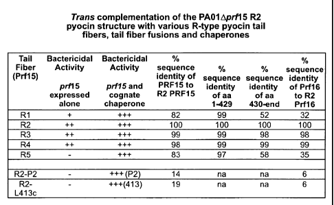
Figure 8 shows the trans complementation of the PAO1Aprf15 R2 pyocin
structure with various R-type pyocin tail fibers, tail fiber fusions and
chaperones. Activities
of the R1 through R5 complemented pyocins were assessed by spotting onto
indicator strain
Pseudomonas aeruginosa 13s, which is sensitive to all pyocin types. The R2-P2
complemented pyocins were tested for activity using E. coli C as the
indicator, and the R2-L-
413c complemented pyocin was tested on Yersinia pestis strain KIM.
The R2, R3, and R4 Prf15 tail fibers could be complemented by the
endogenous Prf16 of the PAO1Aprf15 R2 pyocin. R1 and R5 Prf15 tail fibers,
which differ at
the C-terminus compared to R2, required, for maximal activity, their own
cognate Prfl 6
(which itself differs from the R2 counterpart). Both the R2-P2 and R2-L-413c
fusions, which
contain the C-terminus (RBD) of the phage P2 and L-413c tail fibers,
respectively, require
their cognate tail fiber assembly chaperones encoded by gene G of the phage.
Figure 9 shows the pyocin tail fiber and chaperone expression vector
pUCP3OT. The genes, prf15 and prf16, are expressed using a Pseudomonas/E. coli
shuttle
vector (Schweitzer) with replication origins (on i pR01600, rep, and oriT) for
both species.
Cloning sites are shown by the indicated restriction enzyme sites of cleavage.
The plasmid
confers gentamicin resistance (Gm R) and is maintained by adding gentamicin to
the culture
media. Transcription of both genes is driven by the tac promoter which is
negatively
regulated by lacIQ. When transformed into Pseudomonas aeruginosa strain PA01
Aprf15,
the genes, e.g. prf15 and prfI6, incorporated into the plasmid are expressed
in trans after
being induced with IPTG simultaneously with the mitomycin C induction of those
pyocin
genes remaining in the PA01 Aprf15 host production bacteria.
Figure 10 provides the construction of Yersinia pestis specific pyocin tail
fiber. Similar to the strategy that was used to construct R2-P2, the C-
terminal (RBD)
encoding portion of the L-413c tail fiber gene was fused to an N-terminal
portion (BPAR) of
the R2 tail fiber. When expressed in trans to complement the R2 tail fiber
deletion strain
13
CA 02652450 2008-11-14
WO 2007/134303
PCT/US2007/068908
PAO1Aprf15, modified pyocin particles are produced containing the chimeric R2-
L-413c tail
fibers that can efficiently kill Y. pestis but not Pseudomonas.
Figure 11 provides the amino acid sequences or nucleic acid sequences for
SEQ ID NOS:1-59, provided on pages 11A-11J.
DEFINITIONS
As used herein, an hmw bacteriocin includes an R-type pyocin, tail-like
bacteriocin, R-type bacteriocin, F-type and R-type pyocins, monocins,
meningocins, or other
high molecular weight (hmw) bacteriocins. An hmw bacteriocin includes modified
versions
of R-type and F-type pyocins, enterocoliticins, monocins, and meningocins (see
Kingsbury
"Bacteriocin production by strains of Neisseria meningitidis." J Bacteriol.
91(5):1696-9,
1966). A modified or engineered hmw bacteriocin may be a modified R-type
pyocin selected
from the R1, R2, R3, R4, or R5 pyocin of P. aeruginosa. A bacteriocin of the
disclosure may
be thermolabile, mild acid resistant, trypsin resistant, sedimentable by
centrifugation at about
65,000 x g, and resolvable by electron microscope (see Jabrane et al. Appl.
Environ.
Microbiol. 68:5704-5710, 2002; Daw et al. Micron 27:467-479, 1996; Bradley
Bacteriol.
Revs. 31:230-314, 1967; and Kageyama et al. Life Sciences 9:471-476, 1962. In
many cases,
an engineered hmw bacteriocin disclosed herein has one or more, in any
combination, of
these properties. An additional property common to bacteriocins and engineered
hmw
bacteriocins disclosed herein is that they do not contain nucleic acid and
thus are replication
deficient such that they cannot reproduce themselves after or during the
killing of a target
bacterium as can many bacteriophages.
Pyocins, and other hmw bacteriocins disclosed herein, are complex molecules
comprising multiple protein, or polypeptide, subunits and resemble the tail
structures of
bacteriophages of the myoviridae family. In naturally occurring pyocins, the
subunit
structures are encoded by the bacterial genome such as that of P. aeruginosa
and form
pyocins to serve as natural defenses against other bacteria (Kageyama, 1975).
A sensitive,
target bacterium can be killed by a single pyocin molecule (Kageyama, 1964;
Shinomiya &
Shiga, 1979; Morse et al., 1980; Strauch et al., 2001).
A "target bacterium" or "target bacteria" refer to a bacterium or bacteria
that
are bound by an engineered hmw bacteriocin of the disclosure and/or whose
growth, survival,
or replication is inhibited thereby. The term "growth inhibition" or
variations thereof refers
14
CA 02652450 2008-11-14
WO 2007/134303
PCT/US2007/068908
to the slowing or stopping of the rate of a bacteria cell's division or
cessation of bacterial cell
division, or to death of the bacteria.
As used herein, a "nucleic acid" typically refers to deoxyribonucleotide or
ribonucleotides polymers (pure or mixed) in single-or double-stranded form.
The term may
encompass nucleic acids containing nucleotide analogs or modified backbone
residues or
linkages, which are synthetic, naturally occurring, and non-naturally
occurring, which have
similar binding, structural, or functional properties as the reference nucleic
acid, and which
are metabolized in a manner similar to the reference nucleotides. Non-limiting
examples of
such analogs include, without limitation, phosphorothioates, phosphoramidates,
methyl
phosphonates, chiral-methyl phosphonates, 2-0-methyl ribonucleotides, and
peptide-nucleic
acids (PNAs). The term nucleic acid may, in some contexts, be used
interchangeably with
gene, cDNA, mRNA, oligonucleotide, and polynucleotide.
A particular nucleic acid sequence also encompasses conservatively modified
variants thereof (such as degenerate codon substitutions) and complementary
sequences, as
well as the sequence explicitly indicated. Specifically, degenerate codon
substitutions may
be achieved by generating sequences in which the third ("wobble") position of
one or more
selected (or all) codons is substituted with mixed-base and/or deoxyinosine
residues. Thus a
nucleic acid sequence encoding a protein sequence disclosed herein also
encompasses
modified variants thereof as described herein.
The terms "polypeptide", "peptide", and "protein" are typically used
interchangeably herein to refer to a polymer of amino acid residues. Amino
acids may be
referred to herein by either their commonly known three letter symbols or by
the one-letter
symbols recommended by the IUPAC-IUB Biochemical Nomenclature Commission.
Virulence factors are those molecules that contribute to the pathogenicity of
an
organism but not its general viability. Upon the loss of a virulence factor
the organism is less
pathogenic but not necessarily less viable. Virulence factors may have any one
of numerous
functions, for example, regulating gene expression, providing adhesion or
mobility, pumping
out antibiotic agents, or forming protective coatings including biofilms.
Fitness factors are those molecules that contribute to the organism's general
viability, growth rate or competitiveness in its environment. Upon the loss of
a fitness factor,
the organism is less viable or competitive and because of this compromise,
indirectly less
pathogenic. Fitness factors may also possess any one of numerous functions,
for example,
acquiring nutrients, ions or water, forming components or protectants of cell
membranes or
CA 02652450 2008-11-14
WO 2007/134303
PCT/US2007/068908
cell walls, replicating, repairing or mutagenizing nucleic acids, providing
defense from or
offense towards environmental or competitive insults.
Some virulence and fitness factors are present on the surface of the bacterium
and thereby accessible to an hmw bacteriocin disclosed herein. By binding to
some surface
virulence or fitness factors, an hmw bacteriocin can mediate killing by
puncturing the cell
membranes, compromising the integrity of the cytoplasmic membrane and/or
dissipating the
membrane potential of the cell. Those surface accessible molecules most likely
to support
hmw bacteriocin binding and killing are proteins, polysaccharides, and
lipopolysaccharides of
the outer membrane. Accordingly, potential targets for engineered hmw
bacteriocins are
virulence factors and fitness factors that are proteins, polysaccharides and
lipopolysaccharides of the outer membrane. Some non-limiting examples of
virulence factor
targets for engineered pyocins include intramembrane cleaving protease (iCLIP)
metalloproteases; IL and IIL galactose- and fucose-binding lectins; microbial
surface
components recognizing adhesive matrix molecule (MSCRAMM) proteins; and
adhesin, such
as ACE.
The ultimate success of targeting a specific virulence factor depends on its
topography on the bacterial surface, its density on the surface, perhaps its
two-dimensional
mobility within the outer membrane, and its prevalence in clinical or field
isolates of the
pathogen. For example, OprM is a porin-like outer membrane protein involved in
multiple
efflux pumps, e.g. the MexAB system, and prevalent in many gram-negative
bacteria (Wong
and Hancock, 2000). To1C, similar to OprM, is a required accessory protein for
many efflux
pumps of gram-negative pathogens (Koronakis et al., 2004; Piddock, 2006). In
addition,
several members of the YcrC family of secretins are outer membrane proteins
necessary for
the translocation of pathogenic effector proteins by the type three secretion
system ("T3 SS"),
on which many gram-negative pathogens such as P. aeruginosa and Yersinia
pestis are
dependent for intoxicating their mammalian host (Galan and Collmer. 1999;
Koster et al.,
1997; Cornelis, 2006). In addition, the YscW family members are lipoproteins
also anchored
in the outer membrane to assist the insertion of the secretins into the
membrane (Burghout et
al., 2004).
Additional non-limiting examples of virulence and fitness factors include an
aquaporin, such as the E. colt aquaporin-Z water channel (see Calamita, 2000);
RetS (see
Goodman et al., 2004; and Zolfaghar et al., 2005); members of the 7TMR-DISM
family (see
Anantharaman et al., 2003); OprM (see Wong et al., 2000; and SEQ ID NO:11);
bacterial
proteins such as OprJ (SEQ ID NO:12), OprN (SEQ ID NO:13), AprF (SEQ ID
NO:14),
16
CA 02652450 2012-05-14
OpmM (SEQ ID NO:15), OpmA (SEQ ID NO:16), OpmD (SEQ ID NO:17), OpmE (SEQ ID
NO:18), OpmQ (SE ID NO:35), OpmB (SEQ ID NO:36), Opmj (SEQ ID NO:37), OpmG
(SEQ ID NO:38), OpmI (SEQ ID NO:39), OpmH (SEQ ID NO:40), OpmK (SEQ ID NO:41),
OpmN (SEQ ID NO:42), OpmF (SEQ ID NO:43), or OpmL (SEQ ID NO:44); OprD family
of porins (see Tamber et al., 2006); ACE, or the E. faecalis 0G1RF encoded ACE
gene (see
Sreedhar et al., 2000; and Rich, et al., 1999); PA-IL and PA-IIL galactose-
and fucose-
binding lectins (see Mitchell et al., 2002); plant and animal virulence genes
described by He
et al., 2004; extracellular pyrophosphate moieties (see Bonev et al., 2004);
metalloproteases
(see Rudner et al., 1999); and transposon encoded surface molecules (see
Jacobs 'et al., 2003).
Other non-limiting examples of virulence factors targeted by a disclosed
engineered lunw bacteriocin include those encoded by the open reading frames
(ORFs)
disclosed in U.S. Patent 6,355,411 and WO 99/27129,
In some embodiments, a factor targeted by a bacteriocin
disclosed herein is one encoded by the following ORFs from the U.S. patent:
I Unknown
= I
. 9.1 Unknown .
21 Possib.ly receptor
. 23.1..F.9*.b!1'..!sPc.P::irsP90.r . . . .
. . . =
. .41. 1.p.o..1ibly mucin_ Ilkq.
43 I Unknown . . . . .
. 51.1 Unknown =
53 Poisiblymucin like
89 L Possibly lipp.protgin recpptpr.. _ _
91 ; Unknown
Q ..! _Pcp.ibly.proteppho.sphogly.can, cell_qtyface...
1071 Possitti.ABC
110 I Possibly_membra9a_glyposyltransferase
113 Possibly multidru_g_resistance_protein Me*
. .
132 Possibly muc d
I- - 134 I Possibly 6-UDP mannose dehydrogInase
. 149 I Possibly MDR transnrter_pot9ntial.tamet_____.1
150 I Possibly multidrug. resistance protein Me*
17
CA 02652450 2008-11-14
WO 2007/134303
PCT/US2007/068908
203 Possibly ABC transporter ATPase component
204 Possibly ATPase component of ABC transport
205 Possibly ATPase component of ABC transport
206 Possibly ATPase component of ABC transport
207 Possibly ATPase component of ABC transport
208 Possibly ATPase component of ABC transport
1
209 Possibly ABC
Possibly NhaP-type Na-'-/H+ and K+/H+
213 antiporters
215 Unknown
227 Possibly receptor
239 Possibly deoxycytidine triphosphate deaminase
241 Possibly UTPase
249 Unknown
255 Unknown
261 Possibly 6-phosphoglyconate dehydrogenase
263 Possibly ABC transporter
273 Unknown
277 Possibly PE-PGRS family member
289 Possibly 6-phosphogluconate dehydrogenase
291 Possibly Glycosyl transferase
297 Possibly ligA
301 Possibly glycosyltransferase
309 Possibly cation/multidrug efflux pump
323 Unknown
327 Unknown
;
331 Possibly sensor with putative PiIR kinase
333 Possibly To protein transport
341 Possibly Pil R
349 Possibly Pil A or R
363 Possibly orfz
365 Possibly ABC transporter
375 Possibly mucin
377 Possibly fimT pilus
381 Possibly H1 immobilization antigen
383 Possibly fimU
387 Possibly PilV pilus
393 Possibly pilW et
401 Possibly pil X
18
CA 02652450 2008-11-14
WO 2007/134303
PCT/US2007/068908
403 Possibly antigen cd3
411 Unknown
413 Unknown
419 Possibly pil E
421 Possibly pyl y2
427 Possibly PE-PGRS outer membrane antigen
437 Possibly ABC ligA ..
DETAILED DESCRIPTION OF MODES OF PRACTICING THE DISCLOSURE
General
Hmw bacteriocins have the ability to quickly kill bacteria. A few early
reports
of in vivo studies have shown that they can be effective in mice for this
application (Haas et
al., 1974; Merrikin and Terry, 1972). The inventors have recently determined
that wild type
R2 pyocin can rescue mice from acute peritonitis caused by antibiotic-
resistant Pseudomonas
aeruginosa when administered either intraperitonealy or intravenously and that
R2 pyocins
can act at very low doses, such as 109 pyocins or less than 1 ptg total
protein in a single dose
(data not shown).
For hmw bacteriocins to be clinically useful as antibacterial agents, however,
the problem of their narrow bactericidal spectra must be addressed. While this
can be viewed
as an advantage in that it is possible to specifically target a particular
species or strain without
affecting the normal flora, the types of species/strains that are sensitive to
known bacteriocins
are limited. For example, pyocins currently are known to be produced by some
Pseudomonas
aeruginosa strains, and have activity against a narrow range of other
Pseudomonas strains
and a few other gram negative species. R-type bacteriocins from other species
have been
reported (such as Erwinia, see Jabrane 2002, and Yersinia enterocolitica, see
Strauch) but the
occurrence appears to be limited. Myoviridae phages, on the other hand, are
quite
widespread and common and are found throughout the bacterial class.
This disclosure demonstrates that it is possible to change the spectrum of a
pyocin and so any hmw bacteriocin. A major spectrum determinant among both
pyocins and
their related phages lies in the tail fiber, which binds to the bacterial
surface specifically,
interacting through its C-terminal portion (RBD) with a component of the LPS
or other cell
surface structure. The LPS can be highly variable between different species,
and strains of
bacteria, and bacteriophage tail fibers are themselves highly variable,
particularly in this C-
terminal region that interacts with the cell surface (Tetart, Desplats,). This
variability
19
CA 02652450 2008-11-14
WO 2007/134303
PCT/US2007/068908
apparently reflects phages' constant adaptations to changing host surfaces. It
has been
observed that different phage types that infect the same host (E. coli phages
P2, Mu, and Pl)
have sequence similarity in the C-terminal portion of the tail fiber (Haggard-
Ljungquist E,
Halling C, Calendar R.), indicating that horizontal transfer in these genetic
regions likely
plays a role in host specificity. For example, R2 pyocin has a very high
degree of sequence
similarity to Pseudomonas phage phiCTX, a phage that is also very closely
related to E. coli
phage P2. Comparing the tail fiber sequences of the R2 pyocin and P2, more
sequence
similarity is seen at the N-terminus (BPAR) than with the C-terminus (RBD),
suggesting that
the C-terminus plays the role in host specificity.
As disclosed herein, it is possible to alter the target spectrum of a pyocin
or
other hmw bacteriocin by engineering the C-terminal portion of the tail fiber
gene. It is
notable that this spectrum change can occur across species barriers,
demonstrating that
natural R-type pyocins and other natural hmw bacteriocins can be modified as
disclosed
herein and developed into antimicrobials with broader spectra.
Modified hmw bacteriocins
The disclosure provides engineered hmw bacteriocins with altered binding
specificities and/or affinities. In some embodiments, an hmw bacteriocin of
the disclosure
specifically binds to exposed surface molecules that act as virulence factors
or fitness factors
of pathogenic bacteria. The term "specifically (or selectively) binds" refers
to a binding
reaction that is determinative of the presence of the bound ligand, often in a
heterogeneous
population of proteins and other biological matter. As a result, the
engineered hmw
bacteriocin once bound specifically can generically kill the pathogenic
bacteria.
Furthermore, in order to become resistant to the engineered hmw bacteriocin,
the targeted
pathogenic bacteria must lose its recognition or binding site for the hmw
bacteriocin. Stated
differently, if the modified hmw bacteriocin specifically and exclusively uses
the virulence or
fitness factor as its receptor, the bacteria would be forced to lose its
virulence or fitness in
order to escape killing by the engineered hmw bacteriocin.
A modified hmw bacteriocin of the disclosure resembles a bacteriophage tail
but comprises a binding capability, or receptor binding domain (RBD), that has
been changed
relative to an unmodified, naturally occurring, or native bacteriocin. The RBD
may be
changed in amino acid sequence by use of recombinant DNA techniques as
described herein.
The term "recombinant", typically used with reference to a cell, or nucleic
acid, protein, or
vector, indicates that the cell, nucleic acid, protein or vector, has been
modified by the
CA 02652450 2008-11-14
WO 2007/134303
PCT/US2007/068908
introduction of a heterologous nucleic acid or protein or the alteration of a
native nucleic acid
or protein, or that the cell is derived from a cell so modified. So a
recombinant cell expresses
genes that are not found within the native (non-recombinant) form of the cell
or expresses
native genes that are abnormally expressed, under expressed, or not expressed
at all.
In many embodiments, the RBD may be modified to be that of a tail fiber from
another bacteriocin or a bacteriophage. As one non-limiting example disclosed
herein, the
RBD of R2 pyocin is modified by fusing the C-terminal portion of the tail
fiber protein
(RBD) of a phage (that infects a different host) to the N-terminal portion
(BPAR) of the R2
tail fiber protein. By fusing the C-terminus of the P2 tail fiber to the R2
PRF15 and co-
expressing the P2 cognate chaperone, the target bacteria spectrum of the R2 is
changed to kill
E. coli C. See Figure 2.
In additional embodiments, hmw bacteriocins are engineered otherwise. The
disclosure includes an hmw bacteriocin designed or selected to recognize, or
target, a surface
molecule of a bacterium (such as a pathogenic bacterium). The surface molecule
may be
considered a receptor on a bacterium recognized, or bound, by the lunw
bacteriocin.
The disclosure is based on the properties of an hmw bacteriocin tail fiber to
bind to, or interact with, a receptor to form a binding pair. The binding or
interaction occurs
through the RBD of the tail fiber, which is the first member of the binding
pair, with the
receptor being the second member of the pair. In many embodiments, the
receptor is a
bacterial cell surface molecule or portion thereof In other embodiments, the
receptor is a
molecule with properties of a virulence or fitness factor of a pathogenic
bacterium.
A modified or engineered hmw bacteriocin disclosed herein comprises a tail
fiber having both a base plate attachment region (BPAR) and a modified, or
heterologous,
RBD. As described herein, the tail fiber is a trimeric structure of three tail
fiber protein
subunits, each of which also comprises a first domain corresponding to, and
forming, the
BPAR in a tail fiber and a second domain corresponding to, and forming, a
modified or
heterologous RBD in a tail fiber.
Typically, "heterologous" when used with reference to portions of a protein or
nucleic acid sequence indicates that the sequence comprises two or more
subsequences that
are not usually found in the same relationship to each other in nature. For
instance, a
heterologous protein indicates that the protein comprises two or more
subsequences that are
not found in the same relationship to each other in nature. "Heterologous"
also means that
the amino acid or nucleic acid sequence is not normally found in conjunction
with the other
sequences or is not normally contained in the selected plasmid, vector, or
host. In other
21
CA 02652450 2008-11-14
WO 2007/134303
PCT/US2007/068908
words, it is not native to the system for which it is now utilized. For
example, proteins
produced by an organism that is not the wild type source of those proteins.
So in many embodiments, the disclosure includes an hmw bacteriocin tail fiber
protein comprising a BPAR of the protein and a modified, or heterologous, RBD.
The BPAR
is typically at the N-terminal region of a tail fiber protein, while the RBD
is typically at the
C-terminal region. Other than the modified, or heterologous, RBD, the tail
fiber protein may
be that of any naturally occurring hmw bacteriocin, with a pyocin, monocin,
enterocoliticin,
or meningocin being non-limiting examples. In some embodiments, the tail fiber
protein of
Rl-pyocin, R2-pyocin, R3-pyocin, R4-pyocin, and R5-pyocin, as represented by
SEQ ID
NO:1, 3, 5, 7, 9, respectively, may be used as described herein. In additional
embodiments,
the tail fiber protein may be that or those of theOCTX phage SEQ ID NO:45, or
that of
phage PS17 SEQ ID NO:19 or that of the VHML bacteriophage SEQ ID NO:21 and 22.
Embodiments of the disclosure include combinations of an hmw bacteriocin
tail fiber protein BPAR and a RBD from a bacteriophage tail fiber protein, as
shown in
Figure 3. In some cases, a combination may include the N-terminal amino acids
from
position 1 to about position 164 or position 240 of a bacteriocin tail fiber
protein. This
polypeptide fragment may be fused to a region of a bacteriophage tail fiber
protein including
its C-terminal portion containing an RBD. The region may be a polypeptide
fragment
lacking the N-terminal region from position 1 to about position 150, about
position 170,
about position 190, about position 290, about position 300, or about position
320.
Using the R2 pyocin and the P2 phage tail fiber protein as non-limiting
examples, the BPAR containing fragment may include the N-terminal amino acids
from
position 1 to position 164 or 240. See Figures 4-7. The RBD containing
fragment may
include the C-terminal, and from about 347 to about 755 amino acids in length
of the P2 or
related phage tail fiber proteins. The fusion may be readily prepared by
recombinant DNA
techniques with nucleic acid sequences encoding the R2 tail fiber protein,
such as prf15, and
the P2 phage gene H encoding its tail fiber protein. The cognate chaperone of
the RBD needs
to be co-expressed with the fusion tail fiber genes in order to ensure the
assembly of the
modified tail fibers into a functioning pyocin structure. See Figure 8.
In other embodiments, a modified RBD comprises a change in the amino acid
sequence of the RBD relative to a naturally occurring RBD or relative to the
BPAR present in
the tail fiber protein. Non-limiting examples of a change in amino acid
sequence include
substitution, insertion (or addition), or deletion of one or more amino acids.
22
CA 02652450 2012-05-14
In embodiments comprising the substitution of RBD amino acid residues,
about 1%, about 2 /0, about 3%, about 4%, about 5%, about 6%, about 7%, about
8%, about
9%, about 10%, about 11%, about 12%, about 13%, about .14%, about 15%, about
16%,
about 17%, about 18%, about 19%, about 20%, about 22%, about 24%, about 26%,
about
28%, about 30%, about 35%, about 40%, about 45%, or about 50%, or more, of the
C-
terminal in a tail fiber protein are substituted. In some embodiments, the
substitutions an
within about 245, about 260, about 275, or about 290, or more, residues from
the C-terminal.
The positions for substitution maybe any one or more, in any combination,
within that region. Exemplary positions include, but are not limited to, 448,
449, 452, 453,
454, 455, 459, 460, 462, 463, 464, 469, 472, 473, 474, 475, 478, 480, 484,
485, 486, 491,
494, 496, 497, 498, 499, 505, 506, 507, 508, 510, 512, 514, 517, 518, 519,
520, 521, 523,
527, 528, 530, 531, 533, 535, 537, 538, 541, 543, 546, 548, 561, 603, 604,
605, 606, 610,
618, 621, 624, 626, 627, 628, 629, 631, 632, 633, 638, 641, 642, 645, 646,
647, 648, 649,
650, 651, 652, 653, 654, 655, 657, 659, 663, 664, 665, 666, 667, 668, 669,
670, 671, 672,
673, 674, 675, 676, 677, 678, 679, 680, 681, 682, 683, 684, 685, 686, 687,
688, 689, and 691,
as well as any combination thereof, in SEQ ID NO:1, 3, 5,7, or 9. In some
embodiments, the
substitution is conservative as described herein. In other embodiments, the
substitution is
with a non-conservative substitution.
In further embodiments, insertions and deletions of amino acid residues within
the same region at the C-terminal of a tail fiber protein may be made.
RBD from bacteriophages
Other sources of RBD's include, but are not limited to, T-4 and other T-even
or pseudoT-even phages, phages T-3 and T-7, T-7 super-group of phages, phage
Mu, phage
P22, phage L-413c, and lambdoid phages.
RBD from diversification
In further embodiments, a tail fiber protein comprises a substitution with, or
insertion of, an RBD derived from an organism that diversifies the structure
by deploying a
Diversity Generating Retroelement (DGR), as described in published Patent
Application US
The major tropism determinant (Mtd) of Bordetella bacteriophage BPP-1 is one
such
structure. The sequence of Mtd is represented by SEQ ID NO:24 as disclosed
herein. In
other embodiments, the substitution is with part of the Mtd sequence, such as,
but not limited
23
CA 02652450 2008-11-14
WO 2007/134303
PCT/US2007/068908
to, the region from residue 49 to 381, from residue 171 to 381, or from
residues 306 to 381,
of SEQ ID NO:24. The insertion of the Mtd sequence, or any fragment thereof
(such as those
listed above), to the end of a tail fiber protein, such as after position 691
of SEQ ID NO:3, is
within the embodiments disclosed herein. The substitution of the Mtd sequence,
or any
fragment thereof (such as those listed above), may be for any non-BPAR region
of a tail fiber
protein. Non-limiting examples include the region of SEQ ID NO:1, 3, 5, 7, or
9 beginning at
about position 643, 625, 562, 448, 428, 231, and 163 through to the C-terminus
of the
sequence (see Figures 4-7 for exemplifications of these substitutions).
As described herein, the Mtd sequence in a tail fiber may be diversified to
produce a plurality of modified or heterologous RBDs. The nucleic acid
sequence encoding
Mtd comprises a variable region (VR) which may be operatively linked, in cis
or in trans, to
a template region (TR) such that the TR is a template sequence that directs
site-specific
mutagenesis of the VR. The operative linkage between the VR and TR regions
also includes
an operative linkage to sequences encoding a reverse transcriptase (RT)
activity, which may
be present in trans relative to the VR. Sites of variability in the VR of Mtd
correspond to
adenine residues in the generally homologous template region, TR, which itself
is invariant
and essential for sequence alterations in the VR. So while an initial molecule
may contain a
TR that is identical to the VR, the adenine residues present in the TR will
result in the
mutagenesis or diversification of the corresponding positions in the VR
sequence. So if the
TR sequence is a perfect direct repeat of the sequence in the VR,
diversification of the VR
region results in one or more adenine residues in the VR, also found in the
TR, being mutated
to another nucleotide, that is cytosine, thymine or guanine, without change in
the TR
sequence. This system may be used to alter the VR region, and thus the RBD, of
a modified
tail fiber protein as described herein.
Upon diversification, the tail fiber protein may be varied such that the
resultant RBD has at least 80%, at least 85%, at least 90%, or at least 95%
homology to the
major tropism determinant (Mtd) of Bordetella bacteriophage BPP-1, as
represented by SEQ
ID NO:24. As described herein, the tail fiber protein and Mtd combination may
be a
substitution, or an insertion, of an Mtd sequence or portion thereof into the
tail fiber protein
sequence. Thus the resultant tail fiber protein may be viewed as comprising a
substitution or
insertion with a binding domain with at least 80%, at least 85%, at least 90%,
or at least 95%
homology as recited above.
A nucleic acid molecule encoding a tail fiber and Mtd combination may be
used for diversification and sequence variation. Thus nucleic acid
combinations of sequences
24
CA 02652450 2008-11-14
WO 2007/134303
PCT/US2007/068908
encoding all or part of a tail fiber protein, and all or part of an Mtd, are
within the disclosed
embodiments. Other embodiments include nucleic acid molecules encoding any
tail fiber
protein with a modified or heterologous RBD as disclosed herein. In some
embodiments, the
encoded modified or heterologous RBD comprises a change in the amino acid
sequence of
the RBD relative to a naturally occurring RBD or relative to the BPAR present
in the tail
fiber protein as described above.
In additional embodiments, a tail fiber protein encoding nucleic acid molecule
may be made available for diversification to form a modified tail fiber
protein disclosed
herein. The nucleic acid molecule, under control of a suitable promoter, is
operatively placed
5' to an atd-TR-brt region. The TR sequence may be referred to as TR' and
prepared based
upon the VR sequence as discussed below. The resulting nucleic acid construct
may carry a
deletion of the transcription terminator structure upstream of the atd.
A region of the nucleic acid molecule encoding the C-terminal end of the tail
fiber protein as described above, is selected to be the VR and then
operatively linked to a TR'
sequence containing adenine residues at positions, that when varied, direct
amino acid
changes in the sequence encoded by the VR. Such adenine residues may be
deliberately
designed to be the first or second position of codons within the VR. The TR'
sequence can
initially be identical to the selected VR followed by site directed
mutagenesis or de novo
nucleic acid synthesis to prepare a TR' sequence that contains adenine
residues at the
corresponding positions to direct mutagenesis and diversification in the
encoded tail fiber
protein.
Preparation and use of hmw bacteriocins
The nucleic acid molecules described herein may be used to express and
prepare tail fiber proteins, including modified or engineered proteins, by any
means known to
the skilled person. In some embodiments, the expression is via the use of a
vector containing
the nucleic acid molecule operably linked to a promoter that can direct the
expression of the
encoded tail fiber protein.
In many embodiments, the expression may occur with expression of an
accessory gene, such as a "chaperone" encoding sequence reported for various
bacteriocins
and bacteriophages. The presence of a chaperone facilitates assembly of an hmw
bacteriocin
of the disclosure without necessarily becoming a part of the bacteriocin. The
chaperone may
be the cognate, or corresponding, protein for the RBD used in an hmw
bacteriocin of the
disclosure such as shown in Figure 8. One non-limiting example of a chaperone
is encoded
CA 02652450 2008-11-14
WO 2007/134303
PCT/US2007/068908
by R2 prf16 (SEQ ID NO:4), and it corresponds to (or is the cognate chaperone
for) the R2
pyocin tail fiber protein encoded by prf15 (SEQ ID NO:3). Other examples
include gene G
in the P2 (SEQ ID NO:26), gene G on L-413c (SEQ ID NO:29), the cognate
chaperone, SEQ
ID NO: 20, for the PS17 tail fiber, and the Orf 38 (SEQ ID NO:23) in VHML
bacteriophages
which are cognate chaperones to the tail fiber genes in each phage. These
genes are
homologues to the phage T4 gp38 (SEQ ID NO:32), which is known to be
responsible for
proper folding of the T4 tail fiber (SEQ ID NO:31) into trimers (Burda, Qu,
Hash
emolhosseni).
The use of a cognate chaperone is advantageous because a non-cognate
chaperone may be insufficient to correctly fold a given tail fiber protein
and/or assemble it
into an hmw bacteriocin, as shown in Figure 8. As a non-limiting example, the
R2 prf16
gene product has been observed to be insufficient to complement the folding of
a modified
tail fiber compromising an R2 BPAR fused to a P2 BRD portion of a tail fiber.
Without
being bound by theory, and offered to improve the understanding of the present
disclosure, it
is believed that a chaperone may act specifically on the C-terminal portion of
its cognate tail
fiber protein and that the tail fibers and their chaperones have co-evolved.
However, Qu et
al. isolated a T4 gp37 tail fiber mutant that suppresses the requirement for
gp38, its cognate
chaperone. This mutant had in gp37 a duplication of a coiled-coil motif, which
may itself
play a role in folding. Therefore, it is further believed that a tail fiber
protein may be
designed to contain such a change so that it folds properly without the need
to co-express a
cognate chaperone.
Therefore, embodiments of the disclosure include a bacterial cell transfected
with a nucleic acid molecule encoding a modified or engineered tail fiber
protein, optionally
co-expressed with a chaperone, as described herein. Expression of the nucleic
acid molecule,
optionally with an accessory (chaperone) protein, results in the production of
modified or
engineered tail fibers of the disclosure. The disclosure also includes
expression of more than
modified or engineered tail fiber protein through the use of more than one
nucleic acid
molecule to result in mixed homotrimeric tail fibers or even heterotrimeric
tail fibers.
Additionally, sequences encoding the tail fiber protein and chaperone may be
contained
within a single nucleic acid molecule, such as a plasmid or other vector, or
by separate
molecules. Where a single nucleic acid molecule is used, the sequences
optionally may be
under the control of the same regulatory sequence(s). Alternatively, the
coding sequences
may be under separate regulatory control.
26
CA 02652450 2008-11-14
WO 2007/134303
PCT/US2007/068908
In some embodiments, the bacterial cell is also capable of expressing the
additional subunits to form an lunw bacteriocin comprising a modified or
engineered tail
fiber. In one group of embodiments, the endogenous tail fiber protein coding
sequence of the
bacterial cell is inactivated or deleted. Optionally, the other subunits may
be encoded by
sequences on a nucleic acid molecule, such as a plasmid or other vector,
separate from that
which contains a sequence encoding a tail fiber protein and/or chaperone. Thus
the tail fiber
protein and/or chaperone may be provided one or more nucleic acid molecules in
trans
relative to the other subunits.
The nucleic acids, vectors, and bacterial cells may be used in a method of
producing a modified or engineered hmw bacteriocin as disclosed herein. Such a
method
may comprise culturing bacterial cells containing nucleic acid molecules as
described above
under conditions resulting in the expression and production of the tail fiber
and hmw
bacteriocin. In some embodiments of the disclosure the conditions are in vivo
within an
animal.
In one group of embodiments, a method of preparing an hmw bacteriocin
comprises expressing the bacteriocin subunits, including the modified or
engineered tail fiber
protein, in a host bacterium, and harvesting the hmw bacteriocin from the
bacterial culture.
The host bacterium is a complementary host production bacterium that encodes
and expresses
the other subunits necessary for the production of the bacteriocin. The term
"host bacterium"
or "host bacteria" refers to a bacterium or bacteria used to produce an hmw
bacteriocin
disclosed herein. Host bacteria or bacterium may also be referred to as "host
production
bacterium" or "host production bacteria". The "harvesting an hmw bacteriocin
from a
bacterial culture" generally comprises removing the bacteriocin from the host
bacterial
culture.
In an alternative group of embodiments, a method of preparing an hmw
bacteriocin with a modified tail fiber as described herein is provided. The
method may
comprise preparing a nucleic acid molecule encoding a modified tail fiber
protein by any
means disclosed herein and expressing the nucleic acid molecule in a cell
under conditions
wherein an hmw bacteriocin is produced.
Embodiments of the disclosure include an hmw bacteriocin comprising a tail
fiber protein as described herein. In one group of embodiments, the
bacteriocin comprises a
tail fiber protein comprised in part of the amino acid sequence represented by
SEQ ID NO:1,
3, 5, 7, 9. In other embodiments, the bacteriocin is a modified or engineered
pyocin,
monocin, enterocoliticin, or meningocin comprising a tail fiber with a
heterologous modified
27
CA 02652450 2008-11-14
WO 2007/134303
PCT/US2007/068908
RBD. In many embodiments, the heterologous modified RBD binds a bacterial
virulence or
fitness factor.
In further embodiments, engineered hmw bacteriocins with multivalent tail
fibers are disclosed. Mtd of Bordetella bronchiseptica bacteriophage BPP-1 has
been found
by X-ray crystallographic analysis to be a highly intertwined pyramidal
homotrimer with the
three sets of twelve variable amino acid residues forming three rather flat
receptor-binding
sites at the pyramid's base and located in a convergently evolved C-type
lectin ("CTL")
domain. Comparison of the structures of five Mtd variants at 1.5 angstrom
resolution showed
that the main chain conformation of variable residues is structurally
invariant, with inserts in
the CTL and trimeric assembly both contributing to formation of a static
scaffold for
combinatorial display of variable residues, thereby minimizing the incidence
of protein
misfolding (McMahon et al., 2005). Thus a single tail fiber may be generated
to contain
three properly folded mixed monomers since the structures of the variant Mtd
fibers are
identical except for the non-interacting, solvent-exposed twelve amino acid
residues.
The structure of the dominant Mtd-P1 variant bound to its receptor, the
Bordetella virulence factor pertactin, has also been solved by crystallography
and
characterized. One of the monomers of Mtd binds to onestructural domain on
pertactin; a
second identical monomer of the same Mtd binds a different, non-symmetrical
structural
domain of the same (monomeric) pertactin molecule; a third Mtd monomer remains
unbound.
The above variant Mtd structures and the binding interaction between Mtd and
its target, pertactin, may be applied to the design and selection of
multivalent tail fibers. For
example, it is evident that an Mtd monomer can exhibit affinities for two
different structural
domains and yet in multimeric format possess sufficient avidity to effect
functional phage
binding and infection. Furthermore, not all monomers of a fiber need be bound
to a receptor
to provide adequate avidity for phage binding and infection. These data and
conclusions
along with the knowledge that for at least T4 bacteriophages, also myoviridae,
only three
(homotrimeric) tail fibers need be bound to receptors to trigger tail sheath
contraction and
core penetration of bacterial membranes, indicates several means of generating
a multivalent
hmw bacteriocin. Such engineered multivalent hmw bacteriocins have broader
host ranges
and are capable of binding to more than a single virulence or fitness factor
even on the same
bacterial organism, thereby making it more difficult for targeted bacteria to
develop
resistance by mutational loss of expression of all targeted, relevant
receptors. An R-type
bacteriocin can be engineered to possess two independent sets of three
identical tail fibers,
the fibers of one set comprised of the same three non-identical monomers, and
the fibers of
28
CA 02652450 2008-11-14
WO 2007/134303
PCT/US2007/068908
the other set comprised of three different non-identical monomers. Each
monomer can
possess binding affinities for two different epitopes (e.g. two different
receptors), just as does
Mtd. Thereby any bacterium expressing any one or more of the 12 different
targeted receptor
molecules (2 "epitopes"/monomer times 3 monomers/tail fiber times 2 sets of
different tail
fibers/R-type bacteriocin equals 12 targeted receptors) would bind the
engineered multivalent
hmw bacteriocin and trigger its penetration of the membrane. Such engineered
hmw
bacteriocins have an unnaturally broad host range and, in addition, make it
highly unlikely
that a bacterium expressing more than a single targeted receptor could become
resistant to the
engineered hmw bacteriocins.
In other aspects, methods for the use of an hmw bacteriocin of the disclosure
are provided. In some embodiments, a method of compromising the integrity of
the
cytoplasmic membrane of a bacterium is disclosed. The method may comprise
contacting a
target bacterium with an hmw bacteriocin, or portion thereof, as disclosed
herein.
Alternatively, the contact may be with an lunw bacteriocin containing
composition disclosed
herein.
In one group of embodiments, the contacting occurs in vivo within a subject.
Thus a method of compromising the membrane integrity of a bacterium in a
subject is
disclosed. The method may comprise administering an hmw bacteriocin or a
portion thereof
as described herein to the subject. In another group of embodiments, the
contacting occurs in
vitro.
In yet additional embodiments, a method of forming non-virulent or unfit
bacteria progeny from virulent progenitor bacteria is provided. The method may
comprise
contacting virulent bacteria with an hmw bacteriocin which binds a virulence
or fitness factor
of said virulent progenitor bacteria as disclosed herein. The method then may
continue by
allowing selection of non-virulent bacteria progeny that no longer express the
virulence or
fitness factor.
In an alternative embodiment, a method of maintaining a population of non-
virulent bacteria is provided. The method may comprise contacting the
population with an
hmw bacteriocin which binds a virulence or fitness factor of virulent
bacteria. The method
then continues and prevents propagation of virulent bacteria. Without being
bound by
theory, and offered to improve the understanding of the disclosure, an
emergence of bacterial
resistance to an engineered hmw bacteriocin will be accompanied by a
compromised
virulence or fitness of the pathogenic bacteria.
29
CA 02652450 2008-11-14
WO 2007/134303
PCT/US2007/068908
The methods of the disclosure may also be applied in an environment where
bacterial growth is not desired or is considered to be harmful. Non-limiting
examples include
the sterilizing of environments, including medical settings and operating room
facilities; as
well as food preparation areas, including areas where raw meat or fish is
handled. The
methods may also be used to sterilize heat sensitive objects, medical devices,
and tissue
implants, including transplant organs.
The methods can be used as a stand-alone therapy or as an adjunctive therapy,
such as for the treatment of bacterial populations. Numerous antimicrobial
agents (including
antibiotics and chemotherapeutic agents) are known which would be useful in
combination
with these methods to treating bacteria-based conditions.
Target bacteria
The engineered hmw bacteriocins of the disclosure may be modified to target
a receptor on a variety of bacterial species and strains, including pathogenic
bacteria, such as
nosocomial or pyogenic bacteria, as non-limiting examples. In addition to
targeting the
virulence factors of select bacteria as described herein, bacteria that are
already susceptible to
bacteriophages are one non-limiting group of bacteria that may be inhibited by
an hmw
bacteriocin, such as an engineered pyocin, of the disclosure. These bacteria
include the gram
negative bacteria that are susceptible, as well as not sensitive, to naturally
occurring pyocins.
Additional non-limiting examples include gram negative bacteria as a group as
well as gram
positive bacteria. There are reports of bacteriocin-like entities in gram
positive bacteria
(Thompson & Pattee, 1981; Birmingham & Pattee, 1981; Zink et al., 1995). In
some
embodiments, the target bacterium is identified or diagnosed. Non-limiting
examples of such
bacteria include those of the genus Escherichia, Staphylococcus, Clostridium,
Acinetobacter,
Pseudomonas, or Streptococcus.
As a non-limiting example of targeting a virulence factor, the disclosure
includes the use of a phage tail fiber protein RBD like that of the gp37
protein from a T-
even-like or RB-69-like phage named AV17 that infects E. coli 0157:H7 but does
not infect
a mutant strain derived therefrom that has lost the 0157 antigen. (See Yoichi
et al., 2005)
The binding of this phage appears to require the presence of the 0157 antigen,
a virulence
factor, involved in gut adhesion of the pathogenic E. coli 0157:H7 organism.
Therefore, an
hmw bacteriocin of the disclosure may contain a modified tail fiber protein
containing the
RBD from the gp37 protein (SEQ ID N0:33) of the above described phage AV17
such that
CA 02652450 2008-11-14
WO 2007/134303
PCT/US2007/068908
the modified hmw bacteriocin targets a virulence factor, the 0157 antigen, of
E. coli
0157117. The cognate chaperone for the AV17 tail fiber has SEQ ID N0:34.
Other target bacteria include those responsible for topical or localized P.
aeruginosa infections in humans. An "infection" refers to growth of bacteria,
such as in a
subject or tissue or non-bacterial cell, wherein the bacteria actually or
potentially could cause
disease or a symptom in the subject, tissue or non-bacterial cell. Treatment
of an infection
may include prophylactic treatment of substances or materials. Non-limiting
examples
include donated organs, tissues, and cells; medical equipment, like a
respirator or dialysis
machine; or wounds, such as those during or after surgery. Other uses include
the removal of
target bacteria which may cause problems upon further growth. In additional
embodiments,
an hmw bacteriocin is used to treat plants or harvested parts of plants with
bacterial infections
or contaminations, or to treat environmental occurrences of the target
bacteria, such as in a
hospital or commercial setting.
The disclosure provides for the treatment, by administration or contact with
an
hmw bacteriocin disclosed herein to target the bacteria, of such infections in
tissues and
subjects as follows. The infections include the common infections of the
cornea ("keratitis"
and corneal ulcers), at least two-thirds of which are caused by P. aeruginosa.
Approximately
30% of these pathogens are reported to be resistant to multiple antibiotics
(Mah-Sadorra et
al., 2005). Bacterial infection of the cornea is considered a relatively
uncommon but serious
condition requiring urgent medical attention because of the potential for
reduced vision or
even vision loss in the affected eye(s). Other common infections which may be
treated, and
are caused by antibiotic-resistant P. aeruginosa, include ear infections, e.g.
"swimmer's ear"
(Roland & Stroman, 2002), those secondary to severe burns and wounds (Holder,
1993), and
cystic fibrosis. Cystic fibrosis is consistently aggravated by chronic,
antibiotic-resistant
infections caused by P. aeruginosa and its close relative, Burkholderia
cepacia (Govan &
Deretic, 1996), and these pathogens in cystic fibrosis may be treated by use
of an engineered
hmw bacteriocin. Because bacteriocins like pyocins will tolerate freeze-drying
(Higerd et al.,
1969), the disclosure includes a freeze-dried formulation of a bacteriocin for
administration
to enhance the likelihood of successful delivery to the upper and/or lower
airway of the
respiratory tract.
As described herein, the treatment of a subject is typically treatment of "a
subject in need of treatment". The determination, or diagnosis, of the need
for treatment may
be made by a skilled person, such as a clinician, by use of art recognized
means. In some
31
CA 02652450 2008-11-14
WO 2007/134303
PCT/US2007/068908
embodiments, the subject is an animal or plant with a bacterial infection that
is potentially
life-threatening or that impairs health or shortens the lifespan of the
organism.
In additional embodiments, a method to kill or inhibit the growth of bacteria
in
a biofilm is provided. Such a method may comprise contacting a biofilm with an
hmw
bacteriocin disclosed herein which targets bacteria in the biofilm.
As described herein, an anti-bacterial hmw bacteriocin is used to inhibit
growth, survival, or replication of a particular bacterium. The bacterium may
be a pathogenic
or environmentally deleterious strain, or may be treated in a prophylactic
manner. A
pathogenic microorganism generally causes disease, sometimes only in
particular
circumstances.
The bacteria may also be that of a nosocomial (hospital derived) infection,
environmental bacteria, and pyogenic (pus forming) bacteria. The methods and
compositions
of the disclosure can be used to inhibit growth of nosocomial bacteria,
including bacteria that
populate a typical hospital environment, or bacteria that are present on human
skin or in the
human gastrointestinal tract, or bacteria that infect and form pus in wounds.
Nosocomial
infections are infections which become evident during a hospital stay or are
related to a
procedure performed in a hospital. These procedure-related infections often
become evident
after patients are discharged from the hospital. The most common nosocomial
bacterial
infections are urinary tract infections, surgical-site infections, pneumonia,
C. difficile
associated diarrhea and pseudomembrane colitis, and serious systemic
infections in which
bacteria can be grown from blood.
The methods and compositions of the disclosure may be used to inhibit growth
of gram negative or gram positive bacteria. Non-limiting examples of gram
positive bacteria
include Staphylococcus (pyogenic), Enterococcus (opportunistic),
Streptococcus,
Enterococcus, Bacillus, Micrococcus, Mycobacterium, Corynebacterium, and
Clostridium.
Non-limiting examples of gram negative bacteria include Pseudomonas
(pyogenic), E. coli
(opportunistic), Salmonella (opportunistic), Campylobacter (opportunistic),
Proteus
(pyogenic), Klebsiella (opportunistic), Enterobacter (pyogenic), Citrobacter
(pyogenic),
gram negative non-fermenter rods (such as Acinetobacter), and Shigella. The
pyogenic cocci
are spherical bacteria that cause various suppurative (pus-producing)
infections in animals.
Included are the gram-positive cocci Staphylococcus aureus, Streptococcus pyo
genes, and
Streptococcus pneumoniae, and the gram-negative cocci, Neisseria gonorrhoeae,
and N.
meningitidis.
32
CA 02652450 2008-11-14
WO 2007/134303
PCT/US2007/068908
In additional embodiments, the disclosed methods and compositions of the
disclosure are used to inhibit growth, particularly of antibiotic resistant
bacteria. Non-
limiting examples include numerous bacterial pathogens that have become multi-
drug
resistant (MDR).
Engineering pyocins as a non-limiting representative example
Francois Jacob discovered and first described pyocins as high molecular
weight bacteriocins (Jacob, 1954). Similar bacteriocin-like entities have been
described in
multiple other gram negative bacteria (Coetzee et al., 1968) as well as in
Listeria
moncytogenes (Zink et al. 1995) and Staphylococcus aureus (Thompson and
Pattee, 1981),
both of which are gram positive organisms. While pyocins morphologically
resemble the
tails of contractile (myoviridae) bacteriophages, they are not simple
defective phages; there
are meaningful differences. For example, differences exist in physical and
chemical stability
between pyocins and phage tails (Kageyama & Egami, 1962; Nakayama et al.,
2000).
While the host ranges of pyocins are relatively narrow and usually restricted
to
strains of the same species, there are exceptions (Morse et al, 1976;
Blackwell et al., 1982).
On the other hand, myoviridae bacteriophages can exhibit broad host ranges,
and their host
ranges, like those of pyocins, are determined by the binding specificities of
the tips of their
tail fibers (Tetart et al., 2001).
For numerous phage tail fibers, the distal (3'-terminal) third of the gene
varies
in mutants or variants with altered phage host ranges, or "tropisms"
(Ackermann, 2003). As
a non-limiting example, the major tropism determinant (Mtd), the receptor
binding protein of
Bordetella bacteriophage BPP-1, varies greatly in sequence (Liu et al., 2004;
Doulatov et al.
2004). Variation in Mtd depends on a phage-encoded retroelement (Diversity
Generating
Retroelement, or DGR) that belongs to a family of DGRs implicated in
generating sequence
variation in various phage and bacterial genomes. The Bordetella DGR can
produce more
than 1013 different sequence variants of Mtd, rivaling the 1014-1016 possible
sequences of
antibodies. Mtd variants are produced by a unique adenine-specific mutagenesis
process
involving DGR-encoded reverse transcriptase (bRT) and a stable template region
(TR).
Variability in Mtd is focused to 12 adenine-encoded amino acids that are
scattered across its
C-terminal variable region (VR) (Doulatov et al. 2004). The 3-dimensional
crystal structures
of numerous Bordetella Mtd variants have been solved and confirm, as described
below, that
the tip of the structure determines the binding specificity and thereby the
major tropism (host
33
CA 02652450 2008-11-14
WO 2007/134303
PCT/US2007/068908
range) of the phage (McMahon et al., 2005). Thus, as further described below,
Mtd and its
related DGR system may be used in the practice of the disclosure.
Many Pseudomonas species possess the genes for the R-type pyocins (Takeya
et al., 1969; Kageyama, 1975). The R-type pyocin locus consists of about 16
complementation groups including about 10 structural genes plus regulatory and
chaperone
genes (Shinomiya et al. 1983a; Shinomiya et al., 1983b). Morphologically and
genetically the
R-type pyocins resemble the tails of myoviridae bacteriophages but have no
head structure
and thus no nucleic acid content (Kageyama, 1964; Ishii et al., 1965; Shimizu
et al., 1982).
They are thought to have evolved from the phage tail structure of a P2-related
ancestor, but
they are not simple defective phages, having been further adapted for their
role as defensive
bactericidal agents (Nakayama et al, 2000). Similar to bacteriophages,
however, pyocins do
bind to specific molecular "receptors" on target bacteria and penetrate their
membranes with
a "core" or needle-like structure (Uratani & Hoshino, 1984). As an immediate
consequence
of the core penetration of the membranes, the bacterium is killed by
compromise of the
integrity of its cytoplasmic membrane and dissipation of its membrane
potential, a
bactericidal event that can result from an attack by a single pyocin (Iijima,
1978; Uratani &
Hoshino, 1984; Strauch et al., 2001).
The RBD, or Receptor Binding Determinant of R-pyocin binding, of a typical
R-type pyocin binds to a bacterial surface molecule. In the case of an R2
pyocin isolate, the
RBD resides in the carboxy-terminal portion of its tail fiber. The tail fiber
is a homotrimer of
the product of the prfl 5 gene (Nakayama et al., 2000). Modification of the
RBD in the prf15
gene and recombination of the modified prf15 gene into a system that produces
R-type
pyocins allows production of an engineered pyocin with modified binding
specificity.
The major tropism determinant (Mtd) of Bordetella bacteriophage possesses
several unique and useful properties as a binding domain. The functional form
of Mtd in
Bordetella bacteriophage is a homotrimer that binds the virulence factor
protein, pertactin, in
Bordetella. Thus, the mtd gene may be fused to the distal end of the prf15
gene to take
advantage of the Mtd properties. So as described herein, an aspect of the
disclosure includes
construction of a fusion protein between the P. aeruginosa R-type pyocin tail
fiber protein
(Prfl 5) and the major tropism determinant (Mtd) of Bordetella phage, BPP-1. A
Prf15-Mtd
fusion may be used to complement in trans a P. aeruginosa PA01 pyocin prf15 -
deletion to
bind and kill pertactin-expressing Bordetella bronchiseptica or pertactin-
expressing E. coli.
Additionally, the P2 or P4 bacteriophage may be used as a surrogate to harbor
the prf15-mtd tail fiber fusion gene such that the genotype is coupled to the
binding
34
CA 02652450 2008-11-14
WO 2007/134303
PCT/US2007/068908
phenotype of the tail fiber. This permits efficient transduction, selection,
and isolation of the
tail fiber gene encoding the desired RBD.
Modes of administration
An engineered hmw bacteriocin of the disclosure may be administered by any
suitable means. Non-limiting examples include topical, or localized,
administration as well
as pulmonary (inhalation), gastrointestinal, by catheter or drip tube, or
systemic
administration to a subject. Representative, and non-limiting, examples of
systemic
administration include intraperitoneal and intravenous administration. The
protective effects
of intraperitoneally and intravenously administered pyocins have been
demonstrated in mice
infected systemically with lethal doses P. aeruginosa strains sensitive in
vitro to the
administered pyocins (Merrikin & Terry, 1972; Haas et al., 1974). In some
embodiments,
contact between an hmw bacteriocin disclosed herein and a target bacterial
population results
in a decrease in the population of at least 10, at least 100, at least 1000,
or at least 10,000, or
more, fold decrease relative to the absence of the bacteriocin. In other
embodiments, the
contact may result in a decrease in detectability of the bacteria by at least
5, at least 10, at
least 20, at least 30, at least 40, or at least 50, or more, fold relative to
the absence of the
bacteriocin.
An engineered hmw bacteriocin of the disclosure may be administered to any
subject afflicted with, diagnosed as afflicted with, or suspected of being
afflicted with, an
infection or contamination by bacteria susceptible to the hmw bacteriocin. Non-
limiting
examples of such a subject include animal (mammalian, reptilian, amphibian,
avian, and fish)
species as well as insects, plants and fungi. Representative, and non-
limiting, examples of
mammalian species include humans; non-human primates; agriculturally relevant
species
such as cattle, pigs, goats, and sheep; rodents, such as mice and rats;
mammals for
companionship, display, or show, such as dogs, cats, guinea pigs, rabbits, and
horses; and
mammals for work, such as dogs and horses. Representative, and non-limiting,
examples of
avian species include chickens, ducks, geese, and birds for companionship or
show, such as
parrots and parakeets. An animal subject treated with an engineered
bacteriocin of the
disclosure may also be a quadruped, a biped, an aquatic animal, a vertebrate,
or an
invertebrate, including insects.
In some embodiments, the subject to be treated is a human child or other
young animal which has yet to reach maturity. Thus the disclosure includes the
treatment of
CA 02652450 2008-11-14
WO 2007/134303
PCT/US2007/068908
pediatric conditions comprising infection with bacteria or other microorganism
susceptible to
an hmw bacteriocin of the disclosure.
The disclosure also provides for the treatment or prevention of an
opportunistic infection, such as that resulting from an undesirable growth of
bacteria that are
present in the microbial flora of a human subject or a non-human animal. An
opportunistic
infection may be the result of an immunosuppressed condition in a subject or
the result of
antibiotic treatment that alter the commensal flora of the genitourinary (GU)
or
gastrointestinal (GI) tract. Thus the disclosure also provides for the
treatment or prophylaxis
of immunosuppressed subjects and subjects exposed to other pharmaceutical
agents. An
hmw bacteriocin with its anti-bacterial activity may be used in combination
with another anti-
bacterial or anti-microbial agent, such as an antibiotic or anti-fungal agent
as non-limiting
examples. An "anti microbial agent" is an agent or compound that can be used
to inhibit the
growth of, or to kill, single celled organisms. Anti-microbial agents include
antibiotics,
chemotherapeutic agents, antibodies (with or without complement), chemical
inhibitors of
DNA, RNA, protein, lipid, or cell wall synthesis or functions.
In some embodiments, an hmw bacteriocin is formulated with a
"pharmaceutically acceptable" excipient or carrier. Such a component is one
that is suitable
for use with humans, animals, and/or plants without undue adverse side
effects. Non-limiting
examples of adverse side effects include toxicity, irritation, and/or allergic
response. The
excipient or carrier is typically one that is commensurate with a reasonable
benefit/risk ratio.
In many embodiments, the carrier or excipient is suitable for topical or
systemic
administration. Non-limiting pharmaceutically carriers include sterile aqueous
or non-
aqueous solutions, suspensions, and emulsions. Examples include, but are not
limited to,
standard pharmaceutical excipients such as a phosphate buffered saline
solution, water,
emulsions such as oil/water emulsion, and various types of wetting agents.
Examples of non-
aqueous solvents are propylene glycol, polyethylene glycol, vegetable oils
such as olive oil,
and injectable organic esters such as ethyloleate. Aqueous carriers include
water,
alcoholic/aqueous solutions, emulsions or suspensions, including saline and
buffered media.
Parenteral vehicles include sodium chloride solution, Ringer's dextrose,
dextrose and sodium
chloride, lactated Ringer's or fixed oils. Intravenous vehicles include fluid
and nutrient
replenishers, electrolyte replenishers (such as those based on Ringer's
dextrose), and the like.
Additional formulations and pharmaceutical compositions disclosed herein
comprise an isolated hmw bacteriocin specific for a bacterial host; a mixture
of two, three,
five, ten, or twenty or more bacteriocins that target the same bacterial
hosts; and a mixture of
36
CA 02652450 2008-11-14
WO 2007/134303
PCT/US2007/068908
two, three, five, ten, or twenty or more bacteriocins that target different
bacterial hosts or
different strains of the same bacterial host.
Optionally, a composition comprising an hmw bacteriocin of the disclosure
may also be lyophilized using means well known in the art. Subsequent
reconstitution and
use may be practiced as known in the field.
Also provided are formulations comprising microencapsulated hinw
bacteriocin. In some embodiments, these may provide sustained release kinetics
or allow oral
ingestion to pass through the stomach and into the small or large intestine.
In general, the
pharmaceutical compositions can be prepared in various forms, such as
granules, tablets,
pills, suppositories, capsules (e. g. adapted for oral delivery), microbeads,
microspheres,
liposomes, suspensions, salves, pastes, lotions, and the like. Pharmaceutical
grade organic or
inorganic carriers and/or diluents suitable for oral and topical use can be
used to make up
compositions comprising the therapeutically-active compounds. Stabilizing
agents, wetting
and emulsifying agents, salts for varying the osmotic pressure, or buffers for
securing an
adequate pH value may be included.
An hmw bacteriocin is typically used in an amount or concentration that is
"safe and effective", which refers to a quantity that is sufficient to produce
a desired
therapeutic response without undue adverse side effects like those described
above. An hmw
bacteriocin may also be used in an amount or concentration that is
"therapeutically effective",
which refers to an amount effective to yield a desired therapeutic response,
such as, but not
limited to, an amount effective to slow the rate of bacterial cell division,
or to cause cessation
of bacterial cell division, or to cause death or decrease rate of population
growth of the
bacteria. The safe and effective amount or therapeutically effective amount
will vary with
various factors but may be readily determined by the skilled practitioner
without undue
experimentation. Non-limiting examples of factors include the particular
condition being
treated, the physical condition of the subject, the type of subject being
treated, the duration of
the treatment, the nature of concurrent therapy (if any), and the specific
formulations
employed.
Additionally, and in anticipation of a possible emergence of bacterial
resistance to an engineered hmw bacteriocin, there can be a concomitant
compromise of the
organisms' virulence or fitness where the bacteriocin targets the virulence or
fitness factor of
the targeted bacteria. Because a major, but non-limiting, mechanism by which a
bacterium
may become resistant to an hmw bacteriocin is the loss of its receptor for the
bacteriocin, the
targeting of a virulence or fitness factor as disclosed herein provides many
advantages over
37
CA 02652450 2008-11-14
WO 2007/134303
PCT/US2007/068908
traditional antibiotics and bacteriophages. The resistance to traditional
antibiotics and
bacteriophages can result from many different mechanisms other than loss of
the receptor or
target molecule of the antibacterial agent. As non-limiting examples, an hmw
bacteriocin of
the disclosure would not be subject to a bacterial efflux pump to remove the
bacteriocin from
the cellular environment and would not be subject to a bacterial nucleic acid
deactivation
mechanism.
Having now generally described the inventive subject matter, the same will be
more readily understood through reference to the following examples which are
provided by
way of illustration, and are not intended to be limiting of the disclosure,
unless specified.
EXAMPLES
The following examples are offered to illustrate, but not to limit the claimed
subject matter.
Example 1: Modified hmw bacteriocins containing a fusion protein
a) Complementation system
To facilitate the preparation of a modified hmw bacteriocin as described
herein, construction of a system to complement tail fibers in trans was
established. Using the
R2 pyocin as a representative model, creation of a deletion of the R2 prf15
coding sequence
in the P. aeruginosa PA01 genome was used to create a platform in which a
complementing
tail fiber protein, such as a modified prf15 gene product, was expressed in
trans.
Generally, the deletion was made by the method of Hoang et al. to create P.
aeruginosa strain PA01 Aprf15. The prf16 coding region, SEQ ID NO:4, for the
R2
chaperone overlaps the end of the R2 prf15 gene by 8 nucleotides and the
ribosome binding
site lies within the prf15 coding region, SEQ ID NO:3. The Prfl6 protein,
which is not
necessarily incorporated into the pyocin structure, has been reported to be
required, for
maximum activity, for assembly of the trimeric tail fiber (Figure 8 and
Nakayama et al.,
2000). Therefore, both the transcription start site for prfl 6 and its
ribosome binding site were
left intact such that the chaperone would be produced upon induction of the
modified pyocin
construct encoding a "tail-less," defective pyocin.
Briefly, an in-frame deletion of codons 11-301 of PRF15 was made in PA01
as follows. A 1.1 kb KpnI-AgeI fragment upstream of the desired deletion was
amplified by
PCR from PA01 genomic DNA using primers AV085 (5'-
38
CA 02652450 2008-11-14
WO 2007/134303
PCT/US2007/068908
GCTTCAATGTGCAGCGTTTGC) (SEQ ID NO:46), and AV088 (5'-
GCCACACCGGTAGCGGAAAGGCCACCGTATTTCGGAGTAT) (SEQ ID NO:47), and a
2.2 kb AgeI-EcoRI fragment was amplified using primers AV087 (5'-
ATACTCCGAAATACGGTGGCCTTTCCGCTACCGGTGTGGC) (SEQ ID NO:48) and
AV086 (5'- TCCTTGAATTCCGCTTGCTGCCGAAGTTCTT) (SEQ ID NO:49). The
resulting restriction fragments were cloned into the Kpn/ and EcoR/ sites of
pEX18Gm
(Hoang et al) to make pEXGm-Aprf/5. The finished construct was transformed
into strain
PA01 by electroporation (Chuanchuen et al). Integrants were selected with 100
g/ml
gentamicin, and segregants were then selected in media containing 5 1,1g/m1
sucrose and
lacking NaCl and gentamicin. Deletion candidates were confirmed by PCR
analysis, pyocin
induction, and sequencing of a PCR-amplified fragment.
Strain PA01 Aprf15 grows similarly to its parent strain, PA01, and the pyocin
encoding genes remain inducible through the SOS response, leading to lysis of
the cell.
While there appears to be some production of pyocin gene products, it did not
appear that
stable "tail-less" pyocin particles were produced from PA01 Aprf15.
R2 pyocin Prf15 was expressed in trans by first cloning the coding sequence
into the broad host range Pseudomonas/E. coli shuttle vector, pUCP3OT. See
Figure 9. In
some initial constructs, transcription was driven constitutively or under lad
l control from the
tac promoter. But in other constructs, transcription was modified to be
regulated with an
endogenous prf15 promoter such that expression would be regulated through the
SOS
response. This permitted the expression of the modified prf15 gene to be
induced
synchronously with the expression of the other pyocin genes residing in the
PA01 Aprf15
genome.
Briefly, the broad host-range vector pUCP3OT (Schweizer, H.P et al) was
modified by filling in the unique BspHI site to form pUCP3OTABsp. A tad
promoter was
amplified by PCR from an Mtd expression vector (a gift from Jeffery F. Miller,
UCLA) using
primers AV110 (5'-TTTATTAGCGGAAGAGCCGACTGCACGGTGCACCAATG) (SEQ
ID NO:50) and AV114 (5'-CCCTCGAATTCATGAATACTGTTTCCTGTGTGAAATTG)
(SEQ ID NO:51), then cloned into pUCP3OTABsp to create pUCP-tac.
The R2 PRF15 coding region was amplified from a subclone using primers
AV118 (5'-CTTCCTTTCATGACGACCAATACTCCGAA) (SEQ ID NO:52) and AV116
(5'-ACCACGAATTCTTCATCGTCCAAATGCCTC) (SEQ ID NO:53), while R2 prf15 and
prf16 were amplified using primers AV118 and AV086 (5'-
39
CA 02652450 2008-11-14
WO 2007/134303
PCT/US2007/068908
TCCTTGAATTCCGCTTGCTGCCGAAGTTCTT) (SEQ ID NO:49). The amplified
fragments ofprf15 and prf15 /16 were cloned into pUCPtac digested with BspHI
and EcoRI
to yield pUCP-tac-prf15 and PUCP-tac- prf15 /16.
For expression using the endogenous prfl 5 promoter, prfl 5 and prfl 6 were
amplified together with the 1088 bp sequence upstream ofprf15 from a subclone
using
primers AV107 (5'- CACCATCTAGACAATACGAGAGCGACAAGTC) (SEQ ID NO:54)
and AV091 (5'- TCCTCAAGCTTACGTTGGTTACCGTAACGCCGTG) (SEQ ID NO:55)
and cloned into pUCP3OT digested with XbaI and HindIII to create pUCP-R2p-
prf/5 /16.
Bacteria in log phase suspension growth and containing the expression
plasmids were treated with 3 j.tg mitomycin C/ml to induce pyocin production.
Stable
pyocins were produced upon induction with yields similar to that of wild type
PA01. The
pyocins had the same bactericidal spectrum and level of activity as R2 pyocin
produced from
PA01. It seems that production of a stable pyocin complex requires the
expression of a tail
fiber protein in addition to expression of the other pyocin encoding genes,
and expression of
the tail fiber gene in trans is sufficient.
When prf15 was expressed constitutively from the tac promoter, cell growth
was markedly slower than when it was regulated by lad I or the endogenous
promoter.
Although it appears that production of PRF15 alone in the cell is detrimental,
yields of
pyocins generated from both promoters are comparable.
A plasmid construct was prepared from which R2 prfl 6 was co-expressed
with R2 prfl 5 to insure proper temporal expression prfl 6 for folding of
PRF15 expressed in
trans.
b) Recombinant lunw bacteriocins
As described herein, five different R-type pyocins, based on spectra and
termed R1-5, have been recognized. Because gene sequences encoding the tail
fiber proteins
were known only for R1 (SEQ ID NO:1) and R2 (SEQ ID NO:3), PCR was used to
isolate
and sequence the R3 (SEQ ID NO:5), R4 (SEQ ID NO:7), and R5 (SEQ ID NO:9)
pyocin tail
fiber genes along with their cognate chaperone encoding sequences present in
their producer
strains, SEQ ID NO:6, 8, and 10, respectively. The chaperone genes of pyocins
R1 and R2
were also cloned and sequenced, SEQ ID NO:2 and 4, respectively. To confirm
the
hypothesis that the tail fiber dictates spectra, the sequences encoding R1,
R3, R4, and R5
pyocin tail fiber proteins were obtained and expressed in trans in PA01 Aprf15
such that they
would be incorporated into the R2 pyocin structure. Each of the resulting
recombinant strains
CA 02652450 2008-11-14
WO 2007/134303
PCT/US2007/068908
was then induced to produce pyocins and the spectrum of each was determined by
spot
assays, as shown in Figures 2 and 8.
c) Fusion proteins as tail fiber proteins
A fusion of the R2 tail fiber prf15 gene and bacteriophage P2 gene H
sequences was created, expressed and used to produce additional modified hinw
bacteriocins
of the disclosure. Bacteriophage P2, which infects many E. coli strains, has a
tail fiber
encoding gene H, (SEQ ID NO:25) that has significant sequence similarity to R2
prf15 (SEQ
ID NO:3), particularly at the N-terminus-encoding portion. The portion of gene
H encoding
the C-terminal 551 amino acid residues of the P2 tail fiber protein, which is
the putative
region conferring target specificity (RBD), was fused to the portion ofprf15
encoding the
164 amino acid N-terminal baseplate-binding (BPAR) portion of R2 PRF15 to
encode a
modified tail fiber protein (SEQ ID NO:27).
Bacteriophage P2 also encodes a putative tail fiber chaperone, encoded by
gene G (SEQ ID NO:26), similar to that encoded by R2 pyocin prf16 (SEQ ID
NO:4), and
the chaperones of many of the other myoviridae phages. Because it is likely
that the gene G
encoded chaperone is important for folding the C-terminal portion of the P2
tail protein in the
fusion, constructs were made to co-express P2 gene G.
The portion of R-2 prfl 5 encoding amino acids 1-164 was amplified from a
subclone using primers AV118 and AV127 (5'-
TTCTTTAAGCTTTTCCTTCACCCAGTCCTG) (SEQ ID NO:56) and was digested with
BspHI and HindIII. The protion of P2 gene H encoding amino acids from position
158-669
was amplified from a P2 phage stock (Richard Calendar) using primers AV124 (5'-
CCTCCTGAATTCTTATTGCGGCATTTCCG) (SEQ ID NO:57) and AV126 (5'-
TCCTTCGAATTCTTACACCTGCGCAACGT) (SEQ ID NO:58). P2 gene H158-669 plus
gene G was amplified using primers AV124 and AV125 (5'-
CCTCCTGAATTCTTATTGCGGCATTTCCG) (SEQ ID NO: 59). Each of the PCR
products from P2 were digested with HindIII and EcoRI. pUCP-tac-R2-P2H was
created by
cloning the prf15 fragment encoding the 1-164 amino acid fragment together
with the P2
gene H fragment encoding the 158-669 amino acid fragment into pUCP-tac
digested with
BspHI and EcoRI. pUCP-tac-R2-P2HG was generated by cloning the prf15 fragment
encoding the 1-164 amino acid fragment together with the P2 gene H fragment
encoding the
158-669 amino acid fragment plus gene G into pUCP-tac digested with BspHI and
EcoRI.
Briefly, PA01 Aprf15 was transformed with the prf15-P2 gene H fusion
constructs and pyocin production was induced with mitomycin C. Pyocin
particles were
41
CA 02652450 2008-11-14
WO 2007/134303
PCT/US2007/068908
purified and tested for activity by spot tests and by the bacterial survival
assay (see Figure 2).
The purified pyocin particles containing the R2-P2 fusion tail fiber had
bactericidal activity
against E. coli strain Cla but were incapable of killing P. aeruginosa strain
13s. Furthermore,
the expression of P2 gene G was needed to produce active pyocin. This supports
the
hypothesis that the chaperone is required for proper folding of the C-terminal
portion of the
tail fiber, as shown in Figure 8.
The abilities of a range of different R2-P2 tail fiber protein fusions to form
functional pyocins that kill E. coli Cla were explored by a series of
different R2-P2 fusions.
Representative examples of these fusions are shown in Figures 4-7, along with
the indication
of their bactericidal activities against E. coli Cla.
d) Additional fusions
An additional modified hmw bacteriocin has been produced to target pestis.
L-413c is a yersiniophage that infects most strains of Y. pestis (Richard
Calendar, personal
communication). Most of the L-413c genome is highly similar to P2 with the
notable
exception of the tail fiber gene H, SEQ ID NO:28, which has diverged
considerably. Without
being bound by theory, and offered to improve the understanding of the
disclosure, variation
in the tail fiber gene H, and thus the encoded protein, is the feature that
most likely accounts
for its differing host range.
The N-terminus of L-413c gene H (SEQ ID NO:28), however, shares
considerably sequence similarity to its P2 counterpart (SEQ ID NO:25), likely
due to its
function of baseplate binding. A fusion was constructed to create a fusion
tail fiber with the
N-terminal 1-164 amino acids from R2 PRF15 fused to the C terminal (positions
158-913)
portion of the L-413c tail fiber to create a modified tail fiber, as shown in
Figure 10 (SEQ ID
NO:30). The fusion was expressed in PA01 Aprf15 along with the L-413c tail
fiber cognate
chaperone, gene G (SEQ ID NO:29), as described above. After induction, the
produced
pyocin particles killed Y pestis KIM as well as E. coli C and thus have a
killing spectrum
analogous to the host range of yersiniophage L-413c. The modified pyocins did
not kill any
of the Pseudomonas strains.
A further modified hmw pyocin particle has been made with a novel fusion
tail fiber created between the P. aeruginosa R-type pyocin tail fiber BPAR
(encoded by
prf15) and the tail fiber gene orf34 and/or the RBD from the tail fiber gene
orf35 of VHML
(SEQ ID NO:21 AND 22, respectively). To create the pyocin particles, the VHML
cognate
chaperone gene (SEQ ID NO:23) was co-expressed with the modified tail fiber
fusion genes.
42
CA 02652450 2008-11-14
WO 2007/134303
PCT/US2007/068908
Pyocins with the modified tail fibers were formed and analyzed. The resulting
modified hmw
bacteriocin with the VHML-derived RBD can be subjected to diversification by
the natural
DGR of VHML.
The major tropism determinant (Mtd) of the Bordetella bacteriophage BPP-1
has a C-type lectin (CTL) domain, which serves as a binding determinant for
many different
types of molecules and in many different biological contexts (Drickamer, 1999;
McMahon et
al., 2005). In BPP-1, Mtd is incorporated as a homotrimeric globular domain
located at the
end of the phage tail, where it can bind to the surface protein pertactin, a
virulence factor
expressed on the outer surface of Bordetella bronchiseptica and Bordetella
pertussis (Liu et
al., 2004). In this context, Mtd is also the target of phage-mediated homing
mutagenesis,
which can result in the bacteriophage acquiring a novel binding determinant
for infecting its
ever changing host.
Recent structural studies on the Mtd domain and several of its diversified
variants, have shown how the trimeric fiber tip forms a rigid scaffold that
can contain more
than 10 trillion variant binding ligands (McMahon et al. 2005). Fusing the Mtd
domain onto
the pyocin tail fiber protein and then diversifying the Mtd domain using the
DGR system
described by Miller and colleagues (Liu et al., 2004; Doulatov et al., 2004),
creates a very
large library of variants, from which to select and obtain the genes encoding
pyocin tails with
altered binding specificity.
Example 2: Assays of fusion proteins
a) Pyocin purification and assays
PA01 and derivatives were grown shaking at 200 rpm at 37 C in G medium
supplemented with 50 g/m1 gentamicin when needed to maintain plasmids. When
the
cultures reached 0D600 of 0.250, mitomycin C was added to a final
concentration of 3
Cultures were incubated for an addition 2.5 hours until lysis occurred. Five
I of
DNase 1 was added, and the culture was allowed to incubate an additional 30
mins. Debris
was removed by centrifugation at 12,000 rpm in a Beckman MA-16.250 rotor for 1
hour.
Saturated ammonium sulfate was slowly added, at a rate of 1 ml/min, to the
supernatant
stirring on ice, to a final added volume of 65 ml per 100 ml of the
supernatant of the lysate.
This was stored at 4 C overnight. The ammonium sulfate precipitate was
collected by
centrifugation at 12,000 rpm for 1 hour, and the pellet was resuspended in 10
ml of TN50
buffer (10 mM tris, 50 mM NaC1, pH 7.5). Pyocin particles in the resuspended
solution were
43
CA 02652450 2008-11-14
WO 2007/134303
PCT/US2007/068908
then sedimented at high speed, 20,000 rpm (65,000 x g) in a Beckman JA-25.50
rotor for 1
hour, and resuspended in 3-5 ml of TN50 buffer. Pyocin preps were judged to be
>90% pure
by SDS polyacrylamide gel electrophoretic analysis.
Quantitative pyocin assays were performed by counting bacterial survival in a
slightly modified method as described by Kagayama et al., 1964. Pyocin samples
were
incubated with target bacteria (approximately lx109 CFU/ml) for 40 minutes at
37 C. The
samples were then diluted and plated to count survivors. The number of pyocin
particles is
related to the fraction of bacterial survivors in a Poisson distribution, m = -
1nS, where m = the
average number of lethal events/ cell and S is the fraction of survivors. The
total number of
active pyocin particles/ml = m x cells/ml. Strainl3s was the Pseudomonas
aeruginosa used
in these assays and is a clinical isolate resistant to many anitibiotics, but
sensitive to all 5 R-
type pyocins. The E. coli target was Cla, kindly provided by Richard Calendar.
Semi-quantitative assays were also performed by a spot method where pyocin
samples were serially diluted in TN50 buffer and spotted on lawns of target
bacteria. After
overnight incubation at 37 C, pyocin activity could be observed by a clear
zone of killing on
the lawn. Figure 2 shows representative results from this assay format.
Example 3: Recombinant bacteriophage to screen engineered tail fibers
The P4 bacteriophage is used as a surrogate to harbor the Prf15-based tail
fiber
fusion gene such that the genotype is coupled to the binding phenotype of the
tail fiber. This
allows efficient selection and transduction of the desired tail fiber gene.
Bacteriophage P2 is a temperate coliphage which can infect other enteric
species, and can replicate in, but not infect, P. aeruginosa (Bertani & Six,
1988; Kahn et al.,
1991). R-type pyocins are closely related genetically and structurally to P2,
and the P2 tail
fiber protein, encoded by gene H, shows homology to prf15 at the N-terminal
portion, where
base plate attachment occurs (Haggard-Ljungquist et al., 1992; Nakayama et
al., 2000).
Deploying the P2 or P4 bacteriophage as a surrogate phage, in which plasmid-
encoded tail
fibers are incorporated in place of the P2 phage-encoded fibers, permits the
display and
selection of fusion fibers in a context that closely resembles its intended
functional context in
the pyocin.
The tail fiber genotype can be physically coupled to the binding phenotype in
a transducing phage particle for genetic selection, similar to phage display
technology. When
a P2 phage with an amber mutation in its fiber protein gene (H) infects E.
coli harboring a
plasmid with a cos packaging site, which normally acts as a signal for
packaging
44
CA 02652450 2008-11-14
WO 2007/134303
PCT/US2007/068908
bacteriophage genomic DNA (Ziermann & Calendar, 1991; Kahn et al., 1991), it
will
package the cos-containing plasmid in the heads of newly synthesized P2 phage.
The
plasmid encodes and expresses prf/5-based tail fusion gene and confers
resistance to
gentamicin. The fusion tail fiber expressed from the plasmid in the P2
infected E. coli is
incorporated into the P2 bacteriophage in place of the defective (amber
truncated) gene H
product (P2 tail fiber protein). Upon lysis of the bacteria, the released P2-
based transducing
particles carry the cos-containing plasmid containing the prf/5-based tail
fusion gene rather
than the P2 genome and possess Prf15-fusion tail fibers rather than P2 tail
fibers.
Transducing phage particles with the ability to bind cells and trigger the
bacteriophage injection mechanism then confer gentamicin resistance to
successfully targeted
bacteria, from which the selected fiber fusion gene is isolated from the
plasmid after
replication of the bacteria under gentamicin selection. The tail fiber gene on
the plasmids is
easily further manipulated to create many fusion junctions and to diversify
the RBD in order
to redesign and optimize the function of the modified tail fiber RBD.
This approach also overcomes many of the difficulties imposed by C-terminal
display of a trimeric protein when using conventional phage display systems
(Held & Sidhu,
2004). Bacteriophage P2 has tail fibers that genetically and morphologically
resemble those
of pyocins (Nakayama et al., 2000). Tail fibers attach to the base plates of
P2 and pyocins
via their N-termini, and there is significant sequence similarity of the N-
termini of P2 and R2
pyocin tail fibers (Nakayama et al, 2000; Haggard-Ljungquist et al., 1992).
Furthermore, the
tail fiber gene of the P2-related phage, Psi 7, can complement the R-2 pyocin
tail fiber gene,
prf15 (Shinomiya, 1984; Shinomiya & Ina, 1989).
Alternatively, the RBD is directly fused to the N-terminal domain of the gene
H tail fiber, or the tail fiber genes of VHML phage of Vibrio harveyii (which
like BPP-1 also
contains a functioning DGR) is fused to the N-terminal domain of P2 gene H.
Example 4: Methods to recover the desired tail fiber gene
A P2, P4 or OCTX bacteriophage carrying an engineered tail fiber gene acts as
a surrogate to couple pyocin tail fiber genotype to binding phenotype. By
selecting or
screening for specific binding phenotypes from the diversified or mutagenized
libraries of the
tail fiber genes harbored in surrogate bacteriophages, one can isolate the
tail fiber genes that
encode a desired binding specificity. The selection may be carried out by
single or multiple
enrichment cycles of adsorbing the surrogate bacteriophages or transducing
particles onto
solid-phase target molecules, either by first removing undesired binders and
then isolating,
CA 02652450 2008-11-14
WO 2007/134303
PCT/US2007/068908
from among the remaining surrogates, those that bind to the intended target
molecules, or
visa versa. Alternatively, the selection may occur by applying either binding
step alone.
Ultimately, the surrogate exhibiting the desired binding phenotype can be
subject to DNA
extraction and isolation of the harbored tail fiber gene by cloning
restriction enzyme
fragments or by PCR reactions using oligonucleotide primers that bind specific
DNA
sequences peripheral to the diversified portion of the tail fiber gene.
The desired surrogate bacteriophage can be plaque purified on a lawn of
bacteria expressing the molecular target of interest, for example, a virulence
or fitness factor
by which the surrogate bacteriophages infect the host. The factor-expressing
bacteria may be
the pathogen of interest, e.g. Pseudomonas aeruginosa, or a non-pathogen, e.g.
E coli
engineered to express the targeted virulence or fitness factor. Replicate
plating or serial
plating techniques may be deployed to ensure that the surrogate bacteriophage
does not form
plaques on closely related bacterial strains that do not express the target
factor. For example,
insertional mutants of Pseudomonas aeruginosa that have lost expression of
specific
virulence factors can be used to screen or, to deplete by adsorption,
surrogate bacteriophages
before or after forming plaques on, or panning on, their virulence or fitness
factor-expressing
counterpart (isogenic) bacteria.
Even though the surrogate phages or transducing particles will not form
plaques on the target-expressing bacteria, the infected or transduced bacteria
will still acquire
antibiotic resistance along with the harbored plasmid or phasmid and therefore
can be
selectively grown and subsequently extracted to isolate the multi-copy plasmid
and its desired
tail fiber gene.
These techniques permit the identification and isolation of surrogate
bacteriophages or transducing particles exhibiting the desired, specific
binding phenotypes
from which to extract the desired, specific, unnatural lunw bacteriocin tail
fiber genes.
Furthermore, the binding of surrogates to mammalian molecules, cells or
tissues can be
deployed to deplete from the libraries any genes encoding tail fibers that
might cause adverse
events if incorporated into therapeutic hmw bacteriocins.
There is an available library of insertional mutant Pseudomonas aeruginosa
bacterial strains differing from highly pathogenic parental PA14 Pseudomonas
aeruginosa
only by the lack of expression of a series of specific virulence factors, one
missing from each
non-redundant, isogenic mutant (see the website at
ausubellab.mgh.harvard.edu/cgi-
bin/pal4/home.cgi). These isogenic mutant strains provide tools for ensuring
the specificity
of the surrogate bacteriophages for the targeted virulence factors and not for
other prevalent
46
CA 02652450 2008-11-14
WO 2007/134303
PCT/US2007/068908
surface molecules. For example, the population of surrogate P4 bacteriophages
can be
incubated with a high density culture of a Pseudomonas aeruginosa mutant
missing a
particular targeted virulence factor in order to adsorb and deplete from a
population of
surrogate bacteriophages or transducing particles, those that bind to surface
molecules present
on both the isogenic mutant and the virulent parental strain. The depleted
population will be
enriched in surrogates binding to the desired virulence factor. Once surrogate
bacteriophages
that do bind to and infect the bacteria expressing the particular virulence or
fitness factor are
isolated, each can be screened directly for its inability to infect the
isogenic mutant strain
lacking the targeted factor. The selected plasmid can be repackaged in
surrogate transducing
particles and recycled any number of times through the adsorption-depletion
and infection
process to further enrich and eventually purify the pUC-based plasmid encoding
the desired
tail fibers for targeting the virulence or fitness factor.
Example 5: Methods for producing engineered lunw bacteriocins
The modified tail fiber gene is recombined either (i) into a plasmid under a
regulated promoter for expression in production bacteria also harboring, for
example on a
bacterial artificial chromosome (BAC), the R-pyocin gene cluster (including
the endolysin
genes) from which the resident prtR, prtN, prf15 and holin (prf9 or PA0614)
genes have been
deleted or otherwise disabled, or (ii) into the pyocin cluster containing BAC
vector itself,
using a plasmid-mediated allelic exchange reaction.
Upon induction of the pyocin genes and the engineered tail fiber gene, such as
by inducing prtN directly via an engineered regulatable promoter such as lac
or tac, the host
cells synthesize pyocins until their nutrients are depleted and they cease
growing (Young, Ry,
2006). The producing bacteria do not lyse in the absence of chloroform,
because the holin
gene inactivation prevents cytoplasmic endolysin access to the bacterial cell
wall, as is
necessary for cell lysis. The exhausted cells are harvested by centrifugation
or filtration and
then frozen until one desires to harvest the soluble pyocins that have filled
the cellular
cytoplasm. Upon thawing, the inner cellular membrane ruptures, releasing
endolysin to lyse
the bacteria and thereby release the harvest of modified pyocins. The
disruption of the
bacterial membranes can be accelerated or completed if necessary by the
addition of small
quantities of chloroform to the aqueous solvent in which the bacterial paste
is thawed.
47
CA 02652450 2008-11-14
WO 2007/134303
PCT/US2007/068908
References:
Ackermann HW. 2003. Bacteriophage observations and evolution. Res
Microbiol. 154:245-251
Aiache JM, S Meski, E Beyssac, G Serpin. 1997. The formulation of drug for
ocular administration. J Biomater Appl. 11:329-48
Anantharaman et al. "Application of comparative genomics in the
identification and analysis of novel families of membrane-associated receptors
in bacteria."
BMC Genomics, 4:34, 2003
Bad Bugs, No Drugs: As Antibiotic Discovery Stagnates A Public Health
Crisis Brews, July 2004. Infectious Diseases Society of America
Beisel KW, LD Hazlett, RS Berk. 1983. Dominant susceptibility effect on the
murine corneal response to Pseudomonas aeruginosa. Proc Soc Exp Biol Med.
172:488-491
Bertani LE, and EW Six. 1988. The P2-like phages and their parasite, P4. In
R. Calendar (ed.), The Bacteriophages, vol. 2. Plenum Publishing Corp., New
York. pp 73-
143
Birmingham VA, PA Pattee. 1981. Genetic Transformation in Staphylococcus
aureus: Isolation and Characterization of a Competence-Conferring Factor from
Bacteriophage 80a Lysates. Journal of Bacteriology 148:301-307
Blackwell CC, and Law JA. 1981. Typing of non-serogroupable Neisseria
meningitidis by means of sensitivity to R-type pyocins of Pseudomonas
aeruginosa. J Infect.
3(4):370-8.
Blackwell CC, FP Winstanley, WA Telfer-Brunton. 1982. Sensitivity of
thermophilic campylobacters to R-type pyocins of Pseudomonas aeruginosa. J.
Med
Microbiology. 15:247-51
Bonev et al. "Targeting extracellular pyrophosphates underpins the high
selectivity of nisin." The FASEB Journal. 18:1862-1869, 2004
Burda MR, Miller S.Folding of coliphage T4 short tail fiber in vitro.
Analysing the role of a bacteriophage-encoded chaperone.
Eur J Biochem. 1999 Oct;265(2):771-8.
Burns RP. 1969. Pseudomonas aeruginosa keratitis: mixed infections of the
eye. Am J Ophthalmol. 67:257-262
Calamita, "The Escherichia coli aquaporin-Z water channel." Molecular
Microbiology 37(2):254-262, 2000
48
CA 02652450 2008-11-14
WO 2007/134303
PCT/US2007/068908
Chappell JD, AE Prota, TS Dermody, T Stehle. 2002. The crystal structure of
reovirus attachment protein al reveals evolutionary relationship to adenovirus
fiber. The
EMBO Journal 21:1-11
Cheng KH, SL Leung, HW Hoelcman. 1999. Incidence of contact lens-
associated microbial keratitis and its related morbidity. Lancet. 354:181-185
Choi HK, JB Gaynor, KG White, C Lopez, CM Bosio, RR Karkhoff-
Schweizer, HP Schweizer. 2005. A T-7 based broad-range bacterial cloning and
expression
vector. Nature Methods. 2:443-448
Chuanchuen, R, Narasaki, C.T. and Schweizer, H.P., Benchtop and
microcentrifuge preparation of Pseudomonas aeruginosa competent cells,
BioTechniques
33:760-763 (October 2002).
Coetzee HL, HC De Klerk, IN Coetzee, JA Smit. 1968. Bacteriophage-tail-
like particles associated with intra-species killing of Proteus vulgaris. J
Gen Virol. 2:29-36.
Cole N, MDP Willcox, SMJ Fleiszig. 1998. Different Strains Of Pseudomonas
Aeruginosa Isolated From Ocular Infections Or Inflammation Display Distinct
Corneal
Pathologies In An Animal Model. Curr Eye Res. 17:730-735
Cooper RL, IJ Constable. 1977. Infective keratitis in soft contact lens
wearers.
Br J Ophthalmol. 61:250-254
Cowell BA, C Wu, SMJ Fleiszig. 1999, Use of an Animal Model in Studies of
Bacterial Corneal Infection. Inst Lab Animal Res J. 40:43-50
Desplats C, Krisch HM. The diversity and evolution of the T4-type
bacteriophages. Res Microbiol. 2003 May;154(4):259-67.
Doulatov S, A Hodes, L Dai, N Mandhana, M Liu, R Deora, RWSimons, S
Zimmerly, JF Miller. 2004. Tropism switching in Bordetella bacteriophage
defines a family
of diversity-generating retroelements. Nature. 431:476-481
Drickamer K. 1999. C-type lectin-like domains. Current Opinion in Structural
Biology. 9:585-590 Farmer JJ, LG Herman. 1969.
Dyke J, Berk R S. Growth inhibition and pyocin receptor properties of
endotoxin from Pseudomonas aeruginosa. Proc Soc Exp Biol Med. 1974;145:1405-
1408.
Epidemiologic Fingerprinting of Pseudomonas aeruginosa by the Production
of and Sensitivity to Pyocin and Bacteriophage. Applied Microbiol. 18:760-765
Filiatrault MJ, Munson RS Jr, and Campagnari AA. 2001. Genetic analysis of
a pyocin-resistant lipooligosaccharide (LOS) mutant of Haemophilus ducreyi:
restoration of
full-length LOS restores pyocin sensitivity. J Bacteriol. 183(19):5756-61.
49
CA 02652450 2008-11-14
WO 2007/134303
PCT/US2007/068908
Fleiszig SMJ, DJ Evans. 2002. The pathogenesis of bacterial keratitis: studies
with Pseudomonas aeruginosa. Clin Exp Optom. 85.5:271-278
Gerke JR, MV Magliocco. 1971. Experimental Pseudomonas aeruginosa
infection of the mouse cornea. Infect Immun. 3:209 ¨216
Gillor, 0, LM Nigro, A. Riley. 2005. Genetically engineered bacteriocins and
their potential as the next generation of antimicrobials. Curr. Pharm. Des.
11:1067-1075
Goodman et al. "A Signaling Network Reciprocally Regulates Genes
Associated with Acute Infection and Chronic Persistence in Pseudomonas
aeruginosa."
Developmental Cell 7:745-754, 2004
Govan, JRW & V Deretic. 1996. Microbial Pathogenesis in Cystic Fibrosis:
Mucoid Pseudomonas aeruginosa and Burkholderia cepacia, Microbiological
Reviews.
60:539-574
Haas H, Sacks T, Saltz N. Protective effect of pyocin against lethal
Pseudomonas aeruginosa infections in mice. J Infect Dis. 1974 Apr;129(4):470-
2.
Haggard-Ljungquist E, Hailing C, Calendar R. NA sequences of the tail fiber
genes of bacteriophage P2: evidence for horizontal transfer of tail fiber
genes among
unrelated bacteriophages. J Bacteriol. 1992 Mar;174(5):1462-77.
Hashemolhosseini S, Montag D, Kramer L, Henning U.Determinants of
receptor specificity of coliphages of the T4 family. A chaperone alters the
host range.
J Mol Biol. 1994 Aug 26;241(4):524-33.
Hazlett LD, D Rosen, RS Berk. 1976. Experimental eye infections caused by
Pseudomonas aeruginosa. Ophthalmic Res. 8:311-318
He et al. "The broad host range pathogen Pseudomonas aeruginosa strain
PA14 carries two pathogenicity islands harboring plant and animal virulence
genes." PNAS
101:2530-2535, 2004
Held H, SS Sidhu. 2004. Comprehensive Mutational Analysis of the M13
Major Coat Protein, J Mol Biol. 340:587-97
Hensley S, B Wysocki. As Industry Profits Elsewhere, U.S. Lacks Vaccines,
Antibiotics, The Wall Street Journal November 8, 2005: p Al
Higerd TB, CA Baechler, RS Berk. 1969. Morphological Studies On Relaxed
and Contracted Forms of Purified Pyocin Particles. J. Bacteriology. 98:1378-89
Hoang,T.T., Karkhoff-Schweizer,R.R., Kutchma,A.J. and Schweizer,H.P., A
broad-host-range Flp-FRT recombination system for site-specific excision of
chromosomally-
CA 02652450 2008-11-14
WO 2007/134303
PCT/US2007/068908
located DNA sequences: application for isolation of unmarked Pseudomonas
aeruginosa
mutants Gene 212 (1), 77-86 (1998)
Hobden JA, DS Rootman, RJ O'Callaghan, JM Hill. 1988. Iontophoretic
application of tobramycin to uninfected and Pseudomonas aeruginosa-infected
rabbit corneas.
Antimicrob Agents Chemother. 32:978-981
Holder IA. 1993. Pseudomonas aeruginosa Burn Infections: Pathogenesis and
Treatment,. In M Campa, M Bendinelli, and H Friedman (ed.) Pseudomonas
aeruginosa as an
Opportunistic Pathogen. Plenum Press, New York, N.Y. pp. 275-295
Iijima M. 1978. Mode of Action of Pyocin Rl. J. Biochem (Tokyo) 83:395-
402
Ishii S, Y Nishi, and F Egami. 1965. The fine structure of a pyocin. J. Mol.
Biol. 13:428-431
Ito, S., Kagayama, M. and F. Egami. Isolation and characterization of pyocins
from several strains of Pseudomonas aeruginosa. J. Gen. Appl. Microbiol. 16
205-214
(1970).
Jabrane A, Sabri A,Compere P,Jacques P, Vandenberghe I,Van Beeumen J,
Thonart P Characterization of serracin P, a phage-tail-like bacteriocin, and
its activity against
Erwinia amylovora, the fire blight pathogen. Appl Environ Microbiol. 2002
Nov;68(11):5704-10.
Jacob F. 1954. Biosynthese induite et mode d'action d'une pyocin, antibiotique
de Pseudomonas pyocyanea. Annals Inst. Pasteur. 86:149-60
Jacobs et al. "Comprehensive transposon mutant library of Pseudomonas
aeruginosa." PNAS 100(24):14339-14344, 2003
Kageyama M, F Egami. 1962. On the purification and some properties of a
pyocin, a bacteriocin produced by Pseudomonas aeruginosa. Life Sciences 9: 471-
6
Kageyama M, K Ikeda, and F Egami. 1964. Studies of a pyocin. III.
Biological properties of the pyocin. J. Biochem. 55:59-64.
Kageyama M, Shimomiya T, Aihara Y, Kobayashi M. 1979. Chracterization
of a bacteriophage related to R-type pyocins. J Virol. 32:951-957.
Kageyama M. 1964. Studies of a pyocin I. Physical and chemical properties. J.
Biochem. 55:49-53
Kageyama, M. Bacteriocins and bacteriophages in Pseudomonas aeruginosa,
in: Microbial Drug Resistance, University Park Press, Baltimore. pp 291-305
1975.
51
CA 02652450 2008-11-14
WO 2007/134303
PCT/US2007/068908
Kahn ML, RG Ziermann, DW Deho, M Ow, G Sunshine, R Calendar. 1991.
Bacteriophage P2 and P4. Methods Enzymol. 204:264-280
Kumazaki T, Y Shimizu, SI Ishii. 1982. Isolation and Characterization of
Pyocin R1 Fibers. J. Biochemistry. 91:825-35
Lee EJ, DJ Evans and SMJ Fleiszig. 2003. Role of Pseudomonas aeruginosa
ExsA in Penetration through Corneal Epithelium in a Novel in vivo Model.
Investigative
Ophthalmology & Visual Science. 44:5220-5227
Lee FK, Dudas KC, Hanson JA, Nelson MB, LoVerde PT, Apicella MA.
1999 The R-type pyocin of Pseudomonas aeruginosa C is a bacteriophage tail-
like particle
that contains single-stranded DNA. Infect Immun. 67(2):717-25.
Liu M, M Gingery, SR. Doulatov, Y Liu, A Hodes, S Baker, P Davis, M
Simmonds, C Churcher, K Mungall, MA Quail, A Preston, ET Harvill, DJ Maskell,
FA
Eiserling, J Parkhill, and JF Miller. 2004. Genomic and Genetic Analysis of
Bordetella
Bacteriophages Encoding Reverse Transcriptase-Mediated Tropism-Switching
Cassettes. J.
Bacteriology. 186 476-481
Mah-Sadorra JH, SG Yavuz, D M Najjar, PR Laibson, CJ Rapuano, EJ Cohen.
2005. Trends in contact lens-related corneal ulcers. Cornea. 24:51-58
Matsui H, Sano Y, Ishihara H, Shinomiya T. Regulation of pyocin genes in
Pseudomonas aeruginosa by positive (prtN) and negative (prtR) regulatory
genes.
J Bacteriol. 1993 Mar;175(5):1257-63.
McMahon SA, JL Miller, JA Lawton, DE Kerkow, A Hodes, MA Marti-
Renom, S Doulatov, E Narayanan, A Sali, JF Miller, P Ghosh. 2005. The C-type
Lectin Fold
as an Evolutionary Solution for Massive Sequence Variation. Nature Struct. &
Molecular
Biol. 12:886 - 892
McNamara NA, KA Polse, SA Fukunaga, JS Maebori, RM Suzuki. 1998. Soft
lens extended wear affects epithelial barrier function. Ophthalmology.!
05:2330-2335
Meadow, P.M., and Wells P.L. Receptor sites for R-type pyocins and
bacteriophage E79 in the core part of the lipopolysaccharide of Pseudomonas
aeruginosa
PAC1. J. Gen. Microbiol. 108:339-343. 1978
Merrikin DJ, Terry CS. Use of pyocin 78-C2 in the treatment of Pseudomonas
aeruginosa infection in mice. Appl Microbiol. 1972 Jan;23(1):164-5.
Michel-Briand, Y., and Baysse, C. The pyocins of Pseudomonas aeruginosa.
Biochimie. 2002 May-Jun;84(5-6):499-510.
52
CA 02652450 2008-11-14
WO 2007/134303
PCT/US2007/068908
Microbial Threats To Health: Emergence, Detection, And Response, March
2003 Institute of Medicine, Washington, DC
Mitchell et al. "Structural basis for oligosaccharide-mediated adhesion of
Pseudomonas aeruginosa in the lungs of cystic fibrosis patients." Nature
Structural Biology
9:918-921,2002
Morse SA, Jones By, and Lysko PG. 1980. Pyocin inhibition of Neisseria
gonorrhoeae: mechanism of action. Antimicrob Agents Chemother. 18(3):416-23.
Morse SA, Vaughan P, Johnson D, and Iglewski BH. 1976. Inhibition of
Neisseria gonorrhoeae by a bacteriocin from Pseudomonas aeruginosa. Antimicrob
Agents
Chemother. 10(2):354-62.
Mosig G and F Eiserling. 2006. T4 and Related Phages: Structure and
Development, in The Bacteriophages. Calendar, R. ed. Second edition, Oxford
University
Press, NY, NY. pp225-267
Nakayama K, Kanaya S, Ohnishi M, Terawaki Y, Hayashi T. The complete
nucleotide sequence of phi CTX, a cytotoxin-converting phage of Pseudomonas
aeruginosa:
implications for phage evolution and horizontal gene transfer via
bacteriophages.
Mol Microbiol. 1999 Jan;31(2):399-419.
Nakayama K, Takashima K, Ishihara H, Shinomiya T, Kageyama M, Kanaya
S, Ohnishi M, Murata T, Mori H, Hayashi T. The R-type pyocin of Pseudomonas
aeruginosa
is related to P2 phage, and the F-type is related to lambda phage.
Mol Microbiol. 2000 Oct;38(2):213-31.
Papanikolopoulou K, V Forge, P Goeltz, and A Mitraki. 2004. Formation of
Highly Stable Chimeric Trimers by Fusion of an Adenovirus Fiber Shaft Fragment
with the
Foldon Domain of Bacteriophage T4 Fibritin. Journal of Biological Chemistry.
279: 8991-
8998
Preston MI, SML Fleiszig, TS Zaidi, JB Goldberg, VD Shortridge, MI Vasil,
GB Pier. 1995. Rapid and Sensitive Method for Evaluating Pseudomonas
aeruginosa
Virulence Factors during Corneal Infections in Mice. Infection and Immunity.
63:3497-3501
Qu Y, Hyman P, Harrah T, Goldberg E. In vivo bypass of chaperone by
extended coiled-coil motif in T4 tail fiber. J Bacteriol. 2004
Dec;186(24):8363-9.
Ramphal R, MT McNiece, FM Polack. 1981. Adherence of Pseudomonas
aeruginosa to the injured cornea: a step in the pathogenesis of corneal
infections. Ann.
Ophthalmol. 13:421-425
53
CA 02652450 2008-11-14
WO 2007/134303
PCT/US2007/068908
Rich, et al. "ACE is a collagen binding MSCRAMM from Enterococcus
faecalis." J. Biol. Chem. 274:26939-26945, 1999
Riley MA, JE Wertz. 2002. Bacteriocins: evolution, ecology, and application.
Annu. Rev. Microbiol. 56:117-137
Roland PS, DW Stroman. 2002. Microbiology of Acute Otitis Externa.
Laryngoscope. 112:1166-1177
Rudner et al. "A family of membrane-embedded metalloproteases involved in
regulated proteolysis of membrane-associated transcription factors." PNAS
96(26):14765-
14770, 1999
Schweizer HP. 2001. Vectors to express foreign genes and techniques to
monitor gene expression in Pseudomonads. Current Opinion in Biotechnology.
12:439-445
Schweizer, H.P., Klassen,T. and Hoang,T., Improved methods for gene
analysis and expression in Pseudomonas, Unpublished
Shimizu Y, T Kamazaki, SI Ishii. 1982. Specific Cleavage at Fibers of a
Bacteriophage-Tail-Like Bacteriocin, Pyocin R1 by Successive Treatment with
Organomercurial Compounds and Trypsin. J Virology 44:692-695
Shinomiya T & S Ina. 1989. Genetic Comparison of Bacteriophage PS17 and
Pseudomonas aeruginosa R-Type Pyocin. J. Bacteriology 171:2287-2292
Shinomiya T, S Shiga, A Kikuchi, M Kageyama. 1983b. Genetic determinant
of pyocin R2 in Pseudomonas aeruginosa PAO. II. Physical characterization of
pyocin R2
genes using R-prime plasmids constructed from R68.45. Mol Gen Genet. 189:382-
389
Shinomiya T, S Shiga, M Kageyama. 1983a. Genetic determinant of pyocin
R2 in Pseudomonas aeruginosa PAO. I. Localization of the pyocin R2 gene
cluster between
the trpCD and trpE genes. Mol Gen Genet. 189:375-38
Shinomiya T, S Shiga. 1979. Bactericidal Activity of the Tail of Pseudomonas
aeruginosa Bacteriophage PS17. J of Virology 32:958-967
Shinomiya T. 1984. Phenotypic Mixing of Pyocin R2 and Bacteriophage PS17
in Pseudomonas aeruginosa PAO. J. Virology. 49:310-314
Sreedhar et al. "Enterococcus faecalis Adhesin, ACE, Mediates Attachment to
Extracellular Matrix Proteins Collagen Type IV and Laminin as well as Collagen
Type I."
Infect. Immun. 68(9):5218-5224, 2000
Strauch E, Kaspar H, Schaudinn C, Dersch P, Madela K, Gewinner C, Hertwig
S, Wecke J, Appel B. Characterization of enterocoliticin, a phage tail-like
bacteriocin, and its
54
CA 02652450 2008-11-14
WO 2007/134303
PCT/US2007/068908
effect on pathogenic Yersinia enterocolitica strains. App! Environ Microbiol.
2001
Dec;67(12):5634-42.
Takeya K, Y Minamishima, Y Ohnishi, K Amako. 1969. Rod-shaped
pyocin28, J. Gen. Virol. 4:145-149
Talbot GH, J Bradley, JE Edwards, D Gilbert, M Scheld, JG Bartlett. 2006.
Bad Bugs Need Drugs: An Update on the Development Pipeline from the
Antimicrobial
Availability. Clin Infect. Dis. 42:657-668
Tamber et al. J. Bact. 188(1):45-54,2006
Tetart F, C Desplats, M Kutateladze, C Monod, H-W Ackermann, HM Kirsch.
2001.Phylogeny of the Major Head and tail genes of the Wide-Ranging T4-Type
Bacteriophages. J Bacteriology 183:358-366
Tetart F, Desplats C, Krisch HM. Genome plasticity in the distal tail fiber
locus of the T-even bacteriophage: recombination between conserved motifs
swaps adhesin
specificity. J Mol Biol. 1998 Sep 25;282(3):543-56.
Thompson NE, PA Pattee. 1981. Genetic transformation in Staphylococcus
aureus: demonstration of a competence-conferring factor of bacteriophage
origin in
bacteriophage 80a lysates. J. Bacteriol. 148:294-300
Twining SS, X Zhou, DP Shulte, PM Wilson, B Fish, J Moulder. 1996. Effect
of vitamin A deficiency on the early response to experimental Pseudomonas
keratitis. Invest
Ophthalmol Vis Sci. 37:511-522
Uratani, Y., and Hoshino, T. Pyocin R1 inhibits active transport in
Pseudomonas aeruginosa and depolarizes membrane potential. J Bacteriol. 1984
Feb;157(2):632-6.
van Horn DL, SD Davis, RA Hyndiuk, TVP Alpren. 1978. Pathogenesis of
experimental Pseudomonas keratitis in the guinea pig: bacteriologic, clinical,
and
microscopic observations. Invest Ophthalmol Vis Sci. 17:1076-1086
van Raaij MJ, A Mitraki, G Lavigne, S Cusack. 1999. A triple 0-spiral in the
adenovirus fibre shaft reveals a new structural motif for a fibrous protein.
Nature. 401:935-
38.
van Raaij MJ, G Schoehn, MR Burda, S Miller. 2001. Crystal Structure of a
Heat and Protease-stable Part of the Bacteriophage T4 Short Tail Fibre. J.
Mol. Biol.
314:1137-1146
Weigele PR, E Scanlon, and J King. 2003. Homotrimeric, 13-Stranded Viral
Adhesins and Tail Proteins. J of Bacteriology. 185:4022-4030
CA 02652450 2012-05-14
Wenzel RP. 2004. The Antibiotic Pipeline ¨ Challenges, Costs, and Values
New Engl J Med. 351:523-526
West SHE, HP Schweizer, AK Sample, LJ Runyen-Janecky. 1994.
Construction of Improved Escherichia-Pseudomonas Shuttle Vectors Derived from
pUC18/19 and Sequence of the Region Required for Their Replication in
Pseudomonas
aeruginosa Gene 128:81-86
Wong et al. "Insertion Mutagenesis and Membrane Topology Model of the
Pseudomonas aeruginosa Outer Membrane Protein OprM." J. Bacteriol. 182(9):2402-
2410,
2000
Yoichi M, M Abe, K Miyanaga, H Unno, Y Tanji. 2005. Alteration of tail
fiber protein gp38 enables T2 phage to infect Escherichia coli 0157:H7. J.
Biotech.
115:101-107
Young, Ry, "Phage Lysis" in Phages, Waldor, Friedman and Adhya, eds.
ASM Press, Washington, DC, p95,2006
Ziermann R, R Calendar. 1991. Characterization of the cos site of
bacteriophages P2 and P4. Gene. 96:9-15
Zink R, MJ Loessner and S Scherer. 1995. Characterization of cryptic
prophages (monocins) in Listeria and sequence analysis of a holin/endolysin
gene.
Microbiology. 141:2577-2584
Zolfaelsr et at. "Mutation of retS, encoding a putative hybrid two-component
regulatory protein in Pseudomonas aeruginosa, attenuates multiple virulence
mechanisms."
Microbes Infect Jul 15,2005 Epub ahead of print
As used herein, the terms "a",
"an", and "any" are each intended to include both the singular and plural
forms.
Having now fully described the disclosed subject matter, it will be
appreciated
by those skilled in the art that the same can be performed within a wide range
of equivalent
parameters, concentrations, and conditions without departing from the scope
of the
disclosure and without undue experimentation. While this disclosure has been
described in
connection with specific embodiments thereof, it will be understood that it is
capable of
further modifications. This application is intended to cover any variations,
uses, or
adaptations of the subject matter following, in general, the principles of the
disclosure and
including such departures from the disclosure as come within known or
customary practice
56
CA 02652450 2009-03-24
within the art to which the subject matter pertains and as may be applied to
the essential
features hereinbefore set forth.
This description contains a sequence listing in electronic form in ASCII text
format (file no.
82419-13_ca_seqlist_v1_16Mar2009.txt). A copy of the sequence listing in
electronic form
is available from the Canadian Intellectual Property Office.
57