Note: Descriptions are shown in the official language in which they were submitted.
MICROBURST ELECTRICAL STIMULATION OF CRANIAL NERVES FOR THE
TREATMENT OF MEDICAL CONDITIONS
BACKGROUND OF THE INVENTION
10
1. FIELD OF THE INVENTION
This invention relates generally to medical device systems and, more
particularly, to
medical device systems for applying electrical signals to a cranial nerve for
the treatment of
various medical conditions, and for improved electrical signals in such
systems.
2. DESCRIPTION OF THE RELATED ART
Many advancements have been made in treating diseases such as depression and
epilepsy. Therapies using electrical signals for treating these diseases have
been found to
effective. Implantable medical devices have been effectively used to deliver
therapeutic
stimulation to various portions of the human body (e.g., the vagus nerve) for
treating these
diseases. As used herein, "stimulation" or "stimulation signal" refers to the
application of an
electrical, mechanical, magnetic, electro-magnetic, photonic, audio and/or
chemical signal to a
neural structure in the patient's body. The signal is an exogenous signal that
is distinct from the
endogenous electrical, mechanical, and chemical activity (e.g., afferent
and/or efferent electrical
action potentials) generated by the patient's body and environment. In other
words, the
stimulation signal (whether electrical, mechanical, magnetic, electro-
magnetic, photonic, audio
or chemical in nature) applied to the nerve in the present invention is a
signal applied from an
artificial source, e.g., a neurostimulator.
Pagel
CA 2653110 2017-08-30
= WO 2007/115103
PCTfUS2007/065518
A "therapeutic signal" refers to a stimulation signal delivered to a patient's
body with the
intent of treating a medical condition by providing a modulating effect to
neural tissue. The
effect of a stimulation signal on neuronal activity is termed "modulation";
however, for
simplicity, the terms "stimulating" and "modulating", and variants thereof,
are sometimes used
interchangeably herein. In general, however, the delivery of an exogenous
signal itself refers to
"stimulation" of the neural structure, while the effects of that signal, if
any, on the electrical
activity of the neural structure are properly referred to as "modulation." The
modulating effect
of the stimulation signal upon the neural tissue may be excitatory or
inhibitory, and may
potentiate acute and/or long-term changes in neuronal activity. For example,
the "modulating"
effect of the stimulation signal to the neural tissue may comprise one more of
the following
effects: (a) initiation of an action potential (afferent and/or efferent
action potentials); (b)
inhibition or blocking of the conduction of action potentials, whether
endogenous or
exogenously induced, including hyperpolarizing and/or collision blocking, (c)
affecting changes
in neurotransmitter/neuromodulator release or uptake, and (d) changes in neuro-
plasticity or
neurogenesis of brain tissue.
In some embodiments, electrical neurostimulation may be provided by implanting
an
electrical device underneath the skin of a patient and delivering an
electrical signal to a nerve
such as a cranial nerve. In one embodiment, the electrical neurostimulation
involves sensing or
detecting a body parameter, with the electrical signal being delivered in
response to the sensed
body parameter. This type of stimulation is generally referred to as "active,"
"feedback," or
"triggered" stimulation. In another embodiment, the system may operate without
sensing or
detecting a body parameter once the patient has been diagnosed with a medical
condition that
may be treated by neurostimulation. In this case, the system may apply a
series of electrical
pulses to the nerve (e.g., a cranial nerve such as a vagus nerve)
periodically, intermittently, or
continuously throughout the day, or over another predetermined time interval.
This type of
stimulation is generally referred to as "passive," "non-feedback," or
"prophylactic," stimulation.
The electrical signal may be applied by an IMD that is implanted within the
patient's body. In
another alternative embodiment, the signal may be generated by an external
pulse generator
outside the patient's body, coupled by an RF or wireless link to an implanted
electrode.
Generally, neurostimulation signals that perform neuromodulation are delivered
by the
IMD via one or more leads. The leads generally terminate at their distal ends
in one or more
electrodes, and the electrodes, in turn, are electrically coupled to tissue in
the patient's body.
For example, a number of electrodes may be attached to various points of a
nerve or other tissue
inside a human body for delivery of a neurostimulation signal.
Page 2
CA 2653110 2017-08-30
= WO 200/115103
PCT/US2007/065518
While feedback stimulation schemes have been proposed, conventional vagus
nerve
stimulation (VNS) usually involves non-feedback stimulation characterized by a
number of
parameters. Specifically, conventional vagus nerve stimulation usually
involves a series of
electrical pulses in bursts defined by an "on-time" and an "off-time." During
the on-time,
electrical pulses of a defined electrical current (e.g., 0.5 - 2.0 milliamps)
and pulse width (e.g.,
0.25 ¨ 1.0 milliseconds) are delivered at a defined frequency (e.g., 20¨ 30
Hz) for the on-time
duration, usually a specific number of seconds, e.g., 10 - 60 seconds. The
pulse bursts are
separated from one another by the off-time, (e.g., 30 seconds ¨5 minutes) in
which no electrical
signal is applied to the nerve. The on-time and off-time parameters together
define a duty cycle,
which is the ratio of the on-time to the combination of the on-time and off-
time, and which
describes the percentage of time that the electrical signal is applied to the
nerve.
In conventional VNS, the on-time and off-time may be programmed to define an
intermittent pattern in which a repeating series of electrical pulse bursts
are generated and
applied to the vagus nerve 127. Each sequence of pulses during an on-time may
be referred to
as a "pulse burst." The burst is followed by the off-time period in which no
signals are applied
to the nerve. The off-time is provided to allow the nerve to recover from the
stimulation of the
pulse burst, and to conserve power. If the off-time is set at zero, the
electrical signal in
conventional VNS may provide continuous stimulation to the vagus nerve.
Alternatively, the
idle time may be as long as one day or more, in which case the pulse bursts
are provided only
once per day or at even longer intervals. Typically, however, the ratio of
"off-time" to "on-
time" may range from about 0.5 to about 10.
In addition to the on-time and off-time, the other parameters defining the
electrical signal
in conventional VNS may be programmed over a range of values. The pulse width
for the
pulses in a pulse burst of conventional VNS may be set to a value not greater
than about 1 msec,
such as about 250-500 sec, and the number of pulses in a pulse burst is
typically set by
programming a frequency in a range of about 20-150 Hz (i.e., 20 pulses per
second to 150 pulses
per second). A non-uniform frequency may also be used. Frequency may be
altered during a
pulse burst by either a frequency sweep from a low frequency to a high
frequency, or vice versa.
Alternatively, the timing between adjacent individual signals within a burst
may be randomly
changed such that two adjacent signals may be generated at any frequency
within a range of
frequencies.
Although neurostimulation has proven effective in the treatment of a number of
medical
conditions, it would be desirable to further enhance and optimize
neurostimulation for this
Page 3 ,
CA 2653110 2017-08-30
= WO 2067/115103
PCT/US2007/065518
purpose. For example, it may be desirable to enhance evoked potentials in the
patient's brain to
aid in treating a medical condition.
SUMMARY OF THE INVENTION
Applicant has discovered that it is possible to provide improved therapeutic
neurostimulation treatments for a variety of medical conditions by a new type
of electrical
stimulation of the cranial nerves capable of providing enhanced evoked
potentials in the brain.
"Enhanced" in this context refers to electrical potentials evoked in the
forebrain by
neurostimulation that are higher than those produced by conventional
neurostimulation,
particularly conventional VNS with an interpulse frequency of 20-30 Hz
(resulting in a number
of pulses per burst of 140-1800, at a burst duration from 7-60 sec). The
electrical signal for this
improved therapy is substantially different from the electrical signals in
conventional VNS. In
particular, electrical signals in the present invention are characterized by
very short bursts of a
limited number of electrical pulses. These shorts bursts of less than 1 second
are referred to
hereinafter as "microbursts," and electrical stimulation applying microbursts
to a cranial nerve is
referred to as "microburst stimulation." By applying an electrical signal
comprising a series of
microbursts to, for example, a vagus nerve of a patient, enhanced vagal evoked
potentials
(eVEP) are produced in therapeutically significant areas of the brain.
Significantly, eVEP are
not produced by conventional vagus nerve stimulation.
As used herein, the term "microburst" refers to a portion of a therapeutic
electrical signal
comprising a limited plurality of pulses and a limited duration. More
particularly, in one
embodiment, a microburst may comprise at least two but no more than about 25
electrical
pulses, preferably from 2 to about 20 pulses per burst, more preferably from 2
to about 15 pulses
per burst. In one embodiment, a microburst may last for no more than 1 second,
typically less
than 100 milliseconds, and preferably from about 10 msec to about 80 msec. A
therapeutic
electrical signal may comprise a series of microbursts separated from one
another by time
intervals known as "interburst periods" which allow a refractory interval for
the nerve to recover
from the microburst and again become receptive to eVEP stimulation by another
microburst. In
some embodiments, the interburst period may be as long as or longer than the
adjacent
microbursts separated by the interburst period. In some embodiments the
interburst period may
comprise an absolute time period of at least 100 milliseconds. Adjacent pulses
in a microburst
are separated by a time interval known as an "interpulse interval." The
interpulse interval,
together with the number of pulses and the pulse width of each pulse,
determines a "microburst
Page 4
CA 2653110 2017-08-30
= WO 201)7/115103
PCT/US2007/065518
duration," which is the length of a microburst from the beginning of the first
pulse to the end of
the last pulse (and thus the beginning of a new interburst period). In one
embodiment, a
microburst may have a microburst duration of 1.0 seconds or less (i.e., not
greater than 1 sec),
such as from about 2 msec to about 1 sec, and more preferably 100 msec or
less, such as from
about 5 msec to about 100 msec, and more preferably from about 10 msec to
about 80 msec.
The improved electrical signals of the present invention are thus
characterized by an interburst
period, a microburst duration, a number of pulses per microburst, and an
interpulse interval. The
pulses in a microburst may be further characterized by a current amplitude and
a pulse width.
Electrical stimulation according to the present invention may optionally
include an on-time and
an off-time in which the microbursts are provided and not provided,
respectively, to a cranial
nerve. At least one of the interburst period, the burst duration, the number
of pulses per
microburst, the interpulse interval, the current amplitude, the pulse width,
the on-time, or the
off-time can be selected to enhance cranial nerve evoked potentials.
In one embodiment, the present invention provides a method of treating a
patient having
a medical condition by applying a pulsed electrical signal comprising a series
of microbursts,
wherein each of said microbursts has at least one characteristic selected from
the group
consisting of having from 2 pulses to about 25 pulses per microburst, having
an interpulse
interval of about 1 millisecond to about 50 milliseconds (such as from about 1
msec to about 10
msec), having a microburst duration of less than 1 sec, and being separated
from an adjacent
microburst by an interburst period comprising a time interval selected from
the group consisting
of A) the microburst duration or the microburst duration of the adjacent
microburst and B) at
least 100 milliseconds.
In one embodiment, the present invention provides a method of treating a
medical
condition of a patient with an electrical signal from an implantable medical
device, comprising
applying to a cranial nerve of a patient a pulsed electrical signal comprising
a series of
microbursts separated by interburst periods. Each microburst comprises a
number of pulses per
rnicroburst, an interpulse interval, and a microburst duration. The
microbursts are applied to a
portion of a cranial nerve of said patient, wherein at least one of the
interburst period, the
microburst duration, the number of pulses per microburst, or the interpulse
interval is selected to
enhance cranial nerve evoked potentials.
In one embodiment, the present invention provides a method of treating a
medical
condition of a patient, comprising coupling at least one electrode to at least
one cranial nerve of
the patient, providing a programmable electrical signal generator coupled to
the electrode, and
generating a pulsed electrical signal comprising a series of microbursts
separated by interburst
Page 5
CA 2653110 2017-08-30
WO 2067/115103 PCTILIS2007/065518
periods. Each microburst comprises a number of pulses per microburst and an
interpulse
interval and has a microburst duration. The method further comprises selecting
at least one of
the interburst period, the number of pulses per microburst, the microburst
duration, or the
interpulse interval to enhance cranial nerve evoked potentials, and applying
the pulsed electrical
signal to the at least one electrode to treat the medical condition.
In one embodiment, the present invention provides a computer readable program
storage
device encoded with instructions that, when executed by a computer, perform a
method,
comprising generating an electrical signal comprising a series of microbursts
separated by
interburst periods, with each microburst comprising a number of pulses per
microburst, an
interpulse interval, and a microburst duration, wherein at least one of the
interburst period, the
number of pulses per microburst, the microburst duration, or the interpulse
period is selected to
enhance cranial nerve evoked potentials, and applying the electrical signal to
a cranial nerve of
the patient to treat the medical condition.
In one embodiment, the present invention provides a system for treating a
medical
condition of a patient, comprising at least one electrode coupled to at least
one cranial nerve of a
patient and an implantable device operatively coupled to the electrode and
comprising an
electrical signal generator capable of generating an electrical signal
comprising a series of
microbursts separated by interburst periods, with each microburst comprising a
number of pulses
per microburst, an interpulse interval and a microburst duration, and applying
the electrical
signal to a portion of a cranial nerve of said patient using the electrode,
wherein at least one of
the interburst period, the number of pulses per microburst, the interpulse
interval or the
microburst duration, is selected to enhance cranial nerve evoked potentials.
BRIEF DESCRIPTION OF THE DRAWINGS
The invention may be understood by reference to the following description
taken in
conjunction with the accompanying drawings, in which like reference numerals
identify like
elements, and in which:
Figure 1 provides a stylized diagram of an implantable medical device
implanted into a
patient's body for providing a therapeutic electrical signal to a neural
structure of the patient's
body, in accordance with one illustrative embodiment of the present invention;
Figure 2 is a block diagram of a medical device system that includes an
implantable
medical device and an external device, in accordance with one illustrative
embodiment of the
present invention;
Page 6
CA 2653110 2017-08-30
W02087/115103 PCT/US2007/065518
Figure 3 illustrates an exemplary electrical signal of a firing neuron as a
graph of voltage
at a given location at particular times in response to application of an
electrical signal to the
nerve by the neurostimulator of Figure 2, in accordance with one illustrative
embodiment of the
present invention;
Figures 4A, 4B, and 4C illustrate exemplary waveforms for electrical signals
for
stimulating the cranial nerve for treating a medical condition, according to
one illustrative
embodiment of the present invention;
Figure 5 illustrates a flowchart depiction of a method for treating a medical
condition, in
accordance with an illustrative embodiment of the present invention;
Figure 6 illustrates a flowchart depiction of an alternative method for
treating a medical
condition, in accordance with an alternative illustrative embodiment of the
present invention;
Figure 7 depicts a more detailed flowchart depiction of the step of performing
a detection
process of Figure 6, in accordance with an illustrative embodiment of the
present invention;
Figure 8 shows a comparison of vagal evoked potentials (VEPs) with different
stimulus
timings;
Figure 9 illustrates synchronization of a vagal stimulus burst to the QRS wave
of a
patient's ECG;
Figure 10 illustrates the localization of an early VEP in the right thalamus
and basal
ganglia and a later VEP in the left insular cortex.
While the invention is susceptible to various modifications and alternative
forms,
specific embodiments thereof have been shown by way of example in the drawings
and are
herein described in detail. It should be understood, however, that the
description herein of
specific embodiments is not intended to limit the invention to the particular
forms disclosed, but
on the contrary, the intention is to cover all modifications, equivalents, and
alternatives falling
within the spirit and scope of the invention as defined by the appended
claims.
DETAILED DESCRIPTION OF SPECIFIC EMBODIMENTS
Illustrative embodiments of the invention are described herein. In the
interest of clarity,
not all features of an actual implementation are described in this
specification. In the
development of any such actual embodiment, numerous implementation-specific
decisions must
be made to achieve the design-specific goals, which will vary from one
implementation to
another. It will be appreciated that such a development effort, while possibly
complex and time-
Page 7
CA 2653110 2017-08-30
= WO 2047/115103
PCT/US2007/065518
consuming, would nevertheless be a routine undertaking for persons of ordinary
skill in the art
having the benefit of this disclosure.
This document does not intend to distinguish between components that differ in
name
but not function. In the following discussion and in the claims, the terms
"including" and
"includes" are used in an open-ended fashion, and thus should be interpreted
to mean
"including, but not limited to." Also, the term "couple" or "couples" is
intended to mean either
a direct or an indirect electrical connection. "Direct contact," "direct
attachment," or providing
a "direct coupling" indicates that a surface of a first element contacts the
surface of a second
element with no substantial attenuating medium there between. The presence of
small quantities
of substances, such as bodily fluids, that do not substantially attenuate
electrical connections
does not vitiate direct contact. The word "of' is used in the inclusive sense
(i.e., "and/or")
unless a specific use to the contrary is explicitly stated.
The term "electrode" or "electrodes" described herein may refer to one or more
stimulation electrodes (i.e., electrodes for delivering an electrical signal
generated by an IMD to
a tissue), sensing electrodes (i.e., electrodes for sensing a physiological
indication of a patient's
body), and/or electrodes that are capable of delivering a stimulation signal,
as well as performing
a sensing function.
Cranial nerve stimulation has been proposed to treat a number of medical
conditions
pertaining to or mediated by one or more structures of the nervous system of
the body, including
epilepsy and other movement disorders, depression, anxiety disorders and other
neuropsychiatric
disorders, dementia, head trauma, coma, migraine headache, obesity, eating
disorders, sleep
disorders, cardiac disorders (such as congestive heart failure and atrial
fibrillation),
hypertension, endocrine disorders (such as diabetes and hypoglycemia), and
pain, among others.
See, e.g., U.S. Pats. Nos. 4,867,164; 5,299,569; 5,269,303; 5,571,150;
5,215,086; 5,188,104;
5,263,480; 6,587,719; 6,609,025; 5,335,657; 6,622,041; 5,916,239; 5,707,400;
5,231,988; and
5,330,515. Despite the numerous disorders for which cranial nerve stimulation
has been
proposed or suggested as a treatment option, the fact that detailed neural
pathways for many (if
not all) cranial nerves remain relatively unknown, makes predictions of
efficacy for any given
disorder difficult or impossible. Moreover, even if such pathways were known,
the precise
stimulation parameters that would modulate particular pathways relevant to a
particular disorder
generally cannot be predicted.
In one embodiment, the present invention provides a method of treating a
medical
condition. The medical condition can be selected from the group consisting of
epilepsy,
neuropsychiatric disorders (including but not limited to depression), eating
disorders/obesity,
Page 8
CA 2653110 2017-08-30
WO 2057/115103 PCT/US2007/065518
traumatic brain injury/coma, addiction disorders, dementia, sleep disorders,
pain, migraine,
endocrine/pancreatic disorders (including but not limited to diabetes),
motility disorders,
hypertension, congestive heart failure/cardiac capillary growth, hearing
disorders, angina,
syncope, vocal cord disorders, thyroid disorders, pulmonary disorders, and
reproductive
endocrine disorders (including infertility).
In one embodiment, the present invention provides a method of treating a
medical
condition of a patient using an implantable medical device, comprising
applying to a cranial
nerve of a patient a pulsed electrical signal comprising a series of
microbursts separated by
interburst periods. In one embodiment, the interburst periods comprise at
least 100 milliseconds
each. In another embodiment, the interburst periods comprise at least the
length of one of the
two microbursts separated by the interburst period. In another embodiment, the
interburst period
may be determined on a particular patient by providing microbursts separated
by increasingly
smaller interburst periods. The interburst period may be provided as any time
interval greater
than that at which the eVEP significantly diminishes or disappear. Each
microburst comprises a
number of pulses per microburst, an interpulse interval, and has a microburst
duration. In one
embodiment, the number of pulses per microburst may range from 2 to about 25
pulses, and in
another embodiment the number pulses per microburst may range from 2 to about
20 pulses,
preferably from 2 to about 15 pulses. The microbursts are applied to a portion
of a cranial nerve
of the patient, and at least one of the interburst period, the number of
pulses per microburst, the
interpulse interval, or the microburst duration are selected to enhance
cranial nerve evoked
potentials. Pulses within a microburst may also comprise a pulse width and a
current amplitude.
In an alternate embodiment, the method may also comprise an off-time, during
which
microbursts are not applied to the patient, and an on-time during which
microbursts are applied
to the patient.
It may be convenient to refer to a burst frequency, defined as 1 divided by
the sum of the
microburst duration and the interburst period, and it will be recognized by
persons of skill in the
art that the interburst period may alternatively be described in terms of a
frequency of the pulses
rather than as an absolute time separate one pulse from another.
In another alternate embodiment, the method may comprise, during a first
period,
applying a primary mode of cranial nerve stimulation to a cranial nerve of the
patient, such as
conventional vagus nerve stimulation, and, during a second period, applying a
secondary mode
of cranial nerve stimulation to a cranial nerve of the patient, such as
microburst stimulation. The
conventional vagus nerve stimulation signal may be defined by a current
amplitude, a pulse
width, a frequency, an on-time and an off-time. The conventional vagus nerve
stimulation
Page 9
CA 2653110 2017-08-30
= WO 207/115103
PCT/US2007/065518
signal typically has more than about 50 pulses per burst and a burst duration
of at least about 7
sec. In one embodiment, the first period corresponds to the on-time of
conventional vagus nerve
stimulation and the second time period corresponds to the off-time of
conventional vagus nerve
stimulation, In another embodiment, the first period and the second period can
partially overlap.
In another embodiment, one of the first period or the second period can be
entirely overlapped
by the other of the first period or the second period.
The implantable medical device (IMD) system of one embodiment of the present
invention provides for software module(s) that are capable of acquiring,
storing, and processing
various forms of data, such as patient data/parameters (e.g., physiological
data, side-effects data,
such as heart rate, breathing rate, brain-activity parameters, disease
progression or regression
data, quality of life data, etc.) and therapy parameter data. Therapy
parameters may include, but
are not limited to, electrical signal parameters that define the therapeutic
electrical signals
delivered by the IMD, medication parameters and/or any other therapeutic
treatment parameter.
In an alternative embodiment, the term "therapy parameters" may refer to
electrical signal
parameters defining the therapeutic electrical signals delivered by the IMD.
Therapy parameters
for a therapeutic electrical signal may also include, but are not limited to,
a current amplitude, a
pulse width, an interburst period, a number of pulses per burst, an interpulse
interval, a burst
duration, an on-time, and an off-time.
Although not so limited, a system capable of implementing embodiments of the
present
invention is described below. Figure 1 depicts a stylized implantable medical
system (IMD) 100
for implementing one or more embodiments of the present invention. An
electrical signal
generator 110 is provided, having a main body 112 comprising a case or shell
with a header 116
for connecting to an insulated, electrically conductive lead assembly 122. The
generator 110 is
implanted in the patient's chest in a pocket or cavity formed by the
implanting surgeon just
below the skin (indicated by a dotted line 145), similar to the implantation
procedure for a
pacemaker pulse generator.
A nerve electrode assembly 125, preferably comprising a plurality of
electrodes having
at least an electrode pair, is conductively connected to the distal end of the
lead assembly 122,
which preferably comprises a plurality of lead wires (one wire for each
electrode). Each
electrode in the electrode assembly 125 may operate independently or
alternatively, may operate
in conjunction with the other electrodes. In one embodiment, the electrode
assembly 125
comprises at least a cathode and an anode. In another embodiment, the
electrode assembly
comprises one or more unipolar electrodes.
Page 10
CA 2653110 2017-08-30
' WO 2007/115103 PCT/US2007/065518
Lead assembly 122 is attached at its proximal end to connectors on the header
116 of
generator 110. The electrode assembly 125 may be surgically coupled to the
vagus nerve 127 in
the patient's neck or at another location, e.g., near the patient's diaphragm
or at the
esophagus/stomach junction. Other (or additional) cranial nerves such as the
trigeminal and/or
glossopharyngeal nerves may also be used to deliver the electrical signal in
particular alternative
embodiments. In one embodiment, the electrode assembly 125 comprises a bipolar
stimulating
electrode pair 126, 128 (i.e., a cathode and an anode). Suitable electrode
assemblies are
available from Cyberonics, Inc., Houston, Texas, USA as the Model 302
electrode assembly.
However, persons of skill in the art will appreciate that many electrode
designs could be used in
the present invention. In one embodiment, the two electrodes are wrapped about
the vagus
nerve, and the electrode assembly 125 may be secured to the vagus nerve 127 by
a spiral
anchoring tether 130 such as that disclosed in U.S. Pat. No. 4,979,511 issued
Dec. 25, 1990 to
Reese S. Terry, Jr. and assigned to the same assignee as the instant
application. Lead assembly
122 may be secured, while retaining the ability to flex with movement of the
chest and neck, by
a suture connection to nearby tissue (not shown).
In alternative embodiments, the electrode assembly 125 may comprise
temperature
sensing elements and/or heart rate sensor elements. Other sensors for other
body parameters
may also be employed to trigger active stimulation. Both passive and active
stimulation may be
combined or delivered by a single IMD according to the present invention.
Either or both modes
may be appropriate to treat a specific patient under observation.
In alternative embodiments, a sensor assembly 165, comprising a sensor lead
assembly
162 and a sensor 160, may be employed to detect a body parameter of the
patient.
The electrical pulse generator 110 may be programmed with an external device
(ED)
such as computer 150 using programming software known in the art. A
programming wand 155
may be coupled to the computer 150 as part of the ED to facilitate radio
frequency (RF)
communication between the computer 150 and the pulse generator 110. The
programming wand
155 and computer 150 permit non-invasive communication with the generator 110
after the
latter is implanted. In systems where the computer 150 uses one or more
channels in the
Medical Implant Communications Service (MICS) bandwidths, the programming wand
155 may
be omitted to permit more convenient communication directly between the
computer 150 and
the pulse generator 110.
"lbe therapeutic electrical stimulation signal described herein may be used to
treat a
medical condition by enhancing cranial nerve evoked potentials separately, or
in combination
with another type of treatment. For example, electrical signals according to
the present
Page 11
CA 2653110 2017-08-30
WO 2007/115103 PCT/US2007/065518
invention may be applied in combination with a chemical agent, such as various
drugs, to treat
various medical conditions. Further, the electrical stimulation may be
performed in combination
with treatment(s) relating to a biological or chemical agent. The electrical
stimulation treatment
may also be performed in combination with other types of treatment, such as
magnetic
stimulation treatment.
Turning now to Figure 2, a block diagram depiction of the IMD 200 is provided,
in
accordance with one illustrative embodiment of the present invention. The IMD
200 (such as
generator 110 from Figure 1) may comprise a controller 210 capable of
controlling various
aspects of the operation of the IMD 200. The controller 210 is capable of
receiving internal data
or external data and causing a stimulation unit 220 to generate and deliver an
electrical signal to
target tissues of the patient's body for treating a medical condition. For
example, the controller
210 may receive manual instructions from an operator externally, or may cause
the electrical
signal to be generated and delivered based on internal calculations and
programming. The
controller 210 is capable of affecting substantially all functions of the IMD
200.
The controller 210 may comprise various components, such as a processor 215, a
memory 217, etc. The processor 215 may comprise one or more microcontrollers,
microprocessors, etc., capable of performing various executions of software
components. The
memory 217 may comprise various memory portions where a number of types of
data (e.g.,
internal data, external data instructions, software codes, status data,
diagnostic data, etc.) may be
stored. The memory 217 may comprise one or more of random access memory (RAM)
dynamic
random access memory (DRAM), electrically erasable programmable read-only
memory
(EEPROM), flash memory, etc.
The IMD 200 may also comprise a stimulation unit 220 capable of generating and
delivering electrical signals to one or more electrodes via leads. A lead
assembly such as lead
assembly 122 (Figure 1) may be coupled to the IMD 200. Therapy may be
delivered to the leads
comprising the lead assembly 122 by the stimulation unit 220 based upon
instructions from the
controller 210. The stimulation unit 220 may comprise various circuitry, such
as electrical
signal generators, impedance control circuitry to control the impedance "seen"
by the leads, and
other circuitry that receives instructions relating to the delivery of the
electrical signal to tissue.
The stimulation unit 220 is capable of delivering an electrical signal over
the leads comprising
the lead assembly 122.
The IMD 200 may also comprise a power supply 230. The power supply 230 may
comprise a battery, voltage regulators, capacitors, etc., to provide power for
the operation of the
IMD 200, including delivering the therapeutic electrical signal. The power
supply 230
Page 12
CA 2653110 2017-08-30
' WO 267/115103
PCT/US2007/065518
comprises a power source that in some embodiments may be rechargeable. In
other
embodiments, a non-rechargeable power source may be used. The power supply 230
provides
power for the operation of the IMD 200, including electronic operations and
the electrical signal
generation and delivery functions. The power supply 230 may comprise a
lithium/thionyl
chloride cell or a lithium/carbon monotluoride (LiCFx) cell. Other battery
types known in the
art of implantable medical devices may also be used.
The IMD 200 may also comprise a communication unit 260 capable of facilitating
communications between the IMD 200 and various devices. In particular, the
communication
unit 260 is capable of providing transmission and reception of electronic
signals to and from an
external unit 270, such as computer 150 and wand 155 that may comprise an ED
(Figure 1).
The communication unit 260 may include hardware, software, firmware, or any
combination
thereof.
In one embodiment, the IMD 200 may also comprise a detection unit 295 that is
capable
of detecting various patient parameters. For example, the detection unit 295
may comprise
hardware, software, or firmware that is capable of obtaining and/or analyzing
data relating to
one or more body parameters of the patient. Based upon the data obtained by
the detection unit
295, the IMD 200 may deliver the electrical signal to a portion of the cranial
nerve to treat
epilepsy, depression or other medical conditions. In one embodiment, the
detection unit 295
may be capable of detecting a feedback response from the patient. The feedback
response may
include a magnetic signal input, a tap input, a wireless data input to the IMD
200, etc. The
feedback may be indicative of a pain and/or noxious threshold, wherein the
threshold may be the
limit of tolerance of discomfort for a particular patient. The term "patient
parameters" may refer
to, but is not limited to, various body parameters, which may in some
embodiments involve
sensors coupled to the IMD 200.
In another embodiment, the detection unit 295 may comprise hardware, software,
or
firmware that is capable of obtaining and/or analyzing data relating to one or
more body
parameters of the patient's cardiac cycle. Based upon the data obtained by the
detection unit
295, the IMD 200 may deliver the electrical signal to a portion of the cranial
nerve to teat
epilepsy, depression or other medical conditions.
The external unit 270 may be an ED that is capable of programming electrical
signal
parameters of the IMD 200. In one embodiment, the external unit 270 is a
computer system
capable of executing a data-acquisition program. The external unit 270 may be
controlled by a
healthcare provider, such as a physician, at a base station in, for example, a
doctor's office. In
alternative embodiments, the external unit 270 may be controlled by a patient
in a system
Page 13
CA 2653110 2017-08-30
' WO 20.07/115103 PCT/US2007/065518
providing less control over the operation of the IMD 200 than another external
unit 270
controlled by a healthcare provider. Whether controlled by the patient or by a
healthcare
provider, the external unit 270 may be a computer, preferably a handheld
computer or PDA, but
may alternatively comprise any other device that is capable of electronic
communications and
programming, e.g., hand-held computer system, a PC computer system, a laptop
computer
system, a server, a personal digital assistant (PDA), an Apple-based computer
system, etc. The
external unit 270 may download various parameters and program software into
the IMD 200 for
programming the operation of the IMD, and may also receive and upload various
status
conditions and other data from the IMD 200. Communications between the
external unit 270
and the communication unit 260 in the IMD 200 may occur via a wireless or
other type of
communication, represented generally by line 277 in Figure 2. This may occur
using, e.g., wand
155 (Figure 1) to communicate by RF energy with a generator 110.
Alternatively, the wand may
be omitted in some systems, e.g., systems in which external unit 270 operates
in the MICS
bandwidths.
In one embodiment, the external unit 270 may comprise a local database unit
255.
Optionally or alternatively, the external unit 270 may also be coupled to a
database unit 250,
which may be separate from external unit 270 (e.g., a centralized database
wirelessly linked to a
handheld external unit 270). The database unit 250 and/or the local database
unit 255 are
capable of storing various patient data. This data may comprise patient
parameter data acquired
from a patient's body and/or therapy parameter data. The database unit 250
and/or the local
database unit 255 may comprise data for a plurality of patients, and may be
organized and stored
in a variety of manners, such as in date format, severity of disease format,
etc. The database unit
250 and/or the local database unit 255 may be relational databases in one
embodiment. A
physician may perform various patient management functions using the external
unit 270, which
may include obtaining and/or analyzing data from the IMD 200 and/or data from
the database
unit 250 and/or the local database unit 255. The database unit 250 and/or the
local database unit
255 may store various patient data.
One or more of the blocks illustrated in the block diagram of the IMD 200 in
Figure 2,
may comprise hardware units, software units, firmware units, or any
combination thereof.
Additionally, one or more blocks illustrated in Figure 2 may be combined with
other blocks,
which may represent circuit hardware units, software algorithms, etc.
Additionally, any number
of the circuitry or software units associated with the various blocks
illustrated in Figure 2 may
be combined into a programmable device, such as a field programmable gate
array, an ASIC
device, etc.
Page 14
CA 2653110 2017-08-30
WO 2(107/115103 PCT/US2007/065518
Figure 3 provides a stylized depiction of an exemplary electrical signal of a
firing neuron
as a graph of voltage at a given point on the nerve at particular times during
the propagation of
an action potential along the nerve, in accordance with one embodiment of the
present invention.
A typical neuron has a resting membrane potential of about -70 mV, maintained
by
transmembrane ion channel proteins. When a portion of the neuron reaches a
firing threshold of
about -55 mV, the ion channel proteins in the locality allow the rapid ingress
of extraeellular
sodium ions, which depolarizes the membrane to about +30 mV. The wave of
depolarization
then propagates along the neuron. After depolarization at a given location,
potassium ion
channels open to allow intracellular potassium ions to exit the cell, lowering
the membrane
potential to about -80 mV (hyperpolarization). About 1 msec is required for
transmembrane
proteins to return sodium and potassium ions to their starting intra- and
extracellular
concentrations and allow a subsequent action potential to occur.
Referring again to Figure 1, the IMD 100 may generate a pulsed electrical
signal in
embodiments of the present invention for application to a cranial nerve such
as vagus nerve 127
according to one or more programmed parameters. In one embodiment, the
parameters defining
the electrical signal may be selected from the group consisting of an
interburst period, a number
of pulses per burst, an interpulse interval, a burst duration, a current
magnitude, a pulse width,
an on-time, and an off-time. Suitable ranges for these parameters may comprise
a variety of
values. In particular, the interburst period in microburst signals according
to the present
invention may, in one embodiment, be 100 milliseconds or greater, preferably
100 milliseconds
to 10 minutes, and more preferably 1 second to 5 seconds, In another
embodiment, the
interburst period may be equal to or greater than the microburst duration of
one of the two
adjacent microbursts that the interburst period separates. The number of
pulses comprising a
microburst may range from about 2 to about 25 pulses, such as from 2 to about
20 pulses, and
more specifically from 2 to about 15 pulses. Suitable interpulse intervals in
the present
invention may range from about 1 millisecond to about 50 milliseconds, more
preferably from
about 2 milliseconds to about 10 milliseconds. Suitable microburst durations
may range from
about 2 msec to about 1 sec, preferably less than about 100 msec, more
preferably from about 5
msec to about 100 msec, and even more preferably from about 10 msec to about
80 msec.
Ranges for current magnitude and pulse width may comprise values similar to
those for
conventional VNS signals, e.g., current magnitudes of 0.10 ¨ 6.0 milliamps,
preferably 0.25 ¨
3.0 milliamps, and more preferably 0.5 ¨2.0 milliamps. Pulse widths may range
from about
0.05 to about 1.0 milliseconds, preferably 0.25 to about 0.5 milliseconds. In
view of the stated
values of pulse width and interpulse intervals, a 2-pulse microburst could
comprise a microburst
Page 15
CA 2653110 2017-08-30
= WO 2007/115103
PCT/US2007/065518
duration of as little as 1.1 milliseconds, while a microburst of 25 pulses
could last as long as
about 1275 milliseconds, although microburst durations of 100 milliseconds or
less are
preferred. In embodiments of the present invention, however, the microbursts
are no greater
than 1 second in duration.
In one embodiment, microburst signals of the present invention may be applied
to the
nerve continuously, with microbursts being applied to the nerve separated only
by the interburst
period (e.g., 110 5 seconds in a preferred embodiment). In an alternative
embodiment, the
concepts of "on-time" and "off-time" associated with conventional VNS therapy
may be used to
provide an on-time interval in which microbursts, separated by the interburst
period, are applied
to the nerve for the duration of the on-time, followed by an off-time period
in which no electrical
signal is applied to the nerve. Thus, for example, a series of microbursts
separated by an
interburst period of I second, in which each microburst comprises 3 pulses
separated by an
interpulse interval of 5 msec, may be applied to a vagus nerve of the patient
for an on-time of 5
minutes, followed by an off-time of 10 minutes in which no electrical signal
is applied to the
nerve. In some embodiments, the on-time may range from about 100 msec to about
60 minutes.
In such embodiments, the off-times may range from 200 msec to 24 hours or
more.
In a further embodiment, during the off-time of the microburst stimulation, an
alternative
stimulation technique, such as conventional cranial nerve stimulation, can be
performed.
Conventional cranial nerve stimulation generally also involves an on-time and
an off-time, and
the on-time of the microburst stimulation may be during the off-time of the
conventional cranial
nerve stimulation.
If both microburst stimulation and an alternative stimulation technique are
performed,
the on-times, the off-times, or both of the two stimulation regimes may
partially or wholly
overlap.
Exemplary pulse waveforms in accordance with one embodiment of the present
invention are shown in Figures 4A-4C. Pulse shapes in electrical signals
according to the
present invention may include a variety of shapes known in the art including
square waves,
biphasic pulses (including active and passive charge-balanced biphasic
pulses), triphasic
waveforms, etc. In one embodiment, the pulses comprise a square, biphasic
waveform in which
the second phase is a charge-balancing phase of the opposite polarity to the
first phase.
In microburst stimulation according to the present invention, the microbursts
are
markedly shorter in both the number of pulses and the microburst duration
compared to pulse
bursts in conventional neurostimulation such as vagus nerve stimulation. While
conventional
VNS typically involves pulse bursts at a frequency of 20-30 Hz for a period of
from 7-60
Page 16
CA 2653110 2017-08-30
WO 2007/115103 PCT/US2007/065518
seconds (resulting in a burst having from 140-1800 pulses or more),
microbursts according to
the present invention, by contrast, can have a microburst duration from about
1 msec to no more
than 1 second. Further, each microburst comprises at least 2 and about 25
pulses, with each of
the pulses separated from an adjacent pulse by an interpulse interval of from
about 1 to about 50
milliseconds, more typically from about 2 to about 10 milliseconds. While the
individual pulses
in a microburst according to this aspect of the invention may resemble
conventional VNS signal
pulses in pulse width and pulse current, the number of pulses in a microburst
is markedly
smaller than in a pulse burst in conventional VNS therapy. Consequently,
microbursts are also
much shorter in duration (less than 1 second and typically less than 100 msec,
such as from
about 10 msec to about 80 msec) than pulse bursts in conventional
neurostimulation therapy (at
least 7 seconds and typically 20-60 seconds). Moreover, in most cases, the
interpulse interval
separating the pulses is shorter than in conventional neurostimulation
(typically 2-10 msec for
microbursts compared to 30-50 msec for conventional VNS). Pulse bursts of the
present
invention are termed "microbursts" because they are significantly shorter in
both the number of
pulses and the total microburst duration than conventional neurostimulation
signals.
As noted, it has been discovered by the present inventor that microbursts
according to
this aspect of the invention are capable of providing an enhanced vagal evoked
potential (eVEP)
in the patient's brain that is significantly greater than VEPs produced by
conventional vagus
nerve stimulation signals. This eVEP is attenuated, however, as the number of
pulses increases
beyond an optimal number of pulses. Thus, for example, in the monkey model
discussed below,
where a microburst exceeds 2-5 pulses, the eVEP begins to diminish, and if
more than 15 pulses
are provided, the eVEP is highly diminished. To maintain the eVEP effect, this
aspect of the
present invention requires a small number of pulses in a microburst as well as
an interburst
period separating each microburst from the adjacent microburst in order to
allow the nerve a
refractory space to recover from the microburst. Providing an appropriate
interburst period
ensures that the succeeding microburst in the electrical signal is capable of
generating an eVEP.
In one embodiment the interburst period is as long as or longer than the
duration of the adjacent
microbursts separated by the interburst period. In another embodiment, the
interburst period is
at least 100 milliseconds, such as from about 1 sec to about 5 sec. Each
microburst comprises a
series of pulses that, in some embodiments, are intended to mimic the
endogenous afferent
activity on the vagus nerve. In one embodiment the microbursts may simulate
afferent vagal
action potentials associated with each cardiac and respiratory cycle.
Although evoked potentials have been discussed above in the context of the
vagus nerve,
enhanced evoked potentials can be generated by microburst stimulation of any
cranial nerve, e.g.
Page 17
CA 2653110 2017-08-30
' WO 2607/115103 PCPUS2007/065518
the trigeminal nerve or glossopharyngeal nerve, and remain within the spirit
and scope of the
present invention. Thus, while the present invention is presented, in certain
embodiments, as
providing microburst stimulation to a vagus nerve of a patient, microburst
stimulation may also
be applied to other cranial nerves, and particularly the trigeminal nerve and
the glossopharyngeal
nerve.
The central vagal afferent pathways involve two or more synapses before
producing
activity in the forebrain. Each synaptic transfer is a potential site of
facilitation and a nonlinear
temporal filter, for which the sequence of interpulse intervals in a
microburst can be optimized.
Without being bound by theory, it is believed that the use of microbursts
enhances VNS efficacy
by augmenting synaptic facilitation and "tuning" the input stimulus train to
maximize the
forebrain evoked potential.
For example, as shown in FIG. 8, the vagal evoked potential (VEP) measured in
the
monkey thalamus is barely visible if elicited by a single stimulus pulse on
the vagus nerve (FIG.
8A) and it virtually disappears if the single stimuli are presented in a train
at 30 Hz, as in
conventional neurostimulation (FIG. 8B). However, as shown in the series of
traces in the
middle and lower panels of the figure, the VEP is enormously enhanced
(resulting in eVEP) and
optimized by using a microburst of pulses (2-6 pulses, microburst duration < 1
second, FIG. 8C)
at appropriate interpulse intervals (in this case, 6.7 msec was optimal for
the first interpulse
interval, shown in FIG. 8D) and at an interburst period (i.e., burst
frequency) that approximates
the electrocardiogram R-R cycle (the period between R-waves of consecutive
heartbeats) in the
monkey (in this case 0.3 Hz, shown as FIG. 8E).
The use of pairs of pulses is a standard physiological tool for producing
central responses
by stimulation of small-diameter afferent fibers. However, according to the
present disclosure, a
microburst with an appropriate sequence of interpulse intervals enormously
enhances the effect
of neurostimulation. By selecting an appropriate interburst period, an
electrical signal for
neurostimulation may comprise a series of microbursts that each provide eVEP.
As illustrated in
FIG. 8, a microburst duration of >10 msec produces a maximal VEP in the monkey
and a first
interpulse interval of ¨6-9 msec produces maximal facilitation, and so
according to the present
disclosure, a microburst of pulses with a total duration of ¨10-20 msec and
with an interpulse
interval of ¨6-9 msec and subsequent microbursts of similar duration will
produce an optimal
VEP in the monkey model. Though not to be bound by theory, the eVEP may result
because
such a microburst simulates the pattern of action potentials that occur
naturally in the small-
diameter afferent vagal fibers that elicit the central response that the
present enhanced and
optimized therapy may evoke (see below). Selection of an appropriate
interburst period to
Page 18
CA 2653110 2017-08-30
' WO 2607/115103 PCT/US2007/065518
separate one microburst from the next may be performed experimentally,
although as previously
noted, a refractory period of at least 100 msec (such as from 100 msec to 10
rain, such as 1 sec
to 5 sec) and at least equal to the microburst duration is most desired.
The sequence of interpulse intervals may vary with the patient's heart rate
variability
(HRV) (reflecting cardiac and respiratory timing) and also between individual
patients, and thus,
in one embodiment, the number of pulses, on-time duration, off-time duration,
microburst
frequency, the interpulse interval, the interburst period, and the microburst
duration may be
optimized for each patient. As a standard microburst sequence for initial
usage, a microburst of
2 or 3 pulses at interpulse intervals of 5-10 msec will approximate the short
peak of endogenous
post-cardiac activity. The interburst period may also be determined
empirically by providing
microbursts with a steadily decreasing interburst period until the eVEP begins
to decline. In one
embodiment, the interpulse interval is a series of equal intervals (i.e., the
simplest train) or
increasing intervals, simulating the pattern of a decelerating post-synaptic
potential, as
illustrated in FIG. 9. In an alternative embodiment, the interpulse intervals
may decrease
through the microburst, or may be randomly determined within a preselected
range, e.g., 5-20
msec. This modification of conventional neurostimulation methodology may
produce a
significant enhancement of neurostimulation efficacy that is applicable to
many different
medical conditions.
The optimization may be accomplished by recording, using surface electrodes, a
far-field
VEP, which originates from the thalamus and other regions of the forebrain,
and varying the
stimulus parameters in order to maximize the recorded potential. As
illustrated in Figure 1,
standard EEG recording equipment 194 and 16- or 25- lead electrode placement
(of which five
electrodes 190 are shown, with leads 192 in electrical communication with the
EEG recording
equipment 194), such as typically used clinically for recording somatosensory
or auditory
evoked potentials, will enable the VEP to be recorded and identified as an EEG
recording 198.
Neurostimulation stimulus burst timing can be used to synchronize averages of
8 to 12 epochs, if
desired. By testing the effects of varied numbers of pulses, interpulse
intervals, microburst
durations, and interburst periods in defining the microbursts, the peak-to-
peak amplitude of the
eVEP in a microburst can be optimized in each patient.
Neurostimulation can be optimized in individual patients by selected stimulus
parameters
that produce the greatest effect as measured with EEG surface electrodes. The
current amplitude
and pulse width is first optimized by measuring the size of the VEP elicited
by individual pulses
(as opposed to a microburst). The number of pulses, interpulse intervals,
microburst durations,
and interburst periods for the microbursts are then optimized using the
current amplitude and
Page 19
CA 2653110 2017-08-30
, WO 2007/115103 PCTMS200 7/065518
pulse width previously determined, by measuring the size of the eVEP induced
by the
microbursts.
Because the large eVEPs recorded in the thalamus, striatum, and insular cortex
of the
anesthetized monkey shown in FIG, 8, are large enough that if evoked in a
human patient, the
eVEPs are observable in a standard EEG detected using electrodes adjacent to
the human
patient's scalp, the standard EEG may be used to indicate the effects of
modifications to the
signal parameters of the exogenous electrical signal. In this manner, the EEG
may be used to
optimize or tune the neurostimulation electrical signal parameters for
microbursts empirically.
Without being bound by theory, it is believed that the eVEP recorded in the
right thalamus and
striatum is significant for the anti-epileptic effects of neurostimulation,
whereas another
potential (in the left insular cortex) is most significant for the anti-
depression effects of
neurostimulation. By using regional EEG localization on the right or left
frontal electrodes
(FIG. 10), the neurostimulation electrical signal parameters for microbursts
according to this
aspect of the invention can be optimized appropriately by measuring the eVEP
in these
respective regions for individual patients.
The optimal microburst parameters for eliciting eVEPs from these two areas
(right
thalamus/striatum and left insular cortex, respectively) may differ. Both
eVEPs are identifiable
with EEG recording methods in awake human patients, so that the appropriate
area may easily
be used for parametric optimization in an epilepsy or depression patient.
The regional EEG localization represented in FIG. 10 allows the early VEP in
the right
thalamus and basal ganglia associated with the antiepileptic effects of
neurostimulation to be
distinguished from the later VEP in the left thalamus and insular cortex that
may be associated
with the treatment of other medical conditions.
In one embodiment, the present invention may include coupling of at least one
electrode
to each of two or more cranial nerves. (In this context, two or more cranial
nerves mean two or
more nerves having different names or numerical designations, and do not refer
to the left and
right versions of a particular nerve). In one embodiment, at least one
electrode may be coupled
to either or both vagus nerves or a branch of either or both vagus nerves. The
term "operatively"
coupled may include directly or indirectly coupling. Each of the nerves in
this embodiment or
others involving two or more cranial nerves may be stimulated according to
particular activation
modalities that may be independent between the two nerves.
Another activation modality for stimulation is to program the output of the
IMD 100 to
the maximum amplitude which the patient may tolerate. The stimulation may be
cycled on and
off for a predetermined period of time followed by a relatively long interval
without stimulation.
Page 20
CA 2653110 2017-08-30
WO 2007/115103 PCT/US2007/065518
Where the cranial nerve stimulation system is completely external to the
patient's body, higher
current amplitudes may be needed to overcome the attenuation resulting from
the absence of
direct contact with the cranial nerve, such as vagus nerve 127, and the
additional impedance of
the skin of the patient. Although external systems typically require greater
power consumption
than implantable ones, they have an advantage in that their batteries may be
replaced without
surgery.
Returning to systems for providing cranial nerve stimulation, such as that
shown in
Figures 1 and 2, stimulation may be provided in at least two different
modalities. Where cranial
nerve stimulation is provided based solely on programmed off-times and on-
times, the
stimulation may be referred to as passive, inactive, or non-feedback
stimulation. In contrast,
stimulation may be triggered by one or more feedback loops according to
changes in the body or
mind of the patient. This stimulation may be referred to as active or feedback-
loop stimulation.
In one embodiment, feedback-loop stimulation may be manually-triggered
stimulation, in which
the patient manually causes the activation of a pulse burst outside of the
programmed on-
time/off-time cycle. The patient may manually activate the IMD 100 to
stimulate the cranial
nerve, such as vagus nerve 127, to treat an acute episode of a medical
condition. The patient
may also be permitted to alter the intensity of the signals applied to the
cranial nerve within
limits established by the physician.
Patient activation of an IMD 100 may involve use of an external control magnet
for
operating a reed switch in an implanted device, for example. Certain other
techniques of manual
and automatic activation of implantable medical devices are disclosed in U.S.
Pat. No. 5,304,206
to Baker, Jr., et al., assigned to the same assignee as the present
application ("the 1206 patent").
According to the '206 patent, means for manually activating or deactivating
the electrical signal
generator 110 may include a sensor such as piezoelectric element mounted to
the inner surface
of the generator case and adapted to detect light taps by the patient on the
implant site. One or
more taps applied in fast sequence to the skin above the location of the
electrical signal
generator 110 in the patient's body may be programmed into the implanted
medical device 100
as a signal for activation of the electrical signal generator 110. Two taps
spaced apart by a
slightly longer duration of time may be programmed into the IMD 100 to
indicate a desire to
deactivate the electrical signal generator 110, for example. The patient may
be given limited
control over operation of the device to an extent which may be determined by
the program
dictated or entered by the attending physician. The patient may also activate
the IMD 100 using
other suitable techniques or apparatus.
Page 21
CA 2653110 2017-08-30
' WO 2607/115103 PCT/US2007/065518
In some embodiments, feedback stimulation systems other than manually-
initiated
stimulation may be used in the present invention. A cranial nerve stimulation
system may
include a sensing lead coupled at its proximal end to a header along with a
stimulation lead and
electrode assemblies. A sensor may be coupled to the distal end of the sensing
lead. The sensor
may include a cardiac cycle sensor. The sensor may also include a nerve sensor
for sensing
activity on a nerve, such as a cranial nerve, such as the vagus nerve 127.
In one embodiment, the sensor may sense a body parameter that corresponds to a
symptom of a medical condition. If the sensor is to be used to detect a
symptom of the medical
condition, a signal analysis circuit may be incorporated into the IMD 100 for
processing and
analyzing signals from the sensor. Upon detection of the symptom of the
medical condition, the
processed digital signal may be supplied to a microprocessor in the IMD 100 to
trigger
application of the electrical signal to the cranial nerve, such as vagus nerve
127. In another
embodiment, the detection of a symptom of interest may trigger a stimulation
program including
different stimulation parameters from a passive stimulation program. This may
entail providing
a higher current stimulation signal or providing a higher ratio of on-time to
off-time.
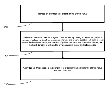
Turning now to Figure 5, a flowchart depiction of a method for treating a
medical
condition, in accordance with one illustrative embodiment of the present
invention is provided.
An electrode may be coupled to a portion of a cranial nerve to enhance cranial
nerve evoked
potentials. In one embodiment, one or more electrodes may be positioned in
electrical contact or
proximate to a portion of the cranial nerve to deliver a stimulation signal to
the portion of the
cranial nerve (block 710). The electrodes may be operatively coupled to at
least one of a main
trunk of the right or left vagus nerve, or any branch thereof. The IMD 100 may
then generate a
controlled electrical signal characterized by an interburst period, a number
of pulses per
microburst, an interpulse interval, and a microburst duration, wherein at
least one of the
interburst period, the number of pulses per microburst, the intemulse
interval, or the microburst
duration is selected to enhance cranial nerve evoked potentials (block 720).
This may include a
predetermined electrical signal that is preprogrammed based upon a particular
condition of a
patient. For example, a physician may pre-program the type of stimulation to
provide in order
to enhance cranial nerve evoked potentials in the patient based upon data
specific to the patient.
The IMD 100 may then generate a signal, such as a controlled-current
microburst signal, to
affect one or more portions of the neurological system of a patient.
The IMD 100 may then deliver the stimulation signal to the portion of the
cranial nerve
(block 730). The application of the electrical signal may be delivered to the
main trunk of the
right or left vagus nerve, or any branch thereof. In one embodiment,
application of the
Page 22
CA 2653110 2017-08-30
' W02607/115103
PCTJUS2007/065518
stimulation signal may be designed to promote an afferent effect. Further, the
stimulation by the
IMD 100 may reduce incidents or symptoms relating to a medical condition.
Additional functions, such as a detection process, may be alternatively
employed with
the embodiment of the present invention. The detection process may be employed
such that an
external detection or an internal detection of a bodily function may be used
to adjust the
operation of the IMD 100.
Turning now to Figure 6, a block diagram depiction of a method in accordance
with an
alternative embodiment of the present invention is illustrated. The IMD 100
may perform a
database detection process (block 810). The detection process may encompass
detecting a
variety of types of vital signs or other body parameters of the patient. A
more detailed depiction
of the steps for performing the detection process is provided in Figure 7, and
accompanying
description below. Upon performing the detection process, the IMD 100 may
determine if
stimulation is indicated (block 820). Upon a determination that stimulation is
not indicated, the
detection process is continued (block 830).
Upon a determination that stimulation is indicated, a determination as to the
type of
stimulation based upon data relating to the patient's condition is made (block
840). The type of
stimulation may be determined in a variety of manners, such as performing a
look-up in a look-
up table that may be stored in the memory 217. Alternatively, the type of
stimulation may be
determined by an input from an external source, such as the external unit 270
or an input from
the patient. Further, determination of the type of stimulation may also
include determining the
location as to where the stimulation is to be delivered. Accordingly, the
selection of particular
electrodes, which may be used to deliver the stimulation signal, is made.
Upon determining the type of stimulation to be delivered, the IMD 100 performs
the
stimulation by delivering the electrical signal to one or more selected
electrodes (block 850).
Upon delivery of the stimulation, the IMD 100 may monitor, store, or compute
the results of the
stimulation (block 860). For example, based upon the calculation, a
determination may be made
that adjustment(s) to the type of signal to be delivered for stimulation, may
be performed.
Further, the calculations may reflect the need to deliver additional
stimulation. Additionally,
data relating to the results of stimulation may be stored in memory 217 for
later extraction or
further analysis. Also, in one embodiment, real time or near real time
communications may be
provided to communicate the stimulation result or the stimulation log to an
external unit 270.
Turning now to Figure 7, a more detailed block diagram depiction of the step
of
performing the detection process of block 810 in Figure 6, is illustrated. The
system 100 may
monitor one or more vital signs or other bodily parameters of the patient
(block 910). This
Page 23
CA 2653110 2017-08-30
W02007/115103 PCT/US2007/065518
detection may be made by sensors residing inside the human body, which may be
operatively
coupled to the IMD 100. In another embodiment, these factors may be performed
by external
means and may he provided to the IMD 100 by an external device via the
communication unit
260. In one embodiment, the sensors include a strain gauge that may be used to
determine
inspiration by identifying chest expansion. By detecting the onset of chest
expansion, the strain
gauge may detect the onset of inspiration. The strain gauge may also detect
expiration by
identifying when the chest is contracting.
Upon acquisition of various signals, a comparison may be performed comparing
the data
relating to the signals to predetermined, stored data (block 920). Based upon
the comparison of
the collected data with theoretical or stored thresholds, the IMD 100 may
determine whether an
appropriate time to commence an on-time block has been reached (block 930).
Based upon the
determination described in Figure 7, the IMD 100 may continue to determine
whether further
stimulation is indicated, as described in Figure 6.
Additionally, external devices may perform such calculations and communicate
the
results or accompanying instructions to the IMD 100. The IMD 100 may also
determine the
specific location or branch of the nerve to stimulate. The IMD 100 may also
indicate the type of
stimulation to be delivered. For example, a microburst electrical signal alone
or in combination
with another type of treatment may be provided based upon the quantifiable
parameter(s) that
are detected. For example, a determination may be made that a microburst
electrical signal by
itself is to be delivered. Alternatively, based upon a particular type of
medical condition, a
determination may be made that a microburst electrical signal, in combination
with a
conventional therapeutic VNS signal, is desirable as a therapy for the
patient.
All of the methods and apparatuses disclosed and claimed herein may be made
and
executed without undue experimentation in light of the present disclosure.
While the methods
and apparatus of this invention have been described in terms of particular
embodiments, it will
be apparent to those skilled in the art that variations may be applied to the
methods and
apparatus and in the steps, or in the sequence of steps, of the method
described herein without
departing from the concept, spirit, and scope of the invention, as defined by
the appended
claims. It should be especially apparent that the principles of the invention
may be applied to
selected cranial nerves other than, or in addition to, the vagus nerve to
achieve particular results
in treating patients having epilepsy, depression, or other medical conditions.
The particular embodiments disclosed above are illustrative only as the
invention may be
modified and practiced in different but equivalent manners apparent to those
skilled in the art
having the benefit of the teachings herein. Furthermore, no limitations are
intended to the
Page 24 ,
CA 2653110 2017-08-30
' WO 2067/115103 PCT/US2007/065518
details of construction or design herein shown other than as described in the
claims below. It is,
therefore, evident that the particular embodiments disclosed above may be
altered or modified
and all such variations are considered within the scope and spirit of the
invention. Accordingly,
the protection sought herein is as set forth in the claims below.
Page 25
CA 2653110 2017-08-30