Note: Descriptions are shown in the official language in which they were submitted.
CA 02653244 2008-11-07
WO 2007/134236 PCT/US2007/068783
1
IMAGING AGENTS AND METHODS
RELATED APPLICATIONS
This application claims priority to U.S. Provisional Patent Application Serial
No. 60/747,180, filed May 12, 2006, and entitled "Imaging Agents and Methods,"
the
contents of which are incorporated herein in their entirety by reference.
This application also claims priority to U.S. Provisional Patent Application
Serial No. 60/819,297, filed July 7, 2006, and entitled "Dual Modality
Mr/Optical
Imaging With A Macromolecular Contrast Agent," the contents of which are
incorporated herein in their entirety by reference.
STATEMENT OF GOVERNMENT INTEREST
This disclosure was developed at least in part using funding from the National
Institute of Health, Grant Numbers RO1 EB000174, RO1 EB003132, and U54
CA90810, and the National Cancer Institute, Grant Number RO1 CA119387. The
U.S.
government may have certain rights in the invention.
TECHNICAL FIELD
The present disclosure relates to medical imaging and imaging agents.
Embodiments relate to near-infrared fluorescence imaging and imaging agents.
Other
embodiments relate to dual modality imaging, such as magnetic resonance and
optical, e.g. near-infrared fluorescence, imaging.
BACKGROUND
Near-infrared fluorescence (NIRF) optical imaging is currently under
development in several laboratories as a diagnostic modality that potentially
allows
imaging of biologic systems at the cellular and molecular level. In the NIRF
wavelength region (700-900 nm), light can travel several centimeters owing to
the
tissue's ability to multiply scatter light and to the relatively low
absorbance associated
with water, fat, hemoglobin and other less contributing biological molecules.
In
addition, endogenous fluorescence is minimal in the NIRF range.
CA 02653244 2008-11-07
WO 2007/134236 PCT/US2007/068783
2
Successful translation of NIRF optical imaging into clinical use requires
advances in several fronts, including development and validation of
fluorescence-
based contrast agents. One approach towards practical use of optical imaging
agents is
the development of "smart"probes, or molecular beacons that change their
optical
properties on interaction with specific molecular processes.
Cathepsin B (CB) are known to be important in normal tissue remodeling, but
also known to play critical roles in many diseases, such as arthritis,
atherosclerosis
and cancer. Elevated level of CB have been found in tumors and shown to
correlate
well with their invasive and metastatic profiles in both experimental cancer
models
and in human malignancies.
Poly(L-glutamic acid) (PG) has been used as a macromolecular carrier for
drug delivery, specifically to target cancer. PG-drug conjugates have been
shown to
be more potent and less toxic than their parent unconjugated drugs. In vivo
degradation of PG by cathepsin B (CB) has been linked to the increased site-
specific
delivery of anticancer drugs and enhanced antitumor activity of such PG-drug
conjugates as PG-paclitaxel (Xyotax ) and PG-camptothecin (CT2003). In
clinical
studies with PG-paclitaxel conjugated, Xyotax , significantly increased
antitumor
activity was noted in women with lung cancer with in elevated estrogen
receptors,
which in turn has been related to increased CB activity. Although the
degradation of
PG by CB has been extensively studied in vitro using either purified CB or
cell
lysates, studies of the kinetics of in vivo degradation of PG in various
tissues in live
animal have not be possible because of the lack of suitable technology.
Determining in vivo degradation of biomaterials and polymeric drug is
traditionally carried out by analyzing the appearance of degradation products
in the
target tissues. This method requires killing animals at each time point so
that tissues
can be removed from the animals. The degradation products are identified often
using
tedious purification scheme in combination with several detection methods
including
UV/Vis spectroscopy and mass spectroscopy. For example, a recent report
confirmed
monoglutamyl-2'-TXL and diglutamyl-2'TXL as the major intracellular
metabolites of
Xyotax using LC-MS technique, and the degradation of the polymer is correlated
to
its enhanced antitumor activity. Imaging technology for monitoring degradation
of
PG-based anticancer drugs in living animals is highly desirable because such
method
CA 02653244 2008-11-07
WO 2007/134236 PCT/US2007/068783
3
may potentially facilitate devising strategies for individualized therapy with
Xyotax
and non-invasive monitoring of treatment response to Xyotax treatment.
Sentinel lymph node (SLN) mapping is a method of determining whether
cancer has metastasized beyond the primary tumor and into the lymph system.
Traditionally, lymph node (LN) status has been assessed using clinical
palpation and
radiographic imaging of macroscopically enlarged nodes. Unfortunately, this
approach is not highly accurate and frequently misses early LN metastases.
Recently,
a new technique to identify early lymph node metastases-lymphatic mapping with
sentinel node biopsy (LSNB)-has been adopted to evaluate microscopic regional
LN
metastases in patients with melanoma, gastrointestinal= or breast cancer who
have no
clinical nodal involvement. In this technique, radiolabeled particles, sulfur
colloid
particles, and blue dye are injected and their localization to the SLN was
visualized by
naked eyes and with the help of hand-held gamma counter. While LSNB has
reduced
morbidity of regional staging by avoiding unnecessary removal of the entire
nodal
basins, LSNB still requires multiple injections, an invasive surgical
procedure, and up
to two weeks of waiting to determine whether or not cancer cells have spread.
The
radionuclide technique is also limited by exposure to ionizing radiation and
the low
spatial and temporal resolution.
To overcome these limitations, several contrast agents for MRI have been
designed to provide a minimally invasive, fast, and sensitive method to detect
SLN.
MRI is being used to characterize lymph nodes abnormalities in cancer patients
because of its excellent spatial and temporal resolution. Published techniques
have
used intravenous and interstitial injection of contrast agents to determine
the
metastatic status of lymph node. This includes using dextran-stabilized iron
oxide
crystals have helped to distinguish between normal and tumor-bearing nodes or
reactive and metastatic nodes with magnetic resonance imaging; using iron
oxide
nanoparticles for strong negative enhancement to identify lymph nodes; and Gd-
DTPA dendrimer-based contrast agent which gives T1-positive contrast
enhancement
of the lymphatic ducts and lymph nodes in mice.
For example, Gd-DTPA labeled polyglucose significantly enhanced T1-
weighted signal intensity of normal but not metastatic nodes in a rabbit model
in
regional nodes 24 hr postinjection. Harika L, Weissleder R, Poss K, et al. MR
lymphography with a lymphotropic Tl-type MR contrast agent: Gd-DTPA-PGM.
CA 02653244 2008-11-07
WO 2007/134236 PCT/US2007/068783
4
Magn Reson Med 1995;33:88-92, MR lymphography performed using dendrimers
visualized regional draining lymph nodes better than small molecular weight
contrast
agents. Kobayashi H, Kawamoto S, Sakai Y, et al. Lymphatic drainage imaging of
breast cancer in mice by micro-magnetic resonance lymphangiography using a
nano-
size paramagnetic contrast agent. J Natl Cancer Inst 2004;96:703-708. However,
the
technique is difficult for real time visualization, which limits the use of
MRI alone in
SLN mapping. Soltesz EG, Kim S, Laurence RG, et al. Intraoperative sentinel
lymph
node mapping of the lung using near-infrared fluorescent quantum dots. Ann
Thorac
Surg 2005;79:269-277; discussion 269-277, all incorporated by reference
herein.
Optical imaging is a relatively new modality that provides distinctly new
diagnostic capabilities while complementing conventional imaging modalities.
Some
advantages of optical imaging methods include the use of non-ionizing
radiation, high
sensitivity with the possibility of detecting micron-sized lesions, capability
of
continuous data acquisition for real time monitoring during surgery, and the
development of potentially cost-effective equipment. It also provides
flexibility in the
mode of chromophore excitation (broadband light source, modulated light,
continuous
wave or pulsed laser and signal detection (transillumination or reflectance,
and
scattering, absorption or fluorescence modes). Optical imaging methods can be
completely non-invasive, especially when endogenous chromophores are used;
minimally invasive, when contrast agents are injected; or invasive, when used
in
conjunction with surgical procedures or catheterization. For example, quantum
dots
(QD) have been used to map sentinel lymph nodes in mice and pigs.
Quantum dots (QD) have been used as NIRF agents to identify SLN.
Questions remained to be addressed before QD-based optical imaging techniques
are
translated into human studies. First, optical imaging is difficult for
visually
identifying deeper SLN owing to light attenuation. Second, potential
toxicities of
QD, which is made of toxic heavy metal ions such as cadmium, telluride,
selenide,
cause considerable concern. Although coating with a layer of biocompatible
materials
on the surface of QD reduces the side effects of QD, long-term effects of QD
in the
body remains to be studied.
Recently, it has been recognized that combination of MRI and optical imaging
can lead to the development of new approaches which will bridge the gaps in
resolution and depth of imaging between these two modalities and at the same
time
CA 02653244 2008-11-07
WO 2007/134236 PCT/US2007/068783
provide complimentary anatomic, functional and molecular information. The
combination of MRI with near-infrared (NIR) optical imaging was evaluated in
tumor
models and in the detection of cancer in human pilot clinical studies. In
those
experiments, MRI was used to obtain precise anatomic information on the
location of
5 tissue structures that were probed optically.
SUMMARY
According to one embodiment, the invention relates to a composition having
the formula:
NH2
O.
NH
+i
N ~ N
SO3H SO3H
According to another embodiment, the invention relates to a composition
including a poly(L-glutamic acid) and a NIRF dye.
According to another embodiment, the invention relates to a method including
providing to a plurality of cells an imaging agent including poly(L-glutamic
acid), a
NIRF dye and then imaging the cells to detect the imaging agent.
According to another embodiment, the invention includes a dual functional
contrast agent. This agent may include an MRI agent comprising Gadolinium
conjugated with an optical imaging agent.
According to another embodiment, the invention includes a method of
detecting cancer. This method include injecting a dual functional contrast
agent into a
patient The dual functional contrast agent may include an MRI agent conjugated
with
an optical imaging agent. The method may also include performing an MRI scan
in
the patient to detect the presence or absence of the contrast agent and
performing an
optical scan on the patient to detect the presence or absence of the contrast
agent. The
CA 02653244 2008-11-07
WO 2007/134236 PCT/US2007/068783
6
presence of the contrast agent in a cell or tissue may correlates with the
presence of
cancer in the cell or tissue.
Finally, according to another embodiment, the invention may include a
method of detecting cancer by injecting PG-DTPA-Gd-NIR813 into a patient, then
detecting the presence or absence of Gd in a cell or tissue of the patient and
detecting
the presence or absence of NIR813 in a cell or tissue of the patient. The
presence of
Gd and NIR813 in a cell or tissue of the patient may be indicative of cancer.
BRIEF DESCRIPTION OF THE DRAWINGS
Some specific example embodiments of the disclosure may be understood by
referring, in part, to the following description and the accompanying
drawings. The
following figures form part of the present specification and are included to
further
demonstrate certain aspects of the present description. The patent or
application file
contains at least one drawing executed in color. Copies of this patent or
patent
application publication with color drawing(s) will be provided by the Office
upon
request and payment of the necessary fee.
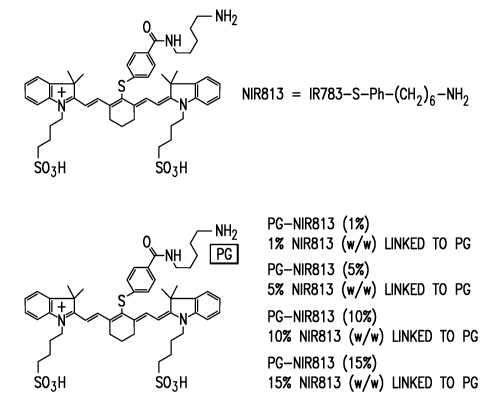
Figure 1 shows the structures of NIR813 and PG-NIR813 conjugates.
Figure 2 shows the fluorescence spectra of IR783 and NIR813. The
excitation/emission wavelengths are 766/798 nm for IR783 and 766/813 nm for
NIR813. Measurements were made in a methanol solution. NIR813 has a longer
emission wavenumber and greater Stokes shift (47 nm) than IR783 (32 nm).
Figure 3 shows images illustrating the effect of NIR813 loading on the
quenching efficiency of PG-NIR813. A. NIRF imaging acquired after 1 h of
incubation at room temperature. B. Fluorescence intensity as a function of
NIR813
loading. Each well contained 100 L PG-NIR813 at a final concentration of 10
M
equivalent NIR813 molecules. The images were acquired and analyzed using a Li-
Cor
Odyssey imaging system. NIR813 loading on PG (17 KDa) is expressed as a
percentage of the number of repeating units in PG.
Figure 4 shows images illustrating the effect of NIR813 loading on
degradation of PG-NIR813 and re-activation of fluorescence signal by cathepsin
B. A.
NIRF imaging. B. Fluorescence intensity as a function of incubation time. Each
well
contained 0.4 unit/mL cathepsin B inlOO L sodium acetate buffer (20 M, pH
5).
Wells were incubated without PG-NIR813 (Cl) or with PG-NIR813 (lO M eq.
CA 02653244 2008-11-07
WO 2007/134236 PCT/US2007/068783
7
NIR813) containing 15% (C2), 10% (C3), 8.3% (C4), 4.4% (C 5), and 1% (C6) of
NIR813 dye. The wellin column 7 contained cathepsin B and NIR813 (10 M) as a
control.
Figure 5 is a comparison of in vitrodegradation of L-PG-NIR813 (abbreviated
as PG-NIR813) and D-PG-NIR813. A. NIRF imaging. B. Fluorescence intensity as a
functionof incubation time. Each well contained 0.8 unit/mL cathepsin B in100
L
sodium acetate buffer (20 M, pH 5). Wells were incubated with 10 M eq. NIR813
of D-PG-NIR813 (C1) or PG-NIR813 (C2). Both conjugates contained 10% of
NIR813 dye.
Figure 6 shows images illustrating the effect of cathepsin B concentration on
the activation of PG-NIR813 (8.3% dye loading, 20 M eq. NIR813). PG-NIR813
(17KDa) was incubated with cathepsin B at room temperature for various times
for up
to 24 hr. Fluorescence intensity increased with increasing concentration of
cathepsin
B and increasing incubation times.
Figure 7 is a graph showing the degradation kinetics of PG-NIR813 (8.3%
loading, 17KDa) by cathepsin B. Product concentrations were derived from the
standard curve produced with the unconjugated NIR813. Non-linear fits of all
data
sets gives the initial velocities, whichwere used to generate Michaelis-Menten
graph.
Figure 8 are Michaelis-Menten graphs for PG-NIR813 (17K Da) and PG-
NIR813 (56K Da). Higher molecular weight conjugate degraded at a slower rate.
Figure 9 shows images illustrating inhibition of PG-NIR813 degradation by
selective cathepsin B inhibitor (inhibitorlI). (Top): NIRF images taken 21 h
after
incubation of PG-NIR813 conjugate (8.3% loading, 10 M eq. NIR813) in the
presence (bottom panel) and absence (top panel) of cathepsin B (0.2 unit/mL).
Microwellsin the bottom panel were added increasing concentrations of
cathepsin
Binhibitor II. (Bottom): Fluorescence signal intensity as a function of
inhibitor
concentration.
Figure 10 shows images illustrating the specificity of PG-NIR813 degradation
by proteinases. PG-NIR813 (10% loading, 40 Meq. NIR813) was incubated with
cathepsin B (0.04 unit), cathepsin D (0.08 unit), cathepsin E (0.08 unit), or
MMP-2
(50 ng) at 37 C over a period of 24 h. The buffer and pH value of the buffer
used in
the degradation studies were selected according to manufacturer provided
procedures.
CA 02653244 2008-11-07
WO 2007/134236 PCT/US2007/068783
8
Fluorescence intensity only increased with the use of cathepsin B. Data are
presented
as an average of duplicate experiments.
Figure 11 shows images illustrating the degradation of PG-NIR813 (10%
loading) by U87 cells in vitro. Cells were seeded (1 x 10 6cells) in 96-well
plate for
24 h. The cells were then treated with PG-INIR813 under the following
conditions:
(A). 0.1 MPG-INIR813 for 24 h without changing culture media; (B) fresh
culture
media followed by 0.1 MPG-NIR813 for 24 h; (C) 24 incubation without PG-
NIR813. Images were taken with culture media.
Figure 12 shows images illustrating the in vivo degradation of PG-NIR813
(10% loading, MW 17K). NIRF images were acquired at various times after
intravenous injection of PG-NIR813 at a dose of 10 nmol eq. NIR813 per mouse.
One
mouse was killed at 4 h after NIRF dye injection to verify tissue
distribution. PG-
NIR813 was primarily degraded in the liver was cleared from the body through
GI
tract.
Figure 13 shows images illustrating the in vivo degradation of PG-NIR813
(10% loading, MW 17K) in human U87/TGL glioma inoculated in the brain. NIRF
images were acquired at 24 hr after intravenous injection of PG-NIR813 at a
dose of
50 nmol eq. NIR813 per mouse. The presence of tumors in the brain was
confirmed
by chemoluminescent optical imaging of luciferase activity in U87/TGL tumors.
Fluorescence signal was detected only the brain of mice injected with L-PGNIR-
813
but not in mice injected with non-degradable D-PG-NIR813.
Figure 14 is an image showing fluorescence spectrum of PG-DTPA-Gd-
NIR813 (1% loading) and NIR813. The polymeric conjugate with low NIR813 dye
loading (<1 %) retained most of thefluorescence signal with minimal quenching
effect.
Figure 15 are images showing PG-DTPA-Gd-NIR813 drained to the sentinel
lymph nodes soon as 5 min after subcutaneous injection at the front paw
(arrow). The
fluorescence signal co-localized with isosulfan blue dye visualized under
bright light
(arrow heads). Isosulfan blue is used as a gold standard for SLN mapping.
Figure 16 are representative microphotography images of H&E stained section
and fluorescence micrography of the same section from a dissected lymph node.
Fluorescence signal was detected only in the lymph node (pseudocolor, red) but
not in
the adjacent muscle tissue (red).
CA 02653244 2008-11-07
WO 2007/134236 PCT/US2007/068783
9
Figure 17 shows comparison of NIRF optical images aquired 1 hr after
subcutaneous injection ofPG-DTPA-Gd-NIR813 at doses of 0.02 mmol Gd/kg (48
nmol eq. NIR813) (A) and 0.002 mmolGd/kg (4.8 nmoleq. NIR813) (B). SLN (arrow
heads) were detected at both doses.
Figure 18 shows comparison of MR images aquired at different times after
subcutaneous injection of PG-DTPA-Gd-NIR813 at doses of 0.02 mmol Gd/kg (A)
and 0.002 mrnolGd/kg (4.8 nmoleq. NIR813) (B). SLN (arrow heads) were clearly
delineated at both doses.
Figure 19 shows the reaction scheme for the synthesis of IR783-NH2 and PG-
benz-DTPA-Gd-IR783.
Figure 20 shows a fluorescence emission spectra of PG-benz-DTPA-Gd-
IR783 (in water) and IR783-NH2 (in ethanol/water). Plot of intensity
(arbitrary units,
AU) vs wavelength (nm) depicting PG-benz-DTPA-Gd-IR783 and IR783-NH2
fluorescence after excitation at 765 nm.
Figure 21 shows images of co-localization of PG-benz-DTPA-Gd-IR783 with
isosulfan blue dye. Male, athymic nude mice were injected subcutaneously with
4.8
nmol IR783/mouse using PG-benzDTPA-Gd-IR783 in the left paw, the pre-injection
of PG-benzDTPA-Gd-IR783 overlay image of white light and NIR fluorescence, and
the 5 min post-injection overlay of white light and NIR fluorescence. The
arrows
indicate the putative axiliary and branchial lymph nodes. Fluorescence images
have
identical exposure times and normalization, image of the mouse after the
injection of
1% isosulfan blue at the same location as the contrast agent, and after 5
minutes with
the exposure of the actual lymph nodes. Isosulfan blue and PG-benzDTPA-Gd-
IR783
were localized in the same lymph nodes: resected lymph nodes for histology
Figure 22 shows images of lymph node (top row) and muscle (bottom row)
after resection. Hematoxylin and eosin (H&E) staining (left) confirmed the
identity of
the lymph node, while the near infra-red fluorescence confirmed the contrast
agent
uptake of PG-benzDTPA-Gd-IR783 into the LN. Overlapping the DIC and
fluorescence indicates the localization of PG-benzDTPA-Gd-IR783 within the LN.
Muscle does not have fluorescence.
Figure 23 shows in vivo optical images of the axial and branchial lymph nodes
in athymic nude mice before and after the injection of PG-benz-DTPA-Gd-IR783
at
CA 02653244 2008-11-07
WO 2007/134236 PCT/US2007/068783
0.02 mmol Gd/kg and 0.002 mmol Gd/kg. NIR fluorescence images have identical
exposure times and normalizations. Also, these lymph nodes were excised for
histological evaluations.
Figure 24 shows T1-weighted axial MR images of PG-benz-DTPA-Gd-IR783
5 at (A) 0.02 mmol Gd/kg and (B) 0.002 mmol Gd/kg. MR signal intensity
increases
with increasing time.
Figure 25 is a graph of the time course of lymph node enhancement using 0.02
mmol Gd/kg and 0.002 mmol Gd/kg of PG-benzDTPA-Gd-IR783. This graph
indicates higher SI in higher concentration than low.
10 Figure 26 illustrates a reactio scheme for the synthises of NIR813 (Figure
26A) and and PG-DTPA-Gd-NIR813 (Figure 26B).
Figure 27 shows the fluorescence emission spectra of NIR8l3 (1 M, in
methanol) and PG-DTPA-Gd-NIR813 contrast agent (1 M, in water). The solutions
were excited at 766 nm.
Figure 28A-D show NIRF images in mice demonstrating co-localization of
PG-DTPA-Gd-NIR813 and isosulfan blue dye in sentinel lymph nodes. Each mouse
was injected subcutaneously with PG-DTPA-Gd-NIR813 contrast agent (10 L, 4.8
nmol eq. NIR813/mouse) in the left paw. Figure 28A shows a pre-contrast
overlay of
white light and NIRF images. Figure 28B shows an overlay of white light and
NIRF
images 5 min post-contrast agent injection. The arrows indicate the putative
sentinel
lymph nodes. Figure 28C shows photography of the same mouse showing the same
lymphatic nodes (arrows) stained blue by isosulfan blue. Figure 28D shows
fluorescence signal in and around resected lymph nodes. Figures 28E-H show
microphotographs of representative resected lymph nodes to evaluate the uptake
of
PG-DTPA-Gd-NIR813 in the lymph nodes. Figure 28E shows an H&E stained tissue
section. Figure 28F shows a DIC image. Figure 28G shows an NIRF image. Figure
28H shows an overlay of the DIC and NIRF images. The NIRF signal is
pseudocolored green, and the DIC pseudocolored red. Original magnification:
50x.
Figure 29 shows dual MR/optical imaging of the axial and branchial lymph
nodes in athymic nude mice. Figures 29A-D are NIRF images. Figure 29A is a pre-
contrast overlay of white light and NIRF images. Figure 29B is an overlay of
white
light and NIRF images 1 hr after injection of PG-DTPA-Gd-NIR813 (0.002 mmol
Gd/kg). Figure 29C is an NIRF image of the same mouse without skin. Figure 29D
CA 02653244 2008-11-07
WO 2007/134236 PCT/US2007/068783
11
shows fluorescence signal of resected lymph nodes. Figures 29E-F show
representative T1-weighted axial MR images at different times. In Figure 29E
PG-
DTPA-Gd-NIR813 was injected at a dose of 0.02 mmol Gd/kg and in Figure 29F at
a
dose of 0.002 mmol Gd/kg. The arrows indicate sentinel nodes.
Figure 30 shows the time course of lymph node enhancement at doses of 0.02
mmol Gd/kg and 0.002 mmol Gd/kg of PG-DTPA-Gd-NIR813. All data were
expressed as mean SD.
Figure 31 shows visualization of cervical lymph nodes after interstitial
injection of PG-DTPA-Gd-NIR813 (0.02 mmol Gd/kg) into the tongue of a normal
mouse (Figures 31A-E) and a mouse with a human DM14 squamous carcinoma tumor
grown in the tongue (Figures 6F-J). Figures 31A&F show T1-weighted coronal
images acquired 2 hr after contrast injection. Figures 31B&G show an overlay
of
white light and NIRF images 24 hr after contrast injection. Figures 31C&H show
NIRF images of mice without skin. Figures 31 D&I show NIRF images of resected
lymph nodes. The arrows indicate sentinel nodes and arrowhead indicates the
primary tumor. Figures 31E&J show microphotographs of H&E stained lymph node
sections. Figure 31 K shows microphotographs of H&E stained tongue section
indicating the presence of micrometastases, presumably in-transit metastases
in the
lymphatic duct.
The patent or application file contains at least one drawing executed in
color.
Copies of this patent or patent application publication with color drawing(s)
will be
provided by the Office upon request and payment of the necessary fee.
While the present disclosure is susceptible to various modifications and
alternative forms, specific example embodiments have been shown in the figures
and
are herein described in more detail. It should be understood, however, that
the
description of specific example embodiments is not intended to limit the
invention to
the particular forms disclosed, but on the contrary, this disclosure is to
cover all
modifications and equivalents as illustrated, in part, by the appended claims.
DETAILED DESCRIPTION
The present disclosure provides, according to certain embodiments, a NIRF
dye having the following structure:
CA 02653244 2008-11-07
WO 2007/134236 PCT/US2007/068783
12
NH2
0
NH
S
+~ / I /
N N
gp3H S03H
This dye is referred to as NIR813. This dye has longer excitation and
fluorescence wavelengths and a greater Stokes shift (difference between the
excitation
wavelength and emission wavelength) than Cy5.5. This means images acquired
using
imaging agents that comprise NIR813 can penetrate deeper into the tissues and
can
have less interference from the excitation light with appropriate filter sets
as
compared to those acquired with Cy5.5 derivatives.
The present disclosure provides, according to certain embodiments, a
composition comprising NIR813. According to certain embodiments such
compositions may be referred to as imaging agents and may comprise poly(L-
glutamic acid) and a NIRF dye, such as for example, NIR813 and IR783.
In general, such imaging agents are present in a quenched (i.e., inactive)
state
in aqueous solution but becomes dequenched (i.e., activated) when cleaved, for
example, upon exposure to a proteinases like CB. Accordingly, these imaging
agents
may be used, among other things, for in vivo molecular optical imaging.
In other embodiments, the imaging agent may further comprise a
paramagnetic metal chelate (e.g., Gd-DTPA). The DTPA-Gd is conjugated to PG so
that the conjugate can be used as an MRI contrast agent in addition to its
NIRF
properties. Accordingly, these imaging agents may be used to detect SLN using
both
optical and MR imaging.
One example of an imaging agent comprises poly(L-glutamic acid) and
NIR813 as the NIRF dye. This imaging agent may be referred to as PG-NIR813 and
has the following structure:
CA 02653244 2008-11-07
WO 2007/134236 PCT/US2007/068783
13
NH _
PG
N H _------
~
S
+i
N N
SO3H SO3H
The NIR813 may be present at from about 1% w/w linked to PG to about 15%
w/w linked to PG. (See Figure 1). PG-NIR813 has excitation and emission
wavenumbers of 766 nm and 813 nm, respectively. The long wavenumber allows
deeper penetration into the tissues and has less interferences from
autofluorescence
(i.e., signal coming from endogenous fluorophores). Such imaging agents may be
used, among other things, for in vivo molecular optical imaging of proteinases
like
CB at diseased sites, and in vitro assays of CB activity in biological
samples.
One example of an imaging agent comprises poly(L-glutamic acid), NIR813
as the NIRF dye, and DTPA-Gd as the a paramagnetic metal chelate. This imaging
agent may be referred to as PG-DTPA-Gd-NIR813 and has the following structure:
cooH co-NH-NIR813
0
H
N
H 1 m ~I n NHZ
O 0
O
NH
DTPA-Gd
The NIR813 may be present at about <4% w/w linked to PG, for example
about 1% w/w linked to PG. The loading of NIR813 should generally be
sufficient to
minimize any quenching effect.
CA 02653244 2008-11-07
WO 2007/134236 PCT/US2007/068783
14
Another example of an imaging agent comprises poly(L-glutamic acid), IR783
as the NIRF dye, and benzDTPA-Gd as the a paramagnetic metal chelate. This
imaging agent may be referred to as PG-DTPA-Gd-NIR783 and has the following
structure:
r
COOH
0 0
H H~
HO Nn H~ N PH
O
O O
HH HH
f
IR783
PGbembTPP1-Cd-IR783
DTPA-Gd
The present disclosure also provides methods for synthesizing NIR813 and
imaging agents.
The present disclosure also provides methods for assessing CB activity
comprising administering to a subject an imaging agent comprising poly(L-
glutamic
acid) and a NIRF dye and measuring a NIRF signal.
The present disclosure also provides methods for detecting inhibition of CB
activity comprising providing to a plurality of cells an imaging agent
comprising
poly(L-glutamic acid) and a NIRF dye and a cell and measuring a NIRF signal.
In one example, PG-NIR813 containing 5%-10% of NIR813 maybe activated
by CB and produce an NIRF signal. The NIRF signal may then be imaged
noninvasively and/or measured in a biological sample (e.g., blood) in vitro.
Tumors are known to secrete cathepsin B and/or to contain membrane-
associated CB, which is thought to be involved in invasion and metastasis.
Therefore,
extracellular CB may be used as a target for tumor detection in certain
embodiments
of the present disclosure. Patients with higher content or increased
proteolytic
activities of CB in tissue homogenates have significantly higher risk of
recurrence or
death than the cases with low content of the enzyme. Therefore, CB activity
also may
be used as aprognostic marker for cancer patients in certain embodiments of
the
present disclosure. Other diseases that are known to have abnormal activity of
CB
include atherosclerosis and arthritis. Therefore, imaging agents of the
present
CA 02653244 2008-11-07
WO 2007/134236 PCT/US2007/068783
disclosure that can be used for the assessment of CB activity in cancer may
also be
used for other diseases.
The present disclosure also provides methods comprising providing to a
plurality of cells an imaging agent comprising poly(L-glutamic acid), a NIRF
dye, and
5 a paramagnetic metal chelate; and imaging the cells to detect the imaging
agent. The
imaging agent may be detected with optical or MR imaging or both. When used
clinically, such methods may be minimally invasive and offer real-time
assessment of
anatomic information. Such methods may be used, for example, for SLN mapping.
SLN mapping is used routinely in the clinics using radiolabeled sulfur
colloid.
10 Imaging agents that avoid the use of radioisotope and provide the
opportunity for
SLN imaging using high resolution MRI and high sensitivity optical imaging are
advantageous. For example, to prepare one example contrast agent, poly(L-
glutamic
acid) (PG) was conjugated with paramagnetic metal chelate DTPA-Gd and a
fluorescence dye NIR813 to obtain PG-DTPA-Gd-NIR813 conjugate. PG-DTPA-Gd-
15 NIR813 can be used to detect SLN using both optical and MR imaging. The
dose
required is as low as 0.002 mmol/kg, about 100-fold lower than the clinical
dose of
Magnevist.
MR and NIRF images were taken before and after subcutaneous injection of
PG-DTPA-Gd-NIR813 into the front paw of healthy nude mice or interstitial
injection
of PG-DTPA-Gd-NIR813 in the tongue of nude mice bearing human DM14
squamous cell carcinoma. After subcutaneous injection, PG-DTPA-Gd-NIR813
colocalized with isosulfan blue dye in the axiliary and branchial lymph nodes,
indicating drainage of the contrast agent to the SLN. These nodes were clearly
visualized with both T1-weighted MR imaging and NIRF optical imaging within 5
min of contrast injection at a dose of 0.02 mmol Gd/kg (4.8 nmol eq. NIR813),
while
the branchial nodes were more readily detected with NIRF imaging than with MRI
at
a lower dose of 0.002 mmol Gd/kg (48 nmol eq. NIR813). In the head and neck
area
after interstitial injection of PG-DTPA-Gd-NIR813 into the tongue (15 L, 0.02
mmol
Gd/kg), optical imaging identified a116 cervical nodes in tumor bearing mice.
In
comparison, 4 of the 6 nodes were detected by MRI, and contrast enhancement of
these nodes were reduced compared to nodes in healthy mice. Histophathologic
examinations of sentinel nodes resected under NIRF imaging guidance revealed
the
.presence of micrometastases in 4 of 6 nodes. The superior spatial resolution
of MRI
CA 02653244 2008-11-07
WO 2007/134236 PCT/US2007/068783
16
combined with high detection sensitivity of NIRF imaging enabled preoperative
visualization of sentinel nodes with accurate anatomic location and detection
of
abnormal contrast enhancement, while intraoperative NIRF imaging permitted
selective removal of SLN and subsequent identification of micrometastases in
these
nodes. This example method represents a minimally invasive approach toward
lymph
node mapping with sentinel node biopsy.
PG-DTPA-Gd-NIR813 is a polymeric contrast agent having hydrodynamic
volume of greater than 20 nm. In general, the size of lymphangiographic agents
for
SLN mapping may be large enough to avoid their leakage into the blood
capillaries
and rapid loss of signal, but small enough to remain mobile for rapid transit
within the
lymphatic tract. Contrast agents having hydrodynamic diameter 5-40 nm usually
satisfy this criterion. Example agents may be derived using the present
disclosure and
Kim S, Lim YT, Soltesz EG, et al. Near-infrared fluorescent type II quantum
dots for
sentinel lymph node mapping. Nat Biotechnol 2004;22:93-97; Moghimi SM.
Bonnemain B. Subcutaneous and intravenous delivery of diagnostic agents to the
lymphatic system: applications in lyrnphoscintigraphy and indirect
lymphography.
Adv Drug Deliv Rev 1999;37:295-312. In addition to a suitable size, it may
also be
desirable to obtain a biocompatible contrast agent that can be metabolized and
eventually cleared from the body. The polymeric carrier in PG-DTPA-Gd-NIR813
is
a biodegradable polymer, which has demonstrated excellent biocompatibility. In
various studies in rodents, PG was used at doses from 200 to 800 mg/kg without
causing apparent toxic effects after intravenous injection. Li C. Poly(L-
glutamic
acid)--anticancer drug conjugates. Adv Drug Deliv Rev 2002;54:695-713. In
fact,
polymeric anticancer agents based on PG have advanced into clinic trial
studies.
Because a large fraction of PG-DTPA-Gd-NIR813 injected interstitially would
eventually be removed by lymph nodes with little to none of the contrast agent
entering systemic circulation, this agent may have acceptable toxicity profile
in SLN
mapping at doses that are 10- to 100-fold less than the dose of conventional
MRI
contrast agent used clinically for intravenous injection.
In yet other embodiments the imaging agents may further comprise a
therapeutic agent. These imaging agents may be referred to as biodegradable
drug
carriers. One example of such imaging agents may comprise a therapeutic agent,
poly(L-glutamic acid), and a NIRF dye.
CA 02653244 2008-11-07
WO 2007/134236 PCT/US2007/068783
17
Biodegradable drug carriers may be used to monitor the delivery of
therapeutic agents. Accordingly, the present disclosure provides, in certain
embodiments, methods for imaging degradation of polymeric drug carriers
comprising introducing to a cell a polymeric drug carrier comprising a
therapeutic
agent, poly(L-glutamic acid), and a NIRF dye; and imaging the cell using near-
infrared fluorescence imaging.
Recently, MRI agents consisting of dendrimers have been developed for
preoperative characterization of lymphatic drainage and lymph node metastases
from
mammary tumors. Kobayashi H, Kawamoto S, Sakai Y, et al. Lymphatic drainage
imaging of breast cancer in mice by micro-magnetic resonance lymphangiography
using a nano-size paramagnetic contrast agent. J Natl Cancer Inst 2004;96:703-
708;
Kobayashi H, Kawamoto S, Bernardo M, et al. Delivery of gadolinium-labeled
nanoparticles to the sentinel lymph node: comparison of the sentinel node
visualization and estimations of intra-nodal gadolinium concentration by the
magnetic
resonance imaging. J Control Release 2006;111:343-351. These studies
demonstrated
that the superior temporal and spatial resolution of micro-MR imaging
facilitates the
identification of lymphatic metastasis in experimental animals.
In embodiments of the present disclosure, using a dual modality contrast agent
in mice with lymph node metastases from squamous carcinoma tumor implanted in
the tongue, T1-weight MR images confirmed that preoperative MRI may allow for
differentiation of normal and metastatic nodes. The different pattern in lymph
node
enhancement may result from differences in macrophage uptake of macromolecular
contrast agents between normal and metastatic lymph nodes, as has been shown
to be
the case for superparamagnetic iron oxide nanoparticles. Anzai Y. Prince MR.
Iron
oxide-enhanced MR lymphography: the evaluation of cervical lymph node
metastases
in head and neck cancer. J Magn Reson Imaging 1997;7:75-81; Anzai Y, Blackwell
KE, Hirschowitz SL, et al. Initial clinical experience with dextran-coated
superparamagnetic iron oxide for detection of lymph node metastases in
patients with
head and neck cancer. Radiology 1994;192:709-715; Harisinghani MG, Barentsz J,
Hahn PF, et al. Noninvasive detection of clinically occult lymph-node
metastases in
prostate cancer. N Engl J Med 2003;348:2491-2499.
Although MRI is a useful method for precise localization and preoperative
characterization for the presence or absence of metastases in SLN, NIRF
imaging
CA 02653244 2008-11-07
WO 2007/134236 PCT/US2007/068783
18
allows detection of SLN at a much higher sensitivity. At an injected dose of
0.02
mmol Gd/kg, one may detect the same sets of SLN as soon as 3 min after the
injection
of PG-DTPA-Gd-NIR813 with both MRI and optical imaging. However, at a reduced
dose of 0.002 mmol Gd/kg, MRI detected only one of the two lymph nodes that
were
visualized with NIRF imaging. Moreover, while NIRF imaging was able to detect
all
6 cervical lymph nodes containing micrometastases in mice with squamous
carcinoma
tumor in the tongue, MRI revealed enhancement in 4 of the 6 nodes. These
findings
are consistent with lower detection sensitivity with MRI than with NIRF
imaging.
The challenge for implementation of sentinel lymph node biopsy is to develop
a reliable minimally invasive technique with high resolution and high
sensitivity.
Embodiments of the present disclosure relate to a dual-functional magnetic
resonance
(MR) and optical, such as near-infrared fluorescence (NIRF) optical imaging
contrast
agent. This agent may, in certain embodiments, be used for both preoperative
and
intraoperative sentinel node detection.
In more specific embodiments, the NIRF imaging agent may include a near
infrared fluorophore, such as a near infrared dye. The near infrared dye may
include a
cyanine or indocyanine derivative such as Cy5.5. The MRI agent may include Gd,
Mn or iron oxide.
Dual MRI and optical imaging of with PG-DTPA-Gd-NIR813 may be of value for the
detection of SLN. NIRF eliminates the need for both a radioactive tracer and a
blue
dye. Kim et al. have shown that lymph flow and the SLN can be identified
optically
and in real time, using intraoperative NIRF imaging and QD. Kim S, Lim YT,
Soltesz EG, et al. Near-infrared fluorescent type II quantum dots for sentinel
lymph
node mapping. Nat Biotechnol 2004;22:93-97.
One example dual modality imaging technique may be used in the following
clinical scenario. Initially, MRI may be used for noninvasive detection of
lymph node
metastases. If the presence of lymph node metastases is confirmed
nonequivocally
with MRI alone, surgery to remove the whole nodal basin may be performed, thus
eliminating the SLN biopsy step and the associated waiting period. If MRI is
unable
to detect metastases with a high degree of certainty, SLN mapping and
subsequent
SLN biopsy may then be performed using NIRF imaging. This may permit
intraoperative dissection without the use of ionizing radiotracer. Because of
its high
CA 02653244 2008-11-07
WO 2007/134236 PCT/US2007/068783
19
detection sensitivity, NIRF imaging may also be used to inspect the surgical
site to
ensure complete removal the SLN.
Accordingly dual functional macromolecular contrast agents according to
embodiments of the present disclosure may be suitable for both MR and NIRF
optical
imaging. Such an agent may be useful not only for precise localization of SLN
and
preoperative. characterization of lymph node abnormalities using MRI, but also
for the
SLN mapping and monitoring the success of complete resection of SLN during
surgical operation.
To facilitate a better understanding of the present invention, the following
examples of specific embodiments are given. In no way should the following
examples be read to limit or define the entire scope of the invention.
EXAMPLES
Example 1 PG-NIR813
Near-infrared fluorescence signal in PG-NIR813 is efficiently quenched when
NIR813 loading is greater than about 4% (based on the number of repeating
glutamic
acid units in the PG polymer) as shown in Figure 3. However, when the loading
is
greater than about 15%, polymer cannot be degraded by CB, as shown in Figure
4.
Therefore, in some examples, the optimal loading for certain activatable NIRF
probe
may be between about 4% and about 15%.
As shown in Figure 5, D-PG-NIR813 is not degradable by CB. Therefore, D-
PG conjugated dye may be used as carrier for the design of activatable NIRF
probe
responsive to other enzymes such as MMP-2. In such design, the NIRF
fluorophore
(NIR813 or others) may be attached to the side chains of D-PG through peptide
linkers that are specific substrate for the enzymes of interest.
As shown in Figure 6, PG-NIR813 is degraded by CB in a dose-dependent
manner. PG-NIR813 is not degraded by other proteinases tested (Figure 10).
Thus,
PG-NIR813 may be used to quantify CB activity in biological fluids (such as
plasma)
in in vitro settings.
The degradation of PG-NIR813 conjugate is generally a function of polymer
molecular weight. Conjugates with higher molecular weight degrade at a slower
rate,
as shown in Figure 7 and Figure 8.
CA 02653244 2008-11-07
WO 2007/134236 PCT/US2007/068783
As shown in Figure 9, degradation of PG-NIR813 by CB can be inhibited by
CB inhibitor in a dose-dependent manner. Accordingly, this property may be
used to
screen for CB inhibitors in a high-throughput setting. PG-NIR813 may also be
used to
image the inhibition of CB activity by CB inhibitors in vivo.
5 As shown in Figure 12, PG-NIR813 degradation in vivo can be monitored
noninvasively. Considering the structural similarity between PG-NIR813 and PG-
paclitaxel that is in advanced clinical trial studies, PG-NIR813 may be used
to select
patients who may benefit the most from PG-paclitaxel therapy, because the
efficacy
of PG-paclitaxel is dependent on the degradation of and release of paclitaxel
at the
10 target site.
As shown in Figure 13, PG-NIR813 can be used to detect the CB activity in
vivo.
PG-DTPA-Gd-NIR813
15 Poly(L-glutamic acid) (PG) was conjugated with paramagnetic metal chelate
Gd-DTPA and a fluorescence dye NIR813 to obtain PG-DTPA-Gd-NIR813
conjugate. The fluorescence spectrum is shown in Figure 14.
To determine its localization in the SLN, PG-DTPA-Gd-NIR813 was co-
injected with isosulfan blue dye, the gold standard for SLN mapping. Pre- and
post-
20 contrast images were taken using 4.7T Bruker Biospec MRI scanner and
Xenogen
optical imaging system. PG-DTPA-Gd-NIR813 was injected subcutaneously into the
front paw of nude mice at doses ranging from 0.002 mmol Gd/kg (4.8 nmol eq.
NIR813) to 0.02 mmol Gd/kg (48 nmol eq. NIR813). When injected together with
isosulfan blue dye, PG-DTPA-Gd-NIR813 co-localized with isosulfan blue dye,
indicating drainage of the contrast agent to the SLN (Figure 15). Axiliary and
branchial lymph nodes did not have sufficient contrast with neighboring tissue
to be
identified without contrast in T-1 weighted acquisitions (Figure 14). However,
these
nodes were clearly visualized as soon as 3 min with both MR and optical
imaging
within 6 min of contrast injection, even at the lowest dose tested (0.002 mmol
Gd/kg)
(Figure 15 and Figure 16). Enhancement remained persistent beyond 24 hr after
injection (Figure 16).
The superior spatial resolution of MRI combined with high detection
sensitivity with NIR optical imaging enabled visualization of lymphatic flow
and SLN
CA 02653244 2008-11-07
WO 2007/134236 PCT/US2007/068783
21
using a minimally invasive imaging procedure requiring no ionizing radiation,
and
may provide a powerful method for SLN mapping.
Example 2 - Materials & Methods
The following materials and methods were used to create the agents in this
example
Poly(L-glutamic acid) sodium salt, 1,3-diisopropylcarbodiimide (DIC),
pyridine, N-hydroxysuccinimde (NHS), N,N-diisopropylethylamine (DIPEA), IR-783
sodium acetate (NaOAc), EDTA, cysteine, PBS (0.01 M phosphate-buffered saline
containing 138 mM NaCI and 2.7 mM KCI, pH 7.4), N,N'-dimethylaminopyridine
(DMAP), and CB were purchased from Sigma-Aldrich (St. Louis, MO). 1-
Hydroxybenzotriazole(HOBt), benzotriazol-l-yl-oxy-tris-pyrrolidino-phosphonium
hexaflurophosphate (PyBOP), and N-tert-butoxycarbonyl-1,5-diaminopentane
toluenesulfonic acid salt was purchased from Novabiochem (San Diego, CA).
Trifluoroacetic acid (TFA) was obtained from Chem-Impex International, Inc.
(Wood
Dale, IL). 4-Mercaptobenzoic acid was purchased from TCI (Portland, Oregon).
Spectra/Pro 7 dialysis tubing with molecular weight cutoff (MWCO) of 10 000
was
purchased from Fisher Scientific (Pittsburgh, PA). PD-10 columns came from
Amersham-Pharmacia Biotech (Piscataway, NJ). CB inhibitor Ac-LVK-CHO
(Inhibitor II) was purchased from Calbiochem (La Jolla, CA). All solvents were
purchased from VWR (San Dimas, CA).
Analytical Methods
Analytical high-performance liquid chromatography (HPLC) was carried out
on an Agilent 1100 system (Wilmington, DE) equipped with a Vydac peptide and
protein analytic C-18 column (Anaheim, CA). Sample was eluted with H20 and
acetonitrile containing 0.1% TFA varying from 10% to 80% over 30 min.
Fluorescence intensity was measured by Licor Odyssey instrument (Lincoln,
Nebraska).
Synthesis of IR-783-S-Ph-COOH
IR-783 (250 mg, 0,33mmol) and 4-mercaptobenzoic acid (104mg, 0,67mmol)
were dissolved in 5 mL DMF and stirred for overnight at room temperature.
After
removing the solvent, the residue was dissolved in methanol and precipitated
in ether.
CA 02653244 2008-11-07
WO 2007/134236 PCT/US2007/068783
22
The solid was collected by filtration and further purified with flash
chromatography
using ethyl acetate and methanol as the mobile phase.
Synthesis of IR- 783-S-Ph-CONH(CH2)5NHBoc
IR-783-S-Ph-COOH (150 mg, 0.18 mmol), NHS (22 mg, 0.21mmol) and were
dissolved in 5 mL DMF. DIC (31 L, 0.21 mmol) and DMAP (2.5 mg, 0.02 mmol)
were added to the solution. The mixture was stirred at room temperature for
4hr. The
solvents were removed under vacuum. The residue was washed with ether. The
resulting activated ester IR-783-S-Ph-CO-NHS and BocNH(CH2)5NH2 (42 mg,
0.21mmol) were dissolved in 5 mL DMF with 5% DIPEA. The mixture was stirred
for 4hr. After removing the solvent, the residue was dissolved in methanol and
precipitated in ether. The solid was filtered out and further purified with
flash
chromatography with ethyl acetate and methanol.
Synthesis of IR-783-S-Ph-CONH(CH2)5NH2 (NIR813)
IR-783-S-Ph-CONH(CH2)5NHBoc was dissolved in 20 mL of 40% TFA in di
chloromethane and stirred for 25 min. The solvent was removed under vacuum.
The
residue was dissolved in methanol and precipitated in ether. The solid was
filtered out
and then dissolved in acetonitrile and water. The product was dried by
lyophilization.
MS: 929.47 (calcl.), 929.43 (found, M).
NIRF dye containing a primary amine, IR-783-S-Ph-CONH(CH2)5NH2, was
synthesized in 3 steps (Fig. 26A). IR-783-S-Ph-COOH was first synthesized
according to Strekowski et al. Strekowski L, Gorecki T, Mason JC, Lee H.
Patonay
G. New Heptamethine Cyanine Reagents for Labeling of Biomolecules with a Near-
Infrared Chromophore. Heterocyclic communications 2001;7:2 117-2122. Briefly,
IR-783 (250 mg, 0.33 mmol) and 4-mercaptobenzoic acid were dissolved in 5 mL
dimethylformamide (DMF). This solution was stirred overnight at room
temperature.
After removing the solvent, the residue was dissolved in methanol and
precipitated in
ether. The solid was collected by filtration and further purified with flash
chromatography using ethyl acetate and methanol as the mobile phase. IR-783-S-
Ph-
COOH was then conjugated to t-Boc protected heterodiamine t-BocNH(CH2)5NH2
using activated ester. Thus, IR-783-S-Ph-COOH (150 mg, 0.18 mmol) and NHS (22
mg, 0.21mmol) were dissolved in 5 mL DMF together with 1,3-
diisopropylcarbodiimide (31 L, 0.21 mmol) and 4-dimethylaminopyridine (2.5
mg,
0.02 mmol). The reaction proceeded at room temperature for 4 hr, after which
the
CA 02653244 2008-11-07
WO 2007/134236 PCT/US2007/068783
23
solvent was removed under vacuum and the residue washed with ether. The
resulting
IR-783-S-Ph-CO-NHS was reacted with BocNH(CH2)5NH2 (42 mg, 0.21mmol) for 4
hr in 5 mL DMF containing 5% N,N-diisopropylethylamine. The product was then
worked up and purified with flash chromatography. Finally, the t-Boc
protection
group in IR-783-S-Ph-CONH(CH2)5NHBoc was removed by treating with 40% TFA
in dichloromethane. After solvent removal, the product was purified by
precipitation
from a methanol solution with ether. IR-783-S-Ph-CONH(CH2)5NH2, was collected
by filtration and dried by lyophilization. MS: 929.47 (calcl.), 929.43 (found,
M).
The fluorescence emission maximum for IR-783-S-Ph-CONH(CH2)5NH2 was 813 nm
(Fig. 27). Consequently, IR-783-S-Ph-CONH(CH2)5NH2 is termed NIR813 dye
throughout this disclosure.
Synthesis of PG-NIR813
Sodium salt of poly-L-glutamic acid (number-average molecular weight M,,,
17,500 and 56,000) was dissolved in H20 and precipitated by acidifying with 1
N
HCI. The polymer precipitate was collected by centrifugation and dried by
lyophilization. The percentage of dye used for each loading was based on molar
number of the side chain glutamic acid residues in pre-weighted PG. The
amounts of
PyBOP and HOBt were 2 eq of the NIR813 dye. All of the reactants PG, NIR813,
PyBOP and HOBt were dissolved in DMF. 2% of DIPEA was added to the solution.
The mixture was stirred until the dye peak disappeared on HPLC (about 2 to 4
hr).
The solvents were removed under vacuum. The residue was dissolved in PBS and
purified using PD-10 columns eluted with PBS. The solution was dialyzed
against
H20 overnight and lyophilized. The yields of polymer were around 60%.
To determine the dynamic range of NIR813 dye, a stock solution of 200 M
of NIR813 in methanol was diluted with assay buffer (20 mM of NaOAc, 1mM
EDTA, 5mM cysteine, pH 5.0) to 2.5, 5, 10, 15, 20 M solutions. 100 L of each
sample was put in each well. The fluorescence intensity for each concentration
was
collected by Licor Odyssey camera. The result was reported by the plot of
concentration vs. fluorescence intensity.
Quenching effect and stability test of PG-NIR813 with different loading (1 %,
4.4%, 8.3%, 10% and I5%)
L-PG-NIR813 with different loading (1%, 4.4%, 8.3%, 10% and 15%) was
dissolved in assay buffer respectively to form 10 M solutions. 100 L of each
CA 02653244 2008-11-07
WO 2007/134236 PCT/US2007/068783
24
sample was put in each well. The fluorescence intensity of each sample was
determined using Li-cor Odyssey NIRF imager. The result of quenching effect
was
showed in the plots of loading percentage vs. fluorescence intensity. The
microwell
assay plate was incubated at 37 C for 48 hr. At predetermined time intervals,
the
stability of each sample in each well was checked through the change on
fluorescence
intensity. The stability of each loading was indicated by the plots of time
vs.
fluorescence intensity.
Biodegradation of L-PG-NIR813 with different loading (1 %, 4.4%, 8.3%, 10%
and 15%)
L-PG-NIR813 with different loading (1%, 4.4%, 8.3%, 10% and 15%) and CB
were dissolved in assay buffer respectively. The reaction mixture in each well
(100
L) was composed of 10 M L-PG-NIR813 probe and 0.4 units/mL CB. The samples
were incubated at 37 C for 24hr. At predetermined time intervals, the
fluorescence
intensity of reaction mixture in each well was measured using Li-Cor Odyssey
imager. The result of each sample was showed in the plots of time vs.
fluorescence
intensity.
Concentration effects on biodegradation of L-PG-NIR813 with loading 8.3%
and 10%.
L-PG-NIR813 with different loading 8.3% and 10% and CB were dissolved in
assay buffer respectively. Three different concentrations 5, 10, and 20 M
were
prepared for each loading of L-PG-NIR813. The concentrations of CB were
serially
arranged from 0.05 to 0.8 units/mL for each concentration of the probe. The
total
volume in each well was 100 L. The reaction mixtures were incubated at 37 C
for
24hr. At predetermined time intervals, the fluorescence intensity of reaction
mixture
in each well was measured by Li-Cor Odyssey imager. The result was showed in
the
plots of time vs. fluorescence intensity.
Inhibition of biodegradation of L-PG-NIR813 in presence of CB inhibitor II
L-PG-NIR813 with different loading 8.3% and 10%, CB and CB inhibitor II
were dissolved in assay buffer respectively. The reaction mixture (100 L) in
each
well was composed of 10 M L-PG-NIR813 probe and 0.2 units/mL CB. The CB
inhibitor II was serially diluted in the assay buffer to obtain concentrations
ranging
from 240 M to 77 nM. The microwell assay plate was incubated at 37 C for
24hr. At
CA 02653244 2008-11-07
WO 2007/134236 PCT/US2007/068783
predetermined time intervals, the inhibition of biodegradation was examined
using Li-
Cor Odyssey imager. The result was showed in the plots of time vs.
fluorescence
intensity.
Physicochemical Properties of Peptidyl MMP-2 Inhibitors are shown in
5 Table 1.
Table 1
Dye Mass Spectrometry HPLC
Molecular Calculated Observed Retention
formula MW (M+1) Time
(min)a
IR-783-S-Ph-COOH C45H53N2O8S3+ 845.31 845.37 18.59
IR-783-S-Ph- C5oH65N407S3+ 929.47 929.43 17.88
CONH(CH2)5NH2 (NIR813)
a Sample was eluted with H20 and acetonitrile containing 0.1% TFA
varying from 10% to 80% over 30 min.
10 Example 3 - Materials and Methods
The following materials and methods were used to create the agents in this
example.
PG sodium salt; 1,3-diisopropylcarbodiimide (DIC); pyridine; 4-
dimethylaminopyridine (DMAP); trifluoroacetic acid (TFA); gadolinium (III)
15 chloride hexahydrate; PBS (0.01 M phosphate buffered saline (PBS)
containing 138
mM NaCI and 2.7 mM KC1, pH 7.4); 1-ethyl-3-(3-dimethylaminopropyl)-
carbodiimide (EDC); 2-morpholinoethanesulfonic acid buffer (MES); IR-783 dye;
N-
hydroxysuccinimide (NHS); N,N-diisopropylethylamine (DIPEA); isosulfan blue;
and
all the other reagents and solvents were purchased from Sigma-Aldrich (St.
Louis,
20 MO). N-tert-butoxycarbonyl-1,5-diaminopentane toluenesulfonic acid salt was
purchased from Novabiochem (San Diego, CA). 4-mercaptobenzoic acid was
purchased from TCI (Portland, Oregon). P-aminobenzyl-diethylenetriaminepenta
(acetic acid-t-butyl ester) was obtained from Macrocyclics (Dallas, TX).
Spectra/Pro 7
dialysis tubing with molecular weight cutoff (MWCO) of 10,000 and PD-10
columns
25 came from Amersham-Pharmacia Biotech (Piscataway, NJ).
CA 02653244 2008-11-07
WO 2007/134236 PCT/US2007/068783
26
Analytical Methods
Gel permeation chromatography (GPC) was performed on a Waters (Milford,
MA) high-performance liquid chromatography (HPLC) system consisting of a 600
controller, a 717 plus auto sampler, and a Viscotek E-Zpr triple detector
(Viscotek,
Houston, TX) that records refractive index, viscosity, and light-scattering
signals. The
samples were separated using an TSK-G4000PW 4.6 mm x 30 cm column (TosoHaas,
Montgomeryville, PA) eluted with PBS containing 0.1% LiBr at a flow rate of
1.0
ml/min. Number-average molecular weights of the polymer conjugates were
calculated using Viscotek TriSEC GPC software. Elemental analysis was
performed
by Galbraith Laboratories, Inc. (Knoxville, TN).
Analytical high-performance liquid chromatography (HPLC) was carried out
on an Agilent 1100 system (Wilmington, DE) equipped with a Vydac peptide and
protein analytic C-18 column (Anaheim, CA). Sample was eluted with water and
acetonitrile containing 0.1 % TFA varying from 10% to 80% over 30 min.
Fluorescence intensity was measured by Spex Fluorolog spectrofluorimeter
(Jobin
Yvon Inc, Edison, NJ).
Synthesis of IR 783 -NH2
IR-783 (250 mg, 0.33 mmol) and 4-mercaptobenzoic acid were dissolved in 5
mL DMF. This solution was stirred overnight at room temperature. After
removing
the solvent, the residue, which is IR-783-S-Ph-COOH, was dissolved in methanol
and
precipitated in ether. The solid was filtered out and further purified with
flash
chromatography with ethyl acetate and methanol.
IR-783-S-Ph-COOH (150 mg, 0.18 mmol) and NHS (22 mg, 0.21mmo1) were
dissolved in 5mL DMF. DIC (31 L, 0.21 mmol) and DMAP (2.5 mg, 0.02 mmol)
were added to the solution and the mixture was stirred for 4 hours. The
solvents were
removed under vacuum and the residue was washed with ether. This gives the
green
residue, IR-783-S-Ph-COOSu.
IR-783-S-Ph-COOSu was dissolved in 5 mL DMF and was added with
BocNH(CH2)5NH2 (42 mg, 0.21mmo1) and 5% DIPEA. The mixture was stirred for 4
hours. After removing the solvent, the residue, which is IR-783-S-Ph-
CONH(CH2)5NHBoc, was dissolved in methanol and precipitated in ether. The
solid
was filtered out and further purified with flash chromatography with ethyl
acetate and
methanol.
CA 02653244 2008-11-07
WO 2007/134236 PCT/US2007/068783
27
The t-Boc protection of IR-783-S-Ph-CONH(CH2)5NHBoc was removed by
dissolving this residue in 20 mL of 40% TFA in DCM and was stirred for 25min.
The
solvent was removed under vacuum and the resulting material was dissolved in
methanol and precipitated in ether. The final product, IR783-NH2, was filtered
out
and then dissolved in acetonitrile and water. The product was dried by
lyophilization
and was characterized using NMR and mass spectrometry (MS).
Synthesis of PG-DTPA-Gd
PG (Mn, 41,400; Ig, 7.75 mmoles of carboxylic unit) andp-aminobenzyl-
diethylenetriaminepenta(acetic acid-t-butyl ester) (2.1 g, 2.79 mmoles) were
dissolved
in lOml of anhydrous DMF, followed by the addition of 1,3-
diisopropylcarbodiimide
(403mg, 3.lmmoles), 1.2m1 of pyridine, and trace amount of 4-
dimethylaminopyridine. The reaction mixture was stirred at 4 C overnight. To
remove
the protecting groups, the reaction mixture was treated with TFA at 4 C
overnight.
After removal of TFA under vacuum, 20 ml of ice-cold 1M NaHCO3 was added into
the residual solid. The pH of the solution was brought up to 7.5 with 1 M NaOH
and
the solution was dialyzed against PBS and water sequentially (MWCO 10,000).
The
resulting solution was filtered through 0.2 m membrane filters and
lyophilized. About
28 of 274 glutamic acid residues were coupled to benzylDTPA-Gd, determined by
elemental analysis. Into a PG-Benz-DTPA (110 mg) solution in 10 ml of sodium
acetate buffered aqueous solution (0.1 M, pH 5.5) was added 0.37 ml of
GdC13'6H2O
(100 mg/ml, 0.1 mmoles) in 0.1 M sodium acetate solution in small fractions.
The
solution was dialyzed against water (MWCO 10,000) until no free Gd3+ was
detectable in the receiving vessel. The solution was lyophilized to yield 1.22
g of
white powder (yield of polymer 81 %). The number-average molecular weight of
Gd3+-chelated polymeric conjugate was about 101,200 as measured by GPC. The
compound contained 10.8% (w/w) of gadolinium.
Synthesis of PG-DTPA-Gd
PG-p-aminobenzyl-DTPA-Gd (PG-DTPA-Gd) was synthesized according to
previously reported procedures. Wen X, Jackson EF, Price RE, et al. Synthesis
and
characterization of poly(L-glutamic acid) gadolinium chelate: a new
biodegradable
MRI contrast agent. Bioconjug Chem 2004; 15:1408-1415. Briefly, p-aminobenzyl-
DTPA(t-butyl ester) (2.1g, 2.79 mmol) was conjugated to PG (Mn, 41,400; 1 g,
7.75
mmol of carboxylic unit) in DMF using 1,3-diisopropylcarbodiimide (403 mg, 3.1
CA 02653244 2008-11-07
WO 2007/134236 PCT/US2007/068783
28
mmol) as the coupling agent. The t-butyl protecting groups were removed by
treating
with TFA at 4 C overnight to give PG-DTPA. To chelate with Gd3+, a solution of
GdC13'6H2O in 0.1 M sodium acetate was added into a solution of PG-DTPA in 0.1
M
sodium acetate (pH 5.5) in small fractions until free Gd3+ was detected. The
solution
was then dialyzed extensively against water (MWCO 10,000) and lyophilized to
yield
1.22 g of white powder (81%). The compound contained 10.8% (w/w) of
gadolinium.
Synthesis of PG-DTPA-Gd-IR 783
PG-Benz-DTPA-Gd (90mg, 0.698 mmol Glu) was dissolved in 2 mL of 0.1 M
MES buffer. IR783-NHZ (4.17 mg, 0.0045 mmol) dissolved in 200 uL of DMF was
added to the PG-Bz-DTPA-Gd solution in the presence of EDC (10 mg, 0.005
mmol).
This was stirred overnight at 4 C while protected from light. The solution was
filtered
in 0.2 m membrane filters and was dialyzed overnight with PBS buffer and water
overnight at 4 C. Yield was 64.6 mg (72%).
Determination of Maximum Emission Wavelength
The fluorescence emission spectra of the synthesized contrast agent was
obtained using a Spex Fluorolog spectrofluorometer (Horiba Yvon Jobin, NJ).
Determination of Relaxivity
Solutions of PG-Benz-DTPA-Gd-IR783 were prepared in water at gadolinium
concentrations of 0.005, 0.01, 0.02, 0.04, 0.08, and 0.16 mM. Spin lattice
(Tl) and
spin-spin (T2) relaxivities were measured at 4.7 Tesla on 4.7T Bruker Biospec
47/40USR (City, State) using inversion recovery and mutiecho T2-weight pulse
sequences. Relaxivities (Ri or R2 in mM-1s-1) were obtained from linear least
square
determination of the slopes of 1/Ti vs [Gd] or l/T2 vs [Gd] plots.
Sentinel lymph node identification
A group of 6 male athymic nude mice (NCI), 6-12 weeks old, were injected
subcutaneously into the front paw with 10 L of 0.002 mmol Gd/kg mouse or 5
nmol
IR783/mouse of PG-benzDTPA-IR783 in PBS at pH 7.4. Optical images are taken
before and at 5 minutes post-contrast and then, 10 L of 1% (17.6 mM)
isosulfan blue
was injected into the same position as the PG-benzDTPA-IR783 was injected.
After 5
minutes, an image-guided removal of lymph nodes and muscle was done. For
histology, OCT-embedded tissue was cryo-sectioned at 10 m thickness.
MR and Optical Imaging
CA 02653244 2008-11-07
WO 2007/134236 PCT/US2007/068783
29
Prior to imaging, mice were anesthetized with 1-2% isoflurane gas, and the
entire animal was imaged for a maximum of 5 min at pre-contrast and at various
times
after subcutaneous injection of the contrast agent. For optical imaging, an
IVIS
imaging system (100 series) (Xenogen Corp., Alameda, CA) was used, while for
MR
imaging, a 4.7T Bruker Biospec 47/40USR MRI experimental scanner was used.
During imaging, mice were maintained in an anesthetized state with 1.5%
isoflurane.
Six mice were divided into two groups having 3 mice in each group. The first
group was injected with 0.02 mmol Gd/kg mouse or 48 nmol IR783/mouse and the
second group with 0.002 mmol Gd/kg mouse or 4.8 nmol IR783/mouse. Pre-contrast
images of the mice were done at first in the optical imaging system and then
the mice
were imaged using MRI. T1-weighted image was set and after the baseline images
were acquired, PG-benzDTPA-Gd-IR783 (0.02 mmol/kg or 0.002 mmol/kg) was
rapidly injected into the front paw of the mice. Images were then taken every
3
minutes thereafter unti130 minutes. After the MR imaging, the mice were imaged
using the optical imaging system and an image-guided removal of the sentinel
lymph
nodes and muscle was done. These tissues were frozen and cut into 10 um thick
slices.
Total photon emissions from defined regions of interest within the optical
images of each mouse were analyzed using the Living Image software (Xenogen
Corporation, Alameda, CA), while imageJ software was used to analyze the MR
images. The relative increase in signal intensity (SI) was calculated
according to the
formula ([SIpost - SIpre]/SIpre) x 100%. For this analysis, the same region of
interest
(ROI) was drawn on the consecutive transaxial MR images. In the lymph nodes,
the
ROI was adapted to encompass as much of this structure as possible with
maximum
enhancement, and the same size of ROI was used in the pre-contrast images. All
the
results of data analysis were expressed as mean SD. Significance of the
differences
of the data comparisons was assessed using a paired or unpaired Student t-
test. A P
value of less than 0.05 was taken to indicate statistical significance.
Example 4- Synthesis and Characterization of PG-benzDTPA-Gd-IR783
The synthetic scheme for the synthesis of PG-benzDTPA-Gd-IR783 is shown
in Figure 19. PG-benzDTPA-Gd was synthesized according to Wen X, et al.
Bioconjugate Chem. 15: 1408-1415, 2004. IR783-NH2 was conjugated to PG-
benzDTPA-Gd using 1-ethyl-3-(3-dimethyl-aminopropyl) carbodiimide
hydrochloride
CA 02653244 2008-11-07
WO 2007/134236 PCT/US2007/068783
(EDC) as the coupling reagent. This conjugate was purified by dialysis against
deionized water and by passing through PD-10 columns. The absence of small
molecular weight contaminant was confirmed by gel permeation chromatography
(GPC). Table 1 gives the summary of the physicochemical properties of the
5 synthesized PG-benzDTPA-Gd and PG-benzDTPA-Gd-IR783. The starting PG has a
molecular weight of 42,100. The molecular weight of the conjugated PG was
calculated in terms of %Gd (w/w) and %IR783 (mol/mol). Percent Gd content by
weight was determined using elemental analysis while %IR783 content was
determined using fluorescence intensity. About 55 out of 274 glutamic acid
units, or
10 0.2 mol/mol of COOH, were attached with Gd as measured by elemental
analysis.
About 3 IR783 units were attached to each PG chain.
Table 2
PG-Benz-DTPA-Gd PG-Benz-DTPA-Gd-IR783
Mw calculated 60,080 62,813
# COOH in PG 274 274
# DTPA per PG 39 39
% Gd (w/w) EA 10.83 10.40
% Gd (w/w) calculated 9.25 -
# DTPA per PG 39 39
% Gd (w/w) EA 10.83 10.40
% Gd (w/w) calculated 9.25 -
% IR783 - 1
# IR783 per PG - 3
Relaxivity (Rl mmol-1 s-1) 8.89 13.23
(R2 mmol-1 s-1) 24.07 39.08
Other physicochemical properties of the Gd3+-chelated PG polymers are also
summarized in Table 2. The reported number average molecular weights were
15 estimated from GPC analyses. For comparison, the theoretical number-average
molecular weights calculated on the basis of starting molecular weight of PG
and the
degree of substitution are also listed. PG-benzDTPA-Gd-IR783 had greater
relaxivity
than that of small molecular weight DTPA-Gd, having T1 value of 4.8 mmol-ls-I
using 4.7T MRI experimental scanner (Table 2).
CA 02653244 2008-11-07
WO 2007/134236 PCT/US2007/068783
31
Comparison of fluorescence intensity of PG-benzDTPA-Gd-IR783 and IR783-
NHZ is presented in Figure 20. A strong emission peak at around 805 nm was
observed for IR783-NH2, while PG-benzDTPA-Gd-IR783 has an emission at 814nm.
Co-localization ofPG-benzDTPA-Gd-IR783 with isosulfan blue dye
The PG-benzDTPA-Gd-IR783 has a maximal fluorescence emission at 814
nm, compared to IR783-NH2 which is at 805 nm. When PG-benzDTPA-Gd-
IR783was injected subcutaneously into the front paw of the mouse, it entered
the
lymphatics and migrated within minutes to the axiliary and branchial lymph
nodes.
Co-injection at the same site with isosulfan blue, the gold standard of SLN
mapping,
resulted in co-localization of the NIR fluorescence signal and the blue dye
(Figure
21). Resection of these brightly fluorescent specimens was proved to be lymph
nodes
as conferred by hematoxylin and eosin (H&E) staining (Figure 22). As a
control, non-
fluorescing muscle was also sectioned and imaged. As expected, muscle showed
no
fluorescence under the NIR fluorescent microscope.
MR and optical imaging findings
To demonstrate the ability of PG-benzDTPA-Gd-IR783 to act as a dual
MR/optical imaging probe, we subcutaneously injected the agent into front paw
of the
mice (n=3) and obtained NIRF and MR images. Figure 23 shows a representative
example of NIRF images using 0.02 mmol Gd/kg or 48 nmol/mouse and 0.002 mmol
Gd/kg or 4.8 nmol/mouse. The bright fluorescent images indicates uptake of the
contrast agent into the axiliary and branchial lymph nodes. MR images also
supports
the NIRF images since branchial and axiliary lymph nodes indicated increase in
signal
enhancement post-contrast (Figure 24a and b). Calculation of the % increase in
signal
intensity reveals a concentration-dependent increase in signal enhancement,
having a
P-value < 0.05 (Figure 25). Examination of the 2 different concentrations
showed that
even at 0.002 mmol Gd/kg or 4.8 nmol/mouse, images can still be taken with
great
sensitivity.
Synthesis of PG-DTPA-Gd-NIR813
The synthetic scheme for the preparation of PG-DTPA-Gd-NIR813 is shown
in Figure 1B. PG-DTPA-Gd was dissolved. NIR813 (4.17 mg, 0.0045 mmol)
dissolved in 200 L of DMF was added to a solution of PG-DTPA-Gd (90 mg, 0.698
mmol Glu) in 0.1 M MES buffer (2 mL) in the presence of EDC (10 mg, 0.005
mmol). The reaction mixture was stirred at 4 C overnight while protected from
light,
CA 02653244 2008-11-07
WO 2007/134236 PCT/US2007/068783
32
filtered through a 0.2- m filter, dialyzed against PBS buffer and water
sequentially,
and lyophilized. Yield: 64.6 mg (72%). The conjugate contained about 4.4%
NIR813
(w/w).
The physicochemical properties of PG-DTPA-Gd and PG-DTPA-Gd-NIR813
are summarized in Table 3. By GPC analysis, PG-DTPA-Gd-NIR813 had a number
average molecular weight of 101,200. For comparison, the theoretical number-
average molecular weight calculated on the basis of starting molecular weight
of PG
is also listed in Table 1. About 51 and 3 of the 274 glutamic acid units per
PG chain
were attached with DTPA-Gd and NIR813 dye, respectively. Table 3 shows the
physico-chemical properties of PG-DTPA-Gd and PG-DTPA-Gd-IR783.
Table 3.
PG-DTPA-Gd PGDTPA-Gd-NIR813
Molecular Weighta 60,080 (274) 62,813 (274)
% Gd (w/w)b 10.83 10.40
Number of DTPA per PG 39 39
% NIR813 (w/w)` - 1
Number of NIR813 per - 3
PG
Relaxivity (Rl mmol-1 s-1) 8.89 13.23
(R2 mmol-1 s-1) 24.07 39.08
aNumber average molecular weight calculated on the basis of starting
molecular weight (42,100 Da) and the percentage of substitution.
bPercentage of Gd by weighted was measured with elemental analysis.
cPercentage of NIR813 measured spectrophotometrically.
The excitation/emission wavelengths were 766/798 nm for IR783 and 766/813
nm for NIR813 in methanol solution. Therefore NIR813 had a greater Stokes
shift
(47 nm) than IR783 (32 nm) did. A comparison of fluorescence emission spectra
of
NIR813 and PG-DTPA-Gd-NIR813 acquired at the same equivalent dye
concentration is presented in Figure 27. Both compounds had the same emission
maximum of 813 nm when excited at 766 nm. However, the fluorescence intensity
of
PG-DTPA-Gd-NIR813 was reduced to approximately 44% of that of unconjugated
NIR813, suggesting the presence of intramolecular interaction among NIR813
dyes
attached to PG in aqueous solution. The presence of a shoulder peak at 765-775
nm
CA 02653244 2008-11-07
WO 2007/134236 PCT/US2007/068783
33
supports that the dequenching effect observed with PG-DTPA-Gd-NIR813 was due
to
7r-staggering of NIR813 in the polymer conjugate. Increasing the loading of
NIR813
dye to more than 15 dye molecules per PG chain caused almost complete
quenching
of fluorescence signal (data not shown). Therefore, we used PG-DTPA-Gd-NIR813
containing on average 3 NIR813 molecules per polymer chain in our dual
modality
imaging studies. The fluorescence intensity of each PG-DTPA-Gd-NIR813
conjugate
was approximately 32% stronger than one NIR813 molecule.
Relaxivity
Solutions of PG-DTPA-Gd-NIR813 were prepared in water at gadolinium
concentrations of 0.005, 0.01, 0.02, 0.04, 0.08, and 0.16 mM. Spin lattice
(T1) and
spin-spin (T2) relaxivities were measured at 4.7 Tesla on 4.7T Bruker Biospec
(Bruker
Biospin Corp., Billerica, MA) using inversion recovery and mutiecho T2-weight
pulse
sequences. Relaxivities (Ri or R2 in mM-ls 1) were obtained from linear least
square
determination of the slopes of 1/T1 vs [Gd] or 1/T2 vs [Gd] plots.
Cell line and animals
Human DM14 squamous carcinoma cells were a soft agar clone derived from
Tu167 cells (a gift from Dr. Clayman, MDACC). Cells were maintained at 37 C in
a
humidified atmosphere containing 5% CO2 in Dulbecco's modified Eagle's medium
and nutrient mixture F-12 Ham (DMEM/F12) containing 10% fetal bovine serum
(GIBCO, Grand Island, NY).
All animal work was carried out in the Small Animal Imaging Facility at The
University of Texas M. D. Anderson Cancer Center in accordance with
institutional
guidelines. For mice with lymph node metastases, 1 x 10⁶ DM14 cells suspended
in
50 L of HBSS were injected directly into the submucosa of the anterior tongue
using
a 1-ml tuberculin syringe (Hamilton Co.) and a 30-gauge needle in male athymic
nude
mice (n = 3). By 20 days after inoculation, mice would die of malnutrition
because
the primary tumors prevented mice from food and water intake. Most mice would
have developed metastases in the cervical lymph nodes by that time. Myers JN,
Holsinger FC, Jasser SA, Bekele BN. Fidler IJ. An orthotopic nude mouse model
of
oral tongue squamous cell carcinoma. Clin Cancer Res 2002;8:293-298. Mice were
used for imaging study on 10 days after tumor cell inoculation
MR and optical imaging
CA 02653244 2008-11-07
WO 2007/134236 PCT/US2007/068783
34
Prior to imaging, mice were anesthetized with 2% isoflurane gas in 1 1/min 02
flow and during imaging, mice were maintained in an anesthetized state with
1.5%
isoflurane. For optical imaging, an IVIS imaging system (100 series) (Xenogen
Corp., Alameda, CA) was used with ICG filter (ex/em, 710-760/810-875 nm) sets.
The field of view was 13.1 cm in diameter. The fluency rates for NIRF
excitation
light was 2 mW/cm2. The camera settings included maximum gain, 2x2 binning,
640
x 480 pixel resolution and an exposure time of 0.8 sec. For MRI, a 4.7T Bruker
Biospec scanner (Bruker Biospin Corp., Billerica, MA) was used. Axial and
coronal
images were obtained using a 950 mT/m, 5.7 cm inner diameter actively shielded
gradient coil system (19,000 mT/m-s slew rate) and a 3.5 cm inner diameter
volume
radiofrequency coil. T1-weighted (TE = 8.5 ms, TR = 1000 ms) MR images were
acquired with a 4x3 cm field of view, 1-mm section thickness, 0.25-mm gap, and
a
256x192 matrix.
SLN identification
A group of 6 male athymic nude mice (NCI, City, State), weighting 20-25 g
each, were injected subcutaneously into the front paw with 10 L of PG-DTPA-Gd-
NIR813 (0.02 mmol Gd/kg, 48 nmol eq. NIR813/mouse) in PBS. Optical images
were taken before and at 5 minutes post-contrast and then, 10 L of 1%
isosulfan blue
(17.6 mM) was injected into the same sites as PG-DTPA-Gd-NIR813 was injected.
Animals were killed 5 min later and the skin in the area where fluorescence
signal
was detected was removed to permit direct visual detection of the dye.
Sentinel nodes
noted for blue coloration under bright light were resected and imaged again
with
NIRF camera. Nodes were then processed for histologic evaluation.
Co-localization of PG-DTPA-Gd-NIR813 with isosulfan blue dye
When PG-DTPA-Gd-NIR813 was injected subcutaneously into the front paw
of the mouse, it entered the lymphatics and migrated within minutes to the
axiliary
and branchial lymph nodes. Injection at the same site with isosulfan blue, the
gold
standard of SLN mapping, resulted in co-localization of the NIR fluorescence
signal
and the blue coloration (n = 6, Fig. 3A-3D). These brightly fluorescent
specimens
were resected and proven to be lymph nodes histologically. No residual
fluorescence
signal was observed in the surrounding areas. Analysis of resected fluorescent
tissues
showed that PG-DTPA-Gd-NIR813 was completely trapped in SLN, but not in the
CA 02653244 2008-11-07
WO 2007/134236 PCT/US2007/068783
surrounding tissues (Fig. 3E-H). Analysis also confirmed uptake of the
contrast agent
by lymph nodes.
Dual MR/optical imaging detection of axiliary and branchial lymph nodes
Each mouse was injected subcutaneously in the left front paw with PG-DTPA-
5 Gd-NIR813 at a dose of 0.02 mmol Gd/kg (48 nmol NIR813/mouse) or 0.002 mmol
Gd/kg (4.8 nmol NIR813/mouse) (n = 3/dose group). Pre-contrast images were
obtained with both optical and MR imaging. T1-weighted MR images were then
acquired every 3 minutes for 30 minutes post-contrast injection, after which
the mice
were imaged again with the NIRF camera. Sentinel lymph nodes were removed
under
10 NIRF guidance. The resected nodes were processed for histologic
examinations.
For analysis of signal enhancement in sentinel nodes, the same region of
interest (ROI), encompassing the whole enhanced axiliary lymph nodes, was
drawn
on the consecutive transaxial MR images. Image J software
(http://rsb.info.nih.gov/ij/) was used to analyze the MR imaging data. The
relative
15 increase in MR signal intensity (SI%), calculated according to the formula
SI% =
(Slpost - SIpie]/SIpre) x 100%, was plotted as a function of time. SI% value
at each time
point was compared between two dose groups using an unpaired Student's t test
with
p < 0.05 considered significant.
To examine whether sentinel auxiliary and branchial nodes could be detected
20 with both MR and NIRF optical imaging, mice were given a single
subcutaneous
injection of PG-DTPA-Gd-NIR813 at a dose of 0.02 mmol Gd/kg as before or at a
lower dose of 0.002 mmol Gd/kg. At both dose levels, the sentinel nodes were
readily
visualized with NIRF imaging. Figures 4A-D shows representative NIRF images
acquired 1 hr after contrast injection at a lower dose of 0.002 mmol Gd/kg,
which
25 clearly revealed the uptake of the contrast agents in the auxiliary and
branchial nodes.
Resected lymph nodes showed bright fluorescence (Figure 4D).
Both auxiliary and branchial nodes and their anatomical location were also
identified as soon as 3 min after contrast injection on MR images at the high
dose
level (Fig. 4E). However, at the low dose level of 0.002 mmol Gd/kg, only the
30 auxiliary node was visualized (Fig. 4F). Calculation of the % increase in
MR signal
intensity for the auxiliary nodes reveals a dose-dependent increase in signal
enhancement. Signal intensities at a dose of 0.02 mmol Gd/kg were
significantly
higher than that at a dose of 0.02 mmol Gd/kg at each time points from 6 min
post-
CA 02653244 2008-11-07
WO 2007/134236 PCT/US2007/068783
36
injection over the 30 min study period (p < 0.05, Fig. 5). MR signal intensity
increased with time in a dose dependent manner.
Identification of cervical lymph nodes and detection of metastases following
imaging-guided nodal resection
Mice were injected with PG-DTPA-Gd-NIR813 interstitially around the
primary tumor at a dose of 0.02 mmol Gd/kg (48 mmol NIR813/mouse). Each mouse
was imaged with optical and MRI before and at different times after contrast
injection
as described previously. At the end of the last imaging session (24 hr post
contrast),
sentinel nodes were removed under the guidance of NIRF imaging, and the
resected
tissues were processed for histologic examinations.
For histopathologic examinations, nodal tissues were embedded in optimal
cutting temperature compound (OCT) (Sakura Finetek USA, Torrance, CA), snap-
frozen, and cryosectioned into 10 m slices, and stained with hematoxylin and
eosin
(H&E). Consecutive unstained sections were photographed on a Leica
fluorescence
microscope (Leica Microsystems, Bannockburn, IL). The microscope was equipped
with a 75-W Xenon lamp, differential interference contrast (DIC) optical
components,
775/845 nm (excitation/emission) filter sets (Chroma Technology, Brattleboro,
VT), a
Hamamatsu black and white chilled charge-coupled device camera (Hamamatsu
Photonics K.K., Hamamatsu City, Japan), and Image-Pro Plus 4.5.1 software
(Media
Cybernetics, Silver Spring, MD).
Whether dual MR/optical imaging using PG-DTPA-Gd-NIR813 could be used
to characterize metastatic SLN preoperatively and postoperatively following
imaging
guided resection in an orthotopic head and neck tumor xenograft model was
investigated. In mice without tumor, both MRI and NIRF imaging readily
detected
uptake of the contrast agent in the cervical lymph nodes after interstitial
injection of
PG-DTPA-Gd-NIR813 into the tongue of the mice at a dose of 0.02 mmol Gd/kg
(Fig. 31A-31E). In mice with orthotopic human DM14 squamous carcinoma tumor
grown in the tongue (n = 3), al16 sentinel nodes were visualized using NIRF
imaging
(Fig. 31 G-I). However, 2 of the 6 nodes visualized with NIRF were not
similarly
identified by the MRI method. The pattern of enhancement of the remaining
nodes
revealed by MRI was different from that observed in normal cervical tissue: in
general the lymph nodes showed less enhancement and the enhancement was
located
at the rim of the lymph nodes (compare Fig. 31A vs. 31F). Histopathologic
CA 02653244 2008-11-07
WO 2007/134236 PCT/US2007/068783
37
examination confirmed micrometastases in these nodes (Fig. 31 J).
Micrometastases
were noted in the lumen of a vascular structure in the tongue of one of the
tumor-
bearing mice (Fig. 31 K).
Notwithstanding that the numerical ranges and parameters setting forth the
broad scope of the invention are approximations, the numerical values set
forth in the
specific examples are reported as precisely as possible. Any numerical value,
however, inherently contain certain errors necessarily resulting from the
standard
deviation found in their respective testing measurements.
While the compositions and methods of this disclosure have been described in
terms of specific embodiments, it will be apparent to those of skill in the
art that
variations may be applied to the compositions and/or methods and in the steps
or in
the sequence of steps of the methods described herein without departing from
the
concept, spirit and scope of the invention. All such similar substitutes and
modifications apparent to those skilled in the art are deemed to be within the
spirit,
scope and concept of the invention.