Note: Descriptions are shown in the official language in which they were submitted.
CA 02654233 2008-12-03
WO 2007/143831 PCT/CA2007/001041
METHOD OF CALIBRATING A BIOMETRIC DEVICE
Field of the Invention
[0001] The present invention relates to a novel method of calibrating a
biometric device, as well as a method of accurately measuring a target
dimension of a
physiological tissue which incorporates calibration.
Background of the Invention
[0002] Measurement of a dimension of a physiological tissue, such as a
mammalian tissue, can have important clinical and research applications in a
variety
of diagnostic and therapeutic fields. For example, measurement of corneal
thickness
may have applications in the diagnosis and/or treatment of conditions in the
field of
optometry or ophthalmology such as glaucoma, corneal pathology, refractive
surgery
and contact lenses. However, despite strong associations among measurements of
central corneal thickness by different techniques,1-6 there is a lack of a
gold standard
for cross-calibration between different instruments.
[0003] Although there is abundant literature on precision (repeatability or
reliability) of the common biometric equipment for measuring different tissues
including corneal thickness3 6-2', no information about accuracy of the
methods exists.
Precision quantifies how multiple measures compare with each other. Accuracy
is an
indicator of the proximity of the measurement to the real physical value that
is being
measured. A measurement method could be precise but not accurate.22 23 For
example, a piece of equipment could always underestimate corneal thickness by,
say,
40 gm and be very precise (repeatable) for this measurement, which is not
accurate.
However, the question is whether a refractive surgeon or a glaucoma specialist
can
make a sound clinical decision based on this measurement, particularly in
borderline
cases. Therefore, in addition to the importance of precision, a measurement
technique
should also be accurate and its calibration should be verifiable using a gold
standard.
[0004] Currently, a non-invasive method for comparing tissue measurements
taken by different biometric devices does not exist. Such a comparison can
only be
CA 02654233 2008-12-03
WO 2007/143831 PCT/CA2007/001041
conducted by obtaining a sample of the subject tissue, for example, by biopsy.
This
method of comparison is neither acceptable nor feasible in the case of certain
tissue
types such as the ocular tissue.
[0005] It would, thus, be desirable to develop a method of using a biometric
device which renders accurate results that can be validly compared with
similar
results obtained using different devices.
Summary of the Invention
[0006] Accordingly, a novel method of calibrating a biometric device useful
to measure dimensions of physiological tissue has now been developed. The
calibration utilizes samples of a reference material that possess a property
of the
physiological tissue that is required for the function of the device.
[0007] Thus, in one aspect, a method of calibrating a biometric device useful
to measure a target dimension of a physiological tissue is provided
comprising:
(i) measuring the target dimension of at least two samples of a
reference material with the device to provide an actual output,
wherein the reference material possesses at least one property
of the tissue required for the function of the device and wherein
each of the samples has a known target dimension;
(ii) calculating a calibration equation based on the actual output of
the device and the known target dimensions of the samples; and
(iii) adjusting the actual output of the device according to the
calibration equation to yield a corrected output.
[0008] In another aspect of the present invention, a method of measuring a
target dimension of a physiological tissue using a biometric device is
provided. The
method comprises the steps o
(i) measuring the target dimension of at least two samples of a
reference material with the device to provide an actual output,
2
CA 02654233 2008-12-03
WO 2007/143831 PCT/CA2007/001041
wherein the reference material possesses at least one property
of the tissue required for the function of the device and wherein
each of the samples has a known target dimension;
(ii) calculating a calibration equation based on the actual output of
the device and the known target dimensions of the samples;
(iii) adjusting the actual output of the device according to the
calibration equation to yield a corrected output; and
(iv) measuring the target dimension of the tissue with the device,
wherein the measured dimension is corrected according to the
calibration equation.
[0009] In another aspect of the invention, a method of cross-calibrating
multiple biometric devices which measure the same dimension of a target
tissue, but
which function differently, is provided. The method comprises calibrating the
biometric devices as described utilizing a reference material having
properties of the
target tissue required for the function of each of the biometric devices.
[0010] The present invention advantageously provides a means of calibrating
a biometric device that can be incorporated into a method of measuring a
target
dimension of a physiological tissue to yield accurate measured values of a
selected
tissue dimension. The present invention allows rapid and simple calibration of
measurements obtained with biometric devices that utilize both the same and
different
working principles so that measurements from different devices may be used
interchangeably when measuring the same target tissue. In addition, device
accuracy
may be verified using the methods disclosed herein.
[0011] These and other aspects of the present invention will be described by
reference to the following drawings in which:
3
CA 02654233 2008-12-03
WO 2007/143831 PCT/CA2007/001041
Brief Description of the Figures
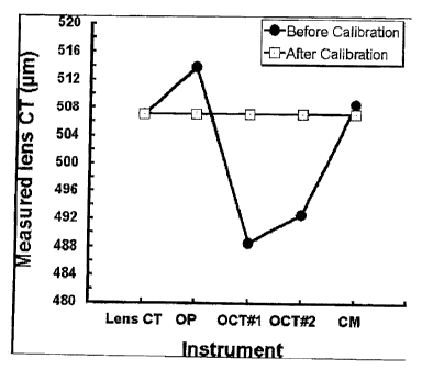
[0012] Figure 1 graphically compares measured corneal thickness with
different instruments before and after instrument calibration according to the
present
invention; and
[0013] Figure 2 provides a graphic comparison of the correlation between
calibrated and uncalibrated instruments for lens center thickness
measurements.
Detailed Description of the Invention
[0014] A method of calibrating a biometric device useful to measure a target
dimension of a physiological tissue is provided. The method makes use of a
reference
material having a known target dimension (a real target dimension) that
possesses at
least one property of the tissue required for the function of the device. The
calibration
method comprises measuring the target dimension of at least two samples of the
reference material with the device to generate an actual output. A calibration
equation
is then calculated based on the actual output of the device and the known or
real target
dimensions of the samples; and adjusting the actual output of the device
according to
the calibration equation to yield a corrected or real output.
[0015] The term "biometric device" is used herein generally to encompass
devices used to measure a dimension of a physiological tissue. An optical
biometric
device, for example, can measure a dimension, such as thickness, of parts of
the eye
including, but not limited to, the retina, iris, crystalline lens and cornea.
Examples of
optical biometric devices include, but are not limited to, pachometers,
interferometric
devices such as optical coherence tomographers (OCT's), scanning slit imaging
devices, confocal microscopes and Scheimpflug devices. Other biometric devices
include, but are not limited to, acoustic biometric devices, ultra-sound
biometric
devices, x-ray imaging devices, which measure the permeability of tissue,
magnetic
resonance imagers, which can be used to measure a number of dimensions of
physiological tissue, and other non-visual electromagnetic devices.
4
CA 02654233 2008-12-03
WO 2007/143831 PCT/CA2007/001041
[0016] The term "dimension" refers to a physical characteristic of a
physiological tissue that can be measured. One of skill in the art will be
familiar with
the various dimensions of tissue that can be measured. Examples include, but
are not
limited to thickness, length, curvature, shape and permeability.
The physiological tissue may be any tissue subject to measurement by a
biometric
device. Examples include, but are not limited to, epidermal tissue, connective
tissue,
muscle tissue, vascular tissue, nervous tissue, and specific tissue types such
as ocular
tissue, including corneal, retinal and lens tissue, and aqueous and/or
vitreous
humour..
[0017] The present method relates to the calibration of a biometric device for
use in measuring a target dimension of a physiological tissue. To calibrate
the device,
the device is used to measure the target dimension of a reference material in
which the
target dimension is already known, i.e. measured by other means known to be
accurate. The calibration will generally involve measurement using the
biometric
device of at least 2, and preferably 3 to 4, samples of a selected reference
material
with a target dimension that is known but which is different in each case. The
known
or real target dimensions generally span the range of expected measurements of
the
target dimension.
[0018] In order to be effective, the reference material will possess at least
one
property of the target physiological tissue that is required for the function
of the
biometric device. For example, for an optical biometric device, the reference
material
will possess a refractive index equal to the refractive index (RI) of the
target tissue.
The overall RI of corneal tissue is generally accepted to be 1.376. Thus, a
reference
material for the measurement of corneal tissue may have an RI of, for example,
1.376.
For the calibration of optical devices, the reference material is preferably a
transparent, semi-transparent or opaque material that is readily measurable by
the
biometric device, for example, plastic, glass, fluid, or gas. The reference
material
may be layered to mimic the composition of the target tissue, for example,
corneal
tissue which comprises multiple layers. For an acoustic biometric device, such
as an
ultrasound pachometer, the reference material will possess an acoustic density
equal
to that of the target tissue. For an x-ray imaging device, the reference
material will
possess the x-ray characteristics of the target tissue.
CA 02654233 2008-12-03
WO 2007/143831 PCT/CA2007/001041
The method is also useful to cross-calibrate biometric devices that measure
the same
dimension of a target tissue, but which function differently, e.g. an optical
biometric
device and a non-optical biometric device including, but not limited to, an
acoustic,
ultrasonic, magnetic, or non-visual electromagnetic biometric device. To
conduct
such a cross-calibration, the reference material must have properties of the
target
tissue required for the function of both devices. To cross-calibrate an
optical
biometric device and a non-optical biometric device, thus, the reference
material must
have both the refractive index and additionally the non-optical characteristic
required
for use of the selected non-optical biometric device. For example, to cross-
calibrate
an optical biometric device and an acoustic biometric device for measuring
ocular
tissue, thus, the reference material must have both the refractive index and
acoustic
density of the target ocular tissue.
[0019] The data obtained from measuring the target dimension of the
reference material samples, i.e. the actual output, and the known target
dimensions of
the samples are both used to prepare a calibration equation. The calibration
equation
defines the relationship between the actual output of the biometric device and
the
actual or known target dimensions, and is used to modify the actual output to
a
corrected output that corresponds with the known or actual target dimension
within an
acceptable amount of error or an amount of error that may not be considered
statistically significant. The accuracy of this calibration method will depend
on the
number of samples of the reference material which are being measured. As one
of
skill in the art will appreciate, the calibration equation will vary with the
biometric
device being calibrated and the target physiological tissue being measured.
[0020] Following measurement of the reference material samples, the
biometric device can be used to measure the same dimension, i.e. the target
dimension, in the selected physiological tissue. The measurements of the
target
dimension of the tissue are then adjusted in accordance with the calibration
equation
in order to yield an accurate measurement of the target dimension, i.e. a
corrected
output. Following calibration according to the present method, the corrected
output
will have a value that corresponds with the known or real target dimension
within an
acceptable range of error, e.g. which may be statistically insignificant.
6
CA 02654233 2008-12-03
WO 2007/143831 PCT/CA2007/001041
[0021] The present invention can be broadly utilized at the manufacturing
level of a biometric device as well as at the user level. The method
advantageously
provides a non-invasive method of calibrating a biometric device as well as a
means
of cross-calibrating between biometric devices that measure the same
characteristic
such that regardless of the device being used, the absolute values of
biometric
measurements are not significantly different between devices. This will aid
manufacturers in accurately cross-calibrating different biometric devices.
Clinicians
and researchers will also have a means to verify a calibration as well as
recalibrating a
device as needed.
[0022] Improved accuracy of biometric measurements using the present
technique provides enhanced validity of data for clinical decision making and
improves the quality of care for patients. In addition, this method will allow
consistent calibration of biometric devices for comparison with historical
data. The
same level of accuracy and consistency can be applied to a research setting
using the
present method to allow accurate comparison and universal interpretation of
data from
different clinics/studies using different biometric devices if they are
similarly
calibrated using the present methodology.
[0023] As one of skill in the art will appreciate, the above disclosure
generally
describes aspects of the invention. It is believed that one of ordinary skill
in the art
may, using the preceding description, make and use these aspects of the
invention. In
addition, certain embodiments of the invention have been described; however,
other
embodiments may exist which also fall within the scope of the appended claims.
For
example, changes in form and substitution of equivalents which do not depart
from
the scope of the claims are also contemplated. All journal articles and other
documents such as patents or patent applications referred to herein are hereby
incorporated by reference.
[0024] Embodiments of the present invention are described by reference to the
following specific example which is not to be construed as limiting.
7
CA 02654233 2008-12-03
WO 2007/143831 PCT/CA2007/001041
Example 1
METHODS - Instrumentation and Lenses
[0025] Fourteen rigid lenses of different thicknesses were manufactured using
a plastic material with refractive index (RI) of 1.3760+/- 0.0005 (at 589 nm).
This
plastic material was developed by Optical Polymer Research, Inc., Gainesville,
Florida. All lenses were made in piano power with a base curve of 8.6 mm and
no
prism. Physical center thickness of the calibration lenses (ranging from 301
to 696
m) was measured using a precision mechanical gauge (Vigor GA-715; Japan) and
the physical thickness of each lens was derived from the average of three
measurements (Table 1).
TABLE 1.
Lens center thickness ( m)
Lens No. Center Thickness ( m)
1 301
2 336
3 362
4 415
470
6 478
7 489
8 527
9 551
580
11 608
12 635
13 650
14 696
Mean 507
Standard deviation 122
[0026] Center thickness (CT) of the same set of lenses was measured using a
computerized optical pachometer (OP) mounted onto a Zeiss 30 SL-M
biomicroscope, two different Zeiss-Humphrey OCTII optical coherence
tomographers
(OCTs), and a Nidek Confoscan3 confocal microscope (CM).
8
CA 02654233 2008-12-03
WO 2007/143831 PCT/CA2007/001041
Procedure
[0027] The lenses were installed onto a wheel in a random order. A number
was assigned to each lens with no reference to the thickness of the lens. All
the
measurements on OP were completed by one operator and all the measurements on
OCT 1, OCT 2 and CM was performed by a second operator. All lenses were
measured once at each station. The number of measurements per lens was
selected
based on the research protocols as used for each device in the Centre for
Contact Lens
Research (CCLR). Therefore, more measurements were required for the OP because
of high measurement variability, which was reported for OP in the
literature.23 z4
[0028] Seven consecutive measurements for each lens were taken by the OP
and the lens CT was derived from the average of five readings after the
computer
trimmed the highest and the lowest readings.
[0029] For each lens, only one measurement was taken by each OCT machine.
One hundred axial scans (1.13-mm width) were processed and lens CT was
obtained
using custom software.
[0030] For the CM, lens CT was measured after applying a drop of a
gonioscopy gel to the posterior surface of the lens and stepping through the
lens
manually from anterior to posterior surfaces. One measurement was taken for
each
lens.
[0031] Accuracy of measurements of the four instruments was determined by
comparison to the physical CT of the lenses.
[0032] Center thickness of the same set of lenses was measured again after
each instrument was calibrated. Accuracy of measurements was compared among
the
four instruments.
9
CA 02654233 2008-12-03
WO 2007/143831 PCT/CA2007/001041
Data Analysis
[0033] Using a repeated-measures analysis of variance, the effects of
measurement device were examined. p values < 0.05 were considered
statistically
significant. Post hoc paired t- tests with Bonferroni correction (significance
level p <
0.01) were used to determine the significance of specific pairs.
RESULTS
[0034] The values quoted in this section are the mean +/- standard deviation
of
lens CT, unless otherwise stated.
[0035] Before calibrating the machines, there was a significant effect of the
measurement device (p < 0.05). There was a significant difference in lens
center
thickness between OP and each OCT as well as between the two OCT machines (all
post hoc tests; p < 0.01). CM was not significantly different from OP (post
hoc test; p
> 0.01) but was significantly different from each OCT (post hoc tests; p <
0.01), (see
Fig. I and Table 2 below).
TABLE 2.
Lens center thickness ( m) by each instrument
(mean +/- standard deviation) before calibration
Lens Optical Optical Optical Confocal
Center Pachometer Coherence Coherence
Thickness Tomographer 1 Tomographer 2
Mean 507.1 513.8 488.6 492.6 508.5
Standard 122.1 118.0 116.4 118.1 120.6
deviation
[0036] The differences between instruments were eliminated (p > 0.05) after
applying calibration equations (Table 3 below) for each device (Fig. 1), which
were
derived through linear regression analysis of lens physical center thickness
(known)
and instrument measured center thickness (actual output of the device).
CA 02654233 2008-12-03
WO 2007/143831 PCT/CA2007/001041
TABLE 3.
Calibration equationsa
Device Calibration Equation
Optical pachometer Calibrated CT = -24.2965 + 1.0342 x measured CT
Optical coherence Calibrated CT =-5.2248 + 1.0486 x measured CT
tomographer I
Optical coherence Calibrated CT =-2.1211 + 1.0338 x measured CT
tomographer 2
Confocal microscope Calibrated CT = 1.4079 + 0.9945 x measured CT
aNote that these are not general equations for the devices.
These equations are specific for individual instruments.
CT, center thickness
[0037] In addition, after each instrument was calibrated with lenses of 1.376
refractive index, there was no significant difference (p > 0.05) between mean
measured values of lens center thickness by OP, each OCT, CM, and the physical
center thickness of the lenses (Table 4 below).
TABLE 4.
Lens center thickness ( m) by each instrument
(mean +/- standard deviation) after calibration
Lens Optical Optical Optical Confocal
Center Pachometer Coherence Coherence
Thickness Tomographer 1 Tomographer 2
Mean 507.1 507.1 507.1 507.1 507.1
Standard 122.1 122.1 122.1 122.1 119.9
deviation
[0038] There were significant correlations (p < 0.05) between each pair of the
instruments for measured CT values both before and after calibration (see Fig.
2).
[0039] Improved accuracy using the present method is clearly shown by
comparing real thickness of the reference material to the measured values by
each
device before and after calibration (Table 2 vs. Table 4 as well as Figure 1).
In
addition, the mean ( standard deviation) percentage difference between
measured
values by each device and the physical thickness of the lenses can be shown by
the
two following tables (Table 5 and 6) which are based on the following formula:
11
CA 02654233 2008-12-03
WO 2007/143831 PCT/CA2007/001041
Percent difference =100 measured value by the device - physical thickness of
the lens
physical thickness of the lens
Table 5
Deviation (%) from physical lens center thickness by each instrument
(mean standard deviation) before calibration
Optical Optical Optical Confocal
Pachometer Coherence Coherence
Tomographer I Tomographer 2
Mean A 1.63% -3.56% -2.83% 0.48%
Standard 1.67% 0.88% 0.53% 5.64%
deviation
Table 6
Deviation (%) from physical lens center thickness by each instrument
(mean standard deviation) after calibration
Optical Optical Optical Confocal
Pachometer Coherence Coherence
Tomographer 1 Tomographer 2
Mean A 0.01 % 0.03% 0.01 % 0.22%
Standard 0.97% 0.78% 0.52% 5.62%
deviation
[0040] The results of the present study demonstrate that using calibration
lenses with the same refractive index as the cornea (1.376) allows rapid and
simple
calibration of the pachometers using different optical principles so that
corneal
thickness measurements from different optical devices may be used
interchangeably.
Example 2
[0041] This is an example of applying the calibration equations to human
central corneal thickness measurements (CCT) by the two OCT machines from
Example 1. The values in the following table are the central corneal thickness
in
microns. The following equations (from Example 1, Table 3) were used which
were
derived from calibrating each machine with the RI 1.376 lenses.
For OCT 1:
Real lens CT =-5.2248 + 1.0486 x measured value of lens CT by the machine.
Therefore:
Human CCT after calibration =-5.2248 + 1.0486 x measured human CCT before
calibration.
12
CA 02654233 2008-12-03
WO 2007/143831 PCT/CA2007/001041
For OCT2:
Real lens CT = -2.1211 + 1.0338 x measured value of lens CT by the machine.
Therefore:
Human CCT after calibration =-2.1211 + 1.0338 x measured human CCT before
calibration.
Table 7
Uncalibrated CT Calibrated CT
OCT# OCT#
ID# OCT#1 2 OCT#1 2
1 500 512 519.1 527.2
2 512 520 531.7 535.5
3 496 500 514.9 514.8
4 492 500 510.7 514.8
472 520 489.7 535.5
6 464 536 481.3 552.0
7 520 480 540.0 494.1
8 528 472 548.4 485.8
9 552 540 573.6 556.1
536 540 556.8 556.1
11 520 516 540.0 531.3
12 504 520 523.3 535.5
13 520 528 540.0 543.7
14 520 528 540.0 543.7
480 484 498.1 498.2
16 488 488 506.5 502.4
17 496 504 514.9 518.9
18 504 512 523.3 527.2
19 480 488 498.1 502.4
496 512 514.9 527.2
21 488 492 506.5 506.5
22 488 496 506.5 510.6
23 512 504 531.7 518.9
24 512 512 531.7 527.2
512 520 531.7 535.5
26 520 520 540.0 535.5
27 480 488 498.1 502.4
28 484 488 502.3 502.4
29 496 504 514.9 518.9
496 504 514.9 518.9
31 520 520 540.0 535.5
32 528 524 548.4 539.6
33 552 552 573.6 568.5
34 552 556 573.6 572.7
504 504 523.3 518.9
36 504 500 523.3 514.8
37 532 528 552.6 543.7
38 536 540 556.8 556.1
13
CA 02654233 2008-12-03
WO 2007/143831 PCT/CA2007/001041
39 456 456 472.9 469.3
40 456 464 472.9 477.6
41 504 496 523.3 510.6
42 496 496 514.9 510.6
43 504 504 523.3 518.9
44 496 500 514.9 514.8
45 520 528 540.0 543.7
46 520 520 540.0 535.5
47 488 496 506.5 510.6
48 488 492 506.5 506.5
49 488 512 506.5 527.2
50 504 508 523.3 523.0
51 528 536 548.4 552.0
52 528 536 548.4 552.0
53 480 488 498.1 502.4
54 488 488 506.5 502.4
55 592 596 615.5 614.0
56 548 552 569.4 568.5
57 532 544 552.6 560.3
58 528 536 548.4 552.0
59 504 504 523.3 518.9
60 504 504 523.3 518.9
61 528 528 548.4 543.7
62 520 532 540.0 547.9
63 496 496 514.9 510.6
64 488 500 506.5 514.8
Mean 507.5 511.9 526.9 527.1
SD 24.6 23.8 25.8 24.6
Paired
t-test p = 0.022 p = 0.928
The "uncalibrated" columns in the above table show human (in vivo) corneal
thickness measurements in two devices "calibrated" by the manufacturer.
Despite
this, there are clear (statistical) differences between measurements
(uncalibrated
OCT# 1 and OCT# 2 columns in the above table).
After applying the present calibration technique , there are no differences
(statistically) between these devices when measuring the corneal thickness
(calibrated
OCT# 1 and OCT# 2 columns in the above table).
14
CA 02654233 2008-12-03
WO 2007/143831 PCT/CA2007/001041
REFERENCES
1. Bechmann M, Thiel MJ, Neubauer AS, Ullrich S, Ludwig K, Kenyon KR,
Ulbig MW. Central corneal thickness measurement with a retinal optical
coherence tomography device versus standard ultrasonic pachymetry. Cornea
2001;20:50-4.
2. Doughty MJ, Zaman ML. Human corneal thickness and its impact on
intraocular pressure measures: a review and meta-analysis approach. Surv
Ophthalmol 2000;44:367-408.
3. Marsich MW, Bullimore MA. The repeatability of corneal thickness
measures. Cornea 2000;17:792-5.
4. Wang J, Fonn D, Simpson TL, Jones L. Relation between optical coherence
tomography and optical pachymetry measurements of corneal swelling
induced by hypoxia. Am J Ophthalmol 2002;134:93-8.
5. Wong AC, Wong CC, Yuen NS, Hui SP. Correlational study of central
corneal thickness measurements on Hong Kong Chinese using optical
coherence tomography, Orbscan and ultrasound pachymetry. Eye
2002;16:715-21.
6. Muscat S, McKay N, Parks S, Kemp E, Keating D. Repeatability and
reproducibility of corneal thickness measurements by optical coherence
tomography. Invest Ophthalmol Vis Sci 2002;43:1791-5.
7. Drexler W, Morgner U, Ghanta RK, Kartner FX, Schuman JS, Fujimoto JG.
Ultrahigh-resolution ophthalmic optical coherence tomography. Nat Med
2001;7:502-7.
8. Lattimore MR Jr, Kaupp S, Schallhorn S, Lewis RT. Orbscan pachymetry:
implications of a repeated measures and diurnal variation analysis.
Ophthalmology 1999:106:977-81.
9. Li HF, Petroll WM, Moller-Pedersen T, Maurer JK, Cavanagh HD, Jester JV.
Epithelial and corneal thickness measurements by in vivo confocal
microscopy through focusing (CMTF). Curr Eye Res 1997;16:214-21.
10. Maldonado MJ, Ruiz-Oblitas L, Munuera JM, Aliseda D, Garcia-Layana A,
Moreno-Montanes J. Optical coherence tomography evaluation of the corneal
cap and stromal bed features after laser in situ keratomileusis for high
myopia
and astigmatism. Ophthalmology 2000;107:81-7.
11. Moezzi AM. Contact lens induced corneal swelling measured with Orbscan II
corneal topographer [Master's thesis]. University of Waterloo, Canada; 2002.
12. Olsen T, Nielsen CB, Ehlers N. On the optical measurement of a corneal
thickness. I. Optical principle and sources of error. Acta Ophthalmol
(Copenh) 1980;58:760-6.
CA 02654233 2008-12-03
WO 2007/143831 PCT/CA2007/001041
13. Reinstein DZ, Silverman RH, Rondeau MJ, Coleman DJ. Epithelial and
corneal thickness measurements by high-frequency ultrasound digital signal
processing. Ophthalmology 1994;101:140-6.
14. Reinstein DZ, Silverman RH, Trokel SL, Coleman DJ. Corneal pachymetric
topography. Ophthalmology 1994;101:432-8.
15. Simpson T, Sin S. Repeatability of corneal and epithelial thickness using
OCT. Optom Vis Sci 2002;79(suppl):7.
16. Stark WJ, Gilbert ML, Gottsch JD, Munnerlyn C. Optical pachometry in the
measurement of anterior corneal disease: an evaluative tool for
phototherapeutic keratectomy. Arch Ophthalmol 1990;108:12-3.
17. Wang J, Fonn D, Simpson TL, Jones L. The measurement of corneal
epithelial thickness in response to hypoxia using optical coherence
tomography. Am J Ophthalmol 2002;133:315-9.
18. Wang J, Fonn D, Simpson TL. Topographical thickness of the epithelium and
total cornea after hydrogel and PMMA contact lens wear with eye closure.
Invest Ophthalmol Vis Sci 2003;44:1070-4.
19. Wilson G, O'Leary DJ, Henson D. Micropachometry: a technique for
measuring the thickness of the corneal epithelium. Invest Ophthalmol Vis Sci
1980;19:414-7.
20. Swarbrick HA, Wong G, O'Leary DJ. Corneal response to orthokeratology.
Optom Vis Sci 1998;75:791-9.
21. Gordon A, Boggess A, Molinari JF. Variability of ultrasonic pachometry.
Optom Vis Sci 1990;67:162-5.
22. Hulley S, Martin JN, Cummings S. Planning the measurements: precision and
accuracy. In: Hulley S, ed. Designing Clinical Research: An Epidemiologic
Approach, 2 d ed. Philadelphia: Lippincott Williams & Wilkins; 2001:37-50.
23. Edmund C, la Cour M. Some components affecting the precision of corneal
thickness measurement performed by optical pachometry. Acta Ophthalmol
(Copenh) 1986;64:499-503.
24. Olsen T, Nielsen CB, Ehlers N. On the optical measurement of corneal
thickness. II. The measuring conditions and sources of error. Acta
Ophthalmol (Copenh) 1980;58:975-84.
16