Note: Descriptions are shown in the official language in which they were submitted.
CA 02654304 2008-12-03
WO 2007/143689 PCT/US2007/070516
COMPOSITIONS AND METHODS FOR MODULATING VASCULAR
DEVELOPMENT
FIELD OF THE INVENTION
The present invention relates generally to compositions and methods that are
useful
for modulating vascular development. In part, the present invention relates to
the use of
Delta-like 4 (DLL4) antagonists for the diagnosis and treatment of disorders
associated with
angiogenesis.
BACKGROUND OF THE INVENTION
Development of a vascular supply is a fundamental requirement for many
physiological and pathological processes. Actively growing tissues such as
embryos and
tumors require adequate blood supply. They satisfy this need by producing pro-
angiogenic
factors, which promote new blood vessel formation via a process called
angiogenesis.
Vascular tube formation is a complex but orderly biological event involving
all or many of
the following steps: a) Endothelial cells (ECs) proliferate from existing ECs
or differentiate
from progenitor cells; b) ECs migrate and coalesce to form cord-like
structures; c) vascular
cords then undergo tubulogenesis to form vessels with a central lumen; d)
existing cords or
vessels send out sprouts to form secondary vessels; e) primitive vascular
plexus undergo
further remodeling and reshaping; and f) peri-endothelial cells are recruited
to encase the
endothelial tubes, providing maintenance and modulatory functions to the
vessels; such
cells including pericytes for small capillaries, smooth muscle cells for
larger vessels, and
myocardial cells in the heart. Hanahan, D. Science 277:48-50 (1997); Hogan, B.
L. &
Kolodziej, P. A. Nature Reviews Genetics. 3:513-23 (2002); Lubarsky, B. &
Krasnow, M.
A. Cell. 112:19-28 (2003).
It is now well established that angiogenesis is implicated in the pathogenesis
of a
variety of disorders. These include solid tumors and metastasis,
atherosclerosis, retrolental
fibroplasia, hemangiomas, chronic inflammation, intraocular neovascular
diseases such as
proliferative retinopathies, e.g., diabetic retinopathy, age-related macular
degeneration
(AMD), neovascular glaucoma, immune rejection of transplanted comeal tissue
and other
tissues, rheumatoid arthritis, and psoriasis. Folkman et al., J. Biol. Chem.,
267:10931-
10934 (1992); Klagsbrun et al., Annu. Rev. Physiol. 53:217-239 (1991); and
Gamer A.,
-1-
CA 02654304 2008-12-03
WO 2007/143689 PCT/US2007/070516
"Vascular diseases ", In: Pathobiology of Ocular Disease. A Dynamic Approach,
Gamer A.,
Klintworth GK, eds., 2nd Edition (Marcel Dekker, NY, 1994), pp 1625-1710.
In the case of tumor growth, angiogenesis appears to be crucial for the
transition
from hyperplasia to neoplasia, and for providing nourishment for the growth
and metastasis
of the tumor. Folkman et al., Nature 339:58 (1989). The neovascularization
allows the
tumor cells to acquire a growth advantage and proliferative autonomy compared
to the
normal cells. A tumor usually begins as a single aberrant cell which can
proliferate only to
a size of a few cubic millimeters due to the distance from available capillary
beds, and it can
stay 'dormant' without further growth and dissemination for a long period of
time. Some
tumor cells then switch to the angiogenic phenotype to activate endothelial
cells, which
proliferate and mature into new capillary blood vessels. These newly formed
blood vessels
not only allow for continued growth of the primary tumor, but also for the
dissemination
and recolonization of metastatic tumor cells. Accordingly, a correlation has
been observed
between density of microvessels in tumor sections and patient survival in
breast cancer as
well as in several other tumors. Weidner et al., N. Engl. J. Med 324:1-6
(1991); Horak et
al., Lancet 340:1120-1124 (1992); Macchiarini et al., Lancet 340:145-146
(1992). The
precise mechanisms that control the angiogenic switch is not well understood,
but it is
believed that neovascularization of tumor mass results from the net balance of
a multitude
of angiogenesis stimulators and inhibitors (Folkman, 1995, Nat Med 1(1):27-
31).
The process of vascular development is tightly regulated. To date, a
significant
number of molecules, mostly secreted factors produced by surrounding cells,
have been
shown to regulate EC differentiation, proliferation, migration and coalescence
into cord-like
structures. For example, vascular endothelial growth factor (VEGF) has been
identified as
the key factor involved in stimulating angiogenesis and in inducing vascular
permeability.
Ferrara et al., Endocr. Rev. 18:4-25 (1997). The finding that the loss of even
a single VEGF
allele results in embryonic lethality points to an irreplaceable role played
by this factor in
the development and differentiation of the vascular system. Furthermore, VEGF
has been
shown to be a key mediator of neovascularization associated with tumors and
intraocular
disorders. Ferrara et al., Endocr. Rev. supra. The VEGF mRNA is overexpressed
by the
majority of human tumors examined. Berkman et al., J. Clin. Invest. 91:153-159
(1993);
Brown et al., Human Pathol. 26:86-91 (1995); Brown et al., Cancer Res. 53:4727-
4735
(1993); Mattem et al., Brit. J. Cancer 73:931-934 (1996); Dvorak et al., Am.
J. Pathol.
146:1029-1039 (1995).
-2-
CA 02654304 2008-12-03
WO 2007/143689 PCT/US2007/070516
Also, the concentration levels of VEGF in eye fluids are highly correlated to
the
presence of active proliferation of blood vessels in patients with diabetic
and other
ischemia-related retinopathies. Aiello et al., N. Engl. J. Med. 331:1480-1487
(1994).
Furthermore, studies have demonstrated the localization of VEGF in choroidal
neovascular
membranes in patients affected by AMD. Lopez et al., Invest. Ophthalmol. Vis.
Sci.
37:855-868 (1996).
Anti-VEGF neutralizing antibodies suppress the growth of a variety of human
tumor
cell lines in nude mice (Kim et al., Nature 362:841-844 (1993); Warren et al.,
J. Clin.
Invest. 95:1789-1797 (1995); Borgstrom et al., Cancer Res. 56:4032-4039
(1996); Melnyk
et al., Cancer Res. 56:921-924 (1996)) and also inhibit intraocular
angiogenesis in models
of ischemic retinal disorders. Adamis et al., Arch. Ophthalmol. 114:66-71
(1996).
Therefore, anti-VEGF monoclonal antibodies or other inhibitors of VEGF action
are
promising candidates for the treatment of tumors and various intraocular
neovascular
disorders. Such antibodies are described, for example, in EP 817,648 published
January 14,
1998; and in W098/45331 and W098/45332, both published October 15, 1998. One
of the
anti-VEGF antibodies, bevacizumab, has been approved by the FDA for use in
combination
with a chemotherapy regimen to treat metastatic colorectal cancer (CRC). And
bevacizumab is being investigated in many ongoing clinical trials for treating
various cancer
indications.
In view of the role of angiogenesis in many diseases and disorders, it is
desirable to
have a means of modulating one or more of the biological effects causing these
processes.
It is clear that there continues to be a need for agents that have clinical
attributes that are
optimal for development as therapeutic agents. The invention described herein
meets this
need and provides other benefits.
All references cited herein, including patent applications and publications,
are
incorporated by reference in their entirety.
SUMMARY OF THE INVENTION
The present invention is based in part on the discovery that vascular
development is
inhibited by treatment with an agent that modulates Delta-like 4
(interchangeably termed
"DLL4") activation of the Notch receptor pathway. Treatment with a DLL4
antagonist
resulted in increased endothelial cell (EC) proliferation, improper
endothelial cell
differentiation and improper arterial development in vasculature, including
tumor
vasculature. Strikingly, treatment with an anti-DLL4 antibody resulted in
inhibition of
-3-
CA 02654304 2008-12-03
WO 2007/143689 PCT/US2007/070516
tumor growth in several different cancers. Accordingly, the invention provides
methods,
compositions, kits and articles of manufacture for modulating (e.g., promoting
or inhibiting)
processes involved in angiogenesis and for use in targeting pathological
conditions
associated with angiogenesis.
In one aspect, the invention provides methods for treating a tumor, a cancer,
and/or a
cell proliferative disorder comprising administering an effective amount of a
DLL4
antagonist to a subject in need of such treatment.
In one aspect, the invention provides methods for reducing, inhibiting,
blocking, or
preventing growth of a tumor or cancer, the methods comprising administering
an effective
amount of an anti-DLL4 antagonist to a subject in need of such treatment.
In one aspect, the invention provides methods for inhibiting angiogenesis
comprising administering an effective amount of a DLL4 antagonist (such as an
anti-DLL4
antibody) to a subject in need of such treatment.
In one aspect, the invention provides methods for treating a pathological
condition
associated with angiogenesis comprising administering an effective amount of a
DLL4
antagonist (such as an anti-DLL4 antibody) to a subject in need of such
treatment. In some
embodiments, the pathological condition associated with angiogenesis is a
tumor, a cancer,
and/or a cell proliferative disorder. In some embodiments, the pathological
condition
associated with angiogenesis is an intraocular neovascular disease.
In one aspect, the invention provides methods for stimulating endothelial cell
proliferation comprising administering an effective amount of a DLL4
antagonist to a
subject in need of such treatment. In some embodiments, the subject has a
pathological
condition associated with angiogenesis (such as a tumor, a cancer and/or a
cell proliferative
disorder).
In one aspect, the invention provides methods for inhibiting endothelial cell
differentiation comprising administering an effective amount of a DLL4
antagonist to a
subject in need of such treatment. In some embodiments, the subject has a
pathological
condition associated with angiogenesis (such as a tumor, a cancer and/or a
cell proliferative
disorder).
In one aspect, the invention provides methods for inhibiting arterial
development
comprising administering an effective amount of DLL4 antagonist to a subject
in need of
such treatment. In some embodiments, the subject has a pathological condition
associated
with angiogenesis (such as a tumor, a cancer and/or a cell proliferative
disorder).
-4-
CA 02654304 2008-12-03
WO 2007/143689 PCT/US2007/070516
In one aspect, the invention provides methods for inhibiting vascular
perfusion
comprising administering an effective amount of a DLL4 antagonist to a subject
in need of
such treatment. In some embodiments, the subject has a pathological condition
associated
with angiogenesis (such as a tumor, a cancer and/or a cell proliferative
disorder).
In another aspect, the invention provides a method of enhancing the efficacy
of an
anti-angiogenic agent treatment in a subject having a pathological condition
associated with
angiogenesis, comprising administering to the subject an effective amount of
DLL4
antagonist in combination with the anti-angiogenic agent. Such a method will
be useful in
treating disorders, for example cancers or intraocular neovascular diseases,
especially those
diseases or stages of the disorders that responded poorly to a treatment with
the anti-
angiogenic agent alone. The anti-angiogenic agent can be any agent capable of
reducing or
inhibiting angiogenesis, including VEGF antagonists such as anti-VEGF
antibody.
In one aspect, the invention provides methods comprising administration of an
effective amount of a DLL4 antagonist (such as an anti-DLL4 antibody) in
combination
with and effective amount of another therapeutic agent (such as an anti-
angiogenesis agent).
For example, DLL4 antagonists are used in combinations with anti-cancer agent
or an anti-
angiogenic agent to treat various neoplastic or non-neoplastic conditions. In
one
embodiment, the neoplastic or non-neoplastic condition is a pathological
condition
associated with angiogenesis. In some embodiments, the other therapeutic agent
is an anti-
angiogenic agent, an anti-neoplastic agent, and/or a chemotherapeutic agent.
The DLL4 antagonist can be administered serially or in combination with the
other
therapeutic agent that is effective for those purposes, either in the same
composition or as
separate compositions. The administration of the DLL4 antagonist and the other
therapeutic
agent (e.g., anti-cancer agent, anti-angiogenic agent) can be done
simultaneously, e.g., as a
single composition or as two or more distinct compositions, using the same or
different
administration routes. Alternatively, or additionally, the administration can
be done
sequentially, in any order. Alternatively, or additionally, the steps can be
performed as a
combination of both sequentially and simultaneously, in any order. In certain
embodiments,
intervals ranging from minutes to days, to weeks to months, can be present
between the
administrations of the two or more compositions. For example, the anti-cancer
agent may
be administered first, followed by the DLL4 antagonist. However, simultaneous
administration or administration of the DLL4 antagonist first is also
contemplated.
Accordingly, in one aspect, the invention provides methods comprising
administration of a
DLL4 antagonist (such as an anti-DLL4 antibody), followed by administration of
an anti-
-5-
CA 02654304 2008-12-03
WO 2007/143689 PCT/US2007/070516
angiogenic agent (such as an anti-VEGF antibody, such as bevacizumab). In
certain
embodiments, intervals ranging from minutes to days, to weeks to months, can
be present
between the administrations of the two or more compositions.
In certain aspects, the invention provides a method of treating a disorder
(such as a
tumor, a cancer, and/or a cell proliferative disorder) by administering
effective amounts of
an antagonist of DLL4 and/or an angiogenesis inhibitor(s) and one or more
chemotherapeutic agents. A variety of chemotherapeutic agents may be used in
the
combined treatment methods of the invention. An exemplary and non-limiting
list of
chemotherapeutic agents contemplated is provided herein under "Definitions."
The
administration of the DLL4 antagonist and the chemotherapeutic agent can be
done
simultaneously, e.g., as a single composition or as two or more distinct
compositions, using
the same or different administration routes. Alternatively, or additionally,
the
administration can be done sequentially, in any order. Alternatively, or
additionally, the
steps can be performed as a combination of both sequentially and
simultaneously, in any
order. In certain embodiments, intervals ranging from minutes to days, to
weeks to months,
can be present between the administrations of the two or more compositions.
For example,
the chemotherapeutic agent may be administered first, followed by the DLL4
antagonist.
However, simultaneous administration or administration of the DLL4 antagonist
first is also
contemplated. Accordingly, in one aspect, the invention provides methods
comprising
administration of a DLL4 antagonist (such as an anti-DLL4 antibody), followed
by
administration of a chemotherapeutic agent. In certain embodiments, intervals
ranging from
minutes to days, to weeks to months, can be present between the
administrations of the two
or more compositions.
In one aspect, the invention provides use of a DLL4 antagonist in the
preparation of
a medicament for the therapeutic and/or prophylactic treatment of a disorder,
such as a
pathological condition associated with angiogenesis. In some embodiments, the
disorder is
a tumor, a cancer, and/or a cell proliferative disorder.
In one aspect, the invention provides methods for treating a disorder
comprising
administering an effective amount of a DLL4 agonist to a subject in need of
such treatment.
In some embodiments, the disorder is associated with expression and/or
activity of the
DLL4-Notch receptor pathways (such as increased activity of the DLL4-Notch
receptor
pathway). In some embodiments, the disorder is a disorder wherein
angiogenesis,
neovascularization and/or hypertrophy is desired, e.g. vascular trauma,
wounds, lacerations,
incisions, burns, ulcers (e.g., diabetic ulcers, pressure ulcers, haemophiliac
ulcers, varicose
-6-
CA 02654304 2008-12-03
WO 2007/143689 PCT/US2007/070516
ulcers), tissue growth, weight gain, peripheral arterial disease, induction of
labor, hair
growth, epidermolysis bullosa, retinal atrophy, bone fractures, bone spinal
fusions, meniscal
tears, etc. In some embodiments, the disorder is a disorder wherein inhibition
of
angiogenesis is desired. In some embodiments, the DLL4 agonist is DBZ.
DLL4 antagonists and agonists are known in the art and some are described and
exemplified herein. In some embodiments, the DLL4 antagonist is a molecule
which binds
to DLL4 and neutralizes, blocks, inhibits, abrogates, reduces or interferes
with one or more
aspects of DLL4-associated effect. In some embodiments, the DLL4 antagonist is
a
molecule which binds to Notch receptor (such as Notchl, Notch2, Notch3 and/or
Notch4)
and neutralizes, blocks, inhibits, abrogates, reduces or interferes with one
or more aspects of
DLL4-associated effects. In some embodiments, the DLL4 antagonist is capable
of
promoting endothelial cell proliferation, inhibiting endothelial cell
differentiation, inhibiting
arterial development and/or reducing vascular perfusion. As is well-
established in the art,
endothelial cell proliferation, endothelial cell differentiation, arterial
development and
vascular function (such as vascular perfusion) can be assessed using any of a
variety of
assays (some of which are described and exemplified herein), and expressed in
terms of a
variety of quantitative values. In some embodiments, the ability of a DLL4
antagonist to
promote endothelial cell proliferation, inhibit endothelial cell
differentiation, inhibit arterial
development and/or reduce vascular function (such as reduced vascular
perfusion) is
assessed relative to level of endothelial cell proliferation, endothelial cell
differentiation,
arterial development and/or vascular function (such as vascular perfusion) in
the absence of
treatment with the DLL4 antagonist. In some embodiments, ability to promote
endothelial
cell proliferation, inhibit endothelial cell differentiation, inhibit arterial
development and/or
reduce vascular function (such as reduced vascular perfusion) is determined in
an in vitro
assay (such as the HUVEC assay described herein). In some embodiments, ability
to
promote endothelial cell proliferation, inhibit endothelial cell
differentiation, inhibit arterial
development and/or reduce vascular function (such as reduced vascular
perfusion) is
determined in an in vivo assay (such as the mouse retinal development assay
described
herein).
The DLL4 antagonist may be an anti-DLL4 antibody. In some embodiments, the
anti-DLL4 antibody is a monoclonal antibody. In some embodiments, the antibody
is a
polyclonal antibody. In some embodiments, the antibody is selected from the
group
consisting of a chimeric antibody, an affinity matured antibody, a humanized
antibody, and
a human antibody. In some embodiments, the antibody is an antibody fragment.
In some
-7-
CA 02654304 2008-12-03
WO 2007/143689 PCT/US2007/070516
embodiments, the antibody is a Fab, Fab', Fab'-SH, F(ab')2, or scFv. In some
embodiments,
the antibody comprises the heavy and light chain variable regions shown in
Table 1.
Table 1
VH
EVQLVESGGGLVQPGGSLRLSCAASGFTFTDNWISWVRQAPGKGLEWVGYI
SPNSGFTYYADSVKGRFTISADTSKNTAYLQMNSLRAEDTAVYYCARDNFGGYFD
YWGQGTLVT (SEQ ID NO: 1)
VL
DIQMTQSPSSLSASVGDRVTITCRASQDVSTAVAWYQQKPGKAPKLLIYSAS
FLYSGVPSRFSGSGSGTDFTLTISSLQPEDFATTYYCQQSYTGTVTFGQGTKVEIKR
(SEQ ID NO: 2)
In one embodiment, the antibody is a chimeric antibody, for example, an
antibody
comprising antigen binding sequences from a non-human donor grafted to a
heterologous
non-human, human or humanized sequence (e.g., framework and/or constant domain
sequences). In one embodiment, the non-human donor is a mouse. In one
embodiment, an
antigen binding sequence is synthetic, e.g. obtained by mutagenesis (e.g.,
phage display
screening, etc.). In one embodiment, a chimeric antibody of the invention has
murine V
regions and human C region. In one embodiment, the murine light chain V region
is fused
to a human kappa light chain. In one embodiment, the murine heavy chain V
region is
fused to a human IgGl C region.
Humanized antibodies include those that have amino acid substitutions in the
FR
and affinity maturation variants with changes in the grafted CDRs. The
substituted amino
acids in the CDR or FR are not limited to those present in the donor or
recipient antibody.
In other embodiments, the antibodies of the invention further comprise changes
in amino
acid residues in the Fc region that lead to improved effector function
including enhanced
CDC and/or ADCC function and B-cell killing. Other antibodies of the invention
include
those having specific changes that improve stability. In other embodiments,
the antibodies
of the invention comprise changes in amino acid residues in the Fc region that
lead to
decreased effector function, e.g. decreased CDC and/or ADCC function and/or
decreased B-
cell killing.
In some embodiment, the DLL4 antagonist is a DLL4 immunoadhesin.
-8-
CA 02654304 2008-12-03
WO 2007/143689 PCT/US2007/070516
In one aspect, the invention provides compositions comprising one or more DLL4
antagonist and a carrier. In one embodiment, the carrier is pharmaceutically
acceptable. In
some embodiments, the DLL4 antagonist is an anti-DLL4 antibody.
In one aspect, the invention provides a composition for use in treating a
tumor, a
cancer and/or a cell proliferative disorder comprising an effective amount of
a DLL4
antagonist and a pharmaceutically acceptable carrier, wherein said use
comprises
simultaneous or sequential administration of an anti-angiogenesis agent. In
some
embodiments, the DLL4 antagonist is an anti-DLL4 antibody. In some
embodiments, the
anti-angiogenesis agent is an anti-VEGF antibody (such as bevacizumab).
In one aspect, the invention provides a composition for use in treating a
tumor, a
cancer and/or a cell proliferative disorder comprising an effective amount of
a DLL4
antagonist and a pharmaceutically acceptable carrier, wherein said use
comprises
simultaneous or sequential administration of an anti-cancer agent. In some
embodiments,
the DLL4 antagonist is an anti-DLL4 antibody. In some embodiments, the anti-
cancer
agent is a chemotherapeutic agent. In some embodiments, the use further
comprises
simultaneous or sequential administration of an anti-angiogenesis agent. In
some
embodiments, the DLL4 antagonist is an anti-DLL4 antibody. In some
embodiments, the
anti-angiogenesis agent is an anti-VEGF antibody (such as bevacizumab).
In one aspect, the invention provides an article of manufacture comprising a
container; and a composition contained within the container, wherein the
composition
comprises one or more DLL4 antagonists or DLL4 agonists.
In one aspect, the invention provides a kit comprising a first container
comprising a
composition comprising one or more DLL4 antagonists or DLL4 agonists; and a
second
container comprising a buffer. In one embodiment, the buffer is
pharmaceutically
acceptable. In one embodiment, the DLL4 antagonist is an anti-DLL4 antibody.
In another aspect, the present invention provides a method for preparing a
composition comprising admixing a therapeutically effective amount of a DLL4
antagonist
or DLL4 agonist with a pharmaceutically acceptable carrier.
BRIEF DESCRIPTION OF THE FIGURES
FIGURE 1: DLL4-mediated Notch signaling regulates EC proliferation. a-c, f,
HUVEC sprouting assays in 3-D fibrin gels. Anti-DLL4 antibody (YW26.82) or DBZ
promoted the sprouting of HUVECs (a). Ki67 staining showed that anti-DLL4
antibody or
DBZ caused hyperproliferation of HUVECs (b). Anti-DLL4 antibody or DBZ
increased the
-9-
CA 02654304 2008-12-03
WO 2007/143689 PCT/US2007/070516
sprouting of HUVECs in the presence of SF conditioned medium (c). d, h,
Systemic
delivery of anti-DLL4 antibody caused massive accumulation of ECs in neonatal
retinas.
Confocal images of low (top) and high magnification (bottom) of retinal
vasculature
(isolectin staining) (d). Ki67 staining shows increased EC proliferation in
the neonatal
retinas treated with anti-DLL4 antibody (h). e, Notch activation by
immobilized DLL4
inhibited HUVEC proliferation. f, Anti-VEGF antibody inhibited HUVEC sprouting
in the
presence or absence of DBZ. g, Regulation of VEGFR2 by Notch. Quantitative PCR
analysis of VEGFR2 expression in response to, Notch blockade in 3-D fibrin gel
culture of
HUVECs (7 d) by anti-DLL4 antibody or DBZ (left), or Notch activation in 2-D
culture of
HUVECs (36 hr) by immobilized DLL4 (right). Anti-DLL4 antibody and DBZ were
used at
5 g/ml and 0.08 M, respectively (a-c, e-g).
Figure 2: DLL4-mediated Notch signaling regulates EC differentiation. a, The
lumen-like structures (white arrows) formed by HUVECs growing in fibrin gels
were lost in
the presence of anti-DLL4 antibody or DBZ. Instead, the spouts were highly
packed with
cells (black arrows). b, Regulation of TGF02 by Notch. Quantitative PCR
analysis of
TGF02 expression in response to, Notch blockade in 3-D fibrin gel culture of
HUVECs for
(7 d) by anti-DLL4 antibody or DBZ (left), or Notch activation in 2-D culture
of HUVECs
(36 hr) by immobilized DLL4 (right). c, Anti-DLL4 antibody blocks arterial
development.
Confocal images of neonatal mouse retinas stained with alpha smooth muscle
actin
(ASMA) and isolectin. Neonatal mice were treated as described in Fig. 1 d. d,
Confocal
images of adult mouse retinas stained with ASMA and isolectin. 8 week-old mice
were
treated with PBS or anti-DLL4 antibody (10 mg/kg, twice weekly) for two weeks.
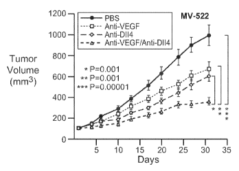
Figure 3: Selective blocking of DLL4 and/or VEGF disrupted tumor angiogenesis
and inhibits tumor growth. a-f, Results of tumor models: HM7 (a), Co1o205 (b),
Calu6 (c),
MDA-MB-435 (d), MV-522 (e) and WEHI3 (f). Mean tumor volumes with SEs are
presented. g-h, Tumor vascular histology studies. Immunohistochemisty of anti-
CD31 in
EL4 tumor sections from control, anti-DLL4 antibody and anti-VEGF treated mice
(g).
Lectin perfusion and anti-CD31 staining in EL4 tumor sections (h). i-p.
Results of tumor
models SK-OV-3X1 (i), LL2 (j), EL4 (k), H1299 (1), SKMES-1(m), MX-1(n), SW620
(o)
and LS174T(p).
Figure 4: DLL4/Notch is dispensable in the homeostasis of mouse intestine.
Immunohistochemical studies of small intestines from control (a, d, g, j),
anti-DLL4
antibody (10 mg/kg, twice weekly for 6 weeks) (b, e, h, k), and DBZ treated
(30 moUkg
daily for 5 days) (c, f, i, 1) mice. As shown by H&E (a, b, c) and Alcian Blue
staining (d, e,
-10-
CA 02654304 2008-12-03
WO 2007/143689 PCT/US2007/070516
f), DBZ caused replacement of the TA population by goblet cells. This change
was entirely
absent from anti-DLL4 antibody treatment. Ki67 (g, h, i) and HES-1 (j, k, 1)
staining further
confirmed that anti-DLL4 antibody failed to replicate the effect of DBZ.
Figure 5: Characterization of anti-DLL4 antibody. a, Epitope mapping of anti-
DLL4 antibody (YW26.82). Schematic representation of a set of DLL4 mutants
expressed
as C-terminal human placental alkaline phosphatase (AP) fusion proteins. 293T
cell
conditioned media containing the fusion proteins were tested on 96-well
microtiter plates
coated with purified anti-DLL4 antibody (YW26.82, 0.5 g/ml). The bound
DLL4.AP was
detected using 1-Step PNPP (Pierce) as substrate and OD 405 nm absorbance
measurement.
b-d, Selective binding of YW26.82 to DLL4. 96-well Nunc Maxisorp plates were
coated
with purified recombinant proteins as indicated (1 g/ml). The binding of
YW26.82 at
indicated concentrations was measured by ELISA assay. Bound antibodies were
detected
with anti-human antibody HRP conjugate using TMB as substrate and OD 450 nm
absorbance measurement. Anti-HER2 and recombinant ErbB2-ECD were used as assay
control (b). FACS analysis of 293 cells transiently transfected with vector,
full length
DLL4, Jagl or DLLl. Significant binding of YW26.82 was only detected on DLL4
transfected cells (top panel). Expression of Jagl and DLLl was confirmed by
the binding of
recombinant rat Notchl -Fc (rrNotchl -Fc, middle panel) and recombinant rat
Notch2-Fc
(rrNotch2-Fc, bottom panel), respectively. YW26.82, rrNotchl-Fc or rrNotch2-Fc
(R& D
system) were used at 2 g/ml followed by goat anti-human IgG-PE (1:500,
Jackson
ImmunoResearch) (c). Anti-DLL4 antibody blocked the binding of DLL4-AP, but
not
DLLl-AP, to coated rNotchl, with a calculated IC50 of -12 nM (left panel).
Anti-DLL4
antibody blocked the binding of DLL4-His, but not Jagl-His, to coated rNotchl,
with a
calculated IC50 of -8 nM (right panel) (d). e, Specific binding of YW26.82 to
endogenously expressed DLL4. FACS analysis of HUVECs transfected with control
or
DLL4-specific siRNA. YW26.82 was used at 2 g/ml, followed by goat anti-human
IgG-PE
(1:500, Jackson ImmunoResearch) (e).
Figure 6: Upregulation of DLL4 by Notch activation. HUVECs were stimulated by
immobilized C-terminal His-tagged human DLL4 (amino acids 1-404) in the
absence or
presence of DBZ (0.08 M). 36 hr after stimulation, endogenous DLL4 expression
was
examined by FACS analysis with anti-DLL4 antibody.
-11-
CA 02654304 2008-12-03
WO 2007/143689 PCT/US2007/070516
DETAILED DESCRIPTION OF THE INVENTION
General techniques
The techniques and procedures described or referenced herein are generally
well
understood and commonly employed using conventional methodology by those
skilled in
the art, such as, for example, the widely utilized methodologies described in
Sambrook et
al., Molecular Cloning: A Laboratory Manual 3rd. edition (2001) Cold Spring
Harbor
Laboratory Press, Cold Spring Harbor, N.Y. CURRENT PROTOCOLS IN MOLECULAR
BIOLOGY (F. M. Ausubel, et al. eds., (2003)); the series METHODS IN ENZYMOLOGY
(Academic Press, Inc.): PCR 2: A PRACTICAL APPROACH (M. J. MacPherson, B. D.
Hames and G. R. Taylor eds. (1995)), Harlow and Lane, eds. (1988) ANTIBODIES,
A
LABORATORY MANUAL, and ANIMAL CELL CULTURE (R. I. Freshney, ed. (1987)).
Definitions
The term "DLL4" (interchangeably termed "Delta-like 4"), as used herein,
refers,
unless specifically or contextually indicated otherwise, to any native or
variant (whether
native or synthetic) DLL4 polypeptide. The term "native sequence" specifically
encompasses naturally occurring truncated or secreted forms (e.g., an
extracellular domain
sequence), naturally occurring variant forms (e.g., alternatively spliced
forms) and
naturally-occurring allelic variants. The term "wild type DLL4" generally
refers to a
polypeptide comprising the amino acid sequence of a naturally occurring DLL4
protein.
The term "wild type DLL4 sequence" generally refers to an amino acid sequence
found in a
naturally occurring DLL4.
The term "Notch receptor" (interchangeably termed "Notch"), as used herein,
refers,
unless specifically or contextually indicated otherwise, to any native or
variant (whether
native or synthetic) Notch receptor polypeptide. Humans have four Notch
receptors
(Notchl, Notch 2, Notch3, and Notch4). As used herein, the term Notch receptor
includes
any one of or all four human Notch receptors. The term "native sequence"
specifically
encompasses naturally occurring truncated or secreted forms (e.g., an
extracellular domain
sequence), naturally occurring variant forms (e.g., alternatively spliced
forms) and
naturally-occurring allelic variants. The term "wild type Notch receptor"
generally refers to
a polypeptide comprising the amino acid sequence of a naturally occurring
Notch receptor
protein. The term "wild type Notch receptor sequence" generally refers to an
amino acid
sequence found in a naturally occurring Notch receptor.
"DLL4 nucleic acid" is RNA or DNA that encodes a DLL4 polypeptide, as defined
above, or which hybridizes to such DNA or RNA and remains stably bound to it
under
-12-
CA 02654304 2008-12-03
WO 2007/143689 PCT/US2007/070516
stringent hybridization conditions and is greater than about 10 nucleotides in
length.
Stringent conditions are those which (1) employ low ionic strength and high
temperature for
washing, for example, 0.15 M NaCI/0.015 M sodium citrate/0.1 % NaDodSO4 at 50
C, or
(2) use during hybridization a denaturing agent such as formamide, for
example, 50%
(vol/vol) formamide with 0.1 % bovine serum albumin/0.1 % FicolU0.1 %
polyvinlypyrrolidone/50 mM sodium phosphate buffer at pH 6.5 with 750 mM
NaC12, 75
mM sodium citrate at 42 C.
A "chimeric DLL4" molecule is a polypeptide comprising full-length DLL4 or one
or more domains thereof fused or bonded to heterologous polypeptide. The
chimeric DLL4
molecule will generally share at least one biological property in common with
naturally
occurring DLL4. An example of a chimeric DLL4 molecule is one that is epitope
tagged
for purification purposes. Another chimeric DLL4 molecule is a DLL4
immunoadhesin.
The term "DLL4 immunoadhesin" is used interchangeably with the term "DLL4-
immunoglobulin chimera", and refers to a chimeric molecule that combines at
least a
portion of a DLL4 molecule (native or variant) with an immunoglobulin
sequence. The
immunoglobulin sequence preferably, but not necessarily, is an immunoglobulin
constant
domain (Fc region). Immunoadhesins can possess many of the valuable chemical
and
biological properties of human antibodies. Since immunoadhesins can be
constructed from
a human protein sequence with a desired specificity linked to an appropriate
human
immunoglobulin hinge and constant domain (Fc) sequence, the binding
specificity of
interest can be achieved using entirely human components. Such immunoadhesins
are
minimally immunogenic to the patient, and are safe for chronic or repeated
use. In some
embodiments, the Fc region is a native sequence Fc region. In some
embodiments, the Fc
region is a variant Fc region. In some embodiments, the Fc region is a
functional Fc region.
Examples of homomultimeric immunoadhesins which have been described for
therapeutic use include the CD4-IgG immunoadhesin for blocking the binding of
HIV to
cell-surface CD4. Data obtained from Phase I clinical trials, in which CD4-IgG
was
administered to pregnant women just before delivery, suggests that this
immunoadhesin
may be useful in the prevention of maternal-fetal transfer of HIV (Ashkenazi
et al., Intern.
Rev. Immunol. 10:219-227 (1993)). An immunoadhesin which binds tumor necrosis
factor
(TNF) has also been developed. TNF is a proinflammatory cytokine which has
been shown
to be a major mediator of septic shock. Based on a mouse model of septic
shock, a TNF
receptor immunoadhesin has shown promise as a candidate for clinical use in
treating septic
shock (Ashkenazi, A. et al. PNAS USA 88:10535-10539 (1991)). ENBREL
(etanercept),
-13-
CA 02654304 2008-12-03
WO 2007/143689 PCT/US2007/070516
an immunoadhesin comprising a TNF receptor sequence fused to an IgG Fc region,
was
approved by the U.S. Food and Drug Administration (FDA), on November 2, 1998,
for the
treatment of rheumatoid arthritis. The new expanded use of ENBREL in the
treatment of
rheumatoid arthritis was approved by FDA on June 6, 2000. For recent
information on TNF
blockers, including ENBREL , see Lovell et al., N. Engl. J. Med. 342:763-169
(2000), and
accompanying editorial on p810-81 l; and Weinblatt et al., N. Engl. J. Med.
340:253-259
(1999); reviewed in Maini and Taylor, Annu. Rev. Med. 51:207-229 (2000).
If the two arms of the immunoadhesin structure have different specificities,
the
immunoadhesin is called a "bispecific immunoadhesin" by analogy to bispecific
antibodies.
Dietsch et al., J. Immunol. Methods 162:123 (1993) describe such a bispecific
immunoadhesin combining the extracellular domains of the adhesion molecules, E-
selectin
and P-selectin, each of which selectins is expressed in a different cell type
in nature.
Binding studies indicated that the bispecific immunoglobulin fusion protein so
formed had
an enhanced ability to bind to a myeloid cell line compared to the
monospecific
immunoadhesins from which it was derived.
The term "heteroadhesin" is used interchangeably with the expression "chimeric
heteromultimer adhesin" and refers to a complex of chimeric molecules (amino
acid
sequences) in which each chimeric molecule combines a biologically active
portion, such as
the extracellular domain of each of the heteromultimeric receptor monomers,
with a
multimerization domain. The "multimerization domain" promotes stable
interaction of the
chimeric molecules within the heteromultimer complex. The multimerization
domains may
interact via an immunoglobulin sequence, leucine zipper, a hydrophobic region,
a
hydrophilic region, or a free thiol that forms an intermolecular disulfide
bond between the
chimeric molecules of the chimeric heteromultimer. The multimerization domain
may
comprise an immunoglobulin constant region. In addition a multimerization
region may be
engineered such that steric interactions not only promote stable interaction,
but further
promote the formation of heterodimers over homodimers from a mixture of
monomers.
"Protuberances" are constructed by replacing small amino acid side chains from
the
interface of the first polypeptide with larger side chains (e.g. tyrosine or
tryptophan).
Compensatory "cavities" of identical or similar size to the protuberances are
optionally
created on the interface of the second polypeptide by replacing large amino
acid side chains
with smaller ones (e.g. alanine or threonine). The immunoglobulin sequence
preferably, but
not necessarily, is an immunoglobulin constant domain. The immunoglobulin
moiety in the
-14-
CA 02654304 2008-12-03
WO 2007/143689 PCT/US2007/070516
chimeras of the present invention may be obtained from IgGi, IgG2, IgG3 or
IgG4 subtypes,
IgA, IgE, IgD or IgM, but preferably IgGi or IgG3.
The term "Fc region" herein is used to define a C-terminal region of an
immunoglobulin heavy chain, including native sequence Fc regions and variant
Fc regions.
Although the boundaries of the Fc region of an immunoglobulin heavy chain
might vary,
the human IgG heavy chain Fc region is usually defined to stretch from an
amino acid
residue at position Cys226, or from Pro230, to the carboxyl-terminus thereof.
The C-
terminal lysine (residue 447 according to the EU numbering system) of the Fc
region may
be removed, for example, during production or purification of the antibody, or
by
recombinantly engineering the nucleic acid encoding a heavy chain of the
antibody.
Accordingly, a composition of intact antibodies may comprise antibody
populations with all
K447 residues removed, antibody populations with no K447 residues removed, and
antibody populations having a mixture of antibodies with and without the K447
residue.
Unless indicated otherwise, herein the numbering of the residues in an
immunoglobulin heavy chain is that of the EU index as in Kabat et al.,
Sequences of
Proteins of Immunological Interest, 5th Ed. Public Health Service, National
Institutes of
Health, Bethesda, MD (1991), expressly incorporated herein by reference. The
"EU index
as in Kabat" refers to the residue numbering of the human IgGl EU antibody.
A "functional Fc region" possesses an "effector function" of a native sequence
Fc
region. Exemplary "effector functions" include Clq binding; complement
dependent
cytotoxicity; Fc receptor binding; antibody-dependent cell-mediated
cytotoxicity (ADCC);
phagocytosis; down regulation of cell surface receptors (e.g. B cell receptor;
BCR), etc.
Such effector functions generally require the Fc region to be combined with a
binding
domain (e.g. an antibody variable domain) and can be assessed using various
assays as
herein disclosed, for example.
A "native sequence Fc region" comprises an amino acid sequence identical to
the
amino acid sequence of an Fc region found in nature. Native sequence human Fc
regions
include a native sequence human IgGl Fc region (non-A and A allotypes); native
sequence
human IgG2 Fc region; native sequence human IgG3 Fc region; and native
sequence human
IgG4 Fc region as well as naturally occurring variants thereof.
A "variant Fc region" comprises an amino acid sequence which differs from that
of
a native sequence Fc region by virtue of at least one amino acid modification,
preferably
one or more amino acid substitution(s). Preferably, the variant Fc region has
at least one
amino acid substitution compared to a native sequence Fc region or to the Fc
region of a
-15-
CA 02654304 2008-12-03
WO 2007/143689 PCT/US2007/070516
parent polypeptide, e.g. from about one to about ten amino acid substitutions,
and
preferably from about one to about five amino acid substitutions in a native
sequence Fc
region or in the Fc region of the parent polypeptide. The variant Fc region
herein will
preferably possess at least about 80% homology with a native sequence Fc
region and/or
with an Fc region of a parent polypeptide, and most preferably at least about
90% homology
therewith, more preferably at least about 95% homology therewith.
An "isolated" antibody is one which has been identified and separated and/or
recovered from a component of its natural environment. Contaminant components
of its
natural environment are materials which would interfere with diagnostic or
therapeutic uses
for the antibody, and may include enzymes, hormones, and other proteinaceous
or
nonproteinaceous solutes. In preferred embodiments, the antibody will be
purified (1) to
greater than 95% by weight of antibody as determined by the Lowry method, and
most
preferably more than 99% by weight, (2) to a degree sufficient to obtain at
least 15 residues
of N-terminal or internal amino acid sequence by use of a spinning cup
sequenator, or (3) to
homogeneity by SDS-PAGE under reducing or nonreducing conditions using
Coomassie
blue or, preferably, silver stain. Isolated antibody includes the antibody in
situ within
recombinant cells since at least one component of the antibody's natural
environment will
not be present. Ordinarily, however, isolated antibody will be prepared by at
least one
purification step.
The terms "antibody" and "immunoglobulin" are used interchangeably in the
broadest sense and include monoclonal antibodies (for e.g., full length or
intact monoclonal
antibodies), polyclonal antibodies, multivalent antibodies, multispecific
antibodies (e.g.,
bispecific antibodies so long as they exhibit the desired biological activity)
and may also
include certain antibody fragments (as described in greater detail herein). An
antibody can
be human, humanized and/or affinity matured.
The term "variable" refers to the fact that certain portions of the variable
domains
differ extensively in sequence among antibodies and are used in the binding
and specificity
of each particular antibody for its particular antigen. However, the
variability is not evenly
distributed throughout the variable domains of antibodies. It is concentrated
in three
segments called complementarity-determining regions (CDRs) or hypervariable
regions
both in the light-chain and the heavy-chain variable domains. The more highly
conserved
portions of variable domains are called the framework (FR). The variable
domains of
native heavy and light chains each comprise four FR regions, largely adopting
a(3-sheet
configuration, connected by three CDRs, which form loops connecting, and in
some cases
-16-
CA 02654304 2008-12-03
WO 2007/143689 PCT/US2007/070516
forming part of, the 0-sheet structure. The CDRs in each chain are held
together in close
proximity by the FR regions and, with the CDRs from the other chain,
contribute to the
formation of the antigen-binding site of antibodies (see Kabat et al.,
Sequences of Proteins
of Immunological Interest, Fifth Edition, National Institute of Health,
Bethesda, MD
(1991)). The constant domains are not involved directly in binding an antibody
to an
antigen, but exhibit various effector functions, such as participation of the
antibody in
antibody-dependent cellular toxicity.
Papain digestion of antibodies produces two identical antigen-binding
fragments,
called "Fab" fragments, each with a single antigen-binding site, and a
residual "Fc"
fragment, whose name reflects its ability to crystallize readily. Pepsin
treatment yields an
F(ab')2 fragment that has two antigen-combining sites and is still capable of
cross-linking
antigen.
"Fv" is the minimum antibody fragment which contains a complete antigen-
recognition and -binding site. In a two-chain Fv species, this region consists
of a dimer of
one heavy- and one light-chain variable domain in tight, non-covalent
association. In a
single-chain Fv species, one heavy- and one light-chain variable domain can be
covalently
linked by a flexible peptide linker such that the light and heavy chains can
associate in a
"dimeric" structure analogous to that in a two-chain Fv species. It is in this
configuration
that the three CDRs of each variable domain interact to define an antigen-
binding site on the
surface of the VH-VL dimer. Collectively, the six CDRs confer antigen-binding
specificity
to the antibody. However, even a single variable domain (or half of an Fv
comprising only
three CDRs specific for an antigen) has the ability to recognize and bind
antigen, although
at a lower affinity than the entire binding site.
The Fab fragment also contains the constant domain of the light chain and the
first
constant domain (CHl) of the heavy chain. Fab' fragments differ from Fab
fragments by
the addition of a few residues at the carboxy terminus of the heavy chain CHl
domain
including one or more cysteines from the antibody hinge region. Fab'-SH is the
designation
herein for Fab' in which the cysteine residue(s) of the constant domains bear
a free thiol
group. F(ab')2 antibody fragments originally were produced as pairs of Fab'
fragments
which have hinge cysteines between them. Other chemical couplings of antibody
fragments
are also known.
The "light chains" of antibodies (immunoglobulins) from any vertebrate species
can
be assigned to one of two clearly distinct types, called kappa (K) and lambda
(k), based on
the amino acid sequences of their constant domains.
-17-
CA 02654304 2008-12-03
WO 2007/143689 PCT/US2007/070516
Depending on the amino acid sequence of the constant domain of their heavy
chains,
immunoglobulins can be assigned to different classes. There are five major
classes of
immunoglobulins: IgA, IgD, IgE, IgG, and IgM, and several of these can be
further divided
into subclasses (isotypes), e.g., IgGl, IgG2, IgG3, IgG4, IgAl, and IgA2. The
heavy-chain
constant domains that correspond to the different classes of immunoglobulins
are called a,
8, E, y, and , respectively. The subunit structures and three-dimensional
configurations of
different classes of immunoglobulins are well known.
"Antibody fragments" comprise only a portion of an intact antibody, wherein
the
portion preferably retains at least one, preferably most or all, of the
functions normally
associated with that portion when present in an intact antibody. Examples of
antibody
fragments include Fab, Fab', F(ab')2, and Fv fragments; diabodies; linear
antibodies; single-
chain antibody molecules; and multispecific antibodies formed from antibody
fragments. In
one embodiment, an antibody fragment comprises an antigen binding site of the
intact
antibody and thus retains the ability to bind antigen. In another embodiment,
an antibody
fragment, for example one that comprises the Fc region, retains at least one
of the biological
functions normally associated with the Fc region when present in an intact
antibody, such as
FcRn binding, antibody half life modulation, ADCC function and complement
binding. In
one embodiment, an antibody fragment is a monovalent antibody that has an in
vivo half
life substantially similar to an intact antibody. For e.g., such an antibody
fragment may
comprise on antigen binding arm linked to an Fc sequence capable of conferring
in vivo
stability to the fragment.
The term "hypervariable region", "HVR", or "HV", when used herein refers to
the
regions of an antibody variable domain which are hypervariable in sequence
and/or form
structurally defined loops. Generally, antibodies comprise six hypervariable
regions; three
in the VH (Hl, H2, H3), and three in the VL (Ll, L2, L3). A number of
hypervariable
region delineations are in use and are encompassed herein. The Kabat
Complementarity
Determining Regions (CDRs) are based on sequence variability and are the most
commonly
used (Kabat et al., Sequences of Proteins of Immunological Interest, 5th Ed.
Public Health
Service, National Institutes of Health, Bethesda, MD. (1991)). Chothia refers
instead to the
location of the structural loops (Chothia and Lesk J. Mol. Biol. 196:901-917
(1987)). The
AbM hypervariable regions represent a compromise between the Kabat CDRs and
Chothia
structural loops, and are used by Oxford Molecular's AbM antibody modeling
software. The
"contact" hypervariable regions are based on an analysis of the available
complex crystal
structures. The residues from each of these hypervariable regions are noted
below.
-18-
CA 02654304 2008-12-03
WO 2007/143689 PCT/US2007/070516
Loop Kabat AbM Chothia Contact
---- ----- --- ------- -------
L1 L24-L34 L24-L34 L26-L32 L30-L36
L2 L50-L56 L50-L56 L50-L52 L46-L55
L3 L89-L97 L89-L97 L91-L96 L89-L96
H1 H31-H35B H26-H35B H26-H32 H30-H35B
(Kabat Numbering)
H1 H31-H35 H26-H35 H26-H32 H30-H35
(Chothia Numbering)
H2 H50-H65 H50-H58 H53-H55 H47-H58
H3 H95-H102 H95-H102 H96-H101 H93-H101
Hypervariable regions may comprise "extended hypervariable regions" as
follows:
24-36 or 24-34 (Ll), 46-56 or 50-56 (L2) and 89-97 (L3) in the VL and 26-35
(Hl), 50-65
or 49-65 (H2) and 93-102, 94-102 or 95-102 (H3) in the VH. The variable domain
residues
are numbered according to Kabat et al., supra for each of these definitions.
"Framework" or "FR" residues are those variable domain residues other than the
hypervariable region residues as herein defined.
The term "monoclonal antibody" as used herein refers to an antibody from a
population of substantially homogeneous antibodies, i.e., the individual
antibodies
comprising the population are identical and/or bind the same epitope(s),
except for possible
variants that may arise during production of the monoclonal antibody, such
variants
generally being present in minor amounts. Such monoclonal antibody typically
includes
an antibody comprising a polypeptide sequence that binds a target, wherein the
target-
binding polypeptide sequence was obtained by a process that includes the
selection of a
single target binding polypeptide sequence from a plurality of polypeptide
sequences. For
example, the selection process can be the selection of a unique clone from a
plurality of
clones, such as a pool of hybridoma clones, phage clones or recombinant DNA
clones. It
should be understood that the selected target binding sequence can be further
altered, for
example, to improve affinity for the target, to humanize the target binding
sequence, to
improve its production in cell culture, to reduce its immunogenicity in vivo,
to create a
multispecific antibody, etc., and that an antibody comprising the altered
target binding
sequence is also a monoclonal antibody of this invention. In contrast to
polyclonal antibody
preparations which typically include different antibodies directed against
different
-19-
CA 02654304 2008-12-03
WO 2007/143689 PCT/US2007/070516
determinants (epitopes), each monoclonal antibody of a monoclonal antibody
preparation is
directed against a single determinant on an antigen. In addition to their
specificity, the
monoclonal antibody preparations are advantageous in that they are typically
uncontaminated by other immunoglobulins. The modifier "monoclonal" indicates
the
character of the antibody as being obtained from a substantially homogeneous
population of
antibodies, and is not to be construed as requiring production of the antibody
by any
particular method. For example, the monoclonal antibodies to be used in
accordance with
the present invention may be made by a variety of techniques, including, for
example, the
hybridoma method (e.g., Kohler et al., Nature, 256:495 (1975); Harlow et al.,
Antibodies: A
Laboratory Manual, (Cold Spring Harbor Laboratory Press, 2nd ed. 1988);
Hammerling et
al., in: Monoclonal Antibodies and T-Cell Hybridomas 563-68 1, (Elsevier,
N.Y., 1981)),
recombinant DNA methods (see, e.g., U.S. Patent No. 4,816,567), phage display
technologies (see, e.g., Clackson et al., Nature, 352:624-628 (1991); Marks et
al., J. Mol.
Biol., 222:581-597 (1991); Sidhu et al., J. Mol. Biol. 338(2):299-310 (2004);
Lee et al.,
J.Mol.Biol.340(5):1073-1093 (2004); Fellouse, Proc. Nat. Acad. Sci. USA
101(34):12467-
12472 (2004); and Lee et al. J. Immunol. Methods 284(1-2):119-132 (2004), and
technologies for producing human or human-like antibodies in animals that have
parts or all
of the human immunoglobulin loci or genes encoding human immunoglobulin
sequences
(see, e.g., WO 1998/24893; WO 1996/34096; WO 1996/33735; WO 1991/10741;
Jakobovits et al., Proc. Natl. Acad. Sci. USA, 90:2551 (1993); Jakobovits et
al., Nature,
362:255-258 (1993); Bruggemann et al., Year in Immuno., 7:33 (1993); U.S.
Patent Nos.
5,545,806; 5,569,825; 5,591,669 (all of GenPharm); 5,545,807; WO 1997/17852;
U.S.
Patent Nos. 5,545,807; 5,545,806; 5,569,825; 5,625,126; 5,633,425; and
5,661,016; Marks
et al., Bio/Technology, 10: 779-783 (1992); Lonberg et al., Nature, 368: 856-
859 (1994);
Morrison, Nature, 368: 812-813 (1994); Fishwild et al., Nature Biotechnology,
14: 845-851
(1996); Neuberger, Nature Biotechnology, 14: 826 (1996); and Lonberg and
Huszar, Intern.
Rev. Immunol., 13: 65-93 (1995).
"Humanized" forms of non-human (e.g., murine) antibodies are chimeric
antibodies
that contain minimal sequence derived from non-human immunoglobulin. For the
most
part, humanized antibodies are human immunoglobulins (recipient antibody) in
which
residues from a hypervariable region of the recipient are replaced by residues
from a
hypervariable region of a non-human species (donor antibody) such as mouse,
rat, rabbit or
nonhuman primate having the desired specificity, affinity, and capacity. In
some instances,
framework region (FR) residues of the human immunoglobulin are replaced by
-20-
CA 02654304 2008-12-03
WO 2007/143689 PCT/US2007/070516
corresponding non-human residues. Furthermore, humanized antibodies may
comprise
residues that are not found in the recipient antibody or in the donor
antibody. These
modifications are made to further refine antibody performance. In general, the
humanized
antibody will comprise substantially all of at least one, and typically two,
variable domains,
in which all or substantially all of the hypervariable loops correspond to
those of a non-
human immunoglobulin and all or substantially all of the FRs are those of a
human
immunoglobulin sequence. The humanized antibody optionally will also comprise
at least a
portion of an immunoglobulin constant region (Fc), typically that of a human
immunoglobulin. For further details, see Jones et al., Nature 321:522-525
(1986);
Riechmann et al., Nature 332:323-329 (1988); and Presta, Curr. Op. Struct.
Biol. 2:593-596
(1992). See also the following review articles and references cited therein:
Vaswani and
Hamilton, Ann. Allergy, Asthma & Immunol. 1:105-115 (1998); Harris, Biochem.
Soc.
Transactions 23:1035-1038 (1995); Hurle and Gross, Curr. Op. Biotech. 5:428-
433 (1994).
"Chimeric" antibodies (immunoglobulins) have a portion of the heavy and/or
light
chain identical with or homologous to corresponding sequences in antibodies
derived from a
particular species or belonging to a particular antibody class or subclass,
while the
remainder of the chain(s) is identical with or homologous to corresponding
sequences in
antibodies derived from another species or belonging to another antibody class
or subclass,
as well as fragments of such antibodies, so long as they exhibit the desired
biological
activity (U.S. Patent No. 4,816,567; and Morrison et al., Proc. Natl. Acad.
Sci. USA
81:6851-6855 (1984)). Humanized antibody as used herein is a subset of
chimeric
antibodies.
"Single-chain Fv" or "scFv" antibody fragments comprise the VH and VL domains
of antibody, wherein these domains are present in a single polypeptide chain.
Generally,
the scFv polypeptide further comprises a polypeptide linker between the VH and
VL
domains which enables the scFv to form the desired structure for antigen
binding. For a
review of scFv see Pluckthun, in The Pharmacology of Monoclonal Antibodies,
vol. 113,
Rosenburg and Moore eds., Springer-Verlag, New York, pp. 269-315 (1994).
An "antigen" is a predetermined antigen to which an antibody can selectively
bind.
The target antigen may be polypeptide, carbohydrate, nucleic acid, lipid,
hapten or other
naturally occurring or synthetic compound. Preferably, the target antigen is a
polypeptide.
The term "diabodies" refers to small antibody fragments with two antigen-
binding
sites, which fragments comprise a heavy-chain variable domain (VH) connected
to a light-
chain variable domain (VL) in the same polypeptide chain (VH - VL). By using a
linker
-21-
CA 02654304 2008-12-03
WO 2007/143689 PCT/US2007/070516
that is too short to allow pairing between the two domains on the same chain,
the domains
are forced to pair with the complementary domains of another chain and create
two antigen-
binding sites. Diabodies are described more fully in, for example, EP 404,097;
WO
93/11161; and Hollinger et al., Proc. Natl. Acad. Sci. USA, 90:6444-6448
(1993).
A "human antibody" is one which possesses an amino acid sequence which
corresponds to that of an antibody produced by a human and/or has been made
using any of
the techniques for making human antibodies as disclosed herein. This
definition of a human
antibody specifically excludes a humanized antibody comprising non-human
antigen-
binding residues.
An "affinity matured" antibody is one with one or more alterations in one or
more
CDRs thereof which result in an improvement in the affinity of the antibody
for antigen,
compared to a parent antibody which does not possess those alteration(s).
Preferred
affinity matured antibodies will have nanomolar or even picomolar affinities
for the target
antigen. Affinity matured antibodies are produced by procedures known in the
art. Marks
et al. Bio/Technology 10:779-783 (1992) describes affinity maturation by VH
and VL
domain shuffling. Random mutagenesis of CDR and/or framework residues is
described
by: Barbas et al. Proc Nat. Acad. Sci, USA 91:3809-3813 (1994); Schier et al.
Gene
169:147-155 (1995); Yelton et al. J. Immunol. 155:1994-2004 (1995); Jackson et
al., J.
Immunol. 154(7):3310-9 (1995); and Hawkins et al, J. Mol. Biol. 226:889-896
(1992).
Antibody "effector functions" refer to those biological activities
attributable to the
Fc region (a native sequence Fc region or amino acid sequence variant Fc
region) of an
antibody, and vary with the antibody isotype. Examples of antibody effector
functions
include: Clq binding and complement dependent cytotoxicity; Fc receptor
binding;
antibody-dependent cell-mediated cytotoxicity (ADCC); phagocytosis; down
regulation of
cell surface receptors (e.g. B cell receptor); and B cell activation.
Antibody-dependent cell-mediated cytotoxicity" or "ADCC" refers to a form of
cytotoxicity in which secreted Ig bound onto Fc receptors (FcRs) present on
certain
cytotoxic cells (e.g. Natural Killer (NK) cells, neutrophils, and macrophages)
enable these
cytotoxic effector cells to bind specifically to an antigen-bearing target
cell and
subsequently kill the target cell with cytotoxins. The antibodies "arm" the
cytotoxic cells
and are absolutely required for such killing. The primary cells for mediating
ADCC, NK
cells, express FcyRIII only, whereas monocytes express FcyRI, FcyRII and
FcyRIII. FcR
expression on hematopoietic cells is summarized in Table 3 on page 464 of
Ravetch and
-22-
CA 02654304 2008-12-03
WO 2007/143689 PCT/US2007/070516
Kinet, Annu. Rev. Immuno19:457-92 (1991). To assess ADCC activity of a
molecule of
interest, an in vitro ADCC assay, such as that described in US Patent No.
5,500,362 or
5,821,337 or Presta U.S. Patent No. 6,737,056 may be performed. Useful
effector cells for
such assays include peripheral blood mononuclear cells (PBMC) and Natural
Killer (NK)
cells. Alternatively, or additionally, ADCC activity of the molecule of
interest may be
assessed in vivo, e.g., in a animal model such as that disclosed in Clynes et
al. PNAS (USA)
95:652-656 (1998).
"Human effector cells" are leukocytes which express one or more FcRs and
perform
effector functions. Preferably, the cells express at least FcyRIII and perform
ADCC effector
function. Examples of human leukocytes which mediate ADCC include peripheral
blood
mononuclear cells (PBMC), natural killer (NK) cells, monocytes, cytotoxic T
cells and
neutrophils; with PBMCs and NK cells being preferred. The effector cells may
be isolated
from a native source, e.g. from blood.
"Fc receptor" or "FcR" describes a receptor that binds to the Fc region of an
antibody. The preferred FcR is a native sequence human FcR. Moreover, a
preferred FcR
is one which binds an IgG antibody (a gamma receptor) and includes receptors
of the FcyRI,
FcyRII, and FcyRIII subclasses, including allelic variants and alternatively
spliced forms of
these receptors. FcyRII receptors include FcyRIIA (an "activating receptor")
and FcyRIIB
(an "inhibiting receptor"), which have similar amino acid sequences that
differ primarily in
the cytoplasmic domains thereof. Activating receptor FcyRIIA contains an
immunoreceptor
tyrosine-based activation motif (ITAM) in its cytoplasmic domain. Inhibiting
receptor
FcyRIIB contains an immunoreceptor tyrosine-based inhibition motif (ITIM) in
its
cytoplasmic domain. (see review M. in Daeron, Annu. Rev. Immunol. 15:203-234
(1997)).
FcRs are reviewed in Ravetch and Kinet, Annu. Rev. Immuno19:457-92 (1991);
Capel et
al., Immunomethods 4:25-34 (1994); and de Haas et al., J. Lab. Clin. Med.
126:330-41
(1995). Other FcRs, including those to be identified in the future, are
encompassed by the
term "FcR" herein. The term also includes the neonatal receptor, FcRn, which
is
responsible for the transfer of maternal IgGs to the fetus (Guyer et al., J.
Immunol. 117:587
(1976) and Kim et al., J. Immunol. 24:249 (1994)) and regulates homeostasis of
immunoglobulins.
W000/42072 (Presta) describes antibody variants with improved or diminished
binding to FcRs. The content of that patent publication is specifically
incorporated herein
by reference. See, also, Shields et al. J. Biol. Chem. 9(2): 6591-6604 (2001).
-23-
CA 02654304 2008-12-03
WO 2007/143689 PCT/US2007/070516
Methods of measuring binding to FcRn are known (see, e.g., Ghetie 1997, Hinton
2004). Binding to human FcRn in vivo and serum half life of human FcRn high
affinity
binding polypeptides can be assayed, e.g., in transgenic mice or transfected
human cell lines
expressing human FcRn, or in primates administered with the Fc variant
polypeptides.
"Complement dependent cytotoxicity" or "CDC" refers to the lysis of a target
cell in
the presence of complement. Activation of the classical complement pathway is
initiated by
the binding of the first component of the complement system (C l q) to
antibodies (of the
appropriate subclass) which are bound to their cognate antigen. To assess
complement
activation, a CDC assay, e.g. as described in Gazzano-Santoro et al., J.
Immunol. Methods
202:163 (1996), may be performed.
Polypeptide variants with altered Fc region amino acid sequences and increased
or
decreased C l q binding capability are described in US patent No. 6,194,551 B
1 and
W099/51642. The contents of those patent publications are specifically
incorporated herein
by reference. See, also, Idusogie et al. J. Immunol. 164: 4178-4184 (2000).
The term "Fc region-comprising polypeptide" refers to a polypeptide, such as
an
antibody or immunoadhesin (see definitions below), which comprises an Fc
region. The C-
terminal lysine (residue 447 according to the EU numbering system) of the Fc
region may
be removed, for example, during purification of the polypeptide or by
recombinant
engineering the nucleic acid encoding the polypeptide. Accordingly, a
composition
comprising a polypeptide having an Fc region according to this invention can
comprise
polypeptides with K447, with all K447 removed, or a mixture of polypeptides
with and
without the K447 residue.
A "blocking" antibody or an "antagonist" antibody is one which inhibits or
reduces
biological activity of the antigen it binds. Preferred blocking antibodies or
antagonist
antibodies substantially or completely inhibit the biological activity of the
antigen.
"Chronic" administration refers to administration of the agent(s) in a
continuous
mode as opposed to an acute mode, so as to maintain the initial therapeutic
effect (activity)
for an extended period of time. "Intermittent" administration is treatment
that is not
consecutively done without interruption, but rather is cyclic in nature.
A "disorder" or "disease" is any condition that would benefit from treatment
with a
substance/molecule or method of the invention. This includes chronic and acute
disorders
or diseases including those pathological conditions which predispose the
mammal to the
disorder in question. Non-limiting examples of disorders to be treated herein
include
malignant and benign tumors; carcinoma, blastoma, and sarcoma.
-24-
CA 02654304 2008-12-03
WO 2007/143689 PCT/US2007/070516
The terms "cell proliferative disorder" and "proliferative disorder" refer to
disorders
that are associated with some degree of abnormal cell proliferation. In one
embodiment, the
cell proliferative disorder is cancer.
"Tumor", as used herein, refers to all neoplastic cell growth and
proliferation,
whether malignant or benign, and all pre-cancerous and cancerous cells and
tissues. The
terms "cancer", "cancerous", "cell proliferative disorder", "proliferative
disorder" and
"tumor" are not mutually exclusive as referred to herein.
The terms "cancer" and "cancerous" refer to or describe the physiological
condition
in mammals that is typically characterized by unregulated cell
growth/proliferation.
Examples of cancer include but are not limited to, carcinoma, lymphoma,
blastoma,
sarcoma, and leukemia. More particular examples of such cancers include
squamous cell
cancer, small-cell lung cancer, non-small cell lung cancer, adenocarcinoma of
the lung,
squamous carcinoma of the lung, cancer of the peritoneum, hepatocellular
cancer,
gastrointestinal cancer, pancreatic cancer, glioblastoma, cervical cancer,
ovarian cancer,
liver cancer, bladder cancer, hepatoma, breast cancer, colon cancer,
colorectal cancer,
endometrial or uterine carcinoma, salivary gland carcinoma, kidney cancer,
liver cancer,
prostate cancer, vulval cancer, thyroid cancer, hepatic carcinoma, gastric
cancer, melanoma,
and various types of head and neck cancer. Dysregulation of angiogenesis can
lead to many
disorders that can be treated by compositions and methods of the invention.
These
disorders include both non-neoplastic and neoplastic conditions. Neoplastics
include but
are not limited those described above. Non-neoplastic disorders include but
are not limited
to undesired or aberrant hypertrophy, arthritis, rheumatoid arthritis (RA),
psoriasis, psoriatic
plaques, sarcoidosis, atherosclerosis, atherosclerotic plaques, diabetic and
other proliferative
retinopathies including retinopathy of prematurity, retrolental fibroplasia,
neovascular
glaucoma, age-related macular degeneration, diabetic macular edema, comeal
neovascularization, comeal graft neovascularization, comeal graft rejection,
retinal/choroidal neovascularization, neovascularization of the angle
(rubeosis), ocular
neovascular disease, vascular restenosis, arteriovenous malformations (AVM),
meningioma,
hemangioma, angiofibroma, thyroid hyperplasias (including Grave's disease),
comeal and
other tissue transplantation, chronic inflammation, lung inflammation, acute
lung
injury/ARDS, sepsis, primary pulmonary hypertension, malignant pulmonary
effusions,
cerebral edema (e.g., associated with acute stroke/ closed head injury/
trauma), synovial
inflammation, pannus formation in RA, myositis ossificans, hypertropic bone
formation,
osteoarthritis (OA), refractory ascites, polycystic ovarian disease,
endometriosis, 3rd
-25-
CA 02654304 2008-12-03
WO 2007/143689 PCT/US2007/070516
spacing of fluid diseases (pancreatitis, compartment syndrome, bums, bowel
disease),
uterine fibroids, premature labor, chronic inflammation such as IBD (Crohn's
disease and
ulcerative colitis), renal allograft rejection, inflammatory bowel disease,
nephrotic
syndrome, undesired or aberrant tissue mass growth (non-cancer), hemophilic
joints,
hypertrophic scars, inhibition of hair growth, Osler-Weber syndrome, pyogenic
granuloma
retrolental fibroplasias, scleroderma, trachoma, vascular adhesions,
synovitis, dermatitis,
preeclampsia, ascites, pericardial effusion (such as that associated with
pericarditis), and
pleural effusion.
As used herein, "treatment" refers to clinical intervention in an attempt to
alter the
natural course of the individual or cell being treated, and can be performed
either for
prophylaxis or during the course of clinical pathology. Desirable effects of
treatment
include preventing occurrence or recurrence of disease, alleviation of
symptoms,
diminishment of any direct or indirect pathological consequences of the
disease, preventing
metastasis, decreasing the rate of disease progression, amelioration or
palliation of the
disease state, and remission or improved prognosis. In some embodiments,
antibodies are
used to delay development of a disease or disorder.
An "individual" is a vertebrate, preferably a mammal, more preferably a human.
Mammals include, but are not limited to, farm animals (such as cows), sport
animals, pets
(such as cats, dogs and horses), primates, mice and rats.
"Mammal" for purposes of treatment refers to any animal classified as a
mammal,
including humans, domestic and farm animals, and zoo, sports, or pet animals,
such as dogs,
horses, cats, cows, etc. Preferably, the mammal is human.
An "effective amount" refers to an amount effective, at dosages and for
periods of
time necessary, to achieve the desired therapeutic or prophylactic result.
A "therapeutically effective amount" of a substance/molecule may vary
according to
factors such as the disease state, age, sex, and weight of the individual, and
the ability of the
substance/molecule, agonist or antagonist to elicit a desired response in the
individual. A
therapeutically effective amount is also one in which any toxic or detrimental
effects of the
substance/molecule, agonist or antagonist are outweighed by the
therapeutically beneficial
effects. A "prophylactically effective amount" refers to an amount effective,
at dosages and
for periods of time necessary, to achieve the desired prophylactic result.
Typically but not
necessarily, since a prophylactic dose is used in subjects prior to or at an
earlier stage of
disease, the prophylactically effective amount will be less than the
therapeutically effective
amount.
-26-
CA 02654304 2008-12-03
WO 2007/143689 PCT/US2007/070516
The term "cytotoxic agent" as used herein refers to a substance that inhibits
or
prevents the function of cells and/or causes destruction of cells. The term is
intended to
include radioactive isotopes (e.g., At2ll, I131, Il2s, Y90, Re186, Relgg, Sm
153, Bi212, P32 and
radioactive isotopes of Lu), chemotherapeutic agents e.g. methotrexate,
adriamicin, vinca
alkaloids (vincristine, vinblastine, etoposide), doxorubicin, melphalan,
mitomycin C,
chlorambucil, daunorubicin or other intercalating agents, enzymes and
fragments thereof
such as nucleolytic enzymes, antibiotics, and toxins such as small molecule
toxins or
enzymatically active toxins of bacterial, fungal, plant or animal origin,
including fragments
and/or variants thereof, and the various antitumor or anticancer agents
disclosed below.
Other cytotoxic agents are described below. A tumoricidal agent causes
destruction of
tumor cells.
A "chemotherapeutic agent" is a chemical compound useful in the treatment of
cancer. Examples of chemotherapeutic agents include alkylating agents such as
thiotepa and
CYTOXAN cyclosphosphamide; alkyl sulfonates such as busulfan, improsulfan and
piposulfan; aziridines such as benzodopa, carboquone, meturedopa, and uredopa;
ethylenimines and methylamelamines including altretamine, triethylenemelamine,
trietylenephosphoramide, triethiylenethiophosphoramide and
trimethylolomelamine;
acetogenins (especially bullatacin and bullatacinone); delta-9-
tetrahydrocannabinol
(dronabinol, MARINOL ); beta-lapachone; lapachol; colchicines; betulinic acid;
a
camptothecin (including the synthetic analogue topotecan (HYCAMTIN ), CPT-11
(irinotecan, CAMPTOSAR ), acetylcamptothecin, scopolectin, and 9-
aminocamptothecin);
bryostatin; callystatin; CC-1065 (including its adozelesin, carzelesin and
bizelesin synthetic
analogues); podophyllotoxin; podophyllinic acid; teniposide; cryptophycins
(particularly
cryptophycin 1 and cryptophycin 8); dolastatin; duocarmycin (including the
synthetic
analogues, KW-2189 and CBl-TMl); eleutherobin; pancratistatin; a sarcodictyin;
spongistatin; nitrogen mustards such as chlorambucil, chlomaphazine,
cholophosphamide,
estramustine, ifosfamide, mechlorethamine, mechlorethamine oxide
hydrochloride,
melphalan, novembichin, phenesterine, prednimustine, trofosfamide, uracil
mustard;
nitrosureas such as carmustine, chlorozotocin, fotemustine, lomustine,
nimustine, and
ranimnustine; antibiotics such as the enediyne antibiotics (e. g.,
calicheamicin, especially
calicheamicin gammall and calicheamicin omegall (see, e.g., Agnew, Chem Intl.
Ed.
Engl., 33: 183-186 (1994)); dynemicin, including dynemicin A; an esperamicin;
as well as
neocarzinostatin chromophore and related chromoprotein enediyne antiobiotic
chromophores), aclacinomysins, actinomycin, authramycin, azaserine,
bleomycins,
-27-
CA 02654304 2008-12-03
WO 2007/143689 PCT/US2007/070516
cactinomycin, carabicin, carminomycin, carzinophilin, chromomycinis,
dactinomycin,
daunorubicin, detorubicin, 6-diazo-5-oxo-L-norleucine, ADRIAMYCIN doxorubicin
(including morpholino-doxorubicin, cyanomorpholino-doxorubicin, 2-pyrrolino-
doxorubicin and deoxydoxorubicin), epirubicin, esorubicin, idarubicin,
marcellomycin,
mitomycins such as mitomycin C, mycophenolic acid, nogalamycin, olivomycins,
peplomycin, potfiromycin, puromycin, quelamycin, rodorubicin, streptonigrin,
streptozocin,
tubercidin, ubenimex, zinostatin, zorubicin; anti-metabolites such as
methotrexate and 5-
fluorouracil (5-FU); folic acid analogues such as denopterin, methotrexate,
pteropterin,
trimetrexate; purine analogs such as fludarabine, 6-mercaptopurine,
thiamiprine,
thioguanine; pyrimidine analogs such as ancitabine, azacitidine, 6-azauridine,
carmofur,
cytarabine, dideoxyuridine, doxifluridine, enocitabine, floxuridine; androgens
such as
calusterone, dromostanolone propionate, epitiostanol, mepitiostane,
testolactone; anti-
adrenals such as aminoglutethimide, mitotane, trilostane; folic acid
replenisher such as
frolinic acid; aceglatone; aldophosphamide glycoside; aminolevulinic acid;
eniluracil;
amsacrine; bestrabucil; bisantrene; edatraxate; defofamine; demecolcine;
diaziquone;
elfomithine; elliptinium acetate; an epothilone; etoglucid; gallium nitrate;
hydroxyurea;
lentinan; lonidainine; maytansinoids such as maytansine and ansamitocins;
mitoguazone;
mitoxantrone; mopidanmol; nitraerine; pentostatin; phenamet; pirarubicin;
losoxantrone; 2-
ethylhydrazide; procarbazine; PSK polysaccharide complex (JHS Natural
Products,
Eugene, OR); razoxane; rhizoxin; sizofiran; spirogermanium; tenuazonic acid;
triaziquone;
2,2',2"-trichlorotriethylamine; trichothecenes (especially T-2 toxin,
verracurin A, roridin A
and anguidine); urethan; vindesine (ELDISINE , FILDESIN ); dacarbazine;
mannomustine; mitobronitol; mitolactol; pipobroman; gacytosine; arabinoside
("Ara-C");
thiotepa; taxoids, e.g., TAXOL paclitaxel (Bristol-Myers Squibb Oncology,
Princeton,
N.J.), ABRAXANETM Cremophor-free, albumin-engineered nanoparticle formulation
of
paclitaxel (American Pharmaceutical Partners, Schaumberg, Illinois), and
TAXOTERE
doxetaxel (Rh6ne-Poulenc Rorer, Antony, France); chloranbucil; gemcitabine
(GEMZAR ); 6-thioguanine; mercaptopurine; methotrexate; platinum analogs such
as
cisplatin and carboplatin; vinblastine (VELBAN ); platinum; etoposide (VP-16);
ifosfamide; mitoxantrone; vincristine (ONCOVIN ); oxaliplatin; leucovovin;
vinorelbine
(NAVELBINE ); novantrone; edatrexate; daunomycin; aminopterin; ibandronate;
topoisomerase inhibitor RFS 2000; difluorometlhylomithine (DMFO); retinoids
such as
retinoic acid; capecitabine (XELODA ); pharmaceutically acceptable salts,
acids or
derivatives of any of the above; as well as combinations of two or more of the
above such as
-28-
CA 02654304 2008-12-03
WO 2007/143689 PCT/US2007/070516
CHOP, an abbreviation for a combined therapy of cyclophosphamide, doxorubicin,
vincristine, and prednisolone, and FOLFOX, an abbreviation for a treatment
regimen with
oxaliplatin (ELOXATINTM) combined with 5-FU and leucovovin.
Also included in this definition are anti-hormonal agents that act to
regulate, reduce,
block, or inhibit the effects of hormones that can promote the growth of
cancer, and are
often in the form of systemic, or whole-body treatment. They may be hormones
themselves. Examples include anti-estrogens and selective estrogen receptor
modulators
(SERMs), including, for example, tamoxifen (including NOLVADEX tamoxifen),
EVISTA raloxifene, droloxifene, 4-hydroxytamoxifen, trioxifene, keoxifene,
LY117018,
onapristone, and FARESTON toremifene; anti-progesterones; estrogen receptor
down-
regulators (ERDs); agents that function to suppress or shut down the ovaries,
for example,
leutinizing hormone-releasing hormone (LHRH) agonists such as LUPRON and
ELIGARD leuprolide acetate, goserelin acetate, buserelin acetate and
tripterelin; other
anti-androgens such as flutamide, nilutamide and bicalutamide; and aromatase
inhibitors
that inhibit the enzyme aromatase, which regulates estrogen production in the
adrenal
glands, such as, for example, 4(5)-imidazoles, aminoglutethimide, MEGASE
megestrol
acetate, AROMASIN exemestane, formestanie, fadrozole, RIVISOR vorozole,
FEMARA letrozole, and ARIMIDEX anastrozole. In addition, such definition of
chemotherapeutic agents includes bisphosphonates such as clodronate (for
example,
BONEFOS or OSTAC ), DIDROCAL etidronate, NE-58095, ZOMETA zoledronic
acid/zoledronate, FOSAMAX alendronate, AREDIA pamidronate, SKELID
tiludronate, or ACTONEL risedronate; as well as troxacitabine (a 1,3-
dioxolane
nucleoside cytosine analog); antisense oligonucleotides, particularly those
that inhibit
expression of genes in signaling pathways implicated in abherant cell
proliferation, such as,
for example, PKC-alpha, Raf, H-Ras, and epidermal growth factor receptor (EGF-
R);
vaccines such as THERATOPE vaccine and gene therapy vaccines, for example,
ALLOVECTIN vaccine, LEUVECTIN vaccine, and VAXID vaccine;
LURTOTECAN topoisomerase 1 inhibitor; ABARELIX rmRH; lapatinib ditosylate
(an
ErbB-2 and EGFR dual tyrosine kinase small-molecule inhibitor also known as
GW572016); and pharmaceutically acceptable salts, acids or derivatives of any
of the
above.
A"growth inhibitory agent" when used herein refers to a compound or
composition
which inhibits growth of a cell (such as a cell expressing DLL4) either in
vitro or in vivo.
Thus, the growth inhibitory agent may be one which significantly reduces the
percentage of
-29-
CA 02654304 2008-12-03
WO 2007/143689 PCT/US2007/070516
cells (such as a cell expressing DLL4) in S phase. Examples of growth
inhibitory agents
include agents that block cell cycle progression (at a place other than S
phase), such as
agents that induce Gl arrest and M-phase arrest. Classical M-phase blockers
include the
vincas (vincristine and vinblastine), taxanes, and topoisomerase II inhibitors
such as
doxorubicin, epirubicin, daunorubicin, etoposide, and bleomycin. Those agents
that arrest
Gl also spill over into S-phase arrest, for example, DNA alkylating agents
such as
tamoxifen, prednisone, dacarbazine, mechlorethamine, cisplatin, methotrexate,
5-
fluorouracil, and ara-C. Further information can be found in The Molecular
Basis of
Cancer, Mendelsohn and Israel, eds., Chapter 1, entitled "Cell cycle
regulation, oncogenes,
and antineoplastic drugs" by Murakami et al. (WB Saunders: Philadelphia,
1995), especially
p. 13. The taxanes (paclitaxel and docetaxel) are anticancer drugs both
derived from the
yew tree. Docetaxel (TAXOTERE , Rhone-Poulenc Rorer), derived from the
European
yew, is a semisynthetic analogue of paclitaxel (TAXOL , Bristol-Myers Squibb).
Paclitaxel and docetaxel promote the assembly of microtubules from tubulin
dimers and
stabilize microtubules by preventing depolymerization, which results in the
inhibition of
mitosis in cells.
"Doxorubicin" is an anthracycline antibiotic. The full chemical name of
doxorubicin is (8S-cis)-10-[(3-amino-2,3,6-trideoxy-a-L-lyxo-
hexapyranosyl)oxy]-7,8,9,10-
tetrahydro-6,8,11-trihydroxy-8-(hydroxyacetyl)-1-methoxy-5,12-
naphthacenedione.
An "intraocular neovascular disease" is a disease characterized by ocular
neovascularization. Examples of intraocular neovascular diseases include, but
are not
limited to, proliferative retinopathies, choroidal neovascularization (CNV),
age-related
macular degeneration (AMD), diabetic and other ischemia-related retinopathies,
diabetic
macular edema, pathological myopia, von Hippel-Lindau disease, histoplasmosis
of the eye,
retinal vein occlusions, including Central Retinal Vein Occlusion (CRVO),
comeal
neovascularization, retinal neovascularization, etc.
The "pathology" of a disease includes all phenomena that compromise the well-
being of the patient. For cancer, this includes, without limitation, abnormal
or
uncontrollable cell growth, metastasis, interference with the normal
functioning of
neighboring cells, release of cytokines or other secretory products at
abnormal levels,
suppression or aggravation of inflammatory or immunological response, etc.
Administration "in combination with" one or more further therapeutic agents
includes simultaneous (concurrent) and consecutive administration in any
order.
-30-
CA 02654304 2008-12-03
WO 2007/143689 PCT/US2007/070516
"Carriers" as used herein include pharmaceutically acceptable carriers,
excipients, or
stabilizers which are nontoxic to the cell or mammal being exposed thereto at
the dosages
and concentrations employed. Often the physiologically acceptable carrier is
an aqueous
pH buffered solution. Examples of physiologically acceptable carriers include
buffers such
as phosphate, citrate, and other organic acids; antioxidants including
ascorbic acid; low
molecular weight (less than about 10 residues) polypeptide; proteins, such as
serum
albumin, gelatin, or immunoglobulins; hydrophilic polymers such as
polyvinylpyrrolidone;
amino acids such as glycine, glutamine, asparagine, arginine or lysine;
monosaccharides,
disaccharides, and other carbohydrates including glucose, mannose, or
dextrins; chelating
agents such as EDTA; sugar alcohols such as mannitol or sorbitol; salt-forming
counterions
such as sodium; and/or nonionic surfactants such as TWEENTM, polyethylene
glycol (PEG),
and PLURONICSTM.
A "liposome" is a small vesicle composed of various types of lipids,
phospholipids
and/or surfactant which is useful for delivery of a drug (such as a DLL4
polypeptide or
antibody thereto) to a mammal. The components of the liposome are commonly
arranged in
a bilayer formation, similar to the lipid arrangement of biological membranes.
The terms "VEGF" and "VEGF-A" are used interchangeably to refer to the 165-
amino acid vascular endothelial cell growth factor and related 121-, 145-, 183-
, 189-, and
206- amino acid vascular endothelial cell growth factors, as described by
Leung et al.
Science, 246:1306 (1989), Houck et al. Mol. Endocrin., 5:1806 (1991), and,
Robinson &
Stringer, Journal of Cell Science, 144(5):853-865 (2001), together with the
naturally
occurring allelic and processed forms thereof.
A "VEGF antagonist" refers to a molecule capable of neutralizing, blocking,
inhibiting, abrogating, reducing or interfering with VEGF activities including
its binding to
one or more VEGF receptors. VEGF antagonists include anti-VEGF antibodies and
antigen-binding fragments thereof, receptor molecules and derivatives which
bind
specifically to VEGF thereby sequestering its binding to one or more
receptors, anti-VEGF
receptor antibodies and VEGF receptor antagonists such as small molecule
inhibitors of the
VEGFR tyrosine kinases, and fusions proteins, e.g., VEGF-Trap (Regeneron),
VEGF121-
gelonin (Peregrine). VEGF antagonists also include antagonist variants of
VEGF, antisense
molecules directed to VEGF, RNA aptamers, and ribozymes against VEGF or VEGF
receptors.
An "anti-VEGF antibody" is an antibody that binds to VEGF with sufficient
affinity
and specificity. The anti-VEGF antibody can be used as a therapeutic agent in
targeting and
-31-
CA 02654304 2008-12-03
WO 2007/143689 PCT/US2007/070516
interfering with diseases or conditions wherein the VEGF activity is involved.
See, e.g.,
U.S. Patents 6,582,959, 6,703,020; W098/45332; WO 96/30046; W094/10202,
W02005/044853; ; EP 0666868B1; US Patent Applications 20030206899,
20030190317,
20030203409, 20050112126, 20050186208, and 20050112126; Popkov et al., Journal
of
Immunological Methods 288:149-164 (2004); and W02005012359. An anti-VEGF
antibody will usually not bind to other VEGF homologues such as VEGF-B or VEGF-
C,
nor other growth factors such as P1GF, PDGF or bFGF. The anti-VEGF antibody
"Bevacizumab (BV)", also known as "rhuMAb VEGF" or "Avastin ", is a
recombinant
humanized anti-VEGF monoclonal antibody generated according to Presta et al.
Cancer
Res. 57:4593-4599 (1997). It comprises mutated human IgGl framework regions
and
antigen-binding complementarity-determining regions from the murine anti-hVEGF
monoclonal antibody A.4.6.1 that blocks binding of human VEGF to its
receptors.
Approximately 93% of the amino acid sequence of Bevacizumab, including most of
the
framework regions, is derived from human IgGl, and about 7% of the sequence is
derived
from the murine antibody A4.6. 1. Bevacizumab has a molecular mass of about
149,000
daltons and is glycosylated. Bevacizumab and other humanized anti-VEGF
antibodies,
including the anti-VEGF antibody fragment "ranibizumab", also known as
"Lucentis ", are
further described in U.S. Pat. No. 6,884,879 issued February 26, 2005.
The term "biological activity" and "biologically active" with regard to a DLL4
polypeptide refer to physical/chemical properties and biological functions
associated with
DLL4. In some embodiments, DLL4 "biological activity" includes one or more of:
binding
a Notch receptor (eg, Notchl, Notch2, Notch3, Notch4), activating a Notch
receptor, and
activating a Notch receptor downstream molecular signaling. In this context,
the term
"modulate" includes both promotion and inhibition.
A "DLL4 antagonist" refers to a molecule capable of neutralizing, blocking,
inhibiting, abrogating, reducing or interfering with the activities of a DLL4
including, for
example, reduction or blocking of Notch receptor activation, reduction or
blocking of Notch
receptor downstream molecular signaling, disruption or blocking of Notch
receptor binding
to DLL4, and/or promotion of endothelial cell proliferation, and/or inhibition
of endothelial
cell differentiation, and/or inhibition of arterial differentiation. DLL4
antagonists include
antibodies and antigen-binding fragments thereof, proteins, peptides,
glycoproteins,
glycopeptides, glycolipids, polysaccharides, oligosaccharides, nucleic acids,
bioorganic
molecules, peptidomimetics, pharmacological agents and their metabolites,
transcriptional
and translation control sequences, and the like. Antagonists also include
small molecule
-32-
CA 02654304 2008-12-03
WO 2007/143689 PCT/US2007/070516
inhibitors of a protein, and fusions proteins, receptor molecules and
derivatives which bind
specifically to protein thereby sequestering its binding to its target,
antagonist variants of
the protein, siRNA molecules directed to a protein, antisense molecules
directed to a
protein, RNA aptamers, and ribozymes against a protein. In some embodiments,
the DLL4
antagonist is a molecule which binds to DLL4 and neutralizes, blocks,
inhibits, abrogates,
reduces or interferes with a biological activity of DLL4. In some embodiments,
the DLL4
antagonist is a molecule which binds to Notch receptor (such as Notchl,
Notch2, Notch3
and/or Notch4) and neutralizes, blocks, inhibits, abrogates, reduces or
interferes with a
biological activity of DLL4. In some embodiments, the DLL4 antagonist
modulates one or
more aspects of DLL4-associated effects, including but not limited to any one
or more of
reduction or blocking of Notch receptor activation, reduction or blocking of
Notch receptor
downstream molecular signaling, disruption or blocking of Notch receptor
binding to
DLL4, and/or promotion of endothelial cell proliferation, and/or inhibition of
endothelial
cell differentiation, and/or inhibition of arterial differentiation, and/or
inhibition of tumor
vascular perfusion, and/or treatment and/or prevention of a tumor, cell
proliferative disorder
or a cancer; and/or treatment or prevention of a disorder associated with DLL4
expression
and/or activity and/or treatment or prevention of a disorder associated with
Notch receptor
expression and/or activity.
The term "anti-neoplastic composition" refers to a composition useful in
treating
cancer comprising at least one active therapeutic agent, e.g., "anti-cancer
agent". Examples
of therapeutic agents (anti-cancer agents, also termed "anti-neoplastic agent"
herein)
include, but are limited to, e.g., chemotherapeutic agents, growth inhibitory
agents,
cytotoxic agents, agents used in radiation therapy, anti-angiogenesis agents,
apoptotic
agents, anti-tubulin agents, toxins, and other-agents to treat cancer, e.g.,
anti-VEGF
neutralizing antibody, VEGF antagonist, anti-HER-2, anti-CD20, an epidermal
growth
factor receptor (EGFR) antagonist (e.g., a tyrosine kinase inhibitor),
HERl/EGFR inhibitor,
erlotinib, a COX-2 inhibitor (e.g., celecoxib), interferons, cytokines,
antagonists (e.g.,
neutralizing antibodies) that bind to one or more of the ErbB2, ErbB3, ErbB4,
or VEGF
receptor(s), inhibitors for receptor tyrosine kinases for platet-derived
growth factor (PDGF)
and/or stem cell factor (SCF) (e.g., imatinib mesylate (Gleevec Novartis)),
TRAIL/Apo2L, and other bioactive and organic chemical agents, etc.
The term "prodrug" as used in this application refers to a precursor or
derivative
form of a pharmaceutically active substance that is less cytotoxic to tumor
cells compared to
the parent drug and is capable of being enzymatically activated or converted
into the more
-33-
CA 02654304 2008-12-03
WO 2007/143689 PCT/US2007/070516
active parent form. See, e.g., Wilman, "Prodrugs in Cancer Chemotherapy"
Biochemical
Society Transactions, 14, pp. 375-382, 615th Meeting Belfast (1986) and Stella
et al.,
"Prodrugs: A Chemical Approach to Targeted Drug Delivery," Directed Drug
Delivery,
Borchardt et al., (ed.), pp. 247-267, Humana Press (1985). The prodrugs of
this invention
include, but are not limited to, phosphate-containing prodrugs, thiophosphate-
containing
prodrugs, sulfate-containing prodrugs, peptide-containing prodrugs, D-amino
acid-modified
prodrugs, glycosylated prodrugs, beta-lactam-containing prodrugs, optionally
substituted
phenoxyacetamide-containing prodrugs or optionally substituted phenylacetamide-
containing prodrugs, 5-fluorocytosine and other 5-fluorouridine prodrugs which
can be
converted into the more active cytotoxic free drug. Examples of cytotoxic
drugs that can be
derivatized into a prodrug form for use in this invention include, but are not
limited to, those
chemotherapeutic agents described above.
An "angiogenic factor or agent" is a growth factor which stimulates the
development
of blood vessels, e.g., promotes angiogenesis, endothelial cell growth,
stability of blood
vessels, and/or vasculogenesis, etc. For example, angiogenic factors, include,
but are not
limited to, e.g., VEGF and members of the VEGF family, P1GF, PDGF family,
fibroblast
growth factor family (FGFs), TIE ligands (Angiopoietins), ephrins, ANGPTL3,
DLL4, etc.
It would also include factors that accelerate wound healing, such as growth
hormone,
insulin-like growth factor-I (IGF-I), VIGF, epidermal growth factor (EGF),
CTGF and
members of its family, and TGF-a and TGF-(3. See, e.g., Klagsbrun and D'Amore,
Annu.
Rev. Physiol., 53:217-39 (1991); Streit and Detmar, Oncogene, 22:3172-3179
(2003);
Ferrara & Alitalo, Nature Medicine 5(12):1359-1364 (1999); Tonini et al.,
Oncogene,
22:6549-6556 (2003) (e.g., Table 1 listing angiogenic factors); and, Sato Int.
J. Clin. Oncol.,
8:200-206 (2003).
An "anti-angiogenesis agent" or "angiogenesis inhibitor" refers to a small
molecular
weight substance, a polynucleotide (including, e.g., an inhibitory RNA (RNAi
or siRNA)), a
polypeptide, an isolated protein, a recombinant protein, an antibody, or
conjugates or fusion
proteins thereof, that inhibits angiogenesis, vasculogenesis, or undesirable
vascular
permeability, either directly or indirectly. For example, an anti-angiogenesis
agent is an
antibody or other antagonist to an angiogenic agent as defined above, e.g.,
antibodies to
VEGF, antibodies to VEGF receptors, small molecules that block VEGF receptor
signaling
(e.g., PTK787/ZK2284, SU6668, SUTENT /SU11248 (sunitinib malate), AMG706, or
those described in, e.g., international patent application WO 2004/113304).
Anti-
-34-
CA 02654304 2008-12-03
WO 2007/143689 PCT/US2007/070516
angiogensis agents also include native angiogenesis inhibitors, e.g.,
angiostatin, endostatin,
etc. See, e.g., Klagsbrun and D'Amore, Annu. Rev. Physiol., 53:217-39 (1991);
Streit and
Detmar, Oncogene, 22:3172-3179 (2003) (e.g., Table 3 listing anti-angiogenic
therapy in
malignant melanoma); Ferrara & Alitalo, Nature Medicine 5(12):1359-1364
(1999); Tonini
et al., Oncogene, 22:6549-6556 (2003) (e.g., Table 2 listing antiangiogenic
factors); and,
Sato Int. J. Clin. Oncol., 8:200-206 (2003) (e.g., Table 1 lists Anti-
angiogenic agents used
in clinical trials).
Methods and compositions of the invention
The present invention is based in part on the discovery that vascular
development is
inhibited by treatment with an agent that modulates Delta-like 4
(interchangeably termed
"DLL4") activation of the Notch receptor pathway. Treatment with a DLL4
antagonist
resulted in increased endothelial cell (EC) proliferation, improper
endothelial cell
differentiation and improper arterial development in vasculature, including
tumor
vasculature. Strikingly, treatment with an anti-DLL4 antibody resulted in
inhibition of
tumor growth in several different cancers. Without being bound by theory, it
is believed
that increased EC proliferation and impaired EC differentiation results in
improper tumor
vascular function, leading to inhibition of tumor growth. Accordingly, DLL4
antagonists
are believed to demonstrate a broadly efficacious approach for the treatment
of cancer.
Accordingly, the invention provides methods, compositions, kits and articles
of
manufacture for modulating (e.g., promoting or inhibiting) processes involved
in
angiogenesis and for use in targeting pathological conditions associated with
angiogenesis,
such as cancer.
It is contemplated that, according to the present invention, the DLL4
modulators
and/or combinations of DLL4 modulators and other therapeutic agents can be
used to treat
various disorders.
Accordingly, the invention encompasses methods for inhibiting angiogenesis
using
an effective amount of a DLL4 antagonist (such as an anti-DLL4 antibody or a
DLL4
immunoadhesin) to inhibit DLL4 activation of Notch receptors (such as Notchl,
Notch2,
Notch3, and/or Notch4). In another aspect, the invention provides methods for
inhibiting
angiogenesis comprising administering an effective amount of a DLL4 antagonist
to a
subject in need of such treatment. In some embodiments, the DLL4 antagonist is
capable of
promoting endothelial cell proliferation, inhibits endothelial cell
differentiation, inhibits
arterial development and/or reduces vascular perfusion. In another aspect, the
invention
provides methods for stimulating endothelial cell proliferation, inhibiting
endothelial cell
-35-
CA 02654304 2008-12-03
WO 2007/143689 PCT/US2007/070516
differentiation, inhibiting arterial development and/or inhibiting tumor
vascular perfusion
comprising administering an effective amount of a DLL4 antagonist to a subject
in need of
such treatment.
Examples of neoplastic disorders to be treated with a DLL4 antagonist (such as
an
anti-DLL4 antibody) include, but are not limited to, those described herein
under the terms
"cancer" and "cancerous." Non-neoplastic conditions that are amenable to
treatment with
antagonists useful in the invention, but are not limited to, e.g., undesired
or aberrant
hypertrophy, arthritis, rheumatoid arthritis (RA), psoriasis, psoriatic
plaques, sarcoidosis,
atherosclerosis, atherosclerotic plaques, edema from myocardial infarction,
diabetic and
other proliferative retinopathies including retinopathy of prematurity,
retrolental fibroplasia,
neovascular glaucoma, age-related macular degeneration, diabetic macular
edema, comeal
neovascularization, comeal graft neovascularization, comeal graft rejection,
retinal/choroidal neovascularization, neovascularization of the angle
(rubeosis), ocular
neovascular disease, vascular restenosis, arteriovenous malformations (AVM),
meningioma,
hemangioma, angiofibroma, thyroid hyperplasias (including Grave's disease),
comeal and
other tissue transplantation, chronic inflammation, lung inflammation, acute
lung
injury/ARDS, sepsis, primary pulmonary hypertension, malignant pulmonary
effusions,
cerebral edema (e.g., associated with acute stroke/ closed head injury/
trauma), synovial
inflammation, pannus formation in RA, myositis ossificans, hypertropic bone
formation,
osteoarthritis (OA), refractory ascites, polycystic ovarian disease,
endometriosis, 3rd
spacing of fluid diseases (pancreatitis, compartment syndrome, bums, bowel
disease),
uterine fibroids, premature labor, chronic inflammation such as IBD (Crohn's
disease and
ulcerative colitis), renal allograft rejection, inflammatory bowel disease,
nephrotic
syndrome, undesired or aberrant tissue mass growth (non-cancer), obesity,
adipose tissue
mass growth, hemophilic joints, hypertrophic scars, inhibition of hair growth,
Osler-Weber
syndrome, pyogenic granuloma retrolental fibroplasias, scleroderma, trachoma,
vascular
adhesions, synovitis, dermatitis, preeclampsia, ascites, pericardial effusion
(such as that
associated with pericarditis), and pleural effusion. Further examples of
disorders to be
treated with a DLL4 antagonist (such as an anti-DLL4 antibody) include an
epithelial or
cardiac disorder.
Modulators of DLL4, e.g., agonists or activators of DLL4, can be utilized for
treatment of pathological disorders. In some embodiments, modulators of DLL4,
e.g.,
agonists of DLL4, can be utilized in the treatment of pathological disorders
where inhibition
of angiogenesis is desired. Modulators of DLL4, e.g. DLL agonists, can also be
used for
-36-
CA 02654304 2008-12-03
WO 2007/143689 PCT/US2007/070516
treatment of pathological disorders where angiogenesis or neovascularization
and/or
hypertrophy is desired, which include, but are not limited to, e.g., vascular
trauma, wounds,
lacerations, incisions, bums, ulcers (e.g., diabetic ulcers, pressure ulcers,
haemophiliac
ulcers, varicose ulcers), tissue growth, weight gain, peripheral arterial
disease, induction of
labor, hair growth, epidermolysis bullosa, retinal atrophy, bone fractures,
bone spinal
fusions, meniscal tears, etc.
Combination Therapies
As indicated above, the invention provides combined therapies in which a DLL4
antagonist (such as an anti-DLL4 antibody) or a DLL4 agonist is administered
with another
therapy. For example, DLL4 antagonists are used in combinations with anti-
cancer agent or
an anti-angiogenic agent to treat various neoplastic or non-neoplastic
conditions. In one
embodiment, the neoplastic or non-neoplastic condition is characterized by
pathological
disorder associated with aberrant or undesired angiogenesis. The DLL4
antagonist can be
administered serially or in combination with another agent that is effective
for those
purposes, either in the same composition or as separate compositions.
Alternatively, or
additionally, multiple inhibitors of DLL4 can be administered.
The administration of the DLL4 antagonist (or DLL4 agonist) and the other
therapeutic agent (e.g., anti-cancer agent, anti-angiogenic agent) can be done
simultaneously, e.g., as a single composition or as two or more distinct
compositions using
the same or different administration routes. Alternatively, or additionally,
the
administration can be done sequentially, in any order. Alternatively, or
additionally, the
steps can be performed as a combination of both sequentially and
simultaneously, in any
order.
In certain embodiments, intervals ranging from minutes to days, to weeks to
months,
can be present between the administrations of the two or more compositions.
For example,
the anti-cancer agent may be administered first, followed by the DLL4
antagonist.
However, simultaneous administration or administration of the DLL4 antagonist
first is also
contemplated. Accordingly, in one aspect, the invention provides methods
comprising
administration of a DLL4 antagonist (such as an anti-DLL4 antibody), followed
by
administration of an anti-angiogenic agent (such as anti-VEGF). In certain
embodiments,
intervals ranging from minutes to days, to weeks to months, can be present
between the
administrations of the two or more compositions.
The effective amounts of therapeutic agents administered in combination with a
DLL4 antagonist (or DLL4 agonist) will be at the physician's or veterinarian's
discretion.
-37-
CA 02654304 2008-12-03
WO 2007/143689 PCT/US2007/070516
Dosage administration and adjustment is done to achieve maximal management of
the
conditions to be treated. The dose will additionally depend on such factors as
the type of
therapeutic agent to be used and the specific patient being treated. Suitable
dosages for the
anti-cancer agent are those presently used and can be lowered due to the
combined action
(synergy) of the anti-cancer agent and the DLL4 antagonist. In certain
embodiments, the
combination of the inhibitors potentiates the efficacy of a single inhibitor.
The term
"potentiate" refers to an improvement in the efficacy of a therapeutic agent
at its common or
approved dose. See also the section entitled Pharmaceutical Compositions
herein.
Typically, the DLL4 antagonists and anti-cancer agents are suitable for the
same or
similar diseases to block or reduce a pathological disorder such as a tumor, a
cancer or a cell
proliferative disorder. In one embodiment the anti-cancer agent is an anti-
angiogenesis
agent.
Antiangiogenic therapy in relationship to cancer is a cancer treatment
strategy aimed
at inhibiting the development of tumor blood vessels required for providing
nutrients to
support tumor growth. Because angiogenesis is involved in both primary tumor
growth and
metastasis, the antiangiogenic treatment provided by the invention is capable
of inhibiting
the neoplastic growth of tumor at the primary site as well as preventing
metastasis of tumors
at the secondary sites, therefore allowing attack of the tumors by other
therapeutics.
Many anti-angiogenic agents have been identified and are known in the arts,
including those listed herein, e.g., listed under Definitions, and by, e.g.,
Carmeliet and Jain,
Nature 407:249-257 (2000); Ferrara et al., Nature Reviews:Drug Discovery,
3:391-400
(2004); and Sato Int. J. Clin. Oncol., 8:200-206 (2003). See also, US Patent
Application
US20030055006. In one embodiment, a DLL4 antagonist is used in combination
with an
anti-VEGF neutralizing antibody (or fragment) and/or another VEGF antagonist
or a VEGF
receptor antagonist including, but not limited to, for example, soluble VEGF
receptor (e.g.,
VEGFR-1, VEGFR-2, VEGFR-3, neuropillins (e.g., NRPl, NRP2)) fragments,
aptamers
capable of blocking VEGF or VEGFR, neutralizing anti-VEGFR antibodies, low
molecule
weight inhibitors of VEGFR tyrosine kinases (RTK), antisense strategies for
VEGF,
ribozymes against VEGF or VEGF receptors, antagonist variants of VEGF; and any
combinations thereof. Alternatively, or additionally, two or more angiogenesis
inhibitors
may optionally be co-administered to the patient in addition to VEGF
antagonist and other
agent. In certain embodiment, one or more additional therapeutic agents, e.g.,
anti-cancer
agents, can be administered in combination with DLL4 antagonist, the VEGF
antagonist,
and an anti-angiogenesis agent.
-38-
CA 02654304 2008-12-03
WO 2007/143689 PCT/US2007/070516
In certain aspects of the invention, other therapeutic agents useful for
combination
tumor therapy with a DLL4 antagonist (or DLL4 agonist) include other cancer
therapies,
(e.g., surgery, radiological treatments (e.g., involving irradiation or
administration of
radioactive substances), chemotherapy, treatment with anti-cancer agents
listed herein and
known in the art, or combinations thereof). Alternatively, or additionally,
two or more
antibodies binding the same or two or more different antigens disclosed herein
can be co-
administered to the patient. Sometimes, it may be beneficial to also
administer one or more
cytokines to the patient.
Chemotherapeutic Agents
In one aspect, the invention provides a method of treating a disorder (such as
a
tumor, a cancer, or a cell proliferative disorder) by administering effective
amounts of an
antagonist of DLL4 (or DLL4 agonist) and/or an angiogenesis inhibitor(s) and
one or more
chemotherapeutic agents. A variety of chemotherapeutic agents may be used in
the
combined treatment methods of the invention. An exemplary and non-limiting
list of
chemotherapeutic agents contemplated is provided herein under "Definitions."
The
administration of the DLL4 antagonist and the chemotherapeutic agent can be
done
simultaneously, e.g., as a single composition or as two or more distinct
compositions, using
the same or different administration routes. Alternatively, or additionally,
the
administration can be done sequentially, in any order. Alternatively, or
additionally, the
steps can be performed as a combination of both sequentially and
simultaneously, in any
order. In certain embodiments, intervals ranging from minutes to days, to
weeks to months,
can be present between the administrations of the two or more compositions.
For example,
the chemotherapeutic agent may be administered first, followed by the DLL4
antagonist.
However, simultaneous administration or administration of the DLL4 antagonist
first is also
contemplated. Accordingly, in one aspect, the invention provides methods
comprising
administration of a DLL4 antagonist (such as an anti-DLL4 antibody), followed
by
administration of a chemotherapeutic agent. In certain embodiments, intervals
ranging
from minutes to days, to weeks to months, can be present between the
administrations of the
two or more compositions.
As will be understood by those of ordinary skill in the art, the appropriate
doses of
chemotherapeutic agents will be generally around those already employed in
clinical
therapies wherein the chemotherapeutics are administered alone or in
combination with
other chemotherapeutics. Variation in dosage will likely occur depending on
the condition
-39-
CA 02654304 2008-12-03
WO 2007/143689 PCT/US2007/070516
being treated. The physician administering treatment will be able to determine
the
appropriate dose for the individual subject.
Relapse Tumor Growth
The invention also provides methods and compositions for inhibiting or
preventing
relapse tumor growth or relapse cancer cell growth. Relapse tumor growth or
relapse
cancer cell growth is used to describe a condition in which patients
undergoing or treated
with one or more currently available therapies (e.g., cancer therapies, such
as
chemotherapy, radiation therapy, surgery, hormonal therapy and/or biological
therapy/immunotherapy, anti-VEGF antibody therapy, particularly a standard
therapeutic
regimen for the particular cancer) is not clinically adequate to treat the
patients or the
patients are no longer receiving any beneficial effect from the therapy such
that these
patients need additional effective therapy. As used herein, the phrase can
also refer to a
condition of the "non-responsive/refractory" patient, e.g., which describe
patients who
respond to therapy yet suffer from side effects, develop resistance, do not
respond to the
therapy, do not respond satisfactorily to the therapy, etc. In various
embodiments, a cancer
is relapse tumor growth or relapse cancer cell growth where the number of
cancer cells has
not been significantly reduced, or has increased, or tumor size has not been
significantly
reduced, or has increased, or fails any further reduction in size or in number
of cancer cells.
The determination of whether the cancer cells are relapse tumor growth or
relapse cancer
cell growth can be made either in vivo or in vitro by any method known in the
art for
assaying the effectiveness of treatment on cancer cells, using the art-
accepted meanings of
"relapse" or "refractory" or "non-responsive" in such a context. A tumor
resistant to anti-
VEGF treatment is an example of a relapse tumor growth.
The invention provides methods of blocking or reducing relapse tumor growth or
relapse cancer cell growth in a subject by administering one or more DLL4
antagonist (or
DLL4 agonist) to block or reduce the relapse tumor growth or relapse cancer
cell growth in
subject. In certain embodiments, the antagonist can be administered subsequent
to the
cancer therapeutic. In certain embodiments, the DLL4 antagonists are
administered
simultaneously with cancer therapy. Alternatively, or additionally, the DLL4
antagonist
therapy alternates with another cancer therapy, which can be performed in any
order. The
invention also encompasses methods for administering one or more inhibitory
antibodies to
prevent the onset or recurrence of cancer in patients predisposed to having
cancer.
Generally, the subject was or is concurrently undergoing cancer therapy. In
one
embodiment, the cancer therapy is treatment with an anti-angiogenesis agent,
e.g., a VEGF
-40-
CA 02654304 2008-12-03
WO 2007/143689 PCT/US2007/070516
antagonist. The anti-angiogenesis agent includes those known in the art and
those found
under the Definitions herein. In one embodiment, the anti-angiogenesis agent
is an anti-
VEGF neutralizing antibody or fragment (e.g., humanized A4.6.1, AVASTIN
(Genentech, South San Francisco, CA), Y0317, M4, G6, B20, 2C3, etc.). See,
e.g., U.S.
Patents 6,582,959, 6,884,879, 6,703,020; W098/45332; WO 96/30046; W094/10202;
EP
0666868B1; US Patent Applications 20030206899, 20030190317, 20030203409, and
20050112126; Popkov et al., Journal of Immunological Methods 288:149-164
(2004); and,
W02005012359. Additional agents can be administered in combination with VEGF
antagonist and a DLL4 antagonist for blocking or reducing relapse tumor growth
or relapse
cancer cell growth, e.g., see section entitled Combination Therapies herein.
DLL4
DLL4 is a transmembrane protein. The extracellular region contains 8 EGF-like
repeats, as well as a DSL domain that is conserved among all Notch ligands and
is
necessary for receptor binding. The predicted protein also contains a
transmembrane region,
and a cytoplasmic tail lacking any catalytic motifs. Human DLL4 protein is a
685 amino
acid protein and contains the following regions: signal peptide (amino acids 1-
25); MNNL
(amino acids 26-92); DSL (amino acids 155-217); EGF-Like (amino acids 221-25
1); EGF-
Like (amino acids 252-282); EGF-Like (amino acids 284-322); EGF-Like (amino
acids
324-360); EGF-Like (amino acids 366-400); EGF-Like (amino acids 402-438); EGF-
Like
(amino acids 440-476); EGF-Like (amino acids 480-518); transmembrane (amino
acids529-
551); cytoplasmic domain (amino acids 552-685). DLL4 nucleic acid and amino
acid
sequences are known in the art and are further discussed herein. Nucleic acid
sequence
encoding the DLL4 can be designed using the amino acid sequence of the desired
region of
DLL4. Alternatively, the cDNA sequence (or fragments thereof) of DLL4 can be
used.
The accession number of human DLL4 is NM 019074, and the accession number of
mouse
DLL4 is NM 019454.
DLL4 binds the Notch receptors. The evolutionarily conserved Notch pathway is
a
key regulator of many developmental processes as well as postnatal self-
renewing organ
systems. From invertebrates to mammals, Notch signaling guides cells through a
myriad of
cell fate decisions and influences proliferation, differentiation and
apoptosis (Miele and
Osborne, 1999). The Notch family consists of structurally conserved cell
surface receptors
that are activated by membrane-bound ligands of the DSL gene family (named for
Delta and
Serrate from Drosophila and Lag-2 from C. elegans). Mammals have four
receptors
(Notchl, Notch2, Notch3, Notch4) and five ligands (Jagl, Jag2, DLLl, D113 and
DLL4).
-41-
CA 02654304 2008-12-03
WO 2007/143689 PCT/US2007/070516
Upon activation by ligands presented on neighboring cells, Notch receptors
undergo
successive proteolytic cleavages. This leads to the release of the Notch Intra-
Cellular
Domain (NICD), which translocates into the nucleus and forms a transcriptional
complex
with the DNA binding protein, RBP-Jk also know as CSL [for CBFl/Su(H)/Lag-1]
and
other transcriptional cofactors. The primary target genes of Notch activation
include the
HES (Hairy/Enhancer of Split) gene family and HES-related genes (Hey, CHF,
HRT,
HESR), which in turn regulate the downstream transcriptional effectors in a
tissue and cell-
type specific manner (Iso et al., 2003; Li and Harris, 2005).
DLL4 Modulators
Modulators of DLL4 are molecules that modulate the activity of DLL4, e.g.,
agonists and antagonists. The term "DLL4 agonist" is used to refer to peptide
and non-
peptide analogs of DLL4 (such as the multimerized DLL4 described herein), and
to other
agents provided they have the ability to signal through a native Notch
receptor (e.g.,
Notchl, Notch2, Notch3, Notch4). The term "agonist" is defined in the context
of the
biological role of a Notch receptor. In certain embodiments, agonists possess
the biological
activities of a DLL4, as defined above, such as binding a Notch receptor
(e.g., Notchl,
Notch2, Notch3, Notch4), activating a Notch receptor, and activating a Notch
receptor
downstream molecular signaling. In some embodiments, DLL4 agonists inhibit
endothelial
cell proliferation, promote epithelial cell differentiation, and/or promote
arterial
development. In some embodiments, DLL4 agonists inhibit vascular development.
DLL4 modulators are known in the art, and some are described and exemplified
herein. An exemplary and non-limiting list of DLL4 antagonists (such as an
anti-DLL4
antibody and a DLL4 immunoadhesin) contemplated is provided herein under
"Definitions."
The modulators useful in the present invention can be characterized for their
physical/chemical properties and biological functions by various assays known
in the art. In
some embodiments, DLL4 antagonists are characterized for any one or more of:
binding to
DLL4, binding to Notch receptor, reduction or blocking of Notch receptor
activation,
reduction or blocking of Notch receptor downstream molecular signaling,
disruption or
blocking of Notch receptor binding to DLL4, and/or promotion of endothelial
cell
proliferation, and/or inhibition of endothelial cell differentiation, and/or
inhibition of arterial
differentiation, and/or inhibition of tumor vascular perfusion, and/or
treatment and/or
prevention of a tumor, cell proliferative disorder or a cancer; and/or
treatment or prevention
of a disorder associated with DLL4 expression and/or activity and/or treatment
or
-42-
CA 02654304 2008-12-03
WO 2007/143689 PCT/US2007/070516
prevention of a disorder associated with Notch receptor expression and/or
activity. In some
embodiments, DLL4 agonists are characterized for any one or more of: binding a
Notch
receptor (e.g., Notchl, Notch2, Notch3, Notch4), activating a Notch receptor,
activating a
Notch receptor downstream molecular signaling, inhibiting endothelial cell
proliferation,
promoting epithelial cell differentiation, and/or promoting arterial
development. Methods
for characterizing DLL4 antagonists and agonists are known in the art, and
some are
described and exemplified herein.
Antibodies
DLL4 antibodies are known in the art and some are described and exemplified
herein. The anti-DLL4 antibodies are preferably monoclonal. Also encompassed
within the
scope of the invention are Fab, Fab', Fab'-SH and F(ab')2 fragments of the
anti-DLL4
antibodies provided herein. These antibody fragments can be created by
traditional means,
such as enzymatic digestion, or may be generated by recombinant techniques.
Such
antibody fragments may be chimeric or humanized. These fragments are useful
for the
diagnostic and therapeutic purposes set forth below.
Monoclonal antibodies are obtained from a population of substantially
homogeneous
antibodies, i.e., the individual antibodies comprising the population are
identical except for
possible naturally occurring mutations that may be present in minor amounts.
Thus, the
modifier "monoclonal" indicates the character of the antibody as not being a
mixture of
discrete antibodies.
The anti-DLL4 monoclonal antibodies can be made using the hybridoma method
first described by Kohler et al., Nature, 256:495 (1975), or may be made by
recombinant
DNA methods (U.S. Patent No. 4,816,567).
In the hybridoma method, a mouse or other appropriate host animal, such as a
hamster, is immunized to elicit lymphocytes that produce or are capable of
producing
antibodies that will specifically bind to the protein used for immunization.
Antibodies to
DLL4 generally are raised in animals by multiple subcutaneous (sc) or
intraperitoneal (ip)
injections of DLL4 and an adjuvant. DLL4 may be prepared using methods well-
known in
the art, some of which are further described herein. For example, recombinant
production
of DLL4 is described below. In one embodiment, animals are immunized with a
derivative
of DLL4 that contains the extracellular domain (ECD) of DLL4 fused to the Fc
portion of
an immunoglobulin heavy chain. In a preferred embodiment, animals are
immunized with
an DLL4-IgGl fusion protein. Animals ordinarily are immunized against
immunogenic
conjugates or derivatives of DLL4 with monophosphoryl lipid A (MPL)/trehalose
-43-
CA 02654304 2008-12-03
WO 2007/143689 PCT/US2007/070516
dicrynomycolate (TDM) (Ribi Immunochem. Research, Inc., Hamilton, MT) and the
solution is injected intradermally at multiple sites. Two weeks later the
animals are
boosted. 7 to 14 days later animals are bled and the serum is assayed for anti-
DLL4 titer.
Animals are boosted until titer plateaus.
Alternatively, lymphocytes may be immunized in vitro. Lymphocytes then are
fused with myeloma cells using a suitable fusing agent, such as polyethylene
glycol, to form
a hybridoma cell (Goding, Monoclonal Antibodies: Principles and Practice,
pp.59-103
(Academic Press, 1986)).
The hybridoma cells thus prepared are seeded and grown in a suitable culture
medium that preferably contains one or more substances that inhibit the growth
or survival
of the unfused, parental myeloma cells. For example, if the parental myeloma
cells lack the
enzyme hypoxanthine guanine phosphoribosyl transferase (HGPRT or HPRT), the
culture
medium for the hybridomas typically will include hypoxanthine, aminopterin,
and
thymidine (HAT medium), which substances prevent the growth of HGPRT-deficient
cells.
Preferred myeloma cells are those that fuse efficiently, support stable high-
level
production of antibody by the selected antibody-producing cells, and are
sensitive to a
medium such as HAT medium. Among these, preferred myeloma cell lines are
murine
myeloma lines, such as those derived from MOPC-21 and MPC-11 mouse tumors
available
from the Salk Institute Cell Distribution Center, San Diego, California USA,
and SP-2 or
X63-Ag8-653 cells available from the American Type Culture Collection,
Rockville,
Maryland USA. Human myeloma and mouse-human heteromyeloma cell lines also have
been described for the production of human monoclonal antibodies (Kozbor, J.
Immunol.,
133:3001 (1984); Brodeur et al., Monoclonal Antibody Production Techniques and
Applications, pp. 51-63 (Marcel Dekker, Inc., New York, 1987)).
Culture medium in which hybridoma cells are growing is assayed for production
of
monoclonal antibodies directed against DLL4. Preferably, the binding
specificity of
monoclonal antibodies produced by hybridoma cells is determined by
immunoprecipitation
or by an in vitro binding assay, such as radioimmunoassay (RIA) or enzyme-
linked
immunoadsorbent assay (ELISA).
The binding affinity of the monoclonal antibody can, for example, be
determined by
the Scatchard analysis of Munson et al., Anal. Biochem., 107:220 (1980).
After hybridoma cells are identified that produce antibodies of the desired
specificity, affinity, and/or activity, the clones may be subcloned by
limiting dilution
procedures and grown by standard methods (Goding, Monoclonal Antibodies:
Principles
-44-
CA 02654304 2008-12-03
WO 2007/143689 PCT/US2007/070516
and Practice, pp.59-103 (Academic Press, 1986)). Suitable culture media for
this purpose
include, for example, D-MEM or RPMI-1640 medium. In addition, the hybridoma
cells
may be grown in vivo as ascites tumors in an animal.
The monoclonal antibodies secreted by the subclones are suitably separated
from the
culture medium, ascites fluid, or serum by conventional immunoglobulin
purification
procedures such as, for example, protein A-Sepharose, hydroxylapatite
chromatography, gel
electrophoresis, dialysis, or affinity chromatography.
The anti-DLL4 antibodies can be made by using combinatorial libraries to
screen for
synthetic antibody clones with the desired activity or activities. In
principle, synthetic
antibody clones are selected by screening phage libraries containing phage
that display
various fragments of antibody variable region (Fv) fused to phage coat
protein. Such phage
libraries are panned by affinity chromatography against the desired antigen.
Clones
expressing Fv fragments capable of binding to the desired antigen are adsorbed
to the
antigen and thus separated from the non-binding clones in the library. The
binding clones
are then eluted from the antigen, and can be further enriched by additional
cycles of antigen
adsorption/elution. Any of the anti-DLL4 antibodies can be obtained by
designing a
suitable antigen screening procedure to select for the phage clone of interest
followed by
construction of a full length anti-DLL4 antibody clone using the Fv sequences
from the
phage clone of interest and suitable constant region (Fc) sequences described
in Kabat et al.,
Sequences of Proteins of Immunological Interest, Fifth Edition, NIH
Publication 91-3242,
Bethesda MD (1991), vols. 1-3.
The antigen-binding domain of an antibody is formed from two variable (V)
regions
of about 110 amino acids, one each from the light (VL) and heavy (VH) chains,
that both
present three hypervariable loops or complementarity-determining regions
(CDRs).
Variable domains can be displayed functionally on phage, either as single-
chain Fv (scFv)
fragments, in which VH and VL are covalently linked through a short, flexible
peptide, or
as Fab fragments, in which they are each fused to a constant domain and
interact non-
covalently, as described in Winter et al., Ann. Rev. Immunol., 12: 433-455
(1994). As used
herein, scFv encoding phage clones and Fab encoding phage clones are
collectively referred
to as "Fv phage clones" or "Fv clones".
Repertoires of VH and VL genes can be separately cloned by polymerase chain
reaction (PCR) and recombined randomly in phage libraries, which can then be
searched for
antigen-binding clones as described in Winter et al., Ann. Rev. Immunol., 12:
433-455
(1994). Libraries from immunized sources provide high-affinity antibodies to
the
-45-
CA 02654304 2008-12-03
WO 2007/143689 PCT/US2007/070516
immunogen without the requirement of constructing hybridomas. Alternatively,
the naive
repertoire can be cloned to provide a single source of human antibodies to a
wide range of
non-self and also self antigens without any immunization as described by
Griffiths et al.,
EMBO J, 12: 725-734 (1993). Finally, naive libraries can also be made
synthetically by
cloning the unrearranged V-gene segments from stem cells, and using PCR
primers
containing random sequence to encode the highly variable CDR3 regions and to
accomplish
rearrangement in vitro as described by Hoogenboom and Winter, J. Mol. Biol.,
227: 381-
388 (1992).
Filamentous phage is used to display antibody fragments by fusion to the minor
coat
protein pIII. The antibody fragments can be displayed as single chain Fv
fragments, in
which VH and VL domains are connected on the same polypeptide chain by a
flexible
polypeptide spacer, e.g. as described by Marks et al., J. Mol. Biol., 222: 581-
597 (1991), or
as Fab fragments, in which one chain is fused to pIII and the other is
secreted into the
bacterial host cell periplasm where assembly of a Fab-coat protein structure
which becomes
displayed on the phage surface by displacing some of the wild type coat
proteins, e.g. as
described in Hoogenboom et al., Nucl. Acids Res., 19: 4133-4137 (1991).
In general, nucleic acids encoding antibody gene fragments are obtained from
immune cells harvested from humans or animals. If a library biased in favor of
anti-DLL4
clones is desired, the subject is immunized with DLL4 to generate an antibody
response,
and spleen cells and/or circulating B cells other peripheral blood lymphocytes
(PBLs) are
recovered for library construction. In a preferred embodiment, a human
antibody gene
fragment library biased in favor of anti-DLL4 clones is obtained by generating
an anti-
DLL4 antibody response in transgenic mice carrying a functional human
immunoglobulin
gene array (and lacking a functional endogenous antibody production system)
such that
DLL4 immunization gives rise to B cells producing human antibodies against
DLL4. The
generation of human antibody-producing transgenic mice is described below.
Additional enrichment for anti-DLL4 reactive cell populations can be obtained
by
using a suitable screening procedure to isolate B cells expressing DLL4-
specific membrane
bound antibody, e.g., by cell separation with DLL4 affinity chromatography or
adsorption
of cells to fluorochrome-labeled DLL4 followed by flow-activated cell sorting
(FACS).
Alternatively, the use of spleen cells and/or B cells or other PBLs from an
unimmunized donor provides a better representation of the possible antibody
repertoire, and
also permits the construction of an antibody library using any animal (human
or non-
human) species in which DLL4 is not antigenic. For libraries incorporating in
vitro
-46-
CA 02654304 2008-12-03
WO 2007/143689 PCT/US2007/070516
antibody gene construction, stem cells are harvested from the subject to
provide nucleic
acids encoding unrearranged antibody gene segments. The immune cells of
interest can be
obtained from a variety of animal species, such as human, mouse, rat,
lagomorpha, luprine,
canine, feline, porcine, bovine, equine, and avian species, etc.
Nucleic acid encoding antibody variable gene segments (including VH and VL
segments) are recovered from the cells of interest and amplified. In the case
of rearranged
VH and VL gene libraries, the desired DNA can be obtained by isolating genomic
DNA or
mRNA from lymphocytes followed by polymerase chain reaction (PCR) with primers
matching the 5' and 3' ends of rearranged VH and VL genes as described in
Orlandi et al.,
Proc. Natl. Acad. Sci. (USA), 86: 3833-3837 (1989), thereby making diverse V
gene
repertoires for expression. The V genes can be amplified from cDNA and genomic
DNA,
with back primers at the 5' end of the exon encoding the mature V-domain and
forward
primers based within the J-segment as described in Orlandi et al. (1989) and
in Ward et al.,
Nature, 341: 544-546 (1989). However, for amplifying from cDNA, back primers
can also
be based in the leader exon as described in Jones et al., Biotechnol., 9: 88-
89 (1991), and
forward primers within the constant region as described in Sastry et al.,
Proc. Natl. Acad.
Sci. (USA), 86: 5728-5732 (1989). To maximize complementarity, degeneracy can
be
incorporated in the primers as described in Orlandi et al. (1989) or Sastry et
al. (1989).
Preferably, the library diversity is maximized by using PCR primers targeted
to each V-
gene family in order to amplify all available VH and VL arrangements present
in the
immune cell nucleic acid sample, e.g. as described in the method of Marks et
al., J. Mol.
Biol., 222: 581-597 (1991) or as described in the method of Orum et al.,
Nucleic Acids
Res., 21: 4491-4498 (1993). For cloning of the amplified DNA into expression
vectors, rare
restriction sites can be introduced within the PCR primer as a tag at one end
as described in
Orlandi et al. (1989), or by further PCR amplification with a tagged primer as
described in
Clackson et al., Nature, 352: 624-628 (1991).
Repertoires of synthetically rearranged V genes can be derived in vitro from V
gene
segments. Most of the human VH-gene segments have been cloned and sequenced
(reported in Tomlinson et al., J. Mol. Biol., 227: 776-798 (1992)), and mapped
(reported in
Matsuda et al., Nature Genet., 3: 88-94 (1993); these cloned segments
(including all the
major conformations of the Hl and H21oop) can be used to generate diverse VH
gene
repertoires with PCR primers encoding H3 loops of diverse sequence and length
as
described in Hoogenboom and Winter, J. Mol. Biol., 227: 381-388 (1992). VH
repertoires
can also be made with all the sequence diversity focused in a long H3 loop of
a single
-47-
CA 02654304 2008-12-03
WO 2007/143689 PCT/US2007/070516
length as described in Barbas et al., Proc. Natl. Acad. Sci. USA, 89: 4457-
4461 (1992).
Human VK and Vk segments have been cloned and sequenced (reported in Williams
and
Winter, Eur. J. Immunol., 23: 1456-1461 (1993)) and can be used to make
synthetic light
chain repertoires. Synthetic V gene repertoires, based on a range of VH and VL
folds, and
L3 and H3 lengths, will encode antibodies of considerable structural
diversity. Following
amplification of V-gene encoding DNAs, germline V-gene segments can be
rearranged in
vitro according to the methods of Hoogenboom and Winter, J. Mol. Biol., 227:
381-388
(1992).
Repertoires of antibody fragments can be constructed by combining VH and VL
gene repertoires together in several ways. Each repertoire can be created in
different
vectors, and the vectors recombined in vitro, e.g., as described in Hogrefe et
al., Gene, 128:
119-126 (1993), or in vivo by combinatorial infection, e.g., the loxP system
described in
Waterhouse et al., Nucl. Acids Res., 21: 2265-2266 (1993). The in vivo
recombination
approach exploits the two-chain nature of Fab fragments to overcome the limit
on library
size imposed by E. coli transformation efficiency. Naive VH and VL repertoires
are cloned
separately, one into a phagemid and the other into a phage vector. The two
libraries are
then combined by phage infection of phagemid-containing bacteria so that each
cell
contains a different combination and the library size is limited only by the
number of cells
present (about 1012 clones). Both vectors contain in vivo recombination
signals so that the
VH and VL genes are recombined onto a single replicon and are co-packaged into
phage
virions. These huge libraries provide large numbers of diverse antibodies of
good affinity
(Kd-i of about 10-g M).
Alternatively, the repertoires may be cloned sequentially into the same
vector, e.g.
as described in Barbas et al., Proc. Natl. Acad. Sci. USA, 88: 7978-7982
(1991), or
assembled together by PCR and then cloned, e.g. as described in Clackson et
al., Nature,
352: 624-628 (1991). PCR assembly can also be used to join VH and VL DNAs with
DNA
encoding a flexible peptide spacer to form single chain Fv (scFv) repertoires.
In yet another
technique, "in cell PCR assembly" is used to combine VH and VL genes within
lymphocytes by PCR and then clone repertoires of linked genes as described in
Embleton et
al., Nucl. Acids Res., 20: 3831-3837 (1992).
The antibodies produced by naive libraries (either natural or synthetic) can
be of
moderate affinity (Kd-1 of about 106 to 107 M-1), but affinity maturation can
also be
mimicked in vitro by constructing and reselecting from secondary libraries as
described in
Winter et al. (1994), supra. For example, mutation can be introduced at random
in vitro by
-48-
CA 02654304 2008-12-03
WO 2007/143689 PCT/US2007/070516
using error-prone polymerase (reported in Leung et al., Technique, 1: 11-15
(1989)) in the
method of Hawkins et al., J. Mol. Biol., 226: 889-896 (1992) or in the method
of Gram et
al., Proc. Natl. Acad. Sci USA, 89: 3576-3580 (1992). Additionally, affinity
maturation can
be performed by randomly mutating one or more CDRs, e.g. using PCR with
primers
carrying random sequence spanning the CDR of interest, in selected individual
Fv clones
and screening for higher affinity clones. WO 9607754 (published 14 March 1996)
described a method for inducing mutagenesis in a complementarity determining
region of
an immunoglobulin light chain to create a library of light chain genes.
Another effective
approach is to recombine the VH or VL domains selected by phage display with
repertoires
of naturally occurring V domain variants obtained from unimmunized donors and
screen for
higher affinity in several rounds of chain reshuffling as described in Marks
et al.,
Biotechnol., 10: 779-783 (1992). This technique allows the production of
antibodies and
antibody fragments with affinities in the 10-9 M range.
DLL4 nucleic acid and amino acid sequences are known in the art and are
further
discussed herein. DNAs encoding DLL4 can be prepared by a variety of methods
known in
the art. These methods include, but are not limited to, chemical synthesis by
any of the
methods described in Engels et al., Agnew. Chem. Int. Ed. Engl., 28: 716-734
(1989), such
as the triester, phosphite, phosphoramidite and H-phosphonate methods. In one
embodiment, codons preferred by the expression host cell are used in the
design of the
DLL4 encoding DNA. Alternatively, DNA encoding the DLL4 can be isolated from a
genomic or cDNA library.
Following construction of the DNA molecule encoding the DLL4, the DNA
molecule is operably linked to an expression control sequence in an expression
vector, such
as a plasmid, wherein the control sequence is recognized by a host cell
transformed with the
vector. In general, plasmid vectors contain replication and control sequences
which are
derived from species compatible with the host cell. The vector ordinarily
carries a
replication site, as well as sequences which encode proteins that are capable
of providing
phenotypic selection in transformed cells. Suitable vectors for expression in
prokaryotic
and eukaryotic host cells are known in the art and some are further described
herein.
Eukaryotic organisms, such as yeasts, or cells derived from multicellular
organisms, such as
mammals, may be used.
Optionally, the DNA encoding the DLL4 is operably linked to a secretory leader
sequence resulting in secretion of the expression product by the host cell
into the culture
medium. Examples of secretory leader sequences include stlI, ecotin, lamB,
herpes GD,
-49-
CA 02654304 2008-12-03
WO 2007/143689 PCT/US2007/070516
lpp, alkaline phosphatase, invertase, and alpha factor. Also suitable for use
herein is the 36
amino acid leader sequence of protein A (Abrahmsen et al., EMBO J., 4: 3901
(1985)).
Host cells are transfected and preferably transformed with the above-described
expression or cloning vectors of this invention and cultured in conventional
nutrient media
modified as appropriate for inducing promoters, selecting transformants, or
amplifying the
genes encoding the desired sequences.
Transfection refers to the taking up of an expression vector by a host cell
whether or
not any coding sequences are in fact expressed. Numerous methods of
transfection are
known to the ordinarily skilled artisan, for example, CaPO4 precipitation and
electroporation. Successful transfection is generally recognized when any
indication of the
operation of this vector occurs within the host cell. Methods for transfection
are well
known in the art, and some are further described herein.
Transformation means introducing DNA into an organism so that the DNA is
replicable, either as an extrachromosomal element or by chromosomal integrant.
Depending on the host cell used, transformation is done using standard
techniques
appropriate to such cells. Methods for transformation are well known in the
art, and some
are further described herein.
Prokaryotic host cells used to produce the DLL4 can be cultured as described
generally in Sambrook et al., supra.
The mammalian host cells used to produce the DLL4 can be cultured in a variety
of
media, which is well known in the art and some of which is described herein.
The host cells referred to in this disclosure encompass cells in in vitro
culture as
well as cells that are within a host animal.
Purification of DLL4 may be accomplished using art-recognized methods, some of
which are described herein.
The purified DLL4 can be attached to a suitable matrix such as agarose beads,
acrylamide beads, glass beads, cellulose, various acrylic copolymers, hydroxyl
methacrylate
gels, polyacrylic and polymethacrylic copolymers, nylon, neutral and ionic
carriers, and the
like, for use in the affinity chromatographic separation of phage display
clones. Attachment
of the DLL4 protein to the matrix can be accomplished by the methods described
in
Methods in Enzymology, vol. 44 (1976). A commonly employed technique for
attaching
protein ligands to polysaccharide matrices, e.g. agarose, dextran or
cellulose, involves
activation of the carrier with cyanogen halides and subsequent coupling of the
peptide
ligand's primary aliphatic or aromatic amines to the activated matrix.
-50-
CA 02654304 2008-12-03
WO 2007/143689 PCT/US2007/070516
Alternatively, DLL4 can be used to coat the wells of adsorption plates,
expressed on
host cells affixed to adsorption plates or used in cell sorting, or conjugated
to biotin for
capture with streptavidin-coated beads, or used in any other art-known method
for panning
phage display libraries.
The phage library samples are contacted with immobilized DLL4 under conditions
suitable for binding of at least a portion of the phage particles with the
adsorbent.
Normally, the conditions, including pH, ionic strength, temperature and the
like are selected
to mimic physiological conditions. The phages bound to the solid phase are
washed and
then eluted by acid, e.g. as described in Barbas et al., Proc. Natl. Acad. Sci
USA, 88: 7978-
7982 (1991), or by alkali, e.g. as described in Marks et al., J. Mol. Biol.,
222: 581-597
(1991), or by DLL4 antigen competition, e.g. in a procedure similar to the
antigen
competition method of Clackson et al., Nature, 352: 624-628 (1991). Phages can
be
enriched 20-1,000-fold in a single round of selection. Moreover, the enriched
phages can
be grown in bacterial culture and subjected to further rounds of selection.
The efficiency of selection depends on many factors, including the kinetics of
dissociation during washing, and whether multiple antibody fragments on a
single phage
can simultaneously engage with antigen. Antibodies with fast dissociation
kinetics (and
weak binding affinities) can be retained by use of short washes, multivalent
phage display
and high coating density of antigen in solid phase. The high density not only
stabilizes the
phage through multivalent interactions, but favors rebinding of phage that has
dissociated.
The selection of antibodies with slow dissociation kinetics (and good binding
affinities) can
be promoted by use of long washes and monovalent phage display as described in
Bass et
al., Proteins, 8: 309-314 (1990) and in WO 92/09690, and a low coating density
of antigen
as described in Marks et al., Biotechnol., 10: 779-783 (1992).
It is possible to select between phage antibodies of different affinities,
even with
affinities that differ slightly, for DLL4. However, random mutation of a
selected antibody
(e.g. as performed in some of the affinity maturation techniques described
above) is likely
to give rise to many mutants, most binding to antigen, and a few with higher
affinity. With
limiting DLL4, rare high affinity phage could be competed out. To retain all
the higher
affinity mutants, phages can be incubated with excess biotinylated DLL4, but
with the
biotinylated DLL4 at a concentration of lower molarity than the target molar
affinity
constant for DLL4. The high affinity-binding phages can then be captured by
streptavidin-
coated paramagnetic beads. Such "equilibrium capture" allows the antibodies to
be selected
according to their affinities of binding, with sensitivity that permits
isolation of mutant
-51-
CA 02654304 2008-12-03
WO 2007/143689 PCT/US2007/070516
clones with as little as two-fold higher affinity from a great excess of
phages with lower
affinity. Conditions used in washing phages bound to a solid phase can also be
manipulated
to discriminate on the basis of dissociation kinetics.
Anti-DLL4 clones may be activity selected. In one embodiment, the invention
provides anti-DLL4 antibodies that block the binding between a Notch receptor
(such as
Notchl, Notch2, Notch3 and/or Notch4) and DLL4, but do not block the binding
between a
Notch receptor and a second protein. Fv clones corresponding to such anti-DLL4
antibodies can be selected by (1) isolating anti-DLL4 clones from a phage
library as
described above, and optionally amplifying the isolated population of phage
clones by
growing up the population in a suitable bacterial host; (2) selecting DLL4 and
a second
protein against which blocking and non-blocking activity, respectively, is
desired; (3)
adsorbing the anti-DLL4 phage clones to immobilized DLL4; (4) using an excess
of the
second protein to elute any undesired clones that recognize DLL4-binding
determinants
which overlap or are shared with the binding determinants of the second
protein; and (5)
eluting the clones which remain adsorbed following step (4). Optionally,
clones with the
desired blocking/non-blocking properties can be further enriched by repeating
the selection
procedures described herein one or more times.
DNA encoding the hybridoma-derived monoclonal antibodies or phage display Fv
clones is readily isolated and sequenced using conventional procedures (e.g.
by using
oligonucleotide primers designed to specifically amplify the heavy and light
chain coding
regions of interest from hybridoma or phage DNA template). Once isolated, the
DNA can
be placed into expression vectors, which are then transfected into host cells
such as E. coli
cells, simian COS cells, Chinese hamster ovary (CHO) cells, or myeloma cells
that do not
otherwise produce immunoglobulin protein, to obtain the synthesis of the
desired
monoclonal antibodies in the recombinant host cells. Review articles on
recombinant
expression in bacteria of antibody-encoding DNA include Skerra et al., Curr.
Opinion in
Immunol., 5: 256 (1993) and Pluckthun, Immunol. Revs, 130: 151 (1992).
DNA encoding the Fv clones can be combined with known DNA sequences
encoding heavy chain and/or light chain constant regions (e.g. the appropriate
DNA
sequences can be obtained from Kabat et al., supra) to form clones encoding
full or partial
length heavy and/or light chains. It will be appreciated that constant regions
of any isotype
can be used for this purpose, including IgG, IgM, IgA, IgD, and IgE constant
regions, and
that such constant regions can be obtained from any human or animal species. A
Fv clone
derived from the variable domain DNA of one animal (such as human) species and
then
-52-
CA 02654304 2008-12-03
WO 2007/143689 PCT/US2007/070516
fused to constant region DNA of another animal species to form coding
sequence(s) for
"hybrid", full length heavy chain and/or light chain is included in the
definition of
"chimeric" and "hybrid" antibody as used herein. In a preferred embodiment, a
Fv clone
derived from human variable DNA is fused to human constant region DNA to form
coding
sequence(s) for all human, full or partial length heavy and/or light chains.
DNA encoding anti-DLL4 antibody derived from a hybridoma can also be modified,
for example, by substituting the coding sequence for human heavy- and light-
chain constant
domains in place of homologous murine sequences derived from the hybridoma
clone (e.g.
as in the method of Morrison et al., Proc. Natl. Acad. Sci. USA, 81: 6851-6855
(1984)).
DNA encoding a hybridoma or Fv clone-derived antibody or fragment can be
further
modified by covalently joining to the immunoglobulin coding sequence all or
part of the
coding sequence for a non-immunoglobulin polypeptide. In this manner,
"chimeric" or
"hybrid" antibodies are prepared that have the binding specificity of the Fv
clone or
hybridoma clone-derived antibodies.
Antibody Fragments
The present invention encompasses antibody fragments. In certain circumstances
there are advantages of using antibody fragments, rather than whole
antibodies. The smaller
size of the fragments allows for rapid clearance, and may lead to improved
access to solid
tumors.
Various techniques have been developed for the production of antibody
fragments.
Traditionally, these fragments were derived via proteolytic digestion of
intact antibodies
(see, e.g., Morimoto et al., Journal of Biochemical and Biophysical Methods
24:107-117
(1992); and Brennan et al., Science, 229:81 (1985)). However, these fragments
can now be
produced directly by recombinant host cells. Fab, Fv and ScFv antibody
fragments can all
be expressed in and secreted from E. coli, thus allowing the facile production
of large
amounts of these fragments. Antibody fragments can be isolated from the
antibody phage
libraries discussed above. Alternatively, Fab'-SH fragments can be directly
recovered from
E. coli and chemically coupled to form F(ab')2 fragments (Carter et al.,
Bio/Technology
10:163-167 (1992)). According to another approach, F(ab')2 fragments can be
isolated
directly from recombinant host cell culture. Fab and F(ab')2 fragment with
increased in vivo
half-life comprising a salvage receptor binding epitope residues are described
in U.S. Pat.
No. 5,869,046. Other techniques for the production of antibody fragments will
be apparent
to the skilled practitioner. In other embodiments, the antibody of choice is a
single chain Fv
fragment (scFv). See WO 93/16185; U.S. Pat. Nos. 5,571,894; and 5,587,458. Fv
and sFv
-53-
CA 02654304 2008-12-03
WO 2007/143689 PCT/US2007/070516
are the only species with intact combining sites that are devoid of constant
regions; thus,
they are suitable for reduced nonspecific binding during in vivo use. sFv
fusion proteins
may be constructed to yield fusion of an effector protein at either the amino
or the carboxy
terminus of an sFv. See Antibody Engineering, ed. Borrebaeck, supra. The
antibody
fragment may also be a "linear antibody", e.g., as described in U.S. Pat. No.
5,641,870 for
example. Such linear antibody fragments may be monospecific or bispecific.
Humanized Antibodies
The present invention encompasses humanized antibodies. Various methods for
humanizing non-human antibodies are known in the art. For example, a humanized
antibody can have one or more amino acid residues introduced into it from a
source which
is non-human. These non-human amino acid residues are often referred to as
"import"
residues, which are typically taken from an "import" variable domain.
Humanization can be
essentially performed following the method of Winter and co-workers (Jones et
al. (1986)
Nature 321:522-525; Riechmann et al. (1988) Nature 332:323-327; Verhoeyen et
al. (1988)
Science 239:1534-1536), by substituting hypervariable region sequences for the
corresponding sequences of a human antibody. Accordingly, such "humanized"
antibodies
are chimeric antibodies (U.S. Patent No. 4,816,567) wherein substantially less
than an intact
human variable domain has been substituted by the corresponding sequence from
a non-
human species. In practice, humanized antibodies are typically human
antibodies in which
some hypervariable region residues and possibly some FR residues are
substituted by
residues from analogous sites in rodent antibodies.
The choice of human variable domains, both light and heavy, to be used in
making
the humanized antibodies is very important to reduce antigenicity. According
to the so-
called "best-fit" method, the sequence of the variable domain of a rodent
antibody is
screened against the entire library of known human variable-domain sequences.
The human
sequence which is closest to that of the rodent is then accepted as the human
framework for
the humanized antibody (Sims et al. (1993) J. Immunol. 151:2296; Chothia et
al. (1987) J.
Mol. Biol. 196:90 1. Another method uses a particular framework derived from
the
consensus sequence of all human antibodies of a particular subgroup of light
or heavy
chains. The same framework may be used for several different humanized
antibodies
(Carter et al. (1992) Proc. Natl. Acad. Sci. USA, 89:4285; Presta et al.
(1993) J. Immunol.,
151:2623.
It is further important that antibodies be humanized with retention of high
affinity
for the antigen and other favorable biological properties. To achieve this
goal, according to
-54-
CA 02654304 2008-12-03
WO 2007/143689 PCT/US2007/070516
one method, humanized antibodies are prepared by a process of analysis of the
parental
sequences and various conceptual humanized products using three-dimensional
models of
the parental and humanized sequences. Three-dimensional immunoglobulin models
are
commonly available and are familiar to those skilled in the art. Computer
programs are
available which illustrate and display probable three-dimensional
conformational structures
of selected candidate immunoglobulin sequences. Inspection of these displays
permits
analysis of the likely role of the residues in the functioning of the
candidate
immunoglobulin sequence, i.e., the analysis of residues that influence the
ability of the
candidate immunoglobulin to bind its antigen. In this way, FR residues can be
selected and
combined from the recipient and import sequences so that the desired antibody
characteristic, such as increased affinity for the target antigen(s), is
achieved. In general,
the hypervariable region residues are directly and most substantially involved
in influencing
antigen binding.
Human antibodies
Human anti-DLL4 antibodies can be constructed by combining Fv clone variable
domain sequence(s) selected from human-derived phage display libraries with
known
human constant domain sequences(s) as described above. Alternatively, human
monoclonal
anti-DLL4 antibodies can be made by the hybridoma method. Human myeloma and
mouse-
human heteromyeloma cell lines for the production of human monoclonal
antibodies have
been described, for example, by Kozbor J. Immunol., 133: 3001 (1984); Brodeur
et al.,
Monoclonal Antibody Production Techniques and Applications, pp. 51-63 (Marcel
Dekker,
Inc., New York, 1987); and Boerner et al., J. Immunol., 147: 86 (1991).
It is now possible to produce transgenic animals (e.g. mice) that are capable,
upon
immunization, of producing a full repertoire of human antibodies in the
absence of
endogenous immunoglobulin production. For example, it has been described that
the
homozygous deletion of the antibody heavy-chain joining region (JH) gene in
chimeric and
germ-line mutant mice results in complete inhibition of endogenous antibody
production.
Transfer of the human germ-line immunoglobulin gene array in such germ-line
mutant mice
will result in the production of human antibodies upon antigen challenge. See,
e.g.,
Jakobovits et al., Proc. Natl. Acad. Sci USA, 90: 2551 (1993); Jakobovits et
al., Nature,
362: 255 (1993); Bruggermann et al., Year in Immunol., 7: 33 (1993).
Gene shuffling can also be used to derive human antibodies from non-human,
e.g.
rodent, antibodies, where the human antibody has similar affinities and
specificities to the
starting non-human antibody. According to this method, which is also called
"epitope
-55-
CA 02654304 2008-12-03
WO 2007/143689 PCT/US2007/070516
imprinting", either the heavy or light chain variable region of a non-human
antibody
fragment obtained by phage display techniques as described above is replaced
with a
repertoire of human V domain genes, creating a population of non-human
chain/human
chain scFv or Fab chimeras. Selection with antigen results in isolation of a
non-human
chain/human chain chimeric scFv or Fab wherein the human chain restores the
antigen
binding site destroyed upon removal of the corresponding non-human chain in
the primary
phage display clone, i.e. the epitope governs (imprints) the choice of the
human chain
partner. When the process is repeated in order to replace the remaining non-
human chain, a
human antibody is obtained (see PCT WO 93/06213 published April 1, 1993).
Unlike
traditional humanization of non-human antibodies by CDR grafting, this
technique provides
completely human antibodies, which have no FR or CDR residues of non-human
origin.
Bispecific Antibodies
Bispecific antibodies are monoclonal, preferably human or humanized,
antibodies
that have binding specificities for at least two different antigens. In the
present case, one of
the binding specificities is for DLL4 and the other is for any other antigen.
Exemplary
bispecific antibodies may bind to two different epitopes of the DLL4 protein.
Bispecific
antibodies may also be used to localize cytotoxic agents to cells which
express DLL4.
These antibodies possess an DLL4-binding arm and an arm which binds the
cytotoxic agent
(e.g. saporin, anti-interferon-a, vinca alkaloid, ricin A chain, methotrexate
or radioactive
isotope hapten). Bispecific antibodies can be prepared as full length
antibodies or antibody
fragments (e.g. F(ab')2 bispecific antibodies).
Methods for making bispecific antibodies are known in the art. Traditionally,
the
recombinant production of bispecific antibodies is based on the co-expression
of two
immunoglobulin heavy chain-light chain pairs, where the two heavy chains have
different
specificities (Milstein and Cuello, Nature, 305: 537 (1983)). Because of the
random
assortment of immunoglobulin heavy and light chains, these hybridomas
(quadromas)
produce a potential mixture of 10 different antibody molecules, of which only
one has the
correct bispecific structure. The purification of the correct molecule, which
is usually done
by affinity chromatography steps, is rather cumbersome, and the product yields
are low.
Similar procedures are disclosed in WO 93/08829 published May 13, 1993, and in
Traunecker et al., EMBO J., 10: 3655 (1991).
According to a different and more preferred approach, antibody variable
domains
with the desired binding specificities (antibody-antigen combining sites) are
fused to
immunoglobulin constant domain sequences. The fusion preferably is with an
-56-
CA 02654304 2008-12-03
WO 2007/143689 PCT/US2007/070516
immunoglobulin heavy chain constant domain, comprising at least part of the
hinge, CH2,
and CH3 regions. It is preferred to have the first heavy-chain constant region
(CHl),
containing the site necessary for light chain binding, present in at least one
of the fusions.
DNAs encoding the immunoglobulin heavy chain fusions and, if desired, the
immunoglobulin light chain, are inserted into separate expression vectors, and
are co-
transfected into a suitable host organism. This provides for great flexibility
in adjusting the
mutual proportions of the three polypeptide fragments in embodiments when
unequal ratios
of the three polypeptide chains used in the construction provide the optimum
yields. It is,
however, possible to insert the coding sequences for two or all three
polypeptide chains in
one expression vector when the expression of at least two polypeptide chains
in equal ratios
results in high yields or when the ratios are of no particular significance.
In a preferred embodiment of this approach, the bispecific antibodies are
composed
of a hybrid immunoglobulin heavy chain with a first binding specificity in one
arm, and a
hybrid immunoglobulin heavy chain-light chain pair (providing a second binding
specificity) in the other arm. It was found that this asymmetric structure
facilitates the
separation of the desired bispecific compound from unwanted immunoglobulin
chain
combinations, as the presence of an immunoglobulin light chain in only one
half of the
bispecific molecule provides for a facile way of separation. This approach is
disclosed in
WO 94/04690. For further details of generating bispecific antibodies see, for
example,
Suresh et al., Methods in Enzymology, 121:210 (1986).
According to another approach, the interface between a pair of antibody
molecules
can be engineered to maximize the percentage of heterodimers which are
recovered from
recombinant cell culture. The preferred interface comprises at least a part of
the CH3
domain of an antibody constant domain. In this method, one or more small amino
acid side
chains from the interface of the first antibody molecule are replaced with
larger side chains
(e.g. tyrosine or tryptophan). Compensatory "cavities" of identical or similar
size to the
large side chain(s) are created on the interface of the second antibody
molecule by replacing
large amino acid side chains with smaller ones (e.g. alanine or threonine).
This provides a
mechanism for increasing the yield of the heterodimer over other unwanted end-
products
such as homodimers.
Bispecific antibodies include cross-linked or "heteroconjugate" antibodies.
For
example, one of the antibodies in the heteroconjugate can be coupled to
avidin, the other to
biotin. Such antibodies have, for example, been proposed to target immune
system cells to
unwanted cells (US Patent No. 4,676,980), and for treatment of HIV infection
(WO
-57-
CA 02654304 2008-12-03
WO 2007/143689 PCT/US2007/070516
91/00360, WO 92/00373, and EP 03089). Heteroconjugate antibodies may be made
using
any convenient cross-linking methods. Suitable cross-linking agents are well
known in the
art, and are disclosed in US Patent No. 4,676,980, along with a number of
cross-linking
techniques.
Techniques for generating bispecific antibodies from antibody fragments have
also
been described in the literature. For example, bispecific antibodies can be
prepared using
chemical linkage. Brennan et al., Science, 229: 81 (1985) describe a procedure
wherein
intact antibodies are proteolytically cleaved to generate F(ab')2 fragments.
These fragments
are reduced in the presence of the dithiol complexing agent sodium arsenite to
stabilize
vicinal dithiols and prevent intermolecular disulfide formation. The Fab'
fragments
generated are then converted to thionitrobenzoate (TNB) derivatives. One of
the Fab'-TNB
derivatives is then reconverted to the Fab'-thiol by reduction with
mercaptoethylamine and
is mixed with an equimolar amount of the other Fab'-TNB derivative to form the
bispecific
antibody. The bispecific antibodies produced can be used as agents for the
selective
immobilization of enzymes.
Recent progress has facilitated the direct recovery of Fab'-SH fragments from
E.
coli, which can be chemically coupled to form bispecific antibodies. Shalaby
et al., J. Exp.
Med., 175: 217-225 (1992) describe the production of a fully humanized
bispecific antibody
F(ab')2 molecule. Each Fab' fragment was separately secreted from E. coli and
subjected to
directed chemical coupling in vitro to form the bispecific antibody. The
bispecific antibody
thus formed was able to bind to cells overexpressing the HER2 receptor and
normal human
T cells, as well as trigger the lytic activity of human cytotoxic lymphocytes
against human
breast tumor targets.
Various techniques for making and isolating bispecific antibody fragments
directly
from recombinant cell culture have also been described. For example,
bispecific antibodies
have been produced using leucine zippers. Kostelny et al., J. Immunol.,
148(5):1547-1553
(1992). The leucine zipper peptides from the Fos and Jun proteins were linked
to the Fab'
portions of two different antibodies by gene fusion. The antibody homodimers
were
reduced at the hinge region to form monomers and then re-oxidized to form the
antibody
heterodimers. This method can also be utilized for the production of antibody
homodimers.
The "diabody" technology described by Hollinger et al., Proc. Natl. Acad. Sci.
USA,
90:6444-6448 (1993) has provided an alternative mechanism for making
bispecific antibody
fragments. The fragments comprise a heavy-chain variable domain (VH) connected
to a
light-chain variable domain (VL) by a linker which is too short to allow
pairing between the
-58-
CA 02654304 2008-12-03
WO 2007/143689 PCT/US2007/070516
two domains on the same chain. Accordingly, the VH and VL domains of one
fragment are
forced to pair with the complementary VL and VH domains of another fragment,
thereby
forming two antigen-binding sites. Another strategy for making bispecific
antibody
fragments by the use of single-chain Fv (sFv) dimers has also been reported.
See Gruber et
al., J. Immunol., 152:5368 (1994).
Antibodies with more than two valencies are contemplated. For example,
trispecific
antibodies can be prepared. Tutt et al. J. Immunol. 147: 60 (1991).
Multivalent Antibodies
A multivalent antibody may be internalized (and/or catabolized) faster than a
bivalent antibody by a cell expressing an antigen to which the antibodies
bind. The
antibodies of the present invention can be multivalent antibodies (which are
other than of
the IgM class) with three or more antigen binding sites (e.g. tetravalent
antibodies), which
can be readily produced by recombinant expression of nucleic acid encoding the
polypeptide chains of the antibody. The multivalent antibody can comprise a
dimerization
domain and three or more antigen binding sites. The preferred dimerization
domain
comprises (or consists of) an Fc region or a hinge region. In this scenario,
the antibody will
comprise an Fc region and three or more antigen binding sites amino-terminal
to the Fe
region. The preferred multivalent antibody herein comprises (or consists of)
three to about
eight, but preferably four, antigen binding sites. The multivalent antibody
comprises at least
one polypeptide chain (and preferably two polypeptide chains), wherein the
polypeptide
chain(s) comprise two or more variable domains. For instance, the polypeptide
chain(s) may
comprise VDl-(Xl)n -VD2-(X2)n -Fc, wherein VDl is a first variable domain, VD2
is a
second variable domain, Fc is one polypeptide chain of an Fc region, Xl and X2
represent
an amino acid or polypeptide, and n is 0 or 1. For instance, the polypeptide
chain(s) may
comprise: VH-CHl-flexible linker-VH-CHl-Fc region chain; or VH-CHl-VH-CHl-Fc
region chain. The multivalent antibody herein preferably further comprises at
least two (and
preferably four) light chain variable domain polypeptides. The multivalent
antibody herein
may, for instance, comprise from about two to about eight light chain variable
domain
polypeptides. The light chain variable domain polypeptides contemplated here
comprise a
light chain variable domain and, optionally, further comprise a CL domain.
Antibody Variants
In some embodiments, amino acid sequence modification(s) of the antibodies
described herein are contemplated. For example, it may be desirable to improve
the binding
affinity and/or other biological properties of the antibody. Amino acid
sequence variants of
-59-
CA 02654304 2008-12-03
WO 2007/143689 PCT/US2007/070516
the antibody are prepared by introducing appropriate nucleotide changes into
the antibody
nucleic acid, or by peptide synthesis. Such modifications include, for
example, deletions
from, and/or insertions into and/or substitutions of, residues within the
amino acid
sequences of the antibody. Any combination of deletion, insertion, and
substitution is made
to arrive at the final construct, provided that the final construct possesses
the desired
characteristics. The amino acid alterations may be introduced in the subject
antibody amino
acid sequence at the time that sequence is made.
A useful method for identification of certain residues or regions of the
antibody that
are preferred locations for mutagenesis is called "alanine scanning
mutagenesis" as
described by Cunningham and Wells (1989) Science, 244:1081-1085. Here, a
residue or
group of target residues are identified (e.g., charged residues such as arg,
asp, his, lys, and
glu) and replaced by a neutral or negatively charged amino acid (most
preferably alanine or
polyalanine) to affect the interaction of the amino acids with antigen. Those
amino acid
locations demonstrating functional sensitivity to the substitutions then are
refined by
introducing further or other variants at, or for, the sites of substitution.
Thus, while the site
for introducing an amino acid sequence variation is predetermined, the nature
of the
mutation per se need not be predetermined. For example, to analyze the
performance of a
mutation at a given site, ala scanning or random mutagenesis is conducted at
the target
codon or region and the expressed immunoglobulins are screened for the desired
activity.
Amino acid sequence insertions include amino- and/or carboxyl-terminal fusions
ranging in length from one residue to polypeptides containing a hundred or
more residues,
as well as intrasequence insertions of single or multiple amino acid residues.
Examples of
terminal insertions include an antibody with an N-terminal methionyl residue
or the
antibody fused to a cytotoxic polypeptide. Other insertional variants of the
antibody
molecule include the fusion to the N- or C-terminus of the antibody to an
enzyme (e.g. for
ADEPT) or a polypeptide which increases the serum half-life of the antibody.
Glycosylation of polypeptides is typically either N-linked or 0-linked. N-
linked
refers to the attachment of the carbohydrate moiety to the side chain of an
asparagine
residue. The tripeptide sequences asparagine-X-serine and asparagine-X-
threonine, where
X is any amino acid except proline, are the recognition sequences for
enzymatic attachment
of the carbohydrate moiety to the asparagine side chain. Thus, the presence of
either of
these tripeptide sequences in a polypeptide creates a potential glycosylation
site. 0-linked
glycosylation refers to the attachment of one of the sugars N-
aceylgalactosamine, galactose,
-60-
CA 02654304 2008-12-03
WO 2007/143689 PCT/US2007/070516
or xylose to a hydroxyamino acid, most commonly serine or threonine, although
5-
hydroxyproline or 5-hydroxylysine may also be used.
Addition of glycosylation sites to the antibody is conveniently accomplished
by
altering the amino acid sequence such that it contains one or more of the
above-described
tripeptide sequences (for N-linked glycosylation sites). The alteration may
also be made by
the addition of, or substitution by, one or more serine or threonine residues
to the sequence
of the original antibody (for 0-linked glycosylation sites).
Where the antibody comprises an Fc region, the carbohydrate attached thereto
may
be altered. For example, antibodies with a mature carbohydrate structure that
lacks fucose
attached to an Fc region of the antibody are described in US Pat Appl No US
2003/0157108
(Presta, L.). See also US 2004/0093621 (Kyowa Hakko Kogyo Co., Ltd).
Antibodies with
a bisecting N-acetylglucosamine (G1cNAc) in the carbohydrate attached to an Fc
region of
the antibody are referenced in WO 2003/011878, Jean-Mairet et al. and US
Patent No.
6,602,684, Umana et al. Antibodies with at least one galactose residue in the
oligosaccharide attached to an Fc region of the antibody are reported in WO
1997/30087,
Patel et al. See, also, WO 1998/58964 (Raju, S.) and WO 1999/22764 (Raju, S.)
concerning
antibodies with altered carbohydrate attached to the Fc region thereof. See
also US
2005/0123546 (Umana et al.) on antigen-binding molecules with modified
glycosylation.
The preferred glycosylation variant herein comprises an Fc region, wherein a
carbohydrate structure attached to the Fc region lacks fucose. Such variants
have improved
ADCC function. Optionally, the Fc region further comprises one or more amino
acid
substitutions therein which further improve ADCC, for example, substitutions
at positions
298, 333, and/or 334 of the Fc region (Eu numbering of residues). Examples of
publications related to "defucosylated" or "fucose-deficient" antibodies
include: US
2003/0157108; WO 2000/61739; WO 2001/29246; US 2003/0115614; US 2002/0164328;
US 2004/0093621; US 2004/0132140; US 2004/0110704; US 2004/0110282; US
2004/0109865; WO 2003/085119; WO 2003/084570; WO 2005/035586; WO 2005/035778;
W02005/053742; Okazaki et al. J. Mol. Biol. 336:1239-1249 (2004); Yamane-
Ohnuki et al.
Biotech. Bioeng. 87: 614 (2004). Examples of cell lines producing
defucosylated
antibodies include Lecl3 CHO cells deficient in protein fucosylation (Ripka et
al. Arch.
Biochem. Biophys. 249:533-545 (1986); US Pat Appl No US 2003/0157108 Al,
Presta, L;
and WO 2004/056312 Al, Adams et al., especially at Example 11), and knockout
cell lines,
such as alpha-l,6-fucosyltransferase gene, FUT8, knockout CHO cells (Yamane-
Ohnuki et
al. Biotech. Bioeng. 87: 614 (2004)).
-61-
CA 02654304 2008-12-03
WO 2007/143689 PCT/US2007/070516
Another type of variant is an amino acid substitution variant. These variants
have at
least one amino acid residue in the antibody molecule replaced by a different
residue. The
sites of greatest interest for substitutional mutagenesis include the
hypervariable regions,
but FR alterations are also contemplated. Conservative substitutions are shown
in Table 2
under the heading of "preferred substitutions". If such substitutions result
in a change in
biological activity, then more substantial changes, denominated "exemplary
substitutions"
in Table 2, or as further described below in reference to amino acid classes,
may be
introduced and the products screened.
-62-
CA 02654304 2008-12-03
WO 2007/143689 PCT/US2007/070516
Table 2
Original Exemplary Preferred
Residue Substitutions Substituti
ons
Ala (A) Val; Leu; Ile Val
Arg (R) Lys; Gln; Asn Lys
Asn (N) Gln; His; Asp, Lys; Arg Gln
Asp (D) Glu; Asn Glu
Cys (C) Ser; Ala Ser
Gln (Q) Asn; Glu Asn
Glu (E) Asp; Gln Asp
Gly (G) Ala Ala
His (H) Asn; Gln; Lys; Arg Arg
Ile (I) Leu; Val; Met; Ala; Leu
Phe; Norleucine
Leu (L) Norleucine; Ile; Val; Ile
Met; Ala; Phe
Lys (K) Arg; Gln; Asn Arg
Met (M) Leu; Phe; Ile Leu
Phe (F) Tip; Leu; Val; Ile; Ala; Tyr Tyr
Pro (P) Ala Ala
Ser (S) Thr Thr
Thr (T) Val; Ser Ser
Trp (W) Tyr; Phe Tyr
Tyr (Y) Tip; Phe; Thr; Ser Phe
Val (V) Ile; Leu; Met; Phe; Leu
Ala; Norleucine
Substantial modifications in the biological properties of the antibody are
accomplished by selecting substitutions that differ significantly in their
effect on
maintaining (a) the structure of the polypeptide backbone in the area of the
substitution, for
example, as a sheet or helical conformation, (b) the charge or hydrophobicity
of the
-63-
CA 02654304 2008-12-03
WO 2007/143689 PCT/US2007/070516
molecule at the target site, or (c) the bulk of the side chain. Naturally
occurring residues are
divided into groups based on common side-chain properties:
(1) hydrophobic: norleucine, met, ala, val, leu, ile;
(2) neutral hydrophilic: Cys, Ser, Thr, Asn, Gln;
(3) acidic: asp, glu;
(4) basic: his, lys, arg;
(5) residues that influence chain orientation: gly, pro; and
(6) aromatic: trp, tyr, phe.
Non-conservative substitutions will entail exchanging a member of one of these
classes for another class.
One type of substitutional variant involves substituting one or more
hypervariable
region residues of a parent antibody (e.g. a humanized or human antibody).
Generally, the
resulting variant(s) selected for further development will have improved
biological
properties relative to the parent antibody from which they are generated. A
convenient way
for generating such substitutional variants involves affinity maturation using
phage display.
Briefly, several hypervariable region sites (e.g. 6-7 sites) are mutated to
generate all
possible amino acid substitutions at each site. The antibodies thus generated
are displayed
from filamentous phage particles as fusions to the gene III product of Ml3
packaged within
each particle. The phage-displayed variants are then screened for their
biological activity
(e.g. binding affinity) as herein disclosed. In order to identify candidate
hypervariable
region sites for modification, alanine scanning mutagenesis can be performed
to identify
hypervariable region residues contributing significantly to antigen binding.
Alternatively,
or additionally, it may be beneficial to analyze a crystal structure of the
antigen-antibody
complex to identify contact points between the antibody and antigen. Such
contact residues
and neighboring residues are candidates for substitution according to the
techniques
elaborated herein. Once such variants are generated, the panel of variants is
subjected to
screening as described herein and antibodies with superior properties in one
or more
relevant assays may be selected for further development.
Nucleic acid molecules encoding amino acid sequence variants of the antibody
are
prepared by a variety of methods known in the art. These methods include, but
are not
limited to, isolation from a natural source (in the case of naturally
occurring amino acid
sequence variants) or preparation by oligonucleotide-mediated (or site-
directed)
mutagenesis, PCR mutagenesis, and cassette mutagenesis of an earlier prepared
variant or a
non-variant version of the antibody.
-64-
CA 02654304 2008-12-03
WO 2007/143689 PCT/US2007/070516
It may be desirable to introduce one or more amino acid modifications in an Fc
region of the immunoglobulin polypeptides, thereby generating a Fc region
variant. The Fc
region variant may comprise a human Fc region sequence (e.g., a human IgGi,
IgG2, IgG3
or IgG4 Fc region) comprising an amino acid modification (e.g. a substitution)
at one or
more amino acid positions including that of a hinge cysteine.
In accordance with this description and the teachings of the art, it is
contemplated
that in some embodiments, an antibody used in methods may comprise one or more
alterations as compared to the wild type counterpart antibody, e.g. in the Fc
region. These
antibodies would nonetheless retain substantially the same characteristics
required for
therapeutic utility as compared to their wild type counterpart. For example,
it is thought
that certain alterations can be made in the Fc region that would result in
altered (i.e., either
improved or diminished) Clq binding and/or Complement Dependent Cytotoxicity
(CDC),
e.g., as described in W099/51642. See also Duncan & Winter Nature 322:738-40
(1988);
US Patent No. 5,648,260; US Patent No. 5,624,821; and W094/29351 concerning
other
examples of Fc region variants. W000/42072 (Presta) and WO 2004/056312
(Lowman)
describe antibody variants with improved or diminished binding to FcRs. The
content of
these patent publications are specifically incorporated herein by reference.
See, also,
Shields et al. J. Biol. Chem. 9(2): 6591-6604 (2001). Antibodies with
increased half lives
and improved binding to the neonatal Fc receptor (FcRn), which is responsible
for the
transfer of maternal IgGs to the fetus (Guyer et al., J. Immunol. 117:587
(1976) and Kim et
al., J. Immunol. 24:249 (1994)), are described in US2005/0014934A1 (Hinton et
al.). These
antibodies comprise an Fc reg on with one or more substitutions therein which
improve
binding of the Fc region to FcRn. Polypeptide variants with altered Fc region
amino acid
sequences and increased or decreased C l q binding capability are described in
US patent No.
6,194,551B1, W099/51642. The contents of those patent publications are
specifically
incorporated herein by reference. See, also, Idusogie et al. J. Immunol. 164:
4178-4184
(2000).
Antibody Derivatives
The antibodies can be further modified to contain additional nonproteinaceous
moieties that are known in the art and readily available. Preferably, the
moieties suitable
for derivatization of the antibody are water soluble polymers. Non-limiting
examples of
water soluble polymers include, but are not limited to, polyethylene glycol
(PEG),
copolymers of ethylene glycol/propylene glycol, carboxymethylcellulose,
dextran,
polyvinyl alcohol, polyvinyl pyrrolidone, poly-l, 3-dioxolane, poly- 1,3,6-
trioxane,
-65-
CA 02654304 2008-12-03
WO 2007/143689 PCT/US2007/070516
ethylene/maleic anhydride copolymer, polyaminoacids (either homopolymers or
random
copolymers), and dextran or poly(n-vinyl pyrrolidone)polyethylene glycol,
propropylene
glycol homopolymers, prolypropylene oxide/ethylene oxide co-polymers,
polyoxyethylated
polyols (e.g., glycerol), polyvinyl alcohol, and mixtures thereof.
Polyethylene glycol
propionaldehyde may have advantages in manufacturing due to its stability in
water. The
polymer may be of any molecular weight, and may be branched or unbranched. The
number of polymers attached to the antibody may vary, and if more than one
polymers are
attached, they can be the same or different molecules. In general, the number
and/or type of
polymers used for derivatization can be determined based on considerations
including, but
not limited to, the particular properties or functions of the antibody to be
improved, whether
the antibody derivative will be used in a therapy under defined conditions,
etc.
Screening for antibodies with desired properties
The antibodies can be characterized for their physical/chemical properties and
biological functions by various assays known in the art. In some embodiments,
antibodies
are characterized for any one or more of binding to DLL4, reduction or
blocking of Notch
receptor activation, reduction or blocking of Notch receptor downstream
molecular
signaling, disruption or blocking of Notch receptor binding to DLL4, and/or
promotion of
endothelial cell proliferation, and/or inhibition of endothelial cell
differentiation, and/or
inhibition of arterial differentiation, and/or inhibition of tumor vascular
perfusion, and/or
treatment and/or prevention of a tumor, cell proliferative disorder or a
cancer; and/or
treatment or prevention of a disorder associated with DLL4 expression and/or
activity
and/or treatment or prevention of a disorder associated with Notch receptor
expression
and/or activity.
The purified antibodies can be further characterized by a series of assays
including,
but not limited to, N-terminal sequencing, amino acid analysis, non-denaturing
size
exclusion high pressure liquid chromatography (HPLC), mass spectrometry, ion
exchange
chromatography and papain digestion.
In certain embodiments of the invention, the antibodies produced herein are
analyzed for their biological activity. In some embodiments, the antibodies of
the present
invention are tested for their antigen binding activity. The antigen binding
assays that are
known in the art and can be used herein include without limitation any direct
or competitive
binding assays using techniques such as western blots, radioimmunoassays,
ELISA (enzyme
linked immunosorbent assay), "sandwich" immunoassays, immunoprecipitation
assays,
-66-
CA 02654304 2008-12-03
WO 2007/143689 PCT/US2007/070516
fluorescent immunoassays, and protein A immunoassays. Illustrative antigen
binding assay
are provided below in the Examples section.
Anti-DLL4 antibodies possessing the unique properties described herein can be
obtained by screening anti-DLL4 hybridoma clones for the desired properties by
any
convenient method, some of which are described and exemplified herein. For
example, if
an anti-DLL4 monoclonal antibody that blocks or does not block the binding of
Notch
receptors to DLL4 is desired, the candidate antibody can be tested in a
binding competition
assay, such as a competitive binding ELISA, wherein plate wells are coated
with DLL4, and
a solution of antibody in an excess of the Notch receptor of interest is
layered onto the
coated plates, and bound antibody is detected enzymatically, e.g. contacting
the bound
antibody with HRP-conjugated anti-Ig antibody or biotinylated anti-Ig antibody
and
developing the HRP color reaction., e.g. by developing plates with
streptavidin-HRP and/or
hydrogen peroxide and detecting the HRP color reaction by spectrophotometry at
490 nm
with an ELISA plate reader.
In one embodiment, the antibody is an altered antibody that possesses some but
not
all effector functions, which make it a desired candidate for many
applications in which the
half life of the antibody in vivo is important yet certain effector functions
(such as
complement and ADCC) are unnecessary or deleterious. In certain embodiments,
the Fc
activities of the produced immunoglobulin are measured to ensure that only the
desired
properties are maintained. In vitro and/or in vivo cytotoxicity assays can be
conducted to
confirm the reduction/depletion of CDC and/or ADCC activities. For example, Fc
receptor
(FcR) binding assays can be conducted to ensure that the antibody lacks FcyR
binding
(hence likely lacking ADCC activity), but retains FcRn binding ability. The
primary cells
for mediating ADCC, NK cells, express FcyRIII only, whereas monocytes express
FcyRI,
FcyRII and FcyRIII. FcR expression on hematopoietic cells is summarized in
Table 3 on
page 464 of Ravetch and Kinet, Annu. Rev. Immuno19:457-92 (1991). An example
of an
in vitro assay to assess ADCC activity of a molecule of interest is described
in US Patent
No. 5,500,362 or 5,821,337. Useful effector cells for such assays include
peripheral blood
mononuclear cells (PBMC) and Natural Killer (NK) cells. Alternatively, or
additionally,
ADCC activity of the molecule of interest may be assessed in vivo, e.g., in a
animal model
such as that disclosed in Clynes et al. PNAS (USA) 95:652-656 (1998). Clq
binding assays
may also be carried out to confirm that the antibody is unable to bind C l q
and hence lacks
CDC activity. To assess complement activation, a CDC assay, e.g. as described
in
-67-
CA 02654304 2008-12-03
WO 2007/143689 PCT/US2007/070516
Gazzano-Santoro et al., J. Immunol. Methods 202:163 (1996), may be performed.
FcRn
binding and in vivo clearance/half life determinations can also be performed
using methods
known in the art, e.g. those described in the Examples section.
Vectors, Host Cells and Recombinant Methods
For recombinant production of an antibody, the nucleic acid encoding it is
isolated
and inserted into a replicable vector for further cloning (amplification of
the DNA) or for
expression. DNA encoding the antibody is readily isolated and sequenced using
conventional procedures (e.g., by using oligonucleotide probes that are
capable of binding
specifically to genes encoding the heavy and light chains of the antibody).
Many vectors are
available. The choice of vector depends in part on the host cell to be used.
Generally,
preferred host cells are of either prokaryotic or eukaryotic (generally
mammalian) origin. It
will be appreciated that constant regions of any isotype can be used for this
purpose,
including IgG, IgM, IgA, IgD, and IgE constant regions, and that such constant
regions can
be obtained from any human or animal species.
a. Generating antibodies using prokaryotic host cells:
i. Vector Construction
Polynucleotide sequences encoding polypeptide components of the antibody can
be
obtained using standard recombinant techniques. Desired polynucleotide
sequences may be
isolated and sequenced from antibody producing cells such as hybridoma cells.
Alternatively, polynucleotides can be synthesized using nucleotide synthesizer
or PCR
techniques. Once obtained, sequences encoding the polypeptides are inserted
into a
recombinant vector capable of replicating and expressing heterologous
polynucleotides in
prokaryotic hosts. Many vectors that are available and known in the art can be
used for the
purpose of the present invention. Selection of an appropriate vector will
depend mainly on
the size of the nucleic acids to be inserted into the vector and the
particular host cell to be
transformed with the vector. Each vector contains various components,
depending on its
function (amplification or expression of heterologous polynucleotide, or both)
and its
compatibility with the particular host cell in which it resides. The vector
components
generally include, but are not limited to: an origin of replication, a
selection marker gene, a
promoter, a ribosome binding site (RBS), a signal sequence, the heterologous
nucleic acid
insert and a transcription termination sequence.
In general, plasmid vectors containing replicon and control sequences which
are
derived from species compatible with the host cell are used in connection with
these hosts.
The vector ordinarily carries a replication site, as well as marking sequences
which are
-68-
CA 02654304 2008-12-03
WO 2007/143689 PCT/US2007/070516
capable of providing phenotypic selection in transformed cells. For example,
E. coli is
typically transformed using pBR322, a plasmid derived from an E. coli species.
pBR322
contains genes encoding ampicillin (Amp) and tetracycline (Tet) resistance and
thus
provides easy means for identifying transformed cells. pBR322, its
derivatives, or other
microbial plasmids or bacteriophage may also contain, or be modified to
contain, promoters
which can be used by the microbial organism for expression of endogenous
proteins.
Examples of pBR322 derivatives used for expression of particular antibodies
are described
in detail in Carter et al., U.S. Patent No. 5,648,237.
In addition, phage vectors containing replicon and control sequences that are
compatible with the host microorganism can be used as transforming vectors in
connection
with these hosts. For example, bacteriophage such as XGEMTM-11 may be utilized
in
making a recombinant vector which can be used to transform susceptible host
cells such as
E. coli LE392.
The expression vector may comprise two or more promoter-cistron pairs,
encoding
each of the polypeptide components. A promoter is an untranslated regulatory
sequence
located upstream (5') to a cistron that modulates its expression. Prokaryotic
promoters
typically fall into two classes, inducible and constitutive. Inducible
promoter is a promoter
that initiates increased levels of transcription of the cistron under its
control in response to
changes in the culture condition, e.g. the presence or absence of a nutrient
or a change in
temperature.
A large number of promoters recognized by a variety of potential host cells
are well
known. The selected promoter can be operably linked to cistron DNA encoding
the light or
heavy chain by removing the promoter from the source DNA via restriction
enzyme
digestion and inserting the isolated promoter sequence into the vector. Both
the native
promoter sequence and many heterologous promoters may be used to direct
amplification
and/or expression of the target genes. In some embodiments, heterologous
promoters are
utilized, as they generally permit greater transcription and higher yields of
expressed target
gene as compared to the native target polypeptide promoter.
Promoters suitable for use with prokaryotic hosts include the PhoA promoter,
the (3-
galactamase and lactose promoter systems, a tryptophan (trp) promoter system
and hybrid
promoters such as the tac or the trc promoter. However, other promoters that
are functional
in bacteria (such as other known bacterial or phage promoters) are suitable as
well. Their
nucleotide sequences have been published, thereby enabling a skilled worker
operably to
-69-
CA 02654304 2008-12-03
WO 2007/143689 PCT/US2007/070516
ligate them to cistrons encoding the target light and heavy chains (Siebenlist
et al. (1980)
Ce1120: 269) using linkers or adaptors to supply any required restriction
sites.
In one aspect of the invention, each cistron within the recombinant vector
comprises
a secretion signal sequence component that directs translocation of the
expressed
polypeptides across a membrane. In general, the signal sequence may be a
component of
the vector, or it may be a part of the target polypeptide DNA that is inserted
into the vector.
The signal sequence selected for the purpose of this invention should be one
that is
recognized and processed (i.e. cleaved by a signal peptidase) by the host
cell. For
prokaryotic host cells that do not recognize and process the signal sequences
native to the
heterologous polypeptides, the signal sequence is substituted by a prokaryotic
signal
sequence selected, for example, from the group consisting of the alkaline
phosphatase,
penicillinase, Ipp, or heat-stable enterotoxin II (STII) leaders, LamB, PhoE,
Pe1B, OmpA
and MBP. In one embodiment of the invention, the signal sequences used in both
cistrons
of the expression system are STII signal sequences or variants thereof.
In another aspect, the production of the immunoglobulins according to the
invention
can occur in the cytoplasm of the host cell, and therefore does not require
the presence of
secretion signal sequences within each cistron. In that regard, immunoglobulin
light and
heavy chains are expressed, folded and assembled to form functional
immunoglobulins
within the cytoplasm. Certain host strains (e.g., the E. coli trxB- strains)
provide cytoplasm
conditions that are favorable for disulfide bond formation, thereby permitting
proper folding
and assembly of expressed protein subunits. Proba and Pluckthun Gene, 159:203
(1995).
Prokaryotic host cells suitable for expressing antibodies include
Archaebacteria and
Eubacteria, such as Gram-negative or Gram-positive organisms. Examples of
useful
bacteria include Escherichia (e.g., E. coli), Bacilli (e.g., B. subtilis),
Enterobacteria,
Pseudomonas species (e.g., P. aeruginosa), Salmonella typhimurium, Serratia
marcescans,
Klebsiella, Proteus, Shigella, Rhizobia, Vitreoscilla, or Paracoccus. In one
embodiment,
gram-negative cells are used. In one embodiment, E. coli cells are used as
hosts for the
invention. Examples of E. coli strains include strain W3110 (Bachmann,
Cellular and
Molecular Biology, vol. 2 (Washington, D.C.: American Society for
Microbiology, 1987),
pp. 1190-1219; ATCC Deposit No. 27,325) and derivatives thereof, including
strain 33D3
having genotype W3110 AfhuA (AtonA) ptr3 lac Iq lacL8 AompTA(nmpc-fepE) degP41
kanR (U.S. Pat. No. 5,639,635). Other strains and derivatives thereof, such as
E. coli 294
(ATCC 31,446), E. coli B, E. coliX 1776 (ATCC 31,537) and E. coli RV308(ATCC
31,608)
-70-
CA 02654304 2008-12-03
WO 2007/143689 PCT/US2007/070516
are also suitable. These examples are illustrative rather than limiting.
Methods for
constructing derivatives of any of the above-mentioned bacteria having defined
genotypes
are known in the art and described in, for example, Bass et al., Proteins,
8:309-314 (1990).
It is generally necessary to select the appropriate bacteria taking into
consideration
replicability of the replicon in the cells of a bacterium. For example, E.
coli, Serratia, or
Salmonella species can be suitably used as the host when well known plasmids
such as
pBR322, pBR325, pACYC177, or pKN410 are used to supply the replicon. Typically
the
host cell should secrete minimal amounts of proteolytic enzymes, and
additional protease
inhibitors may desirably be incorporated in the cell culture.
ii. Antibody Production
Host cells are transformed with the above-described expression vectors and
cultured
in conventional nutrient media modified as appropriate for inducing promoters,
selecting
transformants, or amplifying the genes encoding the desired sequences.
Transformation means introducing DNA into the prokaryotic host so that the DNA
is replicable, either as an extrachromosomal element or by chromosomal
integrant.
Depending on the host cell used, transformation is done using standard
techniques
appropriate to such cells. The calcium treatment employing calcium chloride is
generally
used for bacterial cells that contain substantial cell-wall barriers. Another
method for
transformation employs polyethylene glycol/DMSO. Yet another technique used is
electroporation.
Prokaryotic cells used to produce the polypeptides are grown in media known in
the
art and suitable for culture of the selected host cells. Examples of suitable
media include
luria broth (LB) plus necessary nutrient supplements. In some embodiments, the
media also
contains a selection agent, chosen based on the construction of the expression
vector, to
selectively permit growth of prokaryotic cells containing the expression
vector. For
example, ampicillin is added to media for growth of cells expressing
ampicillin resistant
gene.
Any necessary supplements besides carbon, nitrogen, and inorganic phosphate
sources may also be included at appropriate concentrations introduced alone or
as a mixture
with another supplement or medium such as a complex nitrogen source.
Optionally the
culture medium may contain one or more reducing agents selected from the group
consisting of glutathione, cysteine, cystamine, thioglycollate,
dithioerythritol and
dithiothreitol.
-71-
CA 02654304 2008-12-03
WO 2007/143689 PCT/US2007/070516
The prokaryotic host cells are cultured at suitable temperatures. For E. coli
growth,
for example, the preferred temperature ranges from about 20 C to about 39 C,
more
preferably from about 25 C to about 37 C, even more preferably at about 30 C.
The pH of
the medium may be any pH ranging from about 5 to about 9, depending mainly on
the host
organism. For E. coli, the pH is preferably from about 6.8 to about 7.4, and
more preferably
about 7Ø
If an inducible promoter is used in the expression vector, protein expression
is
induced under conditions suitable for the activation of the promoter. In one
aspect of the
invention, PhoA promoters are used for controlling transcription of the
polypeptides.
Accordingly, the transformed host cells are cultured in a phosphate-limiting
medium for
induction. Preferably, the phosphate-limiting medium is the C.R.A.P medium
(see, e.g.,
Simmons et al., J. Immunol. Methods (2002), 263:133-147). A variety of other
inducers
may be used, according to the vector construct employed, as is known in the
art.
In one embodiment, the expressed polypeptides of the present invention are
secreted
into and recovered from the periplasm of the host cells. Protein recovery
typically involves
disrupting the microorganism, generally by such means as osmotic shock,
sonication or
lysis. Once cells are disrupted, cell debris or whole cells may be removed by
centrifugation
or filtration. The proteins may be further purified, for example, by affinity
resin
chromatography. Alternatively, proteins can be transported into the culture
media and
isolated therein. Cells may be removed from the culture and the culture
supernatant being
filtered and concentrated for further purification of the proteins produced.
The expressed
polypeptides can be further isolated and identified using commonly known
methods such as
polyacrylamide gel electrophoresis (PAGE) and Western blot assay.
In one aspect of the invention, antibody production is conducted in large
quantity by
a fermentation process. Various large-scale fed-batch fermentation procedures
are available
for production of recombinant proteins. Large-scale fermentations have at
least 10001iters
of capacity, preferably about 1,000 to 100,0001iters of capacity. These
fermentors use
agitator impellers to distribute oxygen and nutrients, especially glucose (the
preferred
carbon/energy source). Small scale fermentation refers generally to
fermentation in a
fermentor that is no more than approximately 100 liters in volumetric
capacity, and can
range from about 1 liter to about 1001iters.
In a fermentation process, induction of protein expression is typically
initiated after
the cells have been grown under suitable conditions to a desired density,
e.g., an OD550 of
-72-
CA 02654304 2008-12-03
WO 2007/143689 PCT/US2007/070516
about 180-220, at which stage the cells are in the early stationary phase. A
variety of
inducers may be used, according to the vector construct employed, as is known
in the art
and described above. Cells may be grown for shorter periods prior to
induction. Cells are
usually induced for about 12-50 hours, although longer or shorter induction
time may be
used.
To improve the production yield and quality of the polypeptides, various
fermentation conditions can be modified. For example, to improve the proper
assembly and
folding of the secreted antibody polypeptides, additional vectors
overexpressing chaperone
proteins, such as Dsb proteins (DsbA, DsbB, DsbC, DsbD and or DsbG) or FkpA (a
peptidylprolyl cis,trans-isomerase with chaperone activity) can be used to co-
transform the
host prokaryotic cells. The chaperone proteins have been demonstrated to
facilitate the
proper folding and solubility of heterologous proteins produced in bacterial
host cells. Chen
et al. (1999) J Bio Chem 274:19601-19605; Georgiou et al., U.S. Patent No.
6,083,715;
Georgiou et al., U.S. Patent No. 6,027,888; Bothmann and Pluckthun (2000) J.
Biol. Chem.
275:17100-17105; Ramm and Pluckthun (2000) J. Biol. Chem. 275:17106-17113;
Arie et
al. (2001) Mol. Microbiol. 39:199-210.
To minimize proteolysis of expressed heterologous proteins (especially those
that
are proteolytically sensitive), certain host strains deficient for proteolytic
enzymes can be
used for the present invention. For example, host cell strains may be modified
to effect
genetic mutation(s) in the genes encoding known bacterial proteases such as
Protease III,
OmpT, DegP, Tsp, Protease I, Protease Mi, Protease V, Protease VI and
combinations
thereof. Some E. coli protease-deficient strains are available and described
in, for example,
Joly et al. (1998), supra; Georgiou et al., U.S. Patent No. 5,264,365;
Georgiou et al., U.S.
Patent No. 5,508,192; Hara et al., Microbial Drug Resistance, 2:63-72 (1996).
In one embodiment, E. coli strains deficient for proteolytic enzymes and
transformed with plasmids overexpressing one or more chaperone proteins are
used as host
cells in the expression system.
iii. Antibody Purification
Standard protein purification methods known in the art can be employed. The
following procedures are exemplary of suitable purification procedures:
fractionation on
immunoaffinity or ion-exchange columns, ethanol precipitation, reverse phase
HPLC,
chromatography on silica or on a cation-exchange resin such as DEAE,
chromatofocusing,
SDS-PAGE, ammonium sulfate precipitation, and gel filtration using, for
example,
Sephadex G-75.
-73-
CA 02654304 2008-12-03
WO 2007/143689 PCT/US2007/070516
In one aspect, Protein A immobilized on a solid phase is used for
immunoaffinity
purification of the full length antibody products. Protein A is a 4lkD cell
wall protein from
Staphylococcus aureas which binds with a high affinity to the Fc region of
antibodies.
Lindmark et al (1983) J. Immunol. Meth. 62:1-13. The solid phase to which
Protein A is
immobilized is preferably a column comprising a glass or silica surface, more
preferably a
controlled pore glass column or a silicic acid column. In some applications,
the column has
been coated with a reagent, such as glycerol, in an attempt to prevent
nonspecific adherence
of contaminants.
As the first step of purification, the preparation derived from the cell
culture as
described above is applied onto the Protein A immobilized solid phase to allow
specific
binding of the antibody of interest to Protein A. The solid phase is then
washed to remove
contaminants non-specifically bound to the solid phase. Finally the antibody
of interest is
recovered from the solid phase by elution.
b. Generating antibodies using eukaryotic host cells:
The vector components generally include, but are not limited to, one or more
of the
following: a signal sequence, an origin of replication, one or more marker
genes, an
enhancer element, a promoter, and a transcription termination sequence.
(i) Signal sequence component
A vector for use in a eukaryotic host cell may also contain a signal sequence
or other
polypeptide having a specific cleavage site at the N-terminus of the mature
protein or
polypeptide of interest. The heterologous signal sequence selected preferably
is one that is
recognized and processed (i.e., cleaved by a signal peptidase) by the host
cell. In
mammalian cell expression, mammalian signal sequences as well as viral
secretory leaders,
for example, the herpes simplex gD signal, are available.
The DNA for such precursor region is ligated in reading frame to DNA encoding
the
antibody.
(ii) Origin of replication
Generally, an origin of replication component is not needed for mammalian
expression vectors. For example, the SV40 origin may typically be used only
because it
contains the early promoter.
(iii) Selection gene component
Expression and cloning vectors may contain a selection gene, also termed a
selectable marker. Typical selection genes encode proteins that (a) confer
resistance to
antibiotics or other toxins, e.g., ampicillin, neomycin, methotrexate, or
tetracycline, (b)
-74-
CA 02654304 2008-12-03
WO 2007/143689 PCT/US2007/070516
complement auxotrophic deficiencies, where relevant, or (c) supply critical
nutrients not
available from complex media.
One example of a selection scheme utilizes a drug to arrest growth of a host
cell.
Those cells that are successfully transformed with a heterologous gene produce
a protein
conferring drug resistance and thus survive the selection regimen. Examples of
such
dominant selection use the drugs neomycin, mycophenolic acid and hygromycin.
Another example of suitable selectable markers for mammalian cells are those
that
enable the identification of cells competent to take up the antibody nucleic
acid, such as
DHFR, thymidine kinase, metallothionein-I and -II, preferably primate
metallothionein
genes, adenosine deaminase, ornithine decarboxylase, etc.
For example, cells transformed with the DHFR selection gene are first
identified by
culturing all of the transformants in a culture medium that contains
methotrexate (Mtx), a
competitive antagonist of DHFR. An appropriate host cell when wild-type DHFR
is
employed is the Chinese hamster ovary (CHO) cell line deficient in DHFR
activity (e.g.,
ATCC CRL-9096).
Alternatively, host cells (particularly wild-type hosts that contain
endogenous
DHFR) transformed or co-transformed with DNA sequences encoding an antibody,
wild-
type DHFR protein, and another selectable marker such as aminoglycoside 3'-
phosphotransferase (APH) can be selected by cell growth in medium containing a
selection
agent for the selectable marker such as an aminoglycosidic antibiotic, e.g.,
kanamycin,
neomycin, or G418. See U.S. Patent No. 4,965,199.
(iv) Promoter component
Expression and cloning vectors usually contain a promoter that is recognized
by the
host organism and is operably linked to the antibody polypeptide nucleic acid.
Promoter
sequences are known for eukaryotes. Virtually alleukaryotic genes have an AT-
rich region
located approximately 25 to 30 bases upstream from the site where
transcription is initiated.
Another sequence found 70 to 80 bases upstream from the start of transcription
of many
genes is a CNCAAT region where N may be any nucleotide (SEQ ID NO: 3). At the
3' end
of most eukaryotic genes is an AATAAA sequence that may be the signal for
addition of
the poly A tail to the 3' end of the coding sequence (SEQ ID NO: 4). All of
these sequences
are suitably inserted into eukaryotic expression vectors.
Antibody polypeptide transcription from vectors in mammalian host cells is
controlled, for example, by promoters obtained from the genomes of viruses
such as
polyoma virus, fowlpox virus, adenovirus (such as Adenovirus 2), bovine
papilloma virus,
-75-
CA 02654304 2008-12-03
WO 2007/143689 PCT/US2007/070516
avian sarcoma virus, cytomegalovirus, a retrovirus, hepatitis-B virus and
Simian Virus 40
(SV40), from heterologous mammalian promoters, e.g., the actin promoter or an
immunoglobulin promoter, from heat-shock promoters, provided such promoters
are
compatible with the host cell systems.
The early and late promoters of the SV40 virus are conveniently obtained as an
SV40 restriction fragment that also contains the SV40 viral origin of
replication. The
immediate early promoter of the human cytomegalovirus is conveniently obtained
as a
HindIII E restriction fragment. A system for expressing DNA in mammalian hosts
using
the bovine papilloma virus as a vector is disclosed in U.S. Patent No.
4,419,446. A
modification of this system is described in U.S. Patent No. 4,601,978.
Alternatively, the
Rous Sarcoma Virus long terminal repeat can be used as the promoter.
(v) Enhancer element component
Transcription of DNA encoding the antibody polypeptide of this invention by
higher
eukaryotes is often increased by inserting an enhancer sequence into the
vector. Many
enhancer sequences are now known from mammalian genes (globin, elastase,
albumin, a-
fetoprotein, and insulin). Typically, however, one will use an enhancer from a
eukaryotic
cell virus. Examples include the SV40 enhancer on the late side of the
replication origin
(bp 100-270), the cytomegalovirus early promoter enhancer, the polyoma
enhancer on the
late side of the replication origin, and adenovirus enhancers. See also Yaniv,
Nature
297:17-18 (1982) on enhancing elements for activation of eukaryotic promoters.
The
enhancer may be spliced into the vector at a position 5' or 3' to the antibody
polypeptide-
encoding sequence, but is preferably located at a site 5' from the promoter.
(vi) Transcription termination component
Expression vectors used in eukaryotic host cells will typically also contain
sequences necessary for the termination of transcription and for stabilizing
the mRNA.
Such sequences are commonly available from the 5' and, occasionally 3',
untranslated
regions of eukaryotic or viral DNAs or cDNAs. These regions contain nucleotide
segments
transcribed as polyadenylated fragments in the untranslated portion of the
mRNA encoding
an antibody. One useful transcription termination component is the bovine
growth hormone
polyadenylation region. See W094/11026 and the expression vector disclosed
therein.
(vii) Selection and transformation of host cells
Suitable host cells for cloning or expressing the DNA in the vectors herein
include
higher eukaryote cells described herein, including vertebrate host cells.
Propagation of
-76-
CA 02654304 2008-12-03
WO 2007/143689 PCT/US2007/070516
vertebrate cells in culture (tissue culture) has become a routine procedure.
Examples of
useful mammalian host cell lines are monkey kidney CVl line transformed by
SV40 (COS-
7, ATCC CRL 1651); human embryonic kidney line (293 or 293 cells subcloned for
growth
in suspension culture, Graham et al., J. Gen Virol. 36:59 (1977)) ; baby
hamster kidney cells
(BHK, ATCC CCL 10); Chinese hamster ovary cells/-DHFR (CHO, Urlaub et al.,
Proc.
Natl. Acad. Sci. USA 77:4216 (1980)) ; mouse sertoli cells (TM4, Mather, Biol.
Reprod.
23:243-251 (1980) ); monkey kidney cells (CV l ATCC CCL 70); African green
monkey
kidney cells (VERO-76, ATCC CRL-1587); human cervical carcinoma cells (HELA,
ATCC CCL 2); canine kidney cells (MDCK, ATCC CCL 34); buffalo rat liver cells
(BRL
3A, ATCC CRL 1442); human lung cells (W138, ATCC CCL 75); human liver cells
(Hep
G2, HB 8065); mouse mammary tumor (MMT 060562, ATCC CCL51); TRI cells (Mather
et al., Annals N.Y. Acad. Sci. 383:44-68 (1982)); MRC 5 cells; FS4 cells; and
a human
hepatoma line (Hep G2).
Host cells are transformed with the above-described expression or cloning
vectors
for antibody production and cultured in conventional nutrient media modified
as appropriate
for inducing promoters, selecting transformants, or amplifying the genes
encoding the
desired sequences.
(viii) Culturing the host cells
The host cells used to produce an antibody of this invention may be cultured
in a
variety of media. Commercially available media such as Ham's Fl0 (Sigma),
Minimal
Essential Medium ((MEM), (Sigma), RPMI-1640 (Sigma), and Dulbecco's Modified
Eagle's Medium ((DMEM), Sigma) are suitable for culturing the host cells. In
addition, any
of the media described in Ham et al., Meth. Enz. 58:44 (1979), Barnes et al.,
Anal.
Biochem.102:255 (1980), U.S. Pat. Nos. 4,767,704; 4,657,866; 4,927,762;
4,560,655; or
5,122,469; WO 90/03430; WO 87/00195; or U.S. Patent Re. 30,985 may be used as
culture
media for the host cells. Any of these media may be supplemented as necessary
with
hormones and/or other growth factors (such as insulin, transferrin, or
epidermal growth
factor), salts (such as sodium chloride, calcium, magnesium, and phosphate),
buffers (such
as HEPES), nucleotides (such as adenosine and thymidine), antibiotics (such as
GENTAMYCINTM drug), trace elements (defined as inorganic compounds usually
present
at final concentrations in the micromolar range), and glucose or an equivalent
energy
source. Any other necessary supplements may also be included at appropriate
concentrations that would be known to those skilled in the art. The culture
conditions, such
-77-
CA 02654304 2008-12-03
WO 2007/143689 PCT/US2007/070516
as temperature, pH, and the like, are those previously used with the host cell
selected for
expression, and will be apparent to the ordinarily skilled artisan.
(ix) Purification of antibody
When using recombinant techniques, the antibody can be produced
intracellularly,
or directly secreted into the medium. If the antibody is produced
intracellularly, as a first
step, the particulate debris, either host cells or lysed fragments, are
removed, for example,
by centrifugation or ultrafiltration. Where the antibody is secreted into the
medium,
supematants from such expression systems are generally first concentrated
using a
commercially available protein concentration filter, for example, an Amicon or
Millipore
Pellicon ultrafiltration unit. A protease inhibitor such as PMSF may be
included in any of
the foregoing steps to inhibit proteolysis and antibiotics may be included to
prevent the
growth of adventitious contaminants.
The antibody composition prepared from the cells can be purified using, for
example, hydroxylapatite chromatography, gel electrophoresis, dialysis, and
affinity
chromatography, with affinity chromatography being the preferred purification
technique.
The suitability of protein A as an affinity ligand depends on the species and
isotype of any
immunoglobulin Fc domain that is present in the antibody. Protein A can be
used to purify
antibodies that are based on human yl, y2, or y4 heavy chains (Lindmark et
al., J.
Immunol. Meth. 62:1-13 (1983)). Protein G is recommended for all mouse
isotypes and for
human y3 (Guss et al., EMBO J. 5:15671575 (1986)). The matrix to which the
affinity
ligand is attached is most often agarose, but other matrices are available.
Mechanically
stable matrices such as controlled pore glass or poly(styrenedivinyl)benzene
allow for faster
flow rates and shorter processing times than can be achieved with agarose.
Where the
antibody comprises a CH3 domain, the Bakerbond ABXTMresin (J. T. Baker,
Phillipsburg,
NJ) is useful for purification. Other techniques for protein purification such
as fractionation
on an ion-exchange column, ethanol precipitation, Reverse Phase HPLC,
chromatography
on silica, chromatography on heparin SEPHAROSETM chromatography on an anion or
cation exchange resin (such as a polyaspartic acid column), chromatofocusing,
SDS-PAGE,
and ammonium sulfate precipitation are also available depending on the
antibody to be
recovered.
Following any preliminary purification step(s), the mixture comprising the
antibody
of interest and contaminants may be subjected to low pH hydrophobic
interaction
-78-
CA 02654304 2008-12-03
WO 2007/143689 PCT/US2007/070516
chromatography using an elution buffer at a pH between about 2.5-4.5,
preferably
performed at low salt concentrations (e.g., from about 0-0.25M salt).
Immunoconjugates
The invention contemplates immunoconjugates (interchangeably termed "antibody-
drug conjugates" or "ADC"), comprising a anti-DLL4 antibodies conjugated to a
cytotoxic
agent such as a chemotherapeutic agent, a drug, a growth inhibitory agent, a
toxin (e.g., an
enzymatically active toxin of bacterial, fungal, plant, or animal origin, or
fragments
thereof), or a radioactive isotope (i.e., a radioconjugate).
The use of antibody-drug conjugates for the local delivery of cytotoxic or
cytostatic
agents, i.e. drugs to kill or inhibit tumor cells in the treatment of cancer
(Syrigos and
Epenetos (1999) Anticancer Research 19:605-614; Niculescu-Duvaz and Springer
(1997)
Adv. Drg Del. Rev. 26:151-172; U.S. patent 4,975,278) allows targeted delivery
of the drug
moiety to tumors, and intracellular accumulation therein, where systemic
administration of
these unconjugated drug agents may result in unacceptable levels of toxicity
to normal cells
as well as the tumor cells sought to be eliminated (Baldwin et al., (1986)
Lancet pp. (Mar.
15, 1986):603-05; Thorpe, (1985) "Antibody Carriers Of Cytotoxic Agents In
Cancer
Therapy: A Review," in Monoclonal Antibodies '84: Biological And Clinical
Applications,
A. Pinchera et al. (ed.s), pp. 475-506). Maximal efficacy with minimal
toxicity is sought
thereby. Both polyclonal antibodies and monoclonal antibodies have been
reported as
useful in these strategies (Rowland et al., (1986) Cancer Immunol.
Immunother., 21:183-
87). Drugs used in these methods include daunomycin, doxorubicin,
methotrexate, and
vindesine (Rowland et al., (1986) supra). Toxins used in antibody-toxin
conjugates include
bacterial toxins such as diphtheria toxin, plant toxins such as ricin, small
molecule toxins
such as geldanamycin (Mandler et al (2000) Jour. of the Nat. Cancer Inst.
92(19):1573-
1581; Mandler et al (2000) Bioorganic & Med. Chem. Letters 10:1025-1028;
Mandler et al
(2002) Bioconjugate Chem. 13:786-791), maytansinoids (EP 1391213; Liu et al.,
(1996)
Proc. Natl. Acad. Sci. USA 93:8618-8623), and calicheamicin (Lode et al (1998)
Cancer
Res. 58:2928; Hinman et al (1993) Cancer Res. 53:3336-3342). The toxins may
effect their
cytotoxic and cytostatic effects by mechanisms including tubulin binding, DNA
binding, or
topoisomerase inhibition. Some cytotoxic drugs tend to be inactive or less
active when
conjugated to large antibodies or protein receptor ligands.
ZEVALIN (ibritumomab tiuxetan, Biogen/Idec) is an antibody-radioisotope
conjugate composed of a murine IgGl kappa monoclonal antibody directed against
the
-79-
CA 02654304 2008-12-03
WO 2007/143689 PCT/US2007/070516
CD20 antigen found on the surface of normal and malignant B lymphocytes and 11
lIn or
90Y radioisotope bound by a thiourea linker-chelator (Wiseman et al (2000)
Eur. Jour.
Nucl. Med. 27(7):766-77; Wiseman et al (2002) Blood 99(12):4336-42; Witzig et
al (2002)
J. Clin. Oncol. 20(10):2453-63; Witzig et al (2002) J. Clin. Oncol.
20(15):3262-69).
Although ZEVALIN has activity against B-cell non-Hodgkin's Lymphoma (NHL),
administration results in severe and prolonged cytopenias in most patients.
MYLOTARGTM
(gemtuzumab ozogamicin, Wyeth Pharmaceuticals), an antibody drug conjugate
composed
of a hu CD33 antibody linked to calicheamicin, was approved in 2000 for the
treatment of
acute myeloid leukemia by injection (Drugs of the Future (2000) 25(7):686; US
Patent Nos.
4970198; 5079233; 5585089; 5606040; 5693762; 5739116; 5767285; 5773001).
Cantuzumab mertansine (Immunogen, Inc.), an antibody drug conjugate composed
of the
huC242 antibody linked via the disulfide linker SPP to the maytansinoid drug
moiety, DM 1,
is advancing into Phase II trials for the treatment of cancers that express
CanAg, such as
colon, pancreatic, gastric, and others. MLN-2704 (Millennium Pharm., BZL
Biologics,
Immunogen Inc.), an antibody drug conjugate composed of the anti-prostate
specific
membrane antigen (PSMA) monoclonal antibody linked to the maytansinoid drug
moiety,
DMl, is under development for the potential treatment of prostate tumors. The
auristatin
peptides, auristatin E (AE) and monomethylauristatin (MMAE), synthetic analogs
of
dolastatin, were conjugated to chimeric monoclonal antibodies cBR96 (specific
to Lewis Y
on carcinomas) and cAC 10 (specific to CD30 on hematological malignancies)
(Doronina et
al (2003) Nature Biotechnology 21(7):778-784) and are under therapeutic
development.
Chemotherapeutic agents useful in the generation of immunoconjugates are
described herein (eg., above). Enzymatically active toxins and fragments
thereof that can
be used include diphtheria A chain, nonbinding active fragments of diphtheria
toxin,
exotoxin A chain (from Pseudomonas aeruginosa), ricin A chain, abrin A chain,
modeccin
A chain, alpha-sarcin, Aleurites fordii proteins, dianthin proteins, Phytolaca
americana
proteins (PAPI, PAPII, and PAP-S), momordica charantia inhibitor, curcin,
crotin,
sapaonaria officinalis inhibitor, gelonin, mitogellin, restrictocin,
phenomycin, enomycin,
and the tricothecenes. See, e.g., WO 93/21232 published October 28, 1993. A
variety of
radionuclides are available for the production of radioconjugated antibodies.
Examples
include 212 Bi, 131 I, i3iIn, 90Y, and 186 Re. Conjugates of the antibody and
cytotoxic agent are
made using a variety of bifunctional protein-coupling agents such as N-
succinimidyl-3-(2-
pyridyldithiol) propionate (SPDP), iminothiolane (IT), bifunctional
derivatives of
-80-
CA 02654304 2008-12-03
WO 2007/143689 PCT/US2007/070516
imidoesters (such as dimethyl adipimidate HC1), active esters (such as
disuccinimidyl
suberate), aldehydes (such as glutaraldehyde), bis-azido compounds (such as
bis (p-
azidobenzoyl) hexanediamine), bis-diazonium derivatives (such as bis-(p-
diazoniumbenzoyl)-ethylenediamine), diisocyanates (such as toluene 2,6-
diisocyanate), and
bis-active fluorine compounds (such as 1,5-difluoro-2,4-dinitrobenzene). For
example, a
ricin immunotoxin can be prepared as described in Vitetta et al., Science,
238: 1098 (1987).
Carbon-l4-labeled 1-isothiocyanatobenzyl-3-methyldiethylene
triaminepentaacetic acid
(MX-DTPA) is an exemplary chelating agent for conjugation of radionucleotide
to the
antibody. See W094/11026.
Conjugates of an antibody and one or more small molecule toxins, such as a
calicheamicin, maytansinoids, dolastatins, aurostatins, a trichothecene, and
CC 1065, and the
derivatives of these toxins that have toxin activity, are also contemplated
herein.
i. Maytansine and maytansinoids
In some embodiments, the immunoconjugate comprises an antibody (full length or
fragments) conjugated to one or more maytansinoid molecules.
Maytansinoids are mitototic inhibitors which act by inhibiting tubulin
polymerization. Maytansine was first isolated from the east African shrub
Maytenus serrata
(U.S. Patent No. 3,896,111). Subsequently, it was discovered that certain
microbes also
produce maytansinoids, such as maytansinol and C-3 maytansinol esters (U.S.
Patent No.
4,151,042). Synthetic maytansinol and derivatives and analogues thereof are
disclosed, for
example, in U.S. Patent Nos. 4,137,230; 4,248,870; 4,256,746; 4,260,608;
4,265,814;
4,294,757; 4,307,016; 4,308,268; 4,308,269; 4,309,428; 4,313,946; 4,315,929;
4,317,821;
4,322,348; 4,331,598; 4,361,650; 4,364,866; 4,424,219; 4,450,254; 4,362,663;
and
4,371,533.
Maytansinoid drug moieties are attractive drug moieties in antibody drug
conjugates
because they are: (i) relatively accessible to prepare by fermentation or
chemical
modification, derivatization of fermentation products, (ii) amenable to
derivatization with
functional groups suitable for conjugation through the non-disulfide linkers
to antibodies,
(iii) stable in plasma, and (iv) effective against a variety of tumor cell
lines.
Immunoconjugates containing maytansinoids, methods of making same, and their
therapeutic use are disclosed, for example, in U.S. Patent Nos. 5,208,020,
5,416,064 and
European Patent EP 0 425 235 Bl, the disclosures of which are hereby expressly
incorporated by reference. Liu et al., Proc. Natl. Acad. Sci. USA 93:8618-8623
(1996)
described immunoconjugates comprising a maytansinoid designated DMl linked to
the
-81-
CA 02654304 2008-12-03
WO 2007/143689 PCT/US2007/070516
monoclonal antibody C242 directed against human colorectal cancer. The
conjugate was
found to be highly cytotoxic towards cultured colon cancer cells, and showed
antitumor
activity in an in vivo tumor growth assay. Chari et al., Cancer Research
52:127-131 (1992)
describe immunoconjugates in which a maytansinoid was conjugated via a
disulfide linker
to the murine antibody A7 binding to an antigen on human colon cancer cell
lines, or to
another murine monoclonal antibody TA.l that binds the HER-2/neu oncogene. The
cytotoxicity of the TA.1-maytansinoid conjugate was tested in vitro on the
human breast
cancer cell line SK-BR-3, which expresses 3 x 10⁵ HER-2 surface antigens per
cell. The
drug conjugate achieved a degree of cytotoxicity similar to the free
maytansinoid drug,
which could be increased by increasing the number of maytansinoid molecules
per antibody
molecule. The A7-maytansinoid conjugate showed low systemic cytotoxicity in
mice.
Antibody-maytansinoid conjugates are prepared by chemically linking an
antibody
to a maytansinoid molecule without significantly diminishing the biological
activity of
either the antibody or the maytansinoid molecule. See, e.g., U.S. Patent No.
5,208,020 (the
disclosure of which is hereby expressly incorporated by reference). An average
of 3-4
maytansinoid molecules conjugated per antibody molecule has shown efficacy in
enhancing
cytotoxicity of target cells without negatively affecting the function or
solubility of the
antibody, although even one molecule of toxin/antibody would be expected to
enhance
cytotoxicity over the use of naked antibody. Maytansinoids are well known in
the art and
can be synthesized by known techniques or isolated from natural sources.
Suitable
maytansinoids are disclosed, for example, in U.S. Patent No. 5,208,020 and in
the other
patents and nonpatent publications referred to hereinabove. Preferred
maytansinoids are
maytansinol and maytansinol analogues modified in the aromatic ring or at
other positions
of the maytansinol molecule, such as various maytansinol esters.
There are many linking groups known in the art for making antibody-
maytansinoid
conjugates, including, for example, those disclosed in U.S. Patent No.
5,208,020 or EP
Patent 0 425 235 Bl, Chari et al., Cancer Research 52:127-131 (1992), and U.S.
Patent
Application No. 10/960,602, filed Oct. 8, 2004, the disclosures of which are
hereby
expressly incorporated by reference. Antibody-maytansinoid conjugates
comprising the
linker component SMCC may be prepared as disclosed in U.S. Patent Application
No.
10/960,602, filed Oct. 8, 2004. The linking groups include disulfide groups,
thioether
groups, acid labile groups, photolabile groups, peptidase labile groups, or
esterase labile
groups, as disclosed in the above-identified patents, disulfide and thioether
groups being
preferred. Additional linking groups are described and exemplified herein.
-82-
CA 02654304 2008-12-03
WO 2007/143689 PCT/US2007/070516
Conjugates of the antibody and maytansinoid may be made using a variety of
bifunctional protein coupling agents such as N-succinimidyl-3-(2-
pyridyldithio) propionate
(SPDP), succinimidyl-4-(N-maleimidomethyl) cyclohexane-l-carboxylate (SMCC),
iminothiolane (IT), bifunctional derivatives of imidoesters (such as dimethyl
adipimidate
HC1), active esters (such as disuccinimidyl suberate), aldehydes (such as
glutaraldehyde),
bis-azido compounds (such as bis (p-azidobenzoyl) hexanediamine), bis-
diazonium
derivatives (such as bis-(p-diazoniumbenzoyl)-ethylenediamine), diisocyanates
(such as
toluene 2,6-diisocyanate), and bis-active fluorine compounds (such as 1,5-
difluoro-2,4-
dinitrobenzene). Particularly preferred coupling agents include N-succinimidyl-
3-(2-
pyridyldithio) propionate (SPDP) (Carlsson et al., Biochem. J. 173:723-737
(1978)) and N-
succinimidyl-4-(2-pyridylthio)pentanoate (SPP) to provide for a disulfide
linkage.
The linker may be attached to the maytansinoid molecule at various positions,
depending on the type of the link. For example, an ester linkage may be formed
by reaction
with a hydroxyl group using conventional coupling techniques. The reaction may
occur at
the C-3 position having a hydroxyl group, the C-14 position modified with
hydroxymethyl,
the C-15 position modified with a hydroxyl group, and the C-20 position having
a hydroxyl
group. In a preferred embodiment, the linkage is formed at the C-3 position of
maytansinol
or a maytansinol analogue.
ii. Auristatins and dolastatins
In some embodiments, the immunoconjugate comprises an antibody conjugated to
dolastatins or dolostatin peptidic analogs and derivatives, the auristatins
(US Patent Nos.
5635483; 5780588). Dolastatins and auristatins have been shown to interfere
with
microtubule dynamics, GTP hydrolysis, and nuclear and cellular division (Woyke
et al
(2001) Antimicrob. Agents and Chemother. 45(12):3580-3584) and have anticancer
(US
5663149) and antifungal activity (Pettit et al (1998) Antimicrob. Agents
Chemother.
42:2961-2965). The dolastatin or auristatin drug moiety may be attached to the
antibody
through the N (amino) terminus or the C (carboxyl) terminus of the peptidic
drug moiety
(WO 02/088172).
Exemplary auristatin embodiments include the N-terminus linked
monomethylauristatin drug moieties DE and DF, disclosed in "Monomethylvaline
Compounds Capable of Conjugation to Ligands", US Ser. No. 10/983,340, filed
Nov. 5,
2004, the disclosure of which is expressly incorporated by reference in its
entirety.
Typically, peptide-based drug moieties can be prepared by forming a peptide
bond
between two or more amino acids and/or peptide fragments. Such peptide bonds
can be
-83-
CA 02654304 2008-12-03
WO 2007/143689 PCT/US2007/070516
prepared, for example, according to the liquid phase synthesis method (see E.
Schr6der and
K. Lubke, "The Peptides", volume 1, pp 76-136, 1965, Academic Press) that is
well known
in the field of peptide chemistry. The auristatin/dolastatin drug moieties may
be prepared
according to the methods of: US 5635483; US 5780588; Pettit et al (1989) J.
Am. Chem.
Soc. 111:5463-5465; Pettit et al (1998) Anti-Cancer Drug Design 13:243-277;
Pettit, G.R.,
et al. Synthesis, 1996, 719-725; and Pettit et al (1996) J. Chem. Soc. Perkin
Trans. 1 5:859-
863. See also Doronina (2003) Nat Biotechno121(7):778-784; "Monomethylvaline
Compounds Capable of Conjugation to Ligands", US Ser. No. 10/983,340, filed
Nov. 5,
2004, hereby incorporated by reference in its entirety (disclosing, e.g.,
linkers and methods
of preparing monomethylvaline compounds such as MMAE and MMAF conjugated to
linkers).
iii. Calicheamicin
In other embodiments, the immunoconjugate comprises an antibody conjugated to
one or more calicheamicin molecules. The calicheamicin family of antibiotics
are capable
of producing double-stranded DNA breaks at sub-picomolar concentrations. For
the
preparation of conjugates of the calicheamicin family, see U.S. patents
5,712,374,
5,714,586, 5,739,116, 5,767,285, 5,770,701, 5,770,710, 5,773,001, 5,877,296
(all to
American Cyanamid Company). Structural analogues of calicheamicin which may be
used
include, but are not limited to, yll, a2I, a3I, N-acetyl-yll, PSAG and AIl
(Hinman et al.,
Cancer Research 53:3336-3342 (1993), Lode et al., Cancer Research 58:2925-2928
(1998)
and the aforementioned U.S. patents to American Cyanamid). Another anti-tumor
drug that
the antibody can be conjugated is QFA which is an antifolate. Both
calicheamicin and QFA
have intracellular sites of action and do not readily cross the plasma
membrane. Therefore,
cellular uptake of these agents through antibody mediated internalization
greatly enhances
their cytotoxic effects.
iv. Other cytotoxic agents
Other antitumor agents that can be conjugated to the antibodies include BCNU,
streptozoicin, vincristine and 5-fluorouracil, the family of agents known
collectively LL-
E33288 complex described in U.S. patents 5,053,394, 5,770,710, as well as
esperamicins
(U.S. patent 5,877,296).
Enzymatically active toxins and fragments thereof which can be used include
diphtheria A chain, nonbinding active fragments of diphtheria toxin, exotoxin
A chain (from
Pseudomonas aeruginosa), ricin A chain, abrin A chain, modeccin A chain, alpha-
sarcin,
Aleurites fordii proteins, dianthin proteins, Phytolaca americana proteins
(PAPI, PAPII, and
-84-
CA 02654304 2008-12-03
WO 2007/143689 PCT/US2007/070516
PAP-S), momordica charantia inhibitor, curcin, crotin, sapaonaria officinalis
inhibitor,
gelonin, mitogellin, restrictocin, phenomycin, enomycin and the tricothecenes.
See, for
example, WO 93/21232 published October 28, 1993.
The present invention further contemplates an immunoconjugate formed between
an
antibody and a compound with nucleolytic activity (e.g., a ribonuclease or a
DNA
endonuclease such as a deoxyribonuclease; DNase).
For selective destruction of the tumor, the antibody may comprise a highly
radioactive atom. A variety of radioactive isotopes are available for the
production of
radioconjugated antibodies. Examples include At211, I13111121, Y90, Re186,
Relgg, Sm 153,
Bi212, P32, Pb 212 and radioactive isotopes of Lu. When the conjugate is used
for detection, it
may comprise a radioactive atom for scintigraphic studies, for example tc99m
or 1123, or a
spin label for nuclear magnetic resonance (NMR) imaging (also known as
magnetic
resonance imaging, mri), such as iodine- 123 again, iodine-131, indium-1 11,
fluorine- 19,
carbon-13, nitrogen-15, oxygen-17, gadolinium, manganese or iron.
The radio- or other labels may be incorporated in the conjugate in known ways.
For
example, the peptide may be biosynthesized or may be synthesized by chemical
amino acid
synthesis using suitable amino acid precursors involving, for example,
fluorine- 19 in place
of hydrogen. Labels such as tc99m or I123, Re186, Reigg and In111 can be
attached via a
cysteine residue in the peptide. Yttrium-90 can be attached via a lysine
residue. The
IODOGEN method (Fraker et al (1978) Biochem. Biophys. Res. Commun. 80: 49-57)
can
be used to incorporate iodine-123. "Monoclonal Antibodies in
Immunoscintigraphy"
(Chatal,CRC Press 1989) describes other methods in detail.
Conjugates of the antibody and cytotoxic agent may be made using a variety of
bifunctional protein coupling agents such as N-succinimidyl-3-(2-
pyridyldithio) propionate
(SPDP), succinimidyl-4-(N-maleimidomethyl) cyclohexane-1-carboxylate (SMCC),
iminothiolane (IT), bifunctional derivatives of imidoesters (such as dimethyl
adipimidate
HC1), active esters (such as disuccinimidyl suberate), aldehydes (such as
glutaraldehyde),
bis-azido compounds (such as bis (p-azidobenzoyl) hexanediamine), bis-
diazonium
derivatives (such as bis-(p-diazoniumbenzoyl)-ethylenediamine), diisocyanates
(such as
toluene 2,6-diisocyanate), and bis-active fluorine compounds (such as 1,5-
difluoro-2,4-
dinitrobenzene). For example, a ricin immunotoxin can be prepared as described
in Vitetta
et al., Science 238:1098 (1987). Carbon- 14-labeled 1-isothiocyanatobenzyl-3-
methyldiethylene triaminepentaacetic acid (MX-DTPA) is an exemplary chelating
agent for
conjugation of radionucleotide to the antibody. See W094/11026. The linker may
be a
-85-
CA 02654304 2008-12-03
WO 2007/143689 PCT/US2007/070516
"cleavable linker" facilitating release of the cytotoxic drug in the cell. For
example, an
acid-labile linker, peptidase-sensitive linker, photolabile linker, dimethyl
linker or disulfide-
containing linker (Chari et al., Cancer Research 52:127-131 (1992); U.S.
Patent No.
5,208,020) may be used.
The compounds expressly contemplate, but are not limited to, ADC prepared with
cross-linker reagents: BMPS, EMCS, GMBS, HBVS, LC-SMCC, MBS, MPBH, SBAP,
SIA, SIAB, SMCC, SMPB, SMPH, sulfo-EMCS, sulfo-GMBS, sulfo-KMUS, sulfo-MBS,
sulfo-SIAB, sulfo-SMCC, and sulfo-SMPB, and SVSB (succinimidyl-(4-
vinylsulfone)benzoate) which are commercially available (e.g., from Pierce
Biotechnology,
Inc., Rockford, IL., U.S.A). See pages 467-498, 2003-2004 Applications
Handbook and
Catalog.
v. Preparation of antibody drug conjugates
In the antibody drug conjugates (ADC), an antibody (Ab) is conjugated to one
or
more drug moieties (D), e.g. about 1 to about 20 drug moieties per antibody,
through a
linker (L). The ADC of Formula I may be prepared by several routes, employing
organic
chemistry reactions, conditions, and reagents known to those skilled in the
art, including:
(1) reaction of a nucleophilic group of an antibody with a bivalent linker
reagent, to form
Ab-L, via a covalent bond, followed by reaction with a drug moiety D; and (2)
reaction of a
nucleophilic group of a drug moiety with a bivalent linker reagent, to form D-
L, via a
covalent bond, followed by reaction with the nucleophilic group of an
antibody. Additional
methods for preparing ADC are described herein.
Ab-(L-D)p I
The linker may be composed of one or more linker components. Exemplary linker
components include 6-maleimidocaproyl ("MC"), maleimidopropanoyl ("MP"),
valine-
citrulline ("val-cit"), alanine-phenylalanine ("ala-phe"), p-
aminobenzyloxycarbonyl
("PAB"), N-Succinimidyl 4-(2-pyridylthio) pentanoate ("SPP"), N-Succinimidyl 4-
(N-
maleimidomethyl) cyclohexane-1 carboxylate ("SMCC'), and N-Succinimidyl (4-
iodo-
acetyl) aminobenzoate ("SIAB"). Additional linker components are known in the
art and
some are described herein. See also "Monomethylvaline Compounds Capable of
Conjugation to Ligands", US Ser. No. 10/983,340, filed Nov. 5, 2004, the
contents of which
are hereby incorporated by reference in its entirety.
In some embodiments, the linker may comprise amino acid residues. Exemplary
amino acid linker components include a dipeptide, a tripeptide, a tetrapeptide
or a
-86-
CA 02654304 2008-12-03
WO 2007/143689 PCT/US2007/070516
pentapeptide. Exemplary dipeptides include: valine-citrulline (vc or val-cit),
alanine-
phenylalanine (af or ala-phe). Exemplary tripeptides include: glycine-valine-
citrulline (gly-
val-cit) and glycine-glycine-glycine (gly-gly-gly). Amino acid residues which
comprise an
amino acid linker component include those occurring naturally, as well as
minor amino
acids and non-naturally occurring amino acid analogs, such as citrulline.
Amino acid linker
components can be designed and optimized in their selectivity for enzymatic
cleavage by a
particular enzymes, for example, a tumor-associated protease, cathepsin B, C
and D, or a
plasmin protease.
Nucleophilic groups on antibodies include, but are not limited to: (i) N-
terminal
amine groups, (ii) side chain amine groups, e.g. lysine, (iii) side chain
thiol groups, e.g.
cysteine, and (iv) sugar hydroxyl or amino groups where the antibody is
glycosylated.
Amine, thiol, and hydroxyl groups are nucleophilic and capable of reacting to
form covalent
bonds with electrophilic groups on linker moieties and linker reagents
including: (i) active
esters such as NHS esters, HOBt esters, haloformates, and acid halides; (ii)
alkyl and benzyl
halides such as haloacetamides; (iii) aldehydes, ketones, carboxyl, and
maleimide groups.
Certain antibodies have reducible interchain disulfides, i.e. cysteine
bridges. Antibodies
may be made reactive for conjugation with linker reagents by treatment with a
reducing
agent such as DTT (dithiothreitol). Each cysteine bridge will thus form,
theoretically, two
reactive thiol nucleophiles. Additional nucleophilic groups can be introduced
into
antibodies through the reaction of lysines with 2-iminothiolane (Traut's
reagent) resulting in
conversion of an amine into a thiol. Reactive thiol groups may be introduced
into the
antibody (or fragment thereof) by introducing one, two, three, four, or more
cysteine
residues (e.g., preparing mutant antibodies comprising one or more non-native
cysteine
amino acid residues).
Antibody drug conjugates may also be produced by modification of the antibody
to
introduce electrophilic moieties, which can react with nucleophilic
substituents on the linker
reagent or drug. The sugars of glycosylated antibodies may be oxidized, e.g.
with periodate
oxidizing reagents, to form aldehyde or ketone groups which may react with the
amine
group of linker reagents or drug moieties. The resulting imine Schiff base
groups may form
a stable linkage, or may be reduced, e.g. by borohydride reagents to form
stable amine
linkages. In one embodiment, reaction of the carbohydrate portion of a
glycosylated
antibody with either glactose oxidase or sodium meta-periodate may yield
carbonyl
(aldehyde and ketone) groups in the protein that can react with appropriate
groups on the
drug (Hermanson, Bioconjugate Techniques). In another embodiment, proteins
containing
-87-
CA 02654304 2008-12-03
WO 2007/143689 PCT/US2007/070516
N-terminal serine or threonine residues can react with sodium meta-periodate,
resulting in
production of an aldehyde in place of the first amino acid (Geoghegan & Stroh,
(1992)
Bioconjugate Chem. 3:138-146; US 5362852). Such aldehyde can be reacted with a
drug
moiety or linker nucleophile.
Likewise, nucleophilic groups on a drug moiety include, but are not limited
to:
amine, thiol, hydroxyl, hydrazide, oxime, hydrazine, thiosemicarbazone,
hydrazine
carboxylate, and arylhydrazide groups capable of reacting to form covalent
bonds with
electrophilic groups on linker moieties and linker reagents including: (i)
active esters such
as NHS esters, HOBt esters, haloformates, and acid halides; (ii) alkyl and
benzyl halides
such as haloacetamides; (iii) aldehydes, ketones, carboxyl, and maleimide
groups.
Alternatively, a fusion protein comprising the antibody and cytotoxic agent
may be
made, e.g., by recombinant techniques or peptide synthesis. The length of DNA
may
comprise respective regions encoding the two portions of the conjugate either
adjacent one
another or separated by a region encoding a linker peptide which does not
destroy the
desired properties of the conjugate.
In yet another embodiment, the antibody may be conjugated to a "receptor"
(such
streptavidin) for utilization in tumor pre-targeting wherein the antibody-
receptor conjugate
is administered to the patient, followed by removal of unbound conjugate from
the
circulation using a clearing agent and then administration of a "ligand"
(e.g., avidin) which
is conjugated to a cytotoxic agent (e.g., a radionucleotide).
Covalent Modifications to DLL4 polypeptides
Covalent modifications of polypeptide antagonists or agonists (e.g., a
polypeptide
antagonist fragment, a DLL4 fusion molecule (e.g., a DLL4 imunoadhesin), an
anti-DLL4
antibody), are included within the scope of this invention. They may be made
by chemical
synthesis or by enzymatic or chemical cleavage of the polypeptide, if
applicable. Other
types of covalent modifications of the polypeptide are introduced into the
molecule by
reacting targeted amino acid residues of the polypeptide with an organic
derivatizing agent
that is capable of reacting with selected side chains or the N- or C-terminal
residues, or by
incorporating a modified amino acid or unnatural amino acid into the growing
polypeptide
chain, e.g., Ellman et al. Meth. Enzym. 202:301-336 (1991); Noren et al.
Science 244:182
(1989); and, & US Patent application publications 20030108885 and 20030082575.
Cysteinyl residues most commonly are reacted with a-haloacetates (and
corresponding amines), such as chloroacetic acid or chloroacetamide, to give
carboxymethyl or carboxyamidomethyl derivatives. Cysteinyl residues also are
derivatized
-88-
CA 02654304 2008-12-03
WO 2007/143689 PCT/US2007/070516
by reaction with bromotrifluoroacetone, a-bromo-(3-(5-imidozoyl)propionic
acid,
chloroacetyl phosphate, N-alkylmaleimides, 3-nitro-2-pyridyl disulfide, methyl
2-pyridyl
disulfide, p-chloromercuribenzoate, 2-chloromercuri-4-nitrophenol, or chloro-7-
nitrobenzo-
2-oxa-1,3-diazole.
Histidyl residues are derivatized by reaction with diethylpyrocarbonate at pH
5.5-7.0
because this agent is relatively specific for the histidyl side chain. Para-
bromophenacyl
bromide also is useful; the reaction is typically performed in 0.1 M sodium
cacodylate at pH
6Ø
Lysinyl and amino-terminal residues are reacted with succinic or other
carboxylic
acid anhydrides. Derivatization with these agents has the effect of reversing
the charge of
the lysinyl residues. Other suitable reagents for derivatizing a-amino-
containing residues
include imidoesters such as methyl picolinimidate, pyridoxal phosphate,
pyridoxal,
chloroborohydride, trinitrobenzenesulfonic acid, 0-methylisourea, 2,4-
pentanedione, and
transaminase-catalyzed reaction with glyoxylate.
Arginyl residues are modified by reaction with one or several conventional
reagents,
among them phenylglyoxal, 2,3-butanedione, 1,2-cyclohexanedione, and
ninhydrin.
Derivatization of arginine residues requires that the reaction be performed in
alkaline
conditions because of the high pKa of the guanidine functional group.
Furthermore, these
reagents may react with the groups of lysine as well as the arginine epsilon-
amino group.
The specific modification of tyrosyl residues may be made, with particular
interest
in introducing spectral labels into tyrosyl residues by reaction with aromatic
diazonium
compounds or tetranitromethane. Most commonly, N-acetylimidizole and
tetranitromethane are used to form 0-acetyl tyrosyl species and 3-nitro
derivatives,
respectively. Tyrosyl residues are iodinated using i2sI or 131 I to prepare
labeled proteins for
use in radioimmunoassay.
Carboxyl side groups (aspartyl or glutamyl) are selectively modified by
reaction
with carbodiimides (R-N=C=N-R'), where R and R' are different alkyl groups,
such as 1-
cyclohexyl-3-(2-morpholinyl-4-ethyl) carbodiimide or 1-ethyl-3-(4-azonia-4,4-
dimethylpentyl) carbodiimide. Furthermore, aspartyl and glutamyl residues are
converted
to asparaginyl and glutaminyl residues by reaction with ammonium ions.
Glutaminyl and asparaginyl residues are frequently deamidated to the
corresponding
glutamyl and aspartyl residues, respectively. These residues are deamidated
under neutral
or basic conditions. The deamidated form of these residues falls within the
scope of this
invention.
-89-
CA 02654304 2008-12-03
WO 2007/143689 PCT/US2007/070516
Other modifications include hydroxylation of proline and lysine,
phosphorylation of
hydroxyl groups of seryl or threonyl residues, methylation of the a-amino
groups of lysine,
arginine, and histidine side chains (T.E. Creighton, Proteins: Structure and
Molecular
Properties, W.H. Freeman & Co., San Francisco, pp. 79-86 (1983)), acetylation
of the N-
terminal amine, and amidation of any C-terminal carboxyl group.
Another type of covalent modification involves chemically or enzymatically
coupling glycosides to a polypeptide of the invention. These procedures are
advantageous
in that they do not require production of the polypeptide in a host cell that
has glycosylation
capabilities for N- or 0-linked glycosylation. Depending on the coupling mode
used, the
sugar(s) may be attached to (a) arginine and histidine, (b) free carboxyl
groups, (c) free
sulfhydryl groups such as those of cysteine, (d) free hydroxyl groups such as
those of
serine, threonine, or hydroxyproline, (e) aromatic residues such as those of
phenylalanine,
tyrosine, or tryptophan, or (f) the amide group of glutamine. These methods
are described
in WO 87/05330 published 11 September 1987, and in Aplin and Wriston, CRC
Crit. Rev.
Biochem., pp. 259-306 (1981).
Removal of any carbohydrate moieties present on a polypeptide of the invention
may be accomplished chemically or enzymatically. Chemical deglycosylation
requires
exposure of the polypeptide to the compound trifluoromethanesulfonic acid, or
an
equivalent compound. This treatment results in the cleavage of most or all
sugars except
the linking sugar (N-acetylglucosamine or N-acetylgalactosamine), while
leaving the
polypeptide intact. Chemical deglycosylation is described by Hakimuddin, et
al. Arch.
Biochem. Biophys. 259:52 (1987) and by Edge et al. Anal. Biochem., 118:131
(1981).
Enzymatic cleavage of carbohydrate moieties, e.g., on antibodies, can be
achieved by the
use of a variety of endo- and exo-glycosidases as described by Thotakura et
al. Meth.
Enzymol. 138:350 (1987).
Another type of covalent modification of a polypeptide of the invention
comprises
linking the polypeptide to one of a variety of nonproteinaceous polymers,
e.g., polyethylene
glycol, polypropylene glycol, or polyoxyalkylenes, in the manner set forth in
U.S. Patent
Nos. 4,640,835; 4,496,689; 4,301,144; 4,670,417; 4,791,192 or 4,179,337.
Pharmaceutical Formulations
Therapeutic formulations comprising an antibody are prepared for storage by
mixing
the antibody having the desired degree of purity with optional physiologically
acceptable
carriers, excipients or stabilizers (Remington: The Science and Practice of
Pharmacy 20th
edition (2000)), in the form of aqueous solutions, lyophilized or other dried
formulations.
-90-
CA 02654304 2008-12-03
WO 2007/143689 PCT/US2007/070516
Acceptable carriers, excipients, or stabilizers are nontoxic to recipients at
the dosages and
concentrations employed, and include buffers such as phosphate, citrate,
histidine and other
organic acids; antioxidants including ascorbic acid and methionine;
preservatives (such as
octadecyldimethylbenzyl ammonium chloride; hexamethonium chloride;
benzalkonium
chloride, benzethonium chloride; phenol, butyl or benzyl alcohol; alkyl
parabens such as
methyl or propyl paraben; catechol; resorcinol; cyclohexanol; 3-pentanol; and
m-cresol);
low molecular weight (less than about 10 residues) polypeptides; proteins,
such as serum
albumin, gelatin, or immunoglobulins; hydrophilic polymers such as
polyvinylpyrrolidone;
amino acids such as glycine, glutamine, asparagine, histidine, arginine, or
lysine;
monosaccharides, disaccharides, and other carbohydrates including glucose,
mannose, or
dextrins; chelating agents such as EDTA; sugars such as sucrose, mannitol,
trehalose or
sorbitol; salt-forming counter-ions such as sodium; metal complexes (e.g., Zn-
protein
complexes); and/or non-ionic surfactants such as TWEENTM, PLURONICSTM or
polyethylene glycol (PEG).
The formulation herein may also contain more than one active compound as
necessary for the particular indication being treated, preferably those with
complementary
activities that do not adversely affect each other. Such molecules are
suitably present in
combination in amounts that are effective for the purpose intended.
The active ingredients may also be entrapped in microcapsule prepared, for
example, by coacervation techniques or by interfacial polymerization, for
example,
hydroxymethylcellulose or gelatin-microcapsule and poly-(methylmethacylate)
microcapsule, respectively, in colloidal drug delivery systems (for example,
liposomes,
albumin microspheres, microemulsions, nano-particles and nanocapsules) or in
macroemulsions. Such techniques are disclosed in Remington: The Science and
Practice of
Pharmacy 20th edition (2000).
The formulations to be used for in vivo administration must be sterile. This
is
readily accomplished by filtration through sterile filtration membranes.
Sustained-release preparations may be prepared. Suitable examples of sustained-
release preparations include semipermeable matrices of solid hydrophobic
polymers
containing the immunoglobulin, which matrices are in the form of shaped
articles, e.g.,
films, or microcapsule. Examples of sustained-release matrices include
polyesters,
hydrogels (for example, poly(2-hydroxyethyl-methacrylate), or
poly(vinylalcohol)),
polylactides (U.S. Pat. No. 3,773,919), copolymers of L-glutamic acid and y
ethyl-L-
-91-
CA 02654304 2008-12-03
WO 2007/143689 PCT/US2007/070516
glutamate, non-degradable ethylene-vinyl acetate, degradable lactic acid-
glycolic acid
copolymers such as the LUPRON DEPOTTM (injectable microspheres composed of
lactic
acid-glycolic acid copolymer and leuprolide acetate), and poly-D-(-)-3-
hydroxybutyric acid.
While polymers such as ethylene-vinyl acetate and lactic acid-glycolic acid
enable release
of molecules for over 100 days, certain hydrogels release proteins for shorter
time periods.
When encapsulated immunoglobulins remain in the body for a long time, they may
denature
or aggregate as a result of exposure to moisture at 37 C, resulting in a loss
of biological
activity and possible changes in immunogenicity. Rational strategies can be
devised for
stabilization depending on the mechanism involved. For example, if the
aggregation
mechanism is discovered to be intermolecular S-S bond formation through thio-
disulfide
interchange, stabilization may be achieved by modifying sulfhydryl residues,
lyophilizing
from acidic solutions, controlling moisture content, using appropriate
additives, and
developing specific polymer matrix compositions.
It is further contemplated that an agent useful in the invention can be
introduced to a
subject by gene therapy. Gene therapy refers to therapy performed by the
administration of
a nucleic acid to a subject. In gene therapy applications, genes are
introduced into cells in
order to achieve in vivo synthesis of a therapeutically effective genetic
product, for example
for replacement of a defective gene. "Gene therapy" includes both conventional
gene
therapy where a lasting effect is achieved by a single treatment, and the
administration of
gene therapeutic agents, which involves the one time or repeated
administration of a
therapeutically effective DNA or mRNA. Antisense RNAs and DNAs can be used as
therapeutic agents for blocking the expression of certain genes in vivo. See,
e.g., DLL4-
SiRNA described in the Examples. It has already been shown that short
antisense
oligonucleotides can be imported into cells where they act as inhibitors,
despite their low
intracellular concentrations caused by their restricted uptake by the cell
membrane.
(Zamecnik et al., Proc. Natl. Acad. Sci. USA 83:4143-4146 (1986)). The
oligonucleotides
can be modified to enhance their uptake, e.g. by substituting their negatively
charged
phosphodiester groups by uncharged groups. For general reviews of the methods
of gene
therapy, see, for example, Goldspiel et al. Clinical Pharmacy 12:488-505
(1993); Wu and
Wu Biotherapy 3:87-95 (1991); Tolstoshev Ann. Rev. Pharmacol. Toxicol. 32:573-
596
(1993); Mulligan Science 260:926-932 (1993); Morgan and Anderson Ann. Rev.
Biochem.
62:191-217 (1993); and May TIBTECH 11:155-215 (1993). Methods commonly known
in
the art of recombinant DNA technology which can be used are described in
Ausubel et al.
-92-
CA 02654304 2008-12-03
WO 2007/143689 PCT/US2007/070516
eds. (1993) Current Protocols in Molecular Biology, John Wiley & Sons, NY; and
Kriegler
(1990) Gene Transfer and Expression, A Laboratory Manual, Stockton Press, NY.
Dosage and Administration
The molecules are administered to a human patient, in accord with known
methods,
such as intravenous administration as a bolus or by continuous infusion over a
period of
time, by intramuscular, intraperitoneal, intracerobrospinal, subcutaneous,
intra-articular,
intrasynovial, intrathecal, oral, topical, or inhalation routes, and/or
subcutaneous
administration.
In certain embodiments, the treatment of the invention involves the combined
administration of a DLL4 antagonist and one or more anti-cancer agents, e.g.,
anti-
angiogenesis agents. In one embodiment, additional anti-cancer agents are
present, e.g., one
or more different anti-angiogenesis agents, one or more chemotherapeutic
agents, etc. The
invention also contemplates administration of multiple inhibitors, e.g.,
multiple antibodies
to the same antigen or multiple antibodies to different cancer active
molecules. In one
embodiment, a cocktail of different chemotherapeutic agents is administered
with the DLL4
antagonist and/or one or more anti-angiogenesis agents. The combined
administration
includes coadministration, using separate formulations or a single
pharmaceutical
formulation, and/or consecutive administration in either order. For example, a
DLL4
antagonist may precede, follow, alternate with administration of the anti-
cancer agents, or
may be given simultaneously therewith. In one embodiment, there is a time
period while
both (or all) active agents simultaneously exert their biological activities.
For the prevention or treatment of disease, the appropriate dosage of DLL4
antagonist will depend on the type of disease to be treated, as defined above,
the severity
and course of the disease, whether the inhibitor is administered for
preventive or therapeutic
purposes, previous therapy, the patient's clinical history and response to the
inhibitor, and
the discretion of the attending physician. The inhibitor is suitably
administered to the
patient at one time or over a series of treatments. In a combination therapy
regimen, the
compositions of the invention are administered in a therapeutically effective
amount or a
therapeutically synergistic amount. As used herein, a therapeutically
effective amount is
such that administration of a composition of the invention and/or co-
administration of
DLL4 antagonist and one or more other therapeutic agents, results in reduction
or inhibition
of the targeting disease or condition. The effect of the administration of a
combination of
agents can be additive. In one embodiment, the result of the administration is
a synergistic
effect. A therapeutically synergistic amount is that amount of DLL4 antagonist
and one or
-93-
CA 02654304 2008-12-03
WO 2007/143689 PCT/US2007/070516
more other therapeutic agents, e.g., an angiogenesis inhibitor, necessary to
synergistically or
significantly reduce or eliminate conditions or symptoms associated with a
particular
disease.
Depending on the type and severity of the disease, about 1 g/kg to 50 mg/kg
(e.g.
0.1-20mg/kg) of DLL4 antagonist or angiogenesis inhibitor is an initial
candidate dosage
for administration to the patient, whether, for example, by one or more
separate
administrations, or by continuous infusion. A typical daily dosage might range
from about
1 g/kg to about 100 mg/kg or more, depending on the factors mentioned above.
For
repeated administrations over several days or longer, depending on the
condition, the
treatment is sustained until a desired suppression of disease symptoms occurs.
However,
other dosage regimens may be useful. Typically, the clinician will
administered a
molecule(s) until a dosage(s) is reached that provides the required biological
effect. The
progress of the therapy of the invention is easily monitored by conventional
techniques and
assays.
For example, preparation and dosing schedules for angiogenesis inhibitors,
e.g.,
anti-VEGF antibodies, such as AVASTIN (Genentech), may be used according to
manufacturers' instructions or determined empirically by the skilled
practitioner. In another
example, preparation and dosing schedules for such chemotherapeutic agents may
be used
according to manufacturers' instructions or as determined empirically by the
skilled
practitioner. Preparation and dosing schedules for chemotherapy are also
described in
Chemotherapy Service Ed., M.C. Perry, Williams & Wilkins, Baltimore, MD
(1992).
Efficacy of the Treatment
The efficacy of the treatment of the invention can be measured by various
endpoints
commonly used in evaluating neoplastic or non-neoplastic disorders. For
example, cancer
treatments can be evaluated by, e.g., but not limited to, tumor regression,
tumor weight or
size shrinkage, time to progression, duration of survival, progression free
survival, overall
response rate, duration of response, and quality of life. Because the anti-
angiogenic agents
described herein target the tumor vasculature and not necessarily the
neoplastic cells
themselves, they represent a unique class of anticancer drugs, and therefore
can require
unique measures and definitions of clinical responses to drugs. For example,
tumor
shrinkage of greater than 50% in a 2-dimensional analysis is the standard cut-
off for
declaring a response. However, the inhibitors may cause inhibition of
metastatic spread
without shrinkage of the primary tumor, or may simply exert a tumouristatic
effect.
Accordingly, approaches to determining efficacy of the therapy can be
employed, including
-94-
CA 02654304 2008-12-03
WO 2007/143689 PCT/US2007/070516
for example, measurement of plasma or urinary markers of angiogenesis and
measurement
of response through radiological imaging.
The following Examples are offered for illustrative purposes only, and are not
intended to limit the scope of the present invention in any way.
The disclosures of all patent and literature references cited in the present
specification are hereby incorporated by reference in their entirety.
EXAMPLE S
Commercially available reagents referred to in the Examples were used
according to
manufacturer's instructions unless otherwise indicated. The source of those
cells identified
in the following Examples, and throughout the specification, by ATCC accession
numbers
is the American Type Culture Collection, Manassas, VA 20108. References cited
in the
Examples are listed following the examples. All references cited herein are
hereby
incorporated by reference.
Example 1: Materials and methods
The following materials and methods were used in the Examples.
HUVEC fibrin gel bead assay. Details of the HUVEC fibrin gel bead assay have
been described (Nakatsu, M. N. et al. Microvasc Res 66, 102-12 (2003).
Briefly, Cytodex 3
beads (Amersham Pharmacia Biotech) were coated with 350-400 HUVECs per bead.
About
200 HUVEC-coated beads were imbedded in fibrin clot in one well of 12-well
tissue culture
plate. 8X 10⁴ SF cells were plated on top of the clot. Assays were terminated
between day 7
and day 9 for immunostaining and imaging. In some experiments, HUVEC sprouts
were
visualized by staining with Biotin-anti-CD31 (clone WM59, eBioscience) and
strepavidin-
Cy3. For HUVEC nuclei staining, fibrin gels were fixed overnight in 2%
paraformaldehyde
(PFA), and stained with 4', 6-diamidino-2-phenylindole (DAPI, Sigma). For Ki67
staining,
fibrin gels were treated with l OX trypsin-EDTA for 5 min to remove the top
layer SF,
neutralized with 10% FBS in PBS, and fixed overnight in 4% PFA. Fibrin gels
were then
blocked with 10% goat serum in PBST for 4 hr, incubated overnight with rabbit
anti-mouse
Ki67 (Ready-To-Use, clone Sp6, LabVision), followed by secondary detection
with anti-
rabbit IgG-Cy3 (Jackson ImmunoResearch). All overnight incubations were done
at 4 C.
Mouse neonatal retina study. Neonatal CDl mice from the same litters were
injected i.p. with PBS or YW26.82 (10 mg/kg) on Pl and P3. Eyes were collected
on P5,
and fixed with 4% PFA in PBS overnight. The dissected retinas were blocked
with 10%
goat serum in PBST for 3 hrs, then incubated overnight with primary
antibodies. The
-95-
CA 02654304 2008-12-03
WO 2007/143689 PCT/US2007/070516
primary cocktail included biotinylated isolectin B4 (25 g/ml, Bandeiraea
simplicifolia;
Sigma), and one of the following: rabbit anti-mouse Ki67 (1:1, ready-to-use,
clone Sp6, Lab
Vision), or mouse Cy3-conjugated anti-alpha SMA (1:2000, Sigma-Aldrich), with
10%
serum in PBLEC (1% Triton X-100, 0.1 mM CaC12, 0.1 mM MgC12, 0.1 mM MnC12, in
PBS pH6.8). Retinas were then washed in PBST, and incubated overnight with
secondary
antibody combination of Alexa 488 streptavidin (1:200; Molecular Probes) and
Cy3-anti-
rabbit IgG (1:200; Jackson ImmunoResearch). After staining was completed,
retinas were
post fixed with 4% PFA in PBS. All overnight incubations were done at 4 C.
Images of flat
mounted retinas were captured by confocal fluorescence microscopy.
Tumor models. Beige nude female mice of 8- to 10-week-old were used. To obtain
subcutaneous tumors, mice were injected with 0.1 ml cell suspension containing
50%
matrigel (BD Bioscience) into the right posterior flank. 5X10⁶ human colon
cancer HM7
cells, 10X106 human colon carcinoma Co1o205 cells, 10X106 human lung carcinoma
Calu6
cells, 1OX106 human lung carcinoma MV-522 cells, 1OX106 mouse leukemia WEHI-3
cells,
10X106 mouse lymphoma EL4 cells, 10X106 human ovarian cancer SK-OV-3 Xl cells,
10X106 mouse lung cancer LL2 cells, 10X1061eukemia/lymphoma EL4 cells, or
10X106
non-small lung cancer H1299 cells were injected into each mouse. For human
melanoma
MDA-MB-435 model, mice were injected into the mammary fat pad with 0.1 ml cell
(5X10⁶) suspension containing 50% matrigel. Anti-DLL4 antibody YW26.82 was
administered via i.p. (10 mg/kg body weight, twice weekly). For the following
tumor
models, each test mouse received a subcutaneous tumor fragment (1 mm3)
implanted in the
right flank: non-small lung cancer SKMES-1, human breast cancer MX-1, human
colorectal
cancer SW620 and human adenocarcinoma LS174T. The tumor growth was quantitated
by
caliper measurements. Tumor volume (mm) was determined by measuring the length
(1)
and width (w) and calculating the volume (V = 1w2/2). 10 to 15 animals were
included in
each group. Statistical comparison of treatment groups was performed using two-
tailed
Student's t-test.
Tumor vascular labeling and immunohistochemistry. Mice were anesthetized
with Isoflurane. FITC-labeled Lycopersicon Esculentum Lectin (150 g in 150 1
of 0.9%
NaC1; Vector Laboratories) was injected i.v. and allowed to circulate for 5
min before
systemic perfusion. The vasculature was perfused transcardially with 1% PFA in
PBS for 3
min. Tumors were removed and post fixed by immersion in the same fixative for
2 hr,
followed by an incubation in 30% sucrose overnight for cryoprotection, then
embedded in
-96-
CA 02654304 2008-12-03
WO 2007/143689 PCT/US2007/070516
OCT. Sections (4 m thickness) were stained with anti-mouse CD31 (1:50, BD
Pharmingen), followed by Alexa 594 goat anti-rat IgG (1:800, Molecular
Probes).
Histology and immunohistochemistry of mouse intestines. Formalin-fixed and
paraffin-embedded mouse small intestine tissues were sectioned at 3 m
thickness.
Histochemical identification of intestinal cell types was performed with
Alcian blue as
recommended by the manufacturer (PolyScientific). For anti-Ki67 staining,
sections were
pretreated with Target Retrieval Solution (S 1700, DAKO), and incubated with
rabbit anti-
Ki67 (1:200, clone SP6, Neomarkers). Secondary goat anti-rabbit at 7.5g/ml
(Vector labs)
was detected with the Vectastain ABC Elite Kit (Vector labs). All Ki67 stained
sections
were counterstained with Mayer's hematoxylin. For HES-1 staining, anti-rat HES-
1 (clone
NMl, MBL, International), followed by TSA-HRP, was used.
RNA interference. SMARTpoo1 small interfering RNA (siRNA) duplexes targeting
human DLL4 and Si Control Non-Target siRNA #2 were purchased from Dharmacon.
Transfection of siRNR duplexes (50 nM) was done with HUVECs at 40% confluency
using
Optimem-1 and Lipfectamine 2000 (Invitrogen). FACS analysis was done 48 hr
after
siRNA transfection. The sequences of the 4 anti-DLL4 SMART pool siRNA were as
follows:
CAACTGCCCTTATGGCTTTTT (SEQ ID NO: 5) (Oligo 1, sense),
AAAGCCATAAGGGCAGTTGTT (SEQ ID NO: 6) (Oligo 1, antisense),
CAACTGCCCTTCAATTTCATT (SEQ ID NO: 7) (Oligo 2, sense),
TGAAATTGAAGGGCAGTTGTT (SEQ ID NO: 8) (Oligo 2, antisense),
TGACCAAGATCTCAACTACTT (SEQ ID NO: 9) (Oligo 3, sense),
GTAGTTGAGATCTTGGTCATT (SEQ ID NO: 10) (Oligo 3, antisense),
GGCCAACTATGCTTGTGAATT (SEQ ID NO: 11) (Oligo 4, sense),
TTCACAAGCATAGTTGGCCTT (SEQ ID NO: 12) (Oligo 4, antisense).
Notch ligand : Notch blocking ELISA. 96-well microtiter plates were coated
with
recombinant rat Notchl -Fc (rrNotchl -Fc, R&D Systems) at 0.5 g/ml.
Conditioned
medium containing DLL4-AP (amino acid 1-404 of DLL4 fused to human placenta
alkaline
phosphatase) was used in the assay. To prepare conditioned medium, 293 cells
were
transiently transfected with plasmid expressing DLL4-AP with Fugen6 reagent
(Roche
Molecular Biochemicals). Five days posttransfection, the conditioned medium
was
harvested, filtered and stored at 4 C. Purified antibodies titrated from 0.15
to 25 g/ml
were preincubated for lhr at room temperature with DLL4-AP conditioned medium
at a
dilution that conferred 50% maximally achievable binding to coated rrNotchl-
Fc. The
-97-
CA 02654304 2008-12-03
WO 2007/143689 PCT/US2007/070516
antibody/DLL4-AP mixture was then added to rrNotchl-Fc coated plate for lhr at
room
temperature, after which plates were washed several times in PBS. The bound
DLL4-AP
was detected using 1-Step PNPP (Pierce) as substrate and OD 405 nm absorbance
measurement. Identical assay was performed with DLLl-AP (human DLLl, amino
acid 1-
445). Similar assays were carried out with purified DLL4-His (C-terminal His-
tagged
human DLL4, amino acid 1-404) and Jagl-His (R& D system). The bound His-tagged
ligands was detected with mouse anti-His mAb (1 g/ml, Roche Molecular
Biochemicals),
biotinylated goat-anti-mouse (Jackson ImmunoResearch) and Streptavidin-AP
(Jackson
ImmunoResearch).
RNA extraction and Real-time quantitative RT-PCR. Extraction of total RNA
from HUVECs in 2-D culture was done using RNeasy Mini Kit (Qiagen) as per
instructions
of the manufacturer. To extract total RNA from HUVECs growing in fibrin gels,
fibrin gels
were treated with l OX trypsin-EDTA (Gibco) for 5 min to remove the top layer
fibroblasts,
followed by neutralization with 10% FBS in PBS. The gel clots were then
removed from
tissue culture wells and subjected to centrifugation (10K for 5 min) in
microtubes to remove
excessive fluid. The resulting gel "pellets" were lysed with lysis buffer
(RNeasy Mini Kit),
and further processed as with HUVECs in 2-D culture. The quality of RNA was
assessed
using RNA 6000 Nano Chips and the Agilent 2100 Bioanalyzer (Agilent
Technologies).
Real-time quantitative RT-PCR reactions were done in triplicate using 7500
Real Time PCR
System (Applied Biosystems). Human GAPDH was used as reference gene for
normalization. The expression levels are expressed as the mean ( SEM) fold
mRNA
changes relative to control from 3 separate determinations. The forward and
reverse primer
and probe sequences for VEGFR2, TGF02 and GAPDH were as follows.
TGFb2
Forward: GTA AAG TCT TGC AAA TGC AGC TA (SEQ ID NO: 13)
Reverse: CAT CAT CAT TAT CAT CAT CAT TGT C (SEQ ID NO: 14)
Probe: AAT TCT TGG AAA AGT GGC AAG ACC AAA AT (SEQ ID NO: 15)
VEGFR2
Forward: CTT TCC ACC AGC AGG AAG TAG (SEQ ID NO: 16)
Reverse: TGC AGT CCG AGG TCC TTT (SEQ ID NO: 17)
Probe: CGC ATT TGA TTT TCA TTT CGA CAA CAG A (SEQ ID NO: 18)
GAPDH
Forward: GAA GAT GGT GAT GGG ATT TC (SEQ ID NO: 19)
-98-
CA 02654304 2008-12-03
WO 2007/143689 PCT/US2007/070516
Reverse: GAA GGT GAA GGT CGG AGT C (SEQ ID NO: 20)
Probe: CAA GCT TCC CGT TCT CAG CC (SEQ ID NO: 21)
Example 2: Generation of phage anti-DLL4 antibodies
Synthetic phage antibody libraries were built on a single framework (humanized
anti-ErbB2 antibody, 4D5) by introducing diversity within the complementarity-
determining regions (CDRs) of heavy and light chains (Lee, C. V. et al. J Mol
Bio1340,
1073-93 (2004); Liang, W. C. et al. J Biol Chem 281, 951-61 (2006)). Plate
panning with
naive libraries was performed against His-tagged human DLL4 (amino acid 1-404)
immobilized on maxisorp immunoplates. After four rounds of enrichment, clones
were
randomly picked and specific binders were identified using phage ELISA. The
resulting
hDLL4 binding clones were further screened with His-tagged murine DLL4 protein
to
identify cross-species clones. For each positive phage clone, variable regions
of heavy and
light chains were subcloned into pRK expression vectors that were engineered
to express
full-length IgG chains. Heavy chain and light chain constructs were co-
transfected into 293
or CHO cells, and the expressed antibodies were purified from serum-free
medium using
protein A affinity column. Purified antibodies were tested by ELISA for
blocking the
interaction between DLL4 and rat Notchl-Fc, and by FACS for binding to stable
cell lines
expressing either full-length human DLL4 or murine DLL4. For affinity
maturation, phage
libraries with three different combination of CDR loops (CDR-L3, -Hl, and -H2)
derived
from the initial clone of interest were constructed by soft randomization
strategy so that
each selected position was mutated to a non-wild type residue or maintained as
wild type at
about 50:50 frequency (Liang et al., 2006, above). High affinity clones were
then identified
through four rounds of solution phase panning against both human and murine
His-tagged
DLL4 proteins with progressively increased stringency.
Example 3: Characterization of anti-DLL4 antibody
Epitope mapping of anti-DLL4 Mab YW26.82: Anti-DLL4 Mab 26.82 recognized a
binding determinant present in EGF-like repeat number 2 (EGL2) of the human
DLL4
extracellular domain (ECD). EGL2 comprises amino acids 252-282 of the human
DLL4
ECD. Briefly, DLL4 ECD mutants were expressed as alkaline phosphatase fusion
proteins
and binding to antibody as assessed. Fig 5a depicts a schematic representation
of a set of
DLL4 mutants expressed as C-terminal human placental alkaline phosphatase (AP)
fusion
proteins. Parentheses indicate DLL4 sequences included in the fusion proteins.
293T cell
conditioned media containing the fusion proteins were tested on 96-well
microtiter plates
coated with purified anti-DLL4 Mab (YW26.82, 0.5 g/ml). The bound DLL4.AP was
-99-
CA 02654304 2008-12-03
WO 2007/143689 PCT/US2007/070516
detected using 1-Step PNPP (Pierce) as substrate and OD 405 nm absorbance
measurement.
Mab YW26.82 bound constructs comprising the DLL4 EGL2 domain and did not bind
a
construct lacking the DLL4 EGL2 domain. This demonstrated that anti-DLL4 Mab
YW
26.82 recognized an epitope in the EGL2 domain of the human DLL4 ECD.
Mab YW26.82 selectively binds to mouse and human DLL4. 96-well Nunc
Maxisorp plates were coated with purified recombinant proteins as indicated (1
g/ml). The
binding of YW26.82 at indicated concentrations was measured by ELISA assay.
Bound
antibodies were detected with anti-human antibody HRP conjugate using TMB as
substrate
and OD 450 nm absorbance measurement. Anti-HER2 and recombinant ErbB2-ECD were
used as assay control (Figure 5b). The results of this experiment are shown in
Figure 5b.
The Mab YW26.82 bound human and mouse DLL4, and did not detectably bind to
human
DLLl and human JAGl. These results demonstrated that Mab YW26.82 selectively
binds
to DLL4.
FACS analysis of 293 cells transiently transfected with vector, full length
DLL4,
Jagl or DLLl also demonstrated that Mab YW26.82 selectively bound to DLL4. As
shown
in Figure 5c, significant binding of YW26.82 was only detected on DLL4
transfected cells
(top panel). Significant binding was not detected on DLLl or Jagl transfected
cells.
Expression of Jagl and DLLl was confirmed by the binding of recombinant rat
Notchl-Fc
(rrNotchl -Fc, middle panel) and recombinant rat Notch2-Fc (rrNotch2-Fc,
bottom panel),
respectively. YW26.82, rrNotchl-Fc or rrNotch2-Fc (R& D system) were used at 2
g/ml
followed by goat anti-human IgG-PE (1:500, Jackson ImmunoResearch).
Competition experiments demonstrated that Mab YW26.82 effectively and
selectively blocked the interaction of Notch with DLL4, but not other Notch
ligands. As
shown in Figure 5d, anti-DLL4 Mab blocked the binding of DLL4-AP, but not DLLl-
AP,
to coated rNotchl, with a calculated IC50 of -12 nM (left panel). Anti-DLL4
Mab blocked
the binding of DLL4-His, but not Jagl-His, to coated rNotchl, with a
calculated IC50 of -8
nM (right panel).
Anti-DLL4 Mab YW26.82 specifically bound to endogenously expressed DLL4 in
Human umbilical vein endothelial cells (HUVECs). FACS analysis of HUVECs
transfected
with control or DLL4-specific siRNA was performed. YW26.82 was used at 2
g/ml,
followed by goat anti-human IgG-PE (1:500, Jackson ImmunoResearch). The
results of this
experiment are shown in Figure 5e. Binding was observed to untransfected
HUVECs
(control) and to HUVECs transfected with a control siRNA. By contrast, binding
was
significantly reduced in HUVECs transfected with DLL4 siRNA. These experiments
-100-
CA 02654304 2008-12-03
WO 2007/143689 PCT/US2007/070516
demonstrated that anti-DLL4 Mab YW26.82 specifically binds to endogenously
expressed
DLL4 in HUVEC.
Example 4: Treatment with anti-DLL4 antibody increased endothelial cell
proliferation in vitro
Human umbilical vein endothelial cells (HUVECs) growing in fibrin gels in the
presence of co-cultured human skin fibroblast (SF) cells generate sprouts with
a distinct
lumen-like structure (Nakatsu, M. N. et al. Microvasc Res 66, 102-12 (2003)).
Addition of
anti-DLL4 antibody YW26.82 markedly increased the length and number of sprouts
(Fig.
l a). Proteolytic processing of Notch, catalyzed by the y-secretase activity
of a protein
complex, is an essential step during Notch activation (Baron, M. Semin Cell
Dev Biol 14,
113-9 (2003)). Interestingly, the y-secretase inhibitor dibenzazepine (DBZ)
(van Es, J. H. et
al. Nature 435, 959-63 (2005); Milano, J. et al. Toxicol Sci 82, 341-58
(2004)) had the
same effect on HUVEC sprouting. Given the distinct mechanisms of these two
treatments,
the enhanced sprouting was clearly attributable to the attenuation of Notch
signaling. Ki67
staining revealed that the enhanced EC sprouting was due to elevated cell
proliferation (Fig.
lb). In the original fibrin gel assay, HUVEC sprouting and subsequent lumen
formation are
supported by cocultured SF cells. By replacing SF cells with conditioned
medium, both
anti-DLL4 Mab and DBZ were still able to enhance HUVEC sprouting (Fig. 1 c),
supporting
an EC autonomous role of DLL4/Notch signaling. In the converse experiment,
activation of
Notch by immobilized DLL4 protein resulted in significant growth inhibition
(Fig. l e).
These findings suggest that the activation status of DLL4/Notch signaling is
closely
associated with EC proliferation.
Example 5: Treatment with anti-DLL4 antibody increased endothelial cell
proliferation in vivo
Early postnatal mouse retina develops a stereotypic vascular pattern in a well-
defined sequence of events (Stone, J. & Dreher, Z. J Comp Neuro1255, 35-49
(1987);
Gerhardt, H. et al. J Cell Biol 161, 1163-77 (2003); Fruttiger, M. Invest
Ophthalmol Vis Sci
43, 522-7 (2002)). Prominent and dynamic expression of DLL4 in growing ECs in
the
neonatal retinas suggests a possible role for DLL4 to regulate retinal
vascular development
(Claxton, S. & Fruttiger, M. Gene Expr Patterns 5, 123-7 (2004)). Systemic
delivery of
YW26.82 caused a profound alteration of retinal vasculature. A massive
accumulation of
ECs occurred in the retina, generating a sheet-like structure with primitive
vascular
morphology (Fig. ld). A significant increase of Ki671abeling in ECs was
observed,
-101-
CA 02654304 2008-12-03
WO 2007/143689 PCT/US2007/070516
indicating elevated EC proliferation (Fig. lh). Therefore, this
hyperproliferative phenotype
of retinal ECs upon DLL4 blockade in neonatal mice corroborated the in vitro
findings.
Example 6: Essential role of DLL4/Notch in regulating epithelial cell
proliferation
VEGF controls several fundamental aspects of ECs (Ferrara, N. Exs, 209-31
(2005);
Coultas, L. et al. Nature 438, 937-45 (2005)). It's less understood, however,
how VEGF
signaling is integrated into the complex vascular processes, such as
arteriovenous (AV)
differentiation and hierarchical vascular organization, events evidently
demanding
additional highly coordinated signaling pathways. Genetic studies in zebra
fish suggest that
VEGF acts upstream of the Notch pathway during arterial endothelial
differentiation
(Lawson, N. D. et al. Development 128, 3675-83 (2001)). We found that VEGF
stimulation
of HUVEC caused increased surface expression of DLL4 (data not shown),
consistent with
a recent report on the upregulation of DLL4 mRNA by VEGF stimulation (Patel,
N. S. et al.
Cancer Res 65, 8690-7 (2005)). Intriguingly, DLL4 itself is upregulated
following Notch
activation (Figure 6), suggesting a positive-feedback mechanism by which DLL4
effectively relays VEGF signaling to Notch pathway. Briefly, HUVECs were
stimulated by
immobilized C-terminal His-tagged human DLL4 (amino acids 1-404) in the
absence or
presence of DBZ (0.08 M). 36 hr after stimulation, endogenous DLL4 expression
was
examined by FACS analysis with anti-DLL4 antibody.
Notably, the hyperproliferation of ECs resulting from blocking Notch signaling
was
still dependent on VEGF. In the 3-D fibrin gel culture, treatment with anti-
VEGF Mab
abolished most of the EC sprouting, either in the presence or absence of DBZ
(Fig. 1 f),
raising the possibility that the hyperproliferative behavior could be in part
due to enhanced
VEGF signaling. Indeed, blocking of Notch by YW26.82 or DBZ resulted in
upregulation
of VEGFR2 (Fig. 1 g). Conversely, activation of Notch by immobilized DLL4
suppressed
the expression of VEGFR2 (Fig. lg). Therefore, while VEGF can act upstream of
DLL4/Notch pathway, DLL4/Notch is capable of fine-tuning the response through
negatively regulating VEGFR2 expression.
Example 7: Treatment with anti-DLL4 antibody blocked endothelial cell
differentiation and blocked arterial development
Besides the increase in EC proliferation, antagonizing DLL4/Notch caused a
dramatic morphological change of EC sprouts in fibrin gel. The multicellular
lumen-like
structures were mostly absent (Fig. 2a), suggesting defective EC
differentiation. In the Mab
YW26.82-treated retinas, the characteristic pattern of radially alternating
arteries and veins
-102-
CA 02654304 2008-12-03
WO 2007/143689 PCT/US2007/070516
was severely disrupted. Anti-a smooth muscle actin (ASMA) staining, which is
associated
with the retinal arteries, was completely absent (Fig. 2c). This observation
was remarkably
similar to the defective arterial development in DLL4+/- embryos. These
findings, from
different angles, highlighted the essential role of DLL4/Notch in regulating
EC
differentiation.
Example 8: TFG(3 expression was linked to activation status of Notch
Similar to Notch pathway, TGF(3 signaling is context dependent and has diverse
and
often opposing effects on cell differentiation, proliferation and growth
inhibition. Moreover,
the TGF(3 pathway has been implicated in vascular processes (Umess, L. D. et
al, Nat Genet
26, 328-31 (2000); Oshima, M. et al, Dev Biol 179, 297-302 (1996); Larsson, J.
et al. Embo
J 20, 1663-73 (2001)). For instance, deficiency of Activin receptor-like
kinase 1(ALKl), an
EC specific type I TGF(3 receptor, resulted in a primitive EC network in the
yolk sacs and
arteriovenous malfunction (AVM), a phenotype shared by mice with defective
Notch
signaling (Umess, L. D. et al, Nat Genet 26, 328-31 (2000); Iso, T. et al,
Arterioscler
Thromb Vasc Bio123, 543-53 (2003)). This led us to investigate the possible
connection
between these two pathways. We found that the expression of TGF02 (Fig. 2b)
was tightly
linked to the activation status of Notch, suggesting that TGF(3 pathway could
act
downstream of Notch pathway. Together, our findings support a model wherein
the
DLL4/Notch axis, serving as a "signaling router", integrates VEGF signaling
through
regulating DLL4 expression and engages TGF(3 pathway to promote EC
differentiation.
Example 9: Treatment with anti-DLL4 antibody inhibited tumor growth in
vivo
To directly address the possible role of DLL4/Notch signaling during tumor
angiogenesis, we investigated the impact of blocking DLL4 on tumor growth in
preclinical
tumor models (Figs. 3a-d). In HM7, Co1o205 and Calu6 xenograft tumor models
(Figs. 3a-
c), YW26.82 treatment was initiated after tumor establishment (_250 mm3 in
tumor size). In
all three models, a separation in growth rates between the control and
treatment groups
became evident three days after dosing. The tumor volume of the treatment
group remained
static over two weeks of treatment. In addition to subcutaneous tumors, anti-
DLL4 Mab
also inhibited tumors growing in mouse mammary fat pads. In the MDA-MB-435
tumor
model, treatment was started 14 days post tumor cell injection. A difference
in tumor
growth curves between the control and treatment groups was evident within six
days after
dosing and became increasingly significant as treatment continued (Fig.3d).
-103-
CA 02654304 2008-12-03
WO 2007/143689 PCT/US2007/070516
We also investigated the impact of blocking DLL4 and/or VEGF on numerous
tumor growth in preclinical tumor models (Figs. 3e-f; i-p). In MV-522 and
WEHI3
xenograft tumor models, YW26.82 treatment and/or anti-VEGF treatment was
initiated after
tumor establishment (>250 mm3 in tumor size). In the MV-522 model, both
YW26.82 and
anti-VEGF treatment inhibited tumor growth individually, but the combination
of the two
treatments was most effective. In the WEHI3 model, anti-VEGF treatment showed
no
effect on tumor growth whereas treatment with YW26.82 showed significant
inhibition of
tumor growth. In the SK-OV-3X1, LL2, EL4, H1299, SKMES-1, MX-1, SW620 and
LS 174T models, YW26.82 treatment (5 mg/kg, IP, twice weekly) and/or anti-VEGF
treatment (5 mg/kg, IP, twice weekly) was administered after establishment of
the tumors.
In each of these models, YW26.82 treatment inhibited tumor growth alone.
Furthermore, in
all of these models where the combination was tested, YW26.82 exhibited
enhanced
efficacy in combination with anti-VEGF.
Example 10: Treatment with anti-DLL4 antibody increased tumor endothelial
cell proliferation
In light of the tumor growth inhibition, we used the EL4 mouse lymphoma tumor
model for vascular histology studies. We found that anti-DLL4 Mab treatment
resulted in a
dramatic increase in endothelial cell density (Fig.3g). In contrast, anti-VEGF
had a
completely opposite effect (Fig.3g), although both treatments exhibited
similar efficacy in
this model.
Example 11: Treatment with anti-DLL4 antibody inhibited tumor vascular
perfusion
Since in vitro blocking of DLL4/Notch pathway impaired the formation of lumen-
like structure by ECs (Fig. 2a), it was investigated whether treatment with
anti-DLL4 Mab
caused similar defect in the tumor vasculature and affected efficient blood
flow. Systemic
perfusion with FITC-letin revealed that anti-DLL4 Mab treatment resulted in
marked
reduction of lectin labeling of tumor vessels (Fig. 3h). Notably, it has been
shown that
arteriovenous malfunction in ALKl-deficient mice causes anomalous blood
circulation
(Umess, L. D. et al, Nat Genet 26, 328-31 (2000)). Given the critical role of
DLL4/Notch
signaling in AV differentiation, both in embryos and early postnatal retinas,
anti-DLL4 Mab
could impact cell fate specification of tumor ECs and cause defective
directional blood
flow. Indeed, in anti-DLL4 Mab treated Co1o205 tumors, there were regions
where high EC
density was associated with low viable tumor cell content, implicating poor
vascular
-104-
CA 02654304 2008-12-03
WO 2007/143689 PCT/US2007/070516
function. Further studies utilizing vascular imaging techniques are needed to
gain insights
into the precise vascular defects.
Example 12: DLL4/Notch is dispensable in homeostasis of the mouse intestine
A major concern about global inhibition of Notch is that it may be
deleterious, given
the pleiotropic roles of Notch signaling in regulating the homeostasis of
postnatal self-
renewal systems. For instance, Notch signaling is required to maintain
undifferentiated
crypt progenitor cells in intestines (van Es, J. H. et al. Nature 435, 959-63
(2005); Fre, S. et
al. Nature 435, 964-8 (2005)). Indeed, y-secretase inhibitors, which would
indiscriminately
block all Notch activities, cause unwanted side effects in rodents duo to a
massive increase
in goblet cells within the crypt compartment (Milano, J. et al. Toxicol Sci
82, 341-58
(2004); Wong, G. T. et al. J Biol Chem 279, 12876-82 (2004)). We examined the
small
intestines of mice treated with anti-DLL4 Mab by immunohistochemistry
analyses. In
contrast to DBZ treatment, no differences in epithelial crypt cell
differentiation or
proliferation profiles were identified between anti-DLL4 Mab and control
groups after six
weeks of treatment (Fig.4). Furthermore, anti-DLL4 Mab did not alter the
expression of the
Notch target gene HES-1 in the rapidly dividing transit amplifying (TA)
population (Fig.4).
These results support the notion that DLL4/Notch signaling is largely
restricted to the
vascular system.
Example 13: Treatment with anti-DLL4 antibody does not impact adult retinal
vasculature
While blocking of DLL4 had a profound impact on the retinal vascular
development
in neonatal mouse, administration of anti-DLL4 antibody has no visible impact
on adult
retinal vasculature (Fig. 2d). Thus, DLL4/Notch signaling is critical during
active
angiogenesis, but plays a less important role in normal vessel maintenance. In
agreement
with this notion, during the course of anti-DLL4 Mab treatment, no apparent
weight loss or
animal death was observed in tumor-bearing mice when dosed at 10 mg/kg twice
per week
for up to 8 weeks. In tumor models, anti-DLL4 Mab and anti-VEGF exhibit
opposite effects
on tumor vasculature, suggesting non-overlapping mechanisms of action.
The foregoing written specification is considered to be sufficient to enable
one
skilled in the art to practice the invention. However, various modifications
of the invention
in addition to those shown and described herein will be apparent to those
skilled in the art
from the foregoing description and fall within the scope of the appended
claims.
-105-