Note: Descriptions are shown in the official language in which they were submitted.
CA 02654359 2008-11-19
WO 2007/136783 PCT/US2007/011961
IMPLANTABLE DEVICES FOR CONTROLLING THE SIZE AND SHAPE
OF AN ANATOMICAL STRUCTURE OR LUMEN
CROSS REFERENCE TO RELATED APPLICATIONS
[0001] This application is a continuation-in-part of U.S. patent application
no.
10/651,840, filed August 29, 2003, which application claims priority under 35
U.S.C. 119(e)
from U.S. Provisional Patent Application No. 60/406,841 filed Aug. 29, 2002;
U.S. Provisional
Patent Application No. 60/444,005, filed Jan. 31, 2003; U.S. Provisional
Patent Application No.
60/447,383, filed Feb. 14, 2003; and U.S. Provisional Patent Application No.
60/462,435, filed
Apr. 12, 2003; all of which are incorporated herein by reference. This
application also claims
priority under 35 U.S.C. 119(e) from U.S. Provisional Patent Application No.
60/801,861, filed
on May 19, 2006, which is also incorporated herein by reference.
STATEMENT REGARDING SPONSORED RESEARCH OR DEVELOPMENT
[0002] Not Applicable.
REFERENCE TO SEQUENCE LISTING
[0003] Not Applicable.
BACKGROUND OF THE INVENTION
Field of the Invention
[0004] This invention relates generally to implantable devices for controlling
at least one
of shape and size of an anatomic structure or lumen.
Description of Related Art
[0005] There is often a need to reduce the internal circumference of an
orifice or other
open anatomic structure to narrow or increase the size of the orifice or
opening to achieve a
desired physiologic effect. Often, such surgical procedures require
interruption in the normal
physiologic flow of blood, other physiologic fluids, or other structural
contents through the
orifice or structure. The exact amount of the narrowing or widening required
for the desired
effect often cannot be fully appreciated until physiologic flow through the
orifice oi structure is
resumed. It would be advantageous, therefore, to have an adjustable means of
achieving the
CA 02654359 2008-11-19
WO 2007/136783 PCT/US2007/011961
narrowing or widening effect, such that the degree of narrowing or widening
could be changed
after its implantation, and after the resumption of normal flow in situ.
[0006] One example of a dysfunction within an anatomic lumen is in the area of
cardiac
surgery, and specifically valvular repair. Approximately one million open
heart surgical
procedures are now performed annually in the United States, and twenty percent
of these
operations are related to cardiac valves.
[0007] The field of cardiac surgery was previously transformed by the
introduction of the
pump oxygenator, which allowed open heart surgery to be performed. Valvular
heart surgery
was made possible by the further introduction of the mechanical ball-valve
prosthesis, and many
modifications and different forms of prosthetic heart valves have since been
developed. However,
the ideal prosthetic valve has yet to be designed, which attests to the
elegant form and function of
the native heart valve.
[0008] As a result of the difficulties in engineering a perfect prosthetic
heart valve, there
has been growing interest in repairing a patient's native valve. These efforts
have documented
equal long-term durability to the use of mechanical prostheses, with added
benefits of better
ventricular performance due to preservation of the subvalvular mechanisms and
obviation of the
need for chronic anticoagulation. Mitral valve repair has become one of the
most rapidly
growing areas in adult cardiac surgery today.
[0009] Mitral valve disease can be subdivided into intrinsic valve
disturbances and
pathology extrinsic to the mitral valve ultimately affecting valvular
function. Although these
subdivisions exist, many of the repair techniques and overall operative
approaches are similar in
the various pathologies that exist.
[0010] Historically, most valvular pathology was secondary to rheumatic heart
disease, a
result of a streptococcal infection, most commonly affecting the mitral valve,
followed by the
aortic valve, and least often the pulmonic valve. The results of the
infectious process are mitral
stenosis and aortic stenosis, followed by mitral insufficiency and aortic
insufficiency. With the
advent of better antibiotic therapies, the incidence of rheumatic heart
disease is on the decline,
and accounts for a smaller percentage of valvular heart conditions in the
developed world of the
present day. Commissurotomy of rheumatic mitral stenosis was an early example
of commonly
practiced mitral valve repair outside of the realm of congenital heart
defects. However, the
2
CA 02654359 2008-11-19
WO 2007/136783 PCT/US2007/011961
repairs of rheumatic insufficient valves have not met with good results due to
the underlying
valve pathology and the progression of disease.
[0011] Most mitral valve disease other than rheumatic results in valvular
insufficiency
that is generally amenable to repair. Chordae rupture is a common cause of
mitral insufficiency,
resulting in a focal area of regurgitation. Classically, one of the first
successful and accepted
surgical repairs was for ruptured chordae of the posterior mitral leaflet. The
technical feasibility
of this repair, its reproducible good results, and its long-term durability
led the pioneer surgeons
in the field of mitral valve repair to attempt repairs of other valve
pathologies.
100121 Mitral valve prolapse is a fairly common condition that leads over time
to
valvular insufficiency. In this disease, the plane of coaptation of the
anterior and posterior
leaflets is "atrialized" relative to a normal valve. This problem may readily
be repaired by
restoring the plane of coaptation into the ventricle.
[0013] The papillary muscles within the left ventricle support the mitral
valve and aid in
its function. Papillary muscle dysfunction, whether due to infarction or
ischemia from coronary
artery disease, often leads to mitral insufficiency (commonly referred to as
ischemic mitral
insufficiency). Within the scope of mitral valve disease, this is the most
rapidly growing area for
valve repair. Historically, only patients with severe mitral insufficiency
were repaired or replaced,
but there is increasing support in the surgical literature to support valve
repair in patients with
moderate insufficiency that is attributable to ischemic mitral insufficiency.
Early aggressive
valve repair in this patient population has been shown to increase survival
and improve long-
term ventricular function.
[00141 In addition, in patients with dilated cardiomyopathy the etiology of
mitral
insufficiency is the lack of coaptation of the valve leaflets from a dilated
ventricle. The resultant
regurgitation is due to the lack of coaptation of the leaflets. There is a
growing trend to repair
these valves, thereby repairing the insufficiency and restoring ventricular
geometry, thus
improving overall ventricular function.
100151 Two essential features of mitral valve repair are to fix primary
valvular pathology
(if present) and to support the annulus or reduce the annular dimension using
a prosthesis that is
commonly in the form of a ring or band. The problem encountered in mitral
valve repair is the
surgeon's inability to fully assess the effectiveness of the repair until the
heart has been fully
closed, and the patient is weaned off cardiopulmonary bypass. Once this has
been achieved,
3
CA 02654359 2008-11-19
WO 2007/136783 PCT/US2007/011961
valvular function can be assessed in the operating room using transesophageal
echocardiography
(TEE). If significant residual valvular insufficiency is then documented, the
surgeon must re-
arrest the heart, re-open the heart, and then re-repair or replace the valve.
This increases overall
operative, anesthesia, and bypass times, and therefore increases the overall
operative risks.
[0016] If the prosthesis used to reduce the annulus is larger than the ideal
size, mitral
insufficiency may persist. If the prosthesis is too small, mitral stenosis may
result.
[0017] The need exists, therefore, for an adjustable prosthesis that would
allow a surgeon
to adjust the annular dimension in situ in a beating heart under TEE guidance
or other diagnostic
modalities to achieve optimal valvular sufficiency and function.
100181 Cardiac surgery is but one example of a setting in which adjustment of
the annular
dimension of an anatomic orifice in situ would be desirable. Another example
is in the field of
gastrointestinal surgery, where the Nissen fundoplication procedure has long
been used to
narrow the gastro-esophageal junction for relief of gastric reflux into the
esophagus. In this
setting, a surgeon is conventionally faced with the tension between creating
sufficient narrowing
to achieve reflux control, but avoiding excessive narrowing that may interfere
with the passage
of nutrient contents from the esophagus into the stomach. Again, it would be
desirable to have a
method and apparatus by which the extent to which the gastro-esophageal
junction is narrowed
could be adjusted in situ to achieve optimal balance between these,two
competing interests.
[0019) Aside from the problem of adjusting the internal circumference of body
passages
in situ, there is often a need in medicine and surgery to place a prosthetic
implant at a desired
recipient anatomic site. For example, existing methods proposed for
percutaneous mitral repair
include approaches through either the coronary sinus or percutaneous attempts
to affix the
anterior mitral leaflet to the posterior mitral leaflet. Significant clinical
and logistical problems
attend both of these existing technologies. In the case of the coronary sinus
procedures,
percutaneous access to the coronary sinus is technically difficult and time
consuming to achieve,
with procedures which may require several hours to properly access the
coronary sinus.
Moreover, these procedures employ incomplete annular rings, which compromise
their
physiologic effect. Such procedures are typically not effective for improving
mitral regurgitation
by more than one clinical grade. Finally, coronary sinus procedures carry the
potentially
disastrous risks of either fatal tears or catastrophic thrombosis of the
coronary sinus.
4
CA 02654359 2008-11-19
WO 2007/136783 PCT/US2007/011961
[0020] Similarly, percutaneous procedures which employ sutures, clips, or
other devices
to affix the anterior mitral leaflets to the posterior mitral leaflets also
have limited reparative
capabilities. Such procedures are also typically ineffective in providing a
complete repair of
mitral regurgitation. Furthermore, surgical experience indicates that such
methods are not
durable, with likely separation of the affixed valve leaflets. These
procedures also fail to address
the pathophysiololgy of the dilated mitral annulus in ischemic heart disease.
As a result of the
residual anatomic pathology, no ventricular remodeling or improved ventricular
function is likely
with these procedures.
[0021] The need exists, therefore, for a delivery system and methods for its
use that
would avoid the need for open surgery in such exemplary circumstances, and
allow delivery,
placement, and adjustment of a prosthetic implant to reduce the diameter of
such a mitral annulus
in a percutaneous or other minimally invasive procedure, while still achieving
clinical and
physiologic results that are at least the equivalent of the yields of the best
open surgical
procedures for these same problems.
[0022] The preceding cardiac applications are only examples of some
applications
according to the present invention. Another exemplary application anticipated
by the present
invention is in the field of gastrointestinal surgery, where the
aforementioned Nissen
fundoplication procedure has long been used to nat-row the gastro-esophageal
junction for relief
.of gastric reflux into the esophagus. In this setting, a surgeon is
conventionally faced with the
tension between creating sufficient narrowing to achieve reflux control, but
avoiding excessive
narrowing that may interfere with the passage of nutrient contents from the
esophagus into the
stomach. Additionally, "gas bloat" may cause the inability to belch, a common
complication of
over-narrowing of the GE junction. An adjustable prosthetic implant according
to the present
invention could allow in situ adjustment in such a setting under physiologic
assessment after
primary surgical closure.
[00231 Such an adjustable prosthetic implant according to the present
invention could be
placed endoscopically, percutaneously, or with an endoscope placed within a
body cavity or
organ, or by trans-abdominal or trans-thoracic approaches. In addition, such
an adjustable
prosthetic implant according to the present invention could be coupled with an
adjustment means
capable of being placed in the subcutaneous or other anatomic tissues within
the body, such that
remote adjustments could be made to the implant during physiologic function of
the implant.
CA 02654359 2008-11-19
WO 2007/136783 PCT/US2007/011961
This adjustment means can also be contained within the implant and adjusted
remotely, i.e.
remote control adjustment. Such an adjustment means might be capable of
removal from the
body, or might be retained within the body indefinitely for later adjustment.
[0024] The present invention and the methods for its use anticipate many
alternate
embodiments in other potential applications in the broad fields of medicine
and surgery. Among
the other potential applications anticipated according to the present
invention are adjustable
implants for use in the treatment of morbid obesity, urinary incontinence,
anastomotic strictures,
arterial stenosis, urinary incontinence, cervical incompetence, ductal
strictures, and anal
incontinence. The preceding discussions are intended to be exemplary
embodiments according to
the present invention and should not be construed to limit the present
invention and the methods
for its use in any way.
SUMMARY OF THE INVENTION
[0025] An object of the present invention is to provide an implantable device
for
controlling at least one of shape and size of an anatomical structure or
lumen.
[0026] These and other objects of the present invention are achieved in an
implantable
device for controlling at least on of shape and size of an anatomical
structure or lumen. An
implantable device is provided that, has an adjustable member configured to
adjust the
dimensions of the implantable device. In certain embodiments, a torqueable
adjustment tool is
configured to provide adjustment of the dimensions of the implantable device
for a preferred
dimension. In other embodiments adjustments for a preferred dimension may be
accomplished
remotely through activation of internal adjustment mechanisms.
[0027] In another embodiment of the present invention, an implantable device
is
provided for controlling at least one of shape and size of an anatomical
structure or lumen that
includes an implantable device has an adjustable member configured to adjust
the dimensions of
the implantable device, a particulaTly a preferred dimension. An adjustment
tool is configured to
provide adjustment of the dimensions of the implantable device, the adjustment
tool providing
translated motion through rotation.
[0028] In another embodiment of the present invention, an implantable device
is
provided for controlling at least one of shape and size of an anatomical
structure or lumen. An
implantable device has an adjustable member configured to adjust the
dimensions of the
6
CA 02654359 2008-11-19
WO 2007/136783 PCT/US2007/011961
implantable device and includes first and second bands. An adjustment tool is
configured to
provide adjustment of the dimensions of the implantable device for a preferred
dimension.
[00291 In still another embodiment of the present invention, an implantable
device is
provided for controlling at least one of shape and size of an anatomical
structure or lumen. An
implantable device has an adjustable member configured to adjust the
dimensions of the
implantable device. The implantable device has an anterior portion, a
posterior portion and dual
threads that provide preferential adjustment of one side or the other of the
implantable device.
An adjustment tool is configured to provide adjustment of the dimensions of
the implantable
device.
[0030] In yet another embodiment of the present invention, an implantable
device
controls at least one of shape and size of an anatomical structure or lumen.
An implantable
device has an adjustable member configured to adjust the dimensions of the
implantable device.
An adjustment tool is configured to provide adjustment of the dimensions of
the implantable
device. The adjustment tool provides reciprocating action to provide for the
adjustment.
100311 In another embodiment of the present invention, an implantable device
controls at
least one of shape and size of an anatomical structure or lumen. An
implantable device has an
adjustable member configured to adjust the dimensions of the implantable
device. An
adjustment tool is configured to provide adjustment of the dimensions of the
implantable device.
The adjustment tool provides both course adjustment and fine adjustment.
[00321 Other features and advantages of the present invention will become
apparent upon
reading the following specification, when taken in conjunction with the
drawings and the
appended claims.
BRIEF DESCRIPTION OF FIGURES
[0033J FIG. 1 is a front view of a first embodiment of an implant for reducing
the
circumference of an anatomic orifice.
[00341 FIG. 2 is a front view of the implant of FIG. 1 secured to the annulus
of a mitral
valve, with the implant in an expanded position.
[0035] FIG. 3 is a front view of the implant of FIG. I secured to the annulus
of a mitral
valve, with the implant in a contracted position to reduced the size of the
heart valve opening.
7
CA 02654359 2008-11-19
WO 2007/136783 PCT/US2007/011961
[0036] FIG. 4 is a perspective view of a second embodiment of an implant for
reducing
the circumference of an anatomic orifice, inserted through an open operative
cardiac incision and
secured around the mitral valve.
[0037] FIG. 5 is a perspective view of the implant of FIG. 4, showing the
cardiac incision
closed, an adjustment tool extending through the closed incision, and
adjustment of the implant
possible after the patient has been taken "off pump."
[0038] FIG. 6 is a perspective view of a first embodiment of an adjustment
means for
adjusting the circumference of an implant for reducing the circumference of an
anatomic orifice.
[0039] FIG. 7 is a right side view of the adjustment means of FIG. 6.
100401 FIG. 8 is a left side view of the adjustment means of FIG. 6.
[0041] FIG. 9 is a right side view of a second embodiment of an adjustment
means for
adjusting the circumference of an implant for reducing the circumference of an
anatomic orifice.
100421 FIG. 10 is a perspective view of a first alternate embodiment of an
attachment
means for the implant of FIG. 1.
[0043] FIG. 11 is a perspective view of a second alternate embodiment of an
attachment
means for the implant of FIG. 1.
[0044] FIG. 12 is a perspective view of a third embodiment of an implant for
reducing
the circumference of an anatomic orifice.
[0045] FIG. 13 is a perspective view of one end of the implant of FIG. 12
showing an
optional keyed relationship between three coaxial cannulae to prevent relative
rotation between
the three components.
[0046] FIG. 14 is a perspective view of the implant of FIG. 12 showing the
outer cannula
extended to cover the implant.
[0047] FIG. 15 is a perspective view of the implant of FIG. 12 showing the
outer cannula
retracted to expose the implant.
100481 FIG. 16 is a perspective view of the implant of FIG. 12 showing the
middle
cannula extended to unfold the implant.
[0049] FIGS. 17 and 18 are schematic views illustrating how extension of the
middle
cannula causes the implant to unfold, where FIG. 17 shows the implant in the
folded position,
and FIG. 18 shows the implant in the unfolded position.
8
CA 02654359 2008-11-19
WO 2007/136783 PCT/US2007/011961
[0050] FIG. 19 is a perspective view of the lower end of a touchdown sensor of
the
implant of FIG. 12, showing the sensor in an uncompressed condition.
[0051] . FIG. 20 is a perspective view of the lower end of the touchdown
sensor of FIG. 19,
showing the sensor in a compressed condition,
[0052] FIG. 21 is a perspective end view of a fourth embodiment of an implant
for
reducing the circumference of an anatomic orifice.
[0053] FIG. 22 is a side view of the implant of FIG. 21 with the implant
opened up to
show its full length.
[00541 FIG. 23 is a side view of the adjustment mechanism of the implant of
FIG. 21.
[0055] FIG. 24 is a close-up view of two of the retention barbs of the implant
of FIG. 21.
[0056] FIG. 25 is a front view of a fifth embodiment of an implant for
reducing the
circumference of an anatomic orifice, with the implant shown in its expanded
configuration.
[0057] FIG. 26 is a front view of the implant of FIG. 25, with the implant
shown in its
contracted configuration.
[00581 FIG. 27 is an enlarged view of the area indicated by the circle 27 in
FIG. 25, with
the outer body removed to show interior detail.
100591 FIG. 28 is a schematic view showing the implant of FIG. 12 anatomically
positioned at the mitral annulus in a heart with the implant in a fully
expanded state.
[0060] FIG. 29 is a schematic view showing the implant of FIG. 12 anatomically
positioned at the gastroesophageal opening with the implant in a fully
expanded state.
[0061) FIG. 30 is a schematic view showing the implant of FIG. 29 implanted to
reduce
the circumference of the gastroesophageal opening.
[0062] FIG. 31 is a schematic view of an embodiment of an implantable device
of the
present invention.
[0063] FIG. 32A is a schematic view of another embodiment of an implantable
device of
the present invention.
[00641 FIG. 32B is a schematic view of a threaded member in an embodiment of
an
implantable device of the present invention.
[00651 FIG. 33 is a schematic view of an embodiment of an implantable device
of the
present invention with an outer tubing and an inner tubing in a relative first
position.
9
CA 02654359 2008-11-19
WO 2007/136783 PCT/US2007/011961
[0066] FIG. 34 is a schematic view of an embodiment of an implantable device
of the
present invention with an outer tubing and an inner tubing in a relative
second position.
[0067] FIG. 35 is a schematic view of an embodiment of an implantable device
of the
present invention with an outer tubing and an inner tubing in a relative third
position.
[0068] FIG. 36 is a schematic view of an embodiment of an adjustable member of
the
present invention, with the distal tip of the adjustment tool coupled to the
adjustment member.
[0069] FIG. 37 is a schematic view of an embodiment of an adjustment member of
the
present invention having an integrated pinion gear.
[0070] FIG. 38 is a schematic view of an embodiment of a flexible tube cover
for an
implant device.
(0071] FIG. 39 is a cross-section view of an assembled embodiment of an
adjustable
implant device.
[0072] FIG. 40 is a schematic view of an embodiment of a seal jacket for an
adjustable
member.
[0073] FIG. 41 is a schematic view of an embodiment of an adjustment band in
the
implantable member of the present invention.
(0074) FIG. 42 is a disassembled schematic view of part of the adjustment band
and
adjustment member of FIG. 41.
[00751 FIG. 43 is an assembled view of the adjustment band and adjustment
member of
FIG. 42.
[0076] FIG. 44 is a schematic view of an embodiment of the gearbox for the
adjustment
band of FIG. 41.
[0077] FIG. 45 is a schematic view of an embodiment of the implantable device
of the
present invention with a sliding band that can be opened and closed to effect
a preferential shape
change.
[0078] FIG. 46 is a schematic view of an embodiment of the implantable device
of the
present invention with two adjustable screws used to achieve different pulling
rates.
[0079] FIG. 47 is a schematic view of an embodiment of the implantable device
of the
present invention with reciprocating motion and a clover gear.
[0080] FIG. 48 is a schematic view of an embodiment of the implantable device
system
of the present invention with an adjustment tool having high column strength
and stiffness.
CA 02654359 2008-11-19
WO 2007/136783 PCT/US2007/011961
100811 FIG. 49 is a schematic view of an embodiment of the implantable device
of the
present invention shown in vivo with an adjustment tool having reduced column
stiffness.
100821 FIG. 50 is a cut-away view of an embodiment of the proximal portion of
an
adjustment tool.
[0083J FIG. 51 is a schematic view of an embodiment of the implantable device
of the
present invention with an articulated shape.
DETAILED DESCRIPTION OF THE iNVENTION
[00841 Referring now,to the drawings, in which like numerals indicate like
elements
throughout the several views, an exemplary implant 10 comprising an implant
body 15 is shown
in FIG. 1. The implant body may be provided in a shape and size determined by
the anatomic
needs of an intended native recipient anatomic site within a mammalian
patient. Such a native
recipient anatomic site may be, by way of illustration and not by way of
limitation, a heart valve,
the esophagus near the gastro-esophageal junction, the anus, or other anatomic
sites within a
mammalian body that are creating dysfunction that might be relieved by an
implant capable of
changing the size and shape of that site and maintaining a desired size and
shape after surgery.
[0085) The implant 10 of FIG. 1 comprises a circular implant body 15 which is
provided
with adjustable corrugated sections 20 alternating with intervening grommet-
like attachment
means 25 having narrowed intermediate neck portions. As can be seen in FIGS. 2
and 3, -the
implant body 15 may be secured to the annulus of a heart valve 30 by a
fixation means such as a
suture 35 secured over or through the attachment means 25. The corrugated
sections 20 fold and
unfold as the circumference of the implant body 15 shortens or lengthens.
Adjustment of the
implant 10 in situ may decrease the overall size of the heart valve 30,
increasing the coaptation
of the valve leaflets 40, and changing the configuration from that shown in
FIG. 2 to that shown
in FIG. 3.
100861 An additional exemplary embodiment 100 of the present invention is
shown in
FIGS. 4 and 5, with an open operative cardiac incision 105 in a heart 110
shown in FIG. 4, and
closure of the cardiac incision 105 in FIG. 5. As shown in FIG. 4, the
exemplary adjustable
implant 100 according to the present invention comprises an implant body 115
with attaclunent
means 120 that allows fixation to the annulus of a mitral valve 125. The
exemplary adjustable
implant 100 is further provided with an adjustment means 130 that is
controlled by an attached or
I1
CA 02654359 2008-11-19
WO 2007/136783 PCT/US2007/011961
coupled adjustment tool 135. After closure of the myocardial incision 105 in
FIG. 5, the
adjustment tool 135 remains attached or coupled to the adjustment means 130,
so that the size
and shape of the implant 100 may further be affected after physiologic flow
through the heart
110 is resumed, but with the chest incision still open. Once the desired shape
and function are
achieved, the adjustment tool 135 may be disengaged from the adjustment means
130 and
withdrawn from the myocardial incision 105. In various embodiments according
to the present
invention, the adjustment means 130 may be configured and placed to allow
retention by or re-
introduction of the adjustment tool 135 for adjustment following closure of
the chest incision.
[0087] = To use the implant 100 of FIGS. 4 and 5, the physician makes the open
operative
incision 105 in the heart 110, as shown in FIG. 4, in the conventional manner.
The implant 100,
mounted at the forward end of adjustment tool 135, is then advanced through
the incision 105
and sutured to the annulus of the mitral valve 125. The adjustment tool 135 is
then manipulated,
e.g., rotated, depending upon the design of the adjustment means 130, to cause
the adjustment
means to reduce the size of the implant body 115, and hence the underlying
mitral valve 125 to
which it is sutured, to an approximate size. The myocardial incision 105 can
now be closed, as
shown in FIG. 5, leaving the adjustment tool extending through the incision
for post-operative
adjustment.
[0088] Once the patient has been taken "off pump" and normal flow of blood
through the
heart 110 has resumed, but before the chest incision has been closed, further
adjustments to the
size of the mitral valve 125 can be made by manipulating the adjustment tool
135.
[0089] FIGS. 6-8 show an exemplary adjustment means 200 for adjusting the
circu.mference of an annular implant such as the implant 100 previously
described. The
adjustment means 200 comprises a rack and pinion system in which a first cam
205 with geared
teeth 210 and an engagement coupler 215 turns on a first axel 220. In this
example, the first cam
205 engages a geared rack 225 on one or more surfaces of a first band 230. The
first band 230
passes between the first cam 205 and a second cam 235 that turns on a second
axel 240 that is
joined to a second band 245. As shown in FIG. 8, the first and second axels
220, 240 are
maintained in suitable spaced-apart relation by means of a bracket 250 formed
at the end of the
second band 245.
[0090J The adjustment means 200 is preferably set within a hollow annular
implant 100
of the type previously described, though it is possible to use the adjustment
means in a stand-
12
CA 02654359 2008-11-19
WO 2007/136783 PCT/US2007/011961
alone configuration wherein the first and second bands 230, 245 are opposing
ends of the same
continuous annular structure. In either event, to adjust the length of an
implant comprising the
adjustment means 200, a tool such as a hex wrench engages the engagement
coupler 215 on the
first cam 205 and rotates the first cam in a counterclockwise direction as
shown in FIG. 7, as
indicated by the arrow 255. Rotation of the first cam 205 causes the teeth 210
to drive the rack
225 to move the first band 230 toward the right, as indicated by the arrow 260
in FIG. 7. This
movement of the first band tightens the circumference of the annular implant.
If the physician
inadvertently adjusts the implant too tight, reversing direction of the
engagement coupler 215
will loosen the implant.
[00911 In various embodiments according to the present invention, the first
and second
bands 230, 245 may be separate structures, or they may be opposing ends of the
same continuous
structure. In such an embodiment, when motion is imparted to the engagement
coupler 215, the
first cam 205 is rotated, causing the geared teeth 210 to engage the geared
rack 225, and causing
the first band 230 to move with respect to the second band 245 to adjust the
circumference of an
implant.
[00921 FIG. 9 shows a somewhat different configuration of an exemplary
engagement
means 300 according to the present invention, in which there is no engagement
coupler, and a
bracket'350 is provided on both sides of the cams to maintain the first cam
315 and the- second
cam 320 in close approximation. In one proposed embodiment, the bracket is
designed with close
tolerances so as to press the first band 330 closely against the second band
345, thereby to hold
the bands in fixed relative position by friction. In another proposed
embodiment, the brackets
350 are fabricated from an elastic material such that the cams 315, 320 can be
spread apart to
insert the first band 330 between the cams, whereupon the cams are pulled back
together with
sufficient force to hold the bands 330, 345 in fixed relative position by
friction. In still another
proposed embodiment involving an elastic mounting arrangement between the cams
315, 320,
the lower edge of the first band 330 and the upper edge of the second band 345
have mating
frictional or mechanical surfaces, whereby the cams 315, 320 can be spread
apart to permit
relative movement between the bands or released to clamp the bands together in
fixed relation.
[0093] FIG. 10 shows an exemplary attachment means 400 for an implant
according to
the present invention. The attachment means 400 could be used, for example, in
place of the
attachment means 25 of the implant 10. The attachment means 400 takes the form
of a grommet
13
CA 02654359 2008-11-19
WO 2007/136783 PCT/US2007/011961
410 comprising a wall 415 defining a lumen 420 and an attachment surface 425.
Such an
attachment means would be used with the implant body extending through the
lumen 420 and
with fixation devices such as sutures or wires either tied over or affixed
through the attachment
surface 425.
[0094] FIG. 11 shows another altemate embodiment of an attachment means 500
for an
implant according to the present invention. The attachment means 500 could
also be used, for
example, in place of the attachment means 25 of the implant 10. FIG. 11 shows
an attachment
means 500 in the form of a hollow tube or tube segment 510 comprising a wall
515 defining a
lumen 520, an outer surface 525, and an attachment tab 530. Such an attachment
means would be
used with the implant body extending through the lumen 520 and with fixation
devices such as
sutures or wires either tied or otherwise affixed over or through the
attaclunent tab 530. Such
fixation devices might be placed through holes 535 provided in the attachment
tab 530.
Alternately a solid attachment tab 530 might be provided, and the fixation
devices might be
passed through the solid tab. Modifications of these attachment means may be
used in
conjunction with a sutureless attachment system.
[0095] FIGS. 12-18 show another embodiment of a percutaneous annuloplasty
device
according to the present invention, in which an implant/delivery system array
600 includes a
housing sheath 605 (not seen in FIG. 12), an actuating catheter 610 coaxially
slidably mounted
within the housing sheath 605, and a core catheter 615 coaxially slidably
mounted within the
actuating catheter 610. The core catheter has a central lumen 616 (FIG. 13).
The actuating
catheter 610 and core catheter 615 may be round tubular structures, or as
shown in FIG. 13,
either or both of the actuating and core catheters may be provided with one or
more keyed ridges
618, 620 respectively to be received by one or more reciprocal slots 622, 624
within the inner
lumen of either the housing sheath 605 or the actuating catheter 610,
respectively. Such keyed
ridges 618, 620 would limit internal rotation of an inner element within an
outer element, should
such restriction be desirable to maintain control of the inner contents from
inadvertent
displacement due to undersired rotational motion during use.
[00961 The implant/delivery system array 600 includes a distal tip 625 at the
forward end
of the core catheter 615. One or more radial implant support arms 630 have
their distal ends 632
pivotably or bendably mounted to the core catheter 615 adjacent its distal tip
625. The proximal
14
CA 02654359 2008-11-19
WO 2007/136783 PCT/US2007/011961
ends 634 of the radial implant support arms 630 normally extend along the core
catheter 615 but
are capable of being displaced outward away from the core catheter.
(0097] One or more radial support struts 636 have their proximal ends 638
pivotably or
bendably mounted to the distal end of the actuating catheter 610. The distal
end 640 of each
radial support strut is 636 pivotably or bendably attached to a midpoint of a
corresponding radial
implant support arm 630. As the actuating catheter 610 is advanced with
respect to the core
catheter 615, the radial support struts 636 force the radial implant support
arms 630 upward and
outward in the fashion of an umbrella frame. Thus the actuating catheter 610,
core catheter 615,
radial support struts 636, and radial support arms 630 in combination form a
deployment
umbrella 642.
(0098] A prosthetic implant 645 is releasably attached to the proximal ends
634 of the
radial implant support arms 630. Around the periphery of the prosthetic
implant 645 and
extending proximally therefrom are a plurality of retention barbs 646. In
addition, one or more of
the radial implant support arms 630 comprise touchdown sensors 648 whose
proximal ends
extend proximal to the implant 645. Extending through the central lumen 616
(FIG. 13) of the
core catheter 615 in the exemplary embodiment 600 and out lateral ports 650
(FIG. 12) spaced
proximally from the distal tip 625 are one or more release elements 660, which
serve to release
the implant 645 from the delivery system, and one or more adjustment elements
665 which serve
to adjust the implant's deployed size and effect. Because the release elements
660 and adjustment
elements 665 extend through the proximal end of the core catheter 615, as seen
in FIGS. 14-16,
these elements can be directly or indirectly instrumented or manipulated by
the physician. A
delivery interface 670 (FIGS. 12,16) is defined in this example by the
interaction of the
deployment umbrella 642, the release elements 660, and the implant 645. In the
disclosed
embodiment, the release elements 660 may be a suture, fiber, or wire in a
continuous loop that
passes through laser-drilled bores in the implant 645 and in the radial
implant support arms 630,
and then passes through the length of the core catheter 615. In such an
embodiment, the implant
645 may be released from the delivery system at a desired time by severing the
release element
660 at its proximal end, outside the patient, and then withdrawing the free
end of the release
element 660 through the core catheter 610.
100991 FIGS. 14-16 show the operation of the implant/delivery system array
600, in
which an umbrella-like expansion of the prosthetic implant 645 is achieved by
sliding movement
CA 02654359 2008-11-19
WO 2007/136783 PCT/US2007/011961
of the housing sheath 605, the actuating catheter 610, and the core catheter
615. Referring first to
FIG. 14, the housing sheath 605 is extended to cover the forward ends of the
actuating catheter
610 and core catheter 615 for intravascular insertion of the implant/delivery
system array 600.
From this starting position, the housing sheath 605 is retracted in the
direction indicated by the
arrows 662. In FIG. 15 the housing sheath 605 has been retracted to expose the
forward end of
the actuating catheter 610 and the collapsed deployment umbrella 642. From
this position the
actuating catheter 610 is advanced in the direction indicated by the arrows
664. This will cause
the deployment umbrellas to expand in the directions indicated by the arrows
666. FIG. 16 shows
the expansion of the deployment umbrella 642 produced by distal motion of the
actuating
catheter 610 relative to the core catheter 615. After the implant 645 has been
positioned and
adjusted to the proper size, the housing sheath 605 is advanced in the
direction indicated by the
arrows 668 to collapse and to cover the deployment umbrella 642 for withdrawal
of the device
from the patient.
[00100] FIGS. 17 and 18 are schematic views illustrating the radial implant
support arms
630 and the radial support struts 636 of the implant/delivery system array
600. In FIG. 17, a
radial support strut 636 is pivotably attached at its proximal end 638 at a
first pivotable joint 670
to the actuation catheter 610. The radial siapport strut 636 is attached at
its distal end 640 to a
second pivotable joint 672 at an intermediate point of a corresponding radial
implant support arm
630. The radial implant support arm 630 is attached at its distal end 632 by a
third pivotable joint
674 to the core catheter 620. FIG. 17 shows the assembly in a closed state.
When the actuation
catheter 610 is advanced distally over the core catheter 615, as shown by the
arrows 676, the
radial support strut 636 and the radial implant support arrn. 630 are extended
by the motion at the
first pivotable joint 670, the second pivotable joint 672, and the third
pivotable joint 674, as
shown by the arrow 678. This motion has the effect of expanding the deployment
umbrella and
folded implant (not shown in FIGS. 17 and 18), allowing it to achieve its
greatest radial
dimension, prior to engagement and implantation as previously discussed with
reference to FIGS.
12-16.
[00101] FIGS. 19 and 20 show further details of the touchdown sensors 648
shown
previously in FIG. 12. The touchdown sensor 648 of FIGS. 19 and 20 includes a
distal segment
680, an intermediate segment 682, and a proximal segment 684. The distal
segment 680 is
spring-mounted, so that it is capable of slidable, telescoping displacement
over the intermediate
16
CA 02654359 2008-11-19
WO 2007/136783 PCT/US2007/011961
segment 682 to achieve a seamless junction with the proximal segment 684 upon
maximal
displacement. When the touchdown sensor 648 is in its normal condition, the
spring extends the
proximal segment such that the sensor assumes the orientation shown in FIG.
19. When the
implant 645 (FIG. 12) is seated against the periphery of an anatomical
opening, the proximal
segment 684 of the sensor 648 is compressed against the distal segment 680, as
shown in FIG. 20.
The distal segment 680 and the proximal segment 684 are both constructed of,
are sheathed by,
or otherwise covered with a radio-opaque material. However, the intermediate
segment 682 is
not constructed or coated with such a radio-opaque material. Therefore, when
the distal segment
680 is at rest, it is fully extended from the proximal segment 684, and the
gap represented by the
exposed intermediate segment 682 is visible on radiographic examination.
However, when the
distal segment 680 is brought to maximum closeness with the proximal segment
684, no such
radio-opaque gap is radiographically visible, and the touchdown sensor is said
to be "activated".
This embodiment allows radiographic monitoring of the position of the
touchdown sensor 648
with respect to the degree of extension of the distal catheter segment 680. In
the embodiment
according to the present invention as shown, one or more touchdown detectors
648 are employed
to ascertain that the delivery system for the prosthetic device is located in
the proper position to
deploy the implant into the mitral annulus. As this anatomic structure cannot
be directly
identified on fluoroscopy ox standard radiographic procedures, such precise
location could be
otherwise difficult. At the same time, precise localization and engagement of
the mitral annulus
is critical for proper implant function and safety.
[00102] Touchdown detectors within the embodiments according to the present
invention
can have a multiplicity of forms, including the telescoping, spring-loaded,
radio-opaque elements
joined by a non-radio-opaque element as in the aforementioned examples. In
embodiments
employing magnetic resonance imaging, touchdown detectors according to the
present invention
may utilize metallic segments interposed by nonmetallic segments in a similar
telescoping,
spring-loaded array. Other embodiments include a visually-evident system with
telescoping,
spring-loaded elements with color-coded or other visual features for
procedures in which direct
or endoscopic observation would be possible. Still other embodiments of
touchdown detectors
according to the present invention include touchdown detectors provided with
microswitches at
their tips, such that momentary contact of sufficient pressure completes an
electrical circuit and
signals the activation of the touchdown detector to the operator. Still other
touchdown detectors
17
CA 02654359 2008-11-19
WO 2007/136783 PCT/US2007/011961
according to the present invention are provided with fiberoptic pathways for
Rahmen laser
spectroscopy or other spectral analytical techniques which are capable of
detecting unique tissue
qualities of the tissue at the desired site for implantation. In addition,
still other embodiments
according to the present invention include touchdown detectors containing
electrodes or other
electronic sensors capable of detecting and signaling the operator when a
desired
electrophysiologic, impedance, or other measurable quality of the desired
tissue is detected for
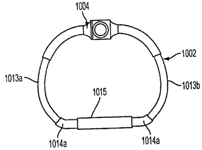
proper implantation. Such electrophysiologic touchdown detectors may include
electrical circuits
that produce visual, auditory, or other signals to the operator that the
detectors are activated and
that the implant is in the proper position for attachment.
[001031 In yet other embodiments according to the present invention, other
intracardiac or
extracardiac imaging techniques including, but not limited to, intravascular
ultrasound, nuclear
magnetic resonance, virtual anatomic positioning systems, or other imaging
techniques may be
employed to confirm proper positioning of the implant, obviating the need for
the touchdown
sensors as previously described.
(001041. FIGS. 21-24 show an implant 700 according to one embodiment of the
present
invention. In this embodiment, the implant body 705 is bandlike and flexible.
Through much of
its length, the implant body 705 is provided with a series of retention barbs
710 which are
oriented to facilitate placement, retention, and removal of the device. The
implant body 705 is
also provided with an adjustable section 715, which is provided in this
example with a series of
adjustment stops 720. The adjustment stops 720 may be slots, holes, detents,
dimples, ridges,
teeth, raised elements, or other mechanical features to allow measured
adjustment of the implant
700 in use. In the embodiment shown in FIGS. 21-24, the adjustment stops 720
are engaged by a
geared connector 725. FIG. 21 is an end view, showing the implant body 705
curved on itself,
with the retention barbs 710 to the exterior, and with the adjustable section
715 passing through
its engagement with the geared connector 725 and curving internally within the
implant body
705 to form a closed, round structure. FIG. 23 shows details of an exemplary
geared connector
725, in which a housing 730 is connected to the implant body 705. The housing
730 contains and
supports a mechanical worm 740 with an attached first geared head 750 which
mates with a
second geared head 755. The second geared head 755 is attached to an
adjustment stem 760
which is machined to receive a screwdriver-like adjustment element. The
various embodiments
according to the present invention may require a number of forms of adjustment
elements. In the
18
CA 02654359 2008-11-19
WO 2007/136783 PCT/US2007/011961
present example, the adjustment element is provided as a finely coiled wire
with a distal tip
machined to be received by a receiving slot in the adjustment stem 760 (not
shown). The
relationship between the distal tip of the adjustment elernent and the
adjustment stem 760 is
mechanically similar to a screwdriver bit and screwhead, such that torsion
imparted to the
adjustment means by the operator will result in the turning of the adjustment
stem 760 and
second geared head 755 allows motion of the first geared head 750 and worm
740, which creates
motion of the adjustable implant section 715 as the worm engages with the
series of adjustment
tops 725. Excess length of the adjustable section 715 passes though a band
slot 735 (FIG. 23),
thus allowing the band to move concentrically inside the closed implant body
705. The
adjustment element in this embodiment may be designed to remain in place after
the deployment
umbrella has been retracted and withdrawn. The connection between the
adjustment element's
distal tip and the adjustment stem 760 may be a simple friction connection, a
mechanical key/slot
formation, or may be magnetically or electronically maintained.
100105) As further shown in FIG. 21, the exemplary embodiment employs
unidirectional
retention barbs 710 which are attached to the outer perimeter of the implant
body 705. The
retention barbs 710 are oriented in a consistent, tangential position with
respect to the implant
body 705 such that rotational motion of the implant body will=either engage or
release the
retention barbs 710 upon contact with the desired tissue at the time of
deployment. This
positioning of the retention barbs 710 allows the operator to "screw in" the
implant 700 by
turning the implant 700 upon its axis, thus engaging the retention barbs 710
into the adjacent
tissue. As shown in FIG_ 24, the retention barbs 710 may each be further
provided with a
terminal hook 775 at the end which would allow for smooth passage through
tissue when
engaging the retention barbs 710 by rotating the implant 700, without
permitting the implant 700
to rotate in the opposite direction, because of the action of the terminal
hooks 775 grasping the
surrounding tissue (much like barbed fish hooks). The terminal hooks 775 thus
ensure the seating
of the implant 700 into the surrounding tissue.
1001061 FIGS. 25-27 illustrate another embodiment of an implant 800 as
contemplated
according to the present invention. The implant 800 includes a band 805 (FIG.
27), but the
retention barbs of the previous example have been eliminated in favor of an
outer fabric implant
sheath 810. The fabric sheath 810 can be sutured or otherwise affixed to the
anatomic tissue in a
desired location. The circumference of the implant body 800 is adjusted
through a geared
19
CA 02654359 2008-11-19
WO 2007/136783 PCT/US2007/011961
connector 825 similar to the geared connector of the bandlike implant array
shown in FIG. 23.
More specifically, adjustment stops 820 on the band are engaged by a
mechanical worm 840
with an attached first geared head 850. The first geared head 850 mates with a
second geared
head 855. The second geared head 855 is attached to an adjustment stem 860
which is machined
to receive a screwdriver-like adjustment element.
[00107) FIG. 28 illustrates an example of the method of use of an
implant/delivery system
array 600 for positioning an implant 645 in a patient with ischemic annular
dilatation and mitral
regurgitation. Peripheral arterial access is obtained via conventional
cutdown, arterial puncture,
or other standard access techniques. After access to the arterial system is
attained, guidewire
placement is performed and intravascular access to the heart 900 is obtained
using fluoroscopic,
ultrasound, three-dimension ultrasound, magnetic resonance, or other real-time
imaging
techniques. The guidewire, deployment device, and implant are passed through
the aortic valve
in a retrograde fashion into the left ventricle 905 and then into the left
atrium 910. At this point,
the operator retracts the housing sheath 605,'thus unsheathing the collapsed
deployment umbrella
642 and implant 645. The deployment umbrella 642 is then distended by the
distal motion of the
actuation catheter, causing the radial support arms and struts to fully
distend. At this point, the
touchdown detectors 648 are not in contact with any solid structures, and are
fully extended with
their radiolucent gaps visible on the imaging system. Once the deployment
umbrella is distended,
the entire assembly is pulled back against the area of the mitral valve 915.
At least two
touchdown detectors 648 are employed in a preferred embodiment according to
the present
invention. When all touchdown detectors show the disappearance of their
intennediate, non-
opaque, intermediate segments and are thus activated, then the deployment
umbrella must be in
contact with the solid tissue in the region of the mitral annulus/atrial
tissue, and further implant
deployment and adjustment may proceed. However, if any one touchdown sensor is
not activated,
and a radiolucent gap persists, then the device is not properly positioned,
and must be
repositioned before further deployment. Thus, the touchdown sensor system may
assist in the
deployment and adjustment of prosthetic devices by the delivery system
according to the present
invention. Once properly positioned, the operator rotates the actuation
catheter in a prescribed
clockwise or counterclockwise manner to engage the retention barbs on the
implant into the
tissue in the region of the.mitral annulus/atrial tissue. Should re-
positioning be required, a
reverse motion would disengage the retention barbs from the annular/atrial
tissue, and
CA 02654359 2008-11-19
WO 2007/136783 PCT/US2007/011961
repositioning may be performed, again using the touchdown detectors for proper
placement.
Once firmly seated, the adjustment element(s) are operated to achieve the
desired degree of
annular reduction. Real-time trans esophageal echocardiography, intravascular
echocardiography,
intracardiac echocardiography, or other modalities for assessing mitral
function may then be
employed to assess the physiologic effect of the repair on mitral function,
and additional =
adjustments may be performed. Once a desired result has been achieved, the
release elements are
activated to detach the implant from the deployment umbrella. The operator
then retracts the
actuation catheter and extends the housing sheath, collapsing the deployment
umbrella and
covering the components for a smooth and atraumatic withdrawal of the device
from the heart
and vascular system.
[00108] If desired, the adjustment elements may be left in position after the
catheter
components are withdrawn for further physiologic adjustment. In yet other
embodiments
according to the present invention, a catheter-based adjustment elements may
subsequently be
re-inserted though a percutaneous or other route. Such an adjustment element
may be steerably
operable by the opperator, and may be provided with magnetic, electronic,
electromagnetic, or
laser-guided systems to allow docking of the adjustment element with the
adjustable mechanism
contained within the implant. In still other embodiments, the adjustment
mechanism may be
driven by implanted electromechanical motors or other systems, which may be
remotely
controlled by electronic flux or ~other remote transcutaneous or percutaneous
methods.
[00109] In the case of pulmonic valve repair, initial catheter access is
achieved through a
peripheral or central vein. Access to the pulmonary valve is also achieved
from below the valve
once central venous access is achieved by traversing the right atrium, the
tricuspid valve, the
right ventricle, and subsequently reaching the pulmonic valve.
[00110] In yet other embodiments according to the present invention, catheter
access to
the left atrium can be achieved from cannulation of central or peripheral
veins, thereby achieving
access to the right atrium. Then a standard atrial trans-septal approach may
be utilized to access
the left atrium by creation of an iatrogenic atria] septal defect (ASD). In
such a situation, the
mitral valve may be accessed from above the valve, as opposed to the
retrograde access
described in Example 1. The implant and a reversed deployment umbrella may be
utilized with
implant placement in the atrial aspect of the mitral annulus, with the same
repair technique
described previously. The iatrogenic ASD may then be closed using standard
device methods.
21
CA 02654359 2008-11-19
WO 2007/136783 PCT/US2007/011961
Access to the aortic valve may also be achieved from above the aortic valve
via arterial access in
a similar retrograde fashion.
100111] Other embodiments of the adjustable implant and methods according to
the
present invention include gastrointestinal disorders such as gastro-esophageal
reflux disease
(GERD), a condition in which the gastro-esophageal (GE) junction lacks
adequate sphincter tone
to prevent the reflux of stomach contents into the esophagus, causing classic
heartburn or acid
reflux. This not only results in discomfort, but may cause trauma to the lower
esophagus over
time that may lead to the development of pre-cancerous lesions (Barrett's
esophagus) or
adenocarcinoma of the esophagus at the GE junction. Surgical repair of the GE
junction has
historically been achieved with the Nissen Fundoplication, an operative
procedure with generally
good results. However, the Nissen procedure requires general anesthesia and a
hospital stay.
Utilizing the devices and methods according to the present invention, an
adjustable implant
would obviate the need for a hospital stay and be performed in a clinic or
gastroenterologist's
office. Referring now to FIGS. 29 and 30, an umbrella deployment device 600
with implant 645
is passed under guidance of an endoscope 1000, through the patient's mouth,
esophagus 1005,
and into the stomach 1010, where the deployment device 600 is opened with
expansion of the
implant 645 and touchdown detectors 648 with a color-coded or otherwise
visible gap. The
touchdown detectors are then engaged onto the stomach around the
gastroesophageal junction
1015 under direct endoscopic control until all touchdown detectors 648 are
visually activated.
The implant is then attached to the stomach wall, 1020 the umbrella 642 is
released and
withdrawn, leaving behind the implant 645 and the adjustment elements. The
implant is then
adjusted until the desired effect is achieved, i.e., minimal acid reflux
either by patient symptoms,
pH monitoring of the esophagus, imaging studies, or other diagnostic means. If
the patient
should suffer from gas bloat, a common complication of gastroesophageal
junction repair in
which the repair is too tight and the patient is unable to belch, the implant
can be loosened until a
more desirable effect is achieved.
[00112] In various embodiments anticipated by the present invention, the
implant body
may be straight, curved, circular, ovoid, polygonal, or some combination
thereof. In various
embodiments anticipated by the present invention the implant may be capable of
providing a
uniform or non-uniform adjustment of an orifice or lumen within the body. The
implant body
may further completely enclose the native recipient anatomic site, or it may
be provided in an
22
CA 02654359 2008-11-19
WO 2007/136783 PCT/US2007/011961
interrupted form that encloses only a pbrtion of the native recipient anatomic
site. In still other
embodiments of the present invention, the implant body may be a solid
structure, while in yet
other embodiments the implant body may form a tubular or otherwise hollow
structure. In one
embodiment of the present invention, the body may further be a structure with
an outer member,
an inner member, and optional attachment members. In such an embodiment, the
outer member
of the implant body may serve as a covering for the implant, and is designed
to facilitate and
promote tissue ingrowth and biologic integration to the native recipient
anatomic site. The outer
member in such an embodiment may be fabricated of a biologically compatible
material, such as
Dacron, PTFE, malleable metals, other biologically compatible materials or a
combination of
such biologically compatible materials in a molded, woven, or non-woven
configuration. The
outer member in such an embodiment also serves to house the inner member. In
this embodiment,
the inner member provides an adjustment means that, when operated by an
adjustment
mechanism, is capable of altering the shape and/or size of the outer member in
a defined manner,
[00113] In alternate embodiments according to the present invention, the
adjustment
means may be located external to or incorporated within the outer member. In
yet additional
alternate embodiments contemplated by the present invention, the implant body
may consist of
an adjustment means without a separate outer member covering said adjustment
means.
[00114] In various embodiments according to the present invention, the
adjustment means
may include a mechanism which may be threaded or non-threaded, and which may
be engaged
by the action of a screw or worm screw, a.friction mechanism, a friction-
detent mechanism, a
toothed mechanism, a ratchet mechanism, a rack and pinion mechanism, or such
other devices to
permit discreet adjustment and retention of desired size a desired position,
once the proper size is
determined.
[00115] In yet other embodiments according to the present invention, the
adjustment
means may comprise a snare or purse string-like mechanism in which a suture, a
band, a wire or
other fiber structure, braided or non-braided, monofilament or multifilament,
is capable of
affecting the anatomic and/or physiologic effects of the implant device on a
native anatomic
recipient site upon varying tension or motion imparted to said wire or fiber
structure by a
surgeon or other operator. Such an adjustment means may be provided as a
circular or non-
circular structure in various embodiments. Changes in tension or motion may
change the size
and/or shape of the implant.
23
CA 02654359 2008-11-19
WO 2007/136783 PCT/US2007/011961
[00116] In various embodiments according to the present invention, the
adjustment means
may be a metallic, plastic, synthetic, natural, biologic, or any other
biologically-compatible
material, or combination thereof. Such adjustment means may further be
fabricated by extrusion
or other molding techniques, machined, or woven. Furthermore, in various
embodiments of the
present invention, the adjustment means may be smooth or may include slots,
beads, ridges, or
any other smooth or textured surface.
[00117] In various embodiments of the present invention, the implant body may
be
provided with one or more attachment members such as grommets or openings or
other
attachment members to facilitate attachment of the implant to the native
recipient site. In
alternate embodiments, the implant body may attach to or incorporate a
mechanical tissue
interface system that allows a sutureless mechanical means of securing the
implant at the native
recipient site. In still other alternate embodiments, sutures or other
attachment means may be
secured around or through the implant body to affix the implant body to the
native recipient site.
In yet other embodiments of the present invention, mechanical means of
securing the implant
body to the native recipient site may be augmented or replaced by use of
fibrin or other
biologically-compatible tissue glues or similar adhesives.
[00118] In additional various embodiments according to the present invention,
the
adjustable implant may be employed to adjustably enlarge or maintain the
circumference or other
dimensions of an orifice, ostium, lumen, or anastomosis in which a disease
process tends to
narrow or constrict such circumference or other dimensions.
[00119] In various embodiments according to the present invention, an
adjustment
mechanism may be provided to interact with the adjustment means to achieve the
desired
alteration in the size and/or position of the adjustment means. Such an
adjustment mechanism
may include one or more screws, worm-screw arrays rollers, gears, frictional
stops, a friction-
detent system, ratchets, rack and pinion arrays, micro-electromechanical
systems, other
mechanical or electromechanical devices or some combination thereof.
1001201 In some embodiments as contemplated by the present invention, an
adjustment
tool may be removably or permanently attached to the adjustment mechanism and
disposed to
impart motion to the adjustment mechanism and, in turn, to the adjustment
means to increase or
decrease the anatomic effect of the implant on the native recipient site.
24
CA 02654359 2008-11-19
WO 2007/136783 PCT/US2007/011961
[00121] In alternate embodiments according to the present invention,
micromotor arrays
with one or more micro-electromechanical motor systems with related electronic
control
circuitry may be provided as an adjustment means, and may be activated by
remote control
through signals convey by electromagnetic radiation or by direct circuitry
though electronic
conduit leads which may be either permanently or removably attached to said
micromotor arrays.
[00122] In still other various embodiments according to the present invention,
the
adjustment mechanism may be provided with a locking mechanism disposed to
maintain the
position of the adjustment means in a selected position upon achievement of
the optimally
desired anatomic and/or physiologic effect upon the native recipient site and
the bodily organ to
which it belongs. In other embodiments, no special locking mechanism may be
necessary due to
the nature of the adjustment means employed.
[00123] In yet other alternate embodiments according to the present invention,
the
adjustment means and/or the outer member structure may be a pliable synthetic
material capable
of rigidification upon exposure to electromagnetic radiation of selected
wavelength, such as
ultraviolet light. In such embodiments, exposure to the desired
electromagnetic radiation may be
achieved by external delivery of such radiation to the implant by the surgeon,
or by internal
delivery of such radiation within an outer implant member using fiberoptic
carriers placed within
said outer member and connected to an appropriate external radiation source.
Such fiberoptic
carriers may be disposed for their removal in whole or in part from the outer
implant member
after suitable radiation exposure and hardening of said adjustment means.
[00124] The present invention also provides methods of using an adjustable
implant
device to selectively alter the anatomic structure and/or physiologic effects
of tissues fonning a
passageway for blood, other bodily fluids, nutrient fluids, semi-solids, or
solids, or wastes within
a mammalian body. Various embodiments for such uses of adjustable implants
include, but are
not limited to, open surgical placement of said adjustable implants at the
native recipient site
through an open surgical incision, percutaneous or intravascular placement of
said implants
under visual control employing fluoroscopic, ultrasound, magnetic resonance
imaging, or other
imaging technologies, placement of said implants through tissue structural
walls, such as the
coronary sinus or esophageal walls, or methods employing some combination of
the above
techniques. In various embodiments as contemplated by the present invention,
adjustable
implants may be placed and affixed in position in a native recipient anatomic
site by trans-atrial,
CA 02654359 2008-11-19
WO 2007/136783 PCT/US2007/011961
trans-ventricular, trans-arterial, trans-venous (i.e., via the pulmonary
veins) or other routes
during beating or non-beating cardiac surgical procedures or endoscopically or
percutaneously in
gastrointestinal surgery.
[00125] Furthermore, alternate methods for use of an adjustable implant device
may
provide for the periodic,. post-implantation adjustment of the size of the
anatomic structure
receiving said implant device as needed to accommodate growth of the native
recipient site in a
juvenile patient or other changes in the physiologic needs of the recipient
patient.
[00126] Adjustment of the adjustable implants and the methods for their use as
disclosed
herein contemplates the use by the surgeon or operator of diagnostic tools to
provide an
assessment of the nature of adjustment needed to achieve a desired effect.
Such diagnostic tools
include, but are not limited to, transesophageal echocardiography,
echocardiography, diagnostic
ultrasound, intravascular ultrasound, virtual anatomic positioning systems
integrated with
magnetic resonance, computerized tomographic, or other imaging technologies,
endoscopy,
mediastinoscopy, laparoscopy, thoracoscopy, radiography, fluoroscopy, magnetic
resonance
imaging, computerized tomographic imaging, intravascular flow sensors, thermal
sensors or
imaging, remote chemical or spectral analysis, or other imaging or
quantitative or qualitative
analytic systems.
[00127] In one aspect, the implant/delivery,system of the present invention
comprises a
collapsible, compressible, or distensible prosthetic implant and a delivery
interface.for such a
prosthetic implant that is capable of delivering the prosthetic implant to a
desired anatomic
recipient site in a collapsed, compressed, or non-distended state, and then
allowing controlled
expansion or distension and physical attachment of such a prosthetic implant
by a user at the
desired anatomic recipient site. Such a system permits the delivery system and
prosthetic implant
to be introduced percutaneously through a trocar, sheath, via Seldinger
technique, needle, or
endoscopically through a natural bodily orifice, body cavity, or region and
maneuvered by the
surgeon or operator to the desired anatomic recipient site, where the delivery
system and
prosthetic implant may be operably expanded for deployment. When desirable,
the
implant/delivery system according to the present invention is also capable of
allowing the user to
further adjust the size or shape of the prosthetic implant once it has been
attached to the desired
anatomic recipient site. The delivery system according to the present
invention is then capable of
detaching from its interface with the prosthetic implant and being removed
from the anatomic
26
CA 02654359 2008-11-19
WO 2007/136783 PCT/US2007/011961
site by the operator. The delivery system and prosthetic implant may be
provided in a shape and
size determined by the anatomic needs of an intended native recipient anatomic
site within a
mammalian patient. Such a native recipient anatomic site may be a heart valve,
the esophagus
near the gastro-esophageal junction, the anus, or other anatomic sites within
a mammalian body
that are creating dysfunction that might be relieved by an implant capable of
changing the size
and shape of that site and maintaining a desired size and shape afi<er
surgery.
[00128] In various embodiments contemplated by the present invention, the
delivery
system may be a catheter, wire, filament, rod, tube, endoscope, or other
mechanism capable of
reaching the desired recipient anatomic site through an incision, puncture,
trocar, or through an
anatomic passageway such as a vessel, orifice, or organ lumen, or trans-
abdominally or trans-
thoracically. In various embodiments according to the present invention, the
delivery system may
be steerable by the operator. The delivery system may further have a delivery
interface that
would retain and convey a prosthetic implant to the desired recipient anatomic
site. Such a
delivery interface may be operably capable of distending, reshaping, or
allowing the independent
distension or expansion of such a prosthetic implant at the desired recipient
anatomic site.
Furthermore, such a delivery interface may provide an operable means to adjust
the distended or
expanded size, shape, or physiologic effect of the prosthetic implant once
said implant has been
attached in situ at the desired recipient anatomic site. In various
embodiments according to the
present invention, such adjustment may be carried out during the procedure in
which the implant
is placed, or at a subsequent time. Depending upon the specific anatomic needs
of a specific
application, the delivery interface and the associated prosthetic implant may
be straight, curved,
circular, helical, tubular, ovoid, polygonal, or some combination thereof. In
still other
embodiments of the present invention, the prosthetic implant may be a solid
structure, while in
yet other embodiments the prosthetic implant may form a tubular, composite, or
otherwise
hollow structure. In one embodiment of the present invention, the prosthetic
implant may further
be a structure with an outer member, an inner member, and optional attachment
members. In
such an embodiment, the outer member of the prosthetic implant may serve as a
covering for the
implant, and is designed to facilitate and promote tissue ingrowth and
biologic integration to the
native recipient anatomic site. The outer member in such an embodiment may be
fabricated of a
biologically compatible material, such as Dacron, PTFE, malleable metals,
other biologically
compatible materials or a combination of such biologically compatible
materials in a molded,
27
CA 02654359 2008-11-19
WO 2007/136783 PCT/US2007/011961
woven, or non-woven configuration. The outer member in such an embodiment also
serves to
house the inner member. In this embodiment, the inner member provides an
adjustment means
that, when operated by an adjustment mechanism, is capable of altering the
shape and/or size of
the outer member in a defined manner.
[00129] In some embodiments according to the present invention, at least some
portions of
the adjustable inner or outer member may be elastic to provide an element of
variable, artificial
muscle tone to a valve, sphincter, orifice, or lumen in settings where such
variability would be
functionally valuable, such as in the treatment of rectal incontinence or
vaginal prolapse.
[00130] In various embodiments according to the present invention, the
delivery interface
would have an attachment means to retain and convey the prosthetic implant en
route to the
native anatomic recipient site and during any in situ adjustment of the
prosthetic implant once it
has been placed by the operator. Such an attachment means would be operably
reversible to
allow detachment of the prosthetic implant from the delivery interface once
desired placement
and adjustment of the prosthetic implant has been accomplished.
[00131] In one embodiment of the present invention, illustrated in FIG. 31, an
implantable
device system 1000 for controlling at least the size or shape of an anatomical
structure or lumen
includes an implantable device 1002 and an adjustment tool 1006. The
anatomical structure or
lumen is an anatomic site with dysfunction that can be relieved by the
implantable device 1002
to change a size or shape of the anatorriic site.
[00132] The implantable device 1002, in one exemplary embodiment, has a
diameter no
larger than 3.5 mm. In another embodiment the implantable device 1002 is
configured to have
variable size relative to its placement at an annulus -of a heart valve. The
implantable device
1002 has an adjustable member 1004 configured to adjust the dimensions of the
implantable
device 1002. In one embodiment, the torqueable adjustment tool 1006 provides
adjustment of
the dimensions of the implantable device 1002. The adjustable member 1004, in
some
embodiments, may be oriented- to receive the adjustment tool from a direction
generally
perpendicular to the primary plane defined by the implant device 1002. Such an
orientation is
advantageous for intravenous access of the tool and in situ adjustment of the
implant device 1002.
The implantable device 1002 can have a configuration where there are different
pulling rates at
different sections of the implantable device 1002. The implantable device 1002
may optionally
include a flexible tube (1032, FIG. 38) and an outer fabric sheath (810, FIGs.
25 and 26), which
28
CA 02654359 2008-11-19
WO 2007/136783 PCT/US2007/011961
are not shown in the subsequent figures for clarity. The outer fabric sheath
can be sutured,
stapled, clipped, coiled, or otherwise affixed to anatomic tissue in a desired
location. Generally
the desired location is considered to be the internal surface of the area to
be controlled, such as
(for example) an interior wall of an organ, artery, or other internal anatomic
passage. Also,
while the implantable device 1002 is generally shown in the subsequent figures
to have a "D"-
shaped configuration, it should be understood that other shapes can be used in
accordance with
embodiments of the present invention.
[00133] Still referring to FIG. 31, in certain embodiments, the adjustment
tool 1006 is at
least partially hollow, and in one specific embodiment at least 50% hollow.
The adjustment tool
1006 may be an elongated tool, which has a proximal end and a distal end
releasably attached to
the adjustable member 1004 of implantable device 1002. The adjustment tool
1006 may extend
from its distal end coupled to the adjustable member 1004 to a control
interface (e.g., handle) at
the proximal end located preferably outside of the patient's body. The
adjustment tool 1006,
when coupled to the adjustable member 1004 of implantable device 1002, can
provide a
preferential shape change of the implantable device 1002 in planar and non-
planar directions.
The adjustment tool 1006 can adjust the implantable device 1002 in terms of
narrowing or
widening the dimensions of the implantable device 1002.
[00134] FIG. 32A is a schematic of the implant device 1002 without showing an
optional
flexible outer tube and fabric sheath. The implantable device includes an
adjustable member
1004 and adjustable tube portions 1013a and 1013b, which slide within hollow
tube portions
1014a and 1014b, and retaining tube 1015. FIG. 32B is a schematic of a
disassembled portion of
implantable device 1002 with retaining tube 1015 removed. As shown in FIG.
32B, in various
embodiments, the implantable device 1002 includes a threaded rod 1008 threaded
with right-
hand helical grooves 1010 and left-hand helical grooves 1012. Other
embodiments may include
a threaded rod 1008 with helical grooves in a single direction (e.g., all
right-hand grooves or all
left-hand grooves). Threaded rod 1008 may be a rigid material such as
titanium, stainless steel,
or a polymer. Adjustable tube portions 1013a and 1013b enclose at least a
portion of grooves
1010 and 1012 so that pins 1016a, 1016b or protuberances on the inside
diameter of the
adjustable tube portions 1013a, 1013b are engaged by the grooves 1010 and
1012, respectively.
In other embodiments, pins 1016a, 1016b may be replaced by threads along the
inside diameter
of the adjustable tube portions 1013a, 1013b. Helical grooves 1010 and 1012
may be single
29
CA 02654359 2008-11-19
WO 2007/136783 PCT/US2007/011961
channels or multiple channels to engage single pins 1016a, 1016b or multiple
pins. Hollow tube
portions 1014a, 1014b are relatively rigid to maintain curvature of the
adjustable tube portions
1013a, 1013b regardless of the adjustment position.
[001351 The implantable device 1002 can have a coating including, but not
limited to,
heparin, and antibiotic, collagen, and an agent that promotes tissue in
growth, PGLA, a de-
calcification agent and the 1 ike. The implantable device 1002 can be made of
a variety of
materials including, but not limited to, a shape memory alloy (SMA), a shape
memory polymer
(SMP), titanium, stainless steel, polymer, a suture-based material, a
biological material and the
like.
[001361 In another embodiment of the present invention, illustrated in FIGs.
33 through 37,
the adjustable member 1004 provides translated motion through rotation. FIGs.
33 through 35
illustrate a theory of operation of an embodiment of the present invention,
while FIGs. 36 and 37
shown details of the adjustment member 1004.
[00137] Referring to now FIG. 33, adjustable member 1004 of implantable device
1102 is
shown including a docking port 1021 to receive the distal tip of the
adjustment tool 1006 (FIG.
31). In this embodiment, implant device includes a set of inner tubing 1028a,
1028b and a set of
outer tubing 1026a, 1026b that can move relative to each other. The ends of
the inner tubing
1028a, 1028b that do not engage the outer tubing 1026a, 1026b are secured to a
set of hollow
tubing 1014a, 1014b so that the inner tubing 1028a, 1028b does not move
relative to the hollow
tubing 1014a, 1014b. Although hollow tube portions 1014a, 1014b may be
separate pieces that
are permanently abutted when assembled, in some embodiments, the hollow tube
portions 1014a,
1014b may be formed from a single tubing piece. An inner cable 1030 passes
through the
various tubing. Thus, the rigidity of the hollow tubing can be used to
maintain the adjustable
implant 1102 shape in certain dimensions so that adjustment of the device can
be restricted to a
preferred dimension, for example, an anterior-posterior dimension.
1001381 As shown in more detail in FIGs. 36 and 37, adjustable member 1004 may
also
include a pinion gear 1022 (which may be integral to a docking port 1021) and
a crown gear
1024. FIG. 36 provides an isometric view of the adjustable member 1004, and
FIG. 37 provides a
cut-away view of the adjustable member 1004. As can be seen in the figures,
the pinion gear
1022 engages the crown gear 1024. In some embodiments, the pinion gear 1022
may be
eliminated from adjustable member 1004, and the distal tip of the adjustment
tool 1006 may
CA 02654359 2008-11-19
WO 2007/136783 PCT/US2007/011961
serve as the pinion gear when the tool is coupled to the docking port 1021.
When coupled to the
docking port 1021, the adjustment tool 1006 can rotate the pinion gear 1022.
[001391 Referring back to FIG. 33, the implantable device 1102 is shown
generally at the
middle of its adjustment range. Outer tubing 1026a, 1026b is affixed to the
adjustable member
1004 and extends along a portion of the circumference of implantable device
1102. Inner tubing
1028a, 1028b is affixed to hollow tubing 1014a, 1014b, respectively. Similar
to the single
threaded rod 1008 of FIG. 32B, threaded rods 1018a, 1018b sit inside the
hollow tubing 1014a,
1014b and are threadedly engaged therewith. Threaded rods 1018a, 1018b may be
a rigid
material such as titanium, stainless steel, or a polymer. Hollow tube portions
1014a, 1014b
enclose the threaded rods 1018a, 101 8b such that rotation of the threaded
rods 1018a, 1018b
causes them to move axially within the hollow tube portions 1014a, 10 14b. The
threaded rod
1018a may have right-handed threads, and the threaded rod 1018b may have left
handed threads.
Other embodiments may include threaded rods 1018a, 1018b with threads in a
single direction
(e.g., all right-hand grooves or all left-hand threads).
[001401 The crown gear 1024, and one end of each threaded rod 1018a, 1018b are
all
attached to an inner cable 1030. Inner cable 1030 may be a cable or tube of
any material with
sufficient flexibility to conform to a shape of the implantable device 1102
while translating
torque. For example, suitable material for inner cable 1030 may include
titanium or stainless
steel. As shown more clearly in FIG. 36 and 37, the rotation of crown gear
1024 imparts rotation
to cable 1030 in the same direction.
[00141] Referring to FIG. 34, when the handle of adjustment tool 1006 (not
shown in this
figure) is rotated clockwise in docking port 1021, it causes clockwise
rotation of the pinion gear
1022 (in FIG 36). Rotation of the pinion gear 1022 in tum rotates crown gear
1024. The
rotation of crown gear 1024 causes rotation of inner cable 1030, which imparts
rotational
movement to each threaded rod 1018a, 1018b. The rotation applied to the
threaded rods 1018a,
1018b causes them to advance into their respective hollow tubing 1014a, 1014b
in the directions
A,, A2 shown. As shown in FIG. 34, when threaded rods 1018a, 101 Sb advance
toward the
middle of the hollow tubing 1014a, 1014b the overall circumference of the
implant device 1002
is reduced. Advancing the threaded rods 1018a, 1018b drives the inner cable
1030 into the
hollow tubing 1014a, 1014b. Translation of inner cable 1030 into the hollow
tubing 1014a,
1014b causes the hollow tubing 1014a, 1014b to move towards adjustable member
1004 in the
31
CA 02654359 2008-11-19
WO 2007/136783 PCT/US2007/011961
direction B, shown. Inner tubing 1028a, 1028b slides into outer tubing 1026a,
1026b to
accommodate movement of the inner cable 1030.
[00142] Referring to FIG_ 35, the handle ofadjustment tool 1006 (not shown in
this figure)
is rotated counter-clockwise in docking port 1021 to cause counter-clockwise
rotation of the
pinion gear 1022 (FIG 36). Rotation of the pinion gear 1022, in turn rotates
crown gear 1024.
The rotation of crown gear 1024 causes rotation of inner cable 1030, which
imparts rotational
movement to each threaded rod 101 8a, 1018b. The rotation applied to the
threaded rods 1018a,
1018b causes them to begin to withdraw from their respective hollow tubing
1014a, 1014b in the
directions A2, Ai shown. As shown in FIG. 35, as threaded rods 1018a, 1018b
withdraw from
the middle of the hollow tubing 1014a, 1014b the overall circumference of the
implant device
1002 is increased. Withdrawal of the threaded rods 1018a, 1018b pushes the
inner cable 1030
out of the hollow tubing 1014a, 1014b. Translation of inner cable 1030 out of
the hollow tubing
1014a, 1014b causes the hollow tubing 1014a, 1014b to move away from
adjustable member
1004 in the direction B2 shown. Inner tubing 1028a, 1028b telescopes out of
outer tubing 1026a,
1026b to acconunodate movement of the inner cable 1030.
[001431 The inner tubing 1028a, 1028b, the outer tubing 1026a, 1026b, and the
hollow
tubing 1014a, 1014b may be covered by a flexible tube 1032, such as a silicone
tube, shown in
FIG. 38. In one embodiment, outer flexible tube 1032 is provided with no seam
in the axial
direction of the tube to allow for better tissue ingrowth after the implant
procedure. In other
embodiments inner tubing 1028a, 1028b may be eliminated, as shown in FIG. 39.
[00144] FIG. 39 provides an assembled cross-section view of an implantable
device 1202
according to an embodiment of the invention. The implant device includes the
adjustable
member 1004, the outer tubing 1026a, 1026b, the hollow tubing 1014a, 1014b,
the inner cable
1030, and the threaded rods 1018a, 1018b as discussed in relation to FIGs. 33-
35. As shown in
FIG. 39, hollow tubing 1014a, 1014b may extend further along the length of
inner cable 1030
than shown in other embodiments of FIGs. 33-35 to better maintain a preferred
shape of the
implant. Hollow tubing 101 4a, 101 4b may be threaded to receive the threaded
rods 1018a,
101 8b; or hollow tubing may optionally include a-threaded insert (spar 1019a,
1019b) affixed to
the inner diameter of hollow tubing 1014a, 1014b. In operation, as previously
described, an
adjustment tool may impart motion to the adjustable member 1004. Gears in the
adjustable
member translate motion to the inner cable 1030 that, in turn translate motion
to the attached
32
CA 02654359 2008-11-19
WO 2007/136783 PCT/US2007/011961
threaded rods 1018a, 1018b. Depending on the direction of rotation, rotation
of threaded rods
1018a, 1018b causes the threaded rods 1018a, 1018b to be drawn toward or away
from the
middle of the hollow tubing 1014a, 1014b, thus reducing or increasing the
overall circumference
of the implant device 1002. The flexible outer tube 1032 and a seal jacket
1100 (also shown in
FIG. 40) encapsulate the device so that no moving parts are exposed. The
flexible outer tube
1032 provide sufficient rigidity to maintain a generally planar dimension,
while allowing the
device to adjust shape generally in a preferred dimension, such as the
anterior-posterior
dimension. As shown in FIG. 39, the flexible outer tube 1032 may be further
covered by an
outer fabric sheath 1110 or thin sewing cuff. Elimination of the inner tubing
(1028a, 1028b of
FIG 35) eliminates the need for telescoping parts and prevents the possibility
of telescoping
tubes being sutured or clipped together during attachment of the implant.
[00145] Referring to FIG. 40, the adjustable member 1004 can include a seal
jacket 1100.
FIG. 40 shows an embodiment of the seal jacket 1100. The seal jacket 1100 may
include a cover
1102 for the docking port 1021 (FIG. 33) of the adjustable member 1004. The
cover 1102 may
be in the form of a slit septum, flaps, elastic material or the like. The seal
jacket cover 1102 may
be included as part of a seal jacket 1100 that covers the entire housing of
the adjustable member
1004 or a separate piece. In one embodiment, the seal jacket 1100 may be
secured to the
flexible tube 1032. The seal jacket 1100 and flexible tube 1032 may be secured
by an adhesive
bond, a wrap, sutures, or the like. The cover 1102 provides access for an
adjustment tool to
couple to the docking port, while reducing the possibility of thrombus. In
some embodiments,
seal jacket cover 1102 and/or the seal jacket 1100 may be made of silicone,
and covered by a
polyester sewing layer or fabric sheath (e.g., 1110 of FIG. 39). In various
embodiments, the seal
jacket fits over the housing of the adjustable member 1004 that includes a
crown gear coupled to
a cable, can provide pinion access, and the like. In operation, the distal tip
of an adjustment tool
passes through the cover 1102 to engage the rotatable gear of adjustable
member 1004.
[001461 FIG. 41 shows an embodiment of implantable device 1302 including a
first
adjustment band 1042a and a second adjustment band 1042b. The first and second
adjustment
bands 1042a, 1042b can be overlapped, and the amount of overlap is effected by
how the
implantable device 1302 is sized. The first and second bands 1042a, 1042b can
be slidable
relative to each other. An adjustable member 1304 is coupled to the first band
1042a and the
second band 1042b, and pulls or pushes them toward or away from each other.
The first band
33
CA 02654359 2008-11-19
WO 2007/136783 PCT/US2007/011961
1042a and the second band 1042b can have flexible portions 1046a, 1046b
configured to create a
flexible zone at the primary bend regions 1047a, 1047b. The flexible portions
1046a, 1046b can
have varying lengths and may also include one or more rigid portions 1044.
These rigid portions
1044 can include welded braids or bands, or have a higher durometer material
than the flexible
portions 1046a, 1046b. The flexible portions 1046a, 1046b and rigid portions
1044 may be part
of the same material as the first and second bands 1042a, 1042b, or one or
more portions may be
separate material that is joined to form continuous piece.
[00147] The first and second bands 1042a, 1042b can have different sizes or
the same
sizes. In one specific embodiment, the first and second bands 1042a, 1042b are
about 0.5 to 3
mm in thickness and about 5 to 10 mm in width. The first and second bands
1042a, 1042b can
be made of a variety of materials including, but not limited to, an SMA, an
SMP, titanium,
stainless steel, polymer, a suture-based material, a biological material and
the like. In one
embodiment, the first and second bands 1042a, 1042b include a plurality of
band layers. At least
a portion of the first and second bands 1042a, 1042b may have superelastic
properties. Implant
1302 may include a flexible, extruded outer layer (not shown) or hollow tube,
such as flexible
tube 1032 of FIG. 38, to encase the structure formed by the first and second
bands 1042a, 1042b
flexible portions 1046a, 1046b, and rigid portions 1044. The parts of the
first and second bands
1042a, 1042b, that extend past adjustable member 1304 can be contained within
the hollow
interior of the outer layer.
[001481 FIG. 42 provides a more detailed schematic view of the unassembled
adjustment
bands and adjustment member of FIG. 41. The first and second bands 1042a,
1042b may include
a series of adjustment stops 1048. Adjustment stops may be in the form of
holes, detents,
dimples, ridges, teeth, raised elements, other mechanical features or the
like. These holes 1048
on each of the bands 1042a, 1042b are coupled to an adjustable member 1304.
The adjustable
member 1304 may be generally cylindrical (such as a spool) with a series of
teeth 1050 or
protrusions radially positioned to engage the adjustment stops 1048.
Adjustable member 1304
may also include a docking port 1320 to receive an adjustment tool to trigger
rotational
movement of the adjustable member.
[00149] FIG. 43 provides an assembled view of the adjustment band and
adjustment
member of FIG. 42. When mounted in a housing (not shown in FIG. 43), the
adjustable member
1304 may be mounted on an axis to allow for rotational movement. The first and
second bands
34
CA 02654359 2008-11-19
WO 2007/136783 PCT/US2007/011961
1042a, 1042b pass on either side of adjustable member 1304 so that the teeth
1050 engage the
adjustment stops 1048 in each of the bands 1042a, 1042b. Rotating the
adjustable member in
turn tightens or loosens the bands.
[00150] FIG. 44 is a cut-away view of an embodiment of the gearbox for the
adjustment
band of FIG. 41. In this embodiment, the adjustable member 1304 rests on a
spring 1052 inside
a housing 1040 for the adjustable member. The housing 1040 includes access and
guidance for
the first and second bands (1042a, 1042b of FIG. 43) to couple with the teeth
1050 of the
adjustable member 1304. The spring 1052 forces the adjustable member 1304
upward so that
teeth 1056 on the top of the adjustable member 1304 engage with teeth 1058 on
the inside upper
surface of the housing 1040. Engagement of the adjustable member teeth 1056
with the housing
teeth 10581ocks the adjustable member 1304 in place to prevent rotational
movement.
Downward force, applied for example by an adjustment tool, against the spring
1052 disengages
the teeth 1056 and 1058 so that the adjustable member 1304 can be rotated to
adjust the size or
shape of implantable device 1302.
1001511 In another embodiment, FIG. 45 provides a schematic view of an
implantable
device 1402 of the present invention with a plurality of sliding bands that
can be opened and
closed to effect a shape change. As with the previous embodiments of FIGs. 41-
44, the first and
second bands 1042a, 1042b pass on either side of adjustable member 13Q4 so
that the teeth 1050
engage the adjustment stops 1048 in each of the bands 1042a, 1042b. Additional
bands 1042c
may be incorporated to increase stiffness at different areas of the implant
device 1402 to provide
preferential shape change. The additional bands 1042c may be secured to the
first and second
bands 1042a, 1042b using welds 1043, adhesive or other mechanical techniques
known in the art.
[001521 As illustrated in FIG. 46, in one embodiment, an implantable device
1502 has an
anterior portion 1060, a posterior portion 1062 and dual threads that provide
preferential
adjustment of one side or the other of implantable device 1002. The
implantable device 1502
has two independently adjustable threaded portions 1064a, 1064b used to
achieve different
pulling rates and/or lateral dimensions. The adjustable threaded portions
1064a, 1064b can be
connected to one or more adjustable member 1004 of the implantable device 1502
and positioned
at either the posterior or anterior portions of the implantable device 1502.
In one embodiment,
the posterior portion 1062 may be a rigid member which includes threaded hex
screws 1066a,
1066b, internal threads or similar structures. In one embodiment, the hex
screws 1066a, 1066b
CA 02654359 2008-11-19
WO 2007/136783 PCT/US2007/011961
are attached in a manner that allows rotation of the hex screws so that the
threads may engage
adjustable threaded portions 1064a, 1064b. Rigid posterior portion 1062 may
include one or
more of adjustable members 1004 that can receive a tool to impart rotational
motion through an
inner tube or cable to one or more of hex screws 1066a, 1066b, as described
above. Anterior
portion 1060 may be a flexible tube to accommodate shape change as the
anterior and posterior
portions 1060, 1062 move relative to each other.
[00153] In another embodiment, differently pitched threads or other mechanisms
may be
used to provide non-symmetrical shape change of the implant device. For
example, referring to
FIG. 46, wider threads on threaded portion 1064b, in relation to the threads
of threaded portion
1064a, would allow an adjustable member 1004 to expand or contract the implant
1502 more
rapidly on the side of threaded portion 1064b to provide preferential shape
change for a selected
region while using a single adjustable member.
[00154] FIG. 47 is a schematic view of an embodiment of an adjustable member
1604 for
an implantable device. An adjustment tool may impart reciprocating motion to
the adjustable
member 1604 that includes a clover gear 1070 mounted in a housing 1072. The
inner cable 1030
(FIG. 33) of the implantable device, for example, is affixed to the clover
gear 1070 such that
rotation of the clover gear transmits torque through the inner cable 1030 to a
screw or other
adjustable portion of the implantable device as previously disclosed. In this
embodiment, the
adjustment tool can provide reciprocating action to provide for adjustment.
The adjustable
member takes an axial force applied to the control portion at the proximal end
of the adjustment
tool and converts it to a rotational force applied to the inner cable 1030 of
the implantable device.
Reciprocating axial force may be provided from an adjustment tool by using
spring-mounted
buttons pressed by the user. Pressing a first button may transmit a downward
axial motion to a
first ribbon 1074 which engages the clover gear 1070 to cause clockwise
rotation of the clover
gear 1070. A spring or other return force pushes the first ribbon back to its
original position after
each click or press of the button. Similarly, pressing a second button may
transmit a downward
axial motion to a second ribbon 1076 that engages the clover gear 1070 to
cause counter-
clockwise rotation of the clover gear 1070.
[00155] In another embodiment, the adjustment tool provides coarse adjustment
and fine
adjustment. This varied adjustment can be achieved with the adjustment tool
having screws with
different threads.
36
CA 02654359 2008-11-19
WO 2007/136783 PCT/US2007/011961
[00156] FIG. 48 provides a schematic view of an embodiment of the implantable
device
system 1000 including an adjustment tool 1706 with high column strength and
stiffness. The
adjustment tool 1706 has a shaft 1794 and a handle 1096 with sufficient column
strength to
ensure a downward axial force on the handle 1096 provides proper engagement
with the
adjustable member 1004 of the implantable device 1002. The handle 1096 may be
a grip-like
handle, as shown, or a smaller pen-type handle. The adjustment tool 1706 can
include
mechanical locking at the distal region 1782 to lock with the adjustable
member 1004. The
mechanical locking is configured to provide engagement and disengagement
tactile feel to the
physician.
[00157] FIG. 49 is a schematic view of another embodiment of the implantable
device
system 1000 including an adjustment tool 1806 with reduced column stiffness.
The adjustment
tool 1806 has a handle 1096 a shaft 1080 with reduced column stiffness for
greater flexibility and
easier articulation of the adjustment tool 1806. The handle 1096 may be a grip-
like handle, as
shown, or a smaller pen-type handle. The easier articulation offered by the
this embodiment may
facilitate user positioning of the device in vivo and clearing adjacent
biological structures,
particularly when it is docketed to the adjustable member 1004 of the implant
1002. Flexibility
may be varied along the length of the adjustment tool shaft 1080. Flexibility
may be increased at
the distal region 1082 of the adjustment tool shaft 1006, particularly in the
region immediately
proximal to the gear/fitting at the distal tip of the adjustment tool 1006.
This gear/fitting is
constrained orthogonally to the adjustable member 1004, and it is important
that the adjustment
tool 1006 be easy to insert/connect and remain clear of biological structures.
1001581 FIG. 50 provides a view of an embodiment of the proximal end of the
adjustment
tool 1006. Referring to FIG. 50, adjustment tool 1006 includes a flexible
cable 1094 or similar
structure that is affixed to and rotates with a handle 1996. In other
embodiments, the adjustment
tool 1006 can have cables, a band, tubes, rods, and the like to impart
rotational and/or axial
motion from the proximal end to the distal tip of the tool 1006. The flexible
cable 1094 may be
enclosed by a flexible, low-friction cable jacket 1098 that allows the cable
1094 to rotate freely
within the jacket 1098. In some embodiments, adjustment tool 1006 may also
include a spring
release mechanism to allow disengagement of the distal tip of the tool from
the docking port
1021 (FIG. 33) with minimal force being applied to the sutures (or other
mechanisms) securing
the implant device to the tissue of an anatomic orifice or lumen. As shown in
FIG. 50, in some
37
CA 02654359 2008-11-19
WO 2007/136783 PCT/US2007/011961
embodiments, an e-clip 1099 or similar device may be used near the handle 1996
of the
adjustment tool 1006 to secure the release mechanism in the docking station
until adjustments
are complete.
[00159] In one embodiment illustrated in FIG. 51, the adjustment tool 1006 may
be
inserted inside a rigid sheath 1092 that reaches the implantable device 1002.
Thus, FIG. 51 is a
schematic view of an embodiment of the implantable device system 1000 of the
present
invention with an articulated shape. The rigidness of the sheath 1092 provides
the necessary
column strength to support the flexible adjustment tool 1006. An added benefit
to this
embodiment is that the sheath may be left in place, docked to the implantable
device 1002. The
flexible adjustment tool 1006 may be removed and then reinserted at some
future time to engage
with the adjustable member 1004 of implantable device 1002.
[00160] The adjustment tool 1006 can have a handle 1096 that can be
adjustable. The
handle 1096 can have a length of at least 8 inches, and in one embodiment at
least 10 inches.
Other embodiments may have a shorter or longer handle length. The handle 1096
may be thick
to provide a hand-grip, or, in other embodiments, smaller to provide a pen-
like grip. The handle
can have a device to quantify a size change of the implantable device 1002.
For example, a half-
turn of the adjustment tool handle can be correlated to a distance of travel
of the threaded rods
1018a, 101$b (FIG. 33) of an implant 1002, thus allowing for measured
adjustment of the
implant. The handle may include a click-counter or other known device to
measure rotational
movement. In one embodiment, the adjustment tool 1006 may be included in a
percutaneous
delivery catheter.
(00161] A sensor, such as the touchdown sensor described in relation to FIGS.
12-18
above, can be coupled to the implantable device 1002. A variety of different
sensors can be
utilized, including but not limited to, sensors that measure pressure,
temperature and flow across
the implantable device 1002. Pacing leads are coupled to the sensor and the
implantable device
1002, and in this embodiment, the sensor is responsive to flow through the
implantable device
1002.
[00162] In another embodiment the implantable device system may include a
micro-
electromechanical motor system in conjunction with or instead of a separate
adjustment tool to
commence rotational movement in an adjustable member. Power and control of the
micro-
39
CA 02654359 2008-11-19
WO 2007/136783 PCT/US2007/011961
electromechanical motor system can be provided by electromagnetic radiation or
through a direct
wire connection and previously described herein.
[00163] Finally, it will be understood that the preferred embodiment has been
disclosed by
way of example, and that other modifications may occur to those skilled in the
art without
departing from the scope and spirit of the appended claims.
39