Note: Descriptions are shown in the official language in which they were submitted.
CA 02654460 2008-11-19
WO 2007/137288 PCT/US2007/069551
TITLE
METHODS AND COMPOSITIONS FOR THE TREATMENT AND
PREVENTION OF STAPHYLOCOCCUS AUREUS INFECTIONS THROUGH
INTERFERENCE WITH OPUC OPERON INTERACTION WITH TRAP
This application claims benefit of the filing date of U.S. Patent application
60/802,517, filed May 23, 2006. The entire disclosure of U.S. Patent
application
60/802,517 is incorporated herein by reference.
BACKGROUND
Technical Field
The invention relates generally to a composition comprising a binding moiety,
and more
particularly antibodies, that inhibit the interaction between a bacterial
protein encoded by the
OpuC operon and Target of RNAIII-Activating Peptide (TRAP), as well as
vaccines and methods
relating to the same to treat or reduce the risk of bacterial infection.
Background of the Technology
Staphylococcus aureus is a major human pathogen and is the most common cause
of
nosocomial pneumonia, surgical site and bloodstream infections, as well as
community-acquired
infections such as osteomyelitis and septic arthritis, skin infections,
endocarditis, and meningitis
(1,2). They cause such fatal diseases due to the expression of toxins like
Toxic-shock syndrome
toxin-l, enterotoxins, hemolysins, and other virulence factors that have been
shown to affect the
outcome of the infective process (2). The expression of virulence factors is
highly regulated and
involves cell-cell communication, otherwise known as quorum sensing.
1
CA 02654460 2008-11-19
WO 2007/137288 PCT/US2007/069551
There are two quorum-sensing systems that have so far been described in S.
aureus (3)
and are referred herein as staphylococcal quorum sensing 1& 2 (SQS 1 and SQS
2). SQS 1
consists of the autoinducer RNAIII-Activating Protein (RAP) and its target
molecule RNAIII-
Activating Protein (TRAP) (3,4). At the mid-exponential phase of growth, SQS 1
induces the
expression of SQS 2, which is encoded by the accessory gene regulator agr and
is composed of
agrABCD and hid (RNAIII). AgrD is a pro-peptide that yields an octapeptide
pheromone
(Autoinducing peptide, AIP) (12) that is processed with the aid of AgrB
(13,14). AgrC and
AgrA are part of a bacterial two-component system, AgrC being the receptor
component
that is phosphorylated in an AIP ligand-dependent manner, and AgrA being a
regulator
(11,15). RNAIII is a polycistronic transcript, coding for delta hemolysin and
acting as a
regulatory RNA molecule that upregulates the expression of multiple exotoxins
(10).
TRAP is a membrane associated 2lkDa protein that is histidine-phosphorylated,
and its
phosphorylation is necessary for activation of SQS 2 at the mid-exponential
phase of growth.
RAP is a 33kDa protein that activates the agr by inducing the phosphorylation
of TRAP
(3,4,5,6). An antagonist of RAP, RNAIII-inhibiting peptide (RIP), inhibits the
phosphorylation
of TRAP and thereby strongly inhibits the downstream production of virulence
factors, bacterial
adhesion, biofilm formation, and infections in vivo (4,25). Upon disruption of
the function of
TRAP expression or phosphorylation, the bacteria lose their tendency to adhere
and/or ability to
form and maintain a biofilm, toxin expression level are reduced and in
general, the development
and worsening of bacterial indiced diseases is suppressed (7). Functional
genomics studies
(7,8) indicate that in the absence of TRAP expression or phosphorylation
(i.e., a TRAP
phenotype), multiple virulence regulatory systems are disrupted, like the
global regulatory
locus agr (agrABCD and hid [RNAIII]), sarH2, otherwise known as sarU, which is
a
2
CA 02654460 2008-11-19
WO 2007/137288 PCT/US2007/069551
transcriptional activator of agr (9), and multiple virulence factors. These
include alpha, beta,
gamma and delta-hemolysin, triacylglycerol lipase precursor, glycerol ester
hydrolase,
hyaluronate lyase precursor, staphylococcal serine protease (V8 protease),
cysteine protease
precursor, cysteine protease, staphopain-cysteine proteinase, 1-
phosphatidylinositol
phosphodiesterase, zinc metalloprotcinase aureolysin precursor, holing-like
proteins, and
capsular polysaccharide synthesis enzymes. Clearly, TRAP belongs to a novel
class of signal
transducers. Thus, preventing TRAP expression or phosphorylation is a desired
result.
The instant invention provides a novel method and composition for prevention
and
treatment of bacterial infections in general and S. aureus infections in
particular.
SUIVIlVIARY
The bacterial protein OpuCA, an intracellular part of an ABC transporter, has
been
shown to interact directly with TRAP. The present invention provides methods
and
compositions directed at interfering with the interaction between OpuCA and
TRAP.
Moreover, the RAP - TRAP interaction noted in the related family cases
identified above
appears to be facilitated through the trans cell membrane protein OpuC. The
resulting
inhibition of TRAP advantageously will reduce pathogenesis of any bacteria,
like S.
aureus, that utilize TRAP in a pathway leading to pathogenesis. As such, the
methods and
compositions identified herein are useful in treating diseases caused by
bacteria that utilize
TRAP in a pathway leading to pathogenesis, such as S. aureus.
3
CA 02654460 2008-11-19
WO 2007/137288 PCT/US2007/069551
The present invention further provides methods and compositions effective to
inhibit the
interaction between TRAP and other proteins encoded by the bacterial OpuC
operon, namely
the extracellular substrate binding protein OpuCC and the membrane-associated
proteins
OpuCB and OpuCD.
In a further aspect of the present invention, a vaccine comprises a protein
encoded by
the OpuC operon or an antigenic fragment thereof. The vaccine is administered
to an individual,
such as a mammal or human, in an amount effective to raise antibodies that are
capable of
blocking or inhibiting the interaction between TRAP and a protein encoded by
the OpuC operon.
In one embodiment, the protein encoded by the OpuC operon is OpuCA; however,
the
protein encoded by the OpuC operon may be any of OpuCA, OpuCB, OpuCC or OpuCD
either individually or collectively.
In another aspect of the invention, a pharmaceutical composition is provided
that
comprises a binding moiety that is capable of binding either a protein encoded
by the OpuC
operon or TRAP, where the binding reduces the interaction between the protein
encoded by the
OpuC operon and TRAP. In one embodiment, the binding moiety is an antibody, a
fragment
thereof, or a compound comprising an epitope-binding region thereof that binds
a protein
encoded by the OpuC operon or TRAP, where the binding inhibits the interaction
between the
protein and TRAP. In another embodiment, the binding moiety is a monoclonal
antibody, a
fragment thereof, or a compound comprising an epitope-binding region of a
monoclonal
antibody. The antibody, fragment thereof, or compound comprising an epitope-
binding region
thereof may be humanized. The pharmaceutical composition is administered to an
individual,
such as a mammal or human, in an amount effective to treat or reduce the risk
of a bacterial
infection. In one embodiment, the protein encoded by the OpuC operon is OpuCA.
4
CA 02654460 2008-11-19
WO 2007/137288 PCT/US2007/069551
BRIEF DESCRIPTION OF THE DRAWINGS
FIG. 1 depicts the results of a comparison of biofilm formation in a rat graft
model by
mutant strains of S. aureus that display a TRAP- or agr- phenotype.
FIG. 2 depicts the generation and validation of the S. aureus 8325-4 genomic
library in
pTRG. PCR products using pTRG primers for the amplification step are shown. No
product
was observed from the pTRG only colonies (lanes 1-3), but a119 pTRG::SAgDNA
colonies
(lanes 4-12) had PCR products of different sizes ranging from 1-4 kb
(Invitrogen ikb ladder
included on right).
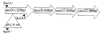
FIG. 3A depicts the opuC locus in S. aureus and the site of pAUL-A
integration. The
position of primers used is indicated by an arrow.
FIG. 3B depicts PCR analysis indicating the disruption of opuC by insertion of
pAUL-A.
Left panel: PCR was done using 5' and 3' opuCA-1 and OpuCA-R primers. Right
panel: PCR
was done using opuCAl and kan1P1 primers. The sequence of the primers is
presented in
Table 2.
FIG. 4 depicts in vivo phosphorylation. S. aureus OpuC+/- cells were in vivo
phosphorylated, total cell homogenate was applied to 15% SDS-PAGE, and the gel
was
Coomassie-stained (left panel) or autoradiographed (right panel).
FIG. 5 depicts real time RT PCR on RNA isolated from OpuC+/- cells using
primers
specific to RNAIII. The sequence of the primers is presented in Table 2.
FIG. 6 depicts a growth curve analysis of OpuC+ (=) and OpuC- (^) cells grown
in LB
(left panel) or in LB containing 10% salt (right panel). Cell density was
determined
spectroscopically at OD 600nm.
CA 02654460 2008-11-19
WO 2007/137288 PCT/US2007/069551
FIG. 7 depicts biofilm formation by OpuC+ and OpuC- cells: Cells were grown in
polystyrene 96 well plates for 2 hrs in static conditions in LB at 42 C.
Adherent bacteria were
stained in gentian violet, solubilized in SDS and OD 595 nm was determined.
Results are
presented as % of maximum absorbance observed in several experiments.
FIG. 8 depicts RNAIII production in OpuC+/- cells treated with quorum sensing
autoinducers RAP and AIP: Cells were grown in the presence of the
autoinducers, RNA isolated,
northern blotted, and RNAIII detected using radiolabeled RNAIII-specific DNA.
Control: cells
grown with < 3 kDa spent culture broth of the agr mutant SA RN6911. Bottom
panel: Ethidium
bromide gel indicating ribosomal RNA loaded.
DETAILED DESCRIPTION
TRAP / OpuC Interaction
The present invention provides methods and compositions directed to disrupting
the
interaction between TRAP and OpuCA as well as TRAP and proteins encoded by the
OpuC
operon. When the OpuC operon is disrupted, the mutant phenotype displays
reduced indicia of
pathogenesis, such as reduced TRAP phosphorylation, reduced agr activity, and
reduced
biofilm formation. Further, OpuCA was shown by a two-hybrid method to interact
directly with
TRAP. Inhibiting the interaction between TRAP and a protein encoded by the
OpuC operon,
particularly OpuCA, will treat a presently occurring disease as well as reduce
the risk of
infection by bacteria in which TRAP plays a role in pathogenesis, such as S.
aureus.
There is evidence that TRAP is conserved among all staphylococcal strains and
species;
therefore, other staphylococcal species should have a quorum sensing mechanism
like that
described above. In addition, there is evidence of TRAP phosphorylation in S.
epidermidis,
6
CA 02654460 2008-11-19
WO 2007/137288 PCT/US2007/069551
indicating that there is a similar quorum sensing mechanism in both S. aureus
and S. epidenmidis.
Other infection-causing bacteria appear to have proteins with sequence
similarity to TRAP,
including Bacillus subtilus, B. anthracis, B. cereus, Listeria innocua, and L.
monoctogenes. An
inhibitor of the TRAP system thus should interfere with biofilm formation and
infections caused
by any of these bacterial species.
To search for components of the TRAP system, in this embodiment of the instant
invention, TRAP-binding proteins were identified by two hybrid experiments
(26, 27), although
a number of other methods could be used. Two-hybrid system techniques are well
known in the
industry. In the present example, S. aureus 8325-4 genomic library was
screened twice and
clones corresponding to ebh, fmtB and opuCA were identified. Ebh (20) encodes
for
extracellular matrix binding that is regulated by agr; therefore, based on the
self-regulatory
nature of the ebh likely acts downstream of TRAP and agr. FmtB (21) encodes a
cell wall
protein and thus likely acts upstream of TRAP. OpuCA is part of the opuC
operon, which
was shown in Listeria to encode for an ABC transporter (23); therefore, opuCA
likely act
upstream of TRAP.
To test which of the candidate proteins interacts with TRAP in a manner
important for its
activity, OpuC+/- cells, Ebh+/- cells, FmtB+/- cells were tested for TRAP
phosphorylation and
for hemolytic activity or RNAIII production. SirA+/- cells were used for
comparison because
SirA encodes for a transporter (22). OpuC- and FmtB- strains were the only
ones defective in
TRAP phosphorylation, so OpuC and FmtB remained candidates for binding
proteins that may
have a role in TRAP activity, e.g., regulation of pathogenesis. However, FmtB-
cells were
hemolytic so this meant that either that the two-hybrid system falsely
identified Ebh and FmtB as
TRAP-binding proteins, or that, even if these proteins do bind TRAP, they do
not disrupt its
7
CA 02654460 2008-11-19
WO 2007/137288 PCT/US2007/069551
function. Moreover, the OpuC- cells demonstrated characteristics similar to
that of the TRAP-.
OpuCA is encoded by the opuC operon that is highly conserved and was shown in
Listeria to encode for a glycine betaine/camitine/ choline ABC transporter
(23) and is an
important osmolyte uptake system contributing to the growth and survival of
Listeria both in
vitro and in vivo (24). OpuC operon consists of four genes encoding for an ATP
binding protein
(OpuCA), an extracellular substrate binding protein (OpuCC), and two membrane-
associated
proteins presumed to form the permease (OpuCB and OpuCD) (23).
In general, ABC (ATP-binding cassette) transporters comprise one of the
largest families
of structurally related membrane proteins. ABC transporters usually consist of
four core
domains. Two transmembrane domains form a tunnel and those usually consist of
six membrane
spanning alpha-helices that contain the substrate binding sites. In addition,
ABC transporters
possess two highly conserved nucleotide binding domains (NBDs) containing the
ATP-binding
and -hydrolyzing 'motor domain' of the transporter (28).
ABC transporters link ATP hydrolysis to the import or export of various
compounds.
Bacterial oligopeptide permeases are members of the large family of ATP
binding cassette
transporters and typically import peptides of 3 to 5 amino acids, apparently
independently of
sequence. Oligopeptide permeases are needed for bacteria to utilize peptides
as nutrient sources
and are sometimes involved in signal transduction pathways. In B. subtilis,
ABC transporters are
also involved in sporulation and competence by importing specific quorum
sensing peptide
molecules (29) and at least in part by importing specific signaling peptides
derived from phr gene
products. In staphylococci, there is no documentation of uptake of quorum
sensing molecules.
The two autoinducers known to date, RAP and AIP, are considered as acting
extracellularly,
RAP by binding to an unknown receptor to phosphorylate its target protein TRAP
and AIP by
8
CA 02654460 2008-11-19
WO 2007/137288 PCT/US2007/069551
binding to its receptor AgrC (15).
S. aureus OpuC- showed reduced TRAP phosphorylation, reduced biofllm formation
and
reduced agr expression, meaning that OpuC acts upstream of TRAP. As OpuC an
upstream
component to TRAP, the extracellular components of OpuC (OpuCB, OpuCC and
OpuCD)
may interact with the quorum sensing activators known to regulate TRAP
phosphorylation, like
RAP and AIP. Indeed, when OpuC+/- cells were grown in the presence of these
quorum sensing
molecules to test if they can affect TRAP activity, both RAP and AIP activated
the production
of RNAIII in OpuC+ cells, but did not activate RNAIII in OpuC- cells. In the
case of RAP, it is
possible that RAP directly interacts with the extracellular components of
OpuC.
When OpuC is mutated, the phosphorylation of TRAP is reduced but not
abolished. This
suggests that there are additional factors regulating TRAP. Such potential
partners could be
SvrA (30), which is a membrane protein (49 kDa) and has recently been shown to
be a
component of MATE family efflux pump (31). TRAP- and SvrA- mutants confer
essentially
identical phenotypes where in both, TRAP is not phosphorylated, agr is not
expressed and
RNAIII is not produced, and both mutant strains are non-hemolytic (32,30).
Other likely TRAP interacting proteins acting downstream of TRAP include
SarH2.
SarH2 is a transcriptional factor (9) which activates the agr. Microarray and
real time PCR
analysis of SarH2 expression in TRAP- cells indicate highly reduced expression
as compared to
TRAP+ cells (7). SarH2 could thus be a possible downstream protein in RAP/TRAP
signaling
pathway.
Antibodies for the Preferred Embodiment of the Instant Invention
The following definitions will be useful for discussing the instant invention:
9
CA 02654460 2008-11-19
WO 2007/137288 PCT/US2007/069551
The term "antibody" refers to an immunoglobulin protein which is capable of
binding an
antigen. "Antibody" as used herein includes the entire antibody as well as any
antibody
fragments, e.g., F(ab)', Fab, Fv, capable of binding the epitope, antigen or
antigenic fragment of
interest. Preferred antibodies for assays and vaccines of the invention are
immunoreactive or
immunospecific for, and therefore specifically and selectively bind to, a
protein of interest, e.g.,
an anti-TRAP antibody. Also, as used herein, antibody encompasses all types of
antibodies,
e.g., polyclonal, monoclonal, humanized, chimeric, and those produced by the
phage display
methodology. Particularly preferred antibodies of the invention are antibodies
which have a
relatively high degree of affinity either for TRAP or a protein encoded by the
OpuC operon and
which inhibit the interaction between these proteins. An "antigenic fragment"
of a protein is a
portion of such a protein that is capable of binding an antibody.
"Binds specifically" means an antibody binding with high avidity and/or high
affinity to
an epitope of a specific polypeptide. "Specific binding" is stronger than
binding of the same
antibody to any other epitope, particularly those which may be present in
molecules in
association with, or in the same sample, as the specific polypeptide of
interest.
A "detectably labeled antibody" is an antibody (or antibody fragment which
retains
binding specificity) having an attached detectable label. The detectable label
is normally
attached by chemical conjugation, but where the label is a polypeptide, it
could alternatively be
attached by genetic engineering techniques. Methods for production of
detectably labeled
proteins are well known in the art. Detectable labels known in the art include
radioisotopes,
fluorophores, paramagnetic labels, enzymes (e.g., horseradish peroxidase), or
other moieties or
compounds which either emit a detectable signal (e.g., radioactivity,
fluorescence, color) or emit
a detectable signal after exposure of the label to its substrate. Various
detectable labeUsubstrate
CA 02654460 2008-11-19
WO 2007/137288 PCT/US2007/069551
pairs (e.g., horseradish peroxidase/diaminobenzidine, avidin/streptavidin,
luciferase/luciferin),
methods for labeling antibodies, and methods for using labeled antibodies are
well known in the
art. See, e.g., Harlow et ah, eds., "Antibodies: A Laboratory Manual," Cold
Spring Harbor
Laboratory Press, Cold Spring Harbor, N.Y. (1988).
The present invention provides an antibody that specifically binds and is
immunoreactive
with TRAP or a protein encoded by the OpuC operon. The antibody may be
monoclonal,
polyclonal or humanized, and is prepared using methods well known in the art.
Polyclonal antibodies of the present invention may be produced by injecting an
animal
with TRAP or a protein encoded by the OpuC operon to initiate an immunogenic
response. The
immunogen may be coupled to a protein carrier such as keyhole limpet
hemocyanin (KLH) or
bovine serum albumin (BSA). The immunogenicity of the protein may be altered
by
administering a fragment of the protein that includes less than the entire
amino acid seqiuence of
the OpuC protein, the sequence of the protein itself, or the protein and
additional sequences.
Immunogenicity may also be altered by chemically modifying any of these
agents, in addition to
the coupling described above, such as by attachment of one or more
polyethylene glyucol (PEG)
moieties, according to methods known in the art. An adjuvant may also be used.
After a suitable
amount of time to establish a high-titer of antibodies, the serum or eggs are
collected. The
presence of antibody in the serum or eggs may be tested by radioimmunoassay
(RIA), by enzyme-
linked immunosorbent assay (ELISA), or by immunoprecipitation. The
immunoglobulins
may be isolated by the sequential precipitation methods, by conventional
methods of "salting
out" the protein fractions from a salt solution, or by chromatographical
methods well known to
those skilled in the art.
11
CA 02654460 2008-11-19
WO 2007/137288 PCT/US2007/069551
Candidate Selection to Treat Staphylococcus Infection
Of particular interest in the present invention are agents inhibit TRAP
activity by
blocking the interaction of TRAP with proteins of the OpuC operon, e.g., any
of OpuCA,
OpuCB, OpuCC or OpuCD individually or collectively. Such agents are candidates
for
development of treatments for infection of pathogenic Staphylococcus and other
bacteria that
utilize the TRAP system. Of particular interest are screening assays for
agents that have a low
toxicity for human cells and/or high specificity for bacteria, preferably with
substantially no or
little pressure for selection of strains resistant to the action of the agent,
and without
substantially affecting normal flora of the host, e.g., as distinguished from
wide-spectrum
antibiotics.
The term "agent" as used herein describes a protein or pharmaceutical with the
capability
of altering the interaction of TRAP with a protein encoded by the OpuC operon.
A plurality of
assay mixtures may be run in parallel with different agent concentrations to
detect differential
responses to the various concentrations of the agent. Typically, one of these
concentrations
serves as a negative control, i.e. at zero concentration or below the level of
detection.
Candidate agents encompass numerous chemical classes, though typically they
are
organic molecules, preferably small organic compounds having a molecular
weight of more than
50 and less than about 2,500 daltons. Candidate agents comprise functional
groups necessary for
structural interaction with proteins, particularly hydrogen bonding, and
typically include at least
an amine, carbonyl, hydroxyl or carboxyl group, preferably at least two of the
functional
chemical groups. The candidate agents often comprise cyclical carbon or
heterocyclic structures
and/or aromatic or polyaromatic structures substituted with one or more of the
above functional
groups. Candidate agents are also found among biomolecules including, but not
limited to:
12
CA 02654460 2008-11-19
WO 2007/137288 PCT/US2007/069551
peptides, saccharides, fatty acids, steroids, pheromones, purines,
pyrimidines, derivatives,
structural analogs or combinations thereof.
Candidate agents are obtained from a wide variety of sources including
libraries of
synthetic or natural compounds. For example, numerous means are available for
random and
directed synthesis of a wide variety of organic compounds and biomolecules,
including
expression of randomized oligopeptides. Alternatively, libraries of natural
compounds in the
form of bacterial (e.g., non-pathogenic Staphylococcus), fungal, plant and
animal extracts are
available or readily produced. Additionally, natural or synthetically produced
libraries and
compounds are readily modified through conventional chemical, physical and
biochemical
means, and may be used to produce combinatorial libraries. Known
pharmacological agents may
be subjected to directed or random chemical modifications, such as acylation,
alkylation,
esterification, amidification, etc. to produce structural analogs.
Screening of Candidate Agents
A wide variety of in vitro assays may be used to screen candidate agents,
including
labeled in vitro binding assays, e.g., protein-protein binding,
electrophoretic mobility shift
assays, immunoassays for protein binding, and the like. Purified naturally-
occurring or
recombinant proteins and/or synthetically produced peptides or fragments of
proteins can be used
in various screening assays to identify ligands or substrates that bind to,
modulate (e.g., increase
or inhibit), or mimic the action of the native proteins. The purified proteins
may also be used for
determination of three-dimensional crystal structure, which can be used for
modeling
intermolecular interactions, transcriptional regulation, etc.
The screening assay can be a binding assay, wherein one or more of the
molecules may
be joined to a label that directly or indirectly provides a detectable signal.
Various labels include
13
CA 02654460 2008-11-19
WO 2007/137288 PCT/US2007/069551
radioisotopes, fluoresces, chemiluminescers, enzymes, specific binding
molecules, particles, e.g.
magnetic particles, and the like. Specific binding molecules include pairs,
such as biotin and
streptavidin, digoxin and antidigoxin etc. For the specific binding members,
the complementary
member would normally be labeled with a molecule that provides for detection,
in accordance
with known procedures. In general, the particular type of screening assay
employed will be
amenable to parallel, simultaneous screening of a large number of candidate
agents.
One screening assay of particular interest involves detection of TRAP
phosphorylation.
TRAP phosphorylation screening assays may be performed in, for example, a cell-
free assay or
in whole cell assays. Phosphorylation screening assays may be performed in a
variety of ways.
For example, the candidate agent may be combined with detectably labeled
phosphate, TRAP,
and varying concentrations of RAP to determine if the candidate agent competes
with RAP to
inhibit TRAP phosphorylation by RAP. Alternatively, the candidate agent may be
combined
with detectably labeled phosphate, RAP, TRAP, and varying concentrations of
RIP to determine
if the candidate agent affects RIP activity. Assays, and methods for
performing such assays, that
can be used in the screening assay of the invention are well known in the art.
See, for example,
Roychoudhury et al., Proc. NatZ Acad. Sci. U.S.A. 90: 965-969 (1993), which
describes
identification of compounds that block the expression of alginate, a virulence
factor for the cystic
fibrosis pathogen Pseudomonas aemginosa, and which is incorporated herein by
reference with
respect to drug screening assays and methods and compositions for performing
same.
Screening assays of the present invention also determine the effect of
candidate agents on
the role of TRAP in RNAIII production and/or virulence factor production. For
example, the
candidate agent may be contacted with pathogenic Staphylococcus, and the
levels of TRAP
phosphorylation and/or RNAIII transcription in the presence of the agent
compared to TRAP
14
CA 02654460 2008-11-19
WO 2007/137288 PCT/US2007/069551
phosphorylation and/or RNAIII transcription levels in the presence of RIP,
RAP, and/or a
combination of RIP and RAP. Such screening assays can utilize recombinant host
cells
containing reporter gene systems such as CAT (chloramphenicol
acetyltransferase), {3-
galactosidase, and the like operably associated with RNAIII or virulence
factor genes to facilitate
detection of RNAIII or virulence gene transcription or to facilitate detection
of RNAIII or
virulence factor production. Alternatively, the screening assay can detect
RNAIII or virulence
factor transcription using hybridization techniques well known in the art,
e.g., Northern blot,
PCR, etc.
A variety of other reagents may be included in the screening assays described
herein.
Where the assay is a binding assay, these include reagents like salts, neutral
proteins, e.g.
albumin, detergents, etc. that are used to facilitate optimal protein-protein
binding, protein-DNA
binding, and/or reduce non-specific or background interactions. Reagents that
improve the
efficiency of the assay, such as protease inhibitors, nuclease inhibitors, or
anti-microbial agents,
may be used. Components are mixed in any order to provide for the requisite
binding.
Incubations are performed at any suitable temperature, typically between 4 C
and 40 C.
Incubation periods are selected for optimum activity, but may also be
optimized to facilitate
rapid high-throughput screening. Typically between 0.1 and 1 hr will be
sufficient.
Screening of Candidate Agents in an Animal Model
Agents having a desired activity as determined in the assays described above
can be
further screened for their ability to affect virulence factor production and
to affect infection in a
non-human animal model. The animal model selected will vary with a number of
factors
including the particular pathogenic strain against which candidate agents are
to be screened, the
ultimate host for which the candidate agents are to serve as therapeutics,
etc. Animals suitable
CA 02654460 2008-11-19
WO 2007/137288 PCT/US2007/069551
for use in screening assays include any animal susceptible to infection by the
selected pathogenic
species. For example, where the bacterial species is S. aureus, the animal
model can be a rodent
model, preferably a mouse model.
In general, the candidate agent is administered to a non-human animal
susceptible to
infection, where the animal has been previously infected with the pathogen or
receives an
infectious does of the pathogen in conjunction with the candidate agent.
Preferably, the animal
has no detectable antibodies against pathogen proteins. The candidate agent
can be administered
in any manner desired and/or appropriate for delivery of the agent in order to
affect a desired
result. For example, the candidate agent can be administered by injection
(e.g., by injection
intravenously, intramuscularly, subcutaneously, or directly into the tissue in
which the desired
affect is to be achieved), topically, orally, or by any other desirable means.
Normally, this screen
will involve a number of animals receiving varying amounts and concentrations
of the candidate
agent (from no agent to an amount of agent hat approaches an upper limit of
the amount that can
be delivered successfully to the animal), and may include delivery of the
agent in different
formulations. The agents can be administered singly or can be combined in
combinations of two
or more, especially where administration of a combination of agents may result
in a synergistic
effect.
The effect of agent administration upon the animal model can be monitored by
any
suitable method, such as assessing the number and size of pathogen-associated
lesions, overall
health, etc. Where the candidate agent affects bacterial infection in a
desirable manner (e.g., by
reducing infectious load, facilitating lesion regression, etc.), the candidate
agent is identified as
an agent suitable for use in treatment of bacterial infection.
16
CA 02654460 2008-11-19
WO 2007/137288 PCT/US2007/069551
Carrier for Candidate Agents
The compounds having the desired pharmacological activity may be administered
in a
physiologically acceptable carrier to a host for treatment of pathogenic
infection. The
therapeutic agents may be administered in a variety of ways including, for
example, orally,
topically, parenterally e.g. subcutaneously, intraperitoneally,
intravascularly, or
intrapulmonary. Depending upon the manner of introduction, the compounds may
be
formulated in a variety of ways. The concentration of therapeutically active
compound in the
formulation may vary from about 0.1 -100 wt. %.
The pharmaceutical compositions can be prepared in various forms, such as
granules,
tablets, pills, suppositories, capsules, suspensions, salves, lotions and the
like. Pharmaceutical
grade organic or inorganic carriers and/or diluents suitable for oral and
topical use can be used to
make up compositions containing the therapeutically-active compounds. Diluents
known to the
art include aqueous media, vegetable and animal oils and fats. Stabilizing
agents, wetting and
emulsifying agents, salts for varying the osmotic pressure or buffers for
securing an adequate pH
value, and skin penetration enhancers can be used as auxiliary agents.
Treating Bacterial Infection
The invention provides a method for preventing or treating a human or an
animal
susceptible to infection by a pathogenic bacteria, e.g., S. aureus in humans,
by administering an
agent that inhibits the interaction of TRAP with a protein encoded by the OpuC
operon. In one
embodiment, the host is treated by administration of an agent, such as anti-
TRAP antibody, that
blocks the interaction. In another embodiment, the agent is co-administered
with other TRAP
inhibitors and/or co-administered with other inhibitors of bacterial virulence
factor production,
e.g., RIP. In another embodiment, a TRAP inhibitor, RIP, and a RAP inhibitor,
e.g., an anti-
17
CA 02654460 2008-11-19
WO 2007/137288 PCT/US2007/069551
RAP antibody, are administered. Such administration of multiple TRAP
inhibitory agents may
involve co-administration or sequential administration of the active
components. The dosage
regimen may be adjusted to provide the optimum therapeutic response. For
example, several
divided doses may be administered daily or the dose may be proportionally
reduced as indicated
by the therapeutic situation. The active compounds may be administered in any
convenient
manner, such as by oral, intravenous, intramuscular, subcutaneous, buccal,
transdermal, or
inhalation routes.
Formulations are administered at a therapeutically effective dosage, e.g.. a
dosage
sufficient to improve the chance of successful prevention or treatment of
infection. Human
dosage levels for treating infections are known and generally include a daily
dose from about 0.1
to 500.0 mg/kg of body weight per day, preferably about 6.0 to 200.0 mg/kg,
and most
preferably about 12.0 to 100.0 mg/kg. Generally, it is sought to obtain a
serum concentration of
such a formulation approximating or greater than circulating levels needed to
reduce or eliminate
any infection in less than 10 days. For administration to a 70 kg person, the
dosage range would
be about 50 mg to 3.5 g per day, preferably about 100 mg to 2 g per day, and
most preferably
about 200 mg to 1 g per day. The amount of formulation administered will, of
course, be
dependent on the subject and the severity of the affliction, the manner and
schedule of
administration and the judgment of the prescribing physician.
In employing formulation for treatment of infections, any pharmaceutically
acceptable
mode of administration can be used. The formulations can be administered
either alone or in
combination with other pharmaceutically acceptable excipients, including
solid, semi-solid,
liquid or aerosol dosage forms, such as, for example, tablets, capsules,
powders, liquids, gels,
suspensions, suppositories, aerosols or the like. The formulations can also be
administered in
18
CA 02654460 2008-11-19
WO 2007/137288 PCT/US2007/069551
sustained or controlled release dosage forms, e.g., employing a slow release
bioerodable delivery
system, including depot injections, osmotic pumps, pills, transdermal and
transcutaneous
patches, and the like, for prolonged administration of a predetermined rate,
preferably in unit
dosage forms suitable for single administration of precise dosages. The
compositions will
typically include a conventional pharmaceutical carrier or excipient and a
formulation of the
invention. In addition, these compositions may include other active agents,
carriers, adjuvants,
etc. Generally, depending on the intended mode of administration, the
pharmaceutically
acceptable composition will contain about 0.1% to 90%, preferably about 0.5%
to 50%, by
weight of active compound, the remainder being suitable pharmaceutical
excipients, carriers, etc.
Methods of preparing such dosage forms are known, or will be apparent, to
those skilled in this
art. See, e.g., "Remington: The Science and Practice of Pharmacy," University
of the Sciences in
Philadelphia, 21st ed., Mack Publishing Co., Easton PA (2005).
Parental administration is generally characterized by injection, either
subcutaneously,
intradermally, intramuscularly, or intravenously, preferably subcutaneously.
Injectables can be
prepared in conventional forms, either as liquid solutions or suspensions,
solid forms suitable for
solution or suspension in liquid prior to injection, or as emulsions. Suitable
excipients are, for
example, water, saline, dextrose, glycerol, ethanol or the like. In addition,
if desired, the
pharmaceutical compositions to be administered may also contain minor amounts
of non-toxic
auxiliary substances such as wetting or emulsifying agents, pH buffering
agents, solubility
enhancers, and the like, such as for example, sodium acetate, sorbitan
monolaurate,
triethanolamine oleate, cyclodextrins, and the like.
The percentage of active ingredient contained in such parental compositions is
highly
dependent on the specific nature thereof, as well as the needs of the subject;
however,
19
CA 02654460 2008-11-19
WO 2007/137288 PCT/US2007/069551
percentages of active ingredient of 0.01% to 10% in solution are employable,
and will be higher
if the composition is a solid which will be subsequently diluted to the above
percentages.
Preferably, the composition will comprise 0.2-2% of the active ingredient in
solution. Slow-
release or sustained-release systems may be used to deliver a constant dosage.
Various matrices,
e.g., polymers, hydrophilic gels, and the like, for controlling the sustained
release and for
progressively diminishing the rate of release of active agents are known in
the art.
Formulations of active components may also be administered to the respiratory
tract as a
nasal or pulmonary inhalation aerosol or solution for a nebulizer, or as a
microfine powder for
inhalation, alone or in combination with an inert carrier such as lactose, or
with other
pharmaceutically acceptable excipients. In such a case, the particles of the
formulation may
advantageously have diameters of less than 50 microns, preferably less than 10
microns. See,
e.g., U.S. Pat. No. 5,364,838, which discloses a method of administration for
insulin that can be
adapted for the administration of formulations of the present invention.
Vaccination
The invention provides a vaccine for inoculating a human or an animal
susceptible to
infection by pathogenic bacteria, e.g., S. aureus, by administering TRAP or a
protein encoded by
the OpuC operon, or an antigenically effective portion of the same, in a
pharmaceutically
acceptable carrier optionally comprising an adjuvant. Formulations appropriate
for eliciting an
immune response are well known in the art. In general, the host is exposed to
the antigen to
perturb the host's immune system and elicit an immune response towards the
antigen. An
adjuvant can be added with the antigen to increase the immune response to the
antigen. The
amount of polypeptide administered is an amount sufficient to elicit a
protective immune
response in the host. Methods for determining such appropriate amounts are
routine and well
CA 02654460 2008-11-19
WO 2007/137288 PCT/US2007/069551
known in the art. For example, an antigenically effective portion can be used
to vaccinate an
animal that can be used as a model of Staphylococcus infection. The amounts
effective in such
animal models can be extrapolated to other hosts, e.g., livestock, humans, to
provide for an
amount effective for vaccination.
Coated Devices
The invention provides for a device, the surface of which is coated with a
composition
having an amount of an agent that inhibits the interaction of TRAP with a
protein of the OpuC
operon in an effective amount to inhibit production of virulence factors by
pathogenic bacteria.
The coated device may be any device which may be associated with a risk of
infection, such as
catheters, needles, surgical instruments, e.g., scalpels, sponges, retractors,
bandages and bandage
materials, e.g., gauze, dressings, artificial joints, heart valves, and
tampons. Such devices have a
tendency to bring bacteria into contact with the host, or to attract
colonizations by bacteria. In
such situations, the coated devices may prevent or reduce infection or prevent
or reduce the
development of serious symptoms associated with exposure to bacterial
virulence factors.
EXAMPLES
1. Rat graft model
A subcutaneous pocket was made on each side of the median line by a 1.5 cm
incision on
anaesthetized 250-300 g male Wistar rats (Experiment 1; n=5. Experiment 2:
n=15).
Aseptically, 1 cro sterile collagen-sealed Dacron grafts (Albograft, Italy)
were implanted into
the pockets. Pockets were closed by skin clips, and 1 ml saline with or
without 2x10
exponentially growing bacterial were inoculated onto the graft surface.
21
CA 02654460 2008-11-19
WO 2007/137288 PCT/US2007/069551
2. In vivo biofilm formation by TRAP and agr mutants
To test biofilm formation by TRAP and agr mutants in vivo, TRAP- and agr-
mutants
and their parent strains were injected onto the graft and number of bacteria
on the graft was
counted after 10 days. The results are depicted in FIG. 1. Almost no biofilm
was formed by
TRAP- mutants (27 5 CFU/ml) compared to the control parent strain S. aureus
8325-4 (5.0
1.1 x 10⁵ CF/ml). Further, mutated agr (S. aureus RN6911) made reduced biofilm
(7.9 1.3 x
103) compared to the parent strain S. aureus RN6390 (4.8 1.7 x 10⁵ CFU/ml).
These results
confirm that inhibiting TRAP or agr effectively suppresses biofilm formation.
Significantly, biofilm studies in vivo do not correspond with long-term in
vitro biofilm
studies, which showed enhancement of biofilm production when agr was deleted
or repressed.
These in vitro studies suggested that QS inhibitors would be inadequate for
inhibiting biofilm,
perhaps because of continued high expression of bacterial adhesion molecules
resulting from the
inhibition of agr or TRAP activity. The present studies show clearly that this
is not the case
in vivo. The present study confirms the efficacy of QS inhibitors to treat
biofilm, perhaps
because biofilm formation in vivo depends more on expression of exotoxins than
on expression
of adhesion molecules.
3. Other bacterial strains and growth media.
E. coli XL 1 Blue MRF' and DH5a were used for cloning purposes and were grown
in
Luria-Bertani (LB) broth. Protein-protein interactions were studied in E. coli
XL 1 Blue MRF'
A(mcrA)183 A(mcrCB-hsdSMR-mrr)173 endAl hisB supE44 thi-1 recAl gyrA96 relAl
lac [V
laqP HIS3 aadA Kari ], grown at 30 C in LB broth supplemented with kanamycin
(50 ug/ ml)
and tetracycline (12.5 ug/ ml), or were grown at 37 C in minimal medium
supplemented with
tetracycline (12,5 ug/ ml), chloramphenicol (25 fig/ ml), stretptomycin (12.5
ug/ ml) and with 3-
22
CA 02654460 2008-11-19
WO 2007/137288 PCT/US2007/069551
amino-1,2,4-triazole (3-AT, 5 mM). S. aureus cells were grown in Tryptic Soy
Broth (TSB) or
LB containing appropriate antibiotics (Table 1).
4. DNA techniques.
Standard DNA techniques including the use of restriction endonucleases and DNA
cloning were performed as described (16). Plasmid DNA from E. coli strains was
isolated using
the QIAprep Miniprep kit (QIAGEN). Genomic DNA from S. aureus was isolated
using the
Wizard genomic DNA purification kit (Promega). Recovery of DNA fragments from
agarose
gels was performed using the QlAquick Gel Extraction kit according to
manufacturer's
instructions. The DNA was end-repaired (i.e., filled in, kinased or
dephosphorylated) using the
DNA Terminator Kit (Lucigen).
5. Bacterial Two-Hybrid system.
A genomic library screening method based on the BacterioMatch two-hybrid
system
(Stratagene) was used to identify proteins that interact with TRAP. For this
purpose, the TRAP
coding sequence was cloned into plasmid pBT (pBT.=: traP) to serve as bait and
S. aureus 8325-4
genomic library (1 - 4 kb) in pTRG plasmid (pTRG::SA gDNA) was used as target.
5.1 Construction of the bait plasmid, pBT:: traP.
S. aureus traP gene (GenBank AF202641) was amplified from S. aureus 8325-4
genomic
DNA (gDNA) by PCR using primers 5'BamHUTRAP and 3'Bg1lI/TRAP, which contain a
Bam
HI site or Bgl II site within the 5' and 3' primers, respectively (Table 2).
Using standard
methodology, the amplified traP was digested with Bam HI and Bgl II, then
cloned into the bait
vector, pBT (Stratagene), which was also digested with Bam HI and Bgl II, to
generate
pBT'. itraP. A (gly4ser)3 linker was added upstream of the traP gene at Not
l/Bam HI site.
23
CA 02654460 2008-11-19
WO 2007/137288 PCT/US2007/069551
5.2 Construction of a S. aureus genomic DNA library in pTRG (pTRG::SA
gDNA) as target vector.
Genomic DNA was isolated from S. aureus strain 8325-4 (a standard laboratory
strain)
using standard methodology. An aliquot of the gDNA was sonicated and aliquots
were removed
at times: 1 sec, 3 sec, 8 sec and 12 sec. A small aliquot of gDNA from each
time point was run
on a 0.8% agarose gel to assess fragment size range. The 8 sec time point had
gDNAs ranging
from about 1-4kb and was ligated into the target vector, pTRG which was
linearized by digestion
with EcoRl and BamHI followed by dephosphorylation.
An aliquot of the final purified DNA preparations was run on a gel to estimate
yields
(FIG. 2). Ligation reactions (2 with different molar ratios and 1 pTRG only)
were set up,
transformed into E. coli XL 1 Blue MRF' Kari competent cells (Stratagene) and
plated onto
kanamycin + tetracycline LB plates (30 C). The pTRG vector only plate had -5
colonies
compared to -120 colonies from the pTRG::SA gDNA ligations. Based on the
number of
colonies, it was estimated that 5,000 colonies were obtained from 1 Ixl of
ligation reaction.
Therefore, from a 20 Ixl ligation reaction, one should get - 105 independent
colonies, which is - 5
genome equivalents (assuming 1 in 6 reading frames is correct, with an average
insert size of
lkb).
A 20 ul ligation reaction was set up and transformed as above. The cells were
plated onto
150 mm plates and an aliquot was diluted in order to calculate actual library
size. The final
library size was estimated to be -4 x 10⁵, which represents -20 genome
equivalents. The
colonies were scrapped off the 150 mm plates and pooled. The cells were
divided into 2
aliquots. One aliquot had glycerol added to a final concentration of 15%, then
was divided
into 1 ml aliquots and stored at -80 C (master stock). The second aliquot of
cells was used to
24
CA 02654460 2008-11-19
WO 2007/137288 PCT/US2007/069551
isolate plasmid DNA (pTRG::SA gDNA), to be used for the two-hybrid experiment.
5.3 Two hybrid protein-protein interaction assays.
For protein-protein interaction assays, transcriptional activation of the HISS
gene was
used as the initial test for interaction of the bait and target hybrid
proteins as follows: Plasmids
pBT, pTRG, pBT':vaP and pTRG::SA gDNA were co-transformed in a 1:1 w/w ratio
in different
combinations in the reporter strain E. coli XL 1 Blue MRF' A(mcrA)183 A(mcrCB-
hsdSMR-
mrr)173 endAl hisB supE44 thi-1 recAl gyrA96 relAl lac [F' laqlq HIS3 aadA
Kanr], The
combination of pBT/pTRG, pBT::/raP/pTRG, and pBT/pTRG::SA gDNA were considered
as
negative controls (Table 3). The co-transformants were plated on both non-
selective minimal
medium and selective minimal medium containing tetracycline (12.5 ligl ml),
chloramphenicol
(34 jig/ ml) and 3-AT (5 mM). The colonies which grew on selective medium were
further
screened for streptomycin resistance on minimal medium containing tetracycline
(12.5 ug/ ml),
chloramphenicol (34 jig/ ml), 3-AT (5 mM) and stretptomycin (12.5 ug/ ml). To
confirrn the
detected protein-protein interactions, plasmids were isolated from positive
clones using Qiagen
miniprep kit (Qiagen) and those plasmids were used to transform the reporter
E. coli strain with
the isolated target plasmid plus bait plasmid on plates containing
chloramphenicol, tetracycline,
3-AT and streptomycin. The presence of genomic fragments from these colonies
which grew on
secondary selection plate was further confirmed by PCR amplification and DNA
fragment was
identified by DNA sequencing using pTRG-F and pTRG-R primers (Table 2).
6. In vivo phosphorylation studies.
S. aureus strains were grown to the early exponential phase (ODeoo 0.2
(equivalent to
about 1x10 cells/ml)). The cells from 2 ml cultures were harvested by
centrifugation at 4000g
and resuspended in 1 ml of low phosphate buffer (PFB), (3) and 20 uCi of
radiolabeled
CA 02654460 2008-11-19
WO 2007/137288 PCT/US2007/069551
orthophosphate (32P) (GE Healthcare). Cells were grown for 40 min at 37 C in
and the cells
were collected by centrifugation at 12,000g for 2 min and resuspended in 100
ul of TE buffer
lysostaphin (25 pg/ml), for 10 min at room temperature. Reducing sample buffer
(Pierce) was
added (without boiling) and sample (total cell lysate) was separated on 15%
SDS-PAGE. The
gels were autoradiographed and stained in Coomassie to ensure that equal
amounts of proteins
were loaded on the gel.
7. RNA isolation and real time PCR analysis.
Total RNA was isolated by using modified Qiagen RNeasyTM protect (Qiagen)
protocol.
S. aureus cells were grown overnight in 3 ml TSB and used as pre-inoculum.
These overnight
cultures were diluted 1:100 in 5 ml of TSB and the cultures were grown to the
post-exponential
phase (from ODeoo of 0.03 for 6 hrs) at 37 C with shaking. The cells were
collected by
centrifugation and treated with 20 ul of lysostaphin (2mg/ml), and 2% SDS
containing
proteinase K. RNA was extracted with TRlzol method (Sigma-Aldrich) and further
purified
using Qiagen RNeasyTM protect protocol followed by treatment with DNAse I
(Ambion Inc) at
37 C for 20 min according to manufacturer's instructions. To verify the
absence of genomic
DNA, PCR was done using these DNAse I treated RNA samples as templates, using
5' and 3'
specific gene primers. Two micrograms of each RNA samples were used for cDNA
synthesis
using ImProm-IITM Reverse Transcription System according to manufacturer's
instructions
(Promega). Random hexamers (Invitrogen) were used to prime the reaction, lpl
of resulting
cDNA reaction was used to set up the real time RT-PCR, using the LightCycler
fast start DNA
master SYBR Green Kit (Roche), according to manufacturer's instructions. The
transcript hid
was amplified using RThld primers (Table 2) and the gyrB transcripts that are
constitutively
expressed were used as an internal control (using RTgyr primers). To monitor
the specificity,
26
CA 02654460 2008-11-19
WO 2007/137288 PCT/US2007/069551
the PCR products were analyzed by melting curves and agarose gel
electrophoresis. The values
were normalized with respect to gyrB expression, and data presented in fold
change in
expression (17) of OpuC-/ OpuC+ cells.
8. Construction of S. aureus OpuC- cells.
The OpuCA gene was insertionally inactivated using the temperature-sensitive
suicide
vector pAUL-A. This 9.2-kb plasmid carries an erythromycin resistance marker,
the pUC19
multicloning site, and a temperature-sensitive replication origin, which
enables chromosomal
integration events to be selected at a non-permissive temperature (18). Full-
length opuCA gene
was amplified by PGR using OpuCA-1 and OpuCA-R primers (Table 2) and was
cloned into PCR
cloning vector pCR2.1 according to manufacturer's instructions (Invitrogen).
Plasmids isolated
from positive clones were digested with Xbal and Sacl and insert was cloned at
Xbal and Sacl
sites of pAUL-A similarly digested. Xbal and Sacl fragment contained full
length OpuCA along
with 40 bp from the vector at the 5' terminal end. This construct was digested
with BamHl
which removes 3' sequence of OpuCA (nearly 750 bp of opuCA). pUTE618 (plasmid
containing the omega kanamycin cassette (19) was digested with BamHl, and
insert was gel-
purified and ligated with the ito/wHI-digested pA JL-A::opuCA (above).
Ligation mix was used
to transform into E. coli DH5a and selected on LB agar plates containing
kanamycin (100
p.g/ml). The plasmid DNA was isolated from positive clones and used to
transform dam- strain
of E, coli GM2163 in order to get non-methylated plasmid DNA that was then
used to electro
transform S. aureus RN4220. Chromosomal integration of pAUL A: : opuCA: : kan
was selected
by repeated plating at 42 C with selection for erythromycin (30 ug/ml)
resistance as described
previously (3). The integration of pAUL A::opuC::Jean into the OpuC locus in
RN4220 was
confirmed by PCR, and the resulting strain was designated RN4220 AopuC (OpuC-
). The
27
CA 02654460 2008-11-19
WO 2007/137288 PCT/US2007/069551
primers opuCA-1 andkanIP-1 (Table 2) were used to confirm the single crossing
over event that
yielded 700-bp DNA fragment,
9. Induction of RNAIII production by RAP and AIP.
RAP and AIP were partially purified as described (3) from culture supernatants
of S.
aureus RN4220. Briefly, S. aureus 4220 cells were grown from early exponential
phases for 6
hrs. Culture broth was collected by centrifugation, lyophilized and
resuspended in water to a
tenth of the original volume (lOx). Material was applied to onto a 3kDa
membrane (Amicon)
and fraction less than 3kDA containing AIP was collected, aliquoted and stored
at -70 C until
use. Fraction greater than 3kDa containing RAP was washed several times with
O.lx PBS using
Amicon 10. As a control for spent media, < 3 kDa fraction of culture broth of
agr null strain S.
aureus RN6911 was used. OpuC+/- cells were grown from early exponential phase
of growth
together with RAP or AIP (final Ix dilution) for 40 min. Cells were collected
by centrifugation,
RNA purified, northern blotted and RNAIII detected as described (3).
10. Biofilm formation.
S. aureus OpuC+/- cells that were grown overnight in TSB at 37 C with shaking
were
diluted 1:100 in TSB, and grown for about 2 hrs to OD6oo 0.2. 100 ul of these
early exponential
cells were placed in polystyrene 96 well plates (Falcon) and further grown at
37 C without
shaking for 2 hr. Unattached cells were removed, wells were gently washed with
PBS several
times, and adherent cells were dried in air. Cells were then fixed with 100%
ethanol, stained for
2 min with 0.4% gentian violet in 12% ethanol. Stain was removed, wells were
washed several
times with PBS, and stained cells were solubilized by the addition of 100 ul
1% SDS. The plate
was read at OD 595 nm.
28
CA 02654460 2008-11-19
WO 2007/137288 PCT/US2007/069551
11. Osmotolerance.
OpuC+/- cells were grown overnight in LB with shaking at 37 C. They were then
diluted 1:100 into LB containing 0, 1%, 5%, 10% and 20% NaC1 and grown for
several hours
with shaking at 37 C. At different time intervals cells were removed and cell
density was
measured by recording the absorbance at 600nm.
12. Identification of TRAP-binding Proteins.
A genomic library screening method based on the BacterioMatch two-hybrid
system
(Stratagene) was used to identify proteins that interact with TRAP. For this
purpose, the TRAP
coding sequence was cloned into plasmid pBT (pBT.=: traP)to serve as bait and
S. aureus 8325-4
genomic library (1-4kb) in pTRG plasmid (pTRG::gDNA) was used as target
vector. The
pTRG::SA gDNA bait library was validated by random selection of 9 clones and
performing
colony PCR (FIG. 2) using pTRG primers. No product was observed from the pTRG-
only
colonies (FIG. 2 lanes 1-3), but all 9 pTRG::SA gDNA colonies (FIG. 2 lanes 4-
12) had PCR
products of different sizes ranging from 1-4kb, indicating that the library is
appropriate for
further analysis. Transcriptional activation of the HIS3 gene was used as the
initial test for
interaction of the bait and target hybrid proteins. As shown in Table 3, no
colonies grew in
control reactions while colonies grew from the pBT.=: traP + pTRG:: SA gDNA
reactions. The
S. aureus 8325-4 genomic library was screened twice and identified two clones
corresponding to
ebh and OpuCA (one clone each) in the first screen mdfintB (three clones) in
the second screen.
These positive clones were re-transformed and those grew on selective minimal
medium,
indicating possible protein-protein interactions. Ebh encodes for
extracellular matrix binding
homologue (clone contained -lkb of 28.6kb), which is reported to be regulated
by agr. FmtB
encodes for a cell wall protein (clone contained -lkb of 7.8 kb). OpuCA is
part of the opuC
29
CA 02654460 2008-11-19
WO 2007/137288 PCT/US2007/069551
operon, which was shown in Listeria to encode for a betaine/ carnitine ABC
transporter.
To test which of the candidate proteins interacts with TRAP in a manner
important for its
signaling (TRAP phosphorylation, agr activation and/or hemolysis), S. aureus
mutant strains of
ebh (20), fintB (21) and opuC were tested for the above phenotypes. Since
there were no reports
of opuC mutant of S. aureus, an opuC mutant of S. aureus was generated. Sir A-
(22) also was
tested because it encodes for an iron siderophore ABC transporter and was used
for comparison.
13. Generation of S. aureus OpuC- mutant.
OpuC operon consists of four genes encoding for an ATP binding protein
(OpuCA), an
extracellular substrate binding protein (OpuCC), and two membrane-associated
proteins
presumed to form the permease (OpuCB and OpuCD) (23). The opuC mutant was
generated by
insertional mutagenesis of pAUL-A containing a kanamycin cassette into S.
aureus 4220 opuCA,
resulting in disruption of the whole operon (FIG. 3A). Transformants in which
a single
crossover recombination event had occurred were further confirmed by PCR using
primers
OpuCA-1 and OpuCA-R (as shown in FIG. 3AB), which did not yield any fragment
in OpuC- as
compared to 1.2 kb fragments in case of the parent OpuC+ strain. Further, PCR
was performed
using primers OpuCA-1 and KanIP-1 (FIG. 3AB), which did not yield any fragment
in case of
OpuC-f cells but did yield 700 bp fragment in case of OpuC- cells. The
sequencing of this 700
bp fragment indicated disruption of the opuC operon. Also, the presence of a
transcriptional
terminator downstream of omega kanamycin and erythromycin resistance gene on
pAUL-A
which is oriented with the direction of opuCA transcription as well as the
fact that pAUL-A is
large (9.2 kb) suggests that the insertion mutation is polar, thereby
inactivating the entire opuC
operon. The stability of the pAUL-A insertion was confirrned by PCR analysis
of cultures grown
without erythromycin selection at 30 C using the above primers. Even after
repeated
CA 02654460 2008-11-19
WO 2007/137288 PCT/US2007/069551
subculturing, no plasmid excision was detected. Of note is that mutant cells
grew like the wild
type in conventional growth media.
14. Phenotypic screens.
Proteins that interact with TRAP should also have a role in phosphorylating
TRAP and/or
in activation of agr and pathogenesis. These were tested by in vivo
phosphorylation assays, by
RNAIII detection, by hemolytic assays and finally biofilm formation.
15. In vivo phosphorylation studies.
OpuC+/- cells, SirA+/- cells, Ebh+/- cells and FmtB+/- cells were in vivo
phosphorylated. As shown in Table 1, OpuC- (Fig 3) and FmtB- (not shown) cells
were the
only ones defective in TRAP phosphorylation and thus OpuC and FmtB remained
candidates for
proteins that interact with TRAP and affect its activity (regulation of
pathogenesis).
16. Production of RNAIII and Hemolytic activity.
Once agr is expressed, RNAIII is made, which in turn upregulates the
production of
hemolysins. Production of hemolysins can be viewed by streaking the cells on
blood agar plates
and if agr is active, hemolysis can be observed. Some strains, however, like
RN4220, are
inherently non hemolytic and then the production of RNAIII has to be tested
instead.
SirA+/- cells, Ebh+/- cells and FmtB+A- cells were tested for hemolytic
activity. As
shown in Table 4, all mutant strains were hemolytic, suggesting either that
the two- hybrid
system falsely identified Ebh and FmtB as TRAP-binding proteins, or that even
if these proteins
do bind TRAP, they do not disrupt its function. Accordingly, further studies
focused only on
OpuC.
OpuC- cells were tested for the production of RNAIII by real time PCR. As
shown in
Table 4 and in FIG. 5, the amount of RNAIII was reduced 40 fold. This suggests
that OpuC,
31
CA 02654460 2008-11-19
WO 2007/137288 PCT/US2007/069551
which affects TRAP phosphorylation, is involved in pathogenesis.
17. Osmotolerance.
Osmolites like glycine betaine and choline chloride are required for salt
stress tolerance
(24). S. aureus OpuC+/- was therefore tested for salt stress tolerance in LB
containing 0, 1, 5, 10
and 20% salt (NaC1). No difference in growth was observed in LB containing
additional NaC1 of
0, 1 and 5% (FIG. 6A shown for no salt addition) but OpuC- cells were not as
tolerant and
growth was retarded in 10% NaC1 as compared to the wild type (FIG. 6B). Both
OpuC+/- cells
did not grow in LB containing 20% NaC1. These results suggest that while
mutating OpuC did
not alter the growth of the cells in LB, it did reduce their ability to grow
under stress.
18. OpuC is important for biofilm formation.
To test for biofilm formation, OpuC+/- cells were grown on 96 well polystyrene
plates
overnight and biofilm formation was detected as described (25). As shown in
FIG. 7,
significantly (p < 0.0091) less biofilm was formed by OpuC- mutants, once
again suggesting that
OpuC plays a role in pathogenesis.
19. Quorum sensing (QS) activators interact with OpuC.
If OpuC is in fact an upstream component to TRAP, the extracellular components
of
OpuC (OpuCB, OpuCC and OpuCD) may interact with the quorum sensing activators
known to
regulate TRAP phosphorylation, like RAP and AIP. To test this hypothesis,
OpuC+/- cells were
grown in the presence of these quorum sensing molecules to test if they can
affect TRAP activity
even in the absence of OpuC. Results shown in FIG. 8 indicate that RAP and AIP
activate the
production of RNAIII in OpuC+ cells, but do not activate RNAIII in OpuC-
cells.
32
CA 02654460 2008-11-19
WO 2007/137288 PCT/US2007/069551
While the present invention has been described with reference to the specific
embodiments thereof, it should be understood by those skilled in the art that
various changes
may be made and equivalents may be substituted without departing from the true
spirit and scope
of the invention. In addition, many modifications may be made to adapt a
particular situation,
material, composition of matter, process, process step or steps, to the
objective, spirit and scope
of the present invention. All such modifications are intended to be within the
scope of the claims
appended hereto.
33
CA 02654460 2008-11-19
WO 2007/137288 PCT/US2007/069551
Table 1.
Plasmid or Strain Characteristics Reference
Plasmids
pAUL-A Temperature-sensitive E. coli-S. aureus shuttle vector (3)
EryR, Ch1R
pUTE619 Source for S2-kan, KanR, Ch1R (19)
pCR2.1 PCR cloning vector, KanR, ApR Invitogen
pUC19 Cloning vector, A PR Lab resource
Strains
E. coli DH5aF' Cloning host, Aps Invitrogen
E. coli GM2163 dcm / dam mutant, Host for isolation of unmethylated (19)
DNA for electroporation into S. aureus RN4220
E. coli TG1 Cloning vector, ApR ZYMO Research
E. coli BL-21 DE3 F ompT gal [dcm] [lon] hsdSb (rB mB ) Novagen
S. aureus RN4220 Accepts foreign DNA (r m+) Lab resource
S. aureus RN4220 opuC mutant strain of S. aureus RN4220 In this study
AopuC
S. aureus RN6390 Prophage-cured wild-type strain Lab resource
S. aureus RN6390 RN6390 sirA::Km, Kmr (22)
AsirA
S. aureus RN6390 sirB::Tet; Tetr (22)
RN6390 AsirB
S. aureus RN450 laboratory strain, Mcs (21)
S. aureus RN450 Tn551 insertion mutant infmtB, Emr (21)
AfmtB
S. aureus 8325-4 Wild-type cured of known prophages Lab resource
S. aureus 8325-4, ebh mutated strain of S. aureus 8325-4 (Emr) (20)
Aebh
S. aureus 8325-4, trap mutated strain of S. aureus 8325-4 (Kanr) (32)
AtraP
34
CA 02654460 2008-11-19
WO 2007/137288 PCT/US2007/069551
Table 2.
Name Oligonucleotide Sequence
opuCA-l 5 ' - ACAATTGCGATAATGGTCTTTTT- 3'
opuCA-R 5 ' - TTACTCGAGTCATGATTTATCATCCC - 3 '
hid-1 5'-GAATTTGTTCACTGTGTCG -3'
hld-2 5 ' - TTTACACCACTCTCCTCAC - 3 '
KYopuC-Y 5 ' - ACGAGACACCATGCAACAAC - 3 '
RTopuC-K 5'-CCCACATGTTCTGTTTGCAC-3'
gyr-U 5 ' - TTATGGTGCTGGGCAAATACA- 3'
gyr-L 5 ' - CACCATGTAAACCACCAGATA- 3'
K a n I P - 1 5 ' - GAATTGATCCGGTGGATGAC - 3 '
pTRG-F 5 ' - TCCGTTGTGGGGAAAGTTATC-3'
pTRG-R 5'-GGGTAGCCAGCAGCATCC-3'
rTRAP-F 5 ' - G A A T T C C A T A T G G C T A TT A A A A A G T A T A A G - 3 '
rTRAP-R 5'-CGCGCGGATCCTTATTTTTTCTTACGTCCACG 3'
rOpuCA-F 5'-ATCTCGAGATGATTATGTTAAGTAT- 3'
rOpuCA-R 5'-ATAGATCTTCATGATTTATCATCTC- 3'
5'BamHI/TRAP 5' - TTAGGATCCAAGAAACTATATACATCTTATGGC
3 'BGLII/TRAP 5' - TAAGATCTTAATTAATTAATTATTCTTTTATTGGGTATAGATA
gly-ser linker A 5' -
ATTCiCGGCCGCTGGTGGAGGCTCAGGCGGAGGTGGCAGCGGCGGTGGCGGATCCTAT
gly-ser linker B 5' -ATAGGATCCGCCACCGCCGCTGCCACCTCCGCCTGAGCCTCCACCAGCGGCCGCAAT
CA 02654460 2008-11-19
WO 2007/137288 PCT/US2007/069551
Table 3: Genetic screen for detection of protein-protein interactions.
3-AT 3AT +
streptomycin
pBT+pTRG - -
pBT+pTRG:: SAgDNA - -
pTRG+pBT;:/rap - -
pBT.=: traP + pTRG::SA gDNA + +
36
CA 02654460 2008-11-19
WO 2007/137288 PCT/US2007/069551
Table 4: Phenotypic characterization of the mutants (N/A=not applicable)
Strains In vivo phosphorylation Hemolytic
activity/RNAIII activity
S. aureus 8325-4 Yes Yes
S. aureus 8325-4 AtraP No No
S. aureus 8325-4 Aebh Yes Yes
S. aureus RN450 Yes Yes
S. aureus RN450 AfmtB Reduced Yes
S. aureus RN6390 Yes Yes
S. aureus RN6390 AsirA Yes Yes
S. aureus RN6390 AsirB Yes Yes
S. aureus RN4220 Yes N/A
S. aureus RN4220 Reduced Reduced by 40 fold
AopuC
REFERENCES
1. Rubin, R. J., Harrington, C. A., Poon, A., Dietrich, K., Greene, J. A., and
Moiduddin, A.
(1999) Emerg Infect Dis 5, 9-17.
2. Lowy, F. D. (1998)NEnglJMed 339, 520-532.
3. Balaban, N., Goldkom, T., Gov, Y., Hirshberg, M., Koyfinan, N., Matthews,
H. R.,
Nhan, R. T., Singh, B., and Uziel, 0. (2001) JBiol Chem 276, 2658-2667.
4. Balaban, N., Goldkom, T., Nhan, R. T., Dang, L. B., Scott, S., Ridgley, R.
M., Rasooly,
A., Wright, S. C, Larrick, J. W., Rasooly, R., and Carlson, J. R. (1998)
Science 280, 438-
440.
5. Korem, M., Sheoran, A. S., Gov, Y., Tzipori, S., Borovok, I., and Balaban,
N. (2003)
FEMSMicrobiol Lett 223, 167-175.
37
CA 02654460 2008-11-19
WO 2007/137288 PCT/US2007/069551
6. Yang, G., Cheng, H., Liu, C, Xue, Y., Gao, Y., Liu, N., Gao, B., Wang, D.,
Li, S., Shen,
B., and Shao, N. (2003) Peptides 24,1823-1828.
7. Balaban, N., Stoodley, P., Fux, C. A., Wilson, S., Costerton, J. W., and
Dell'Acqua, G.
(2005) Clin Orthop Relat Res, 48-54.
8. Korem, ML, Gov, Y., Kiran, M. D., and Balaban, N. (2005) Infect Immun
73,6220-6228.
9. Manna, A. C, and Cheung, A. L. (2003) Infect Immun 71, 343-353.
10. Novick, R. P., Ross, H. F., Projan, S. J., Komblum, J., Kreiswirth, B.,
and Moghazeh, S.
(1993) Embo J12, 3967-3975.
11. Novick, R. P., Projan, S. J., Komblum, J., Ross, H. F., Ji, G.,
Kreiswirth, B., Vandenesch,
F., and Moghazeh, S. (1995) Mol Gen Genet 248, 446-458.
12. Ji, G., Beavis, R., and Novick, R. P. (1997) Science 276,2027-2030
13. Qiu, R., Pei, W., Zhang, L., Lin, J., and Ji, G. (2005) JBiol Chem
280,16695-16704
14. Zhang, L., and Ji, G. (2004) JBacteriol 186, 6706-6713
15. Lina, G., Jarraud, S., Ji, G., Greenland, T., Pedraza, A., Etienne, J.,
Novick, R. P., and
Vandenesch, F. (1998) Mol Microbiol 28, 655-662
16. J Sambrook, E. F. F., T. Maniatis. (1989) Molecular cloning, Second
edition Ed. (Nolan,
C, Ed.), Cold spring harbor Laboratory Press, Plainview
17. Livak, K. J., and Schmittgen, T. D. (2001) Methods 25, 402-408
18. Chakraborty, T., Leimeister-Wachter, M., Domann, E., Haiti, M., Goebel,
W.,
Nichterlein, T., and Notermans, S. (1992) JBacteriol 11 A, 568-574
19. Saile, E., and Koehler, T. M. (2002) JBacteriol 184, 370-380
20. Clarke, S. R., Harris, L. G., Richards, R. G., and Foster, S. J. (2002)
Infect Immun 70,
6680-6687
38
CA 02654460 2008-11-19
WO 2007/137288 PCT/US2007/069551
21. Komatsuzawa, H., Ohta, K., Sugai, M., Fujiwara, T., Glanzmann, P., Berger
Bachi, B.,
and Suginaka, H. (2000) JAntimicrob Chemother 45, 421-431
22. Dale, S. E., Sebulsky, M. T, and Heinrichs, D. E. (2004) JBacteriol 186,
8356-8362
23. Fraser, K. R., Ilarvie, D., Coote, P. J., and O'Byrne, C. P. (2000) Appl
Environ Microbiol
66,4696-4704
24. Sleator, R. D., Wouters, J., Gahan, C. G., Abee, T., and Hill, C. (2001)
Appl Environ
Microbiol 67,2692-2698
25. Balaban, N., Giacometti, A., Cirioni, 0., Gov, Y., Ghiselli, R.,
Mocchegiani, F., Viticchi,
C, Del Prete, M. S., Saba, V., Scalise, G., and Dell'Acqua, G. (2003) Jlnfect
Dis 187,
625-630
26. Sun, A. Q., Balasubramaniyan, N., Liu, C. J., Shahid, ML, and Suchy, F. J.
(2004) JBiol
Chem 279, 16295-16300
27. Slepenkin, A., de la Maza, L. M., and Peterson, E. M. (2005) JBacteriol
187, 473-479
28. Pleban, K., Macchiarulo, A., Costantino, G., Pellicciari, R., Chiba, P.,
and Ecker, G. F.
(2004) Bioorg Med Chem Lett 14, 5823-5826
29. Solomon, J., Su, L., Shyn, S., and Grossman, A. D. (2003) JBacteriol 185,
6425-6433
30. Garvis, S., Mei, J. M, Ruiz-Albert, J, and Holden, D. W. (2002)
Microbiology 148,
3235-3243
31. McAleese, F., Petersen, P., Ruzin, A., Dunman, P. M., Murphy, E., Projan,
S. J., and
Bradford, P. A. (2005) Antimicrob Agents Chemother 49, 1865-1871
32. Gov, Y., Borovok, I., Korem, M, Singh, V. K., Jayaswal, R. K., Wilkinson,
B. J., Rich,
S. M., and Balaban, N. (2004) JBiol Chem 279, 14665-14672
33. Frees, D., Qazi, S. N., Hill, P. J., and Ingmer, H. (2003) Mol Microbiol
48, 1565-1578.
39