Note: Descriptions are shown in the official language in which they were submitted.
CA 02654467 2008-11-19
WO 2007/136837 PCT/US2007/012083
PROSTATE EPITHELIAL ANDROGEN RECEPTOR SUPPRESSES
PROSTATE GROWTH AND TUMOR INVASION
1. BACKGROUND
1. Androgens and epithelial-mesenchymal interactions are necessary for
prostate
growth and development. Androgen signaling occurs through the androgen
receptor (AR)
(Chang, C. S., et al. (1988) Science 240, 324-6; Quigley, C. A. et al. (1995)
Endocr Rev 16,
271-321), which is found in both stroma and epithelium within the prostate of
all mammals.
Mice lacking a functional androgen/AR signaling system fail to develop normal
prostate
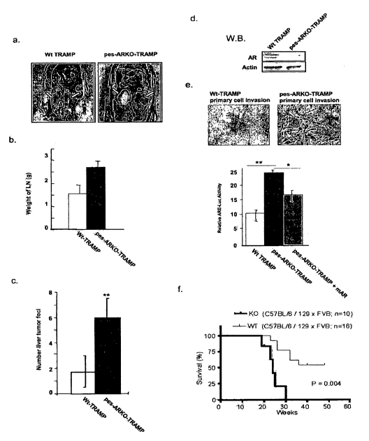
glands (Yeh, S. et al. (2002) Proc Natl Acad Sci U S A 99, 13498-503; Wilson,
J. D., et al.
(1995) Recent Prog Horm Res 50, 349-64). Pioneering developmental studies
showed that
stroma but not epithelial AR is essential for epithelial cell identity,
morphology, bud
formation, ductal branching, proliferation, apoptosis, and regulation of
secretory profile
(Cunha, G. R. & Lung, B. (1978) JExp Zool 205, 181-93; Cunha, G. R. et al.
(2004) J
Steroid Biochem Mol Bio192, 221-36). Experimental evidence has led to the
dogmatically
held assumption that epithelial AR, when activated by androgen, increases
cellular
proliferation (Bello, D., et al.(1997) Carcinogenesis 18, 1215-23; Danielpour,
D., et al.
(1994) Cancer Res 54, 3413-21; Suzuki, H., et al. (2003) Endocr Relat Cancer
10, 209-16).
This notion is the central premise for androgen ablation therapy, a key
treatment for prostate
disease. Although many studies demonstrate that stromal AR mediates key
developmental
events (Cunha, G. R. & Lung, B. (1978) JExp Zoo1205, 181-93; Cunha, G. R. et
al. (2004)
JSteroid Biochem Mol Biol 92, 221-36) these studies were typically evaluated
over short
periods of time, thus recapitulation of events that may take months to
manifest a phenotype
may not have been sufficiently examined.
2. In the adult prostate of all mammals, androgen deprivation leads to
apoptosis and
dedifferentiation of the epithelium, resulting in an increased number of basal
cells and
decreased population of luminal cells (Evans, G. S. & Chandler, J. (1987)
Prostate 11, 339-
51; Mirosevich, J. et al. (1999) J Endocrinol 162, 341-50). In benign
prostatic hyperplasia
and prostate cancer, androgen deprivation decreases growth, increases
apoptosis, and
reduces tumor volume. Often, however, the effect is temporary, and after
removal of
androgens, abnormal epithelial cells persist and inevitably grow independent
of hormone.
Every year more than 30,000 men die and many more suffer from this enigmatic
process. To
-1-
CA 02654467 2008-11-19
WO 2007/136837 PCT/US2007/012083
better understand the role of androgen/AR signaling in prostate biology as
well as
therapeutic targeting, determining the role of epithelia] AR is imperative.
II. SUMMARY
3. Disclosed are methods and compositions related to epithelial Androgen
Receptor
(AR).
III. BRIEF DESCRIPTION OF THE DRAWINGS
4. The accompanying drawings, which are incorporated in and constitute a part
of
this specification, illustrate several embodiments and together with the
description illustrate
the disclosed compositions and methods.
5. Figure 1 shows the characterization of pes-ARKO mice. Figure 1 a shows wild-
type (WT, left) and pes-ARKO mice (KO, right) genotyping. IL-2 RNA occur in
both mice
and serve as internal positive controls. Transgenes: cre (110bps) and floxAR
(540bps) are
only in pes-ARKO mice. Only WT mice have wild-type AR gene. Figure lb shows RT-
PCR
from priming in exon I and exon 3 of the AR gene shows one band for WT AR
(305bps) in
WT mice. In pes-ARKO mice (KO), both WT band (WT AR occurs in stroma) and the
KO
band (153bps; deletion of exon 2) appears in dorso-lateral prostates (DLP) and
ventral
prostate (VP), but only the WT band appears in other tissues. Figure lc shows
the results
from external (c) and internal (d) organs looked the sarne in both strains,
except for VP.
Note larger size of VP from pes-ARKO mice. Figure 1 e shows the expression of
AR (left)
and probasin (right) in VPs of WT vs. pes-ARKO mice with age. Probasin levels
decline
along with epithelial AR. Figure If shows the Pups/litter from WT female x WT
males (red
bar) or x pes-ARKO (blue bar) males were similar, left. Serum testosterone
levels (f, right)
were similar in WT (red bar) and pes-ARKO (blue bar) male mice at weekl2 and
24. Key:
seminal vesicle (SV), kidney (Kid), ureter (U), anterior prostate (AP),
dorsolateral prostate
(DLP), ventral prostate (VP) all lobes of prostate (Pr), testes (T), glans
penis (Pe);
*=P<0.05, * =P<0.001.
6. Figure 2 shows the histomorphological changes in the ventral prostate of
pes-
ARKO mice. Figure 2a shows that the ventral prostate of WT mice show glandular
infoldings (arrowheads) and tall secretory epithelium through week 32. In pes-
ARKO (KO)
littermates, these features were seen only at weeks 3 and 6. In week 9 pes-
ARKO mice,
some ventral prostate ducts lose glandular infoldings (*) and have short,
poorly
differentiated epithelial cells compared with WT littermates. The change in
epithelia is
evident in -50% of ducts within the ventral prostate of week 14 pes-ARKO mice.
At week
-2-
CA 02654467 2008-11-19
WO 2007/136837 PCT/US2007/012083
24 (and older), in pes-ARKO mice nearly all glandular infolding and high
secretory cells are
lost and the enlarged ducts have many sloughed epithelial cells, fragmented
nuclei, and
immune cells. By week 32 pes-ARKO mice lack glandular infolding and have squat
epithelial cells. Figure 2b-e shows that at week 14, in pes-ARKO mice, the
ventral prostate
continues to lose glandular infolding. Layers of sloughed epithelial cells are
abundant in the
prostate lumen. Figure 2b shows that in week 14 pes-ARKO mice, some glandular
infoldings (arrow) appear. Figure 2c shows that in pes-ARKO mice, infoldings
(arrow)
become smaller and shorter, and d, infoldings (arrow) lose cellular polarity
and constrict
(red arrow) at their base. Ultimately, in e, putative glandular infoldings
(arrow) are lost and
sloughed luminal cells (arrows) appear within the lumen.
7. Figure 3 shows the localization of androgen receptor (AR) and probasin
(Prb)
within ventral prostate of wild-type (WT) vs. pes-ARKO (KO) mice. WT prostate
expressed
AR in both epithelial and stromal cells (arrows) at all time points evaluated.
Cytoplasmic
and luminal localization of probasin is first observed at week 3 and decreases
from weeks 6-
24 in KO vs. WT mice.
8. Figure 4 shows that loss of epithelial androgen receptor leads to loss of
androgen
regulated protein and gene expression and increased proliferation. Figure 4a
shows that
Androgen regulated gene transcription decreases as epithelial ARs are lost.
Quantitative
RT-PCR was done on ventral prostates from WT (red bar) and pes-ARKO (blue bar)
mice.
Androgen regulated genes probasin, PSP94, and Nkx3.1 are all down-regulated in
pes-
ARKO prostates compared to WT prostates. Figure 4b shows that ventral
prostates were
collected from WT (red bar) and pes-ARKO (blue bar) mice during different
stages and
analyzed for proliferation. Proliferation, as determined by BrdU positive
nuclei primarily
occurs in epithelial cells at all stages evaluated and c, epithelial cell
proliferation is
significantly (P<0.01) higher in pes-ARKO than WT littermates. *=P<0.05.
9. Figure 5 shows the localization of epithelial specific markers within the
prostate.
To determine which epithelial cell populations were increased due to cellular
proliferation,
immunohistochemistry for basal cell marker p63 a, and luminal cell marker
cytokeratin-8
and 18 and epithelial marker pan-cytokeratin b, were performed. Note that an
increased
number of putative basal cells (arrows; p63 positive) are observed at 32 weeks
in pes-
ARKO vs. WT mice, whereas, epithelial and luminal cell markers appear to
decrease in
ventral prostates from pes-ARKO mice vs WT mice.
-3-
CA 02654467 2008-11-19
WO 2007/136837 PCT/US2007/012083
10. Figure 6 shows the sloughing and apoptosis of epithelia in the prostate of
pes-
ARKO mice. Epithelial cell sloughing is rare in ventral prostates of wild-type
(WT) at any
time point or in pes-ARKO mice prior to week 16. Figure 6a shows hematoxylin
and eosin
staining of pes-ARKO ventral prostates. Note the accumulation of sloughed
layers of
luminal cells (arrows), cellular debris, putative immune cells, and fragmented
nuclei
through week 24. Figure 6b shows the identification of TUNEL-positive cells
was
performed at weeks 16, 24, and 32. Note that the prostatic cell layer (stroma
and epithelium)
is not TUNEL positive, whereas TUNEL-positive cells are found within the
prostatic lumen.
Figure 6c shows efforts to determine if proliferating (BrdU-positive, green)
cells were basal
(CK5-positive, red) or luminal (CK8-positive, red) cells we evaluated double
staining for
BrdU+CK5 vs BrdU+CK8 in pes-ARKO and WT mice. Note that CK8+BrdU-positive
cells
are rarely seen but BrdU positive cells are found within the basal layer
(arrows), whereas
CK8+TUNEL positive cells (arrows) are primarily observed within the lumen of
pes-ARKO
mice with few TUNEL-positive cells observed in the intact epithelial layer.
CK5+BrdU
positive cells are observed accordingly in the basal layer (arrows) however
not all CK5-
positive cells are BrdU-positive (note arrowheads). Note that TUNEL-positive
cells are
CK5-negative (arrows). Figure 6d shows BrdU-labeling index in different
prostatic lobes
from pes-ARKO mice at 24 weeks of age.
11. Figure 7 shows the expression of T857A mutant androgen receptor transgene
in
pes-ARKO mice reverts ventral prostate phenotype to wild-type (WT). Figure 7a
shows
hemotoxylin and eosin staining of 32 week-old ventral prostates from WT, pes-
ARKO, and
pes-ARKO/T857A mice. Note that epithelium in pes-ARKO/T857A mice are very
similar
in morphology, cell height, architecture, and glandular infolding to WT mice.
Figure 7b
shows that pes-ARKO/T857A mice have nonnal AR gene transcription levels and
proliferation rates. Quantitative RT-PCR for probasin (orange bars) expression
in ventral
prostates at 32 weeks. In pes-ARKO/T857A mice probasin expression is
significantly
increased compared to pes-ARKO mice, but not different compared to WT.
Quantitative
RT-PCR for PSP-94 (green bars) expression in ventral prostates at week 32. In
pes-
ARKO/T857A mice PSP-94 expression is significantly increased compared to pes-
ARKO
mice, but not different compared to WT. BrdU-labeling index (blue bars) in
week 32
ventral prostates. In pes-ARKO/T857A mice epithelial cell proliferation is
significantly
decreased compared to pes-ARKO mice, but not different compared to WT;
*=P<0.05,
=P<0.001
- 4 -
CA 02654467 2008-11-19
WO 2007/136837 PCT/US2007/012083
12. Figure 8 shows Probasin cre and ARKO constructs. Figure 8a shows the
structure of the ARR2PB-Cre-SV40 transgene construct_ The plasmid contained
the
ARR2PB composite PB promoter followed by the cDNA for Cre and SV40
polyadenylation
sequence. Arrows indicate the position of the primers used in the PCR-based
identification
of transgenic mice. Figure 8b shows the construction of the floxed AR
fragment. The PKI
vector is modified from the pBluescript plasmid. It contains a T7 promoter at
the 3'end, a T3
promoter at the 5' end, two multiple cloning sites (MCS), two lox sites, a
positive Neo
selective marker (PKG-Neo), and a negative thymidine kinase selective marker
(MCT-TK).
For the cloning, the Xhol site at the 5' end MCS was first destroyed. A 3-kb
intron 2
fragment was introduced into the 3 EcoRl cloning site (R1), followed by a
fragment
containing intron 1, exon 2, and a small fragment of intron 2 sequences into 5
XbaI site (X).
A lox sequence plus an artificial KpnI site were finally inserted into the
Xhol site shortly 5'
to the beginning of exon 2. The constructed plasmid was linearized by NotI
before being
electroporated into ES cells.
13. Figure 9 shows that epithelial AR is a suppressor and stromal AR is a
stimulator
for the prostate cancer cell invasion in vitro. Figure 9(a-c) shows knock-in
AR in human
prostate cancer PC3-v cells results in increased invasion ability. AR protein
expression in
different PC3-v cell lines transfected with AR cDNA under the control of a
natural proximal
AR promoter region PC3-AR9 or strong SV40 promoter PC3-AR2 (upper)(a).
Transcriptional activity of AR using ARE-(4)-Luc in PC3-AR9 increased 5 fold
with 1 nM
DHT, addition of 1 M HF resulted in suppression of DHT-induced
transactivation (b). The
invasion of PC3-AR9 was decreased in the presence of DHT and was increased
with the
addition of HF when compared similarly to PC3-v cells grown Matrigel coated
Boyden
chambers (c). Figure 9(d-f) shows in vitro tissue recombination assays showed
AR played a
positive role in PC3-v and PC3-AR9 cell invasion. PC3-v or PC3-AR9 cells
cultured on the
upper-layer of the Boyden chamber, were co-cultured with WPMY1 vector (WPMY1-
v) or
WPMY1 AR knockdown (WPMY1-ARsi) cells, which were put in the lower-layer of
the
chamber as shown (d). WPMY1 cells with Wt AR significantly increase PC3-v or
PC3-AR9
cells (upper panel) invasion as compared with WPMY1-ARsi cells (lower panel)
(e), and
the data were quantitated (f). Figure 9g shows decreased AR in human prostate
cancer
CWR22R cells results in increased invasion ability. CWR22R-AR~" cells in which
some
alleles of AR gene were genetically disrupted were generated by homologous
recombination
strategy. Western blot shows that expression of AR is low in the presence or
absence of I
- 5 -
CA 02654467 2008-11-19
WO 2007/136837 PCT/US2007/012083
nM DHT in CWR22R-AR+/-'cells compared with CWR22R-AR+/+ (upper).
Transcriptional
activity of AR is diminished in CWR22R-AR+/- compared with CWR22R-AR-"+ in the
presence 1 nM DHT (left lower). The invasion into matrigel increased in CWR22R-
AR+/"
compared with parental CWR22R-AR+/+(right lower). Figure 9h shows knockdown AR
in
CWR22R cells using AR-siRNA increased cell invasion in vitro.
14. Figure 10 shows the addition of functioanal AR in PC3-AR9 cells resulted
in
decreased invasion in in vivo mice models. Figure l0a shows PC3-AR9 cells
formed less
osteoclytic lesions than PC3-v cells. Osteoclasts and osteoclast precursors
(OC) on cortical
bone wafers were cultured with PC3-v and PC3-AR9 cells. After ten days, the
wafers were
scraped, dried, and stained for tartrate-resistant acid phosphatase (TRAP) for
OC cells. The
extent of bone resorption with PC3(AR)9 cells decreased compared to PC3-v via
measurement by the area of osteoclast lacunae on the bone wafers. OC alone and
OC with
PTH were used as a negative and positive control respectively. Data are mean f
SD *P<
0.05, **P< 0.01 of three independent experiment and n = 3 wild type nude rat
(2 day old rat)
to isolate the osteoclast precursors. Figure 10(b, c) shows that PC3-AR9 cells
had less
ability in bone invasion than that of PC3-v cells. Effects of intra-tibial
injection of PC3-v
and PC3-AR9 cells in nude mice. PC3-v cells produced larger and more invasive
tumors as
measured by Dial Caliper in week 12 (b) and higher osteolytic activity in 6-8
weeks
radiograph (X-ray) than PC3-AR9 cells. (c, arrow). Data are mean SD * P<
0.05, **P<
- 0.01. Figure l Od shows that PC3-AR9 cells generated smaller metastatic
tumors in the
lymph nodes than that generated by PC3-v. 5x10⁵ PC3-v and PC3-AR9 cells
suspended in
50u1 Matrigel were directly injected into anterior prostate of nude mice. 12
weeks following
injection, the tumors were developed and the metastatic tumors in the lymph
nodes were
compared. Figure 10e shows that PC3-AR9 cells combined with WPMY1-v and WPMY1-
ARsi cells generated smaller metastatic tumors than their control PC3-v
combine groups.
5x 10⁵ PC3-v or PC3-AR9 cells respectively combined with 5x 10⁵ WPMY1-v or
WPMY1-
ARsi cells were suspended in 50 1 Matrigel, and were directly injected into
anterior prostate
of nude mice. 12 weeks following injection, the tumors were developed and the
metastatic
tumors in the lymph nodes were compared.
15. Figure 11 shows the generation of pes-ARKO-TRAMP Mice. Figure I I a shows
the loss of AR protein expression in prostate epithelium of week 12 pes-ARKO-
TRAMP
mice compared to WT-TRAMP demonstrated by immunohistochemistry using anti-
- 6 -
CA 02654467 2008-11-19
WO 2007/136837 PCT/US2007/012083
AR(C19) antibody. Figure 1 lb shows similar development of internal urogenital
organs of
WT-TRAMP and pes-ARKO-TRAMP mice at 6-weeks-old.
16. Figure 12 shows that pes-ARKO-TRAMP mice that lack AR only in prostate
epithelium develop more aggressive and invasive metastatic tumors. Figure 12a
shows that
Pelvic lymph node (PLN) tumors are significantly larger in week 24- pes-ARKO-
TR.A.MP
mice compared to WT-TRAMP. Figure 12b shows the weight of PLN isolated from
24ws
pes-ARKO-TRAMP and Wt TRAMP mice (n=7 mice in each group). Figure 12c shows
the
number of liver tumor foci was increased in pes-ARKO-TRAIVIP mice compared to
wt-
TRAMP mice (n = 4 mice in each group). Figure 12d shows the expression of AR
determined by Western blot analysis of AR protein in PLN tumor, from either
week 24 WT-
TRAMP or pes-ARKO-TRAMP mice. Figure 12e shows the higher invasion from pes-
ARKO-TRAMP mice PLN tumor primary culture cells compared to those from WT-
TRAMP mice using Boyden chamber invasion assay. Addition of functional AR via
pBabe
virus expressed AR cDNA results in suppression of invasion. The
purity/originality of PLN
tumor primary culture cells was confirmed by the expression of pan-CK
epithelial marker.
Data are meanf SD * P< 0.05, **P< 0.01 of three independent experiment and n =
5 mice
in each group. Figure 12f shows that survival was decreased in pes-ARKO-TRAMP
(C57BU6/129 x TRAMP-FVB, n=10) as compared to WT-TRAMP (C57BL16/129 x
TRAMP-FVB, n=16).
17. Figure 13 shows that ind-ARKO-TRAMP mice delayed in developing
metastasis. Figure 13a shows at 24wks ind-ARKO-TRAMP mice, which significantly
decreased AR expression in both prostate epithelium and stroma, developed
smaller tumor
with less aggression and metastasis comparing with same-aged Wt tumor. And the
metastastic tumor size among groups followed such sequence: pes-ARKO-TR.AMP >
Wt
TRAMP (with and without PIPC) > ind-ARKO-TRAMP. Figure 13b shows that
different
tumor malignancy was demonstrated by comparing the metastasis status of
similar-sized
tumors from groups. In the age of about 22ws, Wt TRAMP mice developed lcm
diameter
tumors, most of which were well-differentiated tumor with small pelvic lymph
node
metastasis. In the contrary, pes-ARKO-TRAMP tumor in the similar size
developed much
larger lymph nodes metastasis in multiple region, even in mesentery, in their
earlier age of
18ws. However, ind-ARKO-TRAMP tumor in 1 cm diameter, although formed as late
as
36ws, invaded into seminal vesicle and migrated to liver. HE staining showed
Wt TRAMP
tumor were much more well-differentiat than pes-ARKO-TRAMP tumor and ind-ARKO-
- 7 -
CA 02654467 2008-11-19
WO 2007/136837 PCT/US2007/012083
TRAMP tumor (the second panel). Although AR staining in the pes-ARKO-TRAMP and
ind-ARKO-TRAMP tumor was significantly reduced (the third panel), the T-
antigen (T-ag)
expression in these tumors were not significantly changed (lowest panel).
18. Figure 14 shows loss of AR expression in human metastatic tumors compared
with primary prostate tumors. Figure 14a shows the area of high-grade primary
tumor
showing positive nuclear staining for AR. Immunostain. 400x. Figure 14b shows
the area of
high-grade metastatic tumor showing negative staining for AR. linmunostain.
400x. Figure
14c shows a summary of AR expression comparing primary versus metastatic
tumor. None
of the tumors displayed neuroendocrine differentiation, and all tissue were
obtained from
live patients, either as biopsies or lymph node dissections during concurrent
prostatectomy.
There was no selection for androgen independent metastatic disease. The data
for treatment
is not available, except in the case for lymph node metastasis, which were all
non-treated
cases. Metastatic sites included lymph nodes (10), bone (12), liver (1), lung
(2),
penis/urethra (2), bowel (1). **P<0.01
19. Figure 15 shows the influence of AR on the different metastasis/invasion-
related
genes. Figure 15a shows that western blot demonstrates a decrease in
expression of NEP
protein from PLN tumor of pes-ARKO-TRAMP and the PC3-v xenograft compared to
those
.from WT-TRAMP and PC3-AR9 respectively. The transcriptional activity of NEP
using
NEP-Luc in PC3-AR9 was decreased in the presence of AR-siRNA (si-AR). Figure
15b
shows increased expression of Cox-2 protein in PLN tumor of pes-ARKO-TRAMP and
the
PC3-v xenograft compare to those from WT-TRAlViP and PC3-AR9 tumor xenograft
(upper). Transcriptional activity of Cox-2 using Cox-2-Luc in PC3-AR9 was
decreased after
restoration of functional AR via pBabe virus carrying AR cDNA (lower). Figure
15c shows
decreased p27 protein expression using Western blot from PLN tumor of pes-ARKO-
TARMP mice and PC3-v xenografts as compared to those from WT-TRAMP and PC3-AR9
(upper). The increased stability of the cdk inhibitor p27 with AR (middle).
PC3-v, and PC3-
AR9 cells were treated 48 hr in presence or absence of 1 nM DHT. Cells were
then treated
with 50 g/ml cycloheximide (CHX) for indicated times and 50 g cell lysates
were
examined by Western blot analysis with an anti-p27 antibody (middle), and the
quantitation
of the data (middle lower). Figure 15d shows decreased MMP-9 expression and
activity
with AR. MMP-9 increased in samples from PLN tumor pes-ARKO-TRAMP and PC3-v
tumor xenografts compared to wt-TR.AMP and PC3-AR9 tumor xenografts (upper).
Enzyme
assays of MMP-9 in PC3-AR9 cells further shows that I nM DHT can suppress and
I M
- 8 -
CA 02654467 2008-11-19
WO 2007/136837 PCT/US2007/012083
HF can restore the gelatinase activity of MMP-9 (middle upper). Zymography
assay for the
activity of MMP-9 were increased after treatment with 10 nM TPA (NF-kB
activator) and
decreased after addition of 1 g/ml parthenolide (NF-kB inhibitor) (middle
lower).
Transactivation assay using a NF-kB-Luc in the presence of 1 nM DHT suppresses
NF-xB
activity in PC-3(AR)9. I M HF restored the effect of DHT (lower). Figure 15e
shows that
Akt activity was increased as measured by phosphorylation at serine 473 using
p-Akt-473
antibody in Western blot assay of PLN tumor from pes-ARKO-TRAMP and PC3-v
xenograft compared to WT-TRAMP and PC3-AR9 (left) and PC3-v and PC3-AR9
derived
tumors cells (right). Data are meanf SD * P< 0.05, **P< 0.01 of three
independent
experiment and n = 3 mice each group. (f) stably knockdown AR expression in
human
stromal WPMY1 cells significantly changed stroma paracrine factor expressions.
Western
blots showed AR expression had been knocked down in WPMY1-ARsi cell lines by
AR-
siRNA (upper panel). Realtime RT-PCR measurement of stroma paracrine factors,
which
infects tumor metastasis (lower panel), showed the decreased expression of
TGFP 1, TGF(32,
TGFP 1, SDF-1 and VEGF.
20. Figure 16 shows knock-in AR in prostate cancer cell line PC3 suppressed
xenograft tumor growth, and knockdown AR in the prostate stroma cell line WPMY
I also
suppressed PC3 xenograft tumor generation in in vivo tissue recombination.
Figure 16a
shows that PC3-AR9 cells grew slowly and generated smaller tumors in the
anterior prostate
than that generated by PC3. 5 x 10⁵ PC3-v and PC3-AR9 cells suspended in 50 1
Matrigel
were directly injected into anterior prostate of nude mice. 12 weeks following
injection, the
tumors were harvested (upper panel). Ki67, which indicated tumor growth
activity, was
stained by immunohistochemistry. Figure 16b shows that PC3 cells combined with
WPMY1-v generated smaller tumors than tumors from PC3 and WPMY1-ARsi
combination in vivo. 5x 10⁵ PC3-v or PC3-AR9 cells respectively combined with
5x 10⁵
WPMY1-v or WPMY1-ARsi cells were suspended in 50 1 Matrigel, and were directly
injected into anterior prostate of nude mice. 12 weeks following injection,
the xenograft
tumors were harvested and compared (upper panel). H&E staining show PC3 +
WPMY1-v
cells formed relatively poor differentiated tumor than tumor from PC3-AR9 +
WPMY1-v in
term of lumen formation (middle panel). Ki67 staining showed PC3 + WPMY1-v
tumor had
higher growth rate than PC3-AR9 + WPMY1-v tumor (lower panel).
- 9 -
CA 02654467 2008-11-19
WO 2007/136837 PCT/US2007/012083
21. Figure 17 shows the generation and confirmation of pes-ARKO-TRAMP mice
and ind-ARKO-TRAMP mice. Figure 17a shows the mating strategy of pes-ARKO-
TRAMP (C57BL/6/129xTRAMP-FVB) and ind-ARKO-TRAMP
mice(C57BIJ6/129xTRAMP-FVB). Figure 17b shows the genotype screening of Mice
from
tail snip DNA. T-ag (SV40) primer was used to identify TRAMP mice at 12 weeks
old
(upper). Primers 2-3 and select that amplify AR exon2 region were used to
identify the
Flox/AR in pes-ARKO-TRAMP mice and ind-A.RKO-TRAMPmice (middle). Primers
specific for Pb-Cre and Mx-Cre were used to identify Pb-Cre and Mx-Cre
transgene mice,
respectively (lower). Figure 17c shows that AR knockout were confirmed by
detecting the
exon2 deletion in AR mRNA. Using exonl and exon3 primers, specific ARKO bands
were
shown by RT-PCR amplifying AR mRNA from different organs. In pes-ARKO-TRAMP
mice, ARKO bands were shown in Dorsal Lateral Prostate (DLP), Ventral Prostate
(VP) and
Anterior Prostate (AP), but not significant in Seminal Vesicles (SV) compared
to WT-
TRAMP. In ind-ARKO-TRAMP mice, ARKO bands were shown in DLP, VP, AP, and SV.
22. Figure 18 shows AR expression in pes-ARKO-TRAMP and ind-ARKO-TRAMP
mice. Figure 18a shows the use of Laser Capture Microdissection (LCM) to
separate
epithelium from stroma, AR exon 2 rnRNA expressed in ventral prostate
epithelium was
amplified by Realtime RT-PCR. pes-ARKO-TRAMP mice lost AR mRNA expression from
6ws (25%), gradually reached about 50% in 12ws, and almost disappeared in
16ws. Figure
18b shows IHC AR (C-19) staining showed AR protein lost in ventral prostate
epithelium
(including basal and luminal cells) but not in stroma of 16ws pes-ARKO-TRAMP
mice
compared to Wt TRAMP mice. Figure 18c shows that AR was only knocked out in
the
epithelium but not in the stroma in ventral prostate of 16ws pes-ARKO-TRAMP
mice using
Real-time RT-PCR of AR exon 2 after LCM separating epithelium from stroma.
Figure 18d
shows 4wks and 8wks following PIPC injection, AR knockout was induced in
different
organs at various degrees by using Real-time RT-PCR to detect relative
expression levels of
AR exon 2 mRNA. Figure 18d shows IHC AR staining showed AR protein partially
lost in
ventral prostate epithelium and stroma of 16ws ind-ARKO-TRAMP mice compared to
Wt
TRAMP mice. Figure 18f shows that AR was partially knocked out in both
epithelium and
stroma in ventral prostate of 16wks ind-ARKO-TRAIVIP mice using Real-time RT-
PCR of
AR exon 2 after LCM separating epithelium from stroma.
23. Figure 19 shows that ARKO leads to reproductive gross looking changes and
cell
population changes in prostate tumor. Figure 19a shows the general gross
looking changes
-10-
CA 02654467 2008-11-19
WO 2007/136837 PCT/US2007/012083
of the reproductive organs were observed among 16wks pes-ARKO-TRAMP, ind-ARKO-
TRAMP, castrated TRAMP and Wt TRAMP mice. pes-ARKO-TRAMP mice had enlarged
prostates compared with Wt TRAMP mice, with other reproductive organs
unchanged. ind-
ARKO-TR.AMP and castrated TRAMP at 12ws significantly shrank the size of all
reproductive organs, including various lobes of prostates, seminal vesicles,
and testis. Figure
19b shows serum testosterone levels were detected sequentially at 12ws (before
PIPC
injection or castration), 16ws, 20ws, and 24ws. The serum T levels remained
unchanged in
pes-ARKO-TRAMP mice and significantly reduced in ind-ARKO-TRAMP and castrated
TRAMP mice. Figure 19c shows more intermediate cell like population had be
observed in
pes-ARKO-TRAMP, ind-ARKO-TR.AMP, castrated TRAMP and castrated pes-ARKO-
TR.AMP mice compared with Wt TRAMP mice by double immunofluorescin staining
CK5(green) and CK8(red) ventral prostate tumor. Figure 19d shows that compared
with Wt
TRAMP tumors, pes-ARKO-TRAMP, ind-ARKO-TRAMP, castrated TRAMP and
castrated pes-ARKO-TRAMP tumors expressed higher levels of CD44 cell marker.
24. Figure 20 shows the AR negative role in the growth of epithelium tumor was
dominated by AR stroma function, which positively stimulates epithelium
proliferation
through epithelium-stroma interaction. Figure 20a shows the gross looking and
H&E
staining of different lobes of the prostates in 16ws and 20ws, pes-ARKO-TRAMP
mice
generated larger tumors than Wt TRAMP mice, while ind-ARKO-TRAMP and castrated
TRAMP mice either didn't generate or generate much smaller tumor than their
littermate
Wt-TRAMP mice. Figure 20b shows that mice had been sacrificed at different
time points
of 16ws, 20ws and 24ws, and tumor weight differences had been measured. Figure
20c
shows the tumor growth rates were detected by BrdU incorporation. 24hs before
sacrificed,
mice were injected intraperitoneally with BrdU for every 6hrs. Paraffin fixed
tissue sections
were stained by special BrdU detecting Kit. Figure 20d shows double
immunofluorescin
staining of Ki67(green) and CK5(red) located the proliferation in CK5 positive
cells in pes-
ARKO-TRAMP mice. Although ind-ARKO-TRAMP and castrated TRAMP also got high
percentage of CK5 positive cells, the proliferation in their prostate was
still low. Figure 20e
shows that using TUNEL assay, the apoptosis signals in pes-ARKO-TRAMP, ind-
ARKO-
TRAMP and castration TRAMP were higher than signals from Wt TRAMP. Figure 20f
shows the life span differences among Wt TRAMP, ind-ARKO-TRAIvIP, pes-ARKO-
TRAMP and castrated TRAMP mice were statistically significant.
-11-
CA 02654467 2008-11-19
WO 2007/136837 PCT/US2007/012083
25. Figure 21 shows the mechanism involved in AR suppressor role in the
prostatic
epithelium. Figure 21 a shows relative expression levels of TGFP 1,
TGFP2,T(.3R-II, FGF2,
FGF7, FGF10, FGF-R1, EGF, EGF-R, SDF1, CXCR4 and VEGF were detected in 16 wks
ventral prostate of WT TRAMP and pes-ARKO-TRAMP by Real-time RT-PCR method.
Figure 21b shows TGF(31, TGFP2 and T(3R-II relative expression levels in 16
wks ventral
prostatic epithelium and stroma, separated by laser capture microdissection
(LCM), were
detected by Real-time RT-PCR method. Figure 21c shows the relative expression
levels of
TGF(31, TGF(32 and T(3R-II in LNCaP cells DHT treated vs. non DHT treated,
CWR22R-
AR+/+ vs. CWR22R-AR+/- were detected by Real-time RT-PCR method. Figure 21d
shows TGFP 1, T(3R-II and phospho-Smad2/3 protein in 16 weeks ventral prostate
and 20
weeks prostate tumor were detected by Western blots and IHC staining.
Consistent with
elevated TGF(31 and T(3R-II protein levels, phospho-Smad2/3 protein was
increased in
cytoplasm (lower panel) in 16 wks and 20 wks pes-ARKO-TRAMP samples. Figure
21e
shows MAPKs signaling pathways including ERK1/2, JNK, and p38, which TGFP
signaling cross-talked with, had been enhanced in pes-ARKO-TRAMP 16 wks
ventral
prostates and 20 wks prostate tumors. Figure 21f shows that in 16 wks ventral
prostates and
20wks prostate tumors of pes-ARKO-TRAMP mice, relatively higher levels of EGF-
R,
FGF-R1 and CXCR4, were observed, which explains why higher levels of phospho-
Akt
(shown by both W.B. and IHC) and phospho-CREB. Figure 21g shows the
consequence of
over-activated MAPKs signaling and elevated phospho-Akt and phospho-CREB in
pes-
ARKO-TRAMP prostate results in lower levels of p16 and p21, and higher levels
of cyclin
Dl comparing with their littermates.
26. Figure 22 shows the mechanism involved in AR stimulator role in the
prostatic
stroma. Figure 22a shows Westem Blots and Real-time RT-PCR showed AR had been
knocked down in WPMY1-ARsi cells. Figure 22b shows Realtime RT-PCR detected
the
relative expression levels of stromal paracrine factor FGF2, FGF7 and FGFIO.
FGF2, FGF7
and FGF 10 expression were lower in WPMY1-ARsi cells and 16 wks ventral
prostate of
ind-ARKO-TRAMP mice compared with in WPMY1-vi cells and 16ws Wt TRAMP
samples respectively. Figure 22c shows the relative higher expressed levels of
INHBA and
BMP4 were observed in 16 wks ventral prostate of ind-ARKO-TRAMP and WPMY1-ARsi
cells compared with 16 wks Wt TRAMP samples and WPMY1-vi cells. TGFP 1 was
also
elevated in ind-ARKO-TRAMP mice. Figure 22d shows the relative lower
expression levels
-12-
CA 02654467 2008-11-19
WO 2007/136837 PCT/US2007/012083
of HB-EGF, 1GF1 and SDF1 were found in 16 wks ventral prostate of ind-ARKO-
TRAMP
mice than that of WT TRAMP mice, and had been confirmed as lower expression
levels in
WPMYI-ARsi cells.
IV. DETAILED DESCRIPTION
27. Before the present compounds, compositions, articles, devices, and/or
methods
are disclosed and described, it is to be understood that they are not limited
to specific
synthetic methods or specific recombinant biotechnology methods unless
otherwise
specified, or to particular reagents unless otherwise specified, as such may,
of course, vary.
It is also to be understood that the terminology used herein is for the
purpose of describing
particular embodiments only and is not intended to be limiting.
A. Definitions
28. As used in the specification and the appended claims, the singular forms
"a,"
"an" and "the" include plural referents unless the context clearly dictates
otherwise. Thus,
for example, reference to "a pharmaceutical carrier" includes mixtures of two
or more such
carriers, and the like.
29. Ranges can be expressed herein as from "about" one particular value,
and/or to
"about" another particular value. When such a range is expressed, another
embodiment
includes from the one particular value and/or to the other particular value.
Similarly, when
values are expressed as approximations, by use of the antecedent "about," it
will be
understood that the particular value forms another embodiment. It will be
further
understood that the endpoints of each of the ranges are significant both in
relation to the
other endpoint, and independently of the other endpoint. It is also understood
that there are
a number of values disclosed herein, and that each value is also herein
disclosed as "about"
that particular value in addition to the value itself. For example, if the
value "10" is
disclosed, then "about 10" is also disclosed. It is also understood that when
a value is
disclosed that "less than or equal to" the value, "greater than or equal to
the value" and
possible ranges between values are also disclosed, as appropriately understood
by the skilled
artisan. For example, if the value "10" is disclosed the "less than or equal
to 10"as well as
"greater than or equal to 10" is also disclosed. It is also understood that
the throughout the
application, data is provided in a number of different formats, and that this
data, represents
endpoints and starting points, and ranges for any combination of the data
points. For
example, if a particular data point "10" and a particular data point 15 are
disclosed, it is
-13-
CA 02654467 2008-11-19
WO 2007/136837 PCT/US2007/012083
understood that greater than, greater than or equal to, less than, less than
or equal to, and
equal to 10 and 15 are considered disclosed as well as between 10 and 15. It
is also
understood that each unit between two particular units are also disclosed. For
example, if
and 15 are disclosed, then 11, 12, 13, and 14 are also disclosed.
5 30. In this specification and in the claims which follow, reference will be
made to a
number of terms which shall be defined to have the following meanings:
31. "Optional" or "optionally" means that the subsequently described event or
circumstance may or may not occur, and that the description includes instances
where said
event or circumstance occurs and instances where it does not.
10 32. "Primers" are a subset of probes which are capable of supporting some
type of
enzymatic manipulation and which can hybridize with a target nucleic acid such
that the
enzymatic manipulation can occur. A primer can be made from any combination of
nucleotides or nucleotide derivatives or analogs available in the art which do
not interfere
with the enzymatic manipulation.
33. "Probes" are molecules capable of interacting with a target nucleic acid,
typically
in a sequence specific manner, for example through hybridization. The
hybridization of
nucleic acids is well understood in the art and discussed herein. Typically a
probe can be
made from any combination of nucleotides or nucleotide derivatives or analogs
available in
the art.
34. Throughout this application, various publications are referenced. The
disclosures of these publications in their entireties are hereby incorporated
by reference into
this application in order to more fully describe the state of the art to which
this pertains.
The references disclosed are also individually and specifically incorporated
by reference
herein for the material contained in them that is discussed in the sentence in
which the
reference is relied upon.
B. Androgen receptor
35. Androgen receptor belongs to a superfamily of steroid hormone receptors
and
was first subcloned in 1988 (Chang, 1988). It contains an N-terminal
transactivation
domain, a central DNA binding domain (DBD) and a C-terminal ligand binding
domain
(LBD) (Umesono, 1995). By forming a homodimer and taking into account of the
ligand
and coregulators, the androgen receptors interact and regulate the
transcription of numerous
target genes (Ing, 1992; Schulman, 1995; Beatp, 1996; Yeh, 1996; Glass, 1997,
Shibata,
1997). Androgen is the strongest ligand of the androgen receptor. However, it
is not the
- 14-
CA 02654467 2008-11-19
WO 2007/136837 PCT/US2007/012083
only ligand_ Estradiol has been found to activate androgen receptor
transactivation through
the interaction with androgen receptor (Yeh, 1998). Also, androgen and
androgen receptor
do not only act in males. The increasing evidence has displayed that the
androgen and
androgen receptor (AR) may also play important role in female physiological
processes,
including the process of folliculogenesis, the bone metabolism and the
maintenance of brain
functions (Miller, 2001).
36. Androgen is the most conspicuous amount of steroid hormone in the ovaries
(Risch HA, 1998). The concentrations of testosterone and estradiol in the late-
follicular
phase when estrogens are at their peak are 0.06-0.10mg/ day and 0.04-
0.08mg.day
respectively (Risch HA, 1998). The ratio of androgens versus estrogens in the
ovarian veins
of postmenopausal women is 15 to 1(Risch, 1998; Doldi N, 1998). Androgen
receptor is
expressed dominantly in granulosa cells of the ovary (Hiller SG, 1992; Hild-
Petito S, 1991).
With the overproduction of ovarian androgen, women with polycystic ovarian
syndrome
suffered from impairment of ovulatory function which is characterized with the
increasing
number of small antral follicles, but arrest in grafian follicles development
(Kase, 1963;
Futterweit W, 1986; Pache TD, 1991; Spinder T, 1989; Spinder T, 1989;
Hughesdon PE,
1982). This symptom has suggested that AR may play a proliferative role in
early
folliculogenesis but turn to inhibitory effect in late folliculogenesis. The
recent studies
conducted in animals have supported this hypothesis (Harlow CR, 1988; Hilllier
S, 1988;
Weil S, 1998; Vendola K, 1998; Weil S, 1999; Vendola K, 1999). Administration
of
dihydroxytestosterone (DHT) in rhesus monkeys has increased the number of
primary,
preantral and small antral follicles. Since DHT is the metabolite of
testosterone and cannot
be aromatized, the result suggested the proliferative effect was through AR
system (Vendola
K, 1999).
C. Method of treating cancer
37. Disclosed herein is the concept that the Androgen ablation therapy
currently used
in the art indiscrimently antagonizes stromal AR to prevent proliferation and
in doing so
ignores the role of epithelial AR in prostate homeostatsis. Epithelial AR acts
as a
suppressor to suppress epithelial proliferation as well as invasiveness and
metastatic
potential of prostate tumors. Loss of epithelial AR through Androgen ablation
therapy
results in a loss of AR and thus enhances invasiveness and metastatic
potential.
Additionally, loss of epithelial AR stimulates mitogenesis. This is in
contrast to the
established effects of androgen ablation therapy on stromal tissue.
-15-
CA 02654467 2008-11-19
WO 2007/136837 PCT/US2007/012083
38. Therefore, disclosed herein are methods of selectively inhibiting cellular
proliferation or treating a cancer through targeted androgen or anti-androgen
therapy. It is
understood and herein contemplated that the disclosed methods can promote AR
driven
epithelial suppression. Thus, for example, disclosed herein are methods of
inhibiting
cellular proliferation in a subject comprising administering to the subject AR
directed to the
epithelial cells. Also disclosed are methods of inhibiting cellular
proliferation in a subject
comprising administering to the subject androgen directed to the epithelial
cells. Also
disclosed are methods of treating cancer in a subject comprising administering
to the subject
androgen directed to the epithelial cells.
39. It is further understood that one way to prevent the inhibitin of the
suppressive
effects epithelial AR on cellular proliferation is through the targeted
application of anti-
androgen therapy directed specifically to the stromal cells. Thus disclosed
herein are
methods of treating a cancer or inhibiting cellular proliferation in a subject
comprising
administering to the an anti-androgen or anti-androgen receptor directed to
the stromal cells.
40. "Inhibit," "inhibiting," and "inhibition" mean to decrease an activity,
response,
condition, disease, or other biological parameter. This can include but is not
limited to the
complete ablation of the activity, response, condition, or disease. This may
also include, for
example, a 10% reduction in the activity, response, condition, or disease as
compared to the
native or control level. Thus, the reduction can be a 10, 20, 30, 40, 50, 60,
70, 80, 90,
100%, or any amount of reduction in between as compared to native or control.
levels.
41. "Treatment," "treat," or "treating" mean a method of reducing the effects
of a
disease or condition. Treatment can also refer to a method of reducing the
disease or
condition itself rather than just the symptoms. The treatment can be any
reduction from
native levels and can be but is not limited to the complete ablation of the
disease, condition,
or the symptoms of the disease or condition. Therefore, in the disclosed
methods,
treatment" can refer to a 10%, 20%, 30%, 40%, 50%, 60%, 70%, 80%, 90%, or 100%
reduction in the severity of an established disease or the disease
progression. For example,
a disclosed method for reducing the effects of prostate cancer is considered
to be a treatment
if there is a 10% reduction in one or more symptoms of the disease in a
subject with the
disease when compared to native levels in the same subject or control
subjects. Thus, the
reduction can be a 10, 20, 30, 40, 50, 60, 70, 80, 90, 100%, or any amount of
reduction in
between as compared to native or control levels. It is understood and herein
contemplated
-16-
CA 02654467 2008-11-19
WO 2007/136837 PCT/US2007/012083
that "treatnient" does not necessarily refer to a cure of the disease or
condition, but an
improvement in the outlook of a disease or condition.
42. A "decrease" can refer to any change that results in a smaller amount of a
composition or compound, such as AR. Thus, a "decrease" can refer to a
reduction in an
activity. A substance is also understood to decrease the genetic output of a
gene when the
genetic output of the gene product with the substance is less relative to the
output of the
gene product without the substance. Also for example, a decrease can be a
change in the
symptoms of a disorder such that the symptoms are less than previously
observed.
43. An "increase" can refer to any change that results in a larger amount of a
composition or compound, such as AR relative to a control. Thus, for example,
an increase
in the amount in AR can include but is not limited to a 10%, 20%, 30%, 40%,
50%, 60%,
70%, 80%, 90%, or 100% increase.
44. It is understood and herein contemplated that the androgen or androgen
receptor
can be administered directly or comprised in a vector. It is also understood
that the vector
can be targeted directly to epithelial cells or the androgen gene or androgen
receptor gene
encoded on the vector can be operably linked to a tissue specific promoter.
Therefore,
disclosed herein are vectors comprising androgen or androgen receptor, wherein
the
androgen or androgen receptor is operably linked to a epithelial tissue
specific promoter
such as probasin. Thus, it is herein contemplated that the epithelial tissue
specific promoter
can be specific the prostatic epithelial tissue. It is also contemplated
herein that the vector
itself can be targeted to a tissue specific site and the androgen gene and/or
androgen receptor
gene is operably linked to its native promoter. Thus, disclosed herein are
methods of
inhibiting cellular proliferation or treating cancer in a subject comprising
administering to
the subject a vector comprising androgen or androgen receptor. Also disclosed
are methods
of inhibiting cellular proliferation or treating cancer in a subject
comprising administering to
the subject a vector comprising androgen or androgen receptor, wherein the
vector is
targeted to the epithelial tissue. Also disclosed are methods of inhibiting
cellular
proliferation or treating cancer in a subject comprising administering to the
subject a vector
comprising androgen or androgen receptor, wherein the androgen or androgen
receptor is
operably linked to a tissue specific promoter.
45. It is understood that androgen promotion of AR driven suppression in
epithelial
cells can inhibit disregulated cellular proliferation such as cancer. It is
also understood that
anti-androgen treatment that targets stromal tissue will also inhibit
disregulated cellular
-17-
CA 02654467 2008-11-19
WO 2007/136837 PCT/US2007/012083
proliferation such as cancer. Disclosed herein are methods of treating a
cancer comprising
administering to a subject an anti-androgen agent, wherein the agent inhibits
the interaction
of androgen and androgen receptor in stromal cells, and wherein the agent does
not inhibit
the interaction of androgen and androgen receptor in epithelial cells. It is
understood that
such agent can be any composition that inhibits the interaction of angrogen
and androgen
receptor. Thus, for example, the agent can comprise a siRNA, small molecule,
antibody or
nonfunctional androgen receptor or androgen. Thus, for example, disclosed
herein are
agents wherein the agent is an anti-androgen or anti-androgen receptor
antibody fusion
protein that is targeted to stromal tissue or an an anti-androgen or anti-
androgen receptor
antibody or siRNA that is delivered to the stromal cells via a vector. Thus,
disclosed herein
are methods of inhibiting cellular proliferation or treating cancer in a
subject comprising
administering to the subject a vector comprising an anti-androgen or anti-
androgen receptor
agent, siRNA, or antibody. Also disclosed are methods of inhibiting cellular
proliferation or
treating cancer in a subject comprising administering to the subject a vector
comprising an
anti-androgen or anti-androgen receptor, wherein the vector is targeted to the
stromal tissue.
Also disclosed are methods of inhibiting cellular proliferation or treating
cancer in a subject
comprising administering to the subject a vector comprising an anti-androgen
or anti-
androgen receptor, wherein the androgen or androgen receptor is operably
linked to a tissue
specific promoter.
46. It is understood and herein contemplated that the disclosed treatment
directed to
epithelial tissue can be combined with treatments directed to stromal tissue.
It is further
understood that the treatments can be administered simultaneously or
sequentially as the
progression of disease dictates. It is understood that those of skill in the
art can determine
whether to administer androgen therapy to epithelial tissue or anti-androgen
therapy to
stromal tissue. For example, one of skill in the art can administer an
androgen therapy that
does not target either epithelial or stromal prostate tissue early in disease
progression.
47. The disclosed compositions can be used to treat any disease where
uncontrolled
cellular proliferation occurs such as cancers. A non-limiting list of
different types of
cancers is as follows: lymphoma, B cell lymphoma, T cell lymphoma, mycosis
fungoides,
Hodgkin's Disease, myeloid leukemia, bladder cancer, brain cancer, nervous
system cancer,
head and neck cancer, squamous cell carcinoma of head and neck, kidney cancer,
lung
cancers such as small cell lung cancer and non-small cell lung cancer,
neuroblastoma/glioblastoma, ovarian cancer, pancreatic cancer, prostate
cancer, skin cancer,
- 18 -
CA 02654467 2008-11-19
WO 2007/136837 PCT/US2007/012083
liver cancer, melanoma, squamous cell carcinomas of the mouth, throat, larynx,
and lung,
colon cancer, cervical cancer, cervical carcinoma, breast cancer, and
epithelial cancer, renal
cancer, genitourinary cancer, pulmonary cancer, esophageal carcinoma, head and
neck
carcinoma, large bowel cancer, hematopoietic cancers; testicular cancer; colon
and rectal
cancers, prostatic cancer, or pancreatic cancer. It is also understood that
the disclosed
treatments can be used to treat any known cancer. Thus, for example, it is
understood that
the disclosed treatments can be used to treat prostate cancer.
48. Compounds disclosed herein may also be used for the treatment of precancer
conditions such as cervical and anal dysplasias, other dysplasias, severe
dysplasias,
hyperplasias, atypical hyperplasias, and neoplasias.
49. It is contemplated herein that a tissue-specific agent that modulates
androgen
androgen receptor interaction or AR-with androgen receptor associated proteins
(ARAs) can
be used to treat cancer or inhibit disregulated cellular proliferation. Thus,
for example, an
agent that inhibits the interaction of androgen with AR or AR-ARA in the
stroma can be
used to treat cancer. Also, for example, an agent that promotes the
interaction of androgen
with AR or AR-ARA in the epithelia can be used to treat cancer. Disclosed
herein are
methods of screening for an agent that inhibits prostate growth comprising
administering the
agent to a prostate cell and monitoring the level of epithelial androgen
receptor on the cell,
wherein an increase in epithelial androgen receptor relative to a control
indicates an agent
that inhibits prostate growth. Also disclosed are methods of screening for an
agent that
inhibits androgen dependent tumor growth comprising administering the agent to
a prostate
cell and monitoring the level of epithelial androgen receptor on the cell,
wherein an increase
in epithelial androgen receptor relative to a control indicates an agent that
inhibits prostate
growth. It is understood that such agents can also be screened for using ex
vivo methods.
Thus, for example, disclosed herein are methods of screening for an agent that
inhibits
prostate growth comprising obtaining a tissue sample from a subject,
administering the
agent to the tissue sample, and monitoring the level of epithelial androgen
receptor on the
cell, wherein an increase in epithelial androgen receptor relative to a
control indicates an
agent that inhibits prostate growth.
50. "Obtaining a tissue sample" or "obtain a tissue sample" means to collect a
sample of tissue from a subject or measure a tissue in a subject. It is
understood and herein
contemplated that tissue samples can be obtained by any means known in the art
including
invasive and non-invasive techniques. It is also understood that methods of
measurement
-19-
CA 02654467 2008-11-19
WO 2007/136837 PCT/US2007/012083
can be direct or indirect. Exainples of methods of obtaining or measuring a
tissue sample
can include but are not limited to tissue biopsy, tissue lavage, aspiration,
tissue swab, spinal
tap, magnetic resonance imaging (MRI), Computed Tomography (CT) scan, Positron
Emission Tomography (PET) scan, and X-ray (with and without contrast media).
D. Compositions
51. Disclosed are the components to be used to prepare the disclosed
compositions
as well as the compositions themselves to be used within the methods disclosed
herein.
These and other materials are disclosed herein, and it is understood that when
combinations,
subsets, interactions, groups, etc. of these materials are disclosed that
while specific
reference of each various individual and collective combinations and
permutation of these
compounds may not be explicitly disclosed, each is specifically contemplated
and described
herein. For example, if a particular AR is disclosed and discussed and a
number of
modifications that can be made to a number of molecules including the AR are
discussed,
specifically contemplated is each and every combination and permutation of AR
and the
modifications that are possible unless specifically indicated to the
coritrary. Thus, if a class
of molecules A, B, and C are disclosed as well as a class of molecules D, E,
and F and an
example of a combination molecule, A-D is disclosed, then even if each is not
individually
recited each is individually and collectively contemplated meaning
combinations, A-E, A-F,
B-D, B-E, B-F, C-D, C-E, and C-F are considered disclosed. Likewise, any
subset or
combination of these is also disclosed. Thus, for example, the sub-group of A-
E, B-F, and
C-E would be considered disclosed. This concept applies to all aspects of this
application
including, but not limited to, steps in methods of making and using the
disclosed
compositions. Thus, if there are a variety of additional steps that can be
performed it is
understood that each of these additional steps can be performed with any
specific
embodiment or combination of embodiments of the disclosed methods.
1. Compositions and methods for disrupting an AR loci
52. The Cre-lox system has been successfully used herein to generate a tissue-
specific androgen receptor knockout mice (ARKO). For example, one tissue
specific
androgen receptor knockout mouse disclosed herein is the prostate epithelial
ARKO mouse
(pes-ARKO ). This principle has been successfully applied for tissue-specific
transgene
expression (Orban PC, 1992), for site specific gene targeting (Gu, 1994) and
for exchange
of gene sequence by the "knock-in" method (Hank M, 1995). Disclosed herein,
the system
has been applied to avoid the infertility problem of male carriers of an
androgen receptor
-20-
CA 02654467 2008-11-19
WO 2007/136837 PCT/US2007/012083
knockout and restrict expression of the knockout phenotype to the prostate
epitheliuni. This
strategy has been used to generate a knock-out model for prostate cancer
progression by
crossing the pes-ARKO mouse with a transgenic adenocarcinoma of mouse prostate
(TRAMP) mouse to make a pes-ARKO-TRAMP mouse. Disclosed herein is the
utilization
of the "knock-in" method to generate pes-ARKO derived mice with restored AR
function
due to the presence of a T857A substitution in the AR gene (pes-ARKO-T857A).
53. Disclosed are methods of generating a cell line wherein the AR loci has
been
disrupted. For example, the AR loci can be disrupted by, for example,
disrupting one of the
exons, such that a stop codon terminates translation of the AR peptide early
or where the
exon is completely taken out. The AR loci would include any exon or intron
associated
with the AR gene on the X chromosome.
54. The AR gene is considered any sequence associated with the AR locus. Thus,
it
would at least include the chromosomal nucleic acid contained within any
organism that
expresses an AR, such as, the introns, exons, 5' upstream sequence involved
with the AR
coding and non-coding sequence, and 3' downstream sequence involved with the
AR coding
and non coding sequence.
55. A disrupted AR loci can be any AR loci that does not produce a native AR
protein. A disrupted AR loci would also include any AR loci wherein the
nucleic acid of
the natural AR gene, including exons and introns has been altered. Typically
the altering of
the AR gene will cause a disruption in AR function, by for example, preventing
DNA
binding in the AR gene product or ligand binding in the AR gene product or
transactivating
activity in the AR gene product. The disrupted AR loci can be made using any
known
technique, including homologous recombination techniques. The disrupted loci
can be an
alteration of any exon to produce a non-functional AR protein. Furthermore,
disclosed are
constructs and methods to mutate any exon in the AR through homologous
recombination
via the surrounding introns. For example, Exon 1 can be floxed through
addition of a lox
site in sequence that will homologously recombine with Intron I and inron 2.
Likewise lox
sites could be inserted into sequence which would homologously recombine with
intron 2
and intron 3 for Exon 2, intron 3 and intron 4 for exon 3, intron 4and intron
5 for exon 4,
intron 5 and intron 6 for exon 5, and so forth for each exon which are
considered disclosed
herein.
56. The disrupted AR loci can be in any cell that contains an AR loci, such as
an
embryonic stem cell, an embryonic germ cell, a breast cell, a breast cancer
cell, an ovary
-21-
CA 02654467 2008-11-19
WO 2007/136837 PCT/US2007/012083
cell, an ovary cancer cell, and any cell line of cells that contain AR genes
which are
expressed, such as prostate cells, testis, bone, brain, neural, and muscle.
57. Disclosed are methods of generating an animal wherein the AR loci has been
disrupted a) wherein the disruption is tissue-specific, b) wherein sequence
associated with
the AR loci is flanked by sites which can be acted upon a recombinase, such as
loxP sites,
and c) wherein the sites can be cleaved by a recombinase, such as cre
recombinase, under
the control of an tissue specific promoter such as, the probasin promoter.
58. Also disclosed are methods wherein the cre recombinase is under the
control of a
promoter specific for breast tissue, such as the WAP promoter, a promoter
specific for
ovarian tissue, such as the ACTB promoter, a prornoter specific for bone
tissue. Any tissues
specific promoter can be used. Promoters specific for prostate, testis, and
neural are also
disclosed.
59. Disclosed are inducible expression systems to generate mice without a
functional
androgen receptor. It is understood that many inducible expression systems
exist in the art
and may be used as disclosed herein. Inducible expression systems can include,
but are not
limited to the Cre-lox system, Flp recombinase, and tetracycline responsive
promoters. The
Cre recombinase system which when used will execute a site-specific
recombination event
at loxP sites. A segment of DNA that is flanked by the loxP sites, floxed, is
excised from
the transcript. To create null mice using the Cre-lox system, two types of
transgenic mice
are created. The first is a mouse transgenic for Cre recombinase under control
of a known
inducible and/or tissue-specific promoter. The second is a mouse that contains
the floxed
gene. These two transgenic mouse strains are then crossed to create one strain
comprising
both mutations. Disclosed are constructs and mice that place the androgen
receptor (AR)
gene in the floxed position such that upon recombination an AR null mutation
is created.
Control of the recombination event, via the Cre Recombinase, can be
constitutive or
inducible, as well as ubiquitous or tissue specific, depending on the promoter
used to control
Cre expression. Disclosed is a constitutive system in which the Cre
recombinase is
expressed from a(3-actin promoter. Other inducible expression systems exist
and can be
used as disclosed herein. Disclosed herein, a non-tissue specific promoter, P-
actin, is used
in the form of the FVB/N-TgN(ACTB-Cre)2Mrt (stock # 003376) mice (Jackson
Laboratory, Bar Harbor, ME). However, the CMV promoter and adenovirus Ella
promoter,
for example, are also examples of ubiquitous promoters and can be substituted
for 0-actin to
achieve the same result. Also disclosed are constructs and their use
comprising the WAP
-22-
CA 02654467 2008-11-19
WO 2007/136837 PCT/US2007/012083
promoter for the establishment of an inducible AR null mutation. Herein, B6129-
TgN(WAPCre)1 1738Mam (stock # 003552) (Jackson Laboratory, Bar Harbor, ME)
mice
are used to establish tissue-specific Cre recombinase expression, with Cre
under the control
of WAP. It is understood that other expression systems may be substituted for
the Cre
expression system disclosed herein. It is anticipated that variations in the
expression system
used can result in a need to change other components of the recombination
event, for
example, the promoter. Commercially available mice (Jackson Laboratory, Bar
Harbor,
ME) that utilize the cre-lox inducible expression system include at least 129-
TgN(PRM-
Cre)580g (stock # 003328),129.Cg-Foxg1""'~c'esk' (stock # 004337), 129S6-
Tg(Pmp-
GFP/Cre) 1 Biw (stock # 003960), B6.129-Tg(Pcp2-Cre)2Mpin (stock # 004146),
B6.129S4-Meox2c'es ' (stock # 003755),, B6.Cg(D2)-TgN(xstpxLacZ)32And (stock #
002982), B6.Cg(SJL)-TgN(NesCre)1K1n (stock # 003771), B6.Cg-Tg(Rbp3-
Cre)528Jxrn
(stock # 003967), B6.Cg-Tg(Synl-Cre)671Jxm (stock # 003966), B6.Cg-Tg(Tek-
Cre)12F1v
(stock # 004128), B6.Cg-TgN(LckCre)548Jxm (stock # 003802), B6.FVB-TgN(EIIa-
Cre)C5379Lmgd (stock # 003724), B6129-TgN(MMTV-Cre)1Mam (stock # 003551),
B6129-TgN(MMTV-Cre)4Mam (stock # 003553), B6129-TgN(WAPCre)11738Mam (stock
# 003552), B6;D2-TgN(Sycpl-Cre)4Min (stock # 003466), B6;FVB-TgN(GZMB-Cre)lJcb
(stock # 003734), B6;SJL-TgN(Col2al-Cre)lBhr (stock # 003554), BALB/c-TgN(CMV-
Cre)#Cgn (stock # 003465), C.129P2-Cd19`m'tcrelos" (stock # 004126), C57BL/6-
TgN(AlbCre)2lMgn (stock # 003574), C57BL/6-TgN(Ins2Cre)25Mgn (stock # 003573),
C57BIJ6-TgN(Zp3-Cre)3Mrt (stock # 003394), C57BL/6-TgN(Zp3-Cre)93Knw (stock #
003651), C57BL/6-TgN(Mxl-Cre)1Cgn (stock # 003556), DBA/2, TgN(xstpxLacZ)36And
(stock # 002981), FVB/N-TgN(ACTB-Cre)2Mrt (stock # 003376), FVB/N-TgN(EIIa-
Cre)C5379Lmgd (stock # 003314), FVB/N-TgN(Zp3-Cre)3Mrt (stock # 003377), STOCK
Mttp""'S83'Ldlr`'"'S8''Apob'"'sgy Tg(Mx-Cre)1 Cgn (stock # 004192), STOCK
TgN(Wntl -
GAL4)1 lRth (stock # 003829), STOCK TgN(Wntl -Cre)11Rth (stock # 003829),
STOCK
TgN(balancerl)2Cgn (stock # 002858), STOCK TgN(balancer2)lCgn (stock #
002859),and
STOCK TgN(hCMV-Cre)140Sau (stock # 002471). Among these mice, B6.Cg(SJL)-
TgN(NesCre)1Kln (stock # 003771), B6.Cg-Tg(Synl-Cre)671Jxm (stock # 003966),
and
C57BLJ6-TgN(Ins2Cre)25Mgn (stock # 003573) are examples of mice that have
tissue
specific Cre promoters. The B6.Cg-TgN(LckCre)548Jxm (stock # 003802) mice
place Cre
under control of the Lck promoter and do not have tissue specificity. The
B6.FVB-
TgN(EIIa-Cre)C5379Lmgd (stock # 003724) and BALB/c-TgN(CMV-Cre)#Cgn (stock #
- 23 -
CA 02654467 2008-11-19
WO 2007/136837 PCT/US2007/012083
003465) also have Cre recombinase under the control of a non-tissue-specific
promoter.
The disclosed floxed AR mice may be crossed with any of the Cre mice available
to take
advantage of additional promoter activity and specificity. Comniercially
available mice
(Jackson Laboratory, Bar Harbor, ME) that utilize the Flp recombinase
expression system
are 129S4/SvJaeSor-Gt(ROSA)26Sor`"'"FLP"Dy'" (stock # 003946) and B6;SJL-
TgN(ACTFLPe)9205Dym (stock # 003800). Also disclosed are the Offspring of the
disclosed floxed AR mice crossed with the disclosed Cre mice. Thus, for
example, are the
AR knock-out mice (ARKO) mice (i.e., pes-ARKO-TRAMP, ind-ARKO-TRAMP, and tgn-
ARKO) disclosed herein.
60. Thus, disclosed herein are transgenic mammals comprising a disrupted AR
gene,
wherein the disrupted gene is produced by action of a recombinase operably
linked to a
tissue specific promoter. It is understood that the tissue specific promoter
can be a prostate
epithelial specific promoter. For example, disclosed herein are transgenic
mammal wherein
the tissue specific promoter is an epithelial prostate specific promoter
selected from the
group consisting of probasin, prostatic promoter, secretory protein-94 (PSP94)
promoter,
and Nloc3.1 promoter. Also, for example, disclosed herein are transgenic
mammal wherein
the tissue specific promoter is the stromal prostate specific promoter such as
the ARA55
promoter, and the transgelin promoter (SM22 including inducivle promoters SM22-
rtTA
and Tagln-cre). It is understood that the transgenic mammals disclosed herein
can be
porcine, bovine, murine, primate (human and non-human), rat, guinea pig, and
rabbit.
Thus, for example disclosed herein are transgenic mice comprising a disrupted
AR gene,
wherein the disrupted gene is produced by action of a recombinase operably
linked to a
tissue specific promoter. It is understood and herein contemplated that the
tissue specific
promoter can be a epithelial specific or stromal specific promoter.
Altematively, it is
understood and herein contemplated that the tissue specific promoter can be a
prostate
specific promoter that does not distinguish between epithelial and stromal
cells.
61. It is also understood that if a particular AR gene is disclosed herein,
specifically
disclosed is each an every species variant of that AR gene. Thus for example
disclosed
herein are transgenic mice comprising a disrupted AR gene, wherein the
disrupted gene is
produced by action of a recombinase operably linked to a tissue specific
promoter, and
wherein the AR gene is a murine gene. It is also contemplated herein that the
disclosed
transgenic mice can be chimeric for a given gene. Thus, for example, the
disclosed
-24-
CA 02654467 2008-11-19
WO 2007/136837 PCT/US2007/012083
transgenic niice can comprise a disrupted human AR gene operably linked to a
tissue
specific promoter.
62. The disclosed transgenic mainmals can also comprise "knock-in" mutations
to
the disrupted AR gene to restore function to the AR knock-out. For example,
the
substitution of Alanine for Threonine at residue 857 of murine AR gene results
in the
constitutively active AR similar to the substitution of Alanine for Threonine
at residue 877
of the human AR gene. It is understood that the resulting transgenic animals
do not lose AR
expression over time due to the point mutation resulting in the amino acid
substitution.
63. As noted above the disruption of the AR gene can result in any number of
ways
known in the art. For example, disclosed herein are disrupted AR genes,
wherein the
disrupted gene comprises a mutation in the AR gene such as a missense or
nonsense
mutation. Also disclosed are disrupted AR genes, wherein the AR is disrupted
through the
insertion of a gene cassette or reporter gene such as neomycin. Thus,
disclosed herein are
transgenic animals comprising a disrupted AR gene, wherein the AR gene
comprises a gene
cassette, missense mutation, or nonsense mutation.
64. The disclosed transgenic animals have disrupted AR gene expression through
the
presence of a mutation or insertion that is flanked by loxP sites such that
upon expression of
cre recombinase, the mutation or insertion is excised from the gene permitting
full
expression of AR. It is understood that by operably linking cre recombinase to
a tissue-
specific promoter, the knock-out phenotype is limited to a particular tissue.
For example,
the disclosed transgenic animals comprising cre recombinase under the control
of a probasin
promoter only lose expression of AR in the prostate epithelia. It also is
understood that as
constructed expression of the promoter controlling cre expression results in
the expression
of a functional AR gene as the cre recombinase will cut out the disrupted area
of the AR
gene at the loxP sites. Loss of promoter expression will leave the loxP sites
intact and thus
Ar function is lost. Thus, for example, as probasin expression -is lost AR
expression
decreases.
65. It is understood that the disclosed transgenic animals can be crossed with
other
transgenic animals to establish a new transgenic animal with the features of
both parents.
For example, disclosed herein are transgenic animals resulting from the cross
of pes-ARKO
mice with TRAMP mice resulting in a pes-ARKO-TRAMP mouse. Such trangeneic mice
can be used as models for the study of cancer progression.
-25-
CA 02654467 2008-11-19
WO 2007/136837 PCT/US2007/012083
66. Disclosed herein are cells, wherein the cell has a disrupted AR gene,
wherein the
disrupted gene is produced by action of a recombinase operably linked to a
tissue specific
promoter. It is understood and herein contemplated that the cell can be an
embryonic stem
cell, an embryonic germ cell, a breast cell, a breast cancer cell, an ovary
cell, an ovary
cancer cell, a prostate cell, a testis cell, a bone cell, a brain cell, a
neural cell, or a muscle
cell. It is also understood that the cell can be derived from a cancer, for
example, a prostate
cancer cell obtained from a subject or prostate cancer cell line.
67. The cells disclosed herein can comprise inducible expression systems such
as the
cre-lox system. It is also understood that AR expression in the cells
disclosed herein can be
tissue specific. One way known to achieve the tissue specific expression of AR
is to disrupt
the AR gene by creating a missense or nonsense mutation in the AR gene or
disrupting the
gene through the insertion of a gene cassette. By flanking the insertion or
mutation in the
AR gene with loxP sites and placing cre recombinase under the control of a
tissue specific
promoter, the resulting cell will only express AR when the tissue specific
promoter is
expressed which will drive cre recombinase expression and remove the
disruption in the AR
gene at the loxP sites. It is understood that the tissue specific promoter can
be a prostate
epithelial promoter selected from the group consisting of probasin promoter,
prostatic
secretory protein-94 (PSP94) promoter, and Nloc3.1 promoter. Thus for example
disclosed
herein are cells comprising a disrupted AR gene wherein the AR gene is
disrupted by the
presence of a gene cassette inserted into the AR gene and flanked by loxP
sites; wherein the
cell further comprises cre recombinase operably linked to a probasin promoter.
68. Also disclosed herein are cells comprising point mutations that restore
function
to a cell comprising a knock out transgene. The "knock-in" can be, for
example, a
Threonine to Alanine substitution. Thus, disclosed herein are cells comprising
a disrupted
AR gene under control of a cre-lox system, further comprising a T857A
substitution of the
mouse AR gene. Also disclosed are cells comprising a disrupted AR gene under
control of
a cre-lox system, further comprising a T877A substitution of the human AR
gene. It is
understood and herein contemplated that the AR gene of the cells disclosed
herein can be
from any mammalian source. Thus, for example, disclosed herein are cells
comprising a
disrupted 'AR gene, wherein the AR gene is a murine AR gene. Also disclosed
are disrupted
human AR genes.
69. Disclosed are mammals comprising the vector and/or cells disclosed herein.
For
example disclosed herein is a mammal comprising a cell, wherein the cell has a
disrupted
-26-
CA 02654467 2008-11-19
WO 2007/136837 PCT/US2007/012083
AR gene, wherein the disrupted gene is produced by action of a recombinase
operably
linked to a tissue specific pronioter. It is understood that the disclosed
mammals can be
transgenic for other genes. Thus, for example, disclosed are mamnials
comprising the cells
disclosed herein further comprising a transgene for adenocarsinoma of mouste
prostate
(TRAMP). Thus, for example, disclosed herein are pes-ARKO-TRAMP and ind-ARKO-
TRAMP mice.
70. Disclosed are mammals, wherein the mammal is bovine, ovine, porcine,
primate
(including human and non-human primates), murine (mouse), rat, hamster, or
rabbit.
71. Also disclosed herein are cells, wherein the cells are an Androgen
Receptor
(AR)-negative prostate metastatic cell, and wherein the cell is stably
transfected with an AR
gene under the control of an AR promoter. For example, disclosed herein are PC-
3 cells
stably transfected with an AR gene under the control of an AR promoter (i.e.,
PC-3(AR)9
cells).
72. Also disclosed herein are cells, wherein the cells are an AR-positive
prostate
metastatic cell, and wherein the cell has AR expression "knocked down." For
example,
disclosed herein is a cell that is stably transfected with an AR siRNA. For
example
disclosed herein are WPMY1 cells stably transfected with an AR siRNA (i.e.,
WPMY1-
ARsi cells). It is understood that an alternative method for creating an AR
"knock-down" is
through gene recombination. Thus, disclosed herein is a cell with knocked-down
AR
expression. For example, disclosed herein are CWR22R-AR-"+ cells with
recombinantly
knocked down AR (i.e., CWR22R-ARC- cells).
2. Homology/identity
73. It is understood that as discussed herein the use of the terms homology
and
identity mean the same thing as similarity. Thus, for example, if the use of
the word
homology is used between two non-natural sequences it is understood that this
is not
necessarily indicating an evolutionary relationship between these two
sequences, but rather
is looking at the similarity or relatedness between their nucleic acid
sequences. Many of the
methods for determining homology between two evolutionarily related molecules
are
routinely applied to any two or more nucleic acids or proteins for the purpose
of measuring
, sequence similarity regardless of whether they are evolutionarily related or
not.
74. It is understood that one way to define any known variants and derivatives
or
those that might arise, of the disclosed genes and proteins herein is through
defining the
variants and derivatives in terms of homology to specific known sequences. For
example
-27-
CA 02654467 2008-11-19
WO 2007/136837 PCT/US2007/012083
SEQ ID NO: 9 sets forth a particular sequence of an AR gene and SEQ ID NO: 8
sets forth a
particular sequence of the protein encoded by SEQ ID NO: 9, an AR protein.
Specifically
disclosed are variants of these and other genes and proteins herein disclosed
which have at
least, 70, 71, 72, 73, 74, 75, 76, 77, 78, 79, 80, 81, 82, 83, 84, 85, 86, 87,
88, 89, 90, 91, 92,
93, 94, 95, 96, 97, 98, 99 percent homology to the stated sequence. Those of
skill in the art
readily understand how to determine the homology of two proteins or nucleic
acids, such as
genes. For example, the homology can be calculated after aligning the two
sequences so
that the homology is at its highest level.
75. Another way of calculating homology can be performed by published
algorithms.
Optimal alignment of sequences for comparison may be conducted by the local
homology
algorithm of Smith and Waterman Adv. Appl. Math. 2: 482 (1981), by the
homology
alignment algorithm of Needleman and Wunsch, J. MoL Biol. 48: 443 (1970), by
the search
for similarity method of Pearson and Lipman, Proc. Natl. Acad. Sci. U.S.A. 85:
2444
(1988), by computerized implementations of these algorithms (GAP, BESTFIT,
FASTA,
and TFASTA in the Wisconsin Genetics Software Package, Genetics Computer
Group, 575
Science Dr., Madison, WI), or by inspection.
76. The same types of homology can be obtained for nucleic acids by for
example
the algorithms disclosed in Zuker, M. Science 244:48-52, 1989, Jaeger et al.
Proc. Natl.
Acad. Sci. USA 86:7706-7710, 1989, Jaeger et al. Methods Enzymol. 183:281-306,
1989
which are herein incorporated by reference for at least material related to
nucleic acid
alignment.
3. Hybridization/selective hybridization
77. The term hybridization typically means a sequence driven interaction
between at
least two nucleic acid molecules, such as a primer or a probe and a gene.
Sequence driven
interaction means an interaction that occurs between two nucleotides or
nucleotide analogs
or nucleotide derivatives in a nucleotide specific manner. For example, G
interacting with
C or A interacting with T are sequence driven interactions. Typically sequence
driven
interactions occur on the Watson-Crick face or Hoogsteen face of the
nucleotide. The
hybridization of two nucleic acids is affected by a number of conditions and
parameters
known to those of skill in the art. For example, the salt concentrations, pH,
and temperature
of the reaction all affect whether two nucleic acid molecules will hybridize.
78. Parameters for selective hybridization between two nucleic acid molecules
are
well known to those of skill in the art. For example, in some embodiments
selective
-28-
CA 02654467 2008-11-19
WO 2007/136837 PCT/US2007/012083
liybridizatioii conditions can be defined as stringent hybridization
conditions. For exanlple,
stringency of hybridization is controlled by both temperature and salt
concentration of either
or both of the hybridization and washing steps. For example, the conditions of
hybridization to achieve selective hybridization may involve hybridization in
high ionic
strength solution (6X SSC or 6X SSPE) at a temperature that is about 12-25 C
below the
Tm (the melting temperature at which half of the molecules dissociate from
their
hybridization partners) followed by washing at a combination of temperature
and salt
concentration chosen so that the washing temperature is about 5 C to 20 C
below the Tm.
The temperature and salt conditions are readily determined empirically in
preliminary
experiments in which samples of reference DNA immobilized on filters are
hybridized to a
labeled nucleic acid of interest and then washed under conditions of different
stringencies.
Hybridization temperatures are typically higher for DNA-RNA and RNA-RNA
hybridizations. The conditions can be used as described above to achieve
stringency, or as
is known in the art. (Sambrook et al., Molecular Cloning: A Laboratory Manual,
2nd Ed.,
Cold Spring Harbor Laboratory, Cold Spring Harbor, New York, 1989; Kunkel et
al.
Methods Enzymol. 1987:154:367, 1987 which is herein incorporated by reference
for
material at least related to hybridization of nucleic acids). A preferable
stringent
hybridization condition for a DNA:DNA hybridization can be at about 68 C (in
aqueous
solution) in 6X SSC or 6X SSPE followed by washing at 68 C. Stringency of
hybridization
and washing, if desired, can be reduced accordingly as the degree of
complementarity
desired is decreased, and further, depending upon the G-C or A-T richness of
any area
wherein variability is searched for. Likewise, stringency of hybridization and
washing, if
desired, can be increased accordingly as homology desired is increased, and
further,
depending upon the G-C or A-T richness of any area wherein high homology is
desired, all
as known in the art.
79. Another way to define selective hybridization is by looking at the amount
(percentage) of one of the nucleic acids bound to the other nucleic acid. For
example, in
some embodiments selective hybridization conditions would be when at least
about, 60, 65,
70, 71, 72, 73, 74, 75, 76, 77, 78, 79, 80, 81, 82, 83, 84, 85, 86, 87, 88,
89, 90, 91, 92, 93,
94, 95,96, 97, 98, 99, 100 percent of the limiting nucleic acid is bound to
the non-limiting
nucleic acid. Typically, the non-limiting primer is in for example, 10 or 100
or 1000 fold
excess. This type of assay can be performed at under conditions where both the
limiting and
non-limiting primer are for example, 10 fold or 100 fold or 1000 fold below
their kd, or
-29-
CA 02654467 2008-11-19
WO 2007/136837 PCT/US2007/012083
where only one of the nucleic acid molecules is 10 fold or 100 fold or 1000
fold or where
one or both nucleic acid molecules are above their kd.
80. Another way to define selective hybridization is by looking at the
percentage of
primer that gets enzymatically manipulated under conditions where
hybridization is required
to promote the desired enzymatic manipulation. For example, in some
embodiments
selective hybridization conditions would be when at least about, 60, 65, 70,
71, 72, 73, 74,
75, 76, 77, 78, 79, 80, 81, 82, 83, 84, 85, 86, 87, 88, 89, 90, 91, 92, 93,
94, 95, 96, 97, 98,
99, 100 percent of the primer is enzymatically manipulated under conditions
which promote
the enzymatic manipulation, for example if the enzymatic manipulation is DNA
extension,
then selective hybridization conditions would be when at least about 60, 65,
70, 71, 72, 73,
74, 75, 76, 77, 78, 79, 80, 81, 82, 83, 84, 85, 86, 87, 88, 89, 90, 91, 92,
93, 94, 95, 96, 97,
98, 99, 100 percent of the primer molecules are extended. Preferred conditions
also include
those suggested by the manufacturer or indicated in the art as being
appropriate for the
enzyme performing the manipulation.
81. Just as with homology, it is understood that there are a variety of
methods herein
disclosed for determining the level of hybridization between two nucleic acid
molecules. It
is understood that these methods and conditions may provide different
percentages of
hybridization between two nucleic acid molecules, but unless otherwise
indicated meeting
the parameters of any of the methods would be sufficient. For example if 80%
hybridization
was required and as long as hybridization occurs within the required
parameters in any one
of these methods it is considered disclosed herein.
82. It is understood that those of skill in the art understand that if a
composition or
method meets any one of these criteria for determining hybridization either
collectively or
singly it is a composition or method that is disclosed herein.
4. Nucleic acids
83. There are a variety of molecules disclosed herein that are nucleic acid
based,
including for example the nucleic acids that encode, for example AR, or any of
the nucleic
acids disclosed herein for making AR knockouts, or fragments thereof, as well
as various
functional nucleic acids. The disclosed nucleic acids are made up of for
example,
nucleotides, nucleotide analogs, or nucleotide substitutes. Non-limiting
examples of these
and other molecules are discussed herein. It is understood that for example,
when a vector
is expressed in a cell, that the expressed mRNA will typically be made up of
A, C, G, and U.
Likewise, it is understood that if, for example, an antisense molecule is
introduced into a
-30-
CA 02654467 2008-11-19
WO 2007/136837 PCT/US2007/012083
cell or cell environment tlu-ough for example exogenous delivery, it is
advantagous that the
antisense molecule be made up of nucleotide analogs that reduce the
degradation of the
antisense molecule in the cellular envirocunent.
a) Nucleotides and related molecules
84. A nucleotide is a molecule that contains a base moiety, a sugar moiety and
a
phosphate moiety. Nucleotides can be linked together through their phosphate
moieties and
sugar moieties creating an intemucleoside linkage. The base moiety of a
nucleotide can be
adenin-9-yl (A), cytosin-l-yl (C), guanin-9-yl (G), uracil-1-yl (U), and
thymin-1-yl (T). The
sugar moiety of a nucleotide is a ribose or a deoxyribose. The phosphate
moiety of a
nucleotide is pentavalent phosphate. An non-limiting example of a nucleotide
would be 3'-
AMP (3'-adenosine monophosphate) or 5'-GMP (5'-guanosine monophosphate). There
are
many varieties of these types of molecules available in the art and available
herein.
85. A nucleotide analog is a nucleotide which contains some type of
modification to
either the base, sugar, or phosphate moieties. Modifications to nucleotides
are well known
in the art and would include for example, 5-methylcytosine (5-me-C), 5-
hydroxymethyl
cytosine, xanthine, hypoxanthine, and 2-aminoadenine as well as modifications
at the sugar
or phosphate moieties. There are many varieties of these types of molecules
available in the
art and available herein.
86. Nucleotide substitutes are molecules having similar functional properties
to
nucleotides, but which do not contain a phosphate moiety, such as peptide
nucleic acid
(PNA). Nucleotide substitutes are molecules that will recognize nucleic acids
in a Watson-
Crick or Hoogsteen manner, but which are linked together through a moiety
other than a
phosphate moiety. Nucleotide substitutes are able to confonn to a double helix
type
structure when interacting with the appropriate target nucleic acid. There are
many varieties
of these types of molecules available in the art and available herein.
87. It is also possible to link other types of molecules (conjugates) to
nucleotides or
nucleotide analogs to enhance for example, cellular uptake. Conjugates can be
chemically
linked to the nucleotide or nucleotide analogs. Such conjugates include but
are not limited
to lipid moieties such as a cholesterol moiety. (Letsinger et al., Proc. Natl.
Acad. Sci. USA,
1989,86, 6553-6556). There are many varieties of these types of molecules
available in the
art and available herein.
88. A Watson-Crick interaction is at least one interaction with the Watson-
Crick
face of a nucleotide, nucleotide analog, or nucleotide substitute. The Watson-
Crick face of
-31-
CA 02654467 2008-11-19
WO 2007/136837 PCT/US2007/012083
a nucleotide, nucleotide analog, or nucleotide substitute includes the C2, NI,
and C6
positions of a purine based nucleotide, nucleotide analog, or nucleotide
substitute and the
C2, N3, C4 positions of a pyrimidine based nucleotide, nucleotide analog, or
nucleotide
substitute.
89. A Hoogsteen interaction is the interaction that takes place on the
Hoogsteen face
of a nucleotide or nucleotide analog, which is exposed in the major groove of
duplex DNA.
The Hoogsteen face includes the N7 position and reactive groups (NH2 or 0) at
the C6
position of purine nucleotides.
b) Sequences
90. There are a variety of sequences related to the protein molecules involved
in the
signaling pathways disclosed herein, all of which are encoded by nucleic acids
or are nucleic
acids. The sequences for the human analogs of these genes, as well as other
anlogs, and
alleles of these genes, and splice variants and other types of variants, are
available in a
variety of protein and gene databases, including Genbank. Those sequences
available at the
time of filing this application at Genbank are herein incorporated by
reference in their
entireties as well as for individual subsequences contained therein. Genbank
can be
accessed at http://www.ncbi.nih.gov/entrez/query.fcgi. Those of skill in the
art understand
how to resolve sequence discrepancies and differences and to adjust the
compositions and
methods relating to a particular sequence to other related sequences. Primers
and/or probes
can be designed for any given sequence given the information disclosed herein
and known
in the art.
c) Primers and probes
91. Disclosed are compositions including primers and probes, which are capable
of
interacting with the disclosed nucleic acids, such as the AR, probasin, Nkx3.
1, and prostatic
secretory protein-94 (PSP94) as disclosed herein. In certain embodiments the
primers are
used to support DNA amplification reactions. Typically the primers will be
capable of
being extended in a sequence specific manner. Extension of a primer in a
sequence specific
manner includes any methods wherein the sequence and/or composition of the
nucleic acid
molecule to which the primer is hybridized or otherwise associated directs or
influences the
composition or sequence of the product produced by the extension of the
primer. Extension
of the primer in a sequence specific manner therefore includes, but is not
limited to, PCR,
DNA sequencing, DNA extension, DNA polymerization, RNA transcription, or
reverse
transcription. Techniques and conditions that amplify the primer in a sequence
specific
-32-
CA 02654467 2008-11-19
WO 2007/136837 PCT/US2007/012083
manner are preferred. In certain embodiments the primers are used for the DNA
amplification reactions, such as PCR or direct sequencing. It is understood
that in certain
embodiments the primers can also be extended using non-enzymatic techniques,
where for
example, the nucleotides or oligonucleotides used to extend the primer are
modified such
that they will chemically react to extend the primer in a sequence specific
manner.
Typically the disclosed primers hybridize with the disclosed nucleic acids or
region of the
nucleic acids or they hybridize with the complement of the nucleic acids or
complement of a
region of the nucleic acids.
92. The size of the primers or probes for interaction with the nucleic acids
in certain
embodiments can be any size that supports the desired enzymatic manipulation
of the
primer, such as DNA amplification or the simple hybridization of the probe or
primer. A
typical primer or probe would be at least 6, 7, 8, 9, 10, 11, 12, 13, 14, 15,
16, 17, 18, 19, 20,
21, 22, 23, 24, 25, 26, 27, 28, 29, 30, 31, 32, 33, 34, 35, 36, 37, 38, 39,
40, 41, 42, 43, 44,
45, 46, 47, 48, 49, 50, 51, 52, 53, 54, 55, 56, 57, 58, 59, 60, 61, 62, 63,
64, 65, 66, 67, 68,
69, 70, 71, 72, 73, 74, 75, 76, 77, 78, 79, 80, 81, 82, 83, 84, 85, 86, 87,
88, 89, 90, 91, 92,
93, 94, 95, 96, 97, 98, 99, 100, 125, 150, 175, 200, 225, 250, 275, 300, 325,
350, 375, 400,
425, 450, 475, 500, 550, 600, 650, 700, 750, 800, 850, 900, 950, 1000, 1250,
1500, 1750,
2000, 2250, 2500, 2750, 3000, 3500, or 4000 nucleotides long.
93. In other embodiments a primer or probe can be less than or equal to 6, 7,
8, 9, 10,
11, 12 13, 14, 15, 16, 17, 18, 19, 20, 21, 22, 23, 24, 25, 26, 27, 28, 29, 30,
31, 32, 33, 34, 35,
36, 37, 38, 39, 40, 41, 42, 43, 44, 45, 46, 47, 48, 49, 50, 51, 52, 53, 54,
55, 56, 57, 58, 59,
60, 61, 62, 63, 64, 65, 66, 67, 68, 69, 70, 71, 72, 73, 74, 75, 76, 77, 78,
79, 80, 81, 82, 83,
84, 85, 86, 87, 88, 89, 90, 91, 92; 93, 94, 95, 96, 97, 98, 99, 100, 125, 150,
175, 200, 225,
250, 275, 300, 325, 350, 375, 400, 425, 450, 475, 500, 550, 600, 650, 700,
750, 800, 850,
900, 950, 1000, 1250, 1500, 1750, 2000, 2250, 2500, 2750, 3000, 3500, or 4000
nucleotides
long.
94. The primers for the AR gene typically will be used to produce an amplified
DNA
product that contains a region of the AR gene or the complete gene. In
general, typically the
size of the product will be such that the size can be accurately determined to
within 3, or 2
or I nucleotides.
95. In certain embodiments this product is at least 20, 21, 22, 23, 24, 25,
26, 27, 28,
29, 30, 31, 32, 33, 34, 35, 36, 37, 38, 39, 40, 41, 42, 43, 44, 45, 46, 47,
48, 49, 50, 51, 52,
53, 54, 55, 56, 57, 58, 59, 60, 61, 62, 63, 64, 65, 66, 67, 68, 69, 70, 71,
72, 73, 74, 75, 76,
-33-
CA 02654467 2008-11-19
WO 2007/136837 PCT/US2007/012083
77, 78, 79, 80, 81, 82, 83, 84, 85, 86, 87, 88, 89, 90, 91, 92, 93, 94, 95,
96, 97, 98, 99, 100,
125, 150, 175, 200, 225, 250, 275, 300, 325, 350, 375, 400, 425, 450, 475,
500, 550, 600,
650, 700, 750, 800, 850, 900, 950, 1000, 1250, 1500, 1750, 2000, 2250, 2500,
2750, 3000,
3500, or 4000 nucleotides long.
96. In other embodiments the product is less than or equal to 20, 21, 22, 23,
24, 25,
26, 27, 28, 29, 30, 31, 32, 33, 34, 35, 36, 37, 38, 39, 40, 41, 42, 43, 44,
45, 46, 47, 48, 49,
50, 51, 52, 53, 54, 55, 56, 57, 58, 59, 60, 61, 62, 63, 64, 65, 66, 67, 68,
69, 70, 71, 72, 73,
74, 75, 76, 77, 78, 79, 80, 81, 82, 83, 84, 85, 86, 87, 88, 89, 90, 91, 92,
93, 94, 95, 96, 97,
98, 99, 100, 125, 150, 175, 200, 225, 250, 275, 300, 325, 350, 375, 400, 425,
450, 475, 500,
550, 600, 650, 700, 750, 800, 850, 900, 950, 1000, 1250, 1500, 1750, 2000,
2250, 2500,
2750, 3000, 3500, or 4000 nucleotides long.
97. Triplex forming functional nucleic acid molecules are molecules that can
interact
with either double-stranded or single-stranded nucleic acid. When triplex
molecules interact
with a target region, a structure called a triplex is formed, in which there
are three strands of
DNA forming a complex dependant on both Watson-Crick and Hoogsteen base-
pairing.
Triplex molecules are preferred because they can bind target regions with high
affinity and
specificity. It is preferred that the triplex forming molecules bind the
target molecule with a
kd less than 10-6, 10-8, 100, or 10-12. Representative examples of how to make
and use
triplex forming molecules to bind a variety of different target molecules can
be found in the
following non-limiting list of United States patents: 5,176,996, 5,645,985,
5,650,316,
5,683,874, 5,693,773, 5,834,185, 5,869,246, 5,874,566, and 5,962,426.
98. External guide sequences (EGSs) are molecules that bind a target nucleic
acid
molecule forming a complex, and this complex is recognized by RNase P, which
cleaves the
target molecule. EGSs can be designed to specifically target a RNA molecule of
choice.
RNAse P aids in processing transfer RNA (tRNA) within a cell. Bacterial RNAse
P can be
recruited to cleave virtually any RNA sequence by using an EGS that causes the
target
RNA:EGS complex to mimic the natural tRNA substrate. (WO 92/03566 by Yale, and
Forster and Altman, Science 238:407-409 (1990)).
99. Similarly, eukaryotic EGS/RNAse P-directed cleavage of RNA can be utilized
to
cleave desired targets within eukarotic cells. (Yuan et al., Proc. Natl. Acad.
Sci. USA
89:8006-8010 (1992); WO 93/22434 by Yale; WO 95/24489 by Yale; Yuan and
Altman,
EMBO J 14:159-168 (1995), and Carrara et al., Proc. Natl. Acad. Sci. (USA)
92:2627-2631
(1995)). Representative examples of how to make and use EGS molecules to
facilitate
-34-
CA 02654467 2008-11-19
WO 2007/136837 PCT/US2007/012083
cleavage of a variety of diffcrent target molecules be found in the following
non-limiting list
of United States patents: 5,168,053, 5,624,824, 5,683,873, 5,728,521,
5,869,248, and
5,877,162.
5. Delivery of the compositions to cells
100. There are a number of compositions and methods which can be used to
deliver nucleic acids to cells, either in vitro or in vivo. These methods and
compositions
can largely be broken down into two classes: viral based delivery systems and
non-viral
based delivery systems. For example, the nucleic acids can be delivered
through a number
of direct delivery systems such as, electroporation, lipofection, calcium
phosphate
precipitation, plasmids, viral vectors, viral nucleic acids, phage nucleic
acids, phages,
cosmids, or via transfer of genetic material in cells or carriers such as
cationic liposomes.
Appropriate means for transfection, including viral vectors, chemical
transfectants, or
physico-mechanical methods such as electroporation and direct diffusion of
DNA, are
described by, for example, Wolff, J. A., et al., Science, 247, 1465-1468,
(1990); and Wolff,
J. A. Nature, 352, 815-818, (1991)Such methods are well known in the art and
readily
adaptable for use with the compositions and methods described herein. In
certain cases, the
methods will be modifed to specifically function with large DNA molecules.
Further, these
methods can be used to target certain diseases and cell populations by using
the targeting
characteristics of the. carrier.
a) Nucleic acid based delivery systems
101. Transfer vectors can be any nucleotide construction used to deliver genes
into cells (e.g., a plasmid), or as part of a general strategy to deliver
genes, e.g., as part of
recombinant retrovirus or adenovirus (Ram et al. Cancer Res. 53:83-88,
(1993)).
102. As used herein, plasmid or viral vectors are agents that transport the
disclosed nucleic acids, such as AR into the cell without degradation and
include a promoter
yielding expression of the gene in the cells into which it is delivered. Viral
vectors are , for
example, Adenovirus, Adeno-associated virus, Herpes virus, Vaccinia virus,
Polio virus,
AIDS virus, neuronal trophic virus, Sindbis and other RNA viruses, including
these.viruses
with the HIV backbone. Also preferred are any viral families which share the
properties of
these viruses which make them suitable for use as vectors. Retroviruses
include Murine
Maloney Leukemia virus, MMLV, and retroviruses that express the desirable
properties of
MMLV as a vector. Retroviral vectors are able to carry a larger genetic
payload, i.e., a
transgene or marker gene, than other viral vectors, and for this reason are a
commonly used
-35-
CA 02654467 2008-11-19
WO 2007/136837 PCT/US2007/012083
vector. However, they are not as useful in non-proliferating cells. Adenovirus
vectors are
relatively stable and easy to work with, have high titers, and can be
delivered in aerosol
formulation, and can transfect non-dividing cells. Pox viral vectors are large
and have
several sites for inserting genes, they are thermostable and can be stored at
room
temperature. A preferred embodiment is a viral vector which has been
engineered so as to
suppress the immune response of the host organism, elicited by the viral
antigens. Preferred
vectors of this type will carry coding regions for Interleukin 8 or 10.
103. Viral vectors can have higher transaction (ability to introduce genes)
abilities
than chemical or physical methods to introduce genes into cells. Typically,
viral vectors
contain, nonstructural early genes, structural late genes, an RNA polymerase
III transcript,
inverted terminal repeats necessary for replication and encapsidation, and
promoters to
control the transcription and replication of the viral genome. When engineered
as vectors,
viruses typically have one or more of the early genes removed and a gene or
gene/promotor
cassette is inserted into the viral genome in place of the removed viral DNA.
Constructs of
this type can carry up to about 8 kb of foreign genetic material. The
necessary functions of
the removed early genes are typically supplied by cell lines which have been
engineered to
express the gene products of the early genes in trans.
(1) Retroviral Vectors
104. A retrovirus is an animal virus belonging to the virus family of
Retroviridae,
including any types, subfamilies, genus, or tropisms. Retroviral vectors, in
general, are
described by Verma, I.M., Retroviral vectors for gene transfer. In
Microbiology-1985,
American Society for Microbiology, pp. 229-232, Washington, (1985), which is
incorporated by reference herein. Examples of methods for using retroviral
vectors for gene
therapy are described in U.S. Patent Nos. 4,868,116 and 4,980,286; PCT
applications WO
90/02806 and WO 89/07136; and Mulligan, (Science 260:926-932 (1993)); the
teachings of
which are incorporated herein by reference.
105. A retrovirus is essentially a package which has packed into it nucleic
acid
cargo. The nucleic acid cargo carries with it a packaging signal, which
ensures that the
replicated daughter molecules will be efficiently packaged within the package
coat. In
addition to the package signal, there are a number of molecules which are
needed in cis, for
the replication, and packaging of the replicated virus. Typically a retroviral
genome,
contains the gag, pol, and env genes which are involved in the making of the
protein coat. It
is the gag, pol, and env genes which are typically replaced by the foreign DNA
that it is to
-36-
CA 02654467 2008-11-19
WO 2007/136837 PCT/US2007/012083
be transferred to the target cell. Retrovirus vectors typically contain a
packaging signal for
incorporation into the package coat, a sequence which signals the start of the
gag
transcription unit, elements necessary for reverse transcription, including a
primer binding
site to bind the tRNA primer of reverse transcription, terminal repeat
sequences that guide
the switch of RNA strands during DNA synthesis, a purine rich sequence 5' to
the 3' LTR
that serve as the priming site for the synthesis of the second strand of DNA
synthesis, and
specific sequences near the ends of the LTRs that enable the insertion of the
DNA state of
the retrovirus to insert into the host genome. The removal of the gag, pol,
and env genes
allows for about 8 kb of foreign sequence to be inserted into the viral
genome, become
reverse transcribed, and upon replication be packaged into a new retroviral
particle. This
amount of nucleic acid is sufficient for the delivery of a one to many genes
depending on the
size of each transcript. It is preferable to include either positive or
negative selectable
markers along with other genes in the insert.
106. Since the replication machinery and packaging proteins in most retroviral
vectors have been removed (gag, pol, and env), the vectors are typically
generated by
placing them into a packaging cell line. A packaging cell line is a cell line
which has been
transfected or transformed with a retrovirus that contains the replication and
packaging
machinery, but lacks any packaging signal. When the vector carrying the DNA of
choice is
transfected into these cell lines, the vector containing the gene of interest
is replicated and
packaged into new retroviral particles, by the machinery provided in cis by
the helper cell.
The genomes for the machinery are not packaged because they lack the necessary
signals.
(2) Adenoviral Vectors
107. The construction of replication-defective adenoviruses has been described
(Berkner et al., J. Virology 61:1213-1220 (1987); Massie et al., Mol. Cell.
Biol. 6:2872-
2883 (1986); Haj-Ahmad et al., J. Virology 57:267-274 (1986); Davidson et al.,
J. Virology
61:1226-1239 (1987); Zhang "Generation and identification of recombinant
adenovirus by
liposome-mediated transfection and PCR analysis" BioTechniques 15:868-872
(1993)).
The benefit of the use of these viruses as vectors is that they are limited in
the extent to
which they can spread to other cell types, since they can replicate within an
initial infected
cell, but are unable to form new infectious viral particles. Recombinant
adenoviruses have
been shown to achieve high efficiency gene transfer after direct, in vivo
delivery to airway
epithelium, hepatocytes, vascular endothelium, CNS parenchyma and a number of
other
tissue sites (Morsy, J. Clin. Invest. 92:1580-1586 (1993); Kirshenbaum, J.
Clin. Invest.
-37-
CA 02654467 2008-11-19
WO 2007/136837 PCT/US2007/012083
92:381-387 (1993); Roessler, J. Clin. Invest. 92:1085-1092 (1993); Moullier,
Nature
Genetics 4:154-159 (1993); La Salle, Science 259:988-990 (1993); Gomez-Foix,
J. Biol.
Chenz. 267:25129-25134 (1992); Rich, Human Gene Therapy 4:461-476 (1993);
Zabner,
Nature Genetics 6:75-83 (1994); Guzman, Circulation Research 73:1201-1207
(1993);
Bout, Human Gene Therapy 5:3-10 (1994); Zabner, Cell 75:207-216 (1993);
Caillaud,
Eur. J. Neuroscience 5:1287-1291 (1993); and Ragot, J. Gen. Virology 74:501-
507 (1993)).
Recombinant adenoviruses achieve gene transduction by binding to specific cell
surface
receptors, after which the virus is internalized by receptor-mediated
endocytosis, in the same
manner as wild type or replication-defective adenovirus (Chardonnet and Dales,
Virology
40:462-477 (1970); Brown and Burlingham, J. Virology 12:386-396 (1973);
Svensson and
Persson, J. Virology 55:442-449 (1985); Seth, et al., J. Virol. 51:650-655
(1984); Seth, et
al., Mol. Cell. Biol. 4:1528-1533 (1984); Varga et al., J. Virology 65:6061-
6070 (1991);
Wickham et al., Cell 73:309-319 (1993)).
108. A viral vector can be one based on an adenovirus which has had the El
gene
removed and these virons are generated in a cell line such as the human 293
cell line. In
another preferred embodiment both the E 1 and E3 genes are removed from the
adenovirus
genome.
(3) Adeno-asscociated viral vectors
109. Another type of viral vector is based on an adeno-associated virus (AAV).
This defective parvovirus is a preferred vector because it can infect many
cell types and is
nonpathogenic to humans. AAV type vectors can transport about 4 to 5 kb and
wild type
AAV is known to stably insert into chromosome 19. Vectors which contain this
site
specific integration property are preferred. An especially preferred
embodiment of this type
of vector is the P4.1 C vector produced by Avigen, San Francisco, CA, which
can contain
the herpes simplex virus thymidine kinase gene, HSV-tk, and/or a marker gene,
such as the
gene encoding the green fluorescent protein, GFP.
110. In another type of AAV virus, the AAV contains a pair of inverted
terminal
repeats (ITRs) which flank at least one cassette containing a promoter which
directs cell-
specific expression operably linked to a heterologous gene. Heterologous in
this context
refers to any nucleotide sequence or gene which is not native to the AAV or B
19 parvovirus.
111. Typically the AAV and B19 coding regions have been deleted, resulting in
a
safe, noncytotoxic vector. The AAV ITRs, or modifications thereof, confer
infectivity and
site-specific integration, but not cytotoxicity, and the promoter directs cell-
specific
-38-
CA 02654467 2008-11-19
WO 2007/136837 PCT/US2007/012083
expression. United states Patent No. 6,261,834 is herein incorproated by
reference for
rnaterial related to the AAV vector.
112. The disclosed vectors tllus provide DNA molecules which are capable of
integration into a mammalian chromosome without substantial toxicity.
113. The inserted genes in viral and retroviral usually contain promoters,
and/or
enhancers to help control the expression of the desired gene product. A
promoter is
generally a sequence or sequences of DNA that function when in a relatively
fixed location
in regard to the transcription start site. A promoter contains core elements
required for basic
interaction of RNA polymerase and transcription factors, and may contain
upstream
elements and response elements.
(4) Large payload viral vectors
114. Molecular genetic experiments with large human herpesviruses have
provided a means whereby large heterologous DNA fragments can be cloned,
propagated
and established in cells permissive for infection with herpesviruses (Sun et
al., Nature
genetics 8: 33-41, 1994; Cotter and Robertson,_ Curr Opin Mol Ther 5: 633-644,
1999).
These large DNA viruses (herpes simplex virus (HSV) and Epstein-Barr virus
(EBV), have
the potential to deliver fragments of human heterologous DNA > 150 kb to
specific cells.
EBV recombinants can maintain large pieces of DNA in the infected B-cells as
episomal
DNA. Individual clones carried human genomic inserts up to 330 kb appeared
genetically
stable The maintenance of these episomes requires a specific EBV nuclear
protein, EBNA1,
constitutively expressed during infection with EBV. Additionally, these
vectors can be used
for transfection, where large amounts of protein can be generated transiently
in vitro.
Herpesvirus amplicon systems are also being used to package pieces of DNA >
220 kb and
to infect cells that can stably maintain DNA as episomes.
115. Other useful systems include, for example, replicating and host-
restricted
non-replicating vaccinia virus vectors.
b) Non-nucleic acid based systems
116. The disclosed compositions can be delivered to the target cells in a
variety of
ways. For example, the compositions can be delivered through electroporation,
or through
lipofection, or through calcium phosphate precipitation. The delivery
mechanism chosen
will depend in part on the type of cell targeted and whether the delivery is
occurring for
example in vivo or in vitro.
-39-
CA 02654467 2008-11-19
WO 2007/136837 PCT/US2007/012083
117. Thus, the compositions can comprise, in addition to the disclosed AR or
vectors for exaniple, lipids such as liposomes, such as cationic liposonies
(e.g., DOTMA,
DOPE, DC-cholesterol) or anionic liposoines. Liposomes can further comprise
proteins to
facilitate targeting a particular cell, if desired. Administration of a
composition comprising
a compound and a cationic liposome can be administered to the blood afferent
to a target
organ or inhaled into the respiratory tract to target cells of the respiratory
tract. Regarding
liposomes, see, e.g., Brigham et al. Am. J. Resp. Cell. Mol. Biol. 1:95-100
(1989); Felgner et
al. Proc. Natl. Acad. Sci USA 84:7413-7417 (1987); U.S. Pat. No.4,897,355.
Furthermore,
the compound can be administered as a component of a microcapsule that can be
targeted to
specific cell types, such as macrophages, or where the diffusion of the
compound or delivery
of the compound from the microcapsule is designed for a specific rate or
dosage.
118. In the methods described above which include the administration and
uptake
of exogenous DNA into the cells of a subject (i.e., gene transduction or
transfection),
delivery of the compositions to cells can be via a variety of mechanisms. As
one example,
delivery can be via a liposome, using commercially available liposome
preparations such as
LIPOFECTIN, LIPOFECTAMINE (GIBCO-BRL, Inc., Gaithersburg, MD), SUPERFECT
(Qiagen, Inc. Hilden, Germany) and TRANSFECTAM (Promega Biotec, Inc., Madison,
WI), as well as other liposomes developed according to procedures standard in
the art. In
addition, the disclosed nucleic acid or vector can be delivered in vivo by
electroporation, the
technology for which is available from Genetronics, Inc. (San Diego, CA) as
well as by
means of a SONOPORATION machine (ImaRx Pharmaceutical Corp., Tucson, AZ).
119. The materials may be in solution, suspension (for example, incorporated
into
microparticles, liposomes, or cells). These may be targeted to a particular
cell type via
antibodies, receptors, or receptor ligands. The following references are
examples of the use
of this technology to target specific proteins to tumor tissue (Senter, et
al., Bioconjugate
Chem., 2:447-451, (1991); Bagshawe, K.D., Br. J. Cancer, 60:275-281, (1989);
Bagshawe,
et al., Br. J. Cancer, 58:700-703, (1988); Senter, et al., Bioconjugate Chem.,
4:3-9, (1993);
Battelli, et al., Cancer Immunol. Immunother., 35:421-425, (1992); Pietersz
and McKenzie,
Immunolog. Reviews, 129:57-80, (1992); and Roffler, et al., Biochem.
Pharmacol,
42:2062-2065, (1991)). These techniques can be used for a variety of other
speciifc cell
types. Vehicles such as "stealth" and other antibody conjugated liposomes
(including lipid
mediated drug targeting to colonic carcinoma), receptor mediated targeting of
DNA through
cell specific ligands, lymphocyte directed tumor targeting, and highly
specific therapeutic
-40-
CA 02654467 2008-11-19
WO 2007/136837 PCT/US2007/012083
retroviral targeting of murine glioma cells in vivo. The following references
are examples of
the use of this technology to target specific proteins to tumor tissue (Hughes
et al., Cancer
Research, 49:6214-6220, (1989); and Litzinger and Huang, Biochimica el
Biophysica Acta,
1104:179-187, (1992)). In general, receptors are involved in pathways of
endocytosis, either
constitutive or ligand induced. These receptors cluster in clathrin-coated
pits, enter the cell
via clathrin-coated vesicles, pass through an acidified endosome in which the
receptors are
sorted, and then either recycle to the cell surface, become stored
intracellularly, or are
degraded in lysosomes. The internalization pathways serve a variety of
functions, such as
nutrient uptake, removal of activated proteins, clearance of macromolecules,
opportunistic
entry of viruses and toxins, dissociation and degradation'of ligand, and
receptor-level
regulation. Many receptors follow more than one intracellular pathway,
depending on the
cell type, receptor concentration, type of ligand, ligand valency, and ligand
concentration.
Molecular and cellular mechanisms of receptor-mediated endocytosis has been
reviewed
(Brown and Greene, DNA and Cell Biology 10:6, 399-409 (1991)).
120. Nucleic acids that are delivered to cells which are to be integrated into
the
host cell genome, typically contain integration sequences. These sequences are
often viral
related sequences, particularly when viral based systems are used. These viral
intergration
systems can also be incorporated into nucleic acids which are to be delivered
using a non-
nucleic acid based system of deliver, such as a liposome, so that the nucleic
acid contained
in the delivery system can be come integrated into the host genome.
121. Other general techniques for integration into the host genome include,
for
example, systems designed to promote homologous recombination with the host
genome.
These systems typically rely on sequence flanking the nucleic acid to be
expressed that has
enough homology with a target sequence within the host cell genome that
recombination
between the vector nucleic acid and the target nucleic acid takes place,
causing the delivered
nucleic acid to be integrated into the host genome. These systems and the
methods
necessary to promote homologous recombination are known to those of skill in
the art.
c) In vivo/ex vivo
122. As described above, the compositions can be administered in a
pharmaceutically acceptable carrier and can be delivered to the subject's
cells in vivo and/or
ex vivo by a variety of mechanisms well known in the art (e.g., uptake of
naked DNA,
liposome fusion, intramuscular injection of DNA via a gene gun, endocytosis
and the like).
-41 -
CA 02654467 2008-11-19
WO 2007/136837 PCT/US2007/012083
123. If ex vivo methods are employed, cells or tissues can be removed and
maintained outside the body according to standard protocols well known in the
art. The
compositions can be introduced into the cells via any gene transfer mechanism,
such as, for
example, calcium phosphate mediated gene delivery, electroporation,
microinjection or
proteoliposomes. The transduced cells can then be infused (e.g., in a
pharmaceutically
acceptable carrier) or homotopically transplanted back into the subject per
standard methods
for the cell or tissue type. Standard methods are known for transplantation or
infusion of
various cells into a subject.
6. Expression systems
124. The nucleic acids that are delivered to cells typically contain
expression
controlling systems. For example, the inserted genes in viral and retroviral
systems usually
contain promoters, and/or enhancers to help control the expression of the
desired gene
product. A promoter is generally a sequence or sequences of DNA that function
when in a
relatively fixed location in regard to the transcription start site. A
promoter contains core
elements required for basic interaction of RNA polymerase and transcription
factors, and
may contain upstream elements and response elements.
a) Viral Promoters and Enhancers
125. Preferred promoters controlling transcription from vectors in mammalian
host cells may be obtained from various sources, for example, the genomes of
viruses such
as: polyoma, Simian Virus 40 (SV40), adenovirus, retroviruses, hepatitis-B
virus and most
preferably cytomegalovirus, or from heterologous mammalian promoters, e.g.
beta actin
promoter. The early and late promoters of the SV40 virus are conveniently
obtained as an
SV40 restriction fragment which also contains the SV40 viral origin of
replication (Fiers et
al., Nature, 273: 113 (1978)). The immediate early promoter of the human
cytomegalovirus is conveniently obtained as a HindIII E restriction fragment
(Greenway,
P.J. et al., Gene 18: 355-360 (1982)). Of course, promoters from the host cell
or related
species also are useful herein.
126. Enhancer generally refers to a sequence of DNA that functions at no fixed
distance from the transcription start site and can be either 5' (Laimins, L.
et al., Proc. Natl.
Acad. Sci. 78: 993 (1981)) or 3' (Lusky, M.L., et al., Mol. Cell Bio. 3: 1108
(1983)) to the
transcription unit. Furthermore, enhancers can be within an intron (Banerji,
J.L. et al., Cell
33: 729 (1983)) as well as within the coding sequence itself (Osbome, T.F_, et
al., Mol. Cell
Bio. 4: 1293 (1984)). They are usually between 10 and 300 bp in length, and
they function
- 42 -
CA 02654467 2008-11-19
WO 2007/136837 PCT/US2007/012083
in cis. Enhanccrs f unction to increase transcription from nearby promoters.
Enhancers
also often contain response elements that mediate the regulation of
transcription. Promoters
can also contain response elements that mediate the regulation of
transcription. Enhancers
often determine the regulation of expression of a gene. While many enhancer
sequences are
now known from mammalian genes (globin, elastase, albumin, -fetoprotein and
insulin),
typically one will use an enhancer from a eukaryotic cell virus for general
expression.
Preferred examples are the SV40 enhancer on the late side of the replication
origin (bp
100-270), the cytomegalovirus early promoter enhancer, the polyoma enhancer on
the late
side of the replication origin, and adenovirus enhancers.
127. The promotor and/or enhancer may be specifically activated either by
light or
specific chemical events which trigger their function. Systems can be
regulated by reagents
such as tetracycline and dexamethasone. There are also ways to enhance viral
vector gene
expression by exposure to irradiation, such as gamma irradiation, or
alkylating
chemotherapy drugs.
128. In certain embodiments the promoter and/or enhancer region can act as a
constitutive promoter and/or enhancer to maximize expression of the region of
the
transcription unit to be transcribed. In certain constructs the promoter
and/or enhancer
region be active in all eukaryotic cell types, even if it is only expressed in
a particular type
of cell at a particular time. A preferred promoter of this type is the CMV
promoter (650
bases). Other preferred promoters are SV40 promoters, cytomegalovirus (full
length
promoter), and retroviral vector LTR.
129. It has been shown that all specific regulatory elements can be cloned and
used to construct expression vectors that are selectively expressed in
specific cell types such
as melanoma cells. The glial fibrillary acetic protein (GFAP) promoter has
been used to
selectively express genes in cells of glial origin.
130. Expression vectors used in eukaryotic host cells (yeast, fungi, insect,
plant,
animal, human or nucleated cells) may also contain sequences necessary for the
termination
of transcription which may affect mRNA expression. These regions are
transcribed as
polyadenylated segments in the untranslated portion of the mRNA encoding
tissue factor
protein. The 3' untranslated regions also include transcription termination
sites. It is
preferred that the transcription unit also contain a polyadenylation region.
One benefit of
this region is that it increases the likelihood that the transcribed unit will
be processed and
transported like mRNA. The identification and use of polyadenylation signals
in
- 43 -
CA 02654467 2008-11-19
WO 2007/136837 PCT/US2007/012083
expression constructs is well established. It is preferred that homologous
polyadenylation
signals be used in the transgene constructs. In certain transcription units,
the
polyadenylation region is derived from the SV40 early polyadenylation signal
and consists
of about 400 bases. It is also preferred that the transcribed units contain
other standard
sequences alone or in combination with the above sequences improve expression
from, or
stability of, the construct.
b) Markers
131. The viral vectors can include nucleic acid sequence encoding a marker
product. This marker product is used to determine if the gene has been
delivered to the cell
and once delivered is being expressed. Preferred marker genes are the E. Coli
lacZ gene,
which encodes f3-galactosidase, and green fluorescent protein.
132. In some embodiments the marker may be a selectable marker. Examples of
suitable selectable markers for mammalian cells are dihydrofolate reductase
(DHFR),
thymidine kinase, neomycin, neomycin analog G418, hydromycin, and puromycin.
When
such selectable markers are successfully transferred into a mammalian host
cell, the
transformed mammalian host cell can survive if placed under selective
pressure. There are
two widely used distinct categories. of selective regimes. The first category
is based on a
cell's metabolism and the use of a mutant cell line which lacks the ability to
grow
independent of a supplemented media. Two examples are: CHO DHFR- cells and
rriouse
LTK- cells. These cells lack the ability to grow without the addition of such
nutrients as
thymidirie or hypoxanthine. Because these cells lack certain genes necessary
for a complete
nucleotide synthesis pathway, they cannot survive unless the missing
nucleotides are
provided in a supplemented media. An altemative to supplementing the media is
to
introduce an intact DHFR or TK gene into cells lacking the respective genes,
thus altering
their growth requirements. Individual cells which were not transformed with
the DHFR or
TK gene will not be capable of survival in non-supplemented media.
133. The second category is dominant selection which refers to a selection
scheme
used in any cell type and does not require the use of a mutant cell line.
These schemes
typically use a drug to arrest growth of a host cell. Those cells which have a
novel gene
would express a protein conveying drug resistance and would survive the
selection.
Examples of such dominant selection use the drugs neomycin, (Southern P. and
Berg, P., J
Molec. Appl. Genet. 1: 327 (1982)), mycophenolic acid, (Mulligan, R.C. and
Berg, P.
Science 209: 1422 (1980)) or hygromycin, (Sugden, B. et al., Mol. Cell. Biol.
5: 410-413
-44-
CA 02654467 2008-11-19
WO 2007/136837 PCT/US2007/012083
(1985)). The three examples employ bacterial genes under eukaryotic control to
convey
resistance to the appropriate drug G418 or neomycin (geneticiii), xgpt
(mycophenolic acid)
or hygromycin, respectively. Others include the neomycin analog G418 and
puramycin.
7. Peptides
a) Protein variants
134. As discussed herein there are numerous variants of the AR protein that
are
known and herein contemplated. In addition, to the known functional AR strain
variants
there are derivatives of the AR proteins which also function in the disclosed
methods and
compositions. Protein variants and derivatives are well understood to those of
skill in the
art and in can involve amino acid sequence modifications. For example, amino
acid
sequence modifications typically fall into one or more of three classes:
substitutional,
insertional or deletional variants. Insertions include amino and/or carboxyl
terminal fusions
as well as intrasequence insertions of single or multiple amino acid residues.
Insertions
ordinarily will be smaller insertions than those of amino or carboxyl terminal
fusions, for
example, on the order of one to four residues. Immunogenic fusion protein
derivatives, such
as those described in the examples, are made by fusing a polypeptide
sufficiently large to
confer immunogenicity to the target sequence by cross-linking in vitro or by
recombinant
cell culture transformed with DNA encoding the fusion. Deletions are
characterized by the
removal of one or more amino acid residues from the protein sequence.
Typically, no more
than about from 2 to 6 residues are deleted at any one site within the protein
molecule.
These variants ordinarily are prepared by site specific mutagenesis of
nucleotides in the
DNA encoding the protein, thereby producing DNA encoding the variant, and
thereafter
expressing the DNA in recombinant cell culture. Techniques for making
substitution
mutations at predetennined sites in DNA having a known sequence are well
known, for
example M13 primer mutagenesis and PCR mutagenesis. Amino acid substitutions
are
typically of single residues, but can occur at a number of different locations
at once;
insertions usually will be on the order of about from I to 10 amino acid
residues; and
deletions will range about from 1 to 30 residues. Deletions or insertions
preferably are
made in adjacent pairs, i.e. a deletion of 2 residues or insertion of 2
residues. Substitutions,
deletions, insertions or any combination thereof may be combined to arrive at
a final
construct. The mutations must not place the sequence out of reading frame and
preferably
will not create complementary regions that could produce secondary mRNA
structure.
Substitutional variants are those in which at least one residue has been
removed and a
-45-
CA 02654467 2008-11-19
WO 2007/136837 PCT/US2007/012083
different residue inserted in its place. Such substitutions generally are made
in accordance
with the following Tables I and 2 and are referred to as conservative
substitutions.
135. TABLE 3:Amino Acid Abbreviations
Amino Acid Abbreviations
alanine A1aA
allosoleucine Alle
arginine ArgR
as ara ine AsnN
aspartic acid AspD
c steine C sC
glutamic acid GluE
glutamine G1nK
glycine Gl
histidine HisH
isolelucine Ilel
leucine LeuL
lysine LysK
hen lalanine PheF
proline ProP
pyroglutamic Glu
acidp
serine SerS
threonine ThrT
tyrosine TyrY
tryptophan T W
valine Va1V
TABLE 4:Amino Acid Substitutions
Oriinal Residue Exemplary Conservative Substitutions, others are known in the
art.
Alaser
Ar 1 s, gln
Asngln; his
Aspglu
Cysser
Glnasn,l s
Gluasp
Glypro
Hisasn; In
Ileleu; val
Leuile; val
L sar ; ln;
MetLeu; ile
Phemet; leu; tyr
Serthr
Thrser
T t
-46-
CA 02654467 2008-11-19
WO 2007/136837 PCT/US2007/012083
he
ogu
136. Substantial changes in function or immunological identity are made by
selecting substitutions that are less conservative than those in Table 4,
i.e., selecting residues
that differ more significantly in their effect on maintaining (a) the
structure of the
polypeptide backbone in the area of the substitution, for example as a sheet
or helical
conformation, (b) the charge or hydrophobicity of the molecule at the target
site or (c) the
bulk of the side chain. The substitutions which in general are expected to
produce the
greatest changes in the protein properties will be those in which (a) a
hydrophilic residue,
e.g. seryl or threonyl, is substituted for (or by) a hydrophobic residue, e.g.
leucyl, isoleucyl,
phenylalanyl, valyl or alanyl; (b) a cysteine or proline is substituted for
(or by) any other
residue; (c) a residue having an electropositive side chain, e.g., lysyl,
arginyl, or histidyl, is
substituted for (or by) an electronegative residue, e.g., glutamyl or
aspartyl; or (d) a residue
having a bulky side chain, e.g., phenylalanine, is substituted for (or by) one
not having a
side chain, e.g., glycine, in this case, (e) by increasing the number of sites
for sulfation
and/or glycosylation.
137. For example, the replacement of one amino acid residue with another that
is
biologically and/or chemically similar is known to those skilled in the art as
a conservative
substitution. For example, a conservative substitution would be replacing one
hydrophobic
residue for another, or one polar residue for another. The substitutions
include
combinations such as, for example, Gly, Ala; Val, Ile, Leu; Asp, Glu; Asn,
Gln; Ser, Thr;
Lys, Arg; and Phe, Tyr. Such conservatively substituted variations of each
explicitly
disclosed sequence are included within the mosaic polypeptides provided
herein.
138. Substitutional or deletional mutagenesis can be employed to insert sites
for
N-glycosylation (Asn-X-Thr/Ser) or 0-glycosylation (Ser or Thr). Deletions of
cysteine or
other labile residues also may be desirable. Deletions or substitutions of
potential
proteolysis sites, e.g. Arg, is accomplished for example by deleting one of
the basic residues
or substituting one by glutaminyl or histidyl residues.
139. Certain post-translational derivatizations are the result of the action
of
recombinant host cells on the expressed polypeptide. Glutaminyl and
asparaginyl residues
are frequently post-translationally deamidated to the corresponding glutamyl
and asparyl
residues. Altematively, these residues are deamidated under mildly acidic
conditions.
Other post-translational modifications include hydroxylation of proline and
lysine,
-47-*
CA 02654467 2008-11-19
WO 2007/136837 PCT/US2007/012083
phosphorylation of hydroxyl groups of seryl or threonyl residues, methylation
of the o-
amino groups of lysine, arginine, and histidine side chains (T.E. Creighton,
Proteins:
Stnicture and Molecular Properties, W. H. Freeman & Co., San Francisco pp 79-
86 [1983]),
acetylation of the N-terminal amine and, in some instances, amidation of the C-
terminal
carboxyl.
140. It is understood that one way to define the variants and derivatives of
the
disclosed proteins herein is through defining the variants and derivatives in
terms of
homology/identity to specific known sequences. For example, SEQ ID NO:9 sets
forth a
particular sequence of AR and SEQ ID NO:8 sets forth a particular sequence of
a AR
protein. Specifically disclosed are variants of these and other proteins
herein disclosed
which have at least, 70% or 75% or 80% or 85% or 90% or 95% homology to the
stated
sequence. Those of skill in the art readily understand how to determine the
homology of
two proteins. For example, the homology can be calculated after aligning the
two sequences
so that the homology is at its highest level.
141. Another way of calculating homology can be performed by published
algorithms. Optimal alignment of sequences for comparison may be conducted by
the local
homology algorithm of Smith and Waterman Adv. Appl. Math. 2: 482 (1981), by
the
homology alignment algorithm of Needleman and Wunsch, J. MoL Biol. 48: 443
(1970), by
the search for similarity method of Pearson and Lipman, Proc. Natl. Acad. Sci.
U.S.A. 85:
2444 (1988), by computerized implementations of these algorithms (GAP,
BESTFIT,
FASTA, and TFASTA in the Wisconsin Genetics Software Package, Genetics
Computer
Group, 575 Science Dr., Madison, WI), or by inspection.
142. The same types of homology can be obtained for nucleic acids by for
example the algorithms disclosed in Zuker, M. Science 244:48-52, 1989, Jaeger
et al. Proc.
Natl. Acad. Sci. USA 86:7706-7710, 1989, Jaeger et al. Methods Enzymol.
183:281-306,
1989 which are herein incorporated by reference for at least material related
to nucleic acid
alignment.
143. It is understood that the description of conservative mutations and
homology
can be combined together in any combination, such as embodiments that have at
least 70%
homology to a particular sequence wherein the variants are conservative
mutations.
144. As this specification discusses various proteins and protein sequences it
is
understood that the nucleic acids that can encode those protein sequences are
also disclosed.
This would include all degenerate sequences related to a specific protein
sequence, i.e. all
-48-
CA 02654467 2008-11-19
WO 2007/136837 PCT/US2007/012083
nucleic acids having a sequence that encodes one particular protein sequence
as well as all
nucleic acids, including degenerate nucleic acids, encoding the disclosed
variants and
derivatives of the protein sequences. Thus, while each particular nucleic acid
sequence may
not be written out herein, it is understood that each and every sequence is in
fact disclosed
and described herein through the disclosed protein sequence. For example, one
of the many
nucleic acid sequences that can encode the protein sequence set forth in SEQ
ID NO:8 is set
forth in SEQ ID NO:9. It is also understood that while no amino acid sequence
indicates
what particular DNA sequence encodes that protein within an organism, where
particular
variants of a disclosed protein are disclosed herein, the known nucleic acid
sequence that
encodes that protein in the particular AR from which that protein arises is
also known and
herein disclosed and described.
145. It is understood that there are numerous amino acid and peptide analogs
which can be incorporated into the disclosed compositions. For example, there
are
numerous D amino acids or amino acids which have a different functional
substituent then
the amino acids shown in Table 3 and Table 4. The opposite stereo isomers of
naturally
occurring peptides are disclosed, as well as the stereo isomers of peptide
analogs. These
amino acids can readily be incorporated into polypeptide chains by charging
tRNA
molecules with the amino acid of choice and engineering genetic constructs
that utilize, for
example, amber codons, to insert the analog amino acid into a peptide chain in
a site
specific way (Thorson et al., Methods in Molec. Biol. 77:43-73 (1991), Zoller,
Current
Opinion in Biotechnology, 3:348-354 (1992); Ibba, Biotechnology & Genetic
Enginerring
Reviews 13:197-216 (1995), Cahill et al., TIBS, 14(10):400-403 (1989); Benner,
TIB Tech,
12:158-163 (1994); Ibba and Hennecke, Bio/technology, 12:678-682 (1994) all of
which are
herein incorporated by reference at least for material related to amino acid
analogs).
146. Molecules can be produced that resemble peptides, but which are not
connected via a natural peptide linkage. For example, linkages for amino acids
or amino
acid analogs can include CH2NH--, --CH2S--, --CH2--CH2 --, --CH=CH-- (cis and
trans), --
COCH2 --, --CH(OH)CH2--, and --CHHZSO-(These and others can be found in
Spatola, A.
F. in Chemistry and Biochemistry of Amino Acids, Peptides, and Proteins, B.
Weinstein,
eds., Marcel Dekker, New York, p. 267 (1983); Spatola, A. F., Vega Data (March
1983),
Vol. 1, Issue 3, Peptide Backbone Modifications (general review); Morley,
Trends Pharm
Sci (1980) pp. 463-468; Hudson, D. et al., Int J Pept Prot Res 14:177-185
(1979) (--CH2NH-
-, CH2CH2--); Spatola et al. Life Sci 38:1243-1249 (1986) (--CH H2--S); Hann
J. Chem. Soc
-49-
CA 02654467 2008-11-19
WO 2007/136837 PCT/US2007/012083
Perkin Trans. I 307-314 (1982) (--CH--CH--, cis and trans); Almquist et al. J.
Med. Chem.
23:1392-1398 (1980) (--COCH2--); Jennings-White et al. Tetrahedron Lett
23:2533 (1982)
(--COCH2--); Szelke et al. European Appln, EP 45665 CA (1982): 97:39405 (1982)
(--
CH(OH)CHZ--); Holladay et al. Tetrahedron. Lett 24:4401-4404 (1983) (--
C(OH)CH2--);
and Hruby Life Sci 31:189-199 (1982) (--CH2--S--); each of which is
incorporated herein by
reference. A particularly preferred non-peptide linkage is --CH2NH--. It is
understood that
peptide analogs can have more than one atom between the bond atoms, such as b-
alanine, g-
aminobutyric acid, and the like.
147. Amino acid analogs and analogs and peptide analogs often have enhanced or
desirable properties, such as, more economical production, greater chemical
stability,
enhanced pharrinacological properties (half-life, absorption, potency,
efficacy, etc.), altered
specificity (e.g., a broad-spectrum of biological activities), reduced
antigenicity, and others.
148. D-amino acids can be used to generate more stable peptides, because D
amino acids are not recognized by peptidases and such. Systematic substitution
of one or
more amino acids of a consensus sequence with a D-amino acid of the same type
(e.g., D-
lysine in place of L-lysine) can be used to generate more stable peptides.
Cysteine residues
can be used to cyclize or attach two or more peptides together. This can be
beneficial to
constrain peptides into particular conformations. (Rizo and Gierasch Ann. Rev.
Biochem.
61:387 (1992), incorporated herein by reference).
8. Antibodies
(1) Antibodies Generally
149. The term "antibodies" is used herein in a broad sense and includes both
polyclonal and monoclonal antibodies. In addition to intact immunoglobulin
molecules,
also included in the term "antibodies" are fragments or polymers of those
inununoglobulin
molecules, and human or humanized versions of immunoglobulin molecules or
fragments
thereof, as long as they are chosen for their ability to interact with AR.
Antibodies that bind
the disclosed regions of AR involved in the interaction between AR and
Androgen are also
disclosed. The antibodies can be tested for their desired activity using the
in vitro assays
described herein, or by analogous methods, after which their in vivo
therapeutic and/or
prophylactic activities are tested according to known clinical testing
methods.
150. The term "monoclonal antibody" as used herein refers to an antibody
obtained from a substantially homogeneous population of antibodies, i.e., the
individual
antibodies within the population are identical except for possible naturally
occurring
-50-
CA 02654467 2008-11-19
WO 2007/136837 PCT/US2007/012083
mutations that may be present in a small subset of the antibody molecules. The
monoclonal
antibodies herein specifically include "chimeric" antibodies in which a
portion of the heavy
and/or light chain is identical with or homologous to corresponding sequences
in antibodies
derived from a particular species or belonging to a particular antibody class
or subclass,
while the remainder of the chain(s) is identical with or homologous to
corresponding
sequences in antibodies derived from another species or belonging to another
antibody class
or subclass, as well as fragments of such antibodies, as long as they exhibit
the desired
antagonistic activity (See, U.S. Pat. No. 4,816,567 and Morrison et al., Proc.
Natl. Acad.
Sci. USA, 81:6851-6855 (1984)).
151. The disclosed monoclonal antibodies can be made using any procedure
which produces mono clonal antibodies. For example, disclosed monoclonal
antibodies can
be prepared using hybridoma methods, such as those described by Kohler and
Milstein,
Nature, 256:495 (1975)_ In a hybridoma method, a mouse or other appropriate
host animal
is typically immunized with an immunizing agent to elicit lymphocytes that
produce or are
capable of producing antibodies that will specifically bind to the immunizing
agent.
Alternatively, the lymphocytes may be immunized in vitro, e.g., using the HIV
Env-CD4-
co-receptor complexes described herein.
152. The monoclonal antibodies may also be made by recombinant DNA
methods, such as those described in U.S. Pat. No. 4,816,567 (Cabilly et al.).
DNA encoding
the disclosed monoclonal antibodies can be readily isolated and sequenced
using
conventional procedures (e.g., by using oligonucleotide probes that are
capable of binding
specifically to genes encoding the heavy and light chains of murine
antibodies). Libraries of
antibodies or active antibody fragments can also be generated and screened
using phage
display techniques, e.g., as described in U.S. Patent No. 5,804,440 to Burton
et al. and U.S.
Patent No. 6,096,441 to Barbas et al.
153. In vitro methods are also suitable for preparing monovalent antibodies.
Digestion of antibodies to produce fragments thereof, particularly, Fab
fragments, can be
accomplished using routine techniques known in the art. For instance,
digestion can be
performed using papain. Examples of papain digestion are described in WO
94/29348
published Dec. 22, 1994 and U.S. Pat. No. 4,342,566. Papain digestion of
antibodies
typically produces two identical antigen binding fragments, called Fab
fragments, each with
a single antigen binding site, and a residual Fc fragment. Pepsin treatment
yields a fragment
that has two antigen combining sites and is still capable of cross-linking
antigen.
- 51 -
CA 02654467 2008-11-19
WO 2007/136837 PCT/US2007/012083
154. The fragments, whether attached to other sequences or not, can also
include
insertions, deletions, substitutions, or other selected modifications of
particular regions or
specific amino acids residues, provided the activity of the antibody or
antibody fragment is
not significantly altered or impaired compared to the non-modified antibody or
antibody
fragment. These modifications can provide for some additional property, such
as to
remove/add amino acids capable of disulfide bonding, to increase its bio-
longevity, to alter
its secretory characteristics, etc. In any case, the antibody or antibody
fragment must
possess a bioactive property, such as specific binding to its cognate antigen.
Functional or
active regions of the antibody or antibody fragment may be identified by
mutagenesis of a
specific region of the protein, followed by expression and testing of the
expressed
polypeptide. Such methods are readily apparent to a skilled practitioner in
the art and can
include site-specific mutagenesis of the nucleic acid encoding the antibody or
antibody
fragment. (Zoller, M.J. Curr. Opin. Biotechnol. 3:348-354, 1992).
155. As used herein, the term "antibody" or "antibodies" can also refer to a
human
antibody and/or a humanized antibody. Many non-human antibodies (e.g., those
derived
from mice, rats, or rabbits) are naturally antigenic in humans, and thus can
give rise to
undesirable immune responses when administered to humans. Therefore, the use
of human
or humanized antibodies in the methods serves to lessen the chance that an
antibody
administered to a human will evoke an undesirable immune response.
(2) Human antibodies
156. The disclosed human antibodies can be prepared using any technique.
Examples of techniques for human monoclonal antibody production include those
described
by Cole et al. (Monoclonal Antibodies and Cancer Therapy, Alan R. Liss, p. 77,
1985) and
by Boemer et al. (J. Immunol., 147(1): 86-95, 1991). Human antibodies (and
fragments
thereof) can also be produced using phage display libraries (Hoogenboom et
al., J. Mol.
Biol., 227:381, 1991; Marks et al., J. Mol. Biol., 222:581, 1991).
157. The disclosed human antibodies can also be obtained from transgenic
animals. For example, transgenic, mutant mice that are capable of producing a
full
repertoire of human antibodies, in response to immunization, have been
described (see, e.g.,
Jakobovits et al., Proc. Natl. Acad. Sci. USA, 90:2551-255 (1993); Jakobovits
et al., Nature,
362:255-258 (1993); Bruggermann et al., Year in Immunol., 7:33 (1993)).
Specifically, the
homozygous deletion of the antibody heavy chain joining region (J(H)) gene in
these
chimeric and germ-line mutant mice results in complete inhibition of
endogenous antibody
- 52 -
CA 02654467 2008-11-19
WO 2007/136837 PCT/US2007/012083
production, and the successful transfer of the human germ-line antibody gene
array into such
germ-line mutant mice results in the production of human antibodies upon
antigen
challenge. Antibodies having the desired activity are selected using Env-CD4-
co-receptor
complexes as described herein.
(3) Humanized antibodies
158_ Antibody humanization techniques generally involve the use of recombinant
DNA technology to manipulate the DNA sequence encoding one or more polypeptide
chains
of an antibody molecule. Accordingly, a humanized form of a non-human antibody
(or a
fragment thereof) is a chimeric antibody or antibody chain (or a fragment
thereof, such as an
Fv, Fab, Fab', or other antigen-binding portion of an antibody) which contains
a portion of
an antigen binding site from a non-human (donor) antibody integrated into the
framework of
a human (recipient) antibody.
159. To generate a humanized antibody, residues from one or more
complementarity determining regions (CDRs) of a recipient (human) antibody
molecule are
replaced by residues from one or more CDRs of a donor (non-human) antibody
molecule
that is known to have desired antigen binding characteristics (e.g., a certain
level of
specificity and affinity for the target antigen). In some instances, Fv
framework (FR)
residues of the human antibody are replaced by corresponding non-human
residues.
Humanized antibodies may also contain residues which are found neither in the
recipient
antibody nor in the imported CDR or framework sequences. Generally, a
humanized
antibody has one or more amino acid residues introduced into it from a source
which is
non-human. In practice, humanized antibodies are typically human antibodies in
which
some CDR residues and possibly some FR residues are substituted by residues
from
analogous sites in rodent antibodies. Humanized antibodies generally contain
at least a
portion of an antibody constant region (Fc), typically that of a human
antibody (Jones et al.,
Nature, 321:522-525 (1986), Reichmann et al., Nature, 332:323-327 (1988), and
Presta,
Curr. Opin. Struct. Biol., 2:593-596 (1992)).
160. Methods for humanizing non-human antibodies are well known in the art.
For example, humanized antibodies can be generated according to the methods of
Winter
and co-workers (Jones et al., Nature, 321:522-525 (1986), Riechmann et al.,
Nature,
332:323-327 (1988), Verhoeyen et al., Science, 239:1534-1536 (1988)), by
substituting
rodent CDRs or CDR sequences for the corresponding sequences of a human
antibody.
Methods that can be used to produce humanized antibodies are also described in
U.S. Patent
-53-
CA 02654467 2008-11-19
WO 2007/136837
PCT/US2007/012083
No. 4,816,567 (Cabilly et at.), U.S. Patent No. 5,565,332 (Hoogendooni Ut a-
.), UØ . aL,,...
No. 5,721,367 (Kay et al.), U.S. Patent No. 5,837,243 (Deo et al.), U.S.
Patent No. 5,
939,598 (Kucherlapati et al.), U.S. Patent No. 6,130,364 (Jakobovits et al.),
and U.S. Patent
No. 6,180,377 (Morgan et al.).
9. Pharmaceutical carriers/Delivery of pharamceutical products
161. As described above, the compositions can also be administered in vivo in
a
pharmaceutically acceptable carrier. By " pharmaceutically acceptable" is
meant a material
that is not biologically or otherwise undesirable, i.e., the material may be
administered to a
subject, along with the nucleic acid or vector, without causing any
undesirable biological
effects or interacting in a deleterious manner with any of the other
components of the
pharmaceutical composition in which it is contained. The.carrier would
naturally be
selected to minimize any degradation of the active ingredient and to minimize
any adverse
side effects in the subject, as would be well known to one of skill in the
art.
162. The compositions may be administered orally, parenterally (e.g.,
intravenously), by intramuscular injection, by intraperitoneal injection,
transdermally,
extracorporeally, topically or the like, including topical intranasal
administration or
administration by inhalant. As used herein, "topical intranasal
administration" means
delivery of the compositions into the nose and nasal passages through one or
both of the
nares and can comprise delivery by a spraying mechanism or droplet mechanism,
or through
aerosolization of the nucleic acid or vector. Administration of the
compositions by inhalant
can be through the nose or mouth via delivery by a spraying or droplet
mechanism.
Delivery can also be directly to any area of the respiratory system (e.g.,
lungs) via
intubation. The exact amount of the compositions required will vary from
subject to
subject, depending on the species, age, weight and general condition of the
subject, the
severity of the allergic disorder being treated, the particular nucleic acid
or vector used, its
mode of administration and the like. Thus, it is not possible to specify an
exact arnount for
every composition. However, an appropriate amount can be determined by one of
ordinary
skill in the art using only routine experimentation given the teachings
herein.
163. Parenteral administration of the composition, if used, is generally
characterized by injection. Injectables can be prepared in conventional forms,
either as
liquid solutions or suspensions, solid forms suitable for solution of
suspension in liquid
prior to injection, or as emulsions. A more recently revised approach for
parenteral
administration involves use of a slow release or sustained release system such
that a
-54-
CA 02654467 2008-11-19
WO 2007/136837 PCT/US2007/012083
constant dosage is maintained. See, e.g., U.S. Patent No. 3,610,795, which is
incorporated
by reference herein.
164. The materials may be in solution, suspension (for example, incorporated
into
microparticles, liposomes, or cells). These may be targeted to a particular
cell type via
antibodies, receptors, or receptor ligands. The following references are
examples of the use
of this technology to target specific proteins to tumor tissue (Senter, et
al., Bioconjugate
Chem., 2:447-451, (1991); Bagshawe, K.D., Br. J. Cancer, 60:275-281, (1989);
Bagshawe,
et al., Br. J. Cancer, 58:700-703, (1988); Senter, et al., Bioconjugate Chem.,
4:3-9, (1993);
Battelli, et al., Cancer Immunol. Immunother., 35:421-425, (1992); Pietersz
and McKenzie,
Immunolog. Reviews, 129:57-80, (1992); and Roffler, et al., Biochem.
Pharmacol, 42:2062-
2065, (1991)). Vehicles such as "stealth" and other antibody conjugated
liposomes
(including lipid mediated drug targeting to colonic carcinoma), receptor
mediated targeting
of DNA through cell specific ligands, lymphocyte directed tumor targeting, and
highly
specific therapeutic retroviral targeting of murine glioma cells in vivo. The
following
references are examples of the use of this technology to target specific
proteins to tumor
tissue (Hughes et al., Cancer Research, 49:6214-6220, (1989); and Litzinger
and Huang,
Biochimica et Biophysica Acta, 1104:179-187, (1992)). In general, receptors
are involved in
pathways of endocytosis, either constitutive or ligand induced. These
receptors cluster in
clathrin-coated pits, enter the cell via clathrin-coated vesicles, pass
through an acidified
endosome in which the receptors are sorted, and then either recycle to the
cell surface,
become stored intracellularly, or are degraded in lysosomes. The
internalization pathways
serve a variety of functions, such as nutrient uptake, removal of activated
proteins, clearance
of macromolecules, opportunistic entry of viruses and toxins, dissociation and
degradation
of ligand, and receptor-level regulation. Many receptors follow more than one
intracellular
pathway, depending on the cell type, receptor concentration, type of ligand,
ligand valency,
and ligand concentration. Molecular and cellular mechanisms of receptor-
mediated
endocytosis has been reviewed (Brown and Greene, DNA and Cell Biology 10:6,
399-409
(1991)).
a) Pharmaceutically Acceptable Carriers
165. The compositions, including antibodies, can be used therapeutically in
combination with a pharmaceutically acceptable carrier.
166. Suitable carriers and their formulations are described in Remington: The
Science and Practice of Pharmacy (19th ed.) ed. A.R. Gennaro, Mack Publishing
Company,
-55-
CA 02654467 2008-11-19
WO 2007/136837 PCT/US2007/012083
Easton, PA 1995. Typically, an appropriate amount of a pharmaceutically-
acceptable salt is
used in the formulation to render the formulation isotonic. Examples of the
pharmaceutically-acceptable carrier include, but are not limited to, saline,
Ringer's solution
and dextrose solution. The pH of the solution is preferably from about 5 to
about 8, and
more preferably from about 7 to about 7.5. Further carriers include sustained
release
preparations such as semipermeable matrices of solid hydrophobic polymers
containing the
antibody, which matrices are in the form of shaped articles, e.g., films,
liposomes or
microparticles. It will be apparent to those persons skilled in the art that
certain carriers may
be more preferable depending upon, for instance, the route of administration
and.
concentration of composition being administered.
167. Pharmaceutical carriers are known to those skilled in the art. These most
typically would be standard carriers for administration of drugs to humans,
including
solutions such as sterile water, saline, and buffered solutions at
physiological pH. The
compositions can be administered intramuscularly or subcutaneously. Other
compounds
will be administered according to standard procedures used by those skilled in
the art.
168. Pharmaceutical compositions may include carriers, thickeners, diluents,
buffers, preservatives, surface active agents and the like in addition to the
molecule of choice.
Pharmaceutical compositions may also include one or more active ingredients
such as
antimicrobial agents, antiinflammatory agents, anesthetics, and the like.
169. The pharmaceutical composition may be administered in a number of ways
depending on whether local or systemic treatment is desired, and on the area
to be treated.
Administration may be topically (including ophthalmically, vaginally,
rectally, intranasally),
orally, by inhalation, or parenterally, for example by intravenous drip,
subcutaneous,
intraperitoneal or intramuscular injection. The disclosed antibodies can be
administered
intravenously, intraperitoneally, intramuscularly, subcutaneously,
intracavity, or
transdermally.
170. Preparations for parenteral administration include sterile aqueous or non-
aqueous solutions, suspensions, and emulsions. Examples of non-aqueous
solvents are
propylene glycol, polyethylene glycol, vegetable oils such as olive oil, and
injectable organic
esters such as ethyl oleate. Aqueous carriers include water, alcoholic/aqueous
solutions,
emulsions or suspensions, including saline and buffered media. Parenteral
vehicles include
sodium chloride solution, Ringer's dextrose, dextrose and sodium chloride,
lactated
Ringer's, or fixed oils. Intravenous vehicles include fluid and nutrient
replenishers,
-56-
CA 02654467 2008-11-19
WO 2007/136837 PCT/US2007/012083
electrolyte replenishers (such as those based on Ringer's dextrose), and the
like.
Preservatives and other additives may also be present such as, for example,
antimicrobials,
anti-oxidants, chelating agents, and inert gases and the like.
171. Formulations for topical administration may include ointments, lotions,
creams, gels, drops, suppositories, sprays, liquids and powders. Conventional
pharmaceutical
carriers, aqueous, powder or oily bases, thickeners and the like may be
necessary or desirable.
172. Compositions for oral administration include powders or granules,
suspensions
or solutions in water or non-aqueous media, capsules, sachets, or tablets.
Thickeners,
flavorings, diluents, emulsifiers, dispersing aids or binders may be
desirable..
173. Some of the compositions may potentially be administered as a
pharmaceutically acceptable acid- or base- addition salt, formed by reaction
with inorganic
acids such as hydrochloric acid, hydrobromic acid, perchloric acid, nitric
acid, thiocyanic
acid, sulfuric acid, and phosphoric acid, and organic acids such as formic
acid, acetic acid,
propionic acid, glycolic acid, lactic acid, pyruvic acid, oxalic acid, malonic
acid, succinic
acid, maleic acid, and fumaric acid, or by reaction with an inorganic base
such as sodium
hydroxide, ammonium hydroxide, potassium hydroxide, and organic bases such as
mono-,
di-, trialkyl and aryl amines and substituted ethanolamines.
b) Therapeutic Uses
174. Effective dosages and schedules for administering the compositions may be
determined empirically, and making such determinations is within the skill in
the art. The
dosage ranges for the administration of the compositions are those large
enough to produce
the desired effect in which the symptoms disorder are effected. The dosage
should not be so
large as to cause adverse side effects, such as unwanted cross-reactions,
anaphylactic
reactions, and the like. Generally, the dosage will vary with the age,
condition, sex and
extent of the disease in the patient, route of administration, or whether
other drugs are
included in the regimen, and can be deternuned by one of skill in the art. The
dosage can be
adjusted by the individual physician in the event of any counterindications.
Dosage can
vary, and can be administered in one or more dose administrations daily, for
one or several
days. Guidance can be found in the literature for appropriate dosages for
given classes of
pharmaceutical products. For example, guidance in selecting appropriate doses
for
antibodies can be found in the literature on therapeutic uses of antibodies,
e.g., Handbook of
Monoclonal Antibodies, Ferrone et al., eds., Noges Publications, Park Ridge,
N.J., (1985)
ch. 22 and pp. 303-357; Smith et al., Antibodies in Human Diagnosis and
Therapy, Haber et
-57-
CA 02654467 2008-11-19
WO 2007/136837 PCT/US2007/012083
al., eds., Raven Press, New York (1977) pp. 365-389. A typical daily dosage of
the
antibody used alone might range from about 1 g/kg to up to 100 mg/kg of body
weight or
more per day, depending on the factors mentioned above.
175. Following administration of a disclosed composition, such as a vector
comprising an AR, for treating, inhibiting, or preventing a cancer (e.g.,
prostate cancer), the
efficacy of the therapeutic vector can be assessed in various ways well known
to the skilled
practitioner. For instance, one of ordinary skill in the art will understand
that a composition,
such as vector comprising AR, disclosed herein is efficacious in treating or
inhibiting an
prostate cancer in a subject by observing that the composition reduces tumor
growth rate or
prevents a further increase in tumor size.
176. The compositions disclosed herein may be administered prophylactically to
patients or subjects who are at risk for a cancer, for example prostate
cancer.
10. Compositions identified by screening with disclosed compositions
177. The disclosed compositions can be used as targets for any molecular
modeling technique to identify either the structure of the disclosed
compositions or to
identify potential or actual molecules, such as small molecules, which
interact in a desired
way with the disclosed compositions. The nucleic acids, peptides, and related
molecules
disclosed herein can be used as targets in any molecular modeling program or
approach.
178. It is understood that when using the disclosed compositions in modeling
techniques, molecules, such as macromolecular molecules, will be identified
that have
particular desired properties such as inhibition or stimulation or the target
molecule's
function. The molecules identified and isolated when using the disclosed
compositions,
such as, AR, are also disclosed. Thus, the products produced using the
molecular modeling
approaches that involve the disclosed compositions, such as, AR, are also
considered herein
disclosed.
179. Thus, one way to isolate molecules that bind a molecule of choice is
through
rational design. This is achieved through structural information and computer
modeling.
Computer modeling technology allows visualization of the three-dimensional
atomic
structure of a selected molecule and the rational design of new compounds that
will interact
with the molecule. The three-dimensional construct typically depends on data
from x-ray
crystallographic analyses or NMR imaging of the selected molecule. The
molecular
dynamics require force field data. The computer graphics systems enable
prediction of how
a new compound will link to the target molecule and allow experimental
manipulation of
-58-
CA 02654467 2008-11-19
WO 2007/136837 PCT/US2007/012083
the structures of the compound and target molecule to perfect binding
specificity.
Prediction of what the molecule-compound interaction will be when small
changes are made
in one or both requires molecular mechanics sofftware and coinputationally
intensive
computers, usually coupled with user-friendly, menu-driven interfaces between
the
molecular design program and the user.
180. Examples of molecular modeling systems are the CHARMm and QUANTA
programs, Polygen Corporation, Waltham, MA. CHARMm performs the energy
minimization and molecular dynamics functions. QUANTA performs the
construction,
graphic modeling and analysis of molecular structure. QUANTA allows
interactive
construction, modification, visualization, and analysis of the behavior of
molecules with
each other.
181. A number of articles review computer modeling of drugs interactive with
specific proteins, such as Rotivinen, et al., 1988 Acta Pharmaceutica Fennica
97, 159-166;
Ripka, New Scientist 54-57 (June 16, 1988); McKinaly and Rossmann, 1989 Annu.
Rev.
Pharmacol.-Toxiciol. 29, 111-122; Perry and Davies, QSAR: Quantitative
Structure-Activity
Relationships in Drug Design pp. 189-193 (Alan R. Liss, Inc. 1989); Lewis and
Dean, 1989
Proc. R. Soc. Lond. 236, 125-140 and 141-162; and, with respect to a model
enzyme for
nucleic acid components, Askew, et al., 1989 J. Am. Chem. Soc. 111, 1082-1090.
Other
computer programs that screen and graphically depict chemicals are available
from
companies such as BioDesign, Inc., Pasadena, CA., Allelix, Inc, Mississauga,
Ontario,
Canada, and Hypercube, Inc., Cambridge, Ontario. Although these are primarily
designed
for application to drugs specific to particular proteins, they can be adapted
to design of
molecules specifically interacting with specific regions of DNA or RNA, once
that region is
identified.
182. Although described above with reference to design and generation of
compounds which could alter binding, one could also screen libraries of known
compounds,
including natural products or synthetic chemicals, and biologically active
materials,
including proteins, for compounds which alter substrate binding or enzymatic
activity.
11. Kits
183. Disclosed herein are kits that are drawn to reagents that can be"used in
practicing the methods disclosed herein. The kits can include any reagent or
combination of
reagent discussed herein or that would be understood to be required or
beneficial in the
practice of the disclosed methods. For example, the kits could include primers
to perform
-59-
CA 02654467 2008-11-19
WO 2007/136837 PCT/US2007/012083
the amplification reactions discussed in certain embodiments of the methods,
as well as the
buffers and enzymes required to use the primers as intended. For exaniple,
disclosed is a kit
for assessing a subject's risk for acquiring prostate cancer, comprising the
oligonucleotides
set forth in SEQ ID Nos: 11 and 12.
12. Compositions with similar funtions
184. It is understood that the compositions disclosed herein have certain
functions, such as enhancing epithelial AR expression. Disclosed herein are
certain
structural requirements for performing the disclosed functions, and it is
understood that
there are a variety of structures which can perform the same function which
are related to
the disclosed.structures, and that these structures will ultimately achieve
the same result, for
example stimulation or inhibition epithelial AR expression.
E. Methods of making the compositions
185. The compositions disclosed herein and the compositions necessary to
perform the disclosed methods can be made using any method known to those of
skill in the
art for that particular reagent or compound unless otherwise specifically
noted.
1. Nucleic acid synthesis
186. For example, the nucleic acids, such as, the oligonucleotides to be used
as
primers can be made using standard chemical synthesis methods or can be
produced using
enzymatic methods or any other known method. Such methods can range from
standard
enzymatic digestion followed by nucleotide fragment isolation (see for
example, Sambrook
et al., Molecular Cloning: A Laboratory Manual, 2nd Edition (Cold Spring
Harbor
Laboratory Press, Cold Spring Harbor, N.Y., 1989) Chapters 5, 6) to purely
synthetic
methods, for example, by the cyanoethyl phosphoramidite method using a
Milligen or
Beckman System 1Plus DNA synthesizer (for example, Model 8700 automated
synthesizer
of Milligen-Biosearch, Burlington, MA or ABI Model 380B). Synthetic methods
useful for
making oligonucleotides are also described by Ikuta et al., Ann. Rev. Biochem.
53:323-356
(1984), (phosphotriester and phosphite-triester methods), and Narang et al.,
Methods
Enzymol., 65:610-620 (1980), (phosphotri ester method). Protein nucleic acid
molecules can
be made using known methods such as those described by Nielsen et al.,
Bioconjug. Chem.
5:3-7 (1994).
2. Peptide synthesis
187. One method of producing the disclosed proteins, such as SEQ ID NO: 8, is
to
link two or more peptides or polypeptides together by protein chemistry
techniques. For
-60-
CA 02654467 2008-11-19
WO 2007/136837 PCT/US2007/012083
example, peptides or polypeptides can be chemically synthesized using
currently available
laboratory equipment using either Fmoc (9-fluorenylmethyloxycarbonyl) or Boc
(terl
-butyloxycarbonoyl) chemistry. (Applied Biosystems, Inc., Foster City, CA).
One skilled in
the art can readily appreciate that a peptide or polypeptide corresponding to
the disclosed
proteins, for example, can be synthesized by standard chemical reactions. For
example, a
peptide or polypeptide can be synthesized and not cleaved from its synthesis
resin whereas
the other fragment of a peptide or protein can be synthesized and subsequently
cleaved from
the resin, thereby exposing a terminal group which is functionally blocked on
the other
fragment. By peptide condensation reactions, these two fragments can be
covalently joined
via a peptide bond at their carboxyl and amino tenmini, respectively, to form
an antibody, or
fragment thereof. (Grant GA (1992) Synthetic Peptides: A User Guide. W.H.
Freeman and
Co., N.Y. (1992); Bodansky M and Trost B., Ed. (1993) Principles of Peptide
Synthesis.
Springer-Verlag Inc., NY (which is herein incorporated by reference at least
for material
related to peptide synthesis). Alternatively, the peptide or polypeptide is
independently
synthesized in vivo as described herein. Once isolated, these independent
peptides or
polypeptides may be linked to form a peptide or fragment thereof via similar
peptide
condensation reactions.
188. For example, enzymatic ligation of cloned or synthetic peptide segments
allow relatively short peptide fragments to be joined to produce larger
peptide fragments,
polypeptides or whole protein domains (Abrahmsen L et al., Biochemistry,
30:4151 (1991)).
Alternatively, native chemical ligation of synthetic peptides can be utilized
to synthetically
construct large peptides or polypeptides from shorter peptide fragments. This
method
consists of a two step chemical reaction (Dawson et al. Synthesis of Proteins
by Native
Chemical Ligation. Science, 266:776-779 (1994)). The first step is the
chemoselective
reaction of an unprotected synthetic peptide--thioester with another
unprotected peptide
segment containing an amino-terminal Cys residue to give a thioester-linked
intermediate as
the initial covalent product. Without a change in the reaction conditions,
this intermediate
undergoes spontaneous, rapid intramolecular reaction to form a native peptide
bond at the
ligation site (Baggiolini M et al. (1992) FEBS Lett. 307:97-101; Clark-Lewis I
et al.,
J.Biol.Chem., 269:16075 (1994); Clark-Lewis I et al., Biochemistry, 30:3128
(1991);
Rajarathnam K et al., Biochemistry 33:6623-30 (1994)).
189. Alternatively, unprotected peptide segments are chemically linked where
the
bond formed between the peptide segments as a result of the chemical ligation
is an
-61-
CA 02654467 2008-11-19
WO 2007/136837 PCT/US2007/012083
unnatural (non-peptide) bond (Schnolzer, M et al. Science, 256:221 (1992)).
This technique
has been used to synthesize analogs of protein domains as well as large
amounts of
relatively pure proteins with full biological activity (deLisle Milton RC et
al., Techniques in
Protein Chemistry N. Academic Press, New York, pp. 257-267 (1992)).
3. Process claims for making the compositions
190. Disclosed are processes for making the compositions as well as making the
intermediates leading to the compositions. For example, disclosed are nucleic
acids in SEQ
ID NOs: 9. There are a variety of methods that can be used for making these
compositions,
such as synthetic chemical methods and standard molecular biology methods. It
is
understood that the methods of making these and the other disclosed
compositions are
specifically disclosed.
191. Disclosed are nucleic acid molecules produced by the process comprising
linking in an operative way a nucleic acid comprising the sequence set forth
in SEQ ID NO:
9 and a sequence controlling the expression of the nucleic acid.
192. Also disclosed are nucleic acid molecules produced by the process
comprising linking in an operative way a nucleic acid molecule comprising a
sequence
having 80% identity to a sequence set forth in in SEQ ID NO: 9, and a sequence
controlling
the expression of the nucleic acid.
193. Disclosed are nucleic acid molecules produced by the process comprising
linking in an operative way a nucleic acid molecule comprising a sequence that
hybridizes
under stringent hybridization conditions to a sequence set forth in SEQ ID NO:
9 and a
sequence controlling the expression of the nucleic acid.
194. Disclosed are nucleic acid molecules produced by the process comprising
linking in an operative way a nucleic acid molecule comprising a sequence
encoding a
peptide set forth in in SEQ ID NO: 9 and a sequence controlling an expression
of the nucleic
acid molecule.
195. Disclosed are nucleic acid molecules produced by the process comprising
linking in an operative way a nucleic acid molecule comprising a sequence
encoding a
peptide having 80% identity to a peptide set forth in in SEQ ID NO: 9 and a
sequence
controlling an expression of the nucleic acid molecule.
196. Disclosed are nucleic acids produced by the process comprising linking in
an
operative way a nucleic acid molecule comprising a sequence encoding a peptide
having
80% identity to a peptide set forth in in SEQ ID NO: 8, wherein any change
from the in
-62-
CA 02654467 2008-11-19
WO 2007/136837 PCT/US2007/012083
SEQ ID NO: 8.are conservative changes and a sequence controlling an expression
of the
nucleic acid molecule.
197. Disclosed are cells produced by the process of transforming the cell with
any
of the disclosed nucleic acids. Disclosed are cells produced by the process of
transforrning
the cell with any of the non-naturally occurring disclosed nucleic acids.
198. Disclosed are any of the disclosed peptides produced by the process of
expressing any of the disclosed nucleic acids. Disclosed are any of the non-
naturally
occurring disclosed peptides produced by the process of expressing any of the
disclosed
nucleic acids. Disclosed are any of the disclosed peptides produced by the
process of
expressing any of the non-naturally disclosed nucleic acids.
199. Disclosed are animals produced by the process of transfecting a cell
within
the animal with any of the nucleic acid molecules disclosed herein. Disclosed
are animals
produced by the process of transfecting a cell within the animal any of the
nucleic acid
molecules disclosed herein, wherein the animal is a mammal. Also disclosed are
animals
produced by the process of transfecting a cell within the animal any of the
nucleic acid
molecules disclosed herein, wherein the mammal is mouse, rat, rabbit, cow,
sheep, pig, or
primate.
200. Also disclose are animals produced by the process of adding to the animal
any of the cells disclosed herein.
201. Throughout this application, various publications are referenced. The
disclosures of these publications in their entireties are hereby incorporated
by reference into
this application in order to more fully describe the state of the art to which
this invention
pertains. The references disclosed are also individually and specifically
incorporated by
reference herein for the material contained in them that is discussed in the
sentence in which
the reference is relied upon.
202. It will be apparent to those skilled in the art that various
modifications and
variations can be made in the present invention without departing from the
scope or spirit of
the invention. Other embodiments of the invention will be apparent to those
skilled in the
art from consideration of the specification and practice of the invention
disclosed herein. It
is intended that the specification and examples be considered as exemplary
only, with a true
scope and spirit of the invention being indicated by the following claims.
F. Examples
-63-
CA 02654467 2008-11-19
WO 2007/136837 PCT/US2007/012083
203. The following examples are put forth so as to provide those of ordinary
skill
in the art with a complete disclosure and description of how the compounds,
compositions,
articles, devices and/or methods claimed herein are made and evaluated, and
are intended to
be purely exemplary and are not intended to limit the disclosure. Efforts have
been made to
ensure accuracy with respect to numbers (e.g., amounts, temperature, etc.),
but some errors
and deviations should be accounted for. Unless indicated otherwise, parts are
parts by
weight, temperature is in C or is at ambient temperature, and pressure is at
or near
atmospheric.
1. Example 1: Increased Prostate Cell Proliferation and Loss of Cell
Differentiation in Mice Lacking Prostate Epithelial Androgen Receptor
a) Generation and characterization of pes-ARKO mice
204. Pes-ARKO mice contain a prostate epithelial specific promoter (Greenberg,
N. M. et al. (1994) Mol Endocrinol8, 230-9) driving cre-recombinase.
Expression of the
probasin promoter transgene has been reported to be increasingly expressed
from.2-7 weeks
and sustained expression is observed throughout life (Wu, X. et al. (2001)
Mech Dev 101,
61-9). To verify AR gene deletion within prostate epithelium of pes-ARKO mice,
candidate
mice were genotyped for probasin-cre transgene and conditional flox-AR allele
(Fig. la).
The specificity of recombination was also evaluated in several key organs by
RT-PCR using
primers directed towards exons I and 3 of the AR gene. Deletion of AR exon 2
was
confirmed by the detectiori of truncated transcripts via RT-PCR present within
the ventral
prostate and dorsal-lateral prostate of pes-ARKO mice only (Fig. lb). This
lobe specific
expression is consistent with the probasin promoter transgene driven
expression in other
models (Greenberg, N. M. et al. (1995) Proc Nat! Acad Sci U S A 92, 3439-43).
No other
tissues in wild-type (WT) or pes-ARKO mice contained truncated forms of AR
DNA.
205. There were no differences in extemal characteristics, including genital-
anal
distances between WT and pes-ARKO mice (Fig. 1 c). The internal urogenital
organs also
showed no differences between WT and pes-ARKO mice (Fig. ld, upper panels). In
contrast, pes-ARKO mice had significantly larger ventral prostates at week 24
(Fig. 1 d,
lower right). Neither the dorso-lateral prostate nor anterior prostate within
pes-ARKO mice
significantly changed in size.
- 206. The progressive loss of AR was confirmed by immunohistochemistry. A
labeling index for epithelial AR was determined and tabulated.(Fig. 1 e,
left). AR protein
localited to epithelial nuclei slowly decreased with age in pes-ARKO mice
compared to WT
-64-
CA 02654467 2008-11-19
WO 2007/136837 PCT/US2007/012083
littermates. By week 24 epithelial AR detection was rare. To indirectly
evaluate epithelial
AR signaling, the staining intensity of probasin, an androgen regulated
protein in mature
animals, was quantified. Probasin intensity was similar in pes-ARKO and WT
littermates
until 12 weeks of age, when the reduced level in pes-ARKO samples compared to
WT
approached significance (P<0.057). By week 24 and thereafter, this difference
was
significant (P<0.027) (Fig. le, right). These data indicate that probasin
expression is normal
before significant loss of AR and epithelial AR decline precedes the loss of
an AR-
dependent secreted protein.
207. To determine if pes-ARKO mice contain abnormalities other than enlarged
ventral prostates, fertility was evaluated. There were no significant
differences in litter-size
when either WT or pes-ARKO males were mated to WT females (Fig. 1 f). To rule
out the
possibility of altered ventral prostate size might be due to circulating
androgen levels, serum
testosterone levels were measured by ELISA. No difference was observed between
the WT
and pes-ARKO males at 12 or 24 weeks of age (Fig. If, right). Together,
results shown in
Fig. I demonstrate an effective deletion of the AR that is confined to the
prostatic
epithelium and that consequences of the AR gene deletion appears to be
restricted to the
prostate and are without the influence of serum testosterone.
b) Loss of differentiated glandular structure in pes-ARKO mice
208. The histomorphology of prostates was checked weekly from 2 through 6
weeks of age and then biweekly thereafter until 32 weeks for the anterior-,
dorso-lateral-,
and ventral-prostates. Early on, the pes-ARKO glands looked normal, showing
considerable
prostatic budding, and differentiation into tall, columnar glandular
epithelium (Fig. 2).
Starting with the inspection at week 9, a progressive loss of this
differentiated state was
observed in ventral prostates. Notably, there was a loss of glandular
infolding, and the
epithelial cells now appeared short and cuboidal, and lacked typical features
of polarized
secretory epithelial cells (Fig. 2).
209. The induction of prostatic budding in ventral prostate of pes-ARKO mice
developed normally and prostatic epithelium reached maturity, containing a
high secretory
columnar epithelium with glandular infoldings at 5-6 weeks of age for both pes-
ARKO mice
and WT littennates (Fig. 2a). In pes-ARKO mice at 9 weeks of age some ducts
within the
ventral prostate contained epithelia that were shorter in height or low
cuboidal as compared
to taller or columnar epithelia from WT littermates. Concurrent with loss of
prostatic
epithelial morphology was the loss of glandular infolding in the pes-ARKO mice
(Fig. 2a).
-65-
CA 02654467 2008-11-19
WO 2007/136837 PCT/US2007/012083
These changes in histo-morphology increased over time until at 24 weeks (and
later) nearly
all ducts contained a dedifferentiated epithelium of low height and lack of
glandular
infolding (Fig. 2a).
210. In pes-ARKO mice 14 weeks of age an increased number of cells were found
as detached layers within the prostatic lumen (Fig. 2e). In ducts where
histological
dedifferentiation appeared, normal glandular infolding could be observed (Fig.
2b) as well
as a range of infoldings with constrictions at their base (Fig. 2 c,d). As the
infoldings
became narrower at their base, cells appeared to have lost polarity, as
observed by nuclei
moving from basal to apical location. Ultimately glandular infoldings were
lost, and
apparently sloughed into the prostatic lumen (Fig. 2e).
c) Loss of epithelial AR decreases androgen regulated gene
expression.
211. Ventral prostates from WT and pes-ARKO mice of different ages were
stained for AR and androgen regulated probasin (Fig. 3). At week 3, AR
staining in both
epithelial and stromal cells was evident in the two strains. At week 6 the pes-
ARKO
prostates start to have noticeably weaker epithelial AR staining and by week
24 AR staining
in the epithetium seemed gone. There were also a small percentage of
epithelial cells within
the dorsal-lateral prostate of pes-ARKO mice that lacked AR protein, however
nearly all
luminal cells of the anterior prostate were positive for AR. The decline of
probasin staining
in ventral prostate epithelium of the pes-ARKO mice lagged behind that of AR,
but was also
gone by week 24. (Fig. 3). Importantly, stromal AR was seen at all stages
evaluated.
212. Probasin, prostatic secretory protein-94 (PSP94), and Nkx3.1 are three
prostate-specific protein known to be transcriptionally regulated by androgens
(He, W. W. et
al. (1997) Genomics 43, 69-77; Kwong, J., (2000) Endocrinology 141, 4543-51 ).
However,
it is unknown if stromal AR or epithelial AR is responsible for their
regulation. To evaluate
loss of epithelial AR signaling on downstream gene expression, RT-PCR was
performed on
ventral prostate RNA from WT and pes-ARKO mice at weeks 6, 12, 18, and 32. In
pes-
ARKO ventral prostates transcriptional down-regulation of probasin and PSP94
were
observed by week 18 and remained lower through the final time point at week 32
(Fig. 4a).
Nkx3.1 is a transcription factor that governs prostate morphogenesis and
patterning(Bhatia-
Gaur, R. et al. (1999) Genes Dev 13, 966-77) as well as a marker in tumor
initiation and
progression(Kim, M. J. et al. (2002) Cancer Res 62, 2999-3004). Nloc3.1 gene
expression is
significantly (P<0.05) decreased by 12 weeks and remains low through 32 weeks
(Fig. 4a).
-66-
CA 02654467 2008-11-19
WO 2007/136837 PCT/US2007/012083
The decreased expression of Nkx3.1 coincides with the marked loss of glandular
infolding.
Thus, AR signaling is lost significantly within prostate epithelia by week 12
and that
epithelial AR is the major transcriptional regulator of these genes. These
data agree with the
report suggesting that epithelial AR govems secretory protein expression
(Donjacour, A. A.
& Cunha, G. R. (1993) Endocrinology 132, 2342-50) and indicate that loss of
epithelial AR
signaling leads to both biochemical and structural dedifferentiation of the
mature prostate.
d) Mature prostate growth is increased in pes-ARKO mice
213. Tissue growth occurs through hypertrophy and/or hyperplasia, and is
balanced by cell death. The normal adult prostate is growth-quiescent, whereas
in prostate
disease organ size and epithelial proliferation increases. Ventral prostates
of the pes-ARKO
mice were larger than those of their WT littermates. As noted in the
histological
examination of ventral prostates from pes-ARKO mice, epithelial cells shrunk
in size,
indicating that ventral prostate enlargement was not due to hypertrophy (Fig.
2a 14-32 wk).
To check for ventral prostate proliferation, bromo-deoxyuridine (BrdU)
incorporation was
evaluated. In prepubescent and mature prostates up to week 14 regardless of
strain, BrdU
incorporation was not different and was primarily localized to epithelial cell
nuclei (Fig.
4b). However, by week 24, concun:ent with nearly complete loss of epithelial
AR, BrdU
incorporation was significantly higher in prostatic epithelium of pes-ARKO
mice than in
WT littermates (Fig. 4 b and c). This finding was confirmed with an
immunohistochemical
test for proliferating cell nuclear antigen (PCNA). It has been widely
accepted that prostatic
epithelial cell proliferation is maintained by androgen-regulated stromal
factors (Chang, C.
S., et al. (1988) Science 240, 324-6; Wang, Y. et al. (2001) Cancer Res 61,
6064-72) and by
androgens having a direct proliferative effect on epithelial cells (Bello, D.,
et al.(1997)
Carcinogenesis 18, 1215-23; Danielpour, D., et al. (1994) Cancer Res 54, 3413-
21).
However, it is now evident that epithelial androgen/AR signaling induces
production of
growth suppressors within the luminal epithelial cells, which act upon the
underlying AR-
negative progenitor cells to inhibit growth. Alternatively, AR directly or
indirectly regulates
epithelial or stromal production of growth factors which in turn regulate
growth of
progenitor cells. Therefore, removal of AR releases growth suppressive effects
of the
luminal cells and proliferation occurs. The lack oÃAR within the epithelium of
pes-ARKO
mice spatially recapitulates what is observed in early prostate development,
in that AR is
present only within the stroma and not in the epithelium (Shannon, J. M. &
Cunha, G. R.
(1983) Prostate 4, 367-373; Takeda, H., et al. (1985) J. Endocrinol. 104, 87-
92).
-67-
CA 02654467 2008-11-19
WO 2007/136837 PCT/US2007/012083
Interestingly, during this time of development epithelial proliferation is
high (Donjacour, A.
A. & Cunha, G. R. (1988) Develop. Biol. 128, 1-14; Sugimura, Y., Cunha, G. R.
&
Donjacour, A. A. (1986) Biol. Reprod. 34, 961-97 l). The results presented
here represent a
new concept in prostate biology, in which epithelial AR is capable of
controlling epithelial
growth by acting as a suppressor of epithelial proliferation in the mature
prostate.
214. Using immunocytochemical markers for basal and luminal epithelial cells,
it
was then determined that, as the pes-ARKO animals matured, the p63-positive
basal
epithelial cell population increased during puberty and then remained elevated
while the
cytokeratins-8 and -18-positive luminal epithelial cell population declined.
In contrast, in
the WT animals, the basal cell number declined with age whereas, the luminal
cell
population remained stable (Fig. 5).
215. To evaluate which population of epithelial cells increased over time in
pes-
ARKO mice, each cell type was identified using cell specific markers. Basal
and luminal
cells were identified histochemically using antibodies directed towards p63
(Signoretti, S. et
al. (2000) Am JPathol 157, 1769-75; Wang, Y., et al. (2001) Differentiation
68, 270-279)
and cytokeratins-8 and -18 (Hayward, S. W. et al. (1996) Acta Anat (Basel)
155, 81-93),
respectively. Expression of p63 is critical for maintaining the progenitor-
cell population
that is necessary to sustain epithelial development and morphogenesis (Yang,
A. et al.
(1999) Nature 398, 714-8; Signoretti, S. & Loda, M. (2006) Cell Cycle 5, 138-
41). As
expected the number of basal cells decreased over time in WT mice, whereas p63
positive
cell numbers remained elevated through 32 weeks of age in pes-ARKO (Fig. 5).
BrdU-
positive cells were primarily colocalized with CK5-positive basal cells and to
a lesser extent
with CK8 positive cells (Fig. 6d). Localization of cytokeratins-8 and -18, as
well as pan-
cytokeratin-positive cells were similar in WT and pes-ARKO through puberty.
However, as
pes-ARKO mice aged and AR protein expression decreased, expression of
cytokeratins-8, -
18 and pan-cytokeratin were diminished (Fig. 5).
216. To evaluate cell death histological and TUNEL staining was used in
ventral
prostates from pes-ARKO mice (Fig. 6b). However, within the lumen, layers of
sloughed
epithelium, immune cells and fragmented DNA were observed. TUNEL analysis
demonstrated that there were no differences in the apoptotic rates between WT
and pes-
ARKO ventral prostatic epithelium and stroma. However numerous TUNEL-positive
cells
or nuclear fragments were observed within the prostatic lumen of pes-ARKO
mice. The lack
of apoptosis within the epithelial layer was not surprising, since lack of
androgen/AR
-68-
CA 02654467 2008-11-19
WO 2007/136837 PCT/US2007/012083
signaling has been shown to be mediated through the stroma. Since there were
few
apoptotic cells within the intact epitllelium, this indicated that TUNEL-
positive epithelial
cells within the lumen had uttdergone anoikis (Reddig, P. J. & Juliano, R.
L.(2005) Cancer
Metastasis Rev 24, 425-39) by detaching from their basement membrane prior to
the
detection of DNA fragmentation, leading to an accumulation of TUNEL-positive
DNA and
scavenging immune cells within the lumen. These data demonstrate that lack
epithelial
AR/signaling leads to sloughing of epithelial cells into the lumen (Fig. 2b-d
and Fig 6a) and
ultimately epithelial cell death (Fig. 6b).
e) Restoring functional AR via knock-in of T857A-AR restores
pes-ARKO to a normal prostate phenotype
217. The determination of whether growth and morphological attributes could be
rescued after "knock-in" of constitutively activated T857A-AR (mouse AR mutant
equivalent to functional human mutant AR, T877A, that is found in LNCaP cells
as well as
human prostate tumors (Han, G. et al. (2005) Proc Natl Acad Sci U S A 102,
1151-6 ; Han,
G. et al. (2001) JBiol Chem 276, 11204-13) into prostate epithelia of pes-ARKO
mice was
wanted since cellular proliferation and lack of differentiation were observed
with removal of
WT AR within the prostate epithelia. Therefore, such knock-in mice were
created, and then
assessed them for restoration of epithelial AR and AR-dependent function.
Using the
criteria described in the studies above, it was found that restoration of AR
to the prostate
epithelium restored the AR-dependent effects on the cell proliferation and
differentiation
(Fig. 7). Collectively, these gain of function via knock-in AR experiments
conclusively
demonstrate that epithelial AR plays essential roles for cell differentiation
and proliferation,
a role that has previously been ascribed to stromal factors (Cunha, G. R. &
Lung, B. (1978)
JExp Zool 205, 181-93; Cunha, G. R. et al. (2004) JSteroid Biochem Mol Biol
92, 221-36).
218. To determine whether restoring AR to prostate epithelia of pes-ARKO mice
reversed the phenotype pesARKO phenotype described, triple mutant mice were
created
containing T857A-AR, floxAR, and ARRPB2-cre resulting in the T857A/pes-ARKO
mice,
which have no WT AR within the prostate epithelium, but express transgenic
T857A-AR_
Genomic DNA (PCR), mRNA (rtPCR), and protein assays (immunohistochemistry) all
demonstrated that deletion of WT AR and appropriate expression of T857A-AR in
T857A/pes-ARKO mice occurred. No external differences were seen between
T857A/pes-
ARKO and WT or pes-ARKO mice. Gross dissection of T857A/pes-ARKO mice revealed
little differences in intemal urogenital organs between WT and pes-ARKO mice
at any
-69-
CA 02654467 2008-11-19
WO 2007/136837 PCT/US2007/012083
stage. lniportantly, ventral prostates were similar in size compared to WT
littermates.
Ventral prostates were much smaller in T857A/pes-ARKO mice than in pes-ARKO
mice.
As anticipated, restoring AR signaling in pes-ARKO mice generated a glandular
epithelial
phenotype quite similar to that of WT mice at week 32 of age (Fig. 7). This
included
presence of glandular infolding and tall secretory columnar cells. In addition
to restoring
normal prostate architecture, the expression of functional AR within the
epithelia of
T857A/pes-ARKO mice stimulated biochemical changes and re-expression of
differentiation markers within epithelium. These changes included increased
gene
expression of secretory proteins, PSP94 and probasin (Fig. 7). To measure
epithelial cell
proliferation in T857A/pes-ARKO mice, the BrdU labeling index was determined
as
described above. Importantly, ventral prostates in T857A/pes-ARKO mice were
smaller
than those in pes-ARKO mice, indicating a lack of proliferation like that in
prostate from
WT mice. The restoration of androgen/AR action within pes-ARKO mice
significantly
reduced epithelial proliferation to levels not different from WT littermates
(Fig. 7b, right).
Collectively, these gain-of-function experiments show that AR can suppress
prostate
epithelial proliferation both in situ and in vivo.
f) Conclusion
219. A key signature of the adult normal prostate gland is the lack of
proliferation
even in the presence of growth stimulating androgens. This is in contrast to
benign prostate
hyperplasia and prostate cancer, in which epithelial cells acquire the ability
to proliferate.
Herein 2 seminal findings in the areas of cell biology and cancer research are
disclosed.
First it is disclosed that in mature prostatic epithelium, AR is critical for
maintaining a
differentiated phenotype and overall homeostasis of the gland. Moreover,
selective removal
of epithelial AR signaling stimulates mitogenesis of the otherwise growth
quiescent
prostate. These data support the hypothesis that AR in differentiated
prostatic epithelium
maintains homeostasis via induction of epithelial growth suppressors (or
decreased
production of growth-stimulatory factors) that can indirectly go through
stromal factors or
act directly on putative AR-negative stem cells or progenitor cells, thereby
inhibiting
epithelial proliferation. Secondly, it is disclosed that through four separate
means from
mice and human prostate cancer cell models that loss of epithelial AR
signaling enhances
invasiveness and metastatic potential. The mechanisms by which AR mediates
these
processes may be multi-factorial, involving paracrine factors. Since
androgen/AR signaling
is aberrant in prostate cancer, it is possible that defects within epithelial
AR-induced growth
-70-
CA 02654467 2008-11-19
WO 2007/136837 PCT/US2007/012083
suppressors allow for growth of the stem cells and/or progenitor cells, which
in hun
facilitate carcinogenesis and ultimately leads to mortality. Normal prostate
growth requires
a delicate temporal and spatial balance between the proliferative role of
stromal AR and the
growth-suppressive role of epithelial AR. The findings recast the role of
androgen/AR
signaling within the prostate, and consequently call into question the current
therapeutic
strategy for prostate disease, which relies solely and indiscriminately on
antagonizing
stromal ARs to prevent proliferation without considering epithelial AR's
suppressive roles.
g) Methods
(1) Generation of transgenic mice
220. To generate pes-ARKO mice, ARRPB2-Cre transgenic mice (Wu, X. et al.
(2001) Mech Dev 101, 61-9) (C57BL/6N, from NIH) were mated with mice
(C57BLl6J)
containing the conditional AR allele (floxed AR; Figure 8) (Yeh, S. et al.
(2002) Proc Natl
Acad Sci U S A 99, 13498-503). To generate pes-ARKO/T857A AR mice, the three
transgenic mice, ARRPB2-Cre mice (C57BL6N), floxed AR mice (C57BL16j), and
T857A
AR mice (FVB) (gift from Dr. N. Greenburg, FHCRC, Seattle, WA) were interbred.
(2) Statistics
221. The data was presented as the mean f standard deviation (SD). Comparisons
were made between groups using a two-sided Student's t test. P values *P< 0.05
or **P<
0.01 were considered significant. Survival curves were analyzed by Kaplan-
Meier analysis
and log-rank tests.
(3) Immunohistochemistry
222. Pes-ARKO specimens: All three lobes were embedded in the same block and
sections prepared 5 um. Immunodetection was performed with the VectaStain kit
(Vector
Laboratories Inc. Burlingame, CA). These antibodies were used: anti-AR (C-19,
1:200),
anti-probasin (1:300), anti-E-cadherin (1:100), anti-CK14 (1:50), anti-CK8/18
(1:100), anti-
p27 (1:200), anti-RhoB (1:300), anti-PCNA (1:500) (Santa Cruz), anti-p63
(1:50) (abcarn),
and anti-SMA (1:100) (Sigma). The ratio of AR-positive to total nuclei was
calculated in at
least 500 cells examined in each of three randomly selected regions.
223. Immunofluorescence stainings were performed by incubation with primary
antibodies (anti-CK8/18, anti-pancytokeratin, anti-calponin) for one hour at
room
temperature. Sections were then incubated with secondary biotinylated
antibodies (Vector
Laboratories, Inc. Burlingame, CA), followed by FITC or Texas red-conjugated
streptavidin
-71- ,
CA 02654467 2008-11-19
WO 2007/136837 PCT/US2007/012083
(Vector Laboratories, Inc. Burlingame, CA). Slides were mounted with
antifading medium
for microscopic examination.
(4) Pes-ARKO-TRAMP and PC-3 tissues:
224. Tissue samples from PLN and liver were fixed ovemight in buffered neutral
formalin (VWR Scientific Products) at room temperature. The tissues was
dehydrated by
passing through 70, 85, 95, and 100% ethanol, cleared in xylene and 1:1
xylene/paraffin for
45 min, and embedded in paraffin. The tissue sections were cut at a 5-7- m
thickness for
mounting onto Probe-On Plus charged slides (Fisher Scientific). For
immunohistochemistry,
sections were heated at 55 C for at least 2 hr, deparaffinized in xylene,
rehydrated, and
washed in Tris-buffered saline (TBS)/pH 8Ø For antigen retrieval, slides
were microwaved
in 0.01 M sodium citrate/pH 6.0, immersed with 1% hydrogen peroxide in
methanol for 30
min, and blocked with 20% normal goat serum in TBS for 60 min. After washing
with PBS,
sections were incubated for 90 min with T-ag, AR and pan-CK-10 antibodies
diluted 1:200
in TBS containing 1% BSA, followed by goat anti-rabbit biotinylated secondary
antibody
diluted 1:300 in TBS containing 1% BSA. Slides were counterstained with
hematoxylin for
30 sec, dehydrated, cleaned in xylene, and mounted and replaced primary
antibody with
normal rabbit IgG or 1% BSA in TBS for negative controls.
(5) RNA isolation and analysis
225. Total cellular RNA was isolated -from each lobe and used to synthesize
random primed first strand complementary DNA for analysis by RT-PCR or real
time PCR.
Amplification of AR exon 2, Nkx3.1, probasin, and PSP 94 (see Table 5 for
primer
sequence) were normalized to beta-actin in each sample.
Table S. Sequence for primers used iu RT-PCR
Gene Sequence
Probasin F: 5'-ATC ATC CTT CTG CTC ACA CTG CAT G-3' SEQ ID NO: 14
R: 5'-ACA GTT GTC CGT GTC CAT GAT ACG C-3' SEQ ID NO: 15
PSP-94 F: 5'-CCT GTA AGG AGT CCT GCT TTG TC-3' SEQ ID NO: 16
R: 5'-ATG CTG GCT CTG CCT TCT GAG T-3' SEQ ID NO: 17
Nkx3.1 F: 5'-AGA CAC GCA CTG AAC CCG AGT CTG ATG CAC-3' SEQ ID NO: 18
R: 5'-AGA CAG TAC AGG TAG GGG TAG TAG GGA TAG C-3' SEQ ID NO: 19
- 72 -
CA 02654467 2008-11-19
WO 2007/136837 PCT/US2007/012083
(6) Apoptosis assay
226. The In Situ Cell Death Detection Kit (Roche Pharmaceuticals, Nutley, NJ)
was used according to the manufacturer's instructions for detection of
apoptotic cells.
(7) BrdU labeling indices
227. Mice were injected, with BrdU (30ug/gm body weight, Sigma) I.P. 24 hours
before sacrificed. The BrdU-labeled epithelial cells were detected employing a
monoclonal
anti-BrdU antibody (Zymed Laboratories, San Francisco, CA) according to
manufacturer's
direction. The labeled cells were calculated from multiple fields of each
slide. Several
sections frorn each prostate were analyzed to obtain the mean of BrdU positive
epithelial
cells. The means of the proliferating cells from each lobe of prostate were
reported.
2. Example 2: Prostate epithelial androgen receptor functions as
suppressor for the prostate cancer metastasis
228. Experimental evidence from study of anaplastic prostate cancer cell lines
has
led to the idea that epithelial AR, when activated by androgen, increases
cellular
proliferation (Bello, D., et al. (1997) Carcinogenesis 18, 1215-1223;
Danielpour, D., et al.
(1994) Cancer Res. 54, 3413-3421; Suzuki, H., et al. (2003) Endocr Relat
Cancer 10, 209-
16)_ Clinical studies also point out that androgen deprivation therapy (ADT)
with
suppression of androgen/androgen receptor (AR) functions, is an effective
treatmerit for
most prostate cancer patients ((1967) Surg. Gynecol. Obstet. 124:1011-1017;
Messing, E.
M., et al. (1999) N. Engl. J. Med. 341, 1781-1788). However, most patients'
tumors re-grow
after 1-2 years of continuous ADT (Eisenberger, M. A., et al. (1998) New Engl.
J. Med. 339,
1036- 1042). The detailed mechanisms of why suppression of androgens/AR
ultimately
fails to suppress prostate tumor metastasis remain unclear.
229. Herein it is disclosed that the generation of the first mouse (pes-ARKO-
TRAlVV) that generates prostate cancer spontaneously which lack AR only in
prostate
epithelia. Notably, it is demonstrated, through AR gain- and loss-of-function
and co-culture
with epithelium-stroma cell experiments, novel suppressive roles of epithelial
AR within the
prostate cancer cells. The counter-intuitive ideas brought forth from herein
revolutionizes
the way prostatic disease is combated.
-73-
CA 02654467 2008-11-19
WO 2007/136837 PCT/US2007/012083
a) Results
(1) Epithelial AR is a suppressor and stromal AR is a
stimulator for the prostate cancer cell invasion.
(a) PC3-v cells vs PC3-AR9 cells
230. To dissect how AR influences prostate cancer metastasis, prostate cancer
PC3 cells that were originally isolated from a bone metastatic tumor from a
prostate cancer
patient were stably transfected with a functional AR cDNA linked with a human
AR
promoter (Mizokami, A., et al. (1994) Mol. Endocrinol. 8, 77-88). Unlike other
cell models
where AR is over-expressed with strong viral promoters (Yuan, S. et al. (1993)
Cancer Res.
53, 1304-1311) and leads to an unnatural build up of AR, these cells,
designated PC3-AR9
express a normal amount of functional AR and are activated by the androgen
dihydrotestosterone (DHT) (Fig. 9a-b). Comparing the two lines using the
invasion assay,
PC3-AR9 cells were found to be significantly less invasive than the parental
PC3 cells that
stably transfected with vector only (named as PC3-v) (Fig. 9c).
(b) WPMY1-vi cells vs WPMY1-ARsi cells
231. These surprising results, when contrasted against the classic concept in
the
prostate field that believes the prostate AR should function as stimulator to
promote prostate
cancer progression, encouraged the further application of co-culturing PC3-v
cells with
stromal WPMY1 (Webb, M. M. et al. (1999) Carcinogenesis 20, 1185-1192) cells
to verify
the AR role in prostate cancer cell invasion. Early reports demonstrated that
functional AR
expressed in the WPMY1 cells led to promotion of androgen-dependent
keratinocyte growth
factor (KGF) gene expressionHeitzer, M. D. & DeFranco, D. B. (2006) Cancer
Res. 66,
7326-7333). Herein, the endogenous AR expression was knockdowned in WPMY1
cells
with stable transfection of AR-siRNA (named as WPMY1-ARsi) and co-cultured
with PC3-
v cells on the different layer of the Boyden chamber (Fig. 9d) for the cell
invasion assay.
The result indicateed that knockdown of stromal AR in WPMYl-ARsi cells result
in the
suppression of epithelial PC3-v cell invasion (Fig. 9e-f). In contrast, result
from co-culture
of WPMY1-vi cells that transfected vector only with either PC3-v or PC3-AR9
cells in
Boyden chamber indicated that addition of epithelial AR in PC3-AR9 cells
result in the
suppression of epithelial cell invasion (Fig. 9e-f), and co-culture of PC3-AR9
cells with
WPMY1-ARsi cells further suppressed cell invasion (Fig. 9e-f): These co-
culture results
confirmed Fig. 9c and indicates that the epithelial AR functions as suppressor
for prostate
-74-
CA 02654467 2008-11-19
WO 2007/136837 PCT/US2007/012083
cancer cell invasion and stromal AR functions as promoter to stimulate
prostate cancer cell
invasion.
(c) CWR22R-AR+/+ cells vs CWR22R-AR+l" cells
232. To further confirm the unexpected results showing epithelial AR functions
as
suppressor for prostate cancer cell invasion, another approach using
homologous gene
recombination strategy (Yeh, S. et al. (2003) J. Exp. Med. 198, 1899-1908) was
applied to
knockdown AR in human CWR22R prostate cancer cells isolated from hormone-
refractory
prostate tumor (Nagabhushan, M., et al. (1996) Cancer Res. 56, 3042-3046). As
shown in
Fig. 9g, CWR22R-AR"' cells expressed much less AR with negligible AR
transactivation as
compared to the parent CWR22R-AR+~ cells. Boyden chamber invasion assay again
demonstrated that knockdown AR in CWR22R-AR~" cells result in more invasive as
compared to parent CWR22R-AR4+ cells (Fig. 9g, low panel). Using different
approach via
AR-siRNA to knockdown AR in CWR22R-AR"" cells also result in the similar
conclusion
with more invasive as compared to parent CWR22R-AR"+ cells and addition of
functional
AR back to CVlTR22R-AR"- cells result in decreased cell invasion as compared
to
CWR22R-AR+l- cells transfected empty vector only (Fig. 9h).
233. Together, results from Fig. 9 using four different approaches (knock-in,
knockdown with genetic recombination, knockdown with siRNA and co-culture
system)
with different prostate cancer cells all demonstrated that epithelial AR
function as
suppressor to suppress the prostate cancer invasion.
(2) Addition of functional AR in PC3=AR9 cells result in
decreased invasion in in vivo mice models.
234. As PC3 cells were isolated from bone metastatic tumor, the invasive
ability
of AR in PC3-AR9 cells contacted with bone was further examined.
Osteoclastogenesis was
assayed in a bone-wafer resorption assay (Goater, J. J., et al. (2002) J.
Orthop. Res. 20, 169-
173). PC3-v or PC3-AR9 cells were co-cultured with bone cells from newborn rat
bone
marrow layered onto bone wafers, and osteoclast formation (Goater, J. J., et
al. (2002) J.
Orthop. Res. 20, 169-173) was quantified. Results from this experiment
demonstrated a
decreased number of osteoclytic lesions (pitted areas) in PC3-AR9 cells as
compared to
parental PC3-v cells (Fig l0a). To evaluate invasion characteristics in vivo,
cells were
injected into the tibia of nude mice (Corey, E. et al. (2002) Prostate 52, 20-
33). PC3-v
tumors grew more aggressively (Fig I Ob) and more invasively (Fig 10c) than
PC3-AR9 as
determined by x-ray analysis. Collectively, these gain-of-function data from
knock-in of
-75-
CA 02654467 2008-11-19
WO 2007/136837 PCT/US2007/012083
functional human AR show that loss of the prostate epithelial AR signaling
directly
promotes invasion and indirectly affects the surrounding micro-environment,
both in vitro
and in vivo.
235. Furthermore, the AR roles in metastatic assays were tested with in vivo
mice
model. Either PC3-v or PC3-AR9 cells were directly injected orthotopically
into anterior
prostate of nude mice. As expected, mice with injected PC3-v cells developed
bigger
metastatic tumor in the lymph node as compared to those from injected PC3-AR9
cells (Fig
10d).
236. Those different combinations of co-cultured PC3-v, PC3-AR9, WPMY1-v
and WPMY 1-ARsi were also injected orthotopically into the anterior prostate
of nude mice.
As expected, these in vivo results are consistent with in vitro data showed in
Fig 9 that
addition of epithelial AR in PC3-AR9 developed less metastatic tumor in lymph
node and
knockdown AR in stromal WPMF 1-ARsi cells also developed less metastatic tumor
in
lymph node (Fig l0e).
237. Together, using either knockdown or knock-in AR in various human prostate
cancer cells, the in vitro cell line data and in vivo mice data all
demonstrate that epithelial
AR functions as suppressor to suppress prostate metastatic tumor invasion and
stromal AR
functions as stimulator to promote prostate metastatic tumor invasion.
(3) Pes-ARKO-TRAMP mice develop more aggressive and
invasive metastatic tumors
238. So far, all above data, either from in vitro cell co-culture system or in
vivo
mice models, were all generated from human prostate cancer cells. Thus the
experiments
were modified to use mice that can spontaneously developed prostate tumor as
another in
vivo animal model to prove the above conclusion. Female flox/AR (C57BU6/128)
mice
(Suzuki, H., et al. (2003) Endocr Relat Cancer 10, 209-16) were mated with
TRAMP
(C57BLJ6/TRAMP x FVB) mice (Hill, R. E., et al (2003) Exp. Biol. Med. 228, 818-
822) to
generate flox/AR-TRAMP (C57BL/6/129 x TRAMP-FVB) mice, these mice were crossed
with Pb-Cre (C57BU6) mice (Wu, X., et al. (2001) Mech. Dev. 101, 61-69) to
generate pes-
ARKO-TRAMP (C57BU6/129 x TRAMP-FVB) mice that lack the AR only in prostate
epithelium. Immunohistochemical analysis of prostate tumors using the C-19
antibody
specific to the AR C-terminal region (Mirosevich, J. et al. (1999) J.
Endocrinol. 162, 341-
350) showed that only the tumors of Wt-TRAMP mice expressed the AR (Fig. 11 a,
left
panel), whereas the tumors of pes-ARKO-TRAMP mice expressed little AR (Fig. 11
a, right
-76-
CA 02654467 2008-11-19
WO 2007/136837 PCT/US2007/012083
panel). As expected, the urogenital organs froni both Wt-TRAMP and pes-ARKO-
TRA.MP
mic develop normally (Fig. I lb).
239. It was found that pes-ARKO-TRAMP mice had significantly larger (P<0.05;
3.0 vs 1.7mg) pelvic lymph nodes (PLN) than WT-TRAMP mice (Fig 12a-12b).
Moreover,
more prostate metastatic foci were observed within the liver of pes-ARKO-TRAMP
mice
(Fig 12c). Western analysis confirmed loss of AR within PLN from pes-ARKO-
TR.AMP
mice (Fig 12d). To evaluate invasiveness of TRAMP cells, the Boyden chamber
matrigel
invasion assay (Pilatus, U. et al. (2000) Neoplasia 2, 273-279) was used on
primary cultured
PLN cells from both strains of mice. PLN cells from pes-ARKO-TRAMP mice were
more
invasive than those from WT-TRAMP mice (Fig. 12e). Moreover, restoring
functional AR
reduced the invasiveness of PLN primary tumor cells (Fig. 12e). Together,
results from pes-
ARKO-TRAMP mice (Fig 12a-d) and primary culture cells from PLN tumor studies
(Fig.
12e) show that loss of prostate epithelial AR leads to the development of more
invasive
metastatic prostate tumors and gain of AR function reverses the increased
invasion in PLN
cells from pes-ARKO-TRAMP mice. The more invasive metastatic prostate tumors
then
lead to lower survival rates in pes-ARKO-TRAMP mice as compared to those from
WT-
TRAMP littermates (Fig 12f).
(4) Ind-ARKO-TRAMP mice develop less aggressive and
invasive metastatic tumors.
240. An inducible knockout system was also used to generate Ind-ARKO-
TRAMP mice that knockdown both prostate epithelial AR (-50%-60%) and stromal
AR
(-50%) via injection of interferon v (PIPC)(Kuhn, R. et al. (1995) Science
269, 1427-1429)
into 12 wks of ind-ARKO-TRAMP mice. A comparison of metastatic tumor size
among 24
wks of pes-ARKO-TRAMP mice, wild type mice (with or without injection of PIPC
at 12
weeks) and ind-A.RKO-TRAMP mice that injected with PIPC at 12 weeks, revealed
that
pes-ARKO-TRAMP mice develop bigger and more aggressive metastatic tumor in
lymph
nodes as compared to their wild type littermates (Fig 13a). In contrast,
knockdown AR in
both epithelial and stromal cells in ind-ARKO-TRAMP mice led to development of
smaller *
and less aggressive metastatic tumor in lymph nodes as compared to their wild
type
littermates injected with IPIC (Fig 13a).
241. Since prostate tumor developed at different rates between pes-ARKO-
TRAMP mice and ind-ARKO-TRAMP mice, another approach was taken to compare the
development of metastatic tumors between these two different'mice. It was
found that pes-
-77-
CA 02654467 2008-11-19
WO 2007/136837 PCT/US2007/012083
ARKO-TRAMP mice need 18 wks to develop prostate piimary tumor with size near 1
cm
diameter. However, it took 36 wks for ind-ARKO-TRAMP to develop the siniilar
size of
prostate primary tumor (Fig. 13b). Moreover, different degree of malignancy
was found in
these two tumors with similar size with more aggressive tumor found in pes-
ARKO-
TRAMP mice. Furthermore, pes-ARKO-TRAMP mice tumor migrated into mesentery
lymph nodes and ind-ARKO-TRAMP mice migrated into seminal vesicle at the stage
when
primary tumors develop to 1 cm diameter (Fig. 13b).
242. Together, results from Fig. 13 clearly demonstrate that knockdown AR in
both epithelial and stromal cells leads to the development of the smaller and
less aggressive
metastatic tumor in lymph nodes as compared to their wild type littermates. In
contrast, loss
of epithelial AR results in more aggressive metastatic tumor as compared to
their wild type
littermates as well as those loss of both epithelial and stromal AR.
Therefore, stromal AR
that functions as stimulator plays more dominant roles as compared to
epithelial AR that
functions as suppressor of prostate metastasis.
(5) AR expression is decreased in metastases as compared
with primary prostate tumor isolated from prostate cancer
patients.
243. Direct clinical data survey via assay of intensity of AR stainirig in
prostate
metastases and primary tumors from prostate cancer patients also supported the
above
conclusion that epithelial AR-functions as suppressor for prostate metastasis.
Prostate
primary tumors (97 cases) and prostate metastases (28 cases), were evaluated
and AR
nuclear staining was found in all AR-positive tumors and a significant
difference was found
in AR expression between primary tumors (91.75%) and metastatic tumors
(67.86%), (P<
0.01) (Fig. 14). These in vivo clinical data are consistent with a recent
clinical study (Li, R.
et al. (2004) Am. J. Surg. Pathol. 28, 928-934) which utilized tissue arrays
from prostate
cancer patients treated with radical prostatectomy where it was concluded that
AR
expression was significantly decreased in metastatic prostate cancer as
compared to primary
prostate cancer or normal prostate (mean 1.30 vs 3.49, P=0.000). The fact that
AR
expression decreased in metastatic tumors as compared to primary tumors in
these two in
vivo clinical studies supports the negative role of AR in prostate metastatic
tumors.
(6) Impact to current clinical treatment to prostate cancer
244. As results from different human prostate tumor cells (Fig. 9, 10) and
various
prostate tumor mice models (Fig. 10-13), as well as human clinical data (Fig.
14) all pointed
-78-
CA 02654467 2008-11-19
WO 2007/136837 PCT/US2007/012083
out that epithelial AR functions as suppressor and stromal AR functions as
stimulator for
the prostate metastasis, it was investigated whether these results which are
surprising and
contradictory to the classic concept of prostate AR roles (shall not function
as suppressor)
influence the current prostate cancer therapy. Based on above conclusions, the
ideal
therapeutic approach to battle the prostate cancer is to target stromal AR,
either via AR-
siRNA or compound, such as ASC-J9 to suppress or degrade AR in stromal cells
only.
Unfortunately, so far no such ideal stromal-specific deliver systein can send
those AR-
killers to target stromal AR only. Furthermore, all current surgical or
chemical castration
with available antiandrogens is target mainly to androgens and not AR (that
can be the
reason why patients with hormone refractory prostate tumor after antiandrogens
treatment
still maintain relative higher amount of AR in prostate tumor (Chen, C. D., et
al. (2004) Nat.
Med. 10, 33-39)). Nevertheless, even when only whole AR can be targeted as was
done in
the ind-ARKO-TRAMP mice, prostate cancer can still be battled effectively with
right
timing. Based on the ind-ARKO-TRAMP mice model that target AR at different
period,
early targeting of AR via knockdown AR (at 4 weeks) results in a much better
suppression
of prostate tumor as compared to those did at later time (at 20 weeks) (see
Table 1). These
results indicate that not only stromal AR plays more dominant roles than
epithelial AR, it
also points out that targeting AR at earlier stage can be a better strategy to
battle prostate
cancer.
-79-
CA 02654467 2008-11-19
WO 2007/136837 PCT/US2007/012083
245. TABLE 1
Tablo 1. AR knockout Induced on ind-ARKO TRAMP mico at early ago (4ws)
significantly roducod
tumor gonusis and progrossion, while at iater ago of 20ivs failed to block
tumor progrossion.
R't TR.kt%liP induced ARKO at 4w-s induced -ARKO at. 2(hs;s
age tumor <?mph node tumor lymph node ttimor 4mph node
24tvs
1- 3mm - - -
2. 3min - - --
3. 5min - - -
4. 71nm - - S. 15mm 4mm - -
24-s
1. 15mm 3min - - 9mm -
2. 18mm - - - 16mm 3mm
3. 20mm 101nm - - 19mm 2Tmn
4. 25nnn 5mm 5mm - 20tnm 5mm
5. 25mm 5mm Die without tumor 25mm lOmin
28ws
1. 20mm 5~nm - - 21.mm 3mm
2. 30mm 71nm - - 72mm 5mm
3. 30mm 5mm - - 25mm Umm
3. Die of tumor 5mm 3mm I?ie of tumor
4. Die of tumor 15mm 7mm Die of tumor
32ws
1. 24mm 10mm - - 25mm 10m
2. Die of tumor - - 28mm 5mm
3. Die of tumor - - Die of tuwnor
4. Die of tumor 3mm - Die of Euinor
5. Die of hunor 6mm 3mm 1}ie of tumor
(7) Molecular mechanisms by which epithelial AR
promotes prostate metastatic tumor invasion
246. To dissect the molecular mechanisms by which AR promotes prostate
metastatic tumor invasion, Q-PCR was used to quantify mRNA expression of most
reported
prostate metastasis/invasion-related genes whose expression closely correlates
with prostate
tumor metastasis. As shown in Table 2, the relative mRNA expression of most
metastasis/invasion-related genes in PLN tumors from WT-TRAMP and pes-ARKO-
TRAMP mice as well as xenografted tumors from PC-3 vs PC3-AR9 correlate well
with
their metastatic status.
-80-
CA 02654467 2008-11-19
WO 2007/136837 PCT/US2007/012083
247. TABLE 2
Table 2. Expression profiles of prostate metastasis/invasion related genes in
pelvic
lymph node tumor (PLN) of pes-ARKO-TRAMP mice and PC3 xenograft tumors
compared to PLN of Wt TRAMP mice and PC3-AR3 xenograft tumors, respectively.
Genes pes-ARKO-T'RAMP(PLNT) PC3 (Xenograft tumors)
NEP -5.9 ~ ** 0.172 -3.7 ~** 0.282
Cox-2 3.3 t ** ~ 0.212 6.2 t** 0.141
P27 -4.6 ~ ** f 0.327 -2.5 ~ ** 0.353
NIMP-2 1.2 N.S. f 0.244 0.54 N.S. 0.494
NIlVIP-9 6.8 1' 0.172 8.5 t** t 0.211
EGF-R 4.7 t 0.172 3.7 t 0.582
IGF-2 3.5 t * 0.333 8.1 t f 0.228
IL-6 3.5 t 0.070 3.9 't' ` 0.499
TNF 3.6 't' 0.472 4.5 t 0.282
IVIENl 5.2 t 0.272 4.5 t 0.707
KRTP -2.9 0.472 -2.6 j 0.482
Quantitative RT-PCR data represent fold increase over PLN tumor Wt TRAMP mice
or PC3-AR9 xenograft tumors. Statistic analysis mean tSD *P<0.05,-P<0.01,
N.S. indicated no significant difference.
248. These results strengthen the in vivo findings showing that loss of the
prostate
epithelial AR promotes prostate metastatic tumor invasion. Further mechanistic
dissection,
which was presented in Fig. 15, indicates that the AR utilizes multiple
mechanisms to
modulate those metastasis/invasion-related genes.
(8) Molecular mechanisms by which epithelial AR
suppresses prostate metastatic tumor invasion
249. Further mechanism dissection indicates that the AR utilizes multiple
mechanisms to modulate those metastasis/invasion-related genes.
- 81 -
CA 02654467 2008-11-19
WO 2007/136837 PCT/US2007/012083
(a) Androgen/AR modulates expression of some
metastasis/invasion-related genes, such as neural
endopeptidase (NEP) and cyclooxygenase 2 (Cox-2)
at the transcriptional level.
250. NEP is capable of degrading neuropeptides, such as bombesin, endothein-1,
and neurotensin that have been implicated in promoting the prostate cancer
cell migration
(Nelson, J. B. & Carducci, M. A. (2000) Cancer Invest. 18, 87-96; Papandreou,
C. N. et al.
(1998) Nat. Med. 4, 50-57; Sehgal, I. et al. (1994) Proc. Natl. Acad. Sci. USA
91, 4673-
4677). Herein, NEP mRNA (Table 1) and protein (Fig. 15a) expression are lower
in both
metastatic tumors lacking AR. Transactivation using a NEP 5' promoter-
Luciferase assay
further confirms that the induced NEP transcription is suppressed by adding
the AR-siRNA
(Fig_ 15a, lower panel).
251. It has been shown that the expression of Cox-2 is elevated in prostate
cancer
(Saunders, M. A., et al. (2001) J. Biol. Chem. 276, 18897-18904), and
inhibition of Cox-2
activity in prostate tumors suppresses their invasion (Attiga, F. A., et al.
(2000) Cancer Res.
60, 4629-4637). Herein, Cox-2 mRNA (Table 1) and protein (Fig. 15b upper
panel)
expression are higher in both metastatic tumors lacking the AR. It was also
shown herein
that transactivation using Cox-2 5' promoter-Luciferase assays that Cox-2
transcription was
suppressed by addition of more functional AR via pBabe virus expressed AR cDNA
into
PC3-AR9 cells (Fig. 15b lower panel).
(b) Androgen/AR enhances the protein levels of
metastasis/invasion-related genes, such as p27.
252. p27 is a cyclin-dependent kinase inhibitor and down-regulation of p27 has
been linked to local invasion or distant metastasis in many tumors (Belletti,
B. et al. (2005)
Curr. Med. Chem. 12, 1589-1605). Recently,-it has been reported that
cytoplasmic p27
protein inhibits tumor migration and invasion (Baldassarre, G. et al. (2005)
Cancer Ce117,
51-63). Herein, both p27 mRNA (Table 1) and protein (Fig. 15c) expressions
were found to
be lower in metastatic tumors lacking AR. Transactivation using p27 5'
promoter-Luciferase
assays, however, show that androgens/AR have little influence on p27
transcription. p27
protein stability was assayed and it was observed that p27 was degraded faster
in PC3 cells
as compared to PC3-AR9 cells in the presence of I nM DHT, indicating that
androgen/AR
up-regulates p27 protein by enhancing its stability.
-82-
CA 02654467 2008-11-19
WO 2007/136837 PCT/US2007/012083
(c) Androgen/AR suppresses metastasis-related
genes, such as MMP-9 via indirect modulation of
NF-xB signals.
253. MMP-9 is known to be involved in tumor metastasis (Wang, X. et al. (2003)
Nat. Med. 9, 1313-1317). Higher expression ofMMP-9 has been found in bone
metastases
originating from several primary tumors (Corey, E. et al. (2003) Clin. Cancer
Res. 9, 295-
306). Both MMP-9 mRNA (Table 1) and protein (Fig. 15d) expressions were found
to be
higher in metastatic tumors lacking AR. Enzyme assays of MMP-9 in PC3-AR9
cells further
show that 1 nM DHT can suppress and 1 M antiandrogen HF can restore the
gelatinase
activity of MMP-9 (Fig. 15d). Transactivation assays using MMP-9 5' prornoter-
Luciferase
assays, however, show that androgen/AR has little influence on MMP-9
transcription. Early
reports suggested that MMP-9 is regulated by NF-KB (Eberhardt, W., et al.
(2000)J.
Immunol. 165, 5788-5797) and androgen/AR can down-regulate NF-xB in prostate
cancer
cells (Altuwaijri, S. et al. (2003) Cancer Res. 63, 7106-7112). Fig. 13d shows
the increased
MMP-9 activity can be enhanced via addition of 10 nM TPA, a NF-xB inducer, yet
further
addition of 1 g Parth, a NF-KB inhibitor, can then reduce the MMP-9 activity
(Fig. 15d).
Furthermore, 1 nM DHT can significantly reduce NF-xB expression and addition
of 1 M
HF can reverse the DHT suppression effect in PC3-AR9 cells. Therefore
androgen/AR can
suppress MMP-9 activity via interruption of NF-tcB signals.
(d) Androgen/AR suppresses Akt activity via
modulating Akt's upstream signals, that are related
to metastasis/invasion genes, such as epidermal
growth factor receptor (EGFR), insulin-like growth
factor b chain (IGF-b), interleukin-6 (IL-6), and
tumor necrosis factor-a (TNF-a).
254. Early reports suggested the activation of Akt signals cause prostate
tumor
invasion (Enomoto, A. et al. (2005) Dev. Cell 9, 389-402). Interestingly,
several reports
documented that the four metastasis/invasion-related genes described in Table
1 (EGFR,
IGF-b, II.-6, and TNF-(x) are able to activate Akt activity (Mizokami, A., et
al. (1994) Mol.
Endocrinol. 8, 77-88; Yeh, S. et al. (2003) J_ Exp. Med. 198, 1899-1908;
Corey, E. et al.
(2002) Prostate 52, 20-33; Pilatus, U. et al. (2000) Neoplasia 2, 273-279).
Previous reports
also demonstrated that suppression of androgen/AR activity results in the
activation of Akt
-83-
CA 02654467 2008-11-19
WO 2007/136837 PCT/US2007/012083
(Yuan, S. et al. (1993) Cancer Res. 53, 1304-1311; Webb, M. M. et al. (1999)
Carcinogeiiesis 20, 1185-1192; Heitzer, M. D. & DeFranco, D. B. (2006) Cancer
Res. 66,
7326-7333; Yeh, S. et al. (2003) J. Exp. Med. 198, 1899-1908; Nagabhushan, M.,
et al.
(1996) Cancer Res. 56, 3042-3046; Goater, J. J., et al. (2002) J. Orthop. Res.
20, 169-173).
Thus, loss of the AR in metastatic tumors results in the activation of Akt,
possibly via
enhancing Akt upstream signals, such as EGFR, IGF-b, IL-6, and TNF-a. As
expected,
Western blot assay with anti-phospho-Akt (Ser473), show Akt activity is higher
in both
PLN-pes-ARKO and PC-3 metastatic tumors lacking AR (Fig. 15e).
(9) Molecular mechanisms by which stromal AR promotes
prostate metastatic tumor invasion
255. Compared to dissecting the mechanisms of why epithelial AR functions in
an
opposite manner by suppressing prostate metastasis as described above, it is
relatively easy
in here to explain why stromal AR promotes prostate metastasis. Knockdown of
AR via
AR-siRNA in WPMY1-ARsi cells result in the decreased mRNA expression of
TGF(31,
TGF02, TGF(33, VEGF, and SDF1 (Fig. 15f), which were studied well for their
vital roles
in the promotion of prostate metastasis (Derynck, R., et al. (2001) Nat.
Genet. 29, 117-129;
Jennbacken, K., et al. (2005) Prostate 65, 110-116; Taichman, R. S., et al.
(2002) Cancer
Res 62, 1832-1837) via stromal-epithelial interaction. Early studies also
documented well
that stromal TGF(3s might play key roles in the promotion of prostate
metastasis via
enhancing angiogenesis and inhibiting immune cells. It is therefore reasonable
to believe
that stromal AR promotes prostate metastasis via modulation of those
metastasis key factors
from stromal cells.
b) Conclusion
256. Herein through four separate means from mice and human prostate cancer
cell models (metastatic tumors from pes-ARKO-TRAMP mice, primary culture cells
from
mice PLN tumors, human CWR22R-AR+~- cells and human PC3-AR9 cells) it is
disclosed
that loss of epithelial AR signaling enhances invasiveness and metastatic
potential. These
findings seem at odds with the classic concept with current clinical treatment
to prostate
cancer that believe androgens/AR stimulates prostate cancer proliferation and
progression
((1967) Surg. Gynecol. Obstet. 124:1011-1017; Messing, E. M., et al. (1999) N.
Engl. J.
Med. 341, 1781-1788). One possible explanation for this odd can that even in
metastatic
sites, ARs in neighboring stromal cells continue to stimulate malignant
prostate epithelial
cells until ADT, by silencing the ARs in stroma, places the disease into
remission, which is
-84-
CA 02654467 2008-11-19
WO 2007/136837 PCT/US2007/012083
eventually reversed when continued ADT silences the prostate epithelial AR's
metastasis
suppressor function, leading to "androgen independeiit" disease progression.
The final
outcome of the AR's influence on prostate tumor progression depends upon the
balaiice of
these two contrasting roles (stromal AR as stimulator versus epithelial AR as
suppressor for
metastasis) and the data indicates that stromal AR plays more dominant roles
than epithelial
AR at early stage of prostate cancer. The implication of these conclusions
challenges the
current therapeutic approaches, because they imply that ADT as currently
utilized,
ultimately induces resistance to its own therapeutic effects.
c) Methods
(1) Cell culture, plasmids, and reagents
257. Human prostate cancer cell lines CWR22R-AR+/+, CWR22R-AR+/-, PC3-v,
PC3-AR2 and PC3-AR9 were maintained in RPMI 1640 media with 10% fetal calf
senun,
25 U/ml penicillin and 25 gg/mi streptomycin. DHT and HF were from Sigma.
Antibodies
to AR (C-19), NEP, Cox-2, p27,1VIMP-9, and actin (Santa Cruz Biotechnology)
and Total
Akt and p-Akt (473) (Cell Signaling) were used. The natural promoter-driven AR
plasmid
was constructed by inserting a 3.6 kb hAR promoter with hAR 5'-UTR, and full-
length AR
cDNA into the pIRES plasmid, and placed the expression of AR under control of
the 3.6 kb
proximal AR promoter region cloned into pIRES. Neomycin resistant cells were
selected by
incubation with 500 g G418/ml.
(2) Construction of mouse pBabe-AR, hARpCDNA3 and
human AR-siRNA.
258. Oligonucleotides (CGGAATTCGTGGAAGCT (SEQ IDNO: 11) and
GAAAGATCTACATCAGTAG-AGG (SEQ ID NO: 12)) were used to clone an AR
fragment from mouse prostate cDNA library using PCR. The retroviral vector
pBabe-AR
was constructed by ligating the BamHUBgl II digested PCR product into
BamHI/Bgl II-
digested pBabe-GFP vector, PCR-generated human AR cDNA fragments into pCDNA3
vector (Invitrogen), full-length AR eDNA, and 310-bp 3'-UTRs followed by 280-
bp bovine
GH poly(A) signals into pBlueScript sk(-) vector (Stratagene). The human AR
small
interfering RNA (siRNA) expression vector that expresses an siRNA-targeting AR
in
mamrnalian cells was constructed by digesting and inserting double-strained
polynucleotide
5'-
GTCGGGCCCTATCCCAGTCCCACTTGCTCGAGCAAGTGGGACTGGGATAGGGCT
- 85 -
CA 02654467 2008-11-19
WO 2007/136837 PCT/US2007/012083
TTTTGAATT-CGC-3' (SEQ ID NO: 13) into the Apal-EcoRl site of a DNA-based
vector
BS/U6 (Wu, X., et al. (2001) Mech. Dev. 101, 61-69).
(3) Invasion assays
259. The invasion assays used Boyden chambers (Becton Dickinson) with 6.4- m
inserts and 8 m pores. Matrigel. Matrix (100 g/cmz surface area) was placed
in medium
(1:5 dilution) on the inner layer of the invasion chamber and incubated at 37
C for 30 min
before adding the cells. The chambers were placed with or without Matrigel
into the wells of
24-well culture plates and added cells in 500 i aliquots to the inner side of
chambers,
incubated chambers for 20 hr at 37 C in a 5% CO2 atmosphere in the presence
and absence
of 1 nM DHT, and harvested for cell invasion and migration assays, which were
quantified
by standard MTT assays as described (Attiga, F. A., et al. (2000) Cancer Res.
60, 4629-
4637).
(4) In vitro bone-wafer resorption and osteoclastogenesis
assay.
260. PC-3 and PC-3(AR)9 cells were added to neonatal rat calvarial bone cells
(osteoclasts and stromal cells) that were cultured on bone wafers, treated for
ten days in the
presence of 10 nM parathyroid hormone (PTH) (Bachem), and replaced the media
every two
days. After 10 days, the wafers were scraped, dried, stained with toluidine
blue, and
examined them at 40x magnification under a light microscope. A digital camera
was used to
capture an image of the wafer, traced pits on the surface of the wafer, and
the enclosed area
determined using Osteometrics software. After 10 days, the wafers were
scraped, dried, and
stained them for TRAP, using the Leukocyte acid phosphatase kit (Sigma). To
quantified
osteoclast formation the number of multinucleated TRAP-positive cells was
counted as
described (Goater, J. J., et al. (2002) J. Orthop. Res. 20, 169-173).
(5) Generation of transgenic mice
261. To generate pes-ARKO mice, ARRPB2-Cre transgenic mice (Wu, X., et al.
(2001) Mech. Dev. 101, 61-69) (C57BL16N, from NIH) were mated with mice
(C57BU6J)
containing the conditional AR allele (floxed AR)(Yeh, S. et al. (2002) Proc.
Natl. Acad. Sci.
USA 99, 13498-13503). To generate pes-ARKO/T857A AR mice, the three transgenic
mice, ARRPB2-Cre mice (C57BL/6N), floxed AR mice (C57BLJ6J), and T857A AR mice
(FVB) (gift from Dr. N. Greenburg, FHCRC, Seattle, WA) were interbred. TRAMP
(C57BL/6-TRAMPxFVB) and Probasin Cre (Pb-Cre) (C57BL/6) mice were obtained
from
Jackson Laboratory.
-86-
CA 02654467 2008-11-19
WO 2007/136837 PCT/US2007/012083
(6) Statistics
262_ Data was presented as the mean t standard deviation (SD)_ Comparisons
between groups were made using a two-sided Student's t test. P values *P<
0.05, **P< 0.01,
***P< 0.001 were considered significant. Survival curves were analyzed by
Kaplan-Meier
analysis and log-rank tests.
3. Example 3: Loss of Epithelial Androgen Receptor Promotes Prostate
Cancer Progression
263. Tumorigenesis in TR.AMP mice is driven by the expression of the SV40
early gene T antigen (T-ag) under control of a minimal probasin promoter.
Although there
are some concerns about the relevance of this model to human prostate cancer
because of
the suppression of p53 and unusually high neuroendocrine cell population in
TRAMP
tumors, this model remains an excellent animal model to study prostate tumor
progression
because these tumors can be initiated from nonmal tissue, and tumor
progression resembles
that of human prostate cancer in that it develops from prostatic
intraepithelial neoplasia
(PIN) to low grade and then to high grade cancers. Importantly, when TRAMP
mice are
crossed with AR knockout (ARKO) mice the probasin continues to be expressed
(from the
1" day to the 5th week after birth), whi.le the AR starts to be knocked out at
the 5`t' week and
this continues until the 2e week with little AR expressed by that time.
Thus,.there is
enough of a time window for T-ag, which is driven by the probasin promoter, to
be
expressed (for the first 5 weeks of life). This observation also is consistent
with early reports
showing that TRAMP mice still develop prostate tumors even when the mice were
castrated
at 4 weeks of age. Using probasin-Cre (Pb-Cre) to knockout the prostate
epithelial AR in
TRAMP mice (with suppressed AR expression starting a week 5 and increasing
through
week 24), therefore provides an excellent animal model to study how loss of
the prostate
epithelial AR influences prostate cancer progression.
a) Result:
(1) Epithelial AR functions as suppressor and stromal AR
functions as stimulator for the prostate cancer progression
in nude mice xenographed with co-cultured epithelial and
stromal cells.
264. To study AR roles in prostate cancer progression, functional human AR
cDNA driven by human AR natural promoter was stably transfected into PC3 cells
(named
as PC3-AR9). AR transactivation assay demonstrated androgen can stimulate AR
activity in
-87-
CA 02654467 2008-11-19
WO 2007/136837 PCT/US2007/012083
these PC3-AR9 cells. PC3-AR9 cells and the parent PC3 cells that stably
transfected vector
only (named as PC3-v) were injected orthotopically into anterior prostate of
nude mice.
Interestingly, significantly bigger primary prostate tumors were found in 12
weeks old mice
injected with PC3-v cells as compared to those with PC3-AR9 cells (Fig. 16a),
indicating
addition of functional AR in PC3-AR9 cells results in the suppression of
prostate cancer
progression.
265. This unexpected result is against classic concept in the prostate field
that
believes prostate AR should function as stimulator and not suppressor for
prostate cancer
progression. These findings were confirmed via another approach with co-
culture of
epithelial and stromal cells: PC3-v or PC3-AR9 were co-cultured with stromal
WPMYI-v
cells that expressed functional AR and injected orthotopically these co-
cultured cells into
anterior prostate of nude mice. The results are consistent with Fig. 16a
showing addition of
AR in PC3-AR9 cells result in the smaller prostate tumor in 12 weeks mice
(Fig. 16b).
266. AR-siRNA that can effectively knockdown endogenous AR was stably
transfected into VWPMYI cells (named WPMY1-ARsi) and co-cultured with either
PC3-v or
PC3-AR9 cells and injected orthotopically into anterior prostate of nude mice.
The results
show knockdown AR in stromal WPMY 1-ARsi cells result in the suppression of
primary
prostate tumor growth (PC3-v + WPMY1-v vs PC3-v + WPMYI-ARsi) and knock-in AR
in PC3-AR9 cells result in the promotion of primary prostate tumor growth (PC3-
v +
WPMY1-v vs PC3-AR9 + WPMY1). HE staining with tumor malignance also showed
primary prostate tumor are more poor differentiation and more malignant in PC3-
AR9 +
WPMY1-Arsi cells. Cell growth assay with proliferation marker Ki67 further
confirms the
above phenotype observation showing stromal AR functions as stimulator to
promote
prostate cancer progression in nude mice.
267. Together, both result from Fig. 16a and 16b clearly demonstrated that
epithelial AR functions as suppressor and stromal AR functions as stimulator
for the
prostate cancer progression.
(2) Generation and confirmation of pes-ARKO-TRAMP
mice that lack AR only in prostate epithelium
268. As all above data from in vitro cell co-culture system together with in
vivo
mice models, were all generated from human prostate cancer cells, use of mice
that
spontaneously developed prostate tumor as another in vivo animal model to
prove the above
conclusion was of interest. First, pes-ARKO mice that lack AR only in prostate
epithelium
-88-
CA 02654467 2008-11-19
WO 2007/136837 PCT/US2007/012083
were generated and demonstrated that loss of epithelial AR in these pes-ARKO
mice result
in increased prostate cell proliferation. Also, tgn-ARKO mice lacking AR only
in prostate
stromal smooth muscle were generated and demonstrated that loss of stromal
smooth
muscle AR in these tgn-ARKO mice result in decreased prostate cell
proliferation.
269. Results from these pes-ARKO mice and tgn-ARKO mice indicated epithelial
AR is suppressor and stromal AR is stimulator for normal prostate
proliferation. Tthose
floxed/AR mice were used to generate pes-ARKO-TRAMP mice that can
spontaneously
developed prostate tumor. Female flox/AR (C57BU6/128) mice were mated with
TRAMP
(C57BU6/TRAMP x FVB) mice to generate flox/AR-TR.AMP (C57BL/6/129 x TRANIP-
FVB) mice, and then crossed with Pb-Cre (C57BLJ6) mice to generate pes-ARKO-
TRAMP
(C57BL/6/129 x TR.AMP-FVB) mice that lack the AR only in prostate epithelium
(Fig.
17a).
270. The pes-ARKO-TRAMP mice were genotyped by PCR from tail snip DNA
as described previously10. As shown in Fig. 17b, both wild type (Wt)-TRAMP and
pes-
ARKO-TRAMP mice expressed T-ag (Fig. 17b), upper panel), whereas only pes-ARKO-
TRAMP mice expressed floxed AR (Fig. 17b, middle panel) and Pb-cre (Fig. 17b,
lower
panel) bands. mRNA levels were analyzed via PCR from anterior prostate (AP),
dorsolateral
prostate (DLP), ventral prostate (VP), and seminal vesicles (SV) with primers
specific for
deleted exon 2 of the AR and demonstrated that AR-exon 2 are excised in AP,
DLP and VP
from pes-ARKO-TRAMP mice, but not in Wt-TRAMP mice (Fig. 17c).
(3) Generation and confirmation of ind-ARKO-TRAMP
mice that knockdown AR in prostate.
271. An inducible knockout system was applied to generate ind-ARKO-TRAMP
mice that can knockdown prostate AR (both in epithelium and stroma) via mating
female
flox/AR-TRAMP (C57BL/6/129xTRAMP-FVB) mice with Mx-Cre (C57BL/6/FVB) mice
(Fig. 17a). Injection of interferon x into ind-ARKO-TRAMP mice then induces
the
knokdown of AR in various tissues, including prostate.
272. To verify the genotype, PCR had been carried out using tail snip DNA as
templates. As shown in Fig. 17b, both wild type (Wt)-TRAMP and ind-ARKO-TRAMP
mice expressed T-ag (Fig. 17b, upper panels), whereas only ind-ARKO-TRAMP mice
expressed floxed AR (Fig. 17b, middle panel) and Mx-Cre (Fig. 17b, lower
panel) bands.
The knockdown of AR in ind-ARKO-TRAMP mice was further confirmed at mRNA level
by RT-PCR via detecting the mRNA deletion of AR exon2 in different organs,
such as
-89-
CA 02654467 2008-11-19
WO 2007/136837 PCT/US2007/012083
anterior prostate (AP), dorsolateral prostate (DLP), ventral prostate (VP),
seminal vesicle
(SV), liver, spleen, and testis (Fig. 17c).
(4) AR expression in pes-ARKO-TRAMP and ind-ARKO-
TRAMP mice
273. To monitor the AR knockout efficiency in pes-ARKO-TRAMP mice,
qlptative realtime RT-PCR was utilized to measure the expression of AR exon2
mRNA
that extracted from prostate epithelium via laser capture microdissection
(LCM). Results
from Fig. 18a showed AR mRNA was knocked out 25%, 50% and over 90% in 6 wks,
12
wks, and 16 wks of pes-ARKO-TRAMP mice. Immunohistochemical staining of AR
expression in prostate from 16 wks pes-ARKO-TRAMP mice confirmed the loss of
AR in
the prostate epithelium, including luminal and basal cells, but not in stromal
cells (Fig. 18b).
Quantitative realtime RT-PCR from ventral prostate isolated via LCM also
confirmed the
loss of AR mRNA in epithelium but not in stromal cells (Fig. 18c).
274. For the monitor of knockdown efficiency in ind-ARKO-TRAMP mice that
injected PIPC at 12 wks for the period of either 4 wks or 8 wks, it was found
that AR
mRNA was knockdowned at 40-50% in prostate, 40% in testis, 20% in seminal
vesicle, and
80% in liver (Fig. 18d). Immunohistochemical staining of AR further confirmed
the
knockdown of AR in 16 wks of ind-ARKO-TRAMP mice that injected PIPC for the
period
of 4 weeks (Fig. 18e). Quantitative realtime RT-PCR of AR mRNA from LCM-
isolated
epithelium or stroma in 16 wks of ind-ARKO-TRAMP mice also confirmed the loss
of 60%
AR mRNA in epithelium and 50% AR mRNA in stromal cells (Fig. 18f).
(5) Prostate size and serum testosterone changes in pes-
ARKO-TRAMP and ind-ARKO-TRAMP mice.
275. The overall of reproductive organs, except prostate in pes-ARKO-TRAMP
mice is larger, are similar between pes-ARKO-TRAMP mice and their wild type
(WT)
littermate (Fig 19a). In contrast, ind-ARKO-TRAMP mice had smaller
reproductive organs,
including prostate as compared to their WT littermate (Fig. 19a). Serum
testosterone
remains compatible between pes-ARKO-TRAMP mice and their WT littermates.
However,
serum testosterone was reduced from 16 to 24 weeks of ind-ARKO-TRAMP mice that
injected PIPC at 12 weeks. Together, results from Fig. 19a and 19b indicated
that knockout
AR in prostate epithelium result in the larger prostate with little change of
serum
testosterone in pes-ARKO-TRAMP mice and inducible knockdown of AR in ind-ARKO-
TRAMP mice result in the smaller prostate with lower serum testosterone.
-90-
CA 02654467 2008-11-19
WO 2007/136837 PCT/US2007/012083
(6) Larger population of intermediate cells found in the
prostate epithelium of pes-ARKO-TRAMP and ind-
ARKO-TRAMP mice.
276. Among the three major cells (reserve stem cells, intennediate cells and
secretory luminal cells) within the prostate epithelium, knockdown of
epithelial AR results
in the loss of most of secretory luminal cells. In contrast, CK5/CK8 double
positive
intermediate cells were increased in both pes-ARKO-TR.AMP and ind-ARKO-TRAMP
mice (Fig 19d). This conclusion is further supported by immunofluorescent
straining of
another intermediate cell marker CD44 showing higher CD44 expression in pes-
ARKO-
TRAMP and ind-ARKO-TRAMP mice (Fig. 19e). Together, results from Fig. 19c to
19e
indicated knockout AR in epithelium result in the cell population changes with
more
intermediate cells and much less secretory luminal cells in prostate of pes-
ARKO-TRAMP
and ind-ARKO-TRAMP mice.
(7) Increased vs decreased prostate cancer progression in
pes-ARKO-TRAMP and ind-ARKO TRAMP mice
277. Examining whether altered cell populations with increased intermediate
cells
within epithelium via knockout of epithelial AR influence prostate cancer
progression,
larger primary prostate tumors were found in in 16, 20 and 24 weeks old pes-
ARKO-
TRAMP mice than the WT littermate. In contrast, smaller primary prostate
tumors were
observed in 24 weeks of ind-ARKO-TRAMP mice that received injections IPIC at
12 weeks
in comparison to their WT littermate. (Fig 20a-b). HE staining also found that
primary
prostate tumors in pes-ARKO-TRAMP mice were poorly differentiated as compared
to their
WT litten-nates at the age of 16 weeks or 20 weeks (Fig. 20a, HE), indicating
tumor in pes-
ARKO-TRAMP mice bear more aggressive behavior as compared to their wt
littermate.
278. Notably, the smaller primary prostate tumor found in ind-ARKO-TRAMP
mice were also poorly differentiated as compared to their WT littermate (Fig.
20a).
279. To correlate the increased size with growth rate in the primary prostate
tumors, both proliferation rate via BrdU staining and apoptosis rate via TUNEL
assay were
assayed. Substantially higher BrdU staining was observed in primary prostate
tumors of pes-
ARKO-TRAMP mice as compared to those in WT littermates (Fig. 20c). In
contrast, less
BrdU staining was found in primary prostate tumors of ind-ARKO-TRAMP mice as
compared to those in their WT littermate (Fig. 20c). Double staining of
another proliferation
marker Ki67 with CK-5-positive intermediate cells also confirm BrdU data
showing higher
-91-
CA 02654467 2008-11-19
WO 2007/136837 PCT/US2007/012083
proliferation activity in primary prostate tumor of pes-ARKO-TRAMP mice and
less
proliferation in ind-ARKO-TRAMP mice as compared to those in their WT
littemiates (Fig.
20c). Using TUNEL assay, a higher apoptosis rate was seen in primary prostate
tumor of
pes-ARKO-TRAMP mice and a lower apoptosis rate was seen in primary prostate
tumor of
ind-ARKO-TRAMP mice as compared to those in their WT littermates (Fig. 20d).
Together,
thisindicates a larger and more aggressive primary prostate tumor with higher
proliferation
and apoptosis rate in pes-ARKO-TRAMP mice and smaller and less aggressive
primary
prostate tumor with lower proliferation and apoptosis rate in ind-ARKO-TRAMP
mice as
compared to those in their WT littermate. The larger and more aggressive
primary prostate
tumors in pes-ARKO-TRAMP mice result in earlier death as compare to their WT
littermate
(Fig. 20e) In contrast, the smaller and less aggressive primary prostate tumor
in ind-ARKO-
TRAMP mice results in longer survival as compared to their WT littermate (Fig.
20e).
280. Together, results from both human prostate cancer cells with either knock-
in
or knockdown AR in the co-culture system (Fig. 16) and mice with either
knockout
epithelial AR or knockdown with epithelial and stromal AR (Fig. 20) all
demonstrate that
epithelial AR functions as suppressor and stromal AR functions as stimulator
for prostate
cancer progression. Furthermore, stromal AR plays a more dominant role than
epithelial AR
so that simultaneously knockdown epithelial and stromal AR results in the
suppression of
prostate cancer progression.
(8) Molecular mechanisms why loss of epithelial AR in
pes-ARKO-TRAMP mice promotes cell proliferation
281. To dissect the molecular mechanisms why loss of epithelial AR results in
the
promotion of prostate cancer progression, several signal pathways that linked
to promotion
of cell proliferation were screened. The results from real-time PCR
quantitation indicates
that signals for TGFps, EGF, SDFI, VEGF and FGF receptor only were increased
in ventral
prostate (that already developed PIN) from 16 weeks of pes-ARKO-TRAMP as
compared to
those from WT littermates (Fig. 21 a) Next, TGF(3 signals were studied that
had been
previously studied for their negative influence on the epithelial growth. The
results from
real-time PCR quantitation confirm the increased mRNA expression for TGF(31,
TGFP2
and their receptor TPR-II from LCM-isolated prostate epithelium of 16 weeks of
pes-
ARKO-TR.AMP mice (Fig. 21b). In contrast, mRNA expression of TGFP 1, TGF02 and
TPR-II from LCM-isolated prostate stroma is compatible between pes-ARKO-TRAMP
and
- 92 -
CA 02654467 2008-11-19
WO 2007/136837 PCT/US2007/012083
their WT littermates (Fig. 21b). These results were ftirther confirmed from 2
human prostate
cancer cells: data from Fig. 21c showed knockdown of AR in CWR22R cells or
deprived of
DHT in LNCaP cells all result in the increased TGF(31 and TGFP2 mRNA
expression.
Westem blot and immunostaining data also found the increased TGF(31 protein
expression
in ventral prostate from 16 and 20 weeks of pes-ARKO-TRAMP mice as compared to
those
in WT littermates (Fig. 21 d).
282. Increased TGFP signals in LCM-isolated epithelial of pes-ARKO-TRAMP
mice results in the increased its downstream target phospho-Smad2/3 expression
in the
cytoplasma. (Fig. 21e). To further examine if accumulation of increased
cytosol phospho-
Smad2/3 expression in prostate epithelium of pes-ARKO-TRAMP mice goes through
MAPK signals to promote cell proliferation, increased phospho-Smad2/3
expression was
found which was accompanied by increased phospho-Erkl/2, phospho-JNK and
phospho-38
in prostate isolated from 16 and 20 weeks of pes-ARKO-TRAMP mice as compared
to
those in their wt littermate (Fig. 21 e).
283. For the other increased signals shown in Fig. 21a, immunostaining was
used
to further confirm the increased protein expression for the EGF-R, EGF-R1 and
CXCR4
(Fig. 21 f) in ventral prostate from 16 and 20 weeks of pes-ARKO-TRAMP mice as
compared to those in WT littermates. The increased signals from these growth
factor
pathways results in the higher expression of phospho-AKT and phospho-CREB
(Fig. 21 f).
284. Collectively, the increased signals from both TGF(3snphospho-
smad2/3nphospho-Erk l/2/phospho-JNK/phospho3 8 and EGFR/FGFR/SDF 1-
CXCR4nphospho-AKT/phospho-CREB all contribute to the decreased expression of
p16
and p21 and increased cyclin D1 expression (Fig. 21 g) that result in the
increased prostate
tumor proliferation in pes-ARKO-TRAMP mice.
(9) Molecular mechanisms why stromal AR functions as
stimulator to promote cell proliferation.
285. To dissect the mechanisms why stromal AR plays opposite roles to the
epithelial AR (stimulator vs suppressor), signals for FGFs, TGF(31, EGF, and
SDF-1 were
studied and found to be decreased, instead of increased as found in pes-ARKO-
TRAMP
mice, in prostate from ind-ARKO-TRAIVIP mice (Fig. 22b-d). The results from
real-time
PCR quantitation also confirmed the decreased mRNA expression of FGF2, FGF7,
FGF10
(Fig. 22b), as well as HB-EGF and SDF-1 (Fig. 22d) in co-culture of PC3-WPMY1-
ARsi as
-93-
CA 02654467 2008-11-19
WO 2007/136837 PCT/US2007/012083
compared to co-culture of PC3-WPMY2-v. Furthermore, decreased expression of
IGFI and
increased expression of INHBA and BMP4 in both co-cultured PC3-WPMY-ARsi and
ind-
ARKO-TRAMP mice (Fig. 22c-d) also support the stimulator roles of stromal AR
to
promote the prostate tumor progression.
b) Methods
(1) Cell culture, plasmids, and reagents
286. A CWR22R-AR+4" cell line was generated from parental CWR22R-AR+4+ cell
line as described previously, using homologous gene recombination strategy to
knockdown
AR expression. w PC3 cells ere stably transfected with AR cDNA driven by human
promoter previously and named this cell line as PC3-AR9. Neomycin resistant
cells were
selected by incubation with 500 g G418/ml. Human prostate cancer cell lines
CWR22R-
AR:+i+, CWR22R-AR+, PC-3, and PC-3(AR)9 were maintained in RPMI 1640 media
with
10% fetal calf serum, 25 U/ml penicillin and 25 g/mi streptomycin. DHT and HF
were
from Sigma.
(2) Establishment of stable transfected cell lines WPMY1-
ARsi.
287. A human AR small interfering RNA (siRNA) expression vector was
constructed that expresses an siRNA-targeting AR mRNA sequence 5'-
GTCGGGCCCTATCCCAGTCCCACTTGCTCGAGCAAGTGGG-3 (SEQ ID NO: 20)
and 5-GCGAATTCAAAAAGCCCTATCCCAGTCCCACTTGCTCGAG-3 (SEQ ID NO:
21) in mammalian cells into the pSuperior vector.
288. WPMY 1 cells, obtained from the American Type Culture Collection, were
cultured to the mid- or late-logarithmic phase of growth. After
trypsinization, the cells were
resuspended and washed twice in 2.5% FBS medium without antibiotics. 400 1 of
the cell
suspension (107 cells) was transfered into the electroporation cuvettes (VWR),
set the
voltages of the electroporator (Bio-RAD GENE PULSER II) to 300V and hinge
capacity to
950 F, added 20 g of total pSuperior-Vector or pSuperior-ARsi DNA to each
cuvette, and
incubated for 5 min at RT. After pulse charge, the cells were incubated on ice
for 5 min and
transferred to a 35-mm culture dish. After culturing in complete medium -at 37
C with 5%
C02 for 72 h, the transfected cells were trypsinized and replated in 5 g/ml
of puromycin to
select infected cells. The selection medium was changed every 2-4 days for 2-3
weeks until
colonies of resistant cells formed. By this method, WPMY1-vector (WPMY1-v) and
WPMY1-ARsi stable cell lines were established.
-94-
CA 02654467 2008-11-19
WO 2007/136837 PCT/US2007/012083
(3) Tumorigenesis in the nude mice
289. To investigate whether AR plays a role in prostate tumor growth in vivo,
a
protocol that introduces PC3 and PC3-AR9 cells directly into anterior prostate
of athyniic
nude mice was utilized. After anesthesia, the abdomens of 8ws old nude mice
were
surgically open in sterile environments. 5x 10⁶ PC3 or PC3-AR9 cells suspended
in 100 l
of Matrigel were directly injected into anterior prostate by fine needles.
After operation, the
abdomens were closed by silk suture stitch. 12ws following the injection, the
mice were
sacrificed to harvest the xenograft tumors. Tissues were fixed in paraffin.
(4) Generation of transgenic mice
290. To generate pes-ARKO-TRAMP or ind-ARKO-TRAMP mice, TRAMP
(C57BLl6-TRAMPxFVB, from Jackson Laboratory) transgenic mice were mated with
floxed AR mice (C57BL/6J) containing the conditional AR allele (floxed AR)3,
to generate
TRAMP-floxAR female mice. Then TRAMP-floxAR female mice were interbred with
either ARRPB2-Cre mice13 (C57BL/6N, from NIH) or Mx Cre (C57BL/6-FVB, from
Jackson Laboratory) to generate the pes-ARKO-TRAMP or ind-ARKO-TRAMP
respectively.
(5) Laser capture microdissection
291. Prostate tissues were embedded O.C.T. on dry ice and stored at -80 C
until
cutting. 5 rn sections were cut onto plain uncoated glass slides and
immediately stained
them by HistoGene LCM Frozen Section Staining Kit (from Arcturus) following
the
manufacturer's instructions. On Pixcell II LCM system, the prostatic
epithelium and stroma
were laser transferred on to different caps. After microdissection, caps were
merged in RNA
extraction buffer of PicoPure RNA Isolation Kit (from Arcturus ) and RNA was
isolated
following the manufacturer's instructions. One round of RNA amplification had
been done
using RiboAmp RNA Amplification Kit (from Arcturus ).
(6) RNA extraction, RT-PCR and Real-time RT-PCR
292. Tissues were harvested in Trizol (Invitrogen) and extracted total RNA
following the manufacturer's instructions. 5 g total RNA was reverse
transcribed into 20 l
cDNA immediately by the SuperScript III kit (Invitrogen) with oligo-dT primer.
PCR and
real-time PCR were performed on the MyCycler thermal cycler (Bio-RAD) with 1
l cDNA
amplified by Taq polymerase (Promega) and on the iCycler IQ multicolor real-
time PCR
detection system with 1/5 l cDNA amplified by SYBR Green PCR Master Mix
respectively.
-95-
CA 02654467 2008-11-19
WO 2007/136837 PCT/US2007/012083
293. Primers were designed using Beacon Designer 2 software as follows: AR
exon2 (forward: 5'- GGACAGTACCAGGGACCATG -3', reverse: 5'-
TCCGTAGTGACAGCCAGAAG -3'); TGF(31 (forward: 5'-
TAATGGTGGACCGCAACAAC -3', reverse: 5'- GTATTCCGTCTCCTTGGTTCAG -3');
TGF,62 (forward: 5'- CAGAGCGGAGGGTGAATG -3'; reverse: 5'-
GGCGAAGGCAGCAATTATC -3'); TGF(33 (forward: 5'-
AGGAGTGGACAATGAAGATG -3', reverse: 5'- TGAGCAGAAGTTGGCATAG -3');
Tj6R-II; Smad2 (forward: 5' -CTACACCCACTCCATTCC -3', reverse: 5'-
GCAGGTTCCGAGTAAGTAA -3'); Smad3 (forward: 5'- GGGCTTTGAGGCTGTCTA -
3', reverse: 5'- AAGGGTCCATTCAGGTGTA -3'); Smad4 (forward: 5'-
CACTATGAGCGGGTTGTC -3', reverse: 5'- GGTGCTGGTGGCGTTAGA -3'), BMP4;
BMP7,; INHBA, mCTGF (forward: 5'- ATCTCCACCCGAGTTACCA -3', reverse: 5'-
AACTTAGCCCTGTATGTCTTCA -3'), hCTGF (forward: 5'-
AATGCTGCGAGGAGTGGG -3', reverse: 5'- GGCTCTAATCATAGTTGGGTCT -3'),
HB-EGF; mHGF(forward: 5'- AGAGGTACGCTACGAAGTC -3', reverse: 5'-
GCTTGCCATCAGGATTGC -3'), hHGF(forward: 5'- AGGGGCACTGTCAATACCATT -
3', reverse: 5'- CGTGAGGATACTGAGAATCCCAA -3'), mIGF (forward: 5'-
GGTGGATGCTCTTCAGTTC -3', reverse: 5'- TTTGTAGGCTTCAGTGGG -3'); hIGF
(forward: 5'- TATTTCAACAAGCCCACAG -3', reverse: 5'-
ATACATCTCCAGCCTCCTTA -3'); NGF, VEGF(forward: 5'- gga aca ccg aca aac eca -3',
reverse: 5'- tcc eca aag cac agc aat -3'); FGF2(forward: 5'-
AACGGCGGCTTCTTCCTG -3',
reverse: 5'- TGGCACACACTCCCTTGATAG -3'); FGF7(forward: 5'-
TCCTGCCAACTCTGCTCTAC -3', reverse: 5'- CTTTCACTTTGCCTCGTTTGTC -3');
FGF 10(forward: 5'- CTGCTGTTGCTGCTTCTTG -3', reverse: 5'-
TGACCTTGCCGTTCTTCTC -3');SFRPI;SDFI (forward: 5'-
CTGTGCCCTTCAGATTGTT -3', reverse: 5'- GGCGGAGTGTCTTTATGC -3'). P-Actin
(forward: 5'- TGTGCCCATCTACGAGGGGTATGC -3', and reverse: 5'-
GGTACATGGTGCCGCCAGACA -3') was used as internal control. The A threshold (CT)
values were calculated by subtracting the control CT value from the
corresponding (3-Actin
CT from each time point. The absence of nonspecific amplification products was
confirmed
by agarose-gel electrophoresis.
-96-
CA 02654467 2008-11-19
WO 2007/136837 PCT/US2007/012083
(7) Immunohistochemistry
294. Samples were fixed in 5% neutral buffered formalin and embedded in
paraffin. The AR, ARA70 and PSA protein expression level were studied by IHC
method in
all 4 pairs of samples. Rabbit anti-Ki67, rabbit anti-Tag, rabbit anti-AR (C
19) (Santa Cruz
Biotechnology), anti-CK5, anti-CK8, anti-CD44, anti-TGF#1, anti-pSmad2/3, and
anti-
pAKT antibodies were used. The bound primary antibody was recognized by the
biotinylated secondary antibody (Vector), and visualized by VECTASTAIN ABC
peroxidase system (Vector) and peroxidase substrate DAB kit (Vector). The
positive
staining were semi-quantitated by Image J software.
(8) Immunofluorescence Staining
295. Samples were fixed in 5% neutral buffered formalin and embedded in
paraffin. Sections were incubated overnight at 4 C with primary antibodies,
mouse anti-
CK5() and chicken anti-CK8 antibody (Abcam). Following 60 min rinse (3 x 20
min, PBS +
1% Triton-X 100), slices were incubated with secondary antibodies (Alexa
Fluors, donkey
anti-chicken 596 and horse anti-mouse 488) for 1 hr at RT. Slices were rinsed
for 60 min (3
x 20 min), and mounted with Vectashield Mounting Medium H1000 (Vector
Laboratories,
Burlingame, CA) and examined on a fluorescein microscope (Leica).
(9) Western blot analysis.
296. Tissue and cell lysates were prepared in RIPA buffer. Then w protein
samples ere separated on SDS-10% PAGE gel and transferred them to a
polyvinylidene
difluoride membrane. After blocking by 5% non-fat milk and 5% FBS in PBST
buffer, the
membrane was immunoblotted with the primary antibody, followed by incubation
with AP-
conjugated second antibody (Santa Cruz). Finally, the membrane was developed
by the AP
color developing reagents (Bio-RAD). Antibodies to AR (C-19), NEP, Cox-2, p27,
MMP-9,
and actin (Santa Cruz Biotechnology) and Total Akt and p-Akt (473) (Cell
Signaling) were
used.
(10) BrdU incorporation assay
297. 5'-Bromo-2'-deoxyuridine (BrdU) was bought from Sigma and resolved in
DDW in 10mg/ml concentration. 24hrs before sacrifice, mice were injected
intraperitoneally
every 6hrs for 10 g BrdU per gram body weight. Following harvest, tissues were
embedded
in paraffin and labeled following the manufacturer's instructions of BrdU
Staining Kit from
Zymed Laboratories Inc.
(11) TUNEL assay
-97-
CA 02654467 2008-11-19
WO 2007/136837 PCT/US2007/012083
298. Fluorescein - Frag ELI"' DNA Fragmentation Detection Kit was bought
from CALBIOCHEM co.. Paraffin-embedded tissue sections were labeled following
the
manufacturer's instructions. The labeled nuclei were counted by using a
standard
fluorescein filter of 465 - 495nm.
(12) Statistics
299. The data was presented as the mean standard deviation (SD). Comparisons
were made between groups using a two-sided Student's t test. P values *P<
0.05, **P< 0.01,
***P< 0.001 were considered significant. Survival curves were analyzed by
Kaplan-Meier
analysis and log-rank tests.
G. References
Altuwaijri, S. et al. Interruption of nuclear factor kappaB signaling by the
androgen receptor
facilitates 12-O-tetradecanoylphorbolacetate-induced apoptosis in androgen-
sensitive prostate cancer LNCaP cells. Cancer Res 63, 7106-12 (2003).
Attiga, F. A., Fernandez, P. M., Weeraratna, A. T., Manyak, M. J. & Patiemo,
S. R.
Inhibitors of prostaglandin synthesis inhibit human prostate tumor cell
invasiveness
and reduce the release of matrix metalloproteinases. Cancer Res 60, 4629-37
(2000).
Baldassarre, G. et al. p27(Kipl)-stathmin interaction influences sarcoma cell
migration and
invasion. Cancer Cell 7, 51-63 (2005).
Belletti, B. et al. p27(kipl) functional regulation in human cancer: a
potential target for
therapeutic designs. Curr Med Chem 12, 1589-605 (2005).
Bello, D., Webber, M. M., Kleinman, H. K., Wartinger, D. D. & Rhim, J. S.
Androgen
responsive adult human prostatic epithelial cell lines immortalized by human
papillomavirus 18. Carcinogenesis 18, 1215-23 (1997).
Bhatia-Gaur, R. et al. Roles for Nkx3.1 in prostate development and cancer.
Genes Dev 13,
966-77 (1999).
Chang, C. S., Kokontis, J. & Liao, S. T. Molecular cloning of human and rat
complementary
DNA encoding androgen receptors. Science 240, 324-6 (1988).
Chen, C. D., Welsbie, D. S., Tran, C., Baek, S.H., Chen, R., Wessella, R.
Rosenfeld, M. G.,
Sawyers. C. L. Molecular determinants of resistance to antiandrogen therapy.
Nat.
Med. 10, 33-39 (2004).
Corey, E. et al. Establishment and characterization of osseous prostate cancer
models: intra-
tibial injection of human -prostate cancer cells. Prostate 52, 20-3 3 (2002).
Corey, E_ et al. Zoledronic acid exhibits inhibitory effects on osteoblastic
and osteolytic
metastases of prostate cancer. Clin Cancer Res 9, 295-306 (2003).
Cunha, G. R. & Lung, B. The possible influence of temporal factors in
androgenic
responsiveness of urogenital tissue recombinants from wild-type and androgen-
insensitive (Tfin) mice. J Exp Zool 205, 181-93 (1978).
-98-
CA 02654467 2008-11-19
WO 2007/136837 PCT/US2007/012083
Cunha, G. R. et al. Homlonal, cellular, and molecular regulation of normal and
neoplastic
prostatic development. J Steroid Biochem Mol Biol 92, 221-36 (2004).
Danielpour, D., Kadomatsu, K., Anzano, M. A., Smith, J. M. & Sporn, M. B.
Development
and characterization of nontumorigenic and tumorigenic epithelial cell lines
from rat
dorsal-lateral prostate. Cancer Res 54, 3413-21 (1994).
Derynck, R., Akhurst R.J. & Balmain, A. TGF(3 signaling in tumor suppression
and cancer
progression. Nat. Genet. 29, 117-129 (2001).
Donjacour, A. A. & Cunha, G. R. Assessment of prostatic protein secretion in
tissue
recombinants made of urogenital sinus mesenchyme and urothelium from nonnal or
androgen-insensitive mice. Endocrinology 132, 2342-50 (1993).
Donjacour, A. A. & Cunha, G. R. The effect of androgen deprivation on
branching
morphogenesis in the mouse prostate. Develop. Biol. 128, 1-14 (1988).
Eberhardt, W., Huwiler, A., Beck, K. F., Walpen, S. & Pfeilschifler, J.
Amplification of II.-
1 beta-induced matrix metalloproteinase-9 expression by superoxide in rat
glomerular mesangial cells is mediated by increased activities of NF-kappa B
and
activating protein-1 and involves activation of the mitogen-activated protein
kinase
pathways. J Immunol 165, 5788-97 (2000).
Eisenberger, M. A., Blumenstein, B. A., Crawford, E. D., Miller, G., McLeod,
D.G.,
Loehrer, P. J., Wilding, G., Sears, K., Thompson, I. M., Bueschen, A. J.,
Lowe, B.
A. Bilateral orchidectomy with or without flutamide for metastatic prostate
cancer.
New Engl. J. Med. 339, 1036- 1042 (1998).
Enomoto, A. et al. Akt/PKB regulates actin organization and cell motility via
Girdin/APE.
Dev Cell 9, 389-402 (2005).
Evans, G. S. & Chandler, J. A. Cell proliferation studies in the rat prostate:
H. The effects of
castration and androgen-induced regeneration upon basal and secretory cell
proliferation. Prostate 11, 339-51 (1987).
Festuccia, C. et al. Epithelial and prostatic marker expression in short-term
primary cultures
of human prostate tissue samples. Int J Oncol 26, 1353-62 (2005).
Fluchter, S. H., Weiser, R., Gamper, C. The role of hormonal treatment in
prostate cancer.
Recent Results Cancer Res. 175, 211-237 (2007).
Goater, J. J., O'Keefe, R. J., Rosier, R. N., Puzas, J. E. & Schwarz, E. M.
Efficacy of ex
vivo OPG gene therapy in preventing wear debris induced osteolysis. J Orthop
Res
20, 169-73 (2002).
Greenberg, N. M. et al. Prostate cancer in a transgenic mouse. Proc Natl Acad
Sci U S A 92,
3439-43 (1995).
Greenberg, N. M. et al. The rat probasin gene promoter directs hormonally and
developmentally regulated expression of a heterologous gene specifically to
the
prostate in transgenic mice. Mol Endocrinol 8, 230-9 (1994).
Han, G. et al. Hormone status selects for spontaneous somatic androgen
receptor variants
that demonstrate specific ligand and cofactor dependent activities in
autochthonous
prostate cancer. J Biol Chem 276, 11204-13 (2001).
-99-
CA 02654467 2008-11-19
WO 2007/136837 PCT/US2007/012083
Han, G. et al. Mutation of the androgen receptor causes oncogenic
transformation of the
prostate. Proc Natl Acad Sci U S A 102, 1151-6 (2005).
Hayward, S. W. et al. Epithelial development in the rat ventral prostate,
anterior prostate
and seminal vesicle. Acta Anat (Basel) 155, 81-93 (1996).
He, W. W. et al. A novel human prostate-specific, androgen-regulated homeobox
gene
(NKX3. 1) that maps to 8p21, a region frequently deleted in prostate cancer.
Genomics 43, 69-77 (1997).
Heitzer, M. D. & DeFranco, D. B. Hic-5/ARA55, a LIM Domain-Containing Nuclear
Receptor Coactivator Expressed in Prostate Stromal Cells. Cancer Res. 66, 7326-
7333 (2006).
Hill, R. E., de Avila, D. M., Bertrand K. P., Greenberg N. M. & Reeves J. J.
Immunization
Against Luteinizing Hormone-Releasing Hormone Fusion Proteins Does Not
Decrease Prostate Cancer in the Transgenic Adenocarcinoma Mouse Prostate
Model.
Exp. Biol. Med. 228, 818-822 (2003).
Ito, H. et al. Remodeling of cortical bone allografts mediated by adherent
rAAV-RANKL
and VEGF gene therapy. Nat Med 11, 291-7 (2005).
Jennbacken, K., Vallbo, C., Wang, W., Damber, J. E. Expression of vascular
endothelial
growth factor C (VEGF-C) and VEGF receptor-3 in human prostate cancer is
associated with regional lymph node metastasis. Prostate 65, 110-116(2005).
Kim, M. J. et al. Nkx3.1 mutant mice recapitulate early stages of prostate
carcinogenesis.
Cancer Res 62, 2999-3004 (2002).
Kuhn, R. Schwenk, F., Aguet, M., Rajewsky, K. Inducible gene targeting in
mice. Science
269, 1427-1429 (1995).
Kwong, J., Xuan, J. W., Chan, P. S., Ho, S. M. & Chan, F. L. A comparative
study of
hormonal regulation of three secretory proteins (prostatic secretory protein-
PSP94,
probasin, and seminal vesicle secretion II) in rat lateral prostate.
Endocrinology 141,
4543-51 (2000).
Li, R. et al. High level of androgen receptor is associated with aggressive
clinicopathologic
features and decreased biochemical recurrence-free survival in prostate:
cancer
patients treated with radical prostatectomy. Am J Surg Pathol 28, 928-34
(2004).
Lin, H. K. et al. Suppression versus induction of androgen receptor functions
by the
phosphatidylinositol 3-kinase/Akt pathway in prostate cancer LNCaP cells with
different passage numbers. J Biol Chem 278, 50902-7 (2003).
Messing, E. M., Manola, J., Sarosdy, M., Wilding, G., Crawford, E.D., Trump,
D.
Inunediate hormonal therapy compared with observation afler radical
prostatectomy
and pelvic lymphadenectomy in men with node-positive prostate cancer. N. Engl.
J.
Med. 341, 1781-1788 (1999).
Mirosevich, J. et al. Androgen receptor expression of proliferating basal and
luniinal cells in
adult murine ventral prostate. J Endocrinol 162, 341-50 (1999).
Mizokami, A., Yeh, S. Y. & Chang, C. Identification of 3',S'-cyclic adenosine
monophosphate response element and other cis-acting elements in the human
androgen receptor gene promoter. Mol Endocrinol 8, 77-88 (1994).
- 100 -
CA 02654467 2008-11-19
WO 2007/136837 PCT/US2007/012083
Murillo, H., Huang, H., Schniidt, L. J., Smith, D. I. & Tindall, D. J. Role
ofPI3K signaling
in survival and progression of LNCaP prostate cancer cells to the androgen
refractory state. Endocrinology 142, 4795-805 (2001).
Nagabhushan, M., Miller, C. M., Pretlow, T. P. et al. CWR22: the first human
prostate
cancer xenograft with strongly androgen-dependent and relapsed strains both in
vivo
and in soft agar. Cancer Res. 56, 3042-3046 (1996).
Nelson, J. B. & Carducci, M. A. Small bioactive peptides and cell surface
peptidases in
androgen-independent prostate cancer. Cancer Invest 18, 87-96 (2000).
Papandreou, C. N. et al. Neutral endopeptidase 24.11 loss in metastatic human
prostate
cancer contributes to androgen-independent progression. Nat Med 4, 50-7
(1998).
Pilatus, U. et al. Imaging prostate cancer invasion with multi-nuclear
magnetic resonance
methods: the Metabolic Boyden Chamber. Neoplasia 2, 273-9 (2000).
Quigley, C. A. et al. Androgen receptor defects: historical, clinical, and
molecular
perspectives. Endocr Rev 16, 271-321 (1995).
Reddig, P. J. & Juliano, R. L. Clinging to life: cell to matrix adhesion and
cell survival.
Cancer Metastasis Rev 24, 425-39 (2005).
Saunders, M. A., Sansores-Garcia, L., Gilroy, D. W. & Wu, K. K. Selective
suppression of
CCAAT/enhancer-binding protein beta binding and cyclooxygenase-2 promoter
activity by sodium salicylate in quiescent human fibroblasts. J Biol Chem 276,
18897-904 (2001).
Sehgal, I. et al. Neurotensin is an autocrine trophic factor stimulated by
androgen
withdrawal in human prostate cancer. Proc Natl Acad Sci U S A 91, 4673-7
(1994).
Shannon, J. M. & Cunha, G. R. Autoradiographic localization of androgen
binding in the
developing mouse prostate. Prostate 4, 367-373 (1983).
Signoretti, S. & Loda, M. Defining cell lineages in the prostate epithelium.
Cell Cycle 5,
138-41 (2006).
Signoretti, S. et al. p63 is a prostate basal cell marker and is required for
prostate
development. Am J Pathol 157, 1769-75 (2000).
Sugimura, Y., Cunha, G. R. & Donjacour, A. A. Morphogenesis of ductal networks
in the
mouse prostate. Biol. Reprod. 34, 961-971 (1986).
Suzuki, H., Ueda, T., Ichikawa, T. & Ito, H. Androgen receptor involvement in
the
progression of prostate cancer. Endocr Relat Cancer 10, 209-16 (2003).
Taichman, R. S., Cooper, C., Keller, E. T., Pienta, K. J., Taichman, N. S.,
McCauley, L. K.
Use of the stromal cell-derived factor-1/CXCR4 pathway in prostate cancer
metastasis to bone. Cancer Res 62, 1832-1837 (2002).
Takeda, H., Mizuno, T. & Lasnitzki, I. Autoradiographic studies of androgen-
binding sites
in the rat urogenital sinus and postnatal prostate. J. Endocrinol. 104, 87-92
(1985).
Tanimura, Y. et al. Tumor necrosis factor alpha promotes invasiveness of
cholangiocarcinoma cells via its receptor, TNFR2. Cancer Lett 219, 205-13
(2005).
The Veterans Administration Co-operative Urological Research Group. Treatment
and
survival of patients with cancer of the prostate. Surg. Gynecol. Obstet.
124:1011-
1017 (1967).
- 101 -
CA 02654467 2008-11-19
WO 2007/136837 PCT/US2007/012083
Wang, X. et al. Lipoprotein receptor-mediated induction of inatrix
metalloproteinase by
tisstie plasminogen activator. Nat Med 9, 1313-7 (2003).
Wang, Y. et al. A human prostatic epithelial model of hormonal carcinogenesis.
Cancer Res
61, 6064-72 (2001).
Wang, Y., Hayward, S., Cao, M., Thayer, K. & Cunha, G. Cell differentiation
lineage in the
prostate. Differentiation 68, 270-279 (2001).
Webb, M. M. et al. A human prostatic stromal myofibroblast cell line WPMY-1: a
model
for stromal-epithelial interactions in prostatic neoplasia. Carcinogenesis 20,
1185-
1192 (1999).
Wilson, J. D., George, F. W. & Renfree, M. B. The endocrine role in mamrnalian
sexual
differentiation. Recent Prog Horm Res 50, 349-64 (1995).
Wu, X. et al. Generation of a prostate epithelial cell-specific Cre transgenic
mouse model
for tissue-specific gene ablation. Mech Dev 101, 61-9 (2001).
Yang, A. et al. p63 is essential for regenerative proliferation in limb,
craniofacial and
epithelial development. Nature 398, 714-8 (1999).
Yang, L. et al. Interleukin-6 differentially regulates androgen receptor
transactivation via
PI3K-Akt, STAT3, and MAPK, three distinct signal pathways in prostate cancer
cells. Biochem Biophys Res Commun 305, 462-9 (2003).
Yeh, S. et al. Abnormal mammary gland development and growth retardation in
female
mice and MCF7 breast cancer cells lacking androgen receptor. J Exp Med 198,
1899-908 (2003).
Yeh, S. et al. Generation and characterization of androgen receptor knockout
(ARKO) mice:
an in vivo model for the study of androgen functions in selective tissues.
Proc Natl
Acad Sci U S A 99, 13498-503 (2002).
Yi, H. K. et al. Impact of PTEN on the expression of insulin-like growth
factors (IGFs) and
IGF-binding proteins in human gastric adenocarcinoma cells. Biochem Biophys
Res
Commttn 330, 760-7 (2005).
Yi, H. K. et al. Impact of PTEN on the expression of insulin-like growth
factors (IGFs) and
IGF-binding proteins in human gastric adenocarcinoma cells. Biochem Biophys
Res
Commun 330, 760-7 (2005).
Yuan, S. et al. Androgen-induced inhibition of cell proliferation in an
androgen-insensitive
prostate cancer cell line (PC-3) transfected with a human androgen receptor
complementary DNA. Cancer Res 53, 1304-11 (1993).
H. Sequences
1. Genbank Accession No. X80172. M.musculus gene for androgen-
receptor 5' untranslated region.
1 ctgcagcttg ttctttaatg tcaggagact ctcccttctg cttgtcctgg tgggccctgg
61 ggggagcggg gagggaatac ctaagagcaa ttggtagctg gtacttctaa tgcctcttcc
121 tcctccaacc tccaagagtc tgttttggga ttgggttcag gaatgaaatt ctgcctgtgc
181 taacctcctg gggagccggt agacttgtct gttaaaaatc gcttctgctt ttggagccta
- 102 -
CA 02654467 2008-11-19
WO 2007/136837 PCT/US2007/012083
241 aagcccggtt ccgaaaaaca agtggtattt aggggaaaga ggggtcttca aaggctacag
301 tgagtcattc cagccttcaa ccatactacg ccagcactac gttctctaaa gccactctgc
361 gctagcttgc ggtgagggga ggggagaaaa ggaaagggga ggggagggga ggggagggag
421 aaaggaggtg ggaaggcaga gaggccggct gcgggggcgg gaccgactca caaactgttc
481 gatttcgttt ccacctccca gcgccccctc ggagatccct aggagccagc ctgctgggag
541 aaccagaggg tccggagcaa acctggaggc tgagagggca tcagagggga aaagactgag
601 ctagccactc cagtgccata cagaagctta agggacgcac cacgccagcc ccagcccagc
661 gacagccaac gcctgttgca gagcggcggc ttcgaagccg ccgcccagga gctgcccttt
721 cctcttcggt gaagtttcta aaagctgcgg gagactcaga ggaagcaagg aaagtgtccg
781 gtaggactac ggctgccttt gtcctcttcc cctctaccct taccccctcc tgggtcccct
841 ctccaggagc tgactaggca ggctttctgg ccaaccctct cccctacacc cccagctctg
901 ccagccagtt tgcacagagg taaactccct ttggctgaga gtaggggagc ttgttgcaca
961 ttgcaaggaa ggcttttggg agcccagaga ctgaggagca acagcacgcc caggagagtc
1021 cctggttcca ggttctcgcc cctgcacctc ctcctgcccg cccctcaccc tgtgtgtggt
1081 gttagaaatg aaaagatgaa aaggcagcta gggtttcagt agtcgaaagc aaaacaaaag
1141 ctaaaagaaa acaaaaagaa aatagcccag ttcttatttg cacctgcttc agtggacttt
1201 gaatttggaa ggcagaggat ttcccctttt ccctcccgtc aaggtttgag catcttttaa
1261 tctgttcttc aagtatttag agacaaactg tgtaagtagc agggcagatc ctgtcttgcg
1321 cgtgccttcc tttactggag actttgaggt tatctgggca ctccccccac ccaccccccc
1381 tcctgcaagt tttcttcccc ggagcttccc gcaggtgggc agctagctgc agatactaca
1441 tcatcagtca ggagaactct tcagagcaag agacgaggag gcaggataag ggaattc
2. Genbank Accession No. X59591. Mouse gene for androgen receptor
promoter region.
I ctgcagcttg ttctttaatg tcaggagact ctcccttctg cttgtcctgg tgggccctgg
61 ggggagcggg gagggaatac ctaagagcaa ttggtagctg gtacttctaa tgcctcttcc
121 tcctccaacc tccaagagtc tgttttggga ttgggttcag gaatgaaatt ctgcctgtgc
181 taacctcctg gggagccggt agacttgtct gttaaaaatc gcttctgctt ttggagccta
241 aagcccggtt ccgaaaaaca agtggtattt aggggaaaga ggggtcttca aaggctacag
301 tgagtcattc cagccttcaa ccatactacg ccagcactac gttctctaaa gccactctgc
361 gctagcttgc ggtgagggga ggggagaaaa ggaaagggga ggggagggga ggggagggag
421 aaaggaggtg ggaaggcaga gaggccggct gcgggggcgg gaccgactca caaactgttc
481 gatttcgttt ccacctccca gcgccccctc ggagatccct aggagccagc ctgctgggag
541 aaccagaggg tccggagcaa acctggaggc tgagagggca tcagagggga aaagactgag
3. Genbank Accession No. X59590. Mouse gene for androgen receptor,
3' UTR.
I cccaagcgct agtgttctgt tctctttttg taatcttgga atcttttgtt gctctaaata
61 caattaaaaa tggcagaaac ttgtttgttg gaatacatgt gtgactcttg gtttgtctct
121 gcgtctggct ttagaaatgt catccattgt gtaaaatact ggcttgttgg tctgccagct
181 aaaacttgcc acagcccctg ttgtgactgc aggctcaagt tattgttaac aaagagcccc
241 aagaaaagct gctaatgtcc tcttatcacc attgttaatt tgttaaaaca taaaacaatc
301 taaaatttca gatgaatgtc atcagagttc ttttcattag ctctttttat tggctgtct
4. Genbank Accession No. X59592. Mouse protein for androgen
receptor.
MEVQLGLGRVYPRPPSKTYRGAFQNLFQSVREAIQNPGPRHPEA
ANIAPPGACLQQRQETSPRRRRRQQHTEDGSPQAHIRGPTGYLALEEEQQPSQQQAAS
EGHPES SCLPEPGAATAPGKGLPQQPPAPPDQDDSAAPSTLSLLGPTFPGLS SCSADI
KDILNEAGTMQLLQ QQQQQQQHQQQHQQHQQQQEVISEGSSARAREATGAPSS SKDSY
LGGNSTISDSAKELCKAV S V SMGLGVEALEHLSPGEQLRGDCMYASLLGGPPAVRPTP
CAPLPECKGLPLDEGPGKSTEETAEYSSFKGGYAKGLEGESLGCSGSSEAGSSGTLEI
PSSLSLYKSGALDEAAAYQNRDYYNFPLALSGPPHPPPPTHPHARIKLENPLDYGSAW
AAAAAQCRYGDLGSLHGG SVAGPSTGSPPATTSSS WHTLFTAEEGQLYGPGGGGGSSS
PSDAGPVAPYGYTRPPQGLTSQESDYSASEV WYPGGV VNRVPYPSPNCVKS EMGPWME
NYSGPYGDMRLDSTRDH VLPIDYYFPPQKTCLICGDEAS GCHYGALTCGSCKVFFKRA
- 103 -
CA 02654467 2008-11-19
WO 2007/136837 PCT/US2007/012083
AEGKQKYLCASRNDCTIDKFRRKNCPSCRLRKCYEAGMTLGARKLKKLGNLKLQEEGE
NSNAGSPTEDPSQKMTVSHIEGYECQPIFLNVLEAIEPGVVCAGHDNNQPDSFAALLS
SLNELGERQLVHVVKWAKALPGFRNLHVDDQMAVIQYSWMGLMVFAMGWRSFTNVNSR
M LYFAPDLVFNEYRMHKSRMYSQCVRMRH LSQEFG WLQITPQEFLCMKALLLFSIIPV
DGLKNQKFFDELRMNYIKELDRIIACKRKNPTSCSRRFYQLTKLLDSVQPIARELHQF
TFDLLIKSHMVSVDFPEMMAEIISVQVPKILSGKVKPIYFHTQ"
5. Genbank Accession No. X59592. Mouse mRNA for androgen
receptor.
I gcttcccgca ggtgggcagc tagctgcaga tactacatca tcagtcagga gaactcttca
61 gagcaagaga cgaggaggca ggataaggga attcggtgga agctacagac aagctcaagg
121 atggaggtgc agttagggct gggaagggtc tacccacggc ccccatccaa gacctatcga
181 ggagcgttcc agaatctgtt ccagagcgtg cgcgaagcga tccagaaccc gggccccagg
241 caccctgagg ccgctaacat agcacctccc ggcgcctgtt tacagcagag gcaggagact
301 agcccccggc ggcggcggcg gcagcagcac actgaggatg gttctcctca agcccacatc
361 agaggcccca caggctacct ggccctggag gaggaacagc agccttcaca gcagcaggca
421 gcctccgagg gccaccctga gagcagctgc ctccccgagc ctggggcggc caccgctcct
481 ggcaaggggc tgccgcagca gccaccagct cctccagatc aggatgactc agctgcccca
541 tccacgttgt ccctgctggg ccccactttc ccaggcttaa gcagctgctc cgccgacatt
601 aaagacattt tgaacgaggc cggcaccatg caacttcttc agcagcagca acaacagcag
661 cagcaccaac agcagcacca acagcaccaa cagcagcagg aggtaatctc cgaaggcagc
721 agcgcaagag ccagggaggc cacgggggct ccctcttcct ccaaggatag ttacctaggg
781 ggcaattcaa ccatatctga cagtgccaag gagttgtgta aagcagtgtc tgtgtccatg
841 ggattgggtg tggaagcatt ggaacatctg agtccagggg aacagcttcg gggagactgc
901 atgtacgcgt cgctcctggg aggtccaccc gcggtgcgtc ccactccttg tgcgccgctg
961 cccgaatgca aaggtcttcc cctggacgaa ggcccaggca aaagcactga agagactgct
1021 gagtattcct ctttcaaggg aggttacgcc aaaggattgg aaggtgagag cttggggtgc
1081 tctggcagca gtgaagcagg tagctctggg acacttgaga tcccgtcctc tctgtctctg
1141 tataaatctg gagcactaga cgaggcagca gcataccaga atcgcgacta ctacaacttt
1201 ccgctggctc tgtccgggcc gccgcacccc ccgcccccta cccatccaca cgcccgtatc
1261 aagctggaga acccattgga ctacggcagc gcctgggctg cggcggcagc gcaatgccgc
1321 tatggggact tgggtagtct acatggaggg agtgtagccg ggcccagcac tggatcgccc
1381 ccagccacca cctcttcttc ctggcatact ctcttcacag ctgaagaagg ccaattatat
1441 gggccaggag gcgggggcgg cagcagcagc ccaagcgatg ccgggcctgt agccccctat
1501 ggctacactc ggccccctca ggggctgaca agccaggaga gtgactactc tgcctccgaa
1561 gtgtggtatc ctggtggagt tgtgaacaga gtaccctatc ccagtcccaa ttgtgtcaaa
1621 agtgaaatgg gaccttggat ggagaactac tccggacctt atggggacat gcgtttggac
1681 agtaccaggg accatgtttt acccatcgac tattactttc caccccagaa gacctgcctg
1741 atctgtggag atgaagcttc tggctgtcac tacggagctc tcacttgtgg cagctgcaag
1801 gtcttcttca aaagagccgc tgaagggaaa cagaagtatc tatgtgccag cagaaacgat
1861 tgtaccattg ataaatttcg gaggaaaaat tgcccatctt gtcgtctccg gaaatgttat
1921 gaagcaggga tgactctggg agctcgtaag ctgaagaaac ttggaaatct aaaactacag
1981 gaggaaggag aaaactccaa tgctggcagc cccactgagg acccatccca gaagatgact
2041 gtatcacaca ttgaaggcta tgaatgtcag cctatctttc ttaacgtcct ggaagccatt
2101 gagccaggag tggtgtgtgc cggacatgac aacaaccaac cagattcctt tgctgccttg
2161 ttatctagcc tcaatgagct tggagagagg cagcttgtgc atgtggtcaa gtgggccaag
2221 gccttgcctg gcttccgcaa cttgcatgtg gatgaccaga tggcggtcat tcagtattcc
2281 tggatgggac tgatggtatt tgccatgggt tggcggtcct tcactaatgt caactccagg
2341 atgctctact ttgcacctga cttggttttc aatgagtacc gcatgcacaa gtctcggatg
2401 tacagccagt gtgtgaggat gaggcacctg tctcaagagt ttggatggct ccaaataacc
2461 ccccaggaat tcctgtgcat gaaagcactg ctgctcttca gcattattcc agtggatggg
2521 ctgaaaaatc aaaaattctt tgatgaactt cgaatgaact acatcaagga actcgatcgc
2581 atcattgcat gcaaaagaaa gaatcccaca tcctgctcaa ggcgcttcta ccagctcacc
2641 aagctcctgg attctgtgca gcctattgca agagagctgc atcagttcac ttttgacctg
2701 ctaatcaagt cccatatggt gagcgtggac tttcctgaaa tgatggcaga gatcatctct
2761 gtgcaagtgc ccaagatcct ttctgggaaa gtcaagccca tctatttcca cacacagtga
2821 agatttggaa accctaatac ccaaaaccca ccttgttccc tttccagatg tcttctgcct
- 104 -
CA 02654467 2008-11-19
WO 2007/136837 PCT/US2007/012083
2881 gttatataac tctgcactac ttctctgcag tgccttgggg gaaattcctc tactgatgta
2941 cagtcagacg tgaacaggtt cctcagttct atttcctggg cttctcct
6. Genbank Accession No. X59592. Mouse protein for androgen
receptor.
MEVQLGLGRVYPRPPSKTYRGAFQNLFQSVREAIQNPGPRHPEA
AN IAPPGACLQQ RQETSPRR.RRRQQHTEDG S PQAHIRGPTGYLALEEEQQ PSQQQAAS
EGHPESSCLPEPGAATAPGKGLPQQPPAPPDQDDSAAPSTLSLLGPTFPGLSSCSADI
KDILNEAGTMQLLQQQQQQQQHQQQHQQHQQQQEVISEGS SARAREATGAPSSSKDSY
LGGNSTISDSAKELCKAVSVSMGLGVEALEHLSPGEQLRGDCMYASLLGGPPAVRPTP
CAPLPECKGLPLDEGPGKSTEETAEYSSFKGGYAKGLEGESLGCSGSSEAGSSGTLEI
P SSLS LYKSGALDEAAAYQNRDYYNFPLALSGPPHPPPPTHPHARIKLENPLDYGSAW
AAAAAQCRYGDLG SLHGGS V AGPSTGSPPATTS SS WHTLFTAEEGQLYGPGGGGGS SS
P SDAGP V APYGYTRPPQGLTS QESDYSASEV WYPGG V VNRVPYPSPNC VKSEMGPWME
NYSGPYGDMRLD STRDHVLPIDYYFPPQKTCLICGDEASGCHYGALTCGSCKVFFKRA
AEGKQKYLCASRNDCTIDKFRRKNCPSCRLRKCYEAGMTLGARKLKKLGNLKLQEEGE
NSNAGSPTEDPS QKM'I'V SHIEGYECQPIFLNVLEAIEPGW CAGHDNNQPDSFAALLS
S LNELGERQLVH V VKWAKALPGFRNLHVDDQMAV IQYS WMGLMVFAMG WRSFTNVNSR
MLYFAPDL VFNEYRMHKS RMY SQC V RMRHLSQEFG W LQITPQEFLCMKALLLFSIIP V
DGLKNQKFFDELRMNYIKELDRIIACKRKNPTSCSRRFYQLTKLLDS VQPIARELHQF
TFDLLIKSHMVSVDFPEMMAEIISVQVPKIISGKVKPIYFHTQ"
7. Genbank Accession No. X59592. Mouse mRNA for androgen
receptor.
1 gcttcccgca ggtgggcagc tagctgcaga tactacatca tcagtcagga gaactcttca
61 gagcaagaga cgaggaggca ggataaggga attcggtgga agctacagac aagctcaagg
121 atggaggtgc agttagggct gggaagggtc tacccacggc ccccatccaa gacctatcga
181 ggagcgttcc agaatctgtt ccagagcgtg cgcgaagcga tccagaaccc gggccccagg
241 caccctgagg ccgctaacat agcacctccc ggcgcctgtt tacagcagag gcaggagact
301 agcccccggc ggcggcggcg gcagcagcac actgaggatg gttctcctca agcccacatc
361 agaggcccca caggctacct ggccctggag gaggaacagc agccttcaca gcagcaggca
421 gcctccgagg gccaccctga gagcagctgc ctccccgagc ctggggcggc caccgctcct
481 ggcaaggggc tgccgcagca gccaccagct cctccagatc aggatgactc agctgcccca
541 tccacgttgt ccctgctggg ccccactttc ccaggcttaa gcagctgctc cgccgacatt
601 aaagacattt tgaacgaggc cggcaccatg caacttcttc agcagcagca acaacagcag
661 cagcaccaac agcagcacca acagcaccaa cagcagcagg aggtaatctc cgaaggcagc
721 agcgcaagag ccagggaggc cacgggggct ccctcttcct ccaaggatag ttacctaggg
781 ggcaattcaa ccatatctga cagtgccaag gagttgtgta aagcagtgtc tgtgtccatg
841 ggattgggtg tggaagcatt ggaacatctg agtccagggg aacagcttcg gggagactgc
901 atgtacgcgt cgctcctggg aggtccaccc gcggtgcgtc ccactccttg tgcgccgctg
961 cccgaatgca aaggtcttcc cctggacgaa ggcccaggca aaagcactga agagactgct
1021 gagtattcct ctttcaaggg aggttacgcc aaaggattgg aaggtgagag cttggggtgc
1081 tctggcagca gtgaagcagg tagctctggg acacttgaga tcccgtcctc tctgtctctg
1141 tataaatctg gagcactaga cgaggcagca gcataccaga atcgcgacta ctacaacttt
1201 ccgctggctc tgtccgggcc gccgcacccc ccgcccccta cccatccaca cgcccgtatc
1261 aagctggaga acccattgga ctacggcagc gcctgggctg cggcggcagc gcaatgccgc
1321 tatggggact tgggtagtct acatggaggg agtgtagccg ggcccagcac tggatcgccc
1381 ccagccacca cctcttcttc ctggcatact ctcttcacag ctgaagaagg ccaattatat
1441 gggccaggag gcgggggcgg cagcagcagc ccaagcgatg ccgggcctgt agccccctat
1501 ggctacactc ggccccctca ggggctgaca agccaggaga gtgactactc tgcctccgaa
1561 gtgtggtatc ctggtggagt tgtgaacaga gtaccctatc ccagtcccaa ttgtgtcaaa
1621 agtgaaatgg gaccttggat ggagaactac tccggacctt atggggacat gcgtttggac
1681 agtaccaggg accatgtttt acccatcgac tattactttc caccccagaa gacctgcctg
1741 atctgtggag atgaagcttc tggctgtcac tacggagctc tcacttgtgg cagctgcaag
1801.gtcttcttca aaagagccgc tgaagggaaa cagaagtatc tatgtgccag cagaaacgat
1861 tgtaccattg ataaatttcg gaggaaaaat tgcccatctt gtcgtctccg gaaatgttat
- 105 -
CA 02654467 2008-11-19
WO 2007/136837 PCT/US2007/012083
1921 gaagcaggga tgactctggg agctcgtaag ctgaagaaac ttggaaatct aaaactacag
1981 gaggaaggag aaaactccaa tgctggcagc cccactgagg acccatccca gaagatgact
2041 gtatcacaca ttgaaggcta tgaatgtcag cctatctttc ttaacgtcct ggaagccatt
2101 gagccaggag tggtgtgtgc cggacatgac aacaaccaac cagattcctt tgctgccttg
2161 ttatctagcc tcaatgagct tggagagagg cagcttgtgc atgtggtcaa gtgggccaag
2221 gccttgcctg gcttccgcaa cttgcatgtg gatgaccaga tggcggtcat tcagtattcc
2281 tggatgggac tgatggtatt tgccatgggt tggcggtcct tcactaatgt caactccagg
2341 atgctctact ttgcacctga cttggttttc aatgagtacc gcatgcacaa gtctcggatg
2401 tacagccagt gtgtgaggat gaggcacctg tctcaagagt ttggatggct ccaaataacc
2461 ccccaggaat tcctgtgcat gaaagcactg ctgctcttca gcattattcc agtggatggg
2521 ctgaaaaatc aaaaattctt tgatgaactt cgaatgaact acatcaagga actcgatcgc
2581 atcattgcat gcaaaagaaa gaatcccaca tcctgctcaa ggcgcttcta ccagctcacc
2641 aagctcctgg attctgtgca gcctattgca agagagctgc atcagttcac ttttgacctg
2701 ctaatcaagt cccatatggt gagcgtggac tttcctgaaa tgatggcaga gatcatctct
2761 gtgcaagtgc ccaagatcct ttctgggaaa gtcaagccca tctatttcca cacacagtga
2821 agatttggaa accctaatac ccaaaaccca ccttgttccc tttccagatg tcttctgcct
2881 gttatataac tctgcactac ttctctgcag tgccttgggg gaaattcctc tactgatgta
2941 cagtcagacg tgaacaggtt cctcagttct atttcctggg cttctcct
8. Genbank Accession No. M37890. Mouse androgen receptor protein
MEVQLGLGRVYPRPPSKTYRGAFQNLFQSVREAIQNPGPRHPEA
ANIAPPGACLQQRQETSPRRRRRQQHTEDGSPQAHIRGPTGYLALEEEQQPSQQQAAS
EGHPES SCLPEPGAATAPGKGLPQ QPPAPPDQDDSAAPSTLSLLGPTFPGLS SCSADI
KDILNEAGTMQLLQQQQQQQQHQQQHQQHQQQQEVISEGSSARAREATGAPS SSKDSY
LGGNSTISDSAKELCKAV SV SMGLGVEALEHLSPGEQLRGDCMYASLLGGPPAV RPTP
CAPLPECKGLPLDEGPGKSTEETAEYSSFKGGYAKGLEGESLGCSGSSEAGSSGTLEI
PSSLSLYKSGALDEAAAYQNRDYYNFPLALSGPPHPPPPTHPHARIKLENPLDYGSA W
AAAAAQCRYGDLGSLHGGSVAGPSTGSPPATTS SS WHTLFTAEEGQLYGPGGGGGSSS
PSDAGPVAPYGYTRPPQGLTSQESDYSASEV WYPGGV VNRVPYPSPNCVKSEMGPWME
NYSGPYGDMRLDSTRDHVLPIDYYFPPQKTCLICGDEASGCHYGALTCGSCKVFFKRA
AEGKQKYLCASRNDCTIDKFRRKNCPSCRLRKCYEAGMTLGARKLKKLGNLKLQEEGE
NSNAGSPTEDPSQKMTV SHIEGYECQPIFLNVLEAIEPGV VCAGHDNNQPDSFAALLS
SLNELGERQLVHV VK WAKALPGFRNLHV DDQMAV IQYS WMGLMVFAMG WRS FTN VN SR
MLYFAPDLVFNEYRMHKSRMYSQC VRM RHLSQEFG WLQITPQEFLCMKALLLFSIIPV
DGLKNQKFFDELRMNYIKELDRIIACKRKNPTS CSRRFYQLTKLLDS VQPIARELHQF
TFDLLIKSHMVSVDFPEMMAEIISVQVPKILSGKVKPIYFHTQ
9. Genbank Accession No. M37890. Mouse androgen receptor mRNA,
complete cds
I atggaggtgc agttagggct gggaagggtc tacccacggc ccccatccaa gacctatcga
61 ggagcgttcc agaatctgtt ccagagcgtg cgcgaagcga tccagaaccc gggccccagg
121 caccctgagg ccgctaacat agcacctccc ggcgcctgtt tacagcagag gcaggagact
181 agcccccggc ggcggcggcg gcagcagcac actgaggatg gttctcctca agcccacatc
241 agaggcccca caggctacct ggccctggag gaggaacagc agccttcaca gcagcaggca
301 gcctccgagg gccaccctga gagcagctgc ctccccgagc ctggggcggc caccgctcct
361 ggcaaggggc tgccgcagca gccaccagct cctccagatc aggatgactc agctgcccca
421 tccacgttgt ccctgctggg ccccactttc ccaggcttaa gcagctgctc cgccgacatt
481 aaagacattt tgaacgaggc cggcaccatg caacttcttc agcagcagca acaacagcag
541 cagcaccaac agcagcacca acagcaccaa cagcagcagg aggtaatctc cgaaggcagc
601 agcgcaagag ccagggaggc cacgggggct ccctcttcct ccaaggatag ttacctaggg
661 ggcaattcaa ccatatctga cagtgccaag gagttgtgta aagcagtgtc tgtgtccatg
721 ggattgggtg tggaagcatt ggaacatctg agtccagggg aacagcttcg gggagactgc
781 atgtacgcgt cgctcctggg aggtccaccc gcggtgcgtc ccactccttg tgcgccgctg
841 cccgaatgca aaggtcttcc cctggacgaa ggcccaggca aaagcactga agagactgct
901 gagtattcct ctttcaaggg aggttacgcc aaaggattgg aaggtgagag cttggggtgc
961 tctggcagca gtgaagcagg tagctctggg acacttgaga tcccgtcctc tctgtctctg
1021 tataaatctg gagcactaga cgaggcagca gcataccaga atcgcgacta ctacaacttt
- 106 -
CA 02654467 2008-11-19
WO 2007/136837 PCT/US2007/012083
1081 ccgctggctc tgtccgggcc gccgcacccc ccgcccccta cccatccaca cgcccgtatc
1 141 aagctggaga acccattgga ctacggcagc gcctgggctg cggcggcagc gcaatgccgc
1201 tatggggact tgggtagtct acatggaggg agtgtagccg ggcccagcac tggatcgccc
1261 ccagccacca cctcttcttc ctggcatact ctcttcacag ctgaagaagg ccaattatat
1321 gggccaggag gcgggggcgg cagcagcagc ccaagcgatg ccgggcctgt agccccctat
1381 ggctacactc ggccccctca ggggctgaca agccaggaga gtgactactc tgcctccgaa
1441 gtgtggtacc ctggtggagt tgtgaacaga gtaccctatc ccagtcccaa ttgtgtcaaa
1501 agtgaaatgg gaccttggat ggagaactac tccggacctt atggggacat gcgtttggac
1561 agtaccaggg accatgtttt acccatcgac tattactttc caccccagaa gacctgcctg
1621 atctgtggag atgaagcttc tggctgtcac tacggagctc tcacttgtgg cagctgcaag
1681 gtcttcttca aaagagccgc tgaagggaaa cagaagtatc tatgtgccag cagaaacgat
1741 tgtaccattg ataaatttcg gaggaaaaat tgcccatctt gtcgtctccg gaaatgttat
1801 gaagcaggga tgactctggg agctcgtaag ctgaagaaac ttggaaatct aaaactacag
1861 gaggaaggag aaaactccaa tgctggcagc cccactgagg acccatccca gaagatgact
1921 gtatcacaca ttgaaggcta tgaatgtcag cctatctttc ttaacgtcct ggaagccatt
1981 gagccaggag tggtgtgtgc cggacatgac aacaaccaac cagattcctt tgctgccttg
2041 ttatctagcc tcaatgagct tggagagagg cagcttgtgc atgtggtcaa gtgggccaag
2101 gccttgcctg gcttccgcaa cttgcatgtg gatgaccaga tggcggtcat tcagtattcc
2161 tggatgggac tgatggtatt tgccatgggt tggcggtcct tcactaatgt caactccagg
2221 atgctctact ttgcacctga cttggttttc aatgagtacc gcatgcacaa gtctcggatg
2281 tacagccagt gtgtgaggat gaggcacctg tctcaagagt ttggatggct ccaaataacc
2341 ccccaggaat tcctgtgcat gaaagcactg ctgctcttca gcattattcc agtggatggg
2401 ctgaaaaatc aaaaattctt tgatgaactt cgaatgaact acatcaagga actcgatcgc
2461 atcattgcat gcaaaagaaa gaatcccaca tcctgctcaa ggcgcttcta ccagctcacc
2521 aagctcctgg attctgtgca gcctattgca agagagctgc atcagttcac ttttgacctg
2581 ctaatcaagt cccatatggt gagcgtggac tttcctgaaa tgatggcaga gatcatctct
2641 gtgcaagtgc ccaagatcct ttctgggaaa gtcaagccca tctatttcca cacacagtga
10. SEQ ID NO:10 Sequence flanking of mouse AR exon2: sequences of
exon 2 are underlined.
5'-
CACCCCCCCAATCCCCTACCCACCCACTCCCCCTTTTTGGCCCTGGCGTTCCCCTGTACTGGG
GCATATAAAGTTTGCAAGTCCAATGGGCCTCTCTCTTTGCCATGATGGCCGACTAGGCCATC
TTTTGATACATATGCAGCTAAAGACAAGAGCTCCCGGGTACTGGTTAGTTCATATTGTTGTTC
CACCTATAGGGTTGCAGTTCCCTTTAGCTCCTTGGGTAATTTCTCTAGCTCCTCCATTAGGGG
CCGTGTGACCCATCCAATAGCTGACTGTGATCATCCACTTCTGTGTTTGCTAGGCCCCGACAT
AGTCTCACAAGAGAGAGCTATAACTGGGTCCTTTCAGCGAAATCTTGCTAGTGTATGCAATG
GTGTCAGCATTTGGAAGCTGATTATGGGATGGATCCCTGCATATGGCATCTATTACATTTTTG
TTACAGAACAGGGAAAGGGACACTGAGAGACTCAAGAAGAAAGAAAAGGAATTAATACAA
AAGAACAGTGAAAGCTGGTATGATAATACTAATITATCCTTTACTTGTATATTAATATCAAG
AGTAACTCATACATCTGATTTATGTTGTCAGAGCAATAACTCAGTACTACTGGTAGCAATAT
TGNTGTTI'ITACAGGGTAAGACTCTAGGCTCCAAGAGCTAAAATATATAAAATTCTTCTGGT
ATTTGATAAGGCTGATCATAGGCCTCTCTCTGGAAGAAGTAAGATAGAGTTATGTTCATGCC
ATTTAATGACTGTATATGTCGTCATTAATGCATCACATTAAGTTGATACCTTAACCTCTGCTt
AACTTCCTTCTCTTACAAATGCAGAGCTCATGAGATTGGCTATTCCCTCAGAACCTGTTTAAT
TCCTTGGCAGGATTCAAAGTGTCCATAGGAAACCTTACAAACACTCTGTCCAGAGAAGGTCT
CAAAAGAGTTCAGCITI'ACACTGATTCACTCGAGCAATCCATAGAATAGTCACTTGGATGTA
TGTACAGTTTCTCAGAAGACCGTAGAATTCTGATCGATGTCTGCCATCCACTGACATATGTTG
CTTTGTTCTCTCTCTGTCTCTGTGTGTGTCI"I'I'I'CAGTTTGGACAGTACCAGGGACCATGTI'IT
ACCCATCGACTATTACTTTCCACCCCAGAAGACCTGCCTGATCTGTGGAGATGAAGCTTCTG
GCTGTCACTACGGAGCTCTCACTTGTGGCAGCTGCAAGGTCTTCITCAAAAGAGCCGCTGAA
GGTAA.AAAGTCTTACCTACTTCCTGATATTTTCCCCTTCTG'TTITGCCTAGCAGAGAATGACA
GTGACCTTCCAGGGCATTCTGATAATCCCAGAGACTGAGTCATTAGCAAGGGCCCTCTCACA
GTACATGTAAGATCAAAGAAGCCCATGGTTATATTTGCTGAGCTGTCTTGGCTGCCCTGGTT
GTACAAGCAATGATGGTGATGTAGGTGGTCCCAGCTGGTGCTFGGTGGCTCCCAGGACTGGA
AGCAAAATTAATGATTTGAAAAATTAAATTTCCTTCCTGCTTGTI7TCAACTCTGCTTCCTAG
- 107 -
CA 02654467 2008-11-19
WO 2007/136837 PCT/US2007/012083
TGAGGAAAAGAAAACTTGTCCTTATTAGAGAGGTTAGAAGTGGAGAAACCCCAACTGAGTA
TACAGG CTGTTTTCTGTAGAGAATATGAGACTGTTCCTTAGCAAAAGCTTCCTGGCTTTAACC
CCAGAAAAGGAAGTG'1'CCTCACTGTfCAGCAGACCATCAGTGTCTGCACCTGCTCCCTCCI'G
CTTGCTGCCTCTTTGGGACCTCTCTTTGCAATAAGGGACTCCAANGCANGAAAAAAACTCAG
AGAGAAGCATCAGAGGACTGCTTTCAGGGCATGACAGTTGGTTCAAGAATCCCAACGTAAC
TTGCATTTTGTATCCAGCTAAGTG GGATGGAGCCTTTACTTGTTATCTGCACTAATTATGATG
TTTCTAACCTACATCATCTAGCAGAAACACCCACTCCAGGCCTTTACTGTAGTCTTAGTGATC
CCTCCCTTCTTAATCACAGGGTGGGGGTGGGAGCTTAAACCTTTATTCATACACTCTACTACC
ATCCCTCAGTCTGGTACTCCTTTCTCAAAGAGTCACTGGAAAGCTGCCCCTACATGGTCTACT
GTGGCTGCAGACTCAGTTTTAAAGATTCCTTTGCAACTCTGCCCTGGTCTCTGGCTTCCCACC
AAGGGGGANCTTCCGGCCAGGGAGGTTITCCTT-3'
11. SEQ ID NO: 11
CGGAATTCGTGGAAGCT
12.SEQIDNO:12
GAAAGATCTACATCAGTAG-AGG
13. SEQ ID NO: 13
GTCGGGCCCTATCCCAGTCCCACTTGCTCGAGCAAGTGGGACTGGGATAGGGCITI'ITGAAT
T-CGC
14. SEQ ID NO: 14 Forward primer for Probasin
ATC ATC CTT CTG CTC ACA CTG CAT G
15. SEQ ID NO: 15 Reverse primer for Probasin
ACA GTT GTC CGT GTC CAT GAT ACG C
16. SEQ ID NO: 16 Forward primer for prostatic secretory protein-94
(PSP94)
CCT GTA AGG AGT CCT GCT TTG TC
17. SEQ ID NO: 17 Reverse primer for prostatic secretory protein-94
(PSP94)
ATG CTG GCT CTG CCT TCT GAG T
18. SEQ ID NO: 18 Forward primer for Nkx3.1
AGA CAC GCA CTG AAC CCG AGT CTG ATG CAC
19. SEQ ID NO: 19 Reverse primer for Nkx3.1
AGA CAG TAC AGG TAG GGG TAG TAG GGA TAG C
hAR siRNA
5-GTCGGGCCCTATCCCAGTCCCACTTGCTCGAGCAAGTGGG-3
5-GCGAATTCAAAAAGCCCTATCCCAGTCCCACTTGCTCGAG-3
MAR exon-2-S 5 i',-GGACAGTACCAGGGACCATG-3 i;
MARexon-2-AS 5 i;-TCCGTAGTGACAGCCAGAAG-3 i;
M-TGF] 1-S TAATGGTGGACCGCAACAAC
M-TGF] 1-AS GTATTCCGTCTCCTTGGTTCAG
-108-
CA 02654467 2008-11-19
WO 2007/136837 PCT/US2007/012083
M-TGF]2-S CAGAGCGGAGGGTGAATG
M-TGF]2-AS GGCGAAGGCAGCAATTATC
M-TGF]3-S AGGAGTGGACAATGAAGATG
M-TGF]3-AS TGAGCAGAAGTTGGCATAG
mSmad4-F CACTATGAGCGGGTTGTC
mSmad4-R GGTGCTGGTGGCGTTAGA
mSmad2-F CTACACCCACTCCATTCC
mSmad2-R GCAGGTTCCGAGTAAGTAA
mSmad3-F GGGCTTTGAGGCTGTCTA
mSmad3-R AAGGGTCCATTCAGGTGTA
mCTGF-S 5 i;-ATCTCCACCCGAGTTACCA -3
mCTGF-AS 5 ;-AACTTAGCCCTGTATGTCTTCA -3 i;
hCTGF-S 5 -AATGCTGCGAGGAGTGGG -3 i;
hCTGF-AS 5 -GGCTCTAATCATAGTTGGGTCT -3 i;
mEGF-S 5 -TGGTCCTGCTGCTCCTCTTG-3
mEGF-AS 5 -CCGCTGCTGCTCACACTTC-3 i;
hEGF-S 5 -TACCGAGACCTGAAGTGG -3 i;
hEGF-AS 5 i ;-TCTGAGTCCTGTAGTAGTGGG -3 i;
mHGF-S 5 -AGAGGTACGCTACGAAGTC-3
mHGF-AS 5 -GCTTGCCATCAGGATTGC-3 i;
h-HGF AGGGGCACTGTCAATACCATT
CGTGAGGATACTGAGAATCCCAA
mIGFI-S 5i;-GGTGGATGCTCTTCAGTTC -3i;
mIGFl -AS 5 ',-TTTGTAGGCTTCAGTGGG -3 J;
hIGF 1-S 5 -TATTTCAACAAGCCCACAG -3
hIGF 1-AS 5 i',-ATACATCTCCAGCCTCCTTA -3 i;
Mouse VEGF F: 5i;- gga aca ccg aca aac cca -3 i;
R: 5i',- tcc cca aag cac agc aat -3i;
mFGF2-S 5 i',-AACGGCGGCTTCTTCCTG-3 i;
mFGF2-AS 5;;-TGGCACACACTCCCTTGATAG-3;;
mFGF7-S 5 -TCCTGCCAACTCTGCTCTAC-3 i;
mFGF7-AS 5 i; CTTTCACTTTGCCTCGTTTGTC-3 i;
- 109 -
CA 02654467 2008-11-19
WO 2007/136837 PCT/US2007/012083
mFGF 10-S 5 -CTGCTGTTGCTGCTTCTTG-3 i;
niFGF 10-AS 5 -TGACCTTGCCGTTCTTCTC-3 i;
SDF 1-S 5 CTGTGCCCTTCAGATTGTT3 i;
SDF 1-AS 5 GGCGGAGTGTCTTTATGC3 i;
beta-actin-S TGTGCCCATCTACGAGGGGTATGC
beta-actin-AS GGTACATGGTGCCGCCAGACA
- 110 -