Note: Descriptions are shown in the official language in which they were submitted.
CA 02654980 2008-12-10
WO 2007/146047 PCT/US2007/013392
PATCHES, SYSTEMS, AND METHODS FOR NON-INVASIVE GLUCOSE
MEASUREMENT
CROSS REFERENCE TO RELATED APPLICATIONS
(0001] This application is a continuation-in-part of USSN 11/159,587, filed
6/22/2005,
which claims priority to USSN 60/585,414, filed on July 1, 2004, both of which
are hereby
incorporated by reference in their entirety.
FIELD
[0002] The devices, methods, and systems described here are in the field of
non-invasive
glucose measurement, and more specifically, non-invasive measurement of
nanogram quantities
of glucose, which have come to the skin surface via sweat.
BACKGROUND
[0003] The American Diabetes Association reports that approximately 6% of the
population in the United States, a group of 16 million people, has diabetes,
and that this number
is growing at a rate of 12-15% per annum. The Association further reports that
diabetes is the
seventh leading cause of death in the United States, contributing to nearly
200,000 deaths per
year. Diabetes is a life-threatening disease with broad complications, which
include blindness,
kidney disease, nerve disease, heart disease, amputation and stroke. Diabetes
is believed to be
the leading cause of new cases of blindness in individuals aging between 20
and 74;
approximately 12,000-24,000 people per year lose their sight because of
diabetes. Diabetes is
also the leading cause of end-stage renal disease, accounting for nearly 40%
of new cases.
Nearly 60-70% of people with diabetes have mild to severe forms of diabetic
nerve damage
which, in severe forms, can lead to lower limb amputations. People with
diabetes are 2-4 times
more likely to have heart disease and to suffer strokes.
[0004] Diabetes results from the inability of the body to produce or properly
use insulin,
a hormone needed to convert sugar, starches, and the like into energy.
Although the cause of
diabetes is not completely understood, genetics, environmental factors, and
viral causes have
been partially identified.
1
CA 02654980 2008-12-10
WO 2007/146047 PCT/US2007/013392
[0005] There are two major types of diabetes: Type 1 and Type 2. Type 1
diabetes (also
known as juvenile diabetes) is caused by an autoimmune process destroying the
beta cells that
secrete insulin in the pancreas. Type 1 diabetes most often occurs in young
adults and children.
People with Type 1 diabetes must take daily insulin injections to stay alive.
[0006] Type 2 diabetes is a metabolic disorder resulting from the body's
inability to
make enough, or properly to use, insulin. Type 2 diabetes is more common,
accounting for 90-
95% of diabetes. In the United States, Type 2 diabetes is nearing epidemic
proportions,
principally due to an increased number of older Americans and a greater
prevalence of obesity
and sedentary lifestyles.
[0007] Insulin, in simple terms, is the hormone that allows glucose to enter
cells and feed
them. In diabetics, glucose cannot enter the cells, so glucose builds up in
the blood to toxic
levels.
[0008] Diabetics having Type 1 diabetes are typically required to self-
administer insulim,
using, e.g., a syringe or a pen with needle and cartridge. Continuous
subcutaneous insulin
infusion via external or implanted pumps is also available. Diabetics having
Type 2 diabetes are
typically treated with changes in diet and exercise, as well as with oral
medications. Many Type
2 diabetics become insulin-dependent at later stages of the disease. Diabetics
using insulin to
help regulate their blood sugar levels are at an increased risk for medically-
dangerous episodes
of low blood sugar due to errors in insulin administration, or unanticipated
changes in insulin
absorption.
[0009] It is highly recommended by the medical profession that insulin-using
patients
practice self-monitoring of blood glucose ("SMBG"). Based upon the level of
glucose in the
blood, individuals may make insulin dosage adjustments before injection.
Adjustments are
necessary since blood glucose levels vary day to day for a variety of reasons,
e.g., exercise,
stress, rates of food absorption, types of food, hormonal changes (pregnancy,
puberty, etc.) and
the like. Despite the importance of SMBG, several studies have found that the
proportion of
individuals who self-monitor at least once a day significantly declines with
age. This decrease is
likely due simply to the fact that the typical, most widely used, method of
SMBG involves
2
CA 02654980 2008-12-10
WO 2007/146047 PCT/US2007/013392
obtaining blood from a capillary finger stick. Many patients consider
obtaining blood to be
significantly more painful than the self-administration of insulin.
[0010] Non- or minimally-invasive techniques are being investigated, some of
which are
beginning to focus on the measurement of glucose on the skin surface or in
interstitial fluid. For
example, U.S. Pat. No. 4,821,733 to Peck describes a process to detect an
analyte that has come
to the skin surface via diffusion. Specifically, Peck teaches a transdermal
detection system for
the detection of an analyte that migrates to the skin surface of a subject by
diffusion in the
absence of a liquid transport medium, such as sweat. As will be described in
more detail below,
because the process of passive diffusion of an analyte to the skin surface
takes an unreasonably
long period of time (e.g., a few hours to several days), Peck does not provide
a practical non-
invasive glucose monitoring solution.
[0011] Similarly, U.S. Pat. No. 6,503,198 to Aronowitz et al. ("Aronowitz")
describes a
transdermal system for analyte extraction from interstitial fluid.
Specifically, Aronowitz teaches
patches containing wet and dry chemistry components. The wet component is used
to form a gel
layer for the extraction and liquid bridge transfer of the analyte from the
biological fluid to the
dry chemistry component. The dry chemistry component is used to quantitatively
or
qualitatively measure the analyte. One disadvantage of the system described in
Aronowitz is the
effect of a wet chemistry interface in providing a liquid phase environment on
the skin in which
different sources of glucose could be irreversibly mixed with one another. A
liquid phase
contact with the skin surface could make it impossible to distinguish between
glucose on the
skin surface originating from many day old epidermal debris, glucose on the
skin surface
originating from many hours old transdermal diffusion, and finally, glucose on
the skin from the
more timely output of the eccrine sweat gland.
[0012] Others have investigated glucose measurement in sweat; however, they
have
failed to demonstrate a correlation between blood glucose levels and sweat
glucose levels, and
have similarly failed to establish or demonstrate that only glucose coming
from sweat is being
measured. For example, U.S. Pat. No. 5,140,985 to Schroeder et al.
("Schroeder") describes a
non-invasive glucose monitoring unit, which uses a wick to absorb the sweat
and
electrochemistry to make glucose measurements. Schroeder relies on an article
by T.C. Boysen,
Shigeree Yanagaun, Fusaho Sato and Uingo Sato published in 1984 in the Journal
of Applied
3
CA 02654980 2008-12-10
WO 2007/146047 PCT/US2007/013392
Psychology to establish the correlation between blood glucose and sweat
glucose levels, but
quantitative analysis of the data provided therein demonstrates that the blood
glucose and sweat
glucose levels of the two subjects described there cannot be correlated
(yielding correlation
coefficients of approximately 0.666 and 0.217 respectively). Additional
methods must be used,
beyond those cited in the paper by Boysen et al., to isolate the glucose in
sweat from other
sources of glucose on the skin.
[00131 Similarly, U.S. Pat. No. 5,036,861 to Sembrowich et al. ("Sembrowich")
describes glucose monitoring technology based on analyzing glucose on the skin
surface from a
localized, modified sweat response. In a like manner, U.S. Pat. No. 5,638,815
to Schoendorfer
("Schoendorfer") describes a dermal patch to be worn on the skin for
increasing the.
concentration of an analyte expressed through the skin in perspiration, to a
conveniently
measurable level. However, similar to Schroeder, Sembrowich and Schoendorfer
each fail to
teach or describe methods or steps for isolating or distinguishing the glucose
in sweat from other
confounding sources of glucose found on the skin surface.
[0014] Because disorders such as diabetes are chronic and have ongoing
effects, there is,
also a need for effective and economical methods of monitoring a subject's
glucose at multiple
time points, and for devices capable of executing these methods.
BRIEF SUMMARY
[0015] Described here are patches, systems, and methods for monitoring
glucose. In
general, the patches comprise a microfluidic collection layer and a detector.
The microfluidic
collection layer may have a number of different configurations. For example,
the microfluidic
collection layer may be serpentine in nature, or may comprise concentric
microfluidic channels.
The microfluidic collection layer may also be composed of a series of micro-
channels that
collect sweat by capillary action in a "wicking" action. Similarly, the
detector may be any
suitable detector. For example, the detector may be an electrochemical
detector (e.g., glucose
oxidase). The detector may be substantially immobilized within the patch, or
may be in
solution. In some variations, the detector is in a detector layer, which may
or may not be in fluid
communication with the collection layer.
4
CA 02654980 2008-12-10
WO 2007/146047 PCT/US2007/013392
[0016] The patch may also comprise a sweat-permeable membrane configured to
act as a
barrier to epidermal contaminants and glucose brought to the skin surface via
diffusion. The
sweat-penneable membrane may be made of a material that is generally
occlusive, but allows
sweat to pass therethrough or may be made of a liquid polymer that cures when
exposed to
oxygen and leaves openings over the sweat gland pores. Other alternative sweat-
permeable
membranes may also be used.
[0017] The patch may also comprise an adhesive or an adhesive layer, for
example, to
help adhere the patch to the skin surface. Similarly, the patch may also
comprise a mechanism
for inducing sweat. The mechanism may be mechanical (e.g., an occlusive
backing layer,
vacuum, etc.), chemical (e.g., sweat inducers such as pilocarpine with or
without a penetration
enhancer or iontophoresis), or thermal (e.g., a heater, etc.). In some
variations, the mechanism
for inducing sweat is in the collection layer.
[0018] Also described here are glucose monitoring systems. In general the
glucose
monitoring system comprises a patch configured to collect a nanogram quantity
of glucose in
sweat, where the patch comprises a microfluidic collection layer and a
detector and a
measurement device configured to measure the nanograny quantity of glucose. As
with the
patches described above, the patches of the system may also comprise a sweat-
permeable
membrane configured to act as a barrier to epidermal contaminants and glucose
brought to the
skin surface via diffusion, an adhesive or an adhesive layer, and a mechanism
for inducing
sweat. That is, any of the patch variations described just above may be used
with the patch
described here as part of the glucose monitoring systems. =
[0019] The systems described here may also include a pump. The pump may be an
active pump (e.g., positional displacement pumps such as gear or peristaltic
pumps, piezoelectric
pumps, membrane pumps, etc.) or a passive pump (e.g., thermal pumps, osmotic
pumps, a
preloaded pressure bolus, etc.). The systems may also comprise a buffer. The
buffer may be at
physiological pH and be isotonic. In some variations, the buffer is Phosphate
Buffered Saline or
"PBS."
[0020] The measurement devices of the systems described here may also comprise
a
display, a process, computer executable code for executing a calibration
algorithm, and a
CA 02654980 2008-12-10
WO 2007/146047 PCT/US2007/013392
measurement mechanism for measuring glucose collected in the patch. In some
variations, the
measurement device is placed on the patch for extended periods of time (e.g.,
the measurement
device is worn by the user), or repeatedly applied to the patch at pre-
determined time intervals.
The system may also comprise a device for measuring relative humidity, which
may or may not
be part of the measurement device.
[0021] As noted above, methods for measuring glucose on the skin surface are
also
provided here. Some methods generally comprise cleaning the skin surface with
a glucose
solvent, collecting sweat from the skin surface using a microfluidic
collection device, and
measuring the collected glucose. The method may also include a step of
inducing sweat prior to
collecting the sweat from the skin surface. The step of inducing sweat may
comprise inducing
sweat mechanically (e.g., by using an occlusive backing layer, a vacuum,
etc.), chemically (e.g.,
by administering sweat inducing agents such as pilocarpine with or without a
penetration
enhancer or iontophoresis), or thermally (e.g., by applying a heater, or
initiating an exothennic
chemical reaction, etc.). In some variations, measuring comprises measuring
nanogram
quantities of glucose.
(0022] Other methods for measuring glucose on the skin surface comprise
cleaning the
skin surface with a glucose solvent, collecting sweat from the skin surface in
a patch comprising
a microfludic collection layer, and measuring glucose collected in the patch.
Again, any of the
patch variations described above may be used with the patch described here as
part of the
methods. In some variations, collecting sweat comprises collecting sweat in a
microfludic
collection layer containing a buffer.
[0023] The method may also include pumping a buffer into the microfluidic
collection
layer (e.g., after collecting the sweat). In these variations, the patch
typically has a collection
layer and a detector layer, which are in fluid communication with each other.
In this way, the
sweat sample may be moved from the collection layer to the detector layer for
glucose detection
and measurement. Of course, it should be understood that any of the steps of
the method may be
repeated (e.g., collecting the sweat and measuring the glucose).
[0024] Still other methods for measuring glucose on a skin surface comprise
cleaning the
skin surface with a glucose solvent, collecting a first sweat sample from the
skin surface in a
6
CA 02654980 2008-12-10
WO 2007/146047 PCT/US2007/013392
patch comprising a microfludic collection layer and a detector layer,
transferring the first sweat
sample from the collection layer to the detector layer, measuring glucose in
the first sweat
sample, and repeating the collection, transferring, and measuring steps at
least once.
[0025] The step of collecting the first sweat sample may comprise collecting
the first
sweat sample in a microfludic collection layer containing a buffer or may
comprise collecting
the first sweat sample in a microfluidic collection layer devoid of a buffer.
Similarly, the -step of
transferring the first sweat sample from the collection layer to the detector
layer may comprise
pumping a buffer into the microfluidic collection layer or may comprise
applying pressure (e.g.,
gas pressure, liquid pressure, or mechanical pressure) within the microfludic
collection layer.
For example, in some variations, pressure is used to transfer the sweat sample
and pressure is
applied with pressurized saline. Other variations for transferring the sweat
sample may also be
used.
[0026] The steps may be repeated after a predetermined period of time, e.g.,
less than
about 60 minutes, less than about 30 minutes, less than about 20 minutes, less
than about 10
minutes, less than about 5 minutes, etc. Similarly, the steps may be repeated
for a predetermined
period of time, e.g., about 1 hour, about 2 hours, about 3 hours, about 4
hours, about 5 hours,
about 6 hours, etc. These periods of time may be set automatically, or may be
set manually.
[0027] The methods described here may also include the step of inducing a
sweat prior to
collecting a first sweat sample. The step of inducing sweat may comprise
inducing sweat
mechanically (e.g., by using an occlusive backing layer, a vacuum, etc.),
chemically (e.g., by
administering sweat inducing agents such as pilocarpine with or without a
penetration enhancer
or iontophoresis), or thermally (e.g., by applying a heater, or initiating an
exothermic chemical
reaction, etc.).
BRIEF DESCRIPTION OF THE DRAWINGS
[0028] FIG. 1 provides a schematic of glucose transport mechanisms from the
blood to
the skin.
[0029] FIGS. 2A and 2B provide cross-sectional views of illustrative patches
described
herein.
7
CA 02654980 2008-12-10
WO 2007/146047 PCT/US2007/013392
[0030] FIGS. 3A, 3B, 3C and 3D provide illustrative microfluidic collection
layers as
described herein.
[0031] FIG. 4 shows the effect of thermal stimulation on the sweat response
over time.
[0032] FIGS. 5A-5G show illustrative variations of how a fixed volume
reservoir may be
used with the patches described herein.
[0033] FIG. 6 provides a schematic representation of an exemplar'y glucose
monitoring
system that may be used herein.
[0034] FIG. 7 provides a flow chart of one exemplary method for measuring
glucose
from the skin surface as described herein.
[0035] FIG. 8 shows the results of glucose measurements with and without the
use of a
sweat-permeable membrane.
[0036] FIG. 9 demonstrates a normalized correlation between blood glucose and
sweat
glucose when a sweat-permeable membrane is used.
[0037] FIG. 10 is a plot of the ratio of sweat flux to glucose flux with and
without a
sweat-permeable membrane. .
[0038], FIG. 11 is a plot demonstrating the sweat and blood glucose levels in
a subject
having falling glucose levels.
[0039] FIGS. 12A and 12B provide regression plots for the data plotted in FIG.
11.
[0040] FIG. 13 is a plot demonstrating the sweat and blood glucose levels in a
subject
having rising glucose levels.
[0041] FIGS. 14A and 14B provide regression plots for the data plotted in FIG.
13.
IDETAILED DESCRIPTION
[0042] Described here are patches, systems, and methods for monitoring
glucose. In
general, the patches comprise a microfluidic collection layer and a detector.
Similarly, the
8
CA 02654980 2008-12-10
WO 2007/146047 PCT/US2007/013392
glucose monitoring systems described herein comprise a patch configured to
collect a nanogram
quantity of glucose in sweat, where the patch comprises a microfluidic
collection layer and a
detector and a measurement device configured to measure the nanogram quantity
of glucose.
Lastly, methods for monitoring glucose are also described here. In some
variations, the methods
generally comprise cleaning the skin surface with a glucose solvent,
collecting sweat from the
skin surface using a microfluidic collection device, and measuring the
collected glucose. These
methods may also include a step of inducing sweat prior to collecting the
sweat from the skin
surface. Other methods for measuring glucose on the skin surface comprise
cleaning the skin
surface with a glucose solvent, collecting sweat from the skin surface in a
patch comprising a
microfludic collection layer; and measuring glucose collected in the patch.
Still other methods
for measuring glucose on a skin surface comprise cleaning the skin surface
with a glucose
solvent, collecting a first sweat sample from the skin surface in a patch
comprising a microfludic
collection layer and a detector layer, transferring the first sweat sample
from the collection layer
to the detector layer, measuring glucose in the first sweat sample, and
repeating the collection,
transferring, and measuring steps at least once. The methods, systems, and
devices described
herein provide a way to measure glucose brought to the skin via sweat, which
is correlatable to
blood glucose as will be described in more detail below. It should be
understood that when
reference is made to the term "skin" herein throughout, that term it is meant
to include, not only
the outermost skin surface, but also, the entire stratum corneum. The patches,
systems and
methods will be described in more detail below.
Patches
[0043] In general, the patches comprise a microfluidic collection layer and a
detector.
The microfluidic collection layer may have a number of different
configurations. For example,
the microfluidic collection layer may be serpentine in nature, or may comprise
concentric
microfluidic channels. Similarly, the detector may be any suitable detector.
For example, the
detector may be an electrochemical detector (e.g., glucose oxidase). The
detector may be
substantially immobilized within the patch, or may be in solution. In some
variations, the
detector is in a detector layer, which may or may not be in fluid
communication with the
collection layer.
9
CA 02654980 2008-12-10
WO 2007/146047 PCT/US2007/013392
[0044] The patch may also comprise a sweat-permeable membrane configured to
act as a
barrier to epidermal contaminants and glucose brought to the skin surface via
diffusion. For
example, as shown in FIG. 1, there are different routes by which the glucose
in blood migrates to
the skin over time. As shown there, the glucose in blood (102) passes to the
interstitial fluid
(104), or to sweat glands (108). After a period of time, the glucose levels in
blood (102) and
glucose levels in the interstitial fluid (104) reach equilibrium. In healthy
subjects, this period of
time is typically on the order of five to ten minutes. This relatively short
time delay for
equilibrium achievement between blood glucose and interstitial fluid glucose
levels has made
interstitial fluid the focus of many efforts to develop continuous glucose
monitoring technology.
[0045] Glucose derived from the interstitial fluid (104) is also transported
by diffusion
(106) through the stratum corneum to the skin surface. However, the relative
impermeability of
the stratum corneum, or alternatively, the high quality of the barrier
function of intact stratum
corneum tissue, results in significant time delays for the passage across the
stratum comeum by
transdermal diffusion. The glucose delivered to the skin surface by
transdermal diffusion lags
behind blood glucose by many hours making it unsuitable for medical diagnostic
uses.
[0046] Glucose may also arrive on the skin surface via the process of stratum
corneum
desquamation resulting in epidermal contaminants (110), and the like. For
example, epidermal
glucose results from the specific enzymatic cleavage of certain lipids. This
produces free
glucose, a source of energy for the upper layers =of the epidermis which are
avascular and
therefore not perfused with blood. This free glucose is not representative of
the corresponding
blood glucose, or of the interstitial glucose values.
[0047] The sweat gland (108) may be considered a shunt that traverses the
stratum
corneum and allows rapid mass transport of material through an otherwise
relatively
impermeable barrier. Glucose from the interstitial fluid is the primary source
of energy for the
work-or-pump function of the eccrine sweat glands (108). The sweat secreted by
the eccrine
sweat gland contains a fraction of glucose from the blood (102), which erupts
from the skin
through tiny pores or orifices on the skin surface. We have discovered that a
fraction of the
secreted sweat may be re-absorbed by the stratum corneum. The amount of sweat,
and
consequently, the amount of glucose, back-absorbed into the stratum corneum
depends on the
hydration state of the skin a.nd varies throughout the day. In addition, the
water in sweat may
CA 02654980 2008-12-10
WO 2007/146047 PCT/US2007/013392
extract glucose from the stratum corneum. Thus, without blocking the back
transfer of glucose
between sweat and the stratum corneum, it may be difficult to develop an
instrument that could
correlate the glucose on the skin with that in the blood.
[0045] Cunningham and Young measured the glucose content in the stratum
corneum
using a variety of methods including serial tape stripping and aqueous
extraction, and found
approximately 10 nanograms per square centimeter per micron of depth of
stratum comeum.
See Cunningham, D.D. and Young, D.F., "Measurements of Glucose on the Skin
Surface, in
Stratum Corneum and in Transcutaneous Extracts: Implications for Physiological
Sampling",
Clin. Chem. Lab Med, 41, 1224-1228, 2003. In their experiments in collecting
and harvesting
glucose from the skin surface, Cunningham and Young found that the stratum
corneum was the
source of epidermal contaminants on the skin surface, and that these
contaminants were not
correlatable to blood glucose.
[0049] The glucose from epidermal contaminants typically reflects glucose
abundance in
the tissue anywhere from days to weeks prior to its appearance during
desquamation (because
epidermal turnover occurs approximately every 28 days). See, e.g., Rao, G.,
Guy, R.H.,
Glikfeld, P., LaCourse, W.R., Leung, L. Tamada, J., Potts, R.O., Azimi, N.
"Reverse
iontophoresis: noninvasive glucose monitoring in vivo in humans," Pharm Res,
12, 1869-1873
(1995). In a like manner, it is unlikely that the glucose brought to the skin
surface via diffusion
(106) can be correlated to blood glucose. In addition, because the glucose has
to traverse the
tortuous path of the skin layers to reach the surface, the glucose brought to
the skin surface via
diffusion often results in a lag time (e.g., in the range of a few hours to
days), which is
undesirable for purposes of glucose monitoring.
[0050) The sweat-permeable membrane may also aid in preventing or minimizing
the re-
absorption of glucose that has been brought to the skin surface via sweat, in
the outer layer of the
stratum comeum. In general, the sweat-permeable membrane may comprise any
material that
allows sweat to pass therethrough, is non-toxic, and prevents glucose brought
to the skin surface
via diffusion or epidermal contamination from entering the collection layer.
As mentioned just
above, it may also prevent reabsorption of the sweat into the skin. For
example, the sweat-
permeable membrane may be made of a hydrophobic coating or a porous
hydrophobic film. The
film should be thick enough to coat the skin, but thin enough to allow sweat
to pass
11
CA 02654980 2008-12-10
WO 2007/146047 PCT/US2007/013392
therethrough. Suitable examples of hydrophobic materials include petrolatum,
paraffin, mineral
oils, silicone oils, vegetable oils, waxes, and the like.
[0051] The sweat permeable membrane may constitute a separate patch layer, but
need
not. For example, in one variation, the sweat-permeable membrane comprises an
oil and/or
petrolatum coating applied to the skin surface. In this way, only that glucose
that comes to the
skin surface via the eccrine sweat gland will be detected. Similarly, a liquid
polymer coating, or
a liquid bandage may be used as a sweat-permeable membrane. Typically, these
materials are
liquid membranes with low surface tension, which leave openings over the sweat
gland pores
when they cure (e.g., silicon polymers such as SILGARD ). The liquid polymer
coating has
significant advantages in that it is impermeable to water everywhere except
the sweat gland
pores, but a solid polymer layer with micropores may also be used, for example
the Whatman
NUCLEOPORE polycarbonate track-etch membrane filters. Other suitable
membranes
include the ANOPORE inorganic membranes consisting of a high-purity alumina
matrix with
a precise non-deformable honeycomb pore structure.
[0052] In some variations, it may be desirable to combine an adhesive polymer
with the
liquid polymers described above. In these variations, the liquid polymer would
begin to cure (or
set up as a solid) when exposed to oxygen (e.g., when the release liner is
removed). The layer
would cover the epidermis, but would leave holes only over the sweat gland
orifices. In this
way, only glucose brought to the skin surface via the sweat glands would be
passed through to
the collection layer. As noted above, in addition to allowing glucose in sweat
to transport to the
skin surface, the sweat-permeable membrane may also be useful in blocking
diffusion and in
blocking the generation of epidermal debris resulting from desquamation.
Accordingly, only the
glucose from the sweat, which can be correlated with blood glucose, will be
measured.
[0053] The patch may also comprise an adhesive or an adhesive layer, for
example, to
help adhere the patch to the skin surface. The adhesive material may comprise
an annular
overlay layer or it may comprise a layer of adhesive contemporaneous and
coextensive with at
least one other patch layer. Any suitable adhesive may be used. For example,
common pressure
sensitive adhesives known in the transdermal patch arts, such as silicone,
polyacrylates, and the
like, may be used. We note here that in some circumstances, it may be
desirable to provide an
adhesive layer, or an adhesive and sweat-permeable barrier combination layer,
that is relatively
12
CA 02654980 2008-12-10
WO 2007/146047 PCT/US2007/013392
dry. This is because it is thought that excessive wetting of the stratum
corneum may inhibit
sweat gland function (see, e.g., Nadel, E.R. and Stolwijk, J.A.J., "Effect of
skin wettedness on
sweat gland response," J. Appl. Physzol., 35, 689-694, 1973). In addition, the
excessive wetting
of the skin may help aid the liberation of glucose on the skin, resulting from
desquamation.
Accordingly, it may be desirable to limit the aqueous or otherwise wet nature
of the interface
between the skin and the patch.
[0054] While variations of patches containing adhesives have just been
described, it is
important to note that in some variations the patch does not comprise an
adhesive. In these
variations, the patch may be otherwise suitably adhered, held, or placed on
the skin surface of a
user. For example, the patch may be held on the skin surface by the user, or
it may be held on
the skin using an elastic material, medical tape, or the like.
[0055] The patch may also comprise a component to induce sweat by physical,
chemical,
or mechanical methods. For example, in one variation, the patch comprises
pilocarpine with or
without a penetration or permeation enhancer to induce sweat chemically or
pharmacologically.
The use of a penetration enhancer may help increase the rate at which the
pilocarpine enters the
body and thereby, increase the onset of the enhanced sweat response. Examples
of suitable
permeation enhancers include, but are not limited to ethanol and other higher
alcohols, N-
decylmethylsulfoxide (nDMS), polyethylene glycol monolaurate, propylene glycol
monolaurate,
dilaurate and related esters, glycerol mono-oleate and related mono, di and
trifunctional
glycerides, diethyl toluamide, alkyl or aryl carboxylic acid esters of
polyethyleneglycol
monoalkyl ether, and polyethyleneglycol alkyl carboxyrnethyl ethers.
Pilocarpine may also be
driven into the skin using iontophoresis. The present inventors have shown
that the infusion of
pilocarpine into the skin using iontophoresis increases the amount of sweat by
about 20 fold per
unit area. Similarly, other chemicals may be introduced into the skin to
increase the sweat
response.
[0056] The patch may also comprise a component that increases the sweat
response by
initiating a local temperature increase. For example, a heater (e.g., an
electrical resistance
heater) may be used to increase the skin surface temperature and thus increase
sweating.
Thermal induction of a sweat response may also be achieved by the application
of energy (e.g.,
in the visible or near infrared regions). For example, a lamp may be used to
generate heat and
13
CA 02654980 2008-12-10
WO 2007/146047 PCT/US2007/013392
induce sweating. Experiments were run to measure the sweat rate (in L/cm2 x
min) as a
function of lamp power (W) versus time (sec). As shown by FIG. 4, there
appears to be a
minimum threshold required to induce a sweat response. In this instance, that
threshold was in
the range of about 2 to about 2.5 Watts (power to the lamp), when a MAGLITE ,
Model
LR00001, 6 Volt halogen lamp was used.
[0057] Direct electrical stimulation (i.e., Faradic stimulation) may also be
used to induce
a sweat response. Similarly, a chemical compound, or combination of compounds
may be used
to initiate a local temperature increase and therefore induce or increase the
sweat response. For
example, two chemical compounds may be used, separated by a thin membrane. The
membrane
may be removed by a pull-tab when the patch is adhered to the skin, thereby
bringing the
compounds into contact with each other, and causing an exothermic reaction. In
this way, a
source of heat is provided.
[0058) Physical mechanisms of inducing or increasing sweat may also be used.
For
example, in one variation, the measurement device, which will be described in
more detail below
with respect to the systems, is brought into contact with the patch and force
is applied to the
patch in a manner sufficient to cause an increase in the transport of sweat to
the skin. The
applied pressure over the collection patch results in fluid from the sweat
gland lumen being
expressed and delivered to the skin surface. In addition, the measurement
device could include a
suction or vacuum mechanism, which in combination with the applied pressure
would result in a
larger amount of sweat being delivered to the collection layer of the patch.
Vibration may also
be used to induce sweat.
[0059] Sweat may also be induced by the use of an occlusive layer within the
patch,
which inhibits evaporative loss from the skin surface and thereby perrnits a
more efficient sweat
accumulation into the patch collection layer. This occlusive layer may
comprise an element
within the patch, or may be a removable overlay which is separated from the
patch prior to use
of the measurement device. This occlusive layer may be, e.g., a thin polyvinyl
film or some
other suitable water vapor-impermeable material.
[0060] It should be understood that the patches may be of any suitable
configuration or
geometry. For example, they may have a rectangular geometry, a circular
geometry, etc. The
14
CA 02654980 2008-12-10
WO 2007/146047 PCT/US2007/013392
patch may also have a fun geometry, or include fun designs thereon (e.g.,
cartoons, shapes,
dinosaurs, etc.), to entertain children. Similarly, the patch may be of any
suitable size. For
example, patches intended for the wrist will typically be larger than those
intended for the
fingertip. Typically, circular patches intended for use on the fingertip will
have diameters in the
range of about 1.0 cm to about 2.5 cm, or areas ranging from about 0.785 cm2
to about 4.91 cm2.
For placement of the patch on other skin surfaces, the patch may have areas
ranging from about
2 cm2 to about 10 cm2.
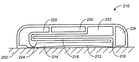
[0061] Making reference now to FIG. 2A, there is shown a cross-sectional view
of patch
(200) on skin (202). The patch (200) comprises an adhesive material in the
form of a layer
(204), a microfluidic collection layer (206), and a detector in the form of a
detector layer (208).
In some variations, the detector layer and the collection layer are in fluid
communication with
each other as shown in cross-sectional form in FIG. 2B. There, patch (210)
comprises adhesive
layer (212), collection layer (214), and a detector in the form of a detector
layer (216). The
collection layer (214) and detector layer (216) are in fluid communication
with each other (218).
As described in more detail below, the patch may also include a buffer and a
buffer reservoir
(220), a waste reservoir (222), and various microfluidic control features,
such as valves (224),
pumps, and the like. The patch may also include a device for measuring
relative humidity (226).
[0062] While not shown in the figures, the patch may also include at least one
release
liner. For example, a release liner on the bottom adhesive surface would
protect the adhesive
layer from losing its adhesive properties during storage and prior to use.
Similarly, a release
liner may be placed on top of the patch to protect any optical or electrical
components contained
therein. In some variations, no release liner is used and the patch is topped
with a backing layer.
In some variations, the backing layer is made from a woven or non-woven
flexible sheet, such as
those known in the art of transdermal patches. In other variations, the
backing layer is made
from a flexible plastic or rubber.
[0063] The microfluidic collection layer (214) may have a number of different
configurations. In general, the microfluidic collection layer comprises one or
more microfluidic
channels. For example, the microfluidic collection layer may include a
serpentine microfluidic
channel (301), as shown in FIG. 3A, or it may comprise concentric microfluidic
channels (303),
as shown in FIG. 3B. In some variations, the microfluidic layer comprises a
spiral microfluidic
CA 02654980 2008-12-10
WO 2007/146047 PCT/US2007/013392
channel (305), as shown in FIG. 3C. Sweat may be collected within the
microfluidic channel or
channels. Serpentine and concentric microfluidic channels may maximize the
surface area of the
collection channel in contact with the subject's skin while also allowing
movement of sweat
and/or buffer through the channel. In some variations, sweat is collected into
a substantially dry
microfluidic channel. In other variations, sweat is collected into buffer that
is present within the
channel. The collection of sweat into the patch is described in greater detail
below.
[0064] Sweat collected in the microfluidic channels is then typically moved
from the
collection layer into a detector layer. Additional microfluidic compartments
(e.g., mixing
compartments, treatment compartments, etc.) may also be included. The
microfluidic channel
may comprise a single channel, or multiple channels, and these channels may be
connected.
Similarly, the microfluidic channel or channels may be of any desirable and
practical size (e.g.,
diameter or cross-sectional area) and length. The microfluidic channels may
also be open to the
skin, or they may communicate with the skin through a sweat-permeable
membrane.
j0065] In some variations, the microfluidic collection layer is combined with
a sweat-
inducing layer, or one or more mechanisms for inducing sweat. For example, the
microfluidic
collection layer may include a mechanism for inducing sweat that acts
mechanically (e.g., by
using an occlusive backing layer, a vacuum, etc.), chemically (e.g., by
administering sweat
inducing agents such as pilocarpine with or without a penetration enhancer or
iontophoresis), or
thermally (e.g., by applying a heater, or initiating an exothermic chemical
reaction, etc.). FIG.
3D shows the microfluidic layer of FIG. 3A with the addition of a mechanism
for inducing sweat
(307) at least partially surrounding the channel (301). In some variations,
the mechanism for
inducing sweat may be included within the microfluidic channel within the
microfluidic
collection layer. For example a buffer within the microfluidic channel may
include a pilocarpine
solution.
[0066] In some variations, it may be necessary to provide a method to minimize
the
effect of variable sweat rates on the amount of glucose accumulation in the
collection layer.
There are several ways in which the effect of variable sweat rates may be
normalized by the
method of collection or the use of various analytes. Measuring the relative
humidity of the skin
under the patch may allow determination of the sweat rate and therefore the
amount of sweat
collected.
16
CA 02654980 2008-12-10
WO 2007/146047 PCT/US2007/013392
[0067] One method of minimizing the effect of a variable sweat rate is to
normalize the
flux of the measured glucose. For example, when glucose is transported to the
skin surface by
sweat, the total amount of glucose deposited on the unit of skin surface per
minute can be
calculated as follows:
GF=SRxSG
[0068] where GF is glucose flux (ng/cma x min), SR is the sweat rate ( L/cm2 x
min),
and SG is the glucose concentration in sweat (ng/ L).
[0069] Often the sweat rate fluctuates over time as the result of physical or
emotional
stimulation, and this fluctuation can result in a variation in the amount of
glucose collected from
the skin surface, and hence the accuracy of the glucose concentration
measurement. This
variation can be significantly reduced if sweat rate is measured as a function
of time and used to
normalized the glucose flux, as follows:
GF/SR = (SRxSG)/SR =SG
[0070] Another method, for example, may comprise configuring the microfluidic
collection layer to collect a constant volume of fluid so that a variable
sweat rate affects only the
time =to fill the collection volume, but not the amount of fluid collected.
For example, the
collection layer may comprise a reservoir having a fixed volume. FIG. 5A shows
a patch (500)
on skin surface (502). In this variation, the adhesive layer and the sweat-
permeable membrane
are combined in a single layer (504). Within the collection layer (508) is a
fixed volume
reservoir (506). The fixed volume reservoir (506) is shown in FIG. 5A as
completely empty. As
sweat begins to transport to the skin surface, and through the sweat-permeable
membrane, the
fixed volume reservoir begins to fill, as depicted in FIG. 5B.
[00711 A number of different techniques may be used to determine when the
fixed
volume reservoir, and hence the collection layer is filled. For example,
electrical capacitance,
electrical conductance, or optical measurements may be used as shown in FIGS.
5C, 5D, and 5E
respectively. For example, shown in FIG. 5C is patch (510) on skin surface
(512). In this FIG.,
sweat has already passed through the adhesive and sweat-permeable membrane
layer (514) to fill
the fixed volume reservoir (516). Conductors (518) for forming a dielectric
filled capacitor are
placed on either side of the patch (510). In this way, the volume within the
fixed volume
17
CA 02654980 2008-12-10
WO 2007/146047 PCT/US2007/013392
reservoir (516) may be determined by a change in capacitance of the dielectric
filled capacitor.
Illustrative conductors suitable for use with the patches described herein
include those made
from silver, platinum, and the like.
[0072] Similarly, electrical conductance may be used to determine when the
reservoir is
filled. Shown in FIG. 5D is patch (520) on skin surface (522). Sweat has
already passed
through the adhesive and sweat-permeable membrane layer (524) to fill the
fixed volume
reservoir (526). A conducting circuit (530) is established with reservoir
(526), here shown at the
top of the reservoir. The circuit may be open or closed. In this way, the
volume within the fixed
volume reservoir (526) may be determined by a change in conductance (e.g., at
the top of the
reservoir). Supports (528) may be provided on either side of patch (520) to
help provide
structurally integrity thereto. These supports may be plastic substrates with
suitably configured
printed circuit elements that could provide a circuit path through the fixed
volume reservoir.
Changes in resistance or conductance at the top of the reservoir could
indicate whether the fluid
volume in the reservoir (or within the microfluidic channel) had reached a
maximum. The
modest power required to drive a current through the circuit described here
could be provided by
an inductive coupling mechanism enclosed within the measurement device, a
plastic battery, and
the like.
[0073] Optical transmission may also be used to determine when the reservoir
is filled.
Shown in FIG. 5E is patch (530) on skin surface (532). Sweat has already
passed through the
adhesive and sweat-permeable membrane layer (534) to fill the fixed volume
reservoir (536).
An optical transmission path (538) is established with reservoir (536), here
shown at the top of
the reservoir. In this way, the volume within the fixed volume reservoir (536)
may be
determined by a change in optical transmission (e.g., at the top of the
reservoir). An optical fiber
path could be provided at the top of the mechanical supports (540) on either
side of patch (530)
connecting an optical source on one side of the patch with an optical detector
on the other.
Changes in the measured transmission could indicate whether the fluid volume
in the reservoir
had reached a maximum. Power for the optical source and detector may be
included in the
measurement device.
[0074] Optical reflection may also be used to determine when the reservoir is
filled. For
example, as shown in FIG. 5F is patch (550) on skin surface (542). Sweat has
already passed
18
CA 02654980 2008-12-10
WO 2007/146047 PCT/US2007/013392
through the adhesive and sweat-permeable membrane layer (544) and partially
filled fixed
volume reservoir (546). A transparent plate (549) is located on the top of the
reservoir. This
plate has an optical index of refraction close to that of sweat (about 1.33).
Incident light (551)
illuminates the interface between reservoirs (546) and plate (549). Here, the
reflected light (552)
has a high intensity because the optical index difference between the plate
(549) and air (which
has an optical index of refraction of about 1.0) is high. Shown in FIG. 5G is
the same patch
(550) where the reservoir (546) is completely filled with sweat. Here, the
reflected light (552)
has a low intensity because the optical index difference between the plate
(549) and sweat is low
(both have an optical index of refraction of about 1.33). Thus, the drop in
reflected light
intensity may be used as an indicator that the reservoir is full. An optical
source and detector
may be included in the measurement device and the patch can be interrogated
via an optical
interface.
[0075] The determination of glucose level in the patch may be normalized for
variable
sweat rates by the use of a non-glucose analyte specific to sweat that is
constant in concentration
(e.g., lactate, urea, sodium chloride, other electrolytes, etc.). In this way,
the glucose
concentration may be normalized to that value. For example, a separate
chemical detector may
be incorporated into the patch to independently determine the amount of the
sweat analyte. The
amount of this sweat analyte accumulated in the collection layer depends only
on the volume of
sweat in the layer. Once this is determined, the amount of glucose measured in
sweat may be
normalized to the total volume of sweat collected, thereby avoiding errors
associated with
measuring an increased accumulation of glucose in the collection layer of the
patch (i.e., due to
increased sweating rather than increased physiological glucose
concentrations). Alternatively,
there may be physiological markers in sweat that increase with increased sweat
rate.
Determination of the concentration of these markers may also serve as a method
for
norinalization of the glucose accumulated in the collection layer.
100761 In some variations, the collection layer may be configured as a
perfusion layer,
wherein a buffer (e.g., phosphate buffered saline, or the like) is used to
assist in the collection of
sweat. For example, the collection layer may include a channel
(e.g.,'microfluidic channel,
tubing, etc.) or passage through which the buffer may be perfused.
19
CA 02654980 2008-12-10
WO 2007/146047 PCT/US2007/013392
[0077] Returning now to Fig. 2B, one variation of a patch includes a buffer
reservoir
(220) which may supply buffer to the microfluidic channel. The buffer
reservoir may be part of
the microfluidic layer, or it may be separate, but fluidly connected to the
microfluidic layer. A
pump may be connected to the buffer reservoir to move buffer from the
reservoir through the
patch (e.g., through the microfluidic collection layer and into and through a
detector layer. Any
appropriate pump may be used, including an active pump or a passive pump. An
active pump
actively applies pressure to move material (e.g., sweat, buffer, air, etc.)
through the device. In
general, the pump may be any pump compatible with the microfluidic channel.
Examples of
microfluidic pumps may include positional displacement pumps such as gear or
peristaltic
pumps, piezoelectric pumps, and membrane pumps.
[0078] Passive pumping methods may also be used (e.g., passive pumps). For
example,
material may be moved through the device by thermal pumps, osmotic pumps, or a
preloaded
pressure bolus. In one variation, buffer is moved through the device by
allowing a pressurized
bolus of buffer to enter the microfluidic channel and push sweat containing
glucose from the
collection layer into, and ultimately, through the detector layer. For
example, buffer may be
preloaded into the device under pressure. After sweat has collected in the
microfluidic channel
to an appropriate level (or for an appropriate period of time), the
pressurized buffer is released
from the buffer reservoir into the microfluidic channel so that the buffer
moves through the
microfluidic channel(s) in the collection layer, and propels the sweat into
the detector layer.
Buffer may be released from the pressurized buffer reservoir by any
appropriate method, such as
by activating a valve, or rupturing a membrane, etc.
[0079] FIG. 2B also illustrates a valve (224) separating the buffer reservoir
(220) from
the microfluidic channel in the collection layer (214). The flow of sweat,
buffer, or other fluids
(including gasses) through the device may be controlled by components such as
valves, pumps,
and switches, which may be controllable by a controller. Thus these components
may include
electronic or manual controls for regulating their operation. A controller may
be part of the
patch (230) or it may be separate from the patch (e.g., part of a measurement
device, as
described in more detail below).
[0080] The device shown in Fig. 2B also includes a waste reservoir for storing
waste that
has passed through the measurement device, such as sweat, buffer, etc. The
waste reservoir may
CA 02654980 2008-12-10
WO 2007/146047 PCT/US2007/013392
also include a pump (e.g., to draw material into the waste reservoir).
Additional pumps may be
used if desirable, to help control the movement of material through the
device. Similarly,
additional valves or switches may also be used if desirable. For example, a
fluid connection
between the collection layer and the detector layer may include a valve so
that fluid (including
sweat or sweat in buffer) does not enter the detector layer until the
appropriate time.
[0081] As described above, the patch may comprise a detector. The detector may
be in
its own layer, adjacent to the collection layer, or, depending on the nature
of the detector, it may
be combined in the collection layer itself. In the absence of thermal,
emotional, physical, or
pharmacological stimulation, typical values of sweat output on the volar
forearm and fingertip
are relatively small. Sweat output varies from one individual to the next and
from one
anatomical site on the body to another. The maximum sweat rate per gland has
been reported to
range from about 2 nL/min to about 20 nL/min. See Sato, K. and Dobson, R.L.
"Regional and
individual variations in the function of the human eccrine sweat gland," J.
Invest. Dermat., 54,
443 , 1970. Assuming insensible perspiration rates per gland of 1 nL/min and
using measured
sweat gland densities at different parts of the body, a total sweat output can
be estimated.
Typical sweat gland densities on the forearm are approximately 100 glands per
square
centimeter, which give 0.1 gL sweat per square centimeter per minute. Typical
sweat gland
densities on the volar fingertip are approximately 500 glands per square
centimeter, which give
0.5 L sweat per square centimeter per minute. In the absence of stimulation,
the number of
active sweat glands per unit area is often reduced by one-half the total
available. Boysen et al.,
described above, found that the glucose concentration in sweat was
approximately one one-
hundredth normal blood glucose values (e.g., 1 mg/dl). Hence the flux of
glucose to the surface
of the volar fingertip may be estimated to be in the range of from about 2.5
nanograms to about 5
nanograms per square centimeter per minute. The flux to the surface of the
volar forearm or
wrist is likely to be even lower. Accordingly, the detector described here
must be capable of
detecting nanogram quantities of glucose and the measurement device described
herein must be
capable of performing ultra-sensitive glucose measurements.
[0082] Indeed, we have demonstrated that the flux of glucose brought to the
skin via
sweat was on the order of 1- 20 nanograms per square centimeter per minute in
the absence of
thermal, pharmacological or other forms of stimulation. These measurements
were made using
21
CA 02654980 2008-12-10
WO 2007/146047 PCT/US2007/013392
the Wescor MACRODUCT (459 South Main Street Logan, Utah 84321) system and in
specially adapted sweat collection chambers. Sweat collected in the Wescor
MACRODUCT
and in the sweat collection chambers was then analyzed using a Dionex
(Sunnyvale, California)
High Performance Anion Exchange with a Pulsed-Amperometric Detector (HPAE-
PAD). The
sensitivity and specificity of the HPAE-PAD system was tested using analytical
samples. We
detected glucose in amounts as low as 1 nanogram using HPAE-PAD.
[0083] Several types of suitably sensitive detectors may be used. For example,
the
detectors may be electrochemical-based, or may be fluorescent-based. Suitable
electrochemical
sensors may be those comprising an immobilized glucose-oxidase or other
enzyme(s) in or on a
polymer or other support, and those comprising glucose-oxidase or other
enzyme(s) in a
microfluidic configuration. Similarly, the detector may be fluorescent-based,
for example, based
on enhanced or suppressed fluorescence of a glucose-sensitive fluorescent
molecule. The
detector may be immobilized within a layer, or may be in solution.
[0084] As noted above, any suitable electrochemical detector may be used. For
example,
the electrochemical detector may be polymer based, based on microfluidics, and
the like. When
the electrochemical detector is polymer based, the polymer is typically
permeable to glucose,
and a glucose-reactive enzyme is immobilized on or within the polymer. In
these variations, the
detector typically comprises at least two electrodes, which are typically
activated by the
measurement device when it is brought into electrical contact with the patch.
In one variation,
the enzyme glucose oxidase is used, which produces hydrogen peroxide that
reacts at the at least
one electrode to produce a measurable electrical current proportional to the
glucose
concentration. That is, using an enzymatic process known in the art, the
glucose oxidase
catalyzes the reaction of glucose and oxygen to produce gluconic acid and
hydrogen peroxide.
The hydrogen peroxide is then electrochemically reduced at the at least one
electrode, producing
two electrons for detection. Electrical contact between the measurement device
and the patch
may also serve to provide power to the patch (although, the patch may comprise
a battery therein
as well if needed). The measurement device, which will be described in more
detail below,
interrogates the patch (i.e., the detector) and provides a glucose measurement
reading.
[0085] When microfluidics based electrochemical detectors are used on the
patch, the
patch typically comprises a fluid reservoir, a flow channel, a gating valve,
and sensor electrodes.
22
CA 02654980 2008-12-10
WO 2007/146047 PCT/US2007/013392
In this variation, the electrochemical enzyme is typically in solution. The
interface layer
comprises at least one electrode, which is activated by the measurement device
when placed into
electrical contact with the patch. As with the case above, electrical contact
between the
measurement device and patch, may serve to power the patch. A microfluidic
sensor may also
comprise a reservoir with a reference analyte to provide in situ calibration
of the detector. As
with the cases above, electrical contact between the measurement device and
patch, may serve to
provide power to the patch, or the patch may comprise a battery therein.
[0086] Sensitivity to these electrochemical detectors may be increased by
increasing the
temperature during the detection cycles, by increasing the length of the
detection cycle, by
increasing the area of the detector, by appropriately selecting the operating
potential, and by the
use of selective membranes to screen interfering substances such as ascorbic
acid, uric acid,
acetaminophen, etc. In addition, differential methods may be used where the
glucose sample is
measured in the presence and absence of a glucose-specific enzyme and the
glucose
concentration is determined from the difference between these two signals.
100871 For example, sensitivity may be increased by heating the sensor
solution from
25 C to 40 C, and such temperature increase is unlikely to affect the enzyme
activity of the
glucose detector. See, e.g., Kriz, D, Berggre, C., Johansson, A. and Ansell,
R.J., "SIRE-
technology. Part I. Amperometric biosensor based on flow injection of the
recognition element
and differential measurements," Instrumentation Science & Technology, 26, 45-
57 (1998).
Similarly, sensitivity may be increased by increasing the area of the
detector, since the detector
current increases linearly with the area of the detector electrode. Extending
the length of time
over which the measurement may be made may also be used to increase the
measured charge
and hence, the overall sensitivity of the detector. Lastly, covering the
electrode with size- and,
or, charge-selective membranes can allow passage of hydrogen peroxide, for
example, while
excluding ascorbate, urate and other material, which can react directly with
the sensor to produce
a spurious signal. Suitable size-selective membranes, for example, include
those made of
polyurethane, polyethylene and other materials as well as charge-selective
membranes made of
polyethylsulfide, NAFION , cellulose acetate, and other materials that can be
used as
interference-screening membranes for electrochemical detectors.
23
CA 02654980 2008-12-10
WO 2007/146047 PCT/US2007/013392
[0088] As noted above, the detector may also be a fluorescent detector. In
this variation,
the detector layer, or the layer immediately adjacent to the measurement
device may be made of
a material that is optically transparent at the relevant excitation and
emission wavelengths for the
particular fluorescent-based detector.used by the patch. In one variation, the
measurement
device need not be brought into direct physical contact, because interrogation
of the patch is
achieved by optically coupling the device and patch. The internal electronics
of the
measurement device may also be configured to record a maximum signal as it is
passed over the
patch, thereby reducing the need for proper static registration between the
measurement device
and the patch itself. The patch may also include a glucose-insensitive
reference fluorescent
molecule to provide a ratiometric, rather than an absolute intensity
measurement. The addition
of a reference molecule may also protect against a spurious signal originating
at the emission
wavelength of the fluorescent-based detector.
[0089] When a fluorescent detector is used, it typically comprises a glucose-
sensitive
fluorescent molecule immobilized in a polymer or suitable solvent, and as
described above, may
be in a separate layer, or dispersed throughout the collection layer. Because
the measurement
device will be measuring the glucose at a specific wavelength, it is desirable
that the materials
used in the patch do not have fluorescence at, or substantially near, the
wavelength of the
fluorescent emission of the glucose transducer molecule. Similarly, it is
often desirable that the
sweat-permeable membrane in these variations be opaque so as to prevent
autofluorescence from
the skin.
[0090] Suitable fluorescent detectors for example may be those described in
U.S. Pat.
No. 6,750,311 to Van Antwerp et al, which section on fluorescent detectors is
hereby
incorporated by reference in its entirety. As described there, fluorescent
detectors may be based
on the attenuation in the fluorescence intensity of labeled lectins or
boronate (germinate or
arsenate) aromatic compounds. Suitable lectins include concanavalin A (Jack
Bean), Vicia faba
(Fava Bean), Vicia sativa, and the like. Such lectins bind glucose with
equilibrium constants of
approximately 100. See, Falasca, et al., Biochim. Biophys. Acta., 577:71
(1979). The lectin may
be labeled with a fluorescent moiety such as fluorescein isothiocyanate or
rhodamine using
commercially available kits. The fluorescence of the labeled lectin decreases
with increasing
glucose concentration.
24
CA 02654980 2008-12-10
WO 2007/146047 PCT/US2007/013392
[0091] Boronate based sugar binding compounds may also be used as the basis
for the
fluorescent detector. Glucose reversibly binds to the boronate group in these
compounds.
Boronate complexes have been described which transduce a glucose signal
through a variety of
means. See, Nakashima, et al., Chem. Lett. 1267 (1994); James, et al., J.
Chem. Soc. Chem.
Commun, 477 (1994); and James, et al., Nature, 374:345 (1995). These include
geometrical
changes in porphyrin or indole type molecules, changes in optical rotation
power in porphyrins,
and photoinduced electron transfer in anthracene type moieties. Similarly, the
fluorescence of 1-
anthrylboronic acid has been shown to be quenched by the addition of glucose.
See, Yoon, et al.,
J. Am. Chem. Soc., 114:5874 (1992).
[0092] The dye used in the above fluorescent-based detector may be, for
example an
anthracene, fluorescein, xanthene (e.g., sulforhodamine, rhodamine), cyanine,
coumarin (e.g.,
coumarin 153), oxazine (e.g., Nile blue), a metal complex or other
polyaromatic hydrocarbon
which produces a fluorescent signal. Unlike previously described applications
of these sensors,
where the sensors are specially-designed for equilibrium-binding with a target
analyte and for
reversibility, the binding constant of the fluorescent-based detectors
described here may be
increased so as to fiu-f.her lower the limit of detection.
Measurement Device
[0093] As noted above, the glucose monitoring systems described here generally
comprise a patch configured to collect a nanogram quantity of glucose in
sweat, where the patch
comprises a microfluidic collection layer and a detector, and a measurement
device configured
to measure the nanogram quantity of glucose collected. The patches were
described in detail
above.
[0094] The measurement device interrogates the patch to measure glucose. The
device
measures the total quantity of glucose present in a fixed volume, and then
converts the glucose
measurement into a concentration. The measurement device may comprise a
display, to display
data. The device may also include warning indicators (e.g., a word prompt,
flashing lights,
sounds, etc.) to indicate that a user's glucose levels are dangerously high or
dangerously low. In
addition, as described briefly above, the measurement device may also be
configured to verify
that a skin-cleaning procedure has been performed. For example, when wipes
with a marker
have been used, (which will be described in more detail below) the marker
remains on the skin
CA 02654980 2008-12-10
WO 2007/146047 PCT/US2007/013392
surface. If the measurement device detects the marker, then the measurement
proceeds. If the
measurement device does not detect the marker, the measurement does not
proceed. The
measurement device may also comprise an iontopheric source, for example, to be
used to help
drive pilocarpine, or other molecules of interest into the skin.
[0095] In general the configuration of the measurement device is dependent on
the
configuration of the detector in the patch. For example, when the measurement
device is to be
used with an electrochemical detector, the measurement device provides an
electrical contact
with the interface layer, and is either powered by the electrical contact, or
is powered by an
independent power source (e.g., a battery within the patch itself, etc.). The
measurement device
also typically comprises a computer processor to analyze data. Conversely,
when the
measurement device is configured for fluorescence detection, the measurement
device is
configured to provide optical contact or interaction with the interface layer.
In this variation, the
measurement device also typically comprises a light source to stimulate
fluorescence. In some
variations, the measurement device comprises both the necessary electrical
contacts and the
necessary optics so that a single measurement device may be used with a patch
having various
configurations of patch layers (e.g., one layer comprising a fluorescent-based
molecule, and
another layer comprising an electrochemical detector).
[0096] The measurement device may further comprise computer executable code
containing a calibration algorithm, which relates measured values of detected
glucose to blood
glucose values. For example, the algorithm may be a multi-point algorithm,
which is typically
valid for about 30 days or longer. For example, the algorithm may necessitate
the performance
of multiple capillary blood glucose measurements (e.g., blood sticks) with
simultaneous patch
measurements over about a 1 day to about a 3 day period. This could be
accomplished using a
separate dedicated blood glucose meter provided with the measurement device
described herein,
which comprises a wireless (or other suitable) link to the measurement device.
In this way, an
automated data transfer procedure is established, and user errors in data
input are minimized.
[0097] Once a statistically significant number of paired data points have been
acquired
having a sufficient range of values (e.g., covering changes in blood glucose
of about 200 mg/dl),
a calibration curve will be generated, which relates the measured sweat
glucose to blood glucose.
26
CA 02654980 2008-12-10
WO 2007/146047 PCT/US2007/013392
Patients can perform,periodic calibration checks with single blood glucose
measurements, or
total recalibrations as desirable or necessary.
[0098] The measurement device may also comprise memory, for saving readings
and the
like. In addition, the measurement device may include a link (wireless, cable,
and the like) to a
computer. In this way, stored data may be transferred from the measurement
device to the
computer, for later analysis, etc. The measurement device may further comprise
various inputs,
to control the various functions of the device and to power the device on and
off when necessary.
[0099] As mentioned above, the system may also include a device for measuring
the
relative humidity of the skin under the patch, which may or may not be part of
the measurement
device (e.g., it may be part of the patch as shown above in FIG. 2B). The
relative humidity may
provide an estimate of the amount of sweat collected by the device, or the
rate of sweat over
time. Any appropriate relative humidity detector may be used. In some
instances, it may be
desirable to use full range (e.g., 0% to 100%) relative humidity sensors.
Examples of
appropriate relative humidity sensors include capacitive humidity sensors,
resistive humidity
sensors, and low-voltage humidity sensors. The relative humidity measured
beneath the patch
reflects the amount of moisture lost by the skin (e.g., sweat) and therefore
the amount and rate of
sweating.
[0100] As mentioned above, the measurement device may also include a
controller for
controlling the patch or its components (e.g., valves, pumps, switches, etc.).
In some variations,
the controller regulates the movement of fluid (e.g., sweat, buffer, and/or
air) through the
collection and detector layers. A controller may achieve this by coordinating
the activity of
pumps, valves, and switches. For example, the controller may open the
connection (e.g., a
switch or valve) between the buffer reservoir and the microfluidic channel and
pump buffer from
the buffer reservoir into the microfluidic channel. Buffer may be added to the
microfluidic
channel either before the collection of sweat (e.g., in "wet" collection
procedures) or after sweat
has been collected (in "dry" collection procedures). One or more switches may
be used to
switch between different regions of the patch. For example, a marker such as a
bolus of gas,
buffer, or marking solution may be applied to one end of the microfluidic
collection channel by
opening a channel between the source of marker material and the end of the
microfluidic
channel. Another switch may also control the movement of material from the
microfluidic
27
CA 02654980 2008-12-10
WO 2007/146047 PCT/US2007/013392
collection chamber to the detector layer. For example, when the collection
layer comprises a
microfluidic channel into which sweat is collecting, the distal end of the
channel may be open to
a reservoir or to the atmosphere, preventing a backpressure within the
channel. After an
appropriate amount of sweat has been collected, a valve or switch may switch
the microfluidic
channel so that it is instead in fluid communication to the detector layer,
allowing sweat
(including sweat in buffer) and other material in the microfluidic channel to
pass into the
detector layer. Sweat may be pumped (passively or actively) from the
collection layer into the
detector layer so that the level of glucose may be determined.
[0101] The measurement device may be worn by the user, but need not be. For
example,
since the patches described here are suitable for both single and repeated
measurements, it may
be desirable in some circumstances to have the measurement device be wearable.
For example,
in the case where the patch will be interrogated multiple times, as will be
described in more
detail below, the measurement device may be worn over the patch in a bracelet
or watch-type
configuration. In these variations, the measurement device should be of a size
suitable to
provide comfort to the wearer, while at the same time being capable of housing
its necessary
components. It should be understood that the size of the measurement device
and how it is
configured for comfortable wear is also dependent upon the patch location
(e.g., fingertip, wrist,
forearm, abdomen, thigh, etc.).
[0102] An exemplary depiction of a glucose monitoring system as described
herein is
shown in FIG. 6. FIG. 6 shows a patch configured as an in-line glucose
detection device that
uses glucose oxidase ("GOx") in solution as part of an electrochemical
detector. In this
variation, the device uses a differential measurement technique to enhance the
glucose signal
while eliminating potential contaminants.
[0103] In the system illustrated in FIG. 6, a sweat sample from the skin is
collected into
the microfluidic collection layer of the patch (614). The collection layer
comprises a
microfluidic channel, which may be a serpentine channel, as described above.
In this example,
the device includes a sweat permeable membrane (612) between a user's skin and
the collection
layer (614). The distal end of the microfluidic chamber is in fluid connection
with a source of
buffer, such as a buffer reservoir (628). The buffer may be pressurized (e.g.,
by a pump) so that
28
CA 02654980 2008-12-10
WO 2007/146047 PCT/US2007/013392
when the valve (630) between the buffer reservoir and the channel is opened,
buffer flows into
the channel.
[0104] As described above, a buffer (which may or may not be different from
the buffer
in the buffer reservoir) may be preloaded into the microfluidic channel so
that sweat is collected
into fluid within the microfluidic channel. In some variations, the sweat is
collected into a
relatively "dry" channel. Typically buffer entering the microfluidic channel
from the buffer
reservoir will drive material (e.g., sweat) from the microfluidic channel and
into the detector
region (616). In some variations, fluid in the microfluidic channel is pumped
into the detector
region (616) by air or by a material other than the buffer (including
immiscible materials such as
oils, etc.) that is added at the proximal end of the microfluidic channel. In
some variations, this
may serve to mark the end of the material collected into the microfluidic
channel as it passes into
the detector. As shown in FIG. 6, the collection layer is fluidly connected to
the detector region
by tubing (618). An additional valve may be used to separate the detector
layer from the
collection layer.
[0105] As mentioned above, different buffers may be used as part of the same
system.
For example, a collection buffer may be used to collect sweat, and a different
buffer (e.g., a
pushing buffer) may be used to move a sweat sample (and/or collection buffer)
within the
system. A different marker buffer may be used to "mark" the microfluidic
solution. In some
variations, the same buffer may be used for all of these. These buffers may
have the same ionic
strengths and pH, or they may have different ionic strengths and pH. In some
variations, the
same buffer is used for all of these different applications.
[0106] As mentioned above, the detector shown in FIG. 6, is a GOx based
detector that
applies a differential detection method to measure glucose. In this way,
glucose can be
accurately measured even in the presence of additional compounds such as
ascorbic acid and
acetaminophen that might otherwise inhibit or interfere with an accurate
measurement. Here,
the detector layer is divided up into two separate regions by a dialysis
membrane (640) that
allows glucose to pass therethrough, but prevents larger molecules (such as
GOx) from passing.
An appropriate differential measurement technique is described in U.S. patents
6,706,160 and
6,214,206, both of which are herein incorporated by reference in their
entirety. Differential
29
CA 02654980 2008-12-10
WO 2007/146047 PCT/US2007/013392
measurement methods typically remove the impact of interfering substances by
recording from a
sweat sample in the presence and absence of GOx, and producing a differential
signal.
(0107] In FIG. 6 the detector layer comprises two regions (644, 646) separated
by the
dialysis membrane (640). The upper region (646) contains three electrodes
(651): a working
electrode, a counter electrode and a reference electrode. This upper region is
also in fluid
communication with a source of GOx (565), and a waste reservoir (658). In some
variations,
(particularly non-differential measurement variations) the GOx may be fixed or
immobilized
(e.g., to the sides of the detection region, or on or near the electrodes),
rather than applied in
solution.
[0108] The sweat collected into the microfluidic channel may be passed (in-
line) into the
lower region of the detector (644), as shown. Once the sweat sample enters the
detection
chamber from the collection region, a signal may be measured from the
electrodes (e.g., a
working electrode and a counter electrode pair). The typical sweat sample may
contain other
non-glucose substances (such as ascorbic acid and acetaminophen) that can
generate a signal on
an electrode, resulting in a background current. These compounds may also pass
through the
dialysis membrane (640) between the upper and lower regions of the detector
layer, and would
be present as background in an electrochemical signal. However, as mentioned
previously and
will be described in more detail below, because a differential measurement
technique is used, the
background signal of the potentially interfering compounds is of no
consequence.
[0109] As mentioned previously, glucose is also free to diffuse across the
dialysis
membrane (640) between the upper and lower chambers. To measure the glucose
concentration,
GOx is then added (e.g., from the GOx reservoir (656) into the upper chamber
where it can react
with glucose and produce a signal proportional to the glucose concentration on
the electrodes.
The enzyme does not pass through the dialysis membrane (640), and converts
glucose into
peroxide resulting in a "peroxide current" local to the upper chamber
electrodes. The difference
in the signals before and after the addition of GOx may accurately reflect the
concentration of
glucose even in the presence of interfering compounds.
[0110] The signal present at the electrodes (651) may be monitored and used by
the
measurement device (not shown), as described above. Furthermore, the
coordination of the
CA 02654980 2008-12-10
WO 2007/146047 PCT/US2007/013392
taking of measurements, addition of GOx, etc. may be performed by a
controller, including a
controller that is part of the patch, or part of the measurement device.
[0111] In some variations, the detector layer comprises a GOx detector that is
not a
differential detector. Thus, the system shown in FIG. 6 may be simplified by
removing the
dialysis membrane (640) and reducing the upper and lower regions (644,646)
into a single
region. This may be particularly desirable if the levels of potentially
interfering compounds is
low.
Methods
[01121 As noted above, methods for measuring glucose on the skin surface are
afso
provided here. Some methods generally comprise cleaning the skin surface with
a glucose
solvent, collecting sweat from the skin surface using a microfluidic
collection device, and
measuring the collected glucose.
[0113] Cleaning the skin surface (e.g., by wiping it clean) is typically
performed to
remove any "old" or residual glucose remaining on the skin. In variations
using a wipe, the wipe
is typically made of a material suitable for wiping the skin and comprises a
solvent for removing
glucose. For ease of description only, the term "wipe" will be used herein to
include any type of
fabric, woven, non-woven, cloth, pad, polymeric or fibrous mixture, and
similar such supports
capable of absorbing a solvent or having a solvent impregnated therein.
[0114] In some variations, the wipe contains a marker that is deposited on the
skin. In
these variations, the measurement device looks for the presence of the marker,
and if the marker
is detected, then the measurement proceeds. If the marker is not detected, the
measurement does
not proceed. In some variations, as will be described in more detail below,
the measurement
device provides an indication to the user that the skin has not been wiped. In
this way, the
possibility that a user obtains and relies upon a clinically dangerous
measurement (e.g., based on
an erroneous reading resulting from food residues or other glucose sources on
the skin that are
not correlated with the user's actual blood glucose) is minimized, and
accurate measurements are
facilitated. The marker may comprise a chemical having a short half-life, so
that it will decay
after a short period of time. In this way, a marker will only be valid for a
single wipe, or a single
use and erroneous detection of a marker on the skin surface will be minimized.
In a like manner,
31
CA 02654980 2008-12-10
WO 2007/146047 PCT/US2007/013392
the marker may also be bound to a volatile compound, and made to evaporate in
a short period
of time.
[0115] It should be noted however, that the wipe should not contain solvents,
markers, or
other chemicals that would interfere with the measurement of glucose. That is,
a suitable
glucose solvent would have the capacity to solubilize glucose without
interfering with either the
electrical or optical measurement of glucose. Polar solvents, and in
particular, a mixture of
distilled water and alcohol, have provided very good results in removing
residual glucose from
the skin surface. The ratio of distilled water to alcohol may be chosen such
that there is
sufficient water to dissolve the glucose, but not so much water as to make the
removal of the
excess water take an inconveniently long period of time relative to the
measurement of glucose
(e.g., more than 25 minutes). As noted above, it is desirable that the
alcohol/water mixture, or
other polar solvent, be selected such that it removes the residual glucose,
but does not interfere
with the glucose measurement.
[0116] In some variations, the skin is cleaned by rinsing or otherwise
treating it with a
glucose solvent to remove potentially contaminating residual glucose. After
cleaning the skin, it
may be dried (or allowed to dry), removing excess cleaning solution. A
separate drying step is
unnecessary in some variations.
[0117] As noted above, after the skin has been cleaned, sweat is collected
from the skin
surface, and this may or may not include placing a patch on the skin surface
for sweat collection.
When a patch is used, it may be placed on any suitable skin surface as
described briefly above.
For example, the patch may be placed on a finger, on the palm, on the wrist,
the forearm, the
thigh, etc. Placement of the patch on the fingertip may provide a convenient,
discrete, and
readily accessible site for testing, particularly non-continuous testing. In
addition, fingertips
have the greatest density of sweat glands. Placement of the patch on the wrist
may provide a
convenient, discrete, and readily accessible site for testing when repeated
measurements are to
be taken from a single patch.
[0118] These methods may also include a step of inducing sweat prior to
collecting the
sweat from the skin surface. The step of inducing sweat may comprise inducing
sweat
32
CA 02654980 2008-12-10
WO 2007/146047 PCT/US2007/013392
mechanically, chemically, physically, or thermally, as described in detail
above. In some
variations, measuring comprises measuring nanogram quantities of glucose.
[0119] Other methods for measuring glucose on the skin surface comprise
cleaning the
skin surface with a glucose solvent, as described just above, collecting sweat
from the skin
surface in a patch comprising a microfludic collection layer; and measuring
glucose collected in
the patch. Again, any of the patch variations described above may be used with
the patch
described here. In some variations, collecting sweat comprises collecting
sweat in a microfludic
collection layer containing a buffer. For example, the patch may be applied to
a user's skin, and
the microfluidic channel may be filled (or it may have been pre-filled) with a
buffer. In some
variations, the buffer includes a mechanism for inducing sweat (e.g.,
pilocarpine). Sweat is
therefore collected into the buffer solution within the niicrofluidic
collection channel. After an
appropriate amount of sweat has been collected, the buffer within the
collection channel is
pumped into the detector layer. The appropriate amount of sweat may be
determined based on
any of the methods descried above. For example, the appropriate amount of
sweat may be
determined by the volume of sweat collected (e.g., when the sweat added to the
buffer within the
collection layer increases by a given amount), or based on the concentration
of another
component of the sweat detected while in the collection channel, or based on
the rate of sweat
determined by the relative humidity of the skin beneath the patch, or based on
a predetermined
lapse of time.
[0120] The sweat (in the buffer) may be moved into the detector layer from the
collection layer. Sweat may be pumped by applying pressure at the proximal end
of the
microfluidic collection passage, when the collection layer is in fluid
communication with the
detector layer. Pressure may be applied by adding additional buffer to the
proximal end of the
collection layer, or by adding any appropriate material (e.g., air, etc.).
Once in the detector
layer, the concentration of glucose in the sweat may be determined by any
appropriate method,
as described above. The detection may occur while the material is entering
into the detector
layer (e.g., continuously), or it may be done at a discrete time periods after
the sweat has entered.
The measurement device may interrogate the detector as (or after) the sweat
has entered the
detector layer. Thus, the measurement device may sample the detector to
determine the
concentration of glucose. As described above, in some variations, the
measurement device may
33
CA 02654980 2008-12-10
WO 2007/146047 PCT/US2007/013392
apply a differential technique to determine a glucose signal, or it may
average, sum, or otherwise
analyze the output of the detector to determine a glucose concentration that
reflects the
concentration of blood glucose. The sweat (and/or buffer) in the detector
layer may be pumped
past the detector (e.g., electrodes) and into a waste reservoir.
[0121] The method may also include pumping a buffer into the microfluidic
collection
layer (e.g., after collecting the sweat). In these variations, the patch
typically has a collection
layer and a detector layer, which are in fluid communication with each other.
Sweat may be
collected into an initially relatively dry microfluidic collection layer. A
sufficient amount of
sweat may then be collected before moving the sweat into the detector layer.
As mentioned
previously, the amount of sweat collected may be measured by the device in any
appropriate
fashion. Any of the steps previously described may then be used to determine
the concentration
of glucose in the sweat.
[0122] Of course, it should be understood that any of the steps of the methods
described
herein may be repeated (e.g., collecting the sweat and measuring the glucose).
Thus, the
devices described herein may be configured for repeated measurements of
glucose from sweat.
[0123] Still other methods for measuring glucose on a skin surface comprise
cleaning the
skin surface with a glucose solvent, as described above, collecting a first
sweat sample from the
skin surface in a patch comprising a microfludic collection layer and a
detector layer,
transferring the first sweat sample from the collection layer to the detector
layer, measuring
glucose in the first sweat sample, and repeating the collection, transferring,
and measuring steps
at least once. This method is shown in flow chart form in FIG. 7.
[0124] In FIG. 7, one example of a method for repeatedly measuring glucose
from sweat
is depicted. The subject's skin is first cleaned (701), as described above,
with an appropriate
glucose solvent, and then the patch is applied (703). Any appropriate skin
region may be used,
preferably a region to which the patch and/or measuring device (e.g., monitor)
may be attached
for the period of time over which repeated measurements are to be taken (e.g.,
minutes, hours,
days). For example, the patch may be applied to the subject's wrist, abdomen,
arm, etc.
[0125] A first sweat sample may then be collected from the skin surface (705),
according
to any of the methods described herein. During or before the collection of
sweat, a mechanism
34
CA 02654980 2008-12-10
WO 2007/146047 PCT/US2007/013392
for inducing sweat may be applied to induce a sweat response from the skin.
For example, the
mechanism for inducing sweat may be chemical (e.g., pilocarpine with or
without penetration
enhancers or iontophoresis), thermal (e.g., heater), or mechanical (e.g.,
occlusive layer).
[0126] Sweat may be collected through a sweat-permeable membrane (but need not
be)
into a microfluidic channel, such as a serpentine microfluidic channel. As
described above, the
step of collecting the first sweat sample may comprise collecting the first
sweat sample in a
microfludic collection layer containing a buffer of may comprise collecting
the first sweat
sample in a microfluidic collection layer devoid of a buffer. In one
variation, the microfluidic
collection layer includes a buffer (e.g., PBS at pH 7.4) into which sweat is
collected. Sweat may
be collected for an appropriate amount of time, or until an appropriate amount
of sweat has
entered the microfluidic channel. In one example, the appropriate amount of
sweat is
determined based on the displacement of fluid within the microfluidic channel.
For example, as
sweat enters the buffer within the channel, the volume of fluid (buffer plus
sweat) within the
channel will increase, and this increase may be detected by the device, using
any of the methods
previously described. For example, when the end (closest to the entrance of
the detector layer)
of the microfluidic channel is blocked, the addition of sweat to the buffer
will extend the front of
the buffer within the microfluidic chamber, which may be detected optically,
electrically, etc. In
some variations, an end of the microfluidic chamber is open to atmosphere via
a valve or switch,
so that backpressure does not develop. Examples of the appropriate amount of
sweat collected
may be less than about 20 l, less than about 10 l, less than about 5 l,
less than about lgl or
less than about 0.5 1.
[0127] After the first sweat sample has been collected, the sweat sample (in
buffer) may
then be transferred from the microfluidic collection layer into the detector
layer (707). As
described above, any appropriate method may be used to transfer the sweat and
buffer into the
detector layer. For example, the step of transferring the first sweat sample
from the collection
layer to the detector layer may comprise pumping a buffer into the
microfluidic collection layer
or may comprise applying pressure (e.g., gas pressure, liquid pressure, or
mechanical pressure)
within the microfludic collection layer. In some variations, pressure is used
to transfer the sweat
sample and pressure is applied with pressurized saline. Other variations for
transferring the
sweat sample may also be used. Pressure is typically applied within the
microfluidic collection
CA 02654980 2008-12-10
WO 2007/146047 PCT/US2007/013392
channel when the channel is in fluid connection with the detector layer. In
one variation,
additional buffer is pumped into the proximal end of the microfluidic
collection layer from a
buffer reservoir after opening a valve to the buffer reservoir from the
microfluidic channel, while
also opening a valve between the microfluidic channel and the detector layer.
[0128] Once the sample is in the detector layer, the concentration of glucose
may be
determined (709) according to any of the methods previously described (e.g.,
electrochemically,
fluorescently, etc.). Thus, if an electrochemical method is used with GOx, the
GOx may react
with glucose in the sample to produce a current that is proportional to the
concentration of
glucose even at very low (e.g., nanogram) levels, as previously described.
[0129] After the glucose reading has been taken, the remaining sample may be
driven
(e.g., by pressure) into a waste reservoir, and the device may be in
preparation for collecting the
next sample (711). For example, the microfluidics channel may be purged with
air, or filled
with fresh buffer (or both). In some variations, clean buffer is run from the
collection layer to
the detector layer until glucose is not detected, and then valves between the
waste reservoir and
the detector layer are shut to prevent later contamination. Valves between the
detector layer and
the collection layer may also be shut. The collection layer may then be primed
to collect a new
sweat sample.
[0130] The steps may be repeated (713) after a predetermined period of time,
e.g., less
than about 60 minutes, less than about 30 minutes, less than about 20 minutes,
less than about 10
minutes, less than about 5 minutes, etc. Similarly, the steps may be repeated
for a predetermined
period of time, e.g., about 1 hour, about 2 hours, about 3 hours, about 4
hours, about 5 hours,
about 6 hours, etc. These periods of time may be set automatically, or may be
set manually.
[0131] As with the methods described above, these methods may also include the
step of
inducing a sweat prior to collecting a first sweat sample.
Examples
Example 1: Investigation of the effects of a sweat-permeable membrane
[0132] A standard pilocarpine iontophoresis was performed simultaneously on
the clean
dry skin of both arms of a 40 year old male type I diabetic. The skin was
wiped after stimulation
36
CA 02654980 2008-12-10
WO 2007/146047 PCT/US2007/013392
and a MedOptix (now VivoMedical) Macrovial surface was applied within 1 min
following the
iontophoresis. The MedOptix Macrovial allows serial samples of sweat to be
collected from the
same site on the skin. It is made from a plate having a hole therethrough for
contact with the
skin surface. On the non-skin contacting side of the plate, a capillary tube
connects the hole to a
collection chamber or vial). A Vaseline-paraffin barrier material (acting as a
sweat-permeable
membrane) was applied to the site on the right arm before the MedOptix
Macrovial was applied.
Samples were collected every 10 minutes from the appearance of the first drop
of sweat on the
end of the MedOptix Macrovial. The subject came in with an initial blood
glucose level of about
220 mg/dl, which then stabilized at about 175 mg/dl during the first 40
minutes of sample
collection. The subject then drank 10 oz of COKE producing a rise in blood
glucose to about
300 mg/dL.
[0133] The first two samples from the left arm (having no sweat-permeable
membrane),
contained approximately 2.0 mg/dl glucose. The glucose concentration of the
sweat increased
monotonically throughout the rest of the experiment to a maximum of
approximately 5.0 mg/dl.
This increase in concentration was not correlated to the increase in blood
glucose, which began
to rise 40 min after the initial rise in glucose in the left arm. In contrast,
the glucose samples
from the right arm, having the sweat-permeable membrane, remained flat at
approximately 1.7
mg/dl and began to rise to a maximum of about 2.5 mg/dl about 10 min after the
blood glucose
started to rise. These results are shown in FIG. 8.
[0134] FIG. 9 shows a fit of blood glucose vs. sweat glucose for the site
having the
sweat-permeable membrane, which has been time-shifted. The blood and sweat
glucose values
were highly correlated, as shown by the R2 of 0.98. The glucose concentration
increased
throughout the experiment on the site having no sweat-permeable membrane,
which is consistent
with a source of glucose independent of sweat. FIG. 10 is a plot of the ratio
of sweat flux to
glucose flux. As shown in that figure, in the case where there is a sweat-
permeable membrane,
the ratio remains constant while the blood glucose level is constant.
Conversely, in the case
where there is no sweat-permeable membrane, the ratio increases during this
time. Accordingly,
this data suggests that the use of a sweat-permeable membrane can act as a
barrier to epidermal
contaminants and glucose brought to the skin surface via diffusion.
37
CA 02654980 2008-12-10
WO 2007/146047 PCT/US2007/013392
Example 2: Correlation of sweat glucose to blood glucose
[0135] Both forearms of the subjects used were wiped with a standard 70%
isopropyl
alcohol swab. Cotton pads soaked in a buffered saline and 1% pilocarpine
solution were applied
respectively to the negative and positive electrodes of a standard
iontophoresis device. A charge
(dose) of 10 mA-min at a current of 1 mA was applied to the electrodes as they
were held tightly
against the skin of the subjects with elastic straps. The skin was wiped after
10 min of
iontophoresis and a MedOptix Macrovial was applied to the site of the positive
electrode within
1 min following the iontophoresis. Sample vials were replaced every 10 or 15
min until sweat
flow became less than about 10 l over the collection interval.
[0136] Blood glucose levels were determined from capillary finger pricks every
10
minutes using a commercial personal blood glucose meter (ACCU-CHECK ADVANTAGE
,
Roche). In some experiments macro-vials were placed simultaneously on the
right and left arms,
while in others macro-vials were placed first on one arm and then after an
hour on the opposite
arm. Samples were filtered, diluted and analyzed on a DIONEX HPAE-PAD system.
The
protocol varied with the initial state of the subject. For example, if the
subject's blood glucose
(BG) was high (>200 mg/dL) the subject was asked to follow his normal insulin
program to
lower BG. Otherwise, the subjects were given a drink containing 35-70g of
glucose at the start
of the experiment to produce a rise in BG over the collection period.
[0137] Subject BCGI701, whose results are shown in FIGS. 11 and 12A-B, is a 48
year
old female Caucasian, type II diabetic. Subject BDW2002, whose results are
shown in FIGS. 13
and 14A-B, is a 39 year old male Asian, non-diabetic.
[0138] FIG. 11 shows a typical result for a falling BG. In this experiment the
subject
arrived with a high (250 mg/dL) BG level. Following the subject's own
treatment regime,
insulin was injected and samples of sweat and blood were collected from both
the left and right
forearms. The data shown in FIG. 11 is uncorrected for the offset some
subjects demonstrate
between their left and right arm. In this figure the BG (circles) decreases
from 250 to 100 over
the 2.5 hr experiment. The sweat glucose (SG) level is shown for the left
forearm (LFA)
followed by the right forearm (RFA). The numbers over the SG points give the
volume in l of
sweat collected for each sample over the collection interval. FIGS. 12A and
12B show a linear
regression plot of interpolated blood glucose vs. sweat glucose for the LFA
and RFA
38
CA 02654980 2008-12-10
WO 2007/146047 PCT/US2007/013392
respectively. These fits have R2 values of 0.83 and 0.92, indicating a high
degree of correlation
between blood and sweat glucose levels.
[0139] FIG. 13 shows experimental results for an experiment with increasing
BG. In this
experiment the subject was given 75 g of glucose which raised his BG from
about 125 to about
200 mg/dL over the course of the experiment. The data plotted in FIG. 13 shows
the sweat
glucose levels (left axis) of "simultaneous" collections of the LFA and RFA
together with the
changing blood levels (right axis). FIGS. 14A and 14B show plots of the linear
regression of
blood vs. sweat glucose for the LFA and RFA. The RZ values were 0.99 and 0.97
for LFA and
RFA respectively demonstrating a strong correlation between blood and sweat
glucose in this
experiment.
39