Note: Descriptions are shown in the official language in which they were submitted.
CA 02655389 2008-12-19
WO 2007/147234 PCT/CA2007/001081
CALCIUM PHOSPHATE COATED IMPLANTABLE MEDICAL DEVICES,
AND ELECTROPHORETIC DEPOSITION PROCESSES FOR MAKING
SAME
FIELD OF THE INVENTION
[0001] This invention relates to novel calcium phosphate coated implantable
medical
devices, and electrophoretic deposition processes for making same.
BACKGROUND OF THE INVENTION
[0002] Hydroxyapatite [Calo(PO4)6(OH)z] (HAP) is a ceramic biomaterial with
excellent bioactivity and biocompatibility with living tissue. The chemical
composition of HAP is very similar to that of bone apatite. HAP is also a bio-
resorbable compound capable of absorbing and binding to a variety of molecules
such
as proteins, enzymes, and other organic components of body fluids such as
blood.
Most investigations of HAP have focussed on processing routes,
characterization
methods, and applications of this material as an enhanced coating for
biomedical
implants, such as orthopaedic and dental implants. The open pore structure of
HAP
enables penetration of the bone tissue into such coatings, which leads to a
higher
mechanical integrity and better osseointegration of the coated implant
surfaces with
host tissue. Several techniques have been utilized for preparing coatings of
HAP and
other calcium phosphates. Techniques include biomimetic processes, plasma
spraying, sputtering, pulsed laser deposition, polymeric route, sol-gel
processing,
electrochemical deposition, and electrophoretic deposition.
[0003] US Patent No. 5,171,326 entitled "Calcium Phosphate Ceramics For Bone
Tissue Calcification Enhancement" discloses electrophoretic deposition (EPD)
coating of oxyhydroxyapatite, and alpha- and beta-tricalcium phosphate, on
metal
surfaces. Materials and processes for enhancing bone ingrowth in porous
surfaces,
such as titanium mesh implants are disclosed. A similar patent (US Patent No.
4,990,163 entitled "Method of Depositing Calcium Phosphate Ceramics for Bone
Tissue Calcification Enhancement") was issued earlier with minor differences.
CA 02655389 2008-12-19
WO 2007/147234 PCT/CA2007/001081
2
[0004] WO Patent No. 03/039609 entitled "Deposition of Coatings on Substrates"
discloses coating a material comprising calcium phosphate by EPD. The methods
of
deposition of calcium phosphate-based materials are disclosed, in general,
through
either coprecipitation of ions, or particles.
[0005] US Patent No. 5,258,044 entitled "Electrophoretic Deposition of Calcium
Phosphate Material on Implant" discloses the deposition of amorphous calcium
phosphate, produced through sol-gel processing in the form of a colloidal
water-based
mixture, on a metallic implant by EPD. The gel-derived material is then
sintered at
relatively high temperatures of up to 1350 C.
SUMMARY OF THE INVENTION
[0006] One aspect of the present invention is directed to a process of coating
an
implantable medical device with a calcium phosphate coating comprising: (a)
pretreating a substrate with an alkaline solution; (b) preparing a slurry
comprising a
solvent and a defined size range of calcium phosphate particles; (c) immersing
the
pretreated substrate in the slurry; and (d) coating the calcium phosphate
particles onto
the pretreated substrate by electrophoretic deposition.
[0007] Further aspects of the present invention are directed to an implantable
medical
device, a flexible implantable medical device, a stent, or a cardiovascular
stent made
by the foregoing process.
DRAWINGS
[0008] Exemplary embodiments are illustrated in referenced figures of the
drawings.
It is intended that the embodiments and figures disclosed herein are to be
considered
illustrative rather than restrictive.
[0009] Figure 1 is a schematic diagram of the experimental setup for
electrophoretic
deposition of HAP powder on coronary stents.
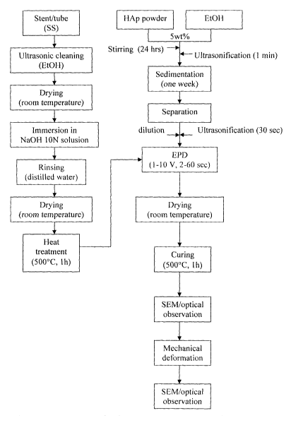
[0010] Figure 2 is a schematic flowchart for the production of HAP-coated
coronary
stents via EPD.
CA 02655389 2008-12-19
WO 2007/147234 PCT/CA2007/001081
3
[0011] Figure 3 is a graph illustrating particle size distribution of calcium
phosphate
particles in a slurry after one week of sedimentation.
[0012] Figure 4 is a micrograph illustrating the surface of a stent after
alkali micro-
etch treatment.
[0013] Figure 5(a) is a micrograph illustrating the microstructure of a
coating
prepared under the following EPD conditions: HAP concentration of 0.5 wt %,
voltage = 2 V, and deposition time = 10 seconds.
[0014] Figure 5(b) is a micrograph illustrating the microstructure of a
coating
prepared under the following EPD conditions: HAP concentration of 0.5 wt %,
voltage = 2 V, and deposition time = 30 seconds.
[0015] Figure 5(c) is a micrograph illustrating the microstructure of a
coating
prepared under the following EPD conditions: HAP concentration of 0.5 wt %,
voltage = 5 V, and deposition time = 10 seconds.
[0016] Figure 5(d) is a micrograph illustrating the microstructure of a
coating
prepared under the following EPD conditions: HAP concentration of 0.5 wt %,
voltage = 5 V, and deposition time = 30 seconds.
[0017] Figure 5(e) is a micrograph illustrating the microstructure of a
coating
prepared under the following EPD conditions: HAP concentration of 1.5 wt %,
voltage = 2 V, and deposition time = 10 seconds.
[0018] Figure 5(f) is a micrograph illustrating the microstructure of a
coating prepared
under the following EPD conditions: HAP concentration of 1.5 wt %, voltage = 2
V,
and deposition time = 30 seconds.
[0019] Figure 5(g) is a micrograph illustrating the microstructure of a
coating
prepared under the following EPD conditions: HAP concentration of 1.5 wt %,
voltage = 5 V, and deposition time = 10 seconds.
CA 02655389 2008-12-19
WO 2007/147234 PCT/CA2007/001081
4
[0020] Figure 5(h) is a micrograph illustrating the microstructure of a
coating
prepared under the following EPD conditions: HAP concentration of 1.5 wt %,
voltage = 5 V, and deposition time = 30 seconds.
[0021] Figure 5(i) is a micrograph illustrating the microstructure of a
coating prepared
under the following EPD conditions: HAP concentration of 2.5 wt %, voltage = 5
V,
and deposition time = 30 seconds.
[0022] Figure 6 are micrographs illustrating at three different magnifications
the
microstructure of a coating prepared under the following EPD conditions: HAP
concentration of 2.5 wt %, voltage = 5 V, and deposition time = 30 seconds.
[0023] Figures 7(a) and 7(b) are micrographs illustrating the microstructure
of a HAP
coating prepared by EPD. The complete uniform coverage of the substrate
surface by
the use of narrow particle size distribution of HAP powder is shown in 7(a),
and as
received powder, i.e., a wide particle size distribution of HAP powder is
shown in
7(b).
[0024] Figures 8(a) and 8(b) are micrographs illustrating the behaviour of a
HAP
coating on an expanded 316L stainless steel stent, without prior surface micro-
etching
through alkali treatment.
[0025] Figures 9(a) and 9(b) are micrographs illustrating the behaviour of a
HAP
coating on an expanded 316L stainless steel stent, with prior surface micro-
etching
through alkali treatment.
[0026] Figures 10(a), 10(b) and 10(c) are micrographs illustrating the
retention of
HAP coatings on an expanded 316L stainless steel stent with surface micro-
etched
through alkali treatment, showing high-strain regions of the expanded stent.
DETAILED DESCRIPTION OF THE INVENTION
[0027]Throughout the following description, specific details are set forth in
order to
provide a more thorough understanding of the invention. However, the invention
may
CA 02655389 2008-12-19
WO 2007/147234 PCT/CA2007/001081
be practiced without these particulars. In other instances, well known
elements have
not been shown or described in detail to avoid unnecessarily obscuring the
invention.
Accordingly, the specification and drawings are to be regarded in an
illustrative,
rather than a restrictive, sense.
5
[0028] In the following description, the term "calcium phosphate" is used
generically
and includes minerals such as HAP, dicalcium phosphate, tricalcium phosphate,
tetracalcium phosphate and amorphous or partially amorphous calcium phosphate.
[0029] The invention in one embodiment is directed to a process of coating an
implantable medical device with calcium phosphate by pretreating a substrate
with an
alkaline solution, preparing a slurry comprising a desired size range of
particles of
calcium phosphate, immersing the pretreated substrate in the slurry, and
coating the
calcium phosphate particles onto the pretreated substrate by electrophoretic
deposition. The process results in a thin, uniform, porous calcium phosphate
coating
that can withstand flexing of the substrate.
[0030] The novel coating process is exemplified below with reference to
stents, such
as cardiovascular stents (e.g. coronary stents). As shown in the examples
below, the
coating withstands simulated stent expansion procedures. However, the
invention has
broad application to virtually any type of implantable device with a metallic
surface
for use in the human or animal body, and particularly to flexible implantable
devices.
For example, the coatings are also useful in ureteral stenting and
catherterisation.
[0031] The novel process involves treating the substrate in an alkaline
solution to
enhance the adhesion of deposited calcium phosphate layer to the substrate.
Alkali
treatment may be performed by soaking the substrate in a NaOH solution or
other
suitable alkaline solution, for example. After the alkali treatment, the
substrate is
rinsed to remove residual alkali material, dried and then heat-treated. Heat
treatment
could, for example, involve heating at 500 C for one hour.
[0032] As shown in the examples below, alkali treatment positively affects the
bonding strength of the calcium phosphate coating. Alkali treatment etches the
surface of the substrate and forms sodium chromate. Both results are believed
to
CA 02655389 2008-12-19
WO 2007/147234 PCT/CA2007/001081
6
account for the subsequent improved bonding of the coating to the substrate.
Sodium
chromate is believed to make a strong bond from one side to the metallic bonds
of the
substrate, and from the other side to the covalent bonds of the calcium
phosphate
particles.
[0033] The novel process also involves the preparation of a stable colloidal
suspension of calcium phosphate particles. The solvent used for the colloidal
suspension may be an alcohol, such as ethanol. The slurry may comprise a
particular
weight percentage range of calcium phosphate, for example ranging from 0.5 to
20 wt
%. The colloidal suspension may also comprise calcium phosphate particles in a
particular size range. A particular size range of calcium phosphate particles
may be
obtained by, for example, by gravity sedimentation andJor centrifuge
sedimentation.
The desired particles may, for example, range in size from 50 nm to 150 nm in
diameter. Fine particles sometimes agglomerate but such agglomeration may be
eliminated by ultrasonification prior to coating.
100341 Figure 1 shows the EPD set-up of one particular embodiment of the
present
invention. The stent is suspended by a stainless steel wire. The
counterelectrode is
cylindrically-shaped to provide a uniform distribution of electrical field and
is made,
for example, from nickel foil. The radial distance between the stent and the
counterelectrode is constant. Deposition can be conducted under a range of
voltages
(e.g. 1 to 5 volts) for a range of times (e.g. 1 to 60 seconds) at room
temperature. The
coating applied may have a thickness no greater than 1 m, for example.
[0035] After EPD, the coating is dried at room temperature, and then cured.
Curing
can comprise heating the coated substrate at 500 C for 1 hour, for example.
This
relatively low curing temperature avoids oxidation damage to certain types of
implantable medical devices such as stainless steel stents.
[0036] The novel process allows the achievement of optimum coating thickness,
coverage uniformity, and maximum coating adhesion. This, in turn, allows the
coatings to withstand stresses applied to the substrate, such as, in the case
of stents,
during and after stent implantation and expansion. Optimum conditions to
achieve a
coating with a maximum mechanical integrity under applied deformation (e.g.
CA 02655389 2008-12-19
WO 2007/147234 PCT/CA2007/001081
7
expansion) can be determined by varying substrate parameters such as the stent
material, pre-treatment conditions such as the concentration of the alkaline
solution,
and coating parameters such as coating thickness, particle size, particle
concentration,
applied voltage, and the deposition duration.
[0037] The present invention provides for uniform distribution of calcium
phosphate
on all outer surfaces of the substrate. For example, all surfaces of a stent,
including
the wall surface of perforated portions of the stent, can be uniformly coated.
Other
methods of deposition, such as aerosol-gel, or plasma spraying are not able to
provide
the same uniform coverage and porous microstructure. Also, in comparison with
other methods of depositing calcium phosphate coatings on implantable devices,
such
as electro-chemical deposition (ECD) technology, the EPD deposits well-
developed
and well-characterized particles of calcium phosphate onto the substrate.
[0038] Further improvement of the functional properties and reliability of the
calcium
phosphate coatings, depending on the type of implantable medical device, can
be
achieved through impregnation with polymers, or polymers containing drug, for
long-
term controlled release. For example, porous calcium phosphate coatings, in
particular HAP coatings, can be used as an inorganic scaffold for carrying
organic
materials, forming a unique organo-ceramic composite. Organic materials may be
either co-deposited with the calcium phosphate particles, or impregnated into
the
coating after calcium phosphate particle deposition.
EXAMPLES
[0039] To demonstrate the feasibility of the novel processing concepts
outlined above,
the following examples are described below for a stainless steel substrate, in
particular, coronary stents. The procedures outlined below can be applied to
other
implantable medical devices.
Example 1
[0040]Figure 2 illustrates the steps taken to coat a stent with HAP according
to this
example. Electropolished cardiovascular (e.g. coronary) stents made of
stainless steel
316L (14 mm length, 1 mm radius) were thoroughly cleaned by immersing in an
EtOH ultrasound bath, and vibrated for 5 minutes.
CA 02655389 2008-12-19
WO 2007/147234 PCT/CA2007/001081
8
[0041] Commercially available HAP powder (Riedel-deHaen) was used as the
source
of calcium phosphate. For comparative purposes, a number of different HAP
powder
suspensions were investigated to assess their relative stability. HAP
concentration in
the slurry was varied from 0.5 to 20 wt %. The solvent used for the suspension
preparation was absolute ethanol, mixed with the HAP for 24 hours, and then
ultrasonicated for 1 minute to break any agglomerates.
[0042] The prepared suspension was allowed to settle and characterized in 24
hour
intervals to determine particle size distribution in the different portions of
the
suspension. Gravitational sedimentation separated the larger particles and
agglomerated granules from the fine particles. An upper portion of the
suspension
containing fine particles was siphoned out by a pipette. It contained
particles with an
average size of approximately 120 nm. This stable colloidal suspension was
found to
possess a long shelf life (> 1 month). The prepared suspension was then
diluted to 10,
30, and 50 vol % of its original concentration to examine the effect of HAP
concentration on coating quality.
[0043] To eliminate any formed agglomerates, the suspensions were subjected to
ultrasonic dispersion for approximately 30 seconds prior to the EPD coating
process.
HAP colloidal particles, suspended in ethanol, are charged positively. Figure
1 shows
schematically the EPD set-up. The cleaned stent was suspended by a stainless
steel
wire within a cylindrically-shaped counterelectrode made of nickel foil. The
constant
radial distance between the stent and the counterelectrode was approximately
1.2 cm.
A uniform distribution of electrical field was achieved due to use of the
cylindrical
counterelectrode. HAP deposition was conducted under conditions of constant
voltage at 1 to 5 volts, for the periods of time 1 to 60 seconds, at room
temperature.
DC current was supplied and controlled by a precise power source. A multimeter
measured the change in electrical current change with time, as the deposited
HAP
layer built up.
[0044] After EPD, the coated stents were dried at room temperature, and then
cured at
500 C for 1 hour.
CA 02655389 2008-12-19
WO 2007/147234 PCT/CA2007/001081
9
[0045] Figures 5(a) to (i) and Figure 6 show the representative appearance and
microstructure of the HAP coating prepared under different EPD conditions.
Example 1 a
[0046] The deposition of HAP was carried out under the following EPD
conditions:
2.5 wt % of HAP in the slurry, 5 V voltage, and 30 second deposition time. The
HAP
in the slurry was in a narrow size distribution, i.e., more than 75% of the
particles
were in the size range of 50-150 nm. This size distribution is shown in Figure
3. This
size distribution was obtained by siphoning off the top portion of the slurry
after a
one-week sedimentation process to separate the agglomerates and coarse
particles
from the finer particles. This separation process may be equally accomplished
through sedimentation using centrifuge in less than one hour. The uniform HAP
coating which resulted from the use of this narrow size distribution of the
particles is
shown in Figure 7a. Figure 7a has been taken at a very large magnification
(40,000X)
in order to visualise the fine (approximately 50 nm to 100 nm), uniformly
distributed
porosity of the HAP coating.
Example lb
[0047] The deposition of HAP coating was executed similarly as in Example 1 a
(2.5
wt % of HAP in the slurry, 5 V voltage, and 30 second deposition time), but
was
carried out by using HAP in the slurry in a broad particle size distribution
(from
approximately 10 nm to 5 m). The coarse HAP coating which resulted from the
use
of this broader range of size distribution of particles is shown in Figure 7b.
Exam lp e lc
[0048] Stability of the prepared HAP slurry was verified by comparing water
and
ethanol as the suspension solvent. The sedimentation time of the HAP
particles, for a
5 wt % suspension was increased from less than an hour in the water-based
suspension to more than a month for the ethanol-based suspension. Although it
was
possible to prepare stable water-based suspensions of HAP by decreasing pH to
a
strongly acidic range, water-based suspensions were not pursued to avoid the
dissolution of HAP in such an acidic environment.
CA 02655389 2008-12-19
WO 2007/147234 PCT/CA2007/001081
Example 1 d
[0049] The stent in this example was processed as in Example 1, and then
expanded
from an initial radius of about 1 mm to a final radius of about 3 mm. The
expansion
test was performed using EncoreTM 26 Inflation Device Kit. The expanded stent
was
5 observed under a scanning electron microscope (SEM). Figure 8 illustrates
the
results. The HAP coating, processed according to the protocol described in
Example
1, separated from the stent surface in areas of significant strain due to
stent expansion.
The flaked coating allowed the assessment of the coating thickness, which was
found
to be in the range of 1.0-1.5 m. The coating was retained in areas
experiencing little
10 or no strain.
Example 1 e
[0050] The stent in this example was modified through alkali pretreatment in
order to
obtain a better adhesion between the coating and the stent. Alkali treatment
was
performed by soaking the stent in 10 mL of lON NaOH solution at 60f5 C for 24
hours. Figure 4 shows the etched surface of a stent after alkali treatment.
After the
alkali treatment, the stent was rinsed with distilled water several times, and
then dried
at room temperature for about 6 hours. The rinsed and dried alkali-treated
stent was
then heated to 500 C at a rate of 10 C/min, maintained at that temperature for
1 hour,
and then cooled to room temperature at a rate of 1.5 C/min.
[0051] The pretreated stent was coated according to the protocol described in
Example 1, and then expanded according to the protocol described in Example
ld.
The expanded stent was observed under SEM. Figures 9 and10 illustrate the
results.
Notably, the HAP coating did not separate from the stent surface, even in
areas of
significant strain resulting from stent expansion.