Note: Descriptions are shown in the official language in which they were submitted.
CA 02655420 2008-12-12
WO 2007/146385 PCT/US2007/013968
ALBUMIN-BOUND PROTEIN/PEPTIDE COMPLEX AS A BIOMARKER FOR
DISEASE
FIELD OF INVENTION
[0001] The invention relates to methods of diagnosis using biomarkers
comprising albumin-
bound protein/peptide complex (ABPPC).
BACKGROUND
100021 Serum albumin is the most abundant protein in serum, typically present
at 45-50
mg/ml. Albumin functions as a "molecular sponge" binding proteins, lipids, and
small
molecules in the intracellular space (1-3) and has been found to form
associations with peptide
hormones, serum amyloid A, interferons, glucagons, bradykinin, insulin, and
Streptococcal
Protein G (4-7) but an extensive list of binding partners, and whether these
partners change with
disease, has not been investigated. Previous studies have shown a higher
recovery of low
molecular weight species when removing high molecular weight species under
denaturing
conditions, further confirming that larger proteins, such as albumin, are
binding peptides (8).
Furthermore, albumin has been reported to bind to a small number of specific
proteins such as
paraoxonase 1 (9), alpha-l-acid glycoprotein (10), and clusterin (11)
(indirect interaction
through paraoxonase 1) and apolipoprotein E'2 in serum. Although albumin
binding peptides
(below 30 kDa) in serum have been studied, the extent of their binding is
currently unknown
(13). To date, a comprehensive study of the whole proteins bound to albumin
has not been
carried out. Additionally, there is no documentation of any changes in the
protein/peptide
composition, ratio or PTM status of the proteins/peptides bound to albumin.
[0003] Albumin has been found to change with disease which alters its binding
to metals
and currently functions as a biomarker for ischemia. A modification of albumin
that has
previously been identified as a biomarker for myocardial ischemia is the N-
terminus N-
acetylation of albumin, which decreases the binding affinity of albumin to
cobalt and nickel (21-
23). Current patents (24,25) cover the usage of this N-terminal modification
of albumin for
ischemia and have led to a clinical assay for albumin cobalt binding (ACB
assay). In addition to
the N-terminal modification, the oxidation of albumin has been proposed to be
a marker for
oxidative stress (26). MALDI-TOF analysis (Matrix Assisted Laser
Desorption/Ionization
Time-of-Flight) of the albumin in patients with renal impairment and end-stage
renal disease
show an increase in the MW of albumin with disease (27). Finally, the fatty
acid transport
i
CA 02655420 2008-12-12
WO 2007/146385 PCT/US2007/013968
function of albumin is modified in atherosclerosis and diabetes (28). In
patients with diabetes,
the binding capacity of albumin for fatty acids is increased, and in patients
with atherosclerosis
the capacity is decreased. In conclusion, the evidence the albumin is changing
with disease is
clear. What has not been investigated or described previously is altered
binding of proteins
and/or peptides to albumin in serum. The current work is unique because it
includes the analysis
of intact proteins, degraded proteins, and peptides, without eliminating any
mass range.
Furthermore, the current work focuses on the changes in the proteins and
peptides that bind to
albumin, a feature not addressed in any previous literature.
SUMMARY
[0004] We examined an albumin-enriched fraction of human serum in order to
determine
any albumin binding proteins in healthy individuals and furthermore whether
the proteins that
bind to albumin change with disease. The study included multiple independent
methods for
isolation of albumin and any bound proteins/peptides (modified and
nonmodified) (albumin
bound protein/peptide complex, ABPPC) (Figure 1). The results show that ABPPC
should be
useful biomarkers for disease.
[0005) Accordingly, a method of diagnosing a disease or disorder is provided,
comprising
measuring the level of specific albumin-bound protein/peptide complex(es)
(ABPPC) in a
subject, and comparing the level to a control level from a normal subject
population. It has been
found that variations in the levels of specific ABPPCs, and variations in
ABPPC profile are
indicative of specific diseases and disorders.
[0006] The aim is to characterize proteins that are differentially bound to
albumin in
diseased and healthy patients in a cost effective, rapid and sensitive manner
that is compatible
with current blood collection protocols. This is based on the hypothesis that
albumin changes
with disease, and therefore the complex of albumin with its bound proteins and
peptides
changes, although the inventors are not bound by any particular hypothesis.
The ABPPC assay
may measure a modification ofalbumin or a change in ABPPC composition (i.e.
the presence or
absence of one or more proteins, altered concentration (or stoichiomery or
molar ratio) of one or
more proteins, change in a protein's PTM (e.g. proteolysis fragment vs. intact
protein including
albumin).
2
CA 02655420 2008-12-12
WO 2007/146385 PCT/US2007/013968
[0007] The method can be used alone, or in conjunction with other diagnostic
tests to
improve the accuracy and specificity of the diagnosis. It can also be used for
screening
purposes, to identify individuals who appear to be "at risk" for further
testing by this or other
means.
[0008] Accordingly, in one aspect, the method comprises (a) measuring the
level of at least
one biomarker in a biological sample obtained from said subject, wherein said
biomarker
comprises an an albumin-bound protein/peptide complex (ABPPC), and (b)
comparing the level
measured in the biological sample to a control level in a normal subject
population, wherein an
increase or decrease in the level, compared to control level, is indicative of
said disease or
disorder.
[0009] In another aspect, the method comprises assaying a subject sample for
the presence
of at least one biomarker comprising an albumin-bound protein/peptide complex
(ABPPC);
wherein the detection of said biomarker(s) is correlated with a diagnosis of
the disease or
disorder, the correlation taking into account the presence and level of
biomarker(s) in the subject
sample as compared to normal subjects.
100101 The biomarkers can be detected by any suitable means known to those of
skill in the
art, for example, using a protein assay, binding assay, or an immunoassay.
Biomarkers may also
be identified as peaks using Mass Spectroscopy, or as gel bands using, for
example size
exclusion chromatography (SEC), optionally after appropriate initial treatment
of the sample.
Exemplary assays are described in detail in the examples which follow. For a
positive
diagnosis, the biomarkers are elevated or lowered as compared to values in
normal healthy
controls.
[0011] The subject sample may be selected, for example, from the group
consisting of
blood, blood plasma, serum. Preferably, the sample is albumin-enriched serum.
[0012] The diagnostic assay can be used, for example, to evaluate patients
presenting to an
emergency room, or for ongoing care within a hospital setting, or in a medical
practitioner's
office. The assay has the advantage that it can be easily and reproducibly
obtained from
individuals since albumin is highly abundant in serum (40-50 mg/ml). Specific
antibodies to
albumin are available and the ABPPC can be captured easily without a
complicated assay.
Other biochemical methods can be used as well, including liquid chromatography
and gel based
3
CA 02655420 2008-12-12
WO 2007/146385 PCT/US2007/013968
methods. Since the capture of ABPPC is based upon targeting albumin, the
proteins (or a PTM
or other modification) that are changing in a particular disease need not be
known in advance,
since the protcol for capturing the ABPPC is universal for all diseases. In
this way, the ABPPC
is a simple targeted assay that casts a wide net over a variety of potential
targets and is,
therefore, very cost effective. There is no requirement for developing
multiple specific
antibodies to detect low abundance proteins, for example. Furthermore,
capturing this naturally-
occurring sub-proteome reduces sample complexity and avoids the problems
associated with
assay sensitivity at low protein concentrations. Since some proteins in the
ABPPC have not
been observed in albumin depleted serum, it appears that some biomarkers are
unique to the
ABPPC.
[0013] After a protein of interest has been identified, downstream clinical
assays could
simply couple one capture antibody for albumin to a different detecting
antibody for the protein
of interest.
[00141 Also provided is a kit for carrying out the method described herein. In
one
embodiment, the kit may comprise, for example, any of: an antibody (or a
chemical moiety) to
specifically capture or enrich for the endogenous albumin), a secondary
antibody (or chemical
moiety) to one or more of the specific protein (or peptide or modified
protein) bound to albumin
and components for detection and/or quantification of the amount of secondary
antibody bound.
In one embodiment, the secondary antibody would be against protein(s) that
change with the
specific protein so that one is quantifying the change in protein content of
the ABPPC.
[0015] An afternative would be a capture of endogenous ABPPC (with an antibody
or
chemical moiety) with a direct detection of the protein(s) of interest using
mass spectrometry of
the intact or enzymatically degraded protein. In this embodiment the kit may
contain the anti-
albumin antibody coupled to a matrix (for example, in a small column or packed
into an end of a
pipette tip) where the ABPPC would be enriched followed elution into MS for
intact mass or
eluted for digestion and subsequent MS analysis (of all peptides or specific
signature peptide for
the analyte(s)). Kits of the invention may contain a plurality of antibodies
so that more than one
ABPPC component could be assessed simultaneously.
[0016] It is also believed that the ratio of bound to free (circulating) ABPPC
may be
important. Methods and kits may be modified so that specific proteins are
measured as bound to
4
CA 02655420 2008-12-12
WO 2007/146385 PCT/US2007/013968
serum albumin or free. For example, in the current work, a number of proteins
have been
observed to be both bound to albumin, but also observed in the albumin-
depleted fraction of
serm, indicating that they could be present in their free form. Examples of
these proteins include
antithrombin III, apolipoprotein All, AIV, CII, clusterin, transthyretin, and
vitamin D binding
protein, for example. Practitioners will be able to determine through routine
experimentation
how the ratio is altered in particular disease states.
[0017] It is a further object to provide a method of identifying biomarkers
comprising
ABPPC for specific diseases and disorders by screening populations of patients
having a disease
or disorder for serum ABPPCs and comparing the ABPPCs thus obtained with those
in a normal
subject population. Such screening can be carried out in the same manner as is
done for the
diagnostic assays described hereinabove. For example, methods such as Mass
Spectroscopy or
SEC can be used to determine a profile of ABPPCs and where differences are
found, specific
ABPPCs identified, e.g. using a protein assay, binding assay, or immunoassay,
that will be
useful as biomarkers. Furthermore, a protein digestion could be carried out
and one or more of
the resulting peptides monitored. Biomarkers so identified can be used in the
diagnostic
method described herein.
[0018] Diseases or disorders for which the methods and compositions of the
invention are
expected to be useful include vasculitis, myocardial infarction, heart
failure, sepsis, cancer, and
diabetes. Using the methods detailed herein, persons of skill in the art will
be able to determine
other diseases and disorders in which ABPPC is altered without undue
experimentation
[0019] This application claims priority to U.S. provisional applications no.
60/813,761,
filed June 14,2007, and 60/813,825, filed June 15, 2007, which are hereby
incorporated by
reference. References and patents cited herein are hereby incorporated by
reference.
BRIEF DESCRIPTION OF THE DRAWINGS
[0020] Figure 1. Methods and corresponding objectives used to separate an
albumin-
enriched fraction and characterize the ABPPC.
[0021] Figure 2. ID SDS-PAGE (AI, BI) and corresponding western blot for
albumin (All,
BII) of the aibumin-enriched fraction of normal pooled serum.
; 5
CA 02655420 2008-12-12
WO 2007/146385 PCT/US2007/013968
[0022] Figure 3. SEC chromatograms. (A) MW standards (A) Albumin-enriched
fraction
overlaid on the MW standards.
[0023] Figure 4. SEC chromatograms (280 nm) of 8 consecutive injections of the
albumin-
enriched fraction from healthy individuals.
[0024] Figure 5. 1 D SDS-PAGE of heated and non-heated SEC fractions, anti-HSA
(human
serum albumin) retentate and the whole albumin-enriched fraction from normal
pooled serum.
[0025] Figure 6. RP-HPLC of three fractions (A-C) of the SEC chromatogram of
the
albumin-enriched fraction from normal pooled serum.
[0026] Figure 7. RP-HPLC chromatograms of the anti-HSA retentate of the
albumin-
enriched fraction from normal pooled serum.
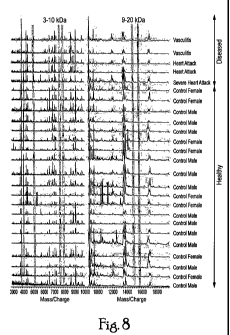
[0027] Figure 8. MALDI-TOF spectra of 20 healthy controls and 5 diseased
patients.
Differences are highlighted in yellow.
[0028] Figure 9. SEC chromatograms of the albumin-enriched fraction of 3
timepoints from
patients (3 MI, 2 SA) who underwent balloon angioplasty.
[0029] Figure 10. Panel A shows RP-BPLC chromatograms (210 nm) of the high MW
SEC
fractions from Figure 9. Yellow bars highlight differences among the 3
timepoints. Panel B
shows 1 D SDS-PAGE of the same fractions. Panel C shows western blot for
albumin of gels in
Panel B.
100301 Figure 11. Zoomed view of a section of the RP-HPLC chromatograms (in
Figure
10A) of the SEC fraction containing the ABPPC.
[0031] Figure 12. SEC chromatograms (280nm) of two healthy controls and a
patient with a
myocardial infarction (A). Panel B shows RP-HPLC chromatograms of the high MW
SEC
fractions from the samples in Panel A.
[0032] Figure 13. 1 D SDS-PAGE of the albumin-enriched fraction of 2 of
healthy controls,
2 patients with AMI and 2 patients with vasculitis.
6
CA 02655420 2008-12-12
WO 2007/146385 PCT/US2007/013968
[0033] Figure 14. RP-HPLC chromatograms of the high MW SEC fractions from
figure
l0A zoomed in to show the albumin peak. Arrows point to change in retention
time for albumin
in time point 8 for 2 MI and 1SA patient. Red circle highlights peak observed
only in time point
8 for the same samples.
7
CA 02655420 2008-12-12
WO 2007/146385 PCT/US2007/013968
DETAILED DESCRIPTION
Definitions
[0034] The following terms are used as defined below throughout this
application, unless
otherwise indicated.
[0035] "Marker" or "biomarker" are used interchangeably herein, and in the
context of the
present invention refer to an ABPPC (of a particular specific identity or
apparent molecular
weight) which is differentially present in a sample taken from patients having
a specific disease
or disorder as compared to a control value, the control value consisting of,
for example, average
or mean values in comparable samples taken from control subjects (e.g., a
person with a
negative diagnosis, normal or healthy subject). Biomarkers may be identified
as specific
peptides or proteins, either presently bound or cleaved from albumin, or as
specific peaks,
bands, fractions, etc. in a mass spectroscopy, SEC, or other separation
process or antibody
detection. In some applications, for example, a mass spectroscopy or other
profile or multiple
antibodies may be used to identify multiple biomarkers, and differences
between individual
biomarkers and/or the partial or complete profile may be used for diagnosis.
[0036] The phrase "differentially present" refers to differences in the
quantity andlor the
frequency of a marker present in a sample taken from patients having a
specific disease or
disorder as compared to a control subject. For example, a marker can be a
ABPPC which is
present at an elevated level or at a decreased level in samples of patients
with the disease or
disorder compared to a control value (e.g. determineed from samples of control
subjects).
Alternatively, a marker can be an ABPPC which is detected at a higher
frequency or at a lower
frequency in samples of patients compared to samples of control subjects. A
marker can be
differentially present in terms of quantity, frequency or both. It may also be
a physical.
change/modification of the protein that is the marker, rather than just an
increase or decrease in
the amount present/detected. For example, it may be the post-translational
modification,
cleavage, or isoform.of the protein that is changing, and it is this change
that is detected by the
assay. This is separate from measuring a different uanti in diseased vs.
control.
[0037] A marker, compound, composition or substance is differentially present
in a sample
if the amount of the marker, compound, composition or substance in the sample
is statistically
significantly different from the amount of the marker, compound, composition
or substance in
8
CA 02655420 2008-12-12
WO 2007/146385 PCT/US2007/013968
another sample, or from a control value. For example, a compound is
differentially present if it
is present at least about 120%, at least about 130%, at least about 150%, at
least about 180%, at
least about 200%, at least about 300%, at least about 500%, at least about
700%, at least about
900%, or at least about 1000% greater or less than it is present in the other
sample (e.g. control),
or if it is detectable in one sample and not detectable in the other.
[0038] Alternatively or additionally, a marker, compound, composition or
substance is
differentially present between samples if the frequency of detecting the
marker, etc. in samples
of patients suffering from a particular disease or disorder, is statistically
significantly higher or
lower than in the control samples or control values obtained from healhty
individuals. For
example, a biomarker is differentially present between the two sets of samples
if it is detected at
least about 120%, at least about 130%, at least about 150%, at least about
180%, at least about
200%, at least about 300%, at least about 500%, at least about 700%, at least
about 900%, or at
least about 1000% more frequently or less frequently observed in one set of
samples than the
other set of samples. These exemplary values notwithstanding, it is expected
that a skilled
practitioner can determine cut-off points, etc. that represent a statistically
significant difference
to determine whether the marker is differentially present
[0039] "Diagnostic" means identifying the presence or nature of a pathologic
condition and
includes identifying patients who are at risk of developing a specific disease
or disorder.
Diagnostic methods differ in their sensitivity and specificity. The
"sensitivity" of a diagnostic
assay is the percentage of diseased individuals who test positive (percent of
"true positives").
Diseased individuals not detected by the.assay are''fals.e negatives."
Subjects:who.are not .
diseased and who test negative in the assay, are termed "true negatives." The
"specificity" of a
diagnostic assay is I minus the false positive rate, where the "false
positive" rate is defined as
the proportion of those without the disease who test positive. While a
particular diagnostic
method may not provide a definitive diagnosis of a condition, it suffices if
the method provides a
positive indication that aids in diagnosis.
[0040] The terms "detection", "detecting" and the like, may be used in the
context of
detecting biomarkers, or of detecting a disease or disorder (e.g. when
positive assay results are
obtained). In the latter context, "detecting" and "diagnosing" are considered
synonymous.
9
CA 02655420 2008-12-12
WO 2007/146385 PCT/US2007/013968
[0041] By "at risk of' is intended to mean at increased risk of, compared to a
normal subject,
or compared to a control group, e.g. a patient population. Thus a subject
carrying a particular
marker may have an increased risk for a specific disease or disorder, and be
identified as
needing further testing. "Increased risk" or "elevated risk" mean any
statistically significant
increase in the probability, e.g., that the subject has the disorder. The risk
is preferably
increased by at least 10%, more preferably at least 20%, and even more
preferably at least 50%
over the control group with which the comparison is being made.
[0042] A "test amount" of a marker refers to an amount of a marker present in
a sample
being tested. A test amount can be either in absolute amount (e.g., g/ml) or
a relative amount
(e.g., relative intensity of signals).
[0043] A "diagnostic amount" of a marker refers to an amount of a marker in a
subject's
sample that is consistent with a diagnosis of a particular disease or
disorder. A diagnostic
amount can be either in absolute amount (e.g., g/ml) or a relative amount
(e.g., relative
intensity of signals).
= = . = == . = . = = ~ .
[0044] A "control amount" of a marker can be any amount or a range of amount
which is to
be compared against a test amount of a marker. For example, a control amount
of a marker can
be the amount of a marker in a person who does not suffer from the disease or
disorder sought to
be diagnosed. A control amount can be either in absolute amount (e.g., g/ml)
or a relative
amount (e.g., relative intensity of signals).
[0045] The terms "polypeptide," "peptide" and "protein" are used
interchangeably herein to
refer to a polymer of a-amino acid residues, in particular, of naturally-
occuring a-amino acids.
The terms apply to amino acid polymers in which one or more amino acid residue
is an analog
or mimetic of a corresponding naturally-occurring amino acid, as well as to
natural[y-occurring
amino acid polymers. Polypeptides can be modified, e.g., by the addition of
carbohydrate
residues to form glycoproteins, phosphorylation to form phosphoproteins, and a
large number of
chemical modifications (oxidation, deamidation, amidation, methylation,
formylation,
hydroxymethylation, guanidination, for example) as well as degraded, reduced,
or crosslinked.
The terms "polypeptide," "peptide" and, "protein" include all unmodified.and
modified forms of
the protein
CA 02655420 2008-12-12
WO 2007/146385 PCT/US2007/013968
[0046] "Detectable moiety" or a "label" refers to a composition detectable by
spectroscopic,
photochemical, biochemical, immunochemical, or chemical means. For example,
useful labels
include 32P, 35S, fluorescent dyes, electron-dense reagents, enzymes (e.g., as
commonly used in
an ELISA), biotin-streptavidin, dioxigenin, haptens and proteins for which
antisera or
monoclonal antibodies are available, or nucleic acid molecules with a sequence
complementary
to a target. The detectable moiety often generates a measurable signal, such
as a radioactive,
chromogenic, or fluorescent signal, that can be used to quantify the amount of
bound detectable
moiety in a sample. Quantitation of the signal is achieved by, e.g.,
scintillation counting,
densitometry, flow cytometry, or direct anlaysis by mass spectreometry of
intact or
subsequentally digested peptides (one or more peptide can be assessed.)
[0047] "Antibody" refers to a polypeptide ligand substantially encoded by an
immunoglobulin gene or immunoglobulin genes, or fragments thereof, which
specifically binds
and recognizes an epitope (e.g., an antigen). The recognized immunoglobulin
genes include the
kappa and lambda light chain constant region genes, the alpha, gamma, delta,
epsilon and mu
heavy chain constant region genes, and the myriad immunoglobulin variable
region genes.
Antibodies exist, e.g., as intact immunoglobulins or as a number of well
characterized fragments
produced by digestion with various peptidases. This includes, e.g., Fab' and
F(ab)'2 fragments.
The term "antibody," as used herein, also includes antibody fragments either
produced by the
modification of whole antibodies or those synthesized de novo using
recombinant DNA
methodologies. It also includes polyclonal antibodies, monoclonal antibodies,
chimeric
antibodies, humanized antibodies, or single chain antibodies. "Fc" portion of
an antibody refers
to that portion of an immunoglobulin heavy chain that comprises one or inore
heavy chain
constant region domains,,CHI, CH2.and,CH3, but does not include the heavy
chain variable
region.
[0048] By "binding assay" is meant a biochemical assay wherein the biomarkers
are detected
by binding to an agent, such as an antibody, through which the detection
process is carried out.
The detection process may involve radioactive or fluorescent labels, and the
like. The assay
may involve immobilization of the biomarker, or may take place in solution.
[0049] "Immunoassay" is an assay that uses an antibody to specifically bind an
antigen (e.g.,
a marker). The immunoassay is characterized by the use of specific binding
properties of a
particular antibody to isolate, target, and/or quantify the antigen.
11
CA 02655420 2008-12-12
WO 2007/146385 PCT/US2007/013968
[0050] The phrase "specifically (or selectively) binds"= to an antibody or
"specifically (or
selectively) immunoreactive with," when referring to a protein or peptide,
refers to a binding
reaction that is determinative of the presence of the protein in a
heterogeneous population of
proteins and other biologics. Thus, under designated immunoassay conditions,
the specified
antibodies bind to a particular protein at least two times the background and
do not substantially
bind in a significant amount to other proteins present in the sample. Specific
binding to an
antibody under such conditions may require an antibody that is selected for
its specificity for a
particular protein. A variety of immunoassay formats may be used to select
antibodies
specifically immunoreactive with a particular protein. For example, solid-
phase ELISA
immunoassays are routinely used to select antibodies specifically
immunoreactive with a protein
(see, e.g., Harlow & Lane, Antibodies, A Laboratory Manual (1988), for a
description of
immunoassay formats and conditions that can be used to determine specific
immunoreactivity).
[0051] The terms "subject", "patient" or "individual" generally refer to a
human, although
the methods of the invention are not limited to humans, and should be useful
in other animals
(e.g. birds, reptiles, amphibians, mammals), particularly in mammals, since
albumin is
homologous among species.
. = . . .. = . - .. . . . = .
100521 "Sample" is used herein in its broadest sense. A saniple may comprise a
bodily fluid
including blood, serum, plasma, tears, aqueous and vitreous humor, spinal
fluid; a soluble
fraction of a cell or tissue preparation, or media in which cells were grown;
a, aorganelle, or
membrane isolated or extracted from a cell or tissue;, polypeptides, or
peptides in solution or
bound to a substrate; a cell; a tissue; a tissue print; a fingerprint, skin or
hair; fragments and
derivatives thereof. Subject samples usually comprise derivatives of blood
products, including
blood, plasma and serum.
[0053] By "albumin-enriched serum or plasma" is meant serum or plasma that has
been
treated to reduce or remove components other than albumin and associated
peptides and proteins
which are bound thereto.
EXAMPLES
[0054] There are two primary methods available for isolating albumin from
serum or
plasma: affinity-based (e.g., antibody, cibacron blue) and chemical-based
methods (e.g.,
. . . . . = , , r ,
NaCI/EtOH [30,31] TCAlacetone [32]). Many of the affinity-based methods have
been
12
CA 02655420 2008-12-12
WO 2007/146385 PCT/US2007/013968
compared and shown to effectively remove albumin [29, 33, 34]. However, these
methods are
vulnerable to non-specific binding of proteins/peptides to the ligand and
column materials and
carryover between experiments in the case of LC columns [29, 31, 33=36].
Alternatively,
albumin has been purified using NaCI/EtOH since the 1940s [37] and this method
is routinely
used for isolating pharmaceutical grade albumin. Recently, this process was
optimized for the
proteomics field to minimize the steps required for effective purification and
removal of albumin
[30], but copurification of other proteins may still be an issue.
Example 1
Isolation of albumin enriched fraction of human serum
[0055] Albumin depletion by chemical extraction was performed as described by
Fu et al.
[30]. Briefly, 100 L normal human serum was depleted of lipids via
centrifugation, followed
by depletion of IgG using a protein G affinity column (Amersham Biosciences,
Piscataway, NJ,
USA). IgG depleted serum was brought to 42% ethanol/100mM NaCI and incubated
at 4 C for
I h followed by centrifugation at ] 6 000 x g for 45 min. The supematant
(albumin-enriched
fraction) was collected and used for the work presented below.
Example 2
Isolation and characterization of ABPPC
[0056] Treatment of whole albumin-enriched fraction is shown schematically in
Figure 1.
The study included multiple independent methods for isolation of albumin and
any bound
proteins/peptides (modified and unmodified)( albumin bound protein/peptide
complex, ABPPC).
[0057] Initial analyses included 1D SDS-PAGE of heated and non-heated samples
and a
western blot for albumin (Figure 2). On 1 D SDS-PAGE, the disappearance of the
116 kDa
band, which contains albumin, and the appearance of several bands only after
severe
denaturation (i.e. heating and treatment with 8M urea) indicate that many of
the
proteins/peptides in the albumin-enriched fraction were associated with
albumin or other protein
(Figure 2 Al, BI). Importantly, these results were seen only when the gel was
overloaded (6-12
,ug/lane). The presence of the lower molecular weight bands were not
visualized in lower loads
of non-heated sample. By western blot, albumin is present in the 116 kDa band
in the non-
heated sample, running higher than its expected MW of 66 kDa (Figure 2AII).
However, upon
heating, this band disappears and smaller MW bands, some containing albumin,
appear (Figure
13
. . . . . = .. . . ' ... . ,
CA 02655420 2008-12-12
WO 2007/146385 PCT/US2007/013968
2 BII). Therefore, it is possible that albumin is appearing at a higher
molecular weight because
it is forming a dimer or it is bound to one or more other proteins/peptides,
and only upon
severely denaturing conditions these proteins/peptides are released.
Consistent with this is the
fact that peptides from proteins other than albumin were identified in this
116kDa band,
including ceruloplasmin, haptoglobin, and alpha-lB-glycoprotein. It is noted
that albumin runs
at a lower molecular weight in the non-heated condition. This could be due to
incomplete
reduction of disulfides such that albumin is not fully saturated with SDS,
which affects the
migration, or that another protein or peptide bound to albumin is altering the
migration of
albumin in the gel. Furthermore, the presence of multiple albumin fragments
after heating
(Figure 21311) indicates that extensive proteolysis of albumin has occurred.
In conclusion, while
the ID SDS-PAGE results are not conclusive evidence of proteins binding to
albumin, these
preliminary results prompted more sophisticated analyses by SEC and
immunoaffinity
chromatography.
10058] Native size-exclusion chromatography (SEC) was used to separate the
albumin-
enriched fraction by size to isolate any protein complexes present in native
conditions. SEC was
chosen because it has minimal non-specific binding coupled with the ability to
sort protein
complexes by size under native conditions. Immunoaffinity by an anti-HSA spin
column was
chosen for its specificity for human albumin, though non-specific binding by,
the.matrix was an
acknowledged drawback. An anti-albumin antibody affinity column (anti-HSA) was
used to
bind albumin and any bound proteins/peptides. The proteins bound to the column
were then
eluted from the column (anti-HSA retentate) prior to further analyses. The
anti-HSA retentate
and high MW SEC fractions were separated by ID SDS-PAGE and reversed phase
high
performance liquid chromatography (RP-HPLC) in order to further separate the
bound
proteins/peptides from albumin prior to tryptic digestion and tandem mass
spectrometry
(MS/MS) for protein/peptide identification.
[0059] Native SEC was used to separate the albumin-enriched fraction by size,
as larger
proteins will spend less time on the column and elute earlier than smaller
proteins and peptides.
Under native conditions, it is expected that those proteins and peptides bound
to albumin will
elute in the fraction/s containing albumin, while those unbound will elute
separately from
albumin, consistent with their native molecular weights. SEC was successful in
separating a
wide range of proteins (29-205 kDa) with good resolution, as illustrated by
well-separated peaks
14
CA 02655420 2008-12-12
WO 2007/146385 PCT/US2007/013968
in Figure 3A. The albumin-enriched fraction separated into 4 regions (A-D) by
SEC, with the
major peak eluting at the time consistent with a mass slightly larger than the
66 kDa standard
protein (Figure 3B). A benefit of SEC is that it is highly reproducible, as
can be seen in Figure
4.
[0060] SEC-A contains fractions eluting near 116 kDa, SEC-B contains fractions
from the
tail of SEC-A and slope of SEC-C, SEC-C contains fractions eluting slightly
above 66 kDa, and
SEC-D contains sample from the lower molecular weight region. Each fraction (A-
D) was then
further separated and desalted prior to analysis by mass spectrometry. The SEC
fractions were
separated by two methods, i D SDS-PAGE (Figure 5) and RP-HPLC (Figure 6) prior
to
MALDI-TOF MS and LC-MS/MS.
Example 3
Proteins identified in the ABPPC
[0061] Analysis by 1D SDS-PAGE and RP-HPLC of the SEC fractions reveals the
presence
of multiple species in addition to albumin eluting in fractions A, B, and C.
Interestingly, many
of these proteins in the high MW SEC fractions have MW well below 66 kDa and
the proteins
are listed in Table 1.
CA 02655420 2008-12-12
WO 2007/146385 PCT/US2007/013968
Table 1. Proteins identified in the albumin-bindin rotein! tide complex
(ABPPC).
. * = .
= r. ='~.
< m o ~- =
. . a~aeln . ~ = ~ - ~ ~ ~ = a~on~e. aa~x .rwnn~n~r, . aa~n~e
'. . .~ =~= .~ , . .
1 qta~++n / J / / Y~g. = /
AI l-ectd teln 1 / ! / / ! yEg:. / C41rdbovascular. ~ ObL" mactwt Amd 2WS
3 1-9tld QiYOOPMto-In 2 / / /' yEg,: / r Asoc. W cardlpvaepdar,
dlaaene/dlat~atm BrfMne 20U8
4 1 `(F.8= / ! / / yE,$_:: / CerdSOVapCader tNUbltOt ptaum essD
200:5
S 1 e n YES I / / / =yO / Cartliovasctder tMibitor Mro Mdareon 2l)tG8
e 1 ! .'yp ' / / / / ! .YEg ' = Cardlovaseuaar Aesoc, wl atterlad thron~holle
dtaease Ga~l 1990
T yEg / r 1 / / y~ CoM)wasadar
Chw1fles essac.WONta r~tinlardlon Bahane ZQQB
g / CardlovcmrLlat = Praauaor blmd Ww3m rncontrol Andereun 2005
r14 AndUqpmbin III ypg= / / / ! y~$ / ~bY~r~~ ~ /~ysyt 2005 ADW-DoProteM A! !
~fel kdarcycn aeaoele Andereon2008
A U yEg Andarorn A tV / / ~õ~~. Fbsh Fcta CHb Ande.yson2pp6
C II V6S YE8 Cermovmcusr wpom~ A~ept =+ppg AmUpm
C IU yEA / / / .y' F,g.. CermOrtCUler= CHD AteAter AMEfean 2006
Vascvtar an0lar Cmputa9on Prohtn: De{~dq vasotlll>KOP
13 carboxymolaw t32 ! / ! YE8 b-tacWNn 2006
111 cerutepboTin / / / / y~: Cardlowawlar Rfsk fartor cartll~ dteaafe And 2005
17 Gustula . YEg ! / / yj$. / Cq,Vasrarsrl yx3Rem 2pU6
/ / / y~. ~w: ~ tadiaNO ayoaarohun ttom
1e .rnent t Inhi4itor YES Ane 2ti
19 Conv4errAnt tu- / / / YT -13
' / Piadan nwvcxrdW Mrbraton~ee Nwerenn 2bob
2D ot:ln I / / / I ' _" / CevoVeaaiar AaeF rtd hl er seNm bfal and hne
dWesltrd
MMTOWOWA. 21 at y6g ' ! / ! / ; ~g. ~~. heaAdeeere Berhnne 2006
J / / / ~g'/
72 ppln p r
YES
y! yEg r / / / /' y~r' / ~ra~tg, Awte Andssa+ 2005
94 Hat+e!yt / / YF$;.
UUer 81phs Mypsln UtWD+2ar heQVX
2s chetn 1=14 / / = / ~; ~ovascular Anaoe Fufte, 2004
26 IW / / J ! ,.,y~;: ~r CeM1eC phOfS
1 / / y~g=. I
27 LOudM 2 fiDERpMe
28 Pa9mrcnass I / / / !' ! Csndlovasnlv Relatfon M cwdWyMMM aise=asp Andason
2606
{j~. J
rewomMm 2 / I / / yj
90 Pleenin / I / ! Cmdlareaeuler d Arrfe~an 2006 anzym- ,h C~y~ / / r / r s
~yr Awadsted 1 t
,ML
Y / / / ! !
33 Vf6vrtn 0 dndn y~ / / ! y~g, =!
94 ZM1C atuM 2 eNmoorctatn yEg / / . f~ps. . !
1 2 a+ri~ ~ ! " ! Cardbv--lar fiWnoivft ovalam
2405
2 ApaggagoWnE / / " / Cartllovasa~lar = E p11YS markr 2006
! ! ! CaRUOVmMdv AnOwmn. 21]06
! r
4 BMD ftwftdo"n
S Carborrc anhvdme I
J ! "
8 mnc~t 3 ! +' " / CncdlovaaLVler tnfercllon aaec. AndamM 2o0b
7 eabM A~ebr 8 / ! d
~ DmvroctaMn / ! / Carmova&aJ Ic ventrlcas eere 2002 dam 9 FI 8 dwM I I "
Cardlavrlo~tnr' Ca~avs:a~= rlslt fatatlon ArMersan 20QU
[4Msdln VES r ! " / gt Pc~tetq In PUmmi C8II4 qunnel inxltvstfan in htpri
13mhnne 2t7oE
/1 Histldlne rich n ! J " ! Cypdiovase~/nr. Aeaec. v y r blood cpaoutuftn yrw
~ s SNQWYO. f 998
12 Lurni[an YEg / ! / CerCloveaNer Aesoc wr woy eme=de.jS gcrhrm ppQa
13 Pmtnrasbin / '/ Caodlaraseutar caaauwiw Andvson 200$
14 Senun / Cerdbvsadlar Markar asute ntnqvew 6Karctlon Beshane 2006
18 Vltronectln s = " Cmdiav~ar Colattar lnhbNan ot BdWiated n C Anderna+ 20Da
AQI hem =P / '^ /
2 Attracsln / / "
tnter elpha trypaln InhllMtw fNtavy / / r
3 f . h a 4 f H 2 - L(751 MsaoCitobul aIj)hA 2 / f =e CacEluvasader tnttiENa
tesee Anderean 2006
Morqcyt~ dlflerentleUOn anilpen
s lntlemrn 13erhnrme 2005 prwAn zane ReUnol lwndt NJ~8 r J InBamrvtlon= Aeeoc.
wtm inttertrre itosat 1 B88
"Protdns tountl In ths Ana.HSA retentats (Indloaanp bound to a(Cumin) tut
absent in 8EC; tharetore not corNfmnd aa bound to atbtrnn
ihefdna faui4ln the hiph AtW 3EC treGbn but not in the enWHAB retanfate end
sre not acnfinned es bamd to dtwnin.
f Rcltnol bindinp pcoteen vras 6amd In the hlph MW 3EC hadlon but nct totnd tn
the ent!=H8A retentate. Howevnr. 11 is a raporied albunitrrbinding protefn.
16
CA 02655420 2008-12-12
WO 2007/146385 PCT/US2007/013968
[0062] Eluting near 116 kDa, SEC-A appeared similar in MW to a band visualized
on I D
SDS-PAGE at 116 kDa. As expected, this fraction contains proteins with MW >100
kDa (n=6).
Additionally, this fraction also contains 26 proteins with MWs well below 100
kDa, indicating
that they must be associated with some other protein/s in order to be eluting
at the higher
molecular weight under native conditions. As can be clearly seen by gel
(Figure 5) several
bands in SEC-A are only present after heating 10 min at 90 C, including
retinol binding protein,
clusterin, and paraoxonase I. SEC-B, containing fractions eluting near 100
kDa, is expected to
be a mixture of proteins found in SEC-A and SEC-C since the tail ends of these
peaks
overlapped in SEC-B, and the overlapping bands are clear in Figure 5. SEC-C
contains fractions
eluting near 66 kDa. As with SEC-A, many bands are present only after heating
of this sample
and the fraction includes many proteins with MWs well below 66 kDa, including
alpha-l-acid
glycoprotein 1, alpha-2HS-glycoprotein, and zinc alpha 2 glycoprotein. In
summary, the SEC
results.show a number of proteins eluting at MWs much higher than their
expected MWs under
native conditions, suggesting that they are associated with another proteins,
potentially albumin,
to form higher molecular weight complexes. Albumin was observed in each of the
SEC
fractions, suggesting that it is possible that albumin is present in a variety
of complexes,
containing different proteins. In other words, albumin complexes may be
heterogeneous. Thus,
the SEC results support the conclusion that there are albumin-protein/peptide
complexes present
under native conditions.
[0063] The anti-HSA immunoaffinity column was used to confirm the SEC results
and to
further probe specifically for interactions of proteins with albumin. The anti-
HSA kit is
designed to specifically remove >95% of albumin from human serum with no cross-
reactivity to
other serum proteins. Therefore, it was predicted that by passing the albumin-
enriched fraction
over the anti-HSA column, those proteins and peptides not bound to albumin
would flow
through and those bound to albumin would remain bound to albumin as it binds
to the column.
The proteins and peptides bound to the anti-HSA column (i.e. retentate) were
analyzed directly
by MALDI-TOF MS, 1D SDS-PAGE (Figure 5), and further separated by RP-HPLC
(Figure 7)
prior to MALDI-TOF MS/MS and.LC-MS/MS. Each of these techniques revealed a
number of
other proteins in addition to albumin present in the retentate (Table 1). 34
of the 49 proteins
identified in the anti-HSA retentate were also observed in the SEC fractions A-
C, confirming
that they are indeed associated with albumin, either directly or indirectly.
17
CA 02655420 2008-12-12
WO 2007/146385 PCT/US2007/013968
[0064] Fifteen proteins were found in the anti-HSA retentate but not in any of
the SEC
fractions and could not be confirmed as bound, but are noted in Table 1 as
potentially bound.
Similarly, seven proteins were found in the SEC-A, but not in the anti-HSA,
and therefore could
not be confirmed as being bound to albumin. However, four (attractin, alpha 2
macroglobulin,
pregnancy zone protein, and complement component 4A) of these seven proteins
in the SEC
fractions have molecular weights above 100 kDa, and are therefore expected to
elute in SEC-A
even if riot associated with other proteins. Actin and monocyte
differentiation antigen CD 14'
have molecular weights below 100 kDa, but are known to associate with other
proteins found in
the albumin-enriched fraction, and therefore these proteins could be forming
complexes,
resulting in their elution at a higher molecular weight. Only one protein,
retinol binding protein,
was found in SEC-A and was expected to be found in the anti-HSA retentate due
to its known
binding to albumin, yet was not observed in the anti-HSA retentate. In
summary, 34 proteins
were confirmed as bound to albumin and 16 additional proteins are potentially
bound. The least
abundant albumin binding proteins range 1.0E+1 - 1.0E+3 pg/ml in normal serum
(carbonic
anyhdrase I, fibrinogen alph chain, beta thromboglobulin). Consequently, the
dynamic range of
proteins bound (i.e. not just high abundance proteins), the fact that the
albumin-protein/peptide
complexes exhibit tight binding (i.e. complexes observed in presence of SDS
and are therefore
not non-specific), and the fact that whole proteins, not just peptides, are
binding, collectively
indicate that albumin is binding proteins specifically. Finally, by combining
the MW observed
by MALDI-TOF MS, location on 1D SDS-PAGE, and sequence coverage observed, we
are able
to confirm that the intact, or nearly-intact version (not merely peptides) is
present for 27 of the
50 bound and potentially bound proteins, and range in MW from 8.7 to 119 kDa.
[0065] The list of proteins identified here was compared to the comprehensive
lists of
cardiovascular biomarkers compiled by Anderson, et al (14) and Berhane, et al
(15).
Additionally, a literature search for other types of biomarkers was also
conducted (14-20). A
summary of the results from these searches is provided in Table 1.
Interestingly, 39 proteins in
the ABPPC have been previously reported to be potential biomarkers, with most
of these related
to cardiovascular diseases. Perhaps the most interesting potential biomarkers
in the ABPPC are
those proteins that were not observed in the alburnin-depleted fraction.
Proteins in this category
are alpha-2HS-glycoprotein, apolipoprotein AI, ceruloplasmin, inter-alpha
trypsin inhibitor H4,
kininogen, apolipoprotein CIII, carboxypeptides B2, fibrinogen, prothrombin,
serum amyloid
A4, and beta thromboglobulin. Interestingly, all of these proteins, except
beta thromboglobulin,
18
CA 02655420 2008-12-12
WO 2007/146385 PCT/US2007/013968
are reported to be potential cardiovascular biomarkers. Beta thromboglobulin
is a chemokine
that is normally present at low levels in serum and is involved in immune
response. Of further
interest is that alpha-2HS-glycoprotein, apolipoprotein Al, apolipoprotein
CIII, and
ceruloplasmin were observed in intact form.
Example 4
Identification of ABPPC Biomarkers in Myocardial Infarction
[0066] The albumin-enriched fraction of healthy and diseased individuals were
compared by
several methods in order to determine if any changes, representative of or
correlating to disease,
could be detected. Comparison of the MALDI-TOF spectra of the whole albumin
enriched
fraction of 20 healthy controls to 5 diseased patients (2 vasculitis, 3 acute
myocardial infarction
(AMI) revealed 5 interesting differences (Figure 8). These peaks were present
only in the
diseased samples, and at higher intensity in the severe AMI than the other
diseased patients.
[0067] In addition to whole albumin-enriched fraction, the ABPPC was compared
among
patients diagnosed with myocardial infarction (MI) and stable angina (SA) who
came to the ER
and underwent a*percutaneous transluminal coronary angioplasty (PTCA)
otherwise known as a
balloon angioplasty. Three timepoints (#1=baseline, #7= 1 hour post procedure
(ischemia), and
#8=24 hours post procedure (necrosis)) were analyzed by SEC followed by RP-
HPLC and I D
SDS-PAGE. The SEC chromatograms of each sample (Figure 9) show similar
patterns for all
samples, illustrating the reproducibility of the albumin-enriched fraction and
of the SEC.
However, distinct differences among times within individuals are visible. A
large peak (yellow
arrow) can be observed below 66 kDa in timepoints I and 7 for 4 of the
samples, and in time
point 7 only for one sample. This peak is noticeably reduced in time point 8
in all samples.
Also visible in the SEC chromatogram are three peaks in the high MW region
(>66 kDa). In
patients with SA, the three peaks look similar among all timepoints. However,
in the MI
patients, the middle peak appears lower in intensity in timepoints I and 7
than it is in time point
8. Also, in one sample, (MI, Male 51 yrs) a 4t' peak appears at time point 8
in the high MW
region (green arrow). Limited resolution of the SEC required further
separation by RP-HPLC
and I D SDS-PAGE of the ABPPC in order to obtain more detail.
[0068] Upon further separation by both RP-HPLC and I D SDS-PAGE, more detail
of the
ABPPC appears. Again, the RP-HPLC profiles have similar patterns among all
samples,
illustrating reproducibility. However, differences are apparent (highlighted
in Figure IOA and
19
CA 02655420 2008-12-12
WO 2007/146385 PCT/US2007/013968
zoomed in Figure 11). Multiple differences are present in time point I vs. 7&8
for all samples.
It appears that fewer proteins are contained in the ABPPC in time point 1 when
compared to 7
and 8. The 1 D SDS-PAGE also reveals differences among timepoints within each
sample as
well as differences among samples. Interestingly, the MI patients contain
multiple small MW
bands (<31 kDa) in the high MW SEC that the SA samples lack. While protein IDs
have not
been obtained for these particular samples, the proteins contained in the
small MW bands of gels
with similar banding patterns are apolipoprotein Al, haptoglobin, retinol
binding protein, and
transthyretin, Also, the band slightly above 116 kDa appears darker in the MI
samples (51, 65
yrs). In previous gels this band was identified as ceruloplasmin. Western blot
analysis (Figure
I OC) of the gels of the high MW SEC fractions show albumin present in the
band near 116 kDa
in addition to multiple smaller MW bands, presumably fragment bands.
Quantitative analysis of
the albumin present in intact form (at 66 kDa) vs. the albumin present in low
MW fragments
from the western blot revealed an interesting trend. The ratio of whole
albumin: albumin
fragments in the MI samples on average was 1.47, while the ratio in SA samples
was 4.57, with
a t-test score of 0.01. Consequently, the selective and specific proteolysis
of albumin, or the
change in albumin that makes it more susceptible to thermal degradation, in MI
vs SA should be
a useful a biomarker.
[0069] More detailed analysis was performed on a different set of samples, 2
healthy
controls and a patient with MI. The ABPPC was isolated by SEC, and split into
two fractions,
SECA* and SECB* (Figure 12A). Differences among diseased and control are
clearly visible in
the reduced peak heights of the 2 large MW peaks. SECA* was then separated by
RP-HPLC
(Figure 12B). The MI sample had significantly reduced peak intensity at
retention times 50-64
min. Further analysis by LC-MS/MS following tryptic digest of fractions 58-61
minutes
revealed 7 proteins present in the healthy controls that are not present in
the MI sample. This
would indicate that the ABPPC. contains fewer proteins in disease than
in.healthy,. It is noted
that this set of samples was normalized by total volume, not protein
concentration, prior to
analysis by SEC.
Example 5
Identification of Biomarkers in Vasculitis
[00701 In addition to patients with MI, the albumin-enriched fraction from
patients with
vasculitis was also examined. The comparison of the albumin-enriched fraction
from patients
CA 02655420 2008-12-12
WO 2007/146385 PCT/US2007/013968
with AMI and vasculitis by iD SDS-PAGE are interesting (Figure 13). Multiple
high MW
bands appear in the diseased but are absent from the controls.
Example 6
100711 The data so far provide evidence for the existence of an ABPPC and that
this
complex changes in disease. This alteration of the ABPPC could be due to
altered availability of
particular proteins in serum in diseased vs. healthy. On the other hand, some
evidence points to
an alteration in the albumin itself (Figure 14). The retention time of albumin
in the RP-HPLC of
the high MW SEC fractions is shifted in time point 8 in 2 patients with MI and
the older patient
with SA (Figure 14, black arrow). Also interesting is the appearance of a
small peak early in the
chromatogram in time point 8 for the same samples (circled in red in figure
14).
Discussion
[0072] These observations that the ABPPC, and albumin itself, change with
disease bring
about important biological concerns regarding the biological role of albumin.
While the cause
and the nature of the change are unknown, the results presented herein provide
sufficient
evidence that there are changes in the ABPPC than can be detected. The
opportunities for the
ABPPC, in particular, to serve as a diagnostic for a variety of diseases is
strengthened by the fact
that it is easy to reproducibly obtain, it binds intact proteins and peptides,
and binds proteins
specifically. The fact that only one capture reagent is required makes an
ABPPC assay
amenable to high throughput analyses. Consequently, an ABPPC assay woiuld be
affordable and
efficient, as one assay can,cast a,wide net for potential biomarkers
of.multiple diseases. .
Furthermore, as albumin is the most abundant protein in human serum, the total
volume of blood
required for an ABPPC assay is small. This translates to minimal invasiveness,
which is
important in neonatology, pediatrics and to those patients where blood loss
has been severe.
Applications of the ABPPC as a diagnostic include multiple scenarios. The
ABPPC can be used
as a single diagnostic for a single disease, or a multiplex diagnostic for
multiple diseases, since
the same capture reagents can be used. This feature increases the ease with
which a clinical
assay may be developed. Adding to this is the availability of the ABPPC in
serum which
therefore aids in robustness of the commercial product. In addition to a
simple yes/no
diagnostic, the ABPPC could also be extended to more sophisticated analyses
such as
differentiating disease stage, progression, or therapeutic regiment. The
specific marker of
disease could be a change in albumin, altered proteolysis of albumin, change
in albumin
21
CA 02655420 2008-12-12
WO 2007/146385 PCT/US2007/013968
affecting its vulnerability to thermal degradation, change in proteins bound
to albumin, change
in stoicheometry of ABPPC, ratio of free protein to that bound in the ABPPC,
ratio of intact
protein vs. protein fragment in the ABPPC, or a combination of any of the
above. Commercial
applications could include a method for capturing the ABPPC, detecting the
specific
proteins/peptides of interest, detecting the modification of the protein of
interest, measuring the
ratio of free: bound protein, measuring the ratio of intact: peptide fragment,
or measuring a
stoicheometry change in the ABPPC. Detection methods could include mass
spectrometry or
anti body systems.
[0073) We have shown several examples hereinabove of how the ABPPC is modified
during
disease progression. Consequently, albumin and ABPPC modifications can be used
to diagnose
a disease state (one state or between two states) or a continuum of the
disease process. In one
example, patients were undergoing induced myocardial ischemia and myocardial
infarction due
to balloon inflation during angioplasty. This experimental condition mimics
the pathological
transition in cardiac patients presenting to the emergency department with
chest pain.
Myocardial ischemia (a potential form of myocardial stunning) occurs when
there is reduced or
no blood flow to a.region of the heart: The heart compensates for this
restricted flow, but
ultimately if the ischemia is sufficiently severe (both.in extent and/or
duration).myocytes will
undergo apoptosis and/or necrosis (myocardial infarction). Thus, the detection
of myocardial
ischemia will allow earlier diagnosis of patients that are at risk of
developing AMI. These
patients can then obtain earlier treatment with tissue-type plasminogen
activator (TPA),
angioplasty or other clot reducing and protective agents, or have their status
elevated for
increased care and monitoring. It is well documented that earlier reperfusion
therapy saves
myocardium. Therefore, early detection of vulnerable myocardium would be
beneficial.
Currently, there are two approaches for diagnostics for early detection i)
development of a more
sensitive myocardial necrosis marker for earlier detection or ii) development
of an ischemic
specific marker. There are only a few proposed markers of ischemia and only
one that has FDA
approval. This is the modified albumin (modified metal binding) which is used
to rule out AMl
when used in conjunction with an absent necrosis marker. In the current
application, we outline
the unique profile in which albumin and its binding complex (ABPPC) changes
with ischemia
and then further changes with AMI (cell necrosis). Thus, the ABPPC allows one
to distinguish
between baseline healthy individuals {and tliose~ with stable angina) and
encroaching ischemia
and AMI. In the second case, we show changes in the ABPPC with patients
already diagnosed
22
CA 02655420 2008-12-12
WO 2007/146385 PCT/US2007/013968
with vasculitis. The majority of patients with vasculitis will go into
remission following
treatment, but most will flare and subsequently need to reestablish therapy. A
valuable
diagnostic for vasculitis, is therefore, one with the ability to predict when
an individual will have
a flare. In the comparison between vasculitis patients in remission and the
subsequent flare,
unique profiles of albumin and the ABPPC were obtained. Thus, the ABPPC could
be used to
distinguish between baseline healthy, individuals with vasculitis in
remission, and those with
vasculitis in flare.
References cited herein are listed below for convenience:
(1) Millea, K.; Krull, 1. Journal ofLiquid Chromatography and Related
Technologies 2003,
26, 2195-2224.
(2) Anderson, N. L.; Anderson, N. G. Mol Cell Proteomics 2002, 1, 845-867.
(3) Carter, D. C.; Ho, J. X. Adv Protein Chem 1994, 45,153-203.
(4) Peters, T., Jr. All About Albumin; Academic Press: San Diego, 1996.
(5) Baczynskyj, L.; Bronson, G. E.; Kubiak, T. M. Rapid Commun Mass Spectrom
1994, 8,
280-286.
(6) Carter, W. A. Methods Enzymol 1981, 78, 576-582.
(7) Sjobring, U.; Bjorck, L.; Kastem, W. JBiol Chem 1991, 266, 399-405.
(8) Tirumalai, R. S.; Chan, K. C.; Prieto, D. A.; Issaq, H. J.; Conrads, T.
P.; Veenstra, T. D.
Mol Cell Proteomics 2003, 2, 1096-1103.
(9) Ortigoza-Ferado, J.; Richter, R. J.; Hornung, S. K.; Motulsky, A. G.;
Furlong, C. E. Am J
Hum Genet 1984, 36, 295-305.
(10) Krauss, E.; Polnaszek, C. F.; Scheeler, D. A.; Halsall, H. B.; Eckfeldt,
J. H.; Holtzman, J.
L. JPharmacol Exp Ther 1986, 239, 754-759.
(11) Kelso, G. J.; Stuart, W. D.; Richter, R. J.; Furlong, C. E.; Jordan-
Starck, T. C.; Harmony,
J. A. Biochemistry 1994, 33, 832-839.
(12) Dergunov, A. D.; Vorotnikova, Y. Y. Int JBiochem 1994, 26, 933-942.
(13) Zhou, M.; I.ucas,'D. A.; Chan, K. C.; Issaq, H. J.; Petricoin; E. F.,
3rd; Liotta, L. A.;
Veenstra, T. D.; Conrads, T. P. Electrophoresis 2004, 25, 1289-1298.
(14) Anderson, L. JPhysio12005, 563, 23-60.
(15) Berhane, B. T.; Zong, C.; Liem, D. A.; Huang, A.; Le, S.; Edmondson, R.
D.; Jones, R.
C.; Qiao, X.; Whitelegge, J. P.; Ping, P.; Vondriska, T. M. Proteomics 2005,
5, 3520-3530.
23
CA 02655420 2008-12-12
WO 2007/146385 PCT/US2007/013968
(16) Gonzalez-Conejero, R.; Lozano, M. L.; Rivera, J.; Corral, J.; Iniesta, J.
A.; Moraleda, J.
M.; Vicente, V. Blood 1998, 92, 2771-2776.
(17) Fujita, Y.; Ezura, Y.; Emi, M.; Sato, K.; Takada, D.; Eno, Y.; Katayama,
Y.; Takahashi,
K.; Kamimura, K.; Bujo, H.; Saito, Y. JHum Gen 2004, 49, 24-28.
(18) Rampazzo, A.; Nava, A.; Malacrida, S.; Beffagna, G.; Bauce, B.; Rossi,
V.; Zimbello,
R.; Simionati, B.; Basso, C.; Thiene, G.; Tobwin, J.; Danieli, G. Am J Hum
Genet 2002, 71,
1200-1206.
(19) Shigeldyo, T.; Yoshida, H.; Matsumoto, K.; Azuma, H.; Wakabayashi, S.;
Saito, S.;
Fujikawa, K.; Koide, T. Blood 1998, 91, 128-133.
(20) Rosales, F.; Ritter, S.; Zolfaghari, R.; Smith, J.; Ross, A. J. Lipid
Res. 1996, 37, 962-971.
(21) Bar-Or, D.; Curtis, G.; Rao, N.; Bampos, N.; Lau, E. EurJBiochern 2001,
268, 42-47.
(22) Takahashi, N.; Takahashi, Y.; Putnam, F. W. Proc Natl Acad Sci USA 1987,
84, 7403-
7407.
(23) Chan, B.; Dodsworth, N.; Woodrow, J.; Tucker, A.; Harris, R. Eur JBiochem
1995, 227,
524-528.
(24) Crosby, P. A. M_, Deborah L In PCT Int. Appl.: USA, 2002.
(25) Bar-or, D. L., Edward; Winkler, James V In PCT Int: US, 2004.
(26) Mera, K.; Anraku, M.; Kitamura, K.; Nakajou, K.; Maruyama, T.; Tomita,
K.; Otagiri,
M. Hypertens Res 2005, 28, 973-980.
(27) Thornalley, P. J.; Argirova, M.; Ahmed, N.; Mann, V. M.; Argirov, 0.;
Dawnay, A.
Kidney Int 2000, 58, 2228-2234.
(28) Muravskaya, E. V.; Lapko, A. G.; Muravskii, V.A. Bull Exp Biol Med 2003,
135, 433-
435.
[29] Zolotarjova, N., Martosella, J., Nicol, G., Bailey, J. et al., Proteomics
2005, 5, 3304-
3313.
[30] Fu, Q., Gamham, C. P., Elliott, S. T., Bovenkamp, D. E. et al.,
Proteomics 2005, 5,
2656-2664.
[31] Colantonio, D. A., Dunkinson, C., Bovenkamp, D. E., Van Eyk, J. E.,
Proteomics 2005,
5, 3831-3835.
[32] Chen, Y. Y., Lin, S. Y., Yeh, Y. Y., Hsiao, H. H. et al., Electrophoresis
2005, 26,2117-
2127.
[33] Bjorhall, K., Miliotis, T., Davidsson, P., Proteomics 2005, 5, 307-317.
24
CA 02655420 2008-12-12
WO 2007/146385 PCT/US2007/013968
[34] Chromy, B. A., Gonzales, A. D., Perkins, J., Choi, M. W. et al., J.
Proteome Res. 2004,
3, 1120-1127.
[35] Steel, L. F., Trotter, M. G., Nakajima, P. B., Mattu, T. S. et al., Mol.
Cell. Proteomics
2003, 2, 262-270.
[36] Stanley, B. A., Gundry, R. L., Cotter, R. J., Van Eyk, J. E., Dis.
Markers 2004, 20, 167-
178.
[37] Cohn, E. J., Strong, L. E., Hughes, W. L., Mulford, D. J. et al., J. Am.
Chem. Soc. 1946,
68,459-475.