Note: Descriptions are shown in the official language in which they were submitted.
CA 02655515 2009-02-25
TEMPERATURE SENSING WITHIN A PATIENT DURING MR IMAGING
This invention relates to a device for sensing temperature within the
body of a patient during MR imaging, which can cause elevated temperatures by
RF
heating of conductive structures.
BACKGROUND OF THE INVENTION
US Patent 5,730,134 (Dumoulin) issued March 24 1998 to General
Electric discloses the use of a system in which automatically an MRI scan is
terminated or reduced in power in response to detection of a raised
temperature
within the body of the patient caused by RF heating of an electrode within the
patient. The electrode is located within an invasive device inserted into the
body
and the temperature sensor is an optical thermocouple mounted also within the
invasive device.
US Patent 5,209,233 (Holland) issued May 11 1993 to Picker
International discloses a similar system in which automatically an MRI scan is
terminated in response to detection of a raised temperature at a cardiac
monitoring
electrode attached to the patient to prevent burning. The temperature sensor
is an
optical thermocouple mounted within the clip.
US Patent 6,185,443 (Crowley) issued February 6 2001 to Boston
Scientific discloses an interventional device for minimally invasive
diagnostic and
therapeutic procedures where the device or probe carries sensors with a
display on
a distal end of the device to display to the user the sensed data including
temperature.
CA 02655515 2009-02-25
2
US Patent 6,270,463 (Morris) issued August 7 2001 to Medrad
discloses a system for measuring temperature in a strong magnetic field.
US Patent 6,939,327 (Hall) issued August 7 2001 to Cardiac
Pacemakers discloses a peal away sheath.
The introduction of Deep Brain Stimulation ( DBS) leads and other
conductive structures under guidance by Magnetic Resonance Imaging is not a
new
clinical procedure. Various papers have been written characterizing the MR
environment and analyzing how the heat is generated by the RF field applied
during
the MRI for implanted devices. Significant efforts have attempted to modify
the
resonant structure of the implant, to detect when energy is being coupled into
an
implanted device or otherwise to detect unsafe conditions.
Thus during MR scanning, it is well known that the high intensity
electric fields created around the tips of long conductive structures inside a
patient's
body will create high current densities in the tissue and thus high
temperatures that
could burn the tissue. It can however be difficult to predict when hazardous
temperatures will be created because of the variability of the electrical
properties of
the patient's tissue, the variability of the geometry of the antenna
structures involved,
the variability of the MR systems that are used and the variability of the
associated
equipment that is used to implant the device.
SUMMARY OF THE INVENTION
It is one object of the invention to provide a temperature sensing
apparatus for use in an invasive procedure for insertion of a conductive
device into a
CA 02655515 2009-02-25
3
patient while the insertion is guided using Magnetic Resonance Imaging.
According to one aspect of the invention there is provided an
apparatus for use in an invasive procedure for insertion of a conductive
device into a
patient while the insertion is guided using Magnetic Resonance Imaging, the
apparatus comprising:
an elongate sheath extending from a first end arranged to be exposed
outside the body of the patient to a distal end arranged to be located within
the body
of the patient at a target location;
the elongate sheath having a peripheral wall surrounding a hollow
interior through which a device can be inserted;
at least one line of weakness formed along the length of the peripheral
wall at which the sheath can be torn longitudinally to allow the peripheral
wall to be
opened to release engagement with a conductive device inserted therethrough;
and a temperature sensing device located in the peripheral wall at or
adjacent the distal end and having a communication medium extending
longitudinally of the peripheral wall from the distal end to a position
exposed from the
patient for communicating the sensed temperature to a display device.
Preferably there are two lines of weakness so that the peripheral wall
splits into two parts longitudinally. These can be at diagonally spaced
positions.
However other arrangements can be used
CA 02655515 2009-02-25
4
Preferably the two parts each have a manually engagable element in
the form of a projecting handle at the first end for manually pulling the two
parts
apart.
Preferably the communication medium is an optical fiber which uses a
fluorescent material at a position adjacent an end of the optical fiber in an
arrangement known as an optical thermocouple. However other temperature
sensing systems can be used although these preferably avoid the use of
conductive
elements which can exacerbate the heating problem.
Preferably the communication medium is embedded in the peripheral
wall.
In another embodiment, instead of using an optical fiber, a regular
thermocouple could be used and its sensitive region is covered with a material
that
is thermally conductive but an electrical insulator.
According to a second aspect of the invention there is provided a
method for embedding a conductive device into patient comprising:
applying a stereotactic positioner to the body of the patient;
defining with a trajectory guide at the positioner a path for insertion of
the conductive device to a target location;
inserting a sheath along the path to the target location so that the
elongate sheath extends from a first end exposed outside the body of the
patient to
a distal end located within the body of the patient at a target location;
CA 02655515 2009-02-25
the elongate sheath having a peripheral wall surrounding a hollow
interior through which the conductive device is inserted;
guiding the insertion using Magnetic Resonance Imaging of the target
location and the conductive device within the sheath;
5 during the MRI, sensing data relating to the temperature of tissue
within the body by a temperature sensing device located in the peripheral wall
at or
adjacent the distal end of the sheath and communicating the data
longitudinally of
the peripheral wall from the distal end to a position exposed from the patient
for
communicating the data to a temperature display device;
and when the conductive element is located at the target location,
tearing the sheath along at least one line of weakness formed along the length
of the
peripheral wall to allow the peripheral wall to be opened to release
engagement of
the sheath with the conductive device, removing the sheath and leaving the
conductive device in place.
The arrangement disclosed herein provides an optical thermocouple
integrated into a peel-away sheath that measures heating at the sheath tip
during
the MR imaging.
As stated hereinbefore, during MR scanning, high intensity electric
fields can be created around the tips of long conductive structures inside a
patient's
body and create high current densities in the tissue and thus high
temperatures that
could burn the tissue. It can be difficult to predict when hazardous
temperatures will
be created because of the variability of the electrical properties of the
patient's
CA 02655515 2009-02-25
6
tissue, the variability of the geometry of the antenna structures involved,
the
variability of the MR systems that could be used and the variability of the
associated
equipment that could be used to implant the device. One consistent aspect of
the
situation is that the heat is always generated at the tip of the conductive
structure, so
a single point measurement will provide verification of the safety of the
procedure.
The rate of temperature increase is proportional to the amount of RF
power used and can also vary widely. Displaying the detected temperature
allows a
physician or other staff member to stop imaging if excessive temperatures are
reached. All MR scanners allow for scan interruption as a patient safety
feature for
patients who suddenly feel claustrophobic, feel ill or otherwise require
interruption of
the scan.
The system disclosed herein is preferably arranged to merely display
the detected temperature and to rely on the discretion of the physician or
staff
member to decide if/when to terminate the scan.
The invention has been described in the context of DBS lead
implantation but it is also relevant to and can be used with the introduction
of any
invasive conducting structure inserted within the body during MR imaging.
BRIEF DESCRIPTION OF THE DRAWINGS
One embodiment of the invention will now be described in conjunction
with the accompanying drawings in which:
Figure 1 is a schematic illustration of a sheath according to the present
CA 02655515 2009-02-25
7
invention and to a method of use of the sheath,
Figure 2 is an isometric view of an outer end of the sheath of Figure 1.
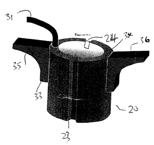
Figure 3 is an isometric view of a distal end of the sheath of Figure 1.
In the drawings like characters of reference indicate corresponding
parts in the different figures.
DETAILED DESCRIPTION
The apparatus of the present invention is mainly shown only
schematically as many of the components are well known to persons skilled in
this
art. In particular there is shown a part of the patient shown at 10 which is
located
during at least a part of the procedure in an MR imaging system 11 including a
magnet 12, RF coils 13 and a control and display system 14 for use in an
invasive
procedure for insertion of a conductive device into a patient while the
insertion is
guided using Magnetic Resonance Imaging. The conventional components further
include a stereotactic positioner 15 and a cannula 16 located by the
positioner 15 so
as to define a path for insertion of a conductive device to a target location.
The conventional components further include a conductive electrode
17 which in regard to DBS can be either a micro-recording electrode that is
temporarily positioned at the target or a stimulation electrode intended to be
located
at and remain in position at a target location in the brain of the patient.
The arrangement of the present invention comprises an elongate
sheath 20 extending from a first end 21 exposed outside the body of the
patient to a
distal end 22 located within the body of the patient 10 at a target location 1
0A.
CA 02655515 2009-02-25
8
The elongate sheath 20 is formed from a plastics material well known
for such invasive procedures and has a wall thickness of the order of 50
microns to
500 microns with an outside diameter of 1mm to 3mm and an interior hollow bore
of
the order of 0.5mm to 2.5mm diameter. The size is adjusted so that the
electrode
17 operates as a sliding fit.
The two ends of the sheath are shown in Figures 2 and 3. The sheath
is molded with at least one line of weakness and as shown there are two
thinner
lines or lines of weakness in the wall as shown at 23 and 24, each extending
along
the full length of the sheath. The thin lines 23 and 24 are formed along the
length of
the peripheral wall at which the sheath can be torn longitudinally to allow
the
peripheral wall to be opened to release engagement with the conductive
electrode
inserted therethrough.
As shown in Figure 3, the distal end of the stimulation electrode 17
includes a contact 26 for communication with surrounding tissue in the brain.
Typical DBS stimulating electrodes contain 5 contacts, the other 4 of which
are
inside the sheath and not visible in Figure 3. The proximal end of the
electrode
contains a connector 28, which connects to a lead extension that is tunneled
under
the skin to a control device (not shown). The control device is located in a
pocket
created near the patient's clavicle (collar bone) after the installation is
complete.
A temperature sensing device 30 is located embedded in the
peripheral wall at or adjacent the distal end and has a communication medium
in the
form of an optical fiber 31 extending longitudinally of the peripheral wall
from the
CA 02655515 2009-02-25
9
distal end to a position exposed from the patient for communicating the sensed
temperature to a control and display device shown schematically at 32.
The two parts 33 and 34 of the sheath defined on each side of the lines
of weakness 23 and 24 each have a manually engagable handle 35 and 36 at the
first end projecting outwardly to the side of the sheath for manually pulling
the two
parts apart.
As previously known, the temperature sensing device 30 includes a
fluorescent material forming a sensor at a position adjacent to but spaced
from an
end 30A of the optical fiber. The sensor in the optical fiber defined by the
fluorescent material is exposed to the tissue at a location 30B adjacent to
but
spaced from the end 30A of the sheath. The optical fiber is otherwise fully
enclosed
by the sheath material. The fluorescent material has a temperature dependent
response allowing the control device 32 to generate optical signals which are
transmitted and returned through the fiber with the return signal being
dependent
upon and indicative of the temperature.
In Figure 3 the end 30A of the fiber is located at the end of the sheath
with the sensor 30 spaced from the end. However the fiber may project beyond
the
end of the sheath so that the sensor portion 30B is located at or slightly
beyond the
end of the sheath to allow detection of the temperature of the tissue just
beyond or
directly at the end of the sheath where the temperature rise generated by the
conductor is most concentrated.
CA 02655515 2009-02-25
In the method for embedding a conductive device into a patient the
following steps are applied:
the stereotactic positioner is attached to the body of the patient and
adjusted until its trajectory guide is oriented along the desired trajectory;
5 the sheath with a stylet inserted therein is fed through the trajectory
guide along the desired trajectory until it reaches the target location, using
Magnetic
Resonance Imaging of the target location and the trajectory;
the stylet is removed and replaced with the electrode;
during the MRI, data relating to the temperature of tissue within the
10 body is obtained by the temperature sensing device and communicated to the
temperature display device;
when imaging has confirmed that the conductive element is indeed
located at the target location, the conductive element is fixed in place and
the sheath
is torn along the lines of weakness to allow the peripheral wall to be opened
to
release engagement of the sheath with the conductive device allowing the
sheath to
be split and removed by pulling on the handles to pull the sheath parts out of
the
body leaving the conductor in place.
The active temperature sensing region of the optical thermocouple is
approximately 0.5 mm back from the tip of the optical fiber. The sheath wall
thickness is in one example approximately 100 microns
The optical fiber can also protrude forward from the sheath so that the
temperature sensing region is at the tip of the sheath. The configuration
would be
CA 02655515 2009-02-25
11
desirable for some clinical purposes because the distance between the tip of
the
conductive element and the sensitive region of the sensor would be reduced. If
the
high temperature region is extremely localized this configuration may be
required.
The configuration where the entire optical fiber is encased within the sheath
is also a
desirable configuration because the minimal amount of tissue is disturbed.
Since various modifications can be made in my invention as herein
above described, and many apparently widely different embodiments of same made
within the spirit and scope of the claims without department from such spirit
and
scope, it is intended that all matter contained in the accompanying
specification shall
be interpreted as illustrative only and not in a limiting sense.
