Note: Descriptions are shown in the official language in which they were submitted.
CA 02655662 2013-07-16
WO 2008/011359 PCT/US2007/073572
Remote Sensing and Actuation of Fluid of Inner Ear
Field of the Invention
The present invention relates to implantable devices, and more particularly to
implantable
devices for mechanical and electrical stimulation and fluid delivery for the
inner ear.
Backeround Art
Fig. 1 shows the anatomy of a normal human car. A normal ear transmits sounds
through
the outer ear 101 to the eardrum 102, which moves the three bones of the
middle ear 103, which in
turn excites the cochlea 104. The cochlea, or inner ear, 104 includes an upper
channel known as
the scala vestibuli 105 and a lower channel known as the scala tympani 106,
which are connected
by the cochlear duct 107. In response to received sounds, the stapes, a bone
of the middle ear 103,
transmits vibrations via the fenestra ovalis, (oval window) 114, to the
perilymph (inner ear fluid)
of the cochlea 104. Vibrations in the inner ear fluid are dissipated out of
the fenestra rotunda
(round window) 115. As a result, the hair cells of the organ of Corti are
excited to initiate
chemical-electric pulses that are transmitted to the cochlear nerve 113, and
ultimately to the brain.
Some patients may have partially or completely impaired hearing for reasons
including:
long term exposure to environmental noise, congenital defects, damage due to
disease or illness,
use of certain medications such as aminoglycosides, or physical trauma.
Hearing impairment may
be of the conductive, sensory neural, or combination types.
There are several types of middle- and inner-ear implants that can restore a
sense of partial
or full hearing. Implants often include various electro-magnetic transducers
that may function as
an actuator, a sensor, and/or a switch. An example of an implant with an
electro-magnetic
actuator is a middle ear implant which mechanically drives the ossicular
chain, the three bones of
the middle car that mechanically connect the eardrum to the oval window.
Another example of an
CA 02655662 2013-07-16
= WO 2008/011359
PCT/1JS2007/073572
implant with an electro-magnetic actuator is a middle ear implant that
mechanically drives the
tympanic membrane.
Another type of implant relies on direct electrical stimulation of the nerves
in the inner ear.
For example, intra-cochlear electrodes can restore some sense of hearing by
direct electrical
stimulation of the neural tissue in proximity of an electrode contact. These
electrodes are typically
located on the end of an electrode carrier that is threaded into the cochlea.
The electrodes are
connected to, for example, an implanted signal processor which communicates
with an external
signal processor that produces an electrical stimulation signal for the
implanted electrodes to
stimulate the cochlear nerve.
In order to treat certain inner ear disorders, it is often necessary to
deliver therapeutic
agents directly into the cochlea. An example of a system for delivering
therapeutic agents to the
inner car is a catheter that is inserted into the cochlea via the round
window. The end of the
catheter might be infused with a therapeutic agent that is released into the
inner ear fluid. The
catheter might also include a fluid reservoir with a solution of the
therapeutic agent that is in fluid
communication with the inner ear fluid. Alternatively, the catheter might
include a fluid filled
lumen containing a solution of the therapeutic agent that is in fluid
communication with the inner
ear fluid. Delivery of therapeutic agents to the cochlea is described further
in U.S. Patent
No. 7,815,615.
Summary of the Invention
In an embodiment of the present invention, a system for communicating with the
inner ear
includes a acoustic transducer that converts between electrical energy and
mechanical energy. An
inner ear catheter has a distal end in vibratory communication with the fluid
of the inner car, a
proximal end in vibratory communication with the acoustic transducer, and a
lumen filled with a
catheter fluid fin- coupling vibratory signals between the distal end and the
proximal end.
In further such embodiments, there may also be a housing chamber enclosing the
acoustic
transducer and filled with a housing fluid in vibratory communication with the
proximal end of the
inner ear catheter. The acoustic transducer may be, for example, a floating
mass transducer.
The distal end of the lumen may be in fluid communication with the fluid of
the inner ear,
the proximal end of the lumen may be in fluid communication with the housing
fluid, and the
2
CA 02655662 2008-12-17
WO 2008/011359
PCT/US2007/073572
housing may further include a fluid port for receiving therapeutic fluid for
delivery to the inner
ear.
In an embodiment, a housing chamber may be filled with a housing fluid in
vibratory
communication with the proximal end of the catheter and may include an outer
housing membrane
in vibratory communication with the housing fluid. The acoustic transducer may
be located
outside the housing chamber in vibratory communication with the housing
membrane.
In such an embodiment, the distal end of the lumen may be in fluid
communication with
the fluid of the inner ear, the proximal end of the lumen may be in fluid
communication with the
housing fluid, and the housing may further include a fluid port for receiving
therapeutic fluid for
delivery to the inner ear.
Embodiments may include a microphone coupled to the housing membrane for
sensing
fluid mechanics associated with the auditory structures. The distal end of the
lumen may be in
fluid communication with the fluid of the inner ear. The lumen may include a
fluid port for
receiving therapeutic fluid for delivery to the inner ear. The distal end of
the lumen may include a
distal membrane in vibratory communication with the fluid of the inner ear.
The acoustic transducer may be adapted for use in the outer ear, the middle
ear, or the
inner ear of a user and/or may be adapted to be secured to the skull of a
user. The distal end of the
inner ear catheter may be adapted for use in the scala tympani of a user.
Any of the foregoing embodiments may also include an electronics module for
producing
an electrical stimulation signal for the inner ear, and an electrode array at
the distal end of the
inner ear catheter and in electrical communication with the electronics module
for stimulating
neural tissue of the inner ear with the electrical stimulation signal.
Brief Description of the Drawings
The foregoing features of the invention will be more readily understood by
reference to the
following detailed description, taken with reference to the accompanying
drawings
Fig. 1 shows the structure of the normal human ear.
Fig. 2A is a graphical illustration of an embodiment of the present invention.
Fig. 2B is a cut-away illustration of a catheter of the present invention.
Fig. 3 is a graphical illustration showing a transducer enclosed in a housing
chamber.
3
CA 02655662 2008-12-17
WO 2008/011359
PCT/US2007/073572
Fig. 4 is a graphical illustration showing a housing chamber having an
external membrane,
with the transducer in contact with the membrane.
Fig. 5 is a pictorial illustration of an embodiment of the present invention
showing a
catheter threaded into the cochlea.
Fig. 6 shows the structure of the normal human ear with an embodiment of the
present
invention implanted in the cochlea.
Detailed Description of Specific Embodiments
In the past, inner ear sensing devices and amplifiers have been brought into
the closest
feasible proximity to the structures of the inner ear. But this approach has
many problems and is
difficult to implement in practice. Embodiments of the present invention
dispose the device
structures within the user in more spacious and accessible locations not
directly adjacent to the
inner rear by using a catheter to establish fluid communication between the
inner ear and the
system devices. The catheter can be filled with a vibration transmitting
liquid, for example, by a
port and/or septum membrane. The distal end of the catheter penetrates the
inner ear and the
proximal end couples to an acoustic transducer. Enclosing the fluid within the
catheter isolates it
from the fluid of the inner ear to avoid leaks and prevent bacterial
contamination while providing
convenient mechanical access to the inner ear. The catheter may include a semi-
permeable
membrane at the distal end to provide pharmacological access by use of
therapeutic drugs adapted
to migrate across the membrane into the fluid of the inner ear. In some
embodiments, the proximal
end of the catheter may also be coupled to a self-sealing semi-permeable
septum membrane that
allows the therapeutic drugs to be introduced in the catheter fluid. For
example, the proximal end
membrane may be located in the middle ear or mastoid cavity for actuation or
sensing of the
catheter fluid. In some embodiments, the membranes may also usefully be
coupled to a
microphone which senses the fluid mechanics associated with the auditory
structures of the middle
and/or inner ear.
Thus, embodiments of the present invention provide a safe and convenient leak
proof and
bacterial resistant interface between an implanted prosthetic system and the
fluid of the inner ear.
Fig. 2A is a graphical illustration of one embodiment of the invention showing
a
transducer-catheter arrangement. Fig. 2B is a cut-away cross-section of a
portion of an inner ear
catheter. In this embodiment, an acoustic transducer 200 is connected to the
proximal end of an
4
CA 02655662 2008-12-17
WO 2008/011359
PCT/US2007/073572
inner ear catheter 202. Wiring 204 may connect the acoustic transducer 200 to
external circuitry.
A fluid port 206 provides access to a catheter lumen 210 within the inner ear
catheter 202. Inner
ear catheter 202 can also include an electrode wire 214 that runs along the
length of the catheter.
Acoustic transducer 200 converts electrical energy into mechanical vibrations,
and vice versa. For
example, acoustic transducer 200 may produce vibrations in the human auditory
range. Catheter
lumen 210 is filled with a catheter fluid 212 (for example via septum port
206), which can
transmit vibrations that are generated by the acoustic transducer 200 to the
fluid of the inner ear.
The acoustic transducer 200 is connected to the proximal end of the inner ear
catheter 202 such
that vibrations generated by the acoustic transducer 200 are transmitted into
the catheter fluid 212.
There is cooperation between the acoustic transducer 200, catheter lumen 210,
and catheter fluid
212 such that a sufficient and appropriate amount of mechanical energy is
generated by the
acoustic transducer 200 and is transmitted by the catheter fluid 212 to the
distal end of the catheter
and into the inner ear fluid to be detected as sound by the inner ear.
Alternatively, fluid movement
generated within the inner ear by stapes movement may be transmitted through
the catheter fluid
212 and detected by a sensitive membrane (e.g., a microphone diaphragm)
associated with the
acoustic transducer 200.
The catheter fluid 212 may be an artificial perilymph, or a physiological
saline when the
catheter lumen 210 is open to the fluid of the inner ear. If the distal end of
the inner ear catheter
202 is to be placed in the scala media, then the catheter fluid 212 may
usefully be an artificial
endolymph. The catheter fluid 212 may be any liquid that facilitates or
emphasizes mechanical
energy transmission. The inner ear catheter 202 may be at least partially in
the form of a channel
through a cochlear implant electrode. Or the inner ear catheter 202 may be a
separate catheter in
parallel with a cochlear implant electrode. The inner ear catheter 202 may be
made of an
incompressible material to optimize transmission through the fluid 212 with
minimal loss of
energy. The volume of the catheter fluid 212 may usefully be minimized in
order to maximize
transmission of mechanical movements in the catheter fluid between the distal
and proximal ends
of the inner ear catheter 200.
The catheter lumen 210 may be open ended to the inner ear fluid, or it may be
at least
partially closed by a sensitive membrane such as a bacterial filter. The
membrane may also
prevent protein transport from the inner ear fluid through the catheter 210,
and inhibit other
5
CA 02655662 2008-12-17
WO 2008/011359
PCT/US2007/073572
diffusion processes. The membrane may be self-sealing and/or semi-porous to
allow semi-
permeable access to therapeutic drugs.
Fig. 3 shows another transducer arrangement in which acoustic transducer 200
is inside a
housing chamber 300 that is filled with a fluid, and disposed such that
vibrations generated by
transducer 200 are transmitted to the chamber fluid. A septum port 302 with
septum can be used
for access to the fluid in housing chamber 300. The septum port 302 allows the
housing chamber
300 and inner ear catheter 202 to be filled with a liquid of chosen
composition. One challenge is to
be able to fill the inner ear catheter 202 with a catheter liquid for optimal
coupling between the
acoustic transducer 200 and the fluid of the inner ear, and also providing an
effective seal between
HI the middle ear and the inner ear. Inner ear catheter 202 connects to
housing chamber 300 so that
mechanical vibrations generated by the acoustic transducer 200 will be
transmitted through the
chamber fluid to the catheter fluid 212. The fluid in the housing chamber 300
may be in fluid
communication with the catheter fluid 212. Vibrations generated by the
acoustic transducer 200
are transmitted through the catheter fluid 212 to the inner ear fluid. In this
arrangement, the
acoustic transducer 200 may be, for example, a floating mass transducer such
as a vibrant FMT.
Fig. 4 shows another transducer arrangement also involving a housing chamber
300. As in
the embodiment of Fig. 3, inner ear catheter 202 connects to the housing
chamber 300 so that
mechanical vibrations will be transmitted through the chamber fluid to the
catheter fluid 212. A
septum port 302 can be used to fill the inner ear catheter 202 with the
catheter fluid 212 and to
provide access to the fluid in the housing chamber 300 through the port septum
302 The fluid in
housing chamber 300 may be in fluid communication with the catheter fluid 212.
In this
embodiment, housing chamber 300 includes a housing membrane 400 through which
vibrations
can be transmitted to the chamber fluid (Figure 4). Acoustic transducer 200 is
external to the
housing chamber 300, and is arranged and mounted with respect to the housing
membrane 400 so
that mechanical vibrations generated by the acoustic transducer 200 will be
transmitted through
the housing membrane 400 via the chamber fluid to the catheter fluid 212.
These vibrations are
then transmitted via the catheter fluid 212 through the distal end of the
catheter to the inner ear
fluid.
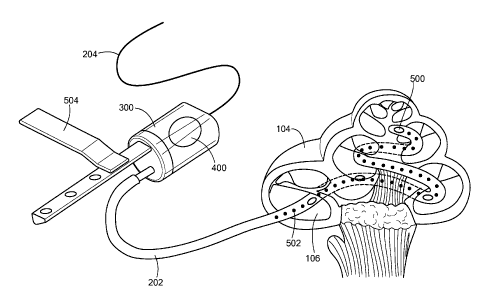
Fig. 5 is a pictorial illustration of a general embodiment of the present
invention showing
the inner ear catheter threaded into the cochlea 104 of a patient user. In
this embodiment, the
acoustic transducer 200 can be situated inside the housing chamber 300 as in
the embodiment of
6
CA 02655662 2008-12-17
WO 2008/011359
PCT/US2007/073572
Fig. 3. The acoustic transducer 200 can also be external to the housing
chamber 300 and mounted
against the housing membrane 400 as in the embodiment of Fig. 4. The housing
membrane 400
can also be used, for example, to monitor the output of the acoustic
transducer 200 when it is
situated inside the housing chamber 300. The housing membrane 400 can also be
of a selectively
porous material such that therapeutic agents may be introduced into the
housing fluid for delivery
via the catheter fluid 212 to the inner ear. A mounting bracket 504 is shown
that can be used to
mount the acoustic transducer 200 to another assembly, or, in another
configuration, directly to the
bone (such as the skull) or other structures in the ear. In the embodiment
shown, the inner ear
catheter 202 also includes catheter membranes 500 and an electrode array 502.
The catheter
membranes 500 transmit the vibrations of the acoustic transducer 200 from the
catheter fluid 212
to the inner ear fluid. In other embodiments, the catheter membranes 500 might
be open ports or
selectively porous membranes that allow therapeutic agents within the catheter
fluid 212 to be
delivered to the inner ear fluid. The electrode array 502 is connected to an
electrode wire 214 and
is used for electrical stimulation of the neural tissue of the inner ear. In
such an arrangement, the
electrode wire 214 may be connected to an implanted audio processor under the
skin of a user near
the outer ear.
Fig. 6 shows the structure of an ear along with an embodiment of the present
invention
implanted in the cochlea. The inner ear catheter 202 is threaded into the
scala tympani 106 of the
cochlea 104 via the round window 115. The acoustic transducer 200 is shown
within the middle
ear. Wiring 204 can be used to connect the acoustic transducer 200 and the
electrode array 502 to
other circuitry. For example, the electrode array 502 may be connected via the
wiring 204 to an
implanted audio processor 600 located under the skin near the outer ear. An
audio processor 600
receives an audio signal and produces an electrical stimulation signal that is
transmitted to the
electrode array 502 via the wiring 204 for electrical stimulation of the
neural tissue of the inner
ear. The audio processor 600 contains electronic components for accepting an
audio input from an
audio source. In various embodiments, the audio processor 600 will accept
analog signals, digital
signals, or both. The audio input may be, but is not limited to, an analog or
digital output from a
microphone, telephone, television, stereo system, mp3 player, radio receiver,
or computer. The
audio input may be accepted via wired or wireless connection.
While the inventive system has been particularly shown and described, it is
not intended to
be exhaustive nor to limit the invention to the embodiments disclosed. It will
be apparent to those
7
CA 02655662 2013-07-16
WO 2008/011359 PCT/US2007/073572
skilled in the art that modifications can be made to the present invention
without departing from
the scope and spirit thereof. For example, while the embodiments shown have
generally described
a system to transmit vibrations produced by a transducer to the inner ear, the
transducer can also
be used to detect vibrations in the inner ear fluid via the catheter fluid.
While the embodiments
shown include wire for connecting various components, the wire is optional.
This connection may
be wireless, or the components may be optional. The scope of the claims should
not be limited
by the preferred embodiments or the examples, but should be given the broadest
interpretation
consistent with the description as a whole.
8