Note: Descriptions are shown in the official language in which they were submitted.
CA 02656192 2008-12-23
WO 2008/005310
PCT/US2007/015089
DETECTABLE NUCLEIC ACID TAG
FIELD
[0001] The subject matter provided herein relates to nucleic acid tags that
are linked to, or
capable of linking to, a protein of interest. In particular, the present
subject matter provided
herein relates to oligonucleotides comprising a reporter function and a
protein tagging
function. Also provided herein, are nucleic acid tag compositions, kits and
methods of use
thereof.
BACKGROUND
=
[0002] Traditional techniques for quantifying and detecting the presence of
proteins include
gel electrophoresis, Western blotting, ELISA based immunoabsorbent assays and
protein
microarrays. Each of these methods are cumbersome and not amenable to high-
throughput
Use. These traditional methods also suffer limitations in detection
sensitivity and specificity.
Provided herein is a nucleic acid tag and a new, highly sensitive and
selective method of
protein detection using the nucleic acid tags.
=
SUMMARY
[0003] Provided herein is a nucleic acid tag that is linked to or capable of
linking to a protein,
which allows the protein to be detected with a high degree of sensitivity. In
one embodiment,
the nucleic acid tag is an oligonucleotide having a reporter function and a
protein tagging
function. In one embodiment, the oligonucleotide (oligomer) is an
oligonucleotide, which
comprises a first nucleic acid sequence that is a PCR amplification sequence
(an amplicon)
recognizable by a PCR probe and a second nucleic acid sequence, which
covalently links,
non-covalently links, complexes or otherwise binds (e.g., binds to or is
capable of binding to)
a protein of interest. In certain embodiments, the amplicon is a randomly
generated, non-
naturally occurring PCR amplification sequence. In one embodiment, the first
nucleic acid
sequence and/or second nucleic acid sequence is not endogenous to a living
organism. In
CA 02656192 2008-12-23
WO 2008/005310 PCT/US2007/015089
other embodiments, first nucleic acid sequence and/or second nucleic acid
sequence is
endogenous to a living organism. In certain embodiments, the first nucleic
acid sequence and
the second nucleic acid sequence are heterologous. As used herein, if two
nucleic acid
sequences are "heterologous," it is meant that the first and second nucleic
acid sequence are
not normally found together. For example, in certain embodiments, the first
and second
nucleic acids do not encode the same protein and/or are not derived from the
same organism.
In some embodiments, the first sequence is a naturally occurring sequence and
the second
sequence is a naturally occurring sequence, wherein the first and second
sequences differ. In
specific embodiments, the first nucleic acid sequence is a nucleic acid
sequence, such as a
synthetic and/or randomly generated nucleic acid sequence, such as a non-
naturally occurring
sequence (e.g., one that is divergent from any naturally occurring sequence).
In certain
embodiments, the first nucleic acid sequence is a nucleic acid sequence, such
as a synthetic
and/or randomly generated nucleic acid sequence, that is not, for example,
found in protein of
interest, fusion protein, nucleic acid-interacting motif, and/or vectors used
in a screening
assay provided herein. In some embodiments, the first nucleic acid sequence is
a nucleic acid
sequence, such as a synthetic and/or randomly generated nucleic acid sequence,
that is not
present in the human kinome, such as when the nucleic acid tag is to be used
in a kinase assay
provided herein (or any other nucleotide sequence used in the given assay).
These
embodiment ensures, for example, that primers used for subsequence PCR
amplification do
not cross react or misprime to a second DNA sequence and/or to any other
(e.g., naturally
occurring) DNA sequence, such as those being used in a given assay. In certain
embodiments, each PCR template is different from the others so that there is
no chance of
primers cross-reacting between templates, such as when used in the multiplex
assays
provided herein.
[0004] In another embodiment, the oligonucleotide comprises a first nucleic
acid sequence
comprising a PCR amplification sequence and a second nucleic acid sequence
comprising a
nucleic acid sequence which is a target sequence for and binds a nucleic acid
interacting
motif. In one example, the target sequence is a recognition sequence for
either a naturally-
occurring or synthetic DNA-binding protein. In specific embodiments, the first
nucleic acid
sequence comprising the PCR amplification sequence is separate and distinct
from the second
nucleic acid comprising the nucleic acid-interacting motif. In such
embodiments, the nucleic
acid tag is capable of binding or otherwise linking to a protein of interest
having a DNA-
binding component specifically recognizing the nucleic acid tag. The nucleic
acid tag may
- 2 -
CA 02656192 2008-12-23
WO 2008/005310 PCT/US2007/015089
then be detected and/or quantified using, e.g., quantitative PCR (qPCR).
Nucleic acid tag
detection by qPCR has the advantage of being not only a reliable quantitative
detection
method but also a highly sensitive and highly selective detection method.
Because of the
highly sensitive nature of the qPCR detection method, this method enables the
detection of
very small amounts of the target protein and reduces the need for scarce and
expensive assay
components, such as recombinant proteins. Because of the highly specific
nature of the qPCR
detection method, qPCR also enables the detection of specific DNA sequences in
complex
heterogeneous mixtures, and obviates the need for any sort of purification
steps normally
done to protein samples to either improve or enhance protein detection.
[0005] The nucleic acid tag provided herein may also be labeled, such as
radiolabeled,
fluorescently labeled or biotinylated. In certain embodiments, provided herein
is a nucleic
acid oligomer that binds a nucleic acid-interacting motif, wherein the nucleic
acid oligomer
comprises (a) a first radiolabeled, fluorescently labeled or biotinylated
nucleic acid sequence,
and (b) a second nucleic acid sequence that binds the nucleic acid-interacting
motif. In other
embodiments, provided herein is a nucleic acid oligomer comprising a nucleic
acid sequence
that binds a nucleic acid-interacting motif, wherein the oligomer is
radiolabeled, fluorescently
labeled or biotinylated. The labeled tags, such as radiolabeled or
fluorescently labeled tags,
may, for example, be used to detect the presence or locality of a protein of
interest in cellular
imaging or in visualization assays. The labeled tags, such as fluorescently
labeled tags, may
also, for example, be used in sorting assays to separate out one or more
proteins of interest
into individual samples. The labeled tags, such as biotinylated tags, also
permit, for example,
the detection of the protein of interest by immunological methods or the
purification of the
labeled protein of interest by affinity chromatography. In certain embodiments
when the
nucleic acid tag is labeled, the nucleic acid tag may or may not also comprise
a PR
amplification sequence.
[0006] Also provided herein is a protein of interest, which is linked or
otherwise complexed
to a nucleic acid tag or capable of linking or otherwise complexing to the
nucleic acid tag,
and which is therefore detectable when, for example, its function, activity or
presence is
being studied or monitored. In one example, the protein of interest is a
chimeric protein
fused to a nucleic acid interacting motif. In one example, the nucleic acid
interacting motif is
a DNA-binding domain. Such a protein of interest may be tagged by a nucleic
acid having a
target sequence that can be recognized by a DNA-binding domain. The chimeric
protein may
- 3 -
CA 02656192 2008-12-23
WO 2008/005310 PCT/US2007/015089
=
be an expressed nucleotide sequence generated by random mutation, an expressed
nucleotide
sequence containing systematically synthesized sequences, an expressed cDNA,
or a
combination of two or more of these possibilities. The protein of interest may
be cloned and
then expressed in an appropriate host cell, such as a bacterial, insect,
mammalian or plant
host cell. In certain embodiments, the host cell gives the protein the benefit
of any post-
translational modifications that may be important for its three dimensional
structure and
function (e.g., glycosylation or prenylation of the protein of interest in a
human host cell).
[0007] Also provided herein is a method of detecting binding between a protein
of interest
and a second molecule, using a nucleic acid tag to label and detect the
protein. In certain
embodiments, the method comprises screening a library of test compounds for
their ability to
bind to a protein of interest, wherein the binding is identified by the
detection of the nucleic
acid tag. In other embodiments, the method comprises competition binding
assays to screen
for and determine the identity of one or more test compounds, which
competitively bind to a
protein of interest in the presence of an immobilized reference ligand (or
"bait") that is
known to bind to the protein of interest. Such a competitive binding assay
allows the
identification of alternative compounds which bind to the protein of interest
in addition to (or
preferentially to) the known reference ligand.
[0008] Also provided herein is a method comprising screening a test compound
against a
panel of proteins of interest for the ability of the test compound to bind to
one or more
proteins in the panel and/or to generate a binding specificity profile for
that compound.
Where the screening is performed against a panel of proteins, in some
embodiments, the
screening is done in a multiplexed format, such as by simultaneously testing
the activity of a
test compound against a pooled sample containing multiple proteins of
interest, and/or at the
detection step by using multiple nucleic acid tags that are each unique for a
specific protein of
interest.
[0009] Also provided herein is a kit comprising one or more of the following
elements: a
detectable nucleic acid tag, a protein capable of being "tagged" by the
nucleic acid tag, an
immobilized reference ligand that binds to the protein of interest, and a PCR
primer pair
capable of initiating amplification of the nucleic acid tag. Such a kit may be
used to identify
molecules that bind to the immobilized reference ligand and/or that compete
with the
immobilized ligand for binding to the protein of interest. Alternatively, the
kit may be used
- 4 -
CA 02656192 2016-01-12
as a diagnostic tool for detecting in a given specimen the presence of a
molecule that binds to
the immobilized reference ligand.
[0009a] In another embodiment of the present invention there is provided a
nucleic acid
oligomer bound to a fusion protein, wherein the fusion protein comprises: (a)
a first domain
comprising a protein of interest, and (b) a second domain comprising a nucleic
acid-
interacting motif, which is a NF-KB binding domain; and wherein the nucleic
acid oligomer
comprises: (a) a first nucleic acid sequence that is a PCR amplification
sequence, and (b) a
second nucleic acid sequence that binds the nucleic acid-interacting motif,
wherein the first
nucleic acid sequence is heterologous to the second nucleic acid sequence.
[0009b] In another embodiment of the present invention there is provided a
nucleic acid
oligomer bound to a fusion protein, wherein the fusion protein comprises: (a)
a first domain
comprising a protein of interest, and (b) a second domain comprising a nucleic
acid-
interacting motif, which is a NF-K13 binding domain; and wherein the nucleic
acid oligomer
comprises a nucleic acid sequence that binds the nucleic acid-interacting
motif, and wherein
the oligomer is radiolabeled, fluorescently labeled or biotinylated.
[0009c] In a further embodiment of the present invention there is provided a
method for
identifying that a protein of interest binds to a ligand, comprising: (a)
contacting the ligand,
which is immobilized on a solid support, with a fusion protein comprising (i)
a first domain
comprising the protein of interest, and (ii) a second domain comprising a
nucleic acid-
interacting motif, wherein the protein of interest and the nucleic acid-
interacting motif differ
from each other; (b) adding to said fusion protein a chimeric nucleic acid
oligomer
comprising (i) a first nucleic acid sequence that is a PCR amplification
sequence, and (ii) a
second nucleic acid sequence that binds to and recognizes the nucleic acid-
interacting motif
in said second domain of said fusion protein, wherein the first nucleic acid
sequence is
heterologous to the second nucleic acid sequence, thereby forming a
composition comprising
a chimeric nucleic acid oligomer bound to a fusion protein; (c) removing
nucleic acid
oligomer unbound to the fusion protein in step (b); and (d) detecting whether
the
composition comprising the chimeric nucleic acid oligomer is bound to the
ligand, after the
nucleic acid oligomer is removed in step (c); whereby detection of bound
nucleic acid
oligomer indicates that the protein of interest binds to the ligand.
- 5 -
CA 02656192 2016-01-12
[0009d] In a further embodiment of the present invention there is provided a
kit for
identifying a test compound that binds to a protein of interest, comprising:
(a) a fusion
protein comprising (i) a first domain comprising the protein of interest, and
(ii) a second
domain comprising a nucleic acid-interacting motif, wherein the protein of
interest and the
nucleic acid-interacting motif differ from each other; (b) a reference ligand,
which binds to
the protein of interest; and (c) a nucleic acid oligomer comprising a first
nucleic acid
sequence that binds the nucleic acid-interacting motif of the fusion protein,
wherein the
chimeric nucleic acid oligomer comprises (i) a first nucleic acid sequence
that is a PCR
amplification sequence, and (ii) a second nucleic acid sequence that binds to
and specifically
recognizes the nucleic acid-interacting motif, wherein the first nucleic acid
sequence is
heterologous to the second nucleic acid sequence.
BRIEF DESCRIPTION OF THE DRAWINGS
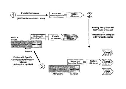
[0010] FIG. 1 is a schematic diagram depicting a competitive binding assay
using a nucleic
acid tag containing a PCR-amplifiable DNA sequence.
[0011] FIG. 2 provides a binding curve with calculated Kas for p38 interaction
with known
kinase inhibitors BIRB-796, SB202190 and VX-745. SB202190 was used as
immobilized
reference ligand, and the nucleic acid tag used was a fusion comprising a GAL4
target DNA
sequence and a PCR-amplifiable DNA sequence.
[0012] FIG. 3 provides a binding curve with calculated Kds for p38 interaction
with known
kinase inhibitors BIRB-796, SB202190 and VX-745. SB202190 was used as
immobilized
bait, and the nucleic acid tag used was a fusion comprising a NF-KB target DNA
sequence
and a PCR-amplifiable DNA sequence.
[0013] FIG. 4 provides a binding curve with calculated Kas for BRAF
interaction with four
internal proprietary compounds. Three of the compounds, A, B and C are kinase
inhibitors
and one of the compounds which is not a kinase inhibitor, served as negative
control. The
interaction was detected using a nucleic acid tag comprising a GAL4 target DNA
sequence
and a PCR-amplifiable DNA sequence.
[0014] FIG. 5 provides a binding curve with calculated Kas for BRAF
interaction with four
internal proprietary compounds. Three of the compounds, A, B and C are kinase
inhibitors
and one of the compounds which is not a kinase inhibitor, served as negative
control. The
- 5a -
CA 02656192 2016-01-12
interaction was detected using a nucleic acid tag comprising a NF-i<13 target
DNA sequence
and a PCR-amplifiable DNA sequence.
[0015] FIGS. 6A-6B show binding curves with calculated Kds for interactions
between the
two forms of Abl (active and inactive) with (A) VX-680 or (B) imatinib. The
interaction was
detected using a nucleic acid tag comprising a NF-KB target DNA sequence and a
PCR-
amplifiable DNA sequence.
- 5b -
CA 02656192 2008-12-23
WO 2008/005310 PCT/US2007/015089
DETAILED DESCRIPTION
[00161 The following embodiments provided herein are exemplary and are not
limitations.
The methods disclosed herein have a range of applications, all of which are
based on the
ability to detect, quantify, or isolate a protein of interest that is tagged
by a detectable nucleic
acid. The compositions and methods provided herein may be used to label
proteins in vitro
and/or in vivo.
[00171 In some embodiments, provided herein is a nucleic acid oligomer (tag)
that binds a
nucleic acid-interacting motif, wherein the nucleic acid oligomer comprises
(a) a first nucleic
acid sequence that is a PCR amplification sequence, and (b) a second nucleic
acid sequence
that binds the nucleic acid-interacting motif, wherein the first nucleic acid
sequence is
heterologous to the second nucleic acid sequence.
[00181 In one embodiment, the length of the nucleic acid oligomer is between
about 50 and
about 100, about 50 and about 200, about 50 and about 300, about 50 and about
400, about 50
and about 500, about 100 and about 200, about 100 and about 300, about 100 and
about 400,
about 100 and about 500, about 200 and about 300, about 200 and about 400,
about 200 and
about 500, about 300 and about 400, about 300 and about 500, or about 400 and
about 500
nucleotides in length.
[00191 As used herein, the term "about" or "approximately" means within 20%,
preferably
within 10%, and more preferably within 5% (or 1% or less) of a given value or
range.
[00201 In some embodiments, the nucleic acid tag has a reporter function and a
protein
tagging function. As used herein, a "reporter" function with reference to a
nucleic acid tag is
the ability to be visualized or otherwise detected or quantitated. In certain
embodiments, the
reporter function of a nucleic acid tag comes from the radiolabeling,
fluorescent labeling or
biotinylation of the nucleic acid tag. As used herein, a "nucleic acid tag" is
a polynucleotide,
e.g., an oligomer, that binds or is capable of binding to a protein of
interest, such as a protein
(e.g., a kinase) fusion comprising a heterologous polynucleotide-binding
domain (also called
a polynucleotide-interacting motif herein), such as a DNA-binding domain
(e.g., NFKB). The
nucleic acid tag may be single- or double-stranded DNA, single- or double-
stranded RNA,
DNA-RNA hybrid, RNA-RNA hybrid, or their native or synthetic derivatives,
analogs and
fragments thereof. In some embodiments, the nucleic acid tag is DNA, and the
reporter
function label can be introduced to the DNA, for example, by any standard
enzymatic
- 6 -
CA 02656192 2008-12-23
WO 2008/005310 PCT/US2007/015089
reaction, such as nick translation, or by terminal labeling, with 32P, 125I or
biotin-labeled =
deoxynucleotide triphosphates (dNTPs), or the label can be introduced as an
intercalating
agent. There are many fluorescent groups that are commercially available and
can be used to
label the nucleic acid tag. Some examples of fluorescent labels that can be
used to label the
nucleic acid tag are fluorescein isothiocyante, rhodamine and coumarin and
their commercial
derivatives such as Texas Red and Alexa Fluor .
[0021] In certain embodiments, the nucleic acid tag is complexed, covalently
linked or non-
covalently linked to a detectable protein or polypeptide, for example, by a
covalent linkage.
Nucleic acid-protein fusions can be produced by any method, for example, by
the method of
Roberts and Szostalc (U.S. Pat. Nos. 6,258,558 and 6,261,804; WO 98/31700;
Roberts &
Szostak (1997) Proc. Natl. Acad. Sci. USA (1997) 94:12297-12302) using a
peptide acceptor,
such as puromycin, as a covalent linking agent. Briefly, such an exemplary
method comprises
an in vitro or in situ transcription/translation protocol that generates
protein covalently linked
to the 3' end of its own mRNA, i.e., an RNA-protein fusion. This is
accomplished by
synthesis and in vitro or in situ translation of an mRNA molecule with a
peptide acceptor
attached to its 3' end. In specific embodiments, the peptide acceptor is
puromycin, a
nucleoside analog that adds to the C-terminus of a growing peptide chain and
terminates
translation. In one embodiment, a DNA sequence is included between the end of
the message
and the peptide acceptor which is designed to cause the ribosome to pause at
the end of the
open reading frame, providing additional time for the peptide acceptor (for
example,
puromycin) to accept the nascent peptide chain before hydrolysis of the
peptidyl-tRNA
linkage.
[0022] As used herein, a "peptide acceptor" is any molecule capable of being
added to the C-
terminus of a growing protein chain by the catalytic activity of the ribosomal
peptidyl
transferase function. In certain embodiments, such molecules contain (i) a
nucleotide or
nucleotide-like moiety (e.g., adenosine or an adenosine analog (di-
methylation at the N-6
amino position is acceptable)), (ii) an amino acid or amino acid-like moiety
(for example, any
of the 20 D- or L-amino acids or any amino acid analog thereof (for example, 0-
methyl
tyrosine or any of the analogs described by Ellman et al., (1991) Meth.
Enzymol. 202:301),
and (iii) a linkage between the two (e.g., an ester, amide, or ketone linkage
at the 3' or 2'
position); preferably, this linkage does not significantly perturb the pucker
of the ring from
the natural ribonucleotide conformation. Peptide acceptors may also possess a
nucleophile,
- 7 -
CA 02656192 2008-12-23
WO 2008/005310 PCT/US2007/015089
which may be, without limitation, an amino group, a hydroxyl group, or a
sulfhydryl group.
In addition, peptide acceptors may be composed of nucleotide mimetics, amino
acid
mimetics, or mimetics of the combined nucleotide-amino acid structure. By a
peptide
acceptor being positioned "at the 3' end" of a protein coding sequence is
meant that the
peptide acceptor molecule is positioned after the final codon of that protein
coding sequence.
This term includes, without limitation, a peptide acceptor molecule that is
positioned
precisely at the 3' end of the protein coding sequence as well as one which is
separated from
the final codon by intervening coding or non-coding sequence (for example, a
sequence
corresponding to a pause site). This term also includes constructs in which
coding or non-
coding sequences follow (that is, are 3' to) the peptide acceptor molecule. In
addition, this
term encompasses, without limitation, a peptide acceptor molecule that is
covalently bonded
(either directly or indirectly through intervening nucleic acid sequence) to
the protein coding
sequence, as well as one that is joined to the protein coding sequence by some
non-covalent
means, for example, through hybridization using a second nucleic acid sequence
that binds at
or near the 3' end of the protein coding sequence and that itself is bound to
a peptide acceptor
molecule.
[0023] In addition to covalently bonded RNA-protein fusions, any other unique,
PCR-
amplifiable nucleic acid (for example, RNA, DNA, PNA, or any other nucleic
acid which
includes two or more covalently bonded, naturally-occurring or modified
ribonucleotides or
deoxyribonucleotides) can be coupled covalently or non-covalently to a
detectable protein or
polypeptide. The protein portions of the fusions are typically composed of
naturally-
occurring amino acid residues, but may also include amino acid analogs or
derivatives, joined
by peptide or peptoid bond(s).
[0024] In other embodiments, the reporter function of a nucleic acid tag is a
nucleic acid
sequence that is amplifiable by PCR (also referred to herein as an
"amplicon"). The
amplifiable sequence hybridizes or is capable of hybridizing to a PCR primer
in a sequence-
specific manner. In certain embodiments, the nucleic acid tag comprises a
plurality of
amplicons, for example, two, three, four, five, six, seven, eight, nine, ten
or more amplicons.
In some embodiments, the plurality of amplicons are tandem repeats of a single
amplicon. In
certain embodiments, the amplicon is amplifiable by quantitative PCR which
permits
quantification of the protein tagged by such a nucleic acid tag. In a specific
amplification
method, amplification of a PCR sequence includes combining the nucleic acid
containing the
- 8 -
CA 02656192 2008-12-23
WO 2008/005310
PCT/US2007/015089
PCR amplification template, PCR primer and qPCR probe in a standard PCR
reaction
mixture (generally, a mixture having a final concentration of 10 mM Tris-HC1
(pH 8.3 at
25 C), 1-4 mM MgC12, 0.1-1 mM dNTP), and treating the sample first under Hot
Start
conditions (for example, heating to 95 C for 5 minutes) to minimize
nonspecific annealing or
mispriming, followed by a denaturation step (for example, 95 C for 45
seconds), followed by
an annealing step (55 C for 1 minute), and followed by an extension step (72 C
for 1
minute), with up to forty rounds of the consecutive steps of denaturation,
annealing and
extension, to complete the amplification of the qPCR signal.
[0025] As used herein, a "protein tagging" function with reference to a
nucleic acid tag is the
ability to target and bind, complex, or otherwise link (e.g., covalently or
non-covalently) to a
nucleic acid-interacting motif, such as a fusion protein comprising (a) a
protein of interest
(e.g., a kinase) and (b) a heterologous polynucleotide-interacting motif, such
as a DNA-
binding protein (e.g., NFKB), which comprises a nucleic acid recognition
sequence. The
nucleic acid-interacting motif of the fusion protein binds to a nucleic acid
oligomer described
elsewhere herein.
[0026] In one embodiment, the target DNA sequence is a transcription factor
binding site
recognizable by the DNA binding domain of a transcription factor. For example,
the nucleic
acid tag may contain target DNA sequences recognized by DNA-binding domain of
transcription factors such as cro repressor, lac repressor, GAL4, GCN4, Lex-
A,
Opaque-2 and TGAla. In one embodiment, the transcription factor binding site
is a naturally
occurring or wildtype sequence. In another embodiment, the transcription
factor binding site
is a mutant sequence. In another embodiment, the transcription factor binding
site may be
characterized by a consensus sequence that encompasses wildtype sequences and
optionally,
mutant sequences. In yet another embodiment, the transcription factor binding
site is a
synthetic or genetically engineered sequence capable of forming a complex with
either a
naturally occurring, modified or synthetic DNA binding protein. In yet another
embodiment,
the target DNA sequence is characterized by having palindromic sequences
usually
recognized by protein dimers. The target sequence for Ga14 or LexA are two
such examples.
In yet another embodiment, the transcription factor binding site is
characterized by having a
GC rich region such as the target site for the transcription factor Spl. In
another
embodiment, the transcription factor binding site is characterized by having a
DNA-protein
- 9 -
CA 02656192 2008-12-23
WO 2008/005310 PCT/US2007/015089
complex half-life of more than one, two, three, four, five or six hours with
its associated
DNA binding protein.
[0027] A fusion protein provided herein comprising a protein of interest and a
nucleic acid-
interacting motif, such as a DNA-binding protein may therefore by "tagged" by
the nucleic
acid oligomer provided herein through, for example, a DNA-protein complex
formation. In
certain embodiments, the fusion protein comprising a nucleic acid-interacting
motif and a
protein of interest are derived from the same organism, such as a human. In
one particular
embodiment, the nucleic acid tag comprises an amplicon linked to a target DNA
sequence
specifically recognizable by a DNA-binding protein (e.g., NFicB, cro
repressor, GAL4,
GCN4, LexA, Opaque-2 and TGAla). In another embodiment, the nucleic acid tag
comprises an amplicon linked to the cognate DNA sequence for the DNA-binding
domain of
a transcription factor. Cognate DNA sequences for such DNA-binding domains are
known in
the art, and exemplary sequences are provided in Table 1.
[0028] In other embodiments, a protein tagging function of a nucleic acid tag
is a target DNA
sequence recognized by DNA metabolizing enzyme, such as a methyltransferase,
alkyltransferase and/or glycosydase. These enzymes can interact with
chemically-modified
DNA bases and create a covalent bond between an amino acid of the protein and
the DNA
sequence of the nucleic acid tag. For example, if the protein fusion contained
a functional
fragment of an 06-alkylguanine-DNA alkyltransferase (AGT), the
alkyltransferase function
can be used to transfer the nucleic acid tag attached either to an 06-
alkylguanine or an 06-
benzylguanine to the AGT fusion protein to create a covalent linkage between
the nucleic
acid tag and the fusion protein to form a nucleic acid-protein complex (See,
e.g., PCT
Application No. W002/083937). OGT can be used to label, and optionally
subsequently
manipulate and/or detect a protein of interest in a system in which a fusion
of the protein and
AGT is contacted with a labeled substrate so that the AGT transfers the label
from the
substrate to the AGT fusion, thereby allowing the labeled AGT-protein fusion
to be
manipulated and or detected by virtue of the transferred label. The label part
of the substrate
can be chosen by those skilled in the art dependent on the application for
which the fusion
protein is intended. Non-inclusive examples of labels include: (1) a
spectroscopic probe such
as a fluorophore, a chromophore, a magnetic probe or a contrast reagent; (2) a
radioactively
labeled molecule; (3) a molecule which is one part of a specific binding pair
which is capable
of specifically binding to a partner. Such specific binding pairs are well
known in the art and
- 1 0 -
CA 02656192 2008-12-23
WO 2008/005310 PCT/US2007/015089
include, for example, biotin, which can bind to avidin or streptavidin; (4) a
molecule that are
suspected to interact with other biomolecules; (5) a library of molecules that
are suspected to
interact with other biomolecules; (6) a molecule which is capable of
crosslinking to other
biomolecules as known to those skilled in the art (see, e.g., Nadeau et al.
(2002) in Protein-
Protein interactions: a molecular cloning manual; Ed. E Golemis, Cold Spring
Harbor
Laboratory Press; pp. 75-92); (7) A molecule which is capable of generating
hydroxyl
radicals upon exposure to 11202 and ascorbate such as a tethered metal-chelate
(see, e.g., Hon
et al. (2002) in Protein-Protein interactions: a molecular cloning manual; Ed.
E Golemis,
Cold Spring Harbor Laboratory Press; pp. 288-311) (8) a molecule which is
capable of
generating reactive radicals upon irradiation with light such as malachite
green (see, e.g., Jay
eta?. (1999) Biochim. Biophys. Acta M39-48); (9) a molecule covalently
attached to a solid
support, where the support may be a glass slide, a microtiter plate or any
polymer in general
known to those proficient in the art; (10) a nucleic acid or a derivative
thereof capable of
undergoing base-pairing with its complementary strand; (11) a lipid or other
hydrophobic
molecule with membrane-inserting properties; (12) a biomolecule with desirable
enzymatic,
chemical or physical properties; or (13) a molecule possessing a combination
of any of the
properties listed above.
[00291 As used herein, a "protein of interest" can be any conceivable
polypeptide or protein
that may be of interest, such as to study or otherwise characterize. In some
embodiments, the
protein of interest is a transferase, oxidoreductase, hydrolase, ligase,
isomerase or lyase. In
one embodiment, the protein of interest is a human polypeptide or protein. In
certain
embodiments, the protein of interest is a transferase having transferase
activities, such as an
acyltransferase, glycosyltransferase, amidotransferase or sulfurtransferase.
In another
embodiment, the protein of interest is a hydrolase, peptidase, protease or
phosphatase.
[00301 In certain embodiments, the kinase is a lipid kinase, such as a lipid
kinase of the P13K
family (e.g., mTOR). In specific embodiments, the protein of interest is a
protein kinase (see,
e.g., Manning (2002) Science 298:1912). In specific embodiments, the protein
of interest is a
tyrosine kinase, or a serine/threonine kinase. In some embodiments, the
protein of interest is
a human non-receptor tyrosine kinase, for example, a non-receptor tyrosine
kinase that is a
member of the ABL, ACK, CSK, MATK, FAK, PYK2 , FES, FRK, JAK, SRC-A, SRC-B,
TEC, and/or SYK families. In other embodiments, the protein of interest is a
human receptor
tyrosine kinase, for example, a receptor tyrosine kinase that is member of the
ALK, AXL,
-11-
CA 02656192 2008-12-23
WO 2008/005310 PCT/US2007/015089
DDR, EGFR, EPH, FGFR, INSR, MET, MUSK, PDGFR, PTK7, RET, ROR, ROS, RYK,
TIE, TRK, VEGFR, AATYK, and/or SuRTK106 families.
[0031] In some embodiments, a protein of interest is a transmembrane protein,
such as a 7-
transmembrane helix protein, such as a G-protein coupled receptor (GPCR). A
protein of
interest may also be transmembrane ion channel protein, and in certain
embodiments, a
ligand gated ion channel protein. In other embodiments, a protein of interest
is a nuclear
hormone receptor protein, such as a classic steroid hormone receptor and/or a
receptor in the
orphan class of nuclear hormone receptors.
[0032] In yet other embodiments, a protein of interest is an extracellular
signaling molecule
or factor, such as a cytokine (e.g., an interferon and/or an inierleulcin),
growth factor, and/or
hormone (e.g., insulin, glucagon or prostaglandins). In certain embodiments, a
protein of
interest is a protein involved in intracellular signal cascades, such as an
enzyme or cofactor
involved in phosphatidinyl-inositol signaling, cAMP, or cGMP generation.
[0033] In some embodiments, a protein of interest is an antibody, small chain
variable
fragment (scFv), antigen or epitope.
[0034] The protein of interest can, in some embodiments, be the expression of
a nucleotide
sequence generated by random mutation, the expression of a nucleotide sequence
containing
systematically synthesized sequences, or it may be an expressed cDNA. In one
example, the
protein of interest being studied or characterized is derived from a human
cDNA library (i.e.,
a human protein).
=
[0035] In certain embodiments, the protein of interest is a chimeric fusion
between a protein
of interest and a heterologous DNA-binding protein. In such chimeric fusions,
at least two
gene sequences representing each half of the chimera can be fused in-frame,
cloned into the
appropriate vector and expressed in a host cell of choice. In certain
embodiments, the protein
of interest is 5' of the nucleotide-binding domain (e.g., DNA-binding
protein). In other
embodiments, the protein of interest is 3' of the nucleotide-binding domain
(e.g., DNA-
binding protein). In specific embodiments, the protein of interest and/or the
nucleotide-
binding domain (e.g., DNA-binding protein) retain the respective activity of
the wildtype
protein. The protein of interest, including chimeric fusions, may be expressed
in any of a
variety of host cells, including bacterial, insect, mammalian or plant host
cells. When the
protein of interest is expressed in the appropriate eukaryotic host cell, it
can exhibit post-
- 12 -
CA 02656192 2008-12-23
WO 2008/005310 PCT/US2007/015089
translational eukaryotic modification that is present in native protein and is
therefore
expected to have the structure and function of a native protein.
Alternatively, the protein of
interest may be otherwise synthetically linked (e.g., using a polypeptide
linker) to the
nucleotide-binding domain
[00361 Also provided herein is a library of fusion proteins, comprising a
plurality of fusion
proteins provided herein, wherein at least two or more of the fusion proteins
differ from each
other. In certain embodiments, provided herein is a library of oligomers,
comprising a
plurality of oligomers provided herein, wherein at least two or more of the
oligomers differ
from each other. Also provided herein is a nucleic acid encoding a fusion
protein provided
herein, as well as a vector comprising a nucleic acid encoding a fusion
protein provided
herein. Additionally, provided herein is a host cell comprising a vector
comprising a nucleic
acid encoding a fusion protein provided herein. In certain embodiments, the
host cell is a
bacterial, insect, mammalian or plant host cell.
[0037] In certain embodiments, also provided herein is a functional assay
which studies the
activity of the protein of interest. In some embodiments, the activity of a
protein of interest is
assessed using a nucleic acid tag, such as by detecting the presence of the
nucleic acid tag.
Such a functional assay may be used to study the effects of test compounds as
inhibitors,
agonists, antagonists or more generally, as modulators, of protein activity.
[0038] The protein of interest can be part of a chimera comprised of (a) a
nucleic acid
interacting motif and (b) the protein being studied or characterized (the
portion of the protein
that is the true "protein of interest"). In one embodiment of the invention,
the nucleic acid
recognition motif may be a DNA-binding protein. Exemplary motifs are shown in
Table 1.
DNA-binding protein may include the DNA-binding domain of transcription
factors,
including transcriptional activators and repressors. Examples of suitable DNA-
binding
domains include NF--03 (eukaryotic), cro repressor ( X bacteriophage), lac
repressor (yeast),
GAL4 (yeast), GCN4 (yeast), Lex-A (E. coli), Opaque-2 (maize) and TGAla
(tobacco).
Suitability of the DNA-binding domain may also depend of the association times
of a
particular DNA-binding domain to its target sequence. For example, NF--K13 is
considered to
form a strong association with its target DNA sequence, with a dissociation
half-life of over 4
hours. (See Speight et al. (2001) Chem. Biol. 8:951-965). Suitable DNA-binding
domains
also include synthetic DNA-binding domains constructed by combining different
pieces of
naturally occurring and/or engineered DNA-binding motifs, such as synthetic
zinc fingers,
- 13 -
CA 02656192 2008-12-23
WO 2008/005310 PCT/US2007/015089
leucine zippers, winged helix, helix-loop-helix, homeodomain and POU domain.
The
chimeric protein may be "tagged" through the recognition of the DNA-binding-
domain to a
certain binding recognition sequence of the nucleic acid tag. In another
embodiment of the
invention, the nucleic acid recognition motif may be a full-length, partial-
length or a
functional fragment of a DNA-metabolizing enzyme already mentioned above, such
as DNA
ligases, DNA repair enzymes, restriction enzymes or DNA methyltransferases.
Table 1: Exemplary Nucleic Acid Tag, Binding Domain and Binding Domain
Recognition
Motif Sequences
Nucleic acid tags for NF-KB binding
TTGTGAATTGCTGACCGTAGATGTCAACTTTGACCATCAGACAACGTT
TCTCCATTCCAATTATGCGAGAATCCTAGGGAATTCCCCTAGATCGCA
TG (SEQ ID NO:1); amplicon sequence is the sequence preceding the
underlined region, the NFKB recognition sequence is the underlined
region.
CGGCGTAAAAACGAATACCATGTCTCTCATCGCTCGACTCATTCTTTC
CAAAATTTCGCGGAACCAGGGGGAATTCCCCTAGATCGCATG (SEQ
ID NO:2); amplicon sequence is the sequence preceding the underlined
region, the NFKB recognition sequence is the underlined region
AAACAATGAGACACCAGGGATTAGATATCAGTACAATGTGCTICCACA
AAGGATCACCAGCAATATTCCAAAGGGAATTCCCCTAGATCGCATG
(SEQ ID NO:3); amplicon sequence is the sequence preceding the
underlined region, the NFKB recognition sequence is the underlined
region
Nucleic acid tag for GAL4 binding
CATGCGACAGCGGAGTTACGTCCAGAAGGACAACATCTTTGACATCG
CCTCTTGAATTGCTGCACCAAGGGCTACTGCCGGAGTACTGTCCTCC
GCTAGATCGCATG (SEQ ID NO:4); amplicon sequence Is the sequence
preceding the underlined region, the GAL4 recognition sequence is the
underlined region.
NF-KB DNA binding domain
MAGPYLQILEQPKQRGFRFRYVCEGPSHGGLPGASSEKNKKSYPQVKI
CNYVGPAKVIVQLVTNGKNIHLHAHSLVGKHCEDGICTVTAGPKDMVVG
FANLGILHVTKKKVFETLEARMTEAC IRGYNPG LLVH PDLAYLQA EGGGD
RQLGDREKELIRQAALQQTKEMDLSWRLMFTAFLPDSTGSFTRRLEPV
VSDAIYDSKAPNASNLKIVRMDRTAGCVTGGEEIYLLCDKVQKDDIQIRFY
EEEENGGVWEGFGDFSPTDVHROFAIVFKTPKYKDINITKPASVFVOLRR
KSDLETSEPKPFLYYPEIKDKEEVD (SEQ ID NO:5)
GAL4 DNA binding domain
MKLLSSIEQACDICRLKKLKCSKEKPKCAKCLKNNWECRYSPKTKRSPLT
RAHLTEVESRLERLEOLFLLIFPREDLDMILKMDSLQDIKALLTGLFVQDN
VNKDAVTDRLASVETDMPLTLRQHRISATSSSEESSNKGQRQLTVS
(SEQ ID NO: 6)
NFKB recognition sequence GGGAATTCCC (SEQ ID NO:7)
NF-KB recognition sequence GGGAAATTCCC (SEQ ID NO:6)
- 14 -
CA 02656192 2008-12-23
WO 2008/005310 PCT/US2007/015089
NF-KB recognition sequence GGGACTTTCC (SEQ ID NO:9)
NF-KB consensus sequence GGGRNNYYCC (SEQ ID NO:10) (R=purine;
Y=pyrimidine) (N = any
amino acid)
Ga14 recognition sequence CGGAGTACTGTCCTCCG (SEQ ID NO:11)
Ga14 consensus sequence CGGNNNNNNNNNNNCCG (SEQ ID NO:12) (N = any
amino acid)
ReIA/c-Rel consensus sequence HGGARNYYCC (SEQ ID NO:13) (H=A,C or T;
R=purine; Y=pyrimidine)
Cro repressor recognition sequence TCTATCACCGCGGGTGATAAA (SEQ ID NO:14)
Lac repressor recognition sequence GAATTGTGAGCGCTCACAATT (SEQ ID NO:15)
GCN4 recognition sequence AGTGACTCAT (SEQ ID NO:16)
Opaque-2 recognition sequence TGTCATTCCACGTAGATGAAAA (SEQ ID NO:17)
Opaque-2 recognition sequence TCCACGTAGA (SEQ ID NO: 18)
Lex-A recognition sequence CTGTATATATATACAG (SEQ ID NO:19)
TGA1a recognition sequence GACGTC (SEQ ID NO:20)
EGR-1 or Zif 268 recognition sequence GCGTGGGCGT (SEQ ID NO:21)
[0039] In vitro methods provided herein include using a nucleic acid tag to
visualize one or
more proteins for the study of subcellular localization of the labeled
proteins, for the study of
labeled organelles, for the monitoring of the movement of labeled proteins
including
tTanslocation, internalization or secretion of proteins, and/or for the
monitoring of spatial and
temporal expression profiles of labeled proteins.
[00401 Other methods provided herein comprise the use of a nucleic acid tag
for detecting,
quantifying and/or sorting labeled protein using flow cytometry. In such an
application, the
nucleic acid tag can, in certain embodiments, be fiuorescently labeled for
fluorescent-
activated cell sorting (FACS).
[00411 In yet other methods provided herein, a nucleic acid tag is
biotinylated, which permits
the detection of the protein of interest by immunological methods.
Alternatively, purification
of the labeled protein of interest may be achieved by affinity chromatography.
- 15 -
CA 02656192 2008-12-23
WO 2008/005310 PCT/US2007/015089
[0042] In other methods provided herein, a nucleic acid tag is immobilized in
an array. Such
an array can be used in certain embodiments to create an addressable protein
array, such as
for a protein expression profiling analysis.
[0043] In one embodiment, provided herein is a method for identifying a
protein of interest
that binds to a ligand, comprising (i) contacting the ligand with a fusion
protein comprising
(a) a first domain comprising the protein of interest, and (b) a second domain
comprising a
nucleic acid-interacting motif, wherein the protein of interest and the
nucleic acid-interacting
motif differ from each other (e.g., different proteins from the same organism
or different
proteins from different organism); (ii) adding a nucleic acid oligomer
comprising a nucleic
acid sequence that binds the nucleic acid-interacting motif of the fusion
protein; (iii)
removing unbound nucleic acid oligomer and/or unbound fusion protein; and (iv)
detecting
whether the nucleic acid oligomer is bound to the fusion protein; whereby
detection of bound
nucleic acid oligomer indicates the protein of interest binds to the ligand.
[0044] The methods and assays provided herein can be practiced in any order.
For example,
in certain embodiments, the nucleic acid tag is contacted with the fusion
protein before,
during (e.g. simultaneously), or after contact of the fusion protein with the
reference ligand.
In certain embodiments of the methods provided herein, the nucleic acid
oligomer is
contacted with a nucleic acid-interacting motif under conditions in which the
nucleic acid-
interacting motif binds to the oligomer.
[0045] In another embodiment, provided herein is a method of identifying a
test compound
that binds to a protein of interest, comprising (i) in the presence and
absence of test
compound, contacting an immobilized reference ligand, which binds the protein
of interest,
with a fusion protein comprising (a) a first domain comprising the protein of
interest, and (b)
a second domain comprising a nucleic acid-interacting motif, wherein the
protein of interest
and the nucleic acid-interacting motif differ from each other; (ii) adding a
nucleic acid
oligomer comprising a nucleic acid sequence that binds the nucleic acid-
interacting motif of
the fusion protein; (iii) removing unbound nucleic acid oligomer and/or
unbound fusion
protein; and (iv) detecting whether the nucleic acid oligomer is bound to the
fusion protein;.
wherein a reduction in the amount fusion protein bound to the immobilized
reference ligand
in the presence of test compound as compared to the absence of test compound
indicates the
test compound binds the protein of interest.
-16-
CA 02656192 2008-12-23
WO 2008/005310 PCT/US2007/015089
[0046] In some embodiments' , provided herein is a method of identifying a
test compound
that binds to a protein of interest, the method comprising: (i)contacting a
fusion protein to an
oligomer under conditions wherein said fusion protein binds to said detectable
oligomer,
wherein said fusion protein comprises said protein of interest is fused to a
nucleic acid
interacting motif, and wherein said detectable oligomer comprises a nucleic
acid sequence
that binds to said nucleic acid-interacting motif, (ii) contacting the mixture
in step (i), to an
immobilized reference ligand capable of binding said protein of interest, in
the presence and
in the absence of said test compound; (iii) removing unbound oligomer and/or
unbound
fusion protein; (iv) quantifying the fusion protein bound to the immobilized
reference ligand
by detecting said nucleic acid oligomer; wherein a reduction in the amount of
fusion protein
bound to the immobilized bait in the presence of compound as compared to the
absence of
compound indicates that said test compound binds to said protein of interest.
[0047] In another embodiment, provided herein is a method of identifying a
test compound
that binds to a protein of interest, the method comprising (i) contacting a
fusion protein to an
oligomer under conditions wherein said fusion protein binds to said oligomer,
wherein said
fusion protein comprises said protein of interest fused to a nucleic acid
interacting motif, and
wherein said oligomer comprises a PCR amplification sequence and a nucleic
acid sequence
that binds to said nucleic acid-interacting motif, (ii) contacting the mixture
in step (i), to an
immobilized reference ligand capable of binding said protein of interest, in
the presence and
in the absence of said test compound; (iii) removing unbound oligomer and/or
unbound
fusion protein; (iv) detecting or quantifying the fusion protein bound to the
immobilized
reference ligand by qPCR; wherein a reduction in the amount of fusion protein
bound to the
immobilized bait in the presence of compound as compared to the absence of
compound
indicates that said test compound binds to said protein of interest.
[0048] In specific embodiments, a nucleic acid tag is employed in a screening
assay to
identify from a large number of candidate ligands (or "test compounds"), those
ligands that
will competitively bind to the protein of interest, in the presence of a
competing reference
ligand that is known to bind to the protein of interest. Candidate test
compounds may include
one or more organic chemical compounds, inorganic chemical compounds,
synthetic nucleic
acids, natural nucleic acids, synthetic polypeptides, natural polypeptides,
peptide fragments
and/or proteins. Likewise, the competing reference ligand may be organic
chemical
-17-
CA 02656192 2008-12-23
WO 2008/005310 PCT/US2007/015089
compounds, inorganic chemical compounds, synthetic nucleic acids, natural
nucleic acids,
synthetic polypeptides, natural polypeptides, peptide fragments and/or
proteins.
[0049] For example, in a screen for a pharmaceutical compound, one or more
test
compounds, which can be free in solution, are evaluated for an ability to
compete with an
immobilized reference ligand or "bait" for binding a protein of interest. In
certain
embodiments, the immobilized reference ligand is a pharmaceutical compound. In
specific
embodiments, baits may be selected based on their promiscuity rather than
selective
interaction with a plurality of proteins of interest. In some embodiments, the
baits are
selected such that the bait binds to two, three, four, five, ten, fifteen,
twenty, thirty, forty, fifty
or more proteins of interest, such as when the bait is used against a panel or
library
comprising a plurality of proteins of interest.
[0050] In one embodiment, the screen is for a kinase inhibitor (or other
modulator). The
immobilized reference can be any known inhibitor or other binder of a kinase.
In
embodiments, in which competitive binding assays for a panel of kinases is
created, baits
may be selected based on their promiscuity rather than selective interaction
with multiple
kinases. Exemplary baits having promiscuity profiles are known, such as
SB202190,
staurosporine, purvalanol B, SU5402, imatinib mesylate, SU6668, Iressa and PD-
173955.
Techniques for immobilizing such reference compounds are known, see, e.g.,
U.S.
Publication No. 20050153371 (e.g, Example 11). As used herein, a "solid
support" is,
without limitation, any column (or column material), bead, test tube,
microtiter dish, solid
particle (for example, magnetic, agarose or sepharose beads), microchip (for
example, glass,
fiberglass, latex, silicon, silicon-glass, or gold chip), or membrane (for
example, the
membrane of a liposome or vesicle). a plastic material (for example,
polystyrene or
polyvinylchloride, or sensor chip (for example, those used with a BIAcore
system) to which a
ligand, such as a reference ligand, may be bound, either directly or
indirectly (for example,
through other binding partner intermediates such as other antibodies or
Protein A), or in
which a ligand, such as a reference ligand may be embedded (for example,
through a receptor
or channel).
[0051] The reference ligand (bait) can be captured using any standard
procedure, for
example, by biotinylation of the reference ligand, followed by capture of
biotinylated =
reference ligand using immobilized streptavidin (for example, streptavidin
immobilized on
magnetic beads or a column). Proteins of interest that bind to the reference
ligand (and
- 18 -
CA 02656192 2008-12-23
WO 2008/005310 PCT/US2007/015089
nucleic acid tags, which bind to the proteins of interest) will remain bound
to the solid
support, while unbound binding reagents (proteins of interest and/or nucleic
acid tags) are
washed away. Following capture of bound protein of interest, a nucleic acid
tag that has
bound a target in the sample (e.g., or protein of interest of a panel of
proteins of interest) is
detected simply by performing a PCR reaction using primers which hybridize to
the amplicon
portion of the nucleic acid tag. In certain embodiments, the PCR reaction is
carried out using
standard quantitative methods (for example, using Taq Man by Perkin-Elmer). In
some
embodiments, multiple protein of interest-nucleic acid tag complexes are
retained by the solid
support, in which case the individual members of the isolated pool can be
identified, such as
through the amplification of each unique nucleic acid tag, which is specific
for a particular
protein of interest, e.g., in a panel.
[0052] In one embodiment, the immobilized reference ligand binds to the ATP-
binding site
of a kinase, and the screen enables the identification of compounds that
competitively bind to
the ATP-binding site of the kinase.
100531 In another embodiment, the immobilized reference binds to a site
comprising the
ATP-binding site and a site adjacent to or adjoining the ATP-binding site.
Such a reference
"bait" may be used to determine whether a test compound binds in an ATP-
competitive or
non ATP-competitive manner, such as by running a competitive binding assay in
the presence
or absence of ATP and determining the effect of ATP on the apparent Kd of the
test
compound to the kinase. In the situation where the test compound binds to the
ATP-bound
kinase in a cooperative fashion, a test compound that is ATP-competitive will
display an
upward shift in apparent Kd in the presence of ATP, while a test compound that
is non-ATP
competitive will show either no change in apparent Kd or, in the situation
where the test
compound and ATP binds cooperatively, a downward shift in apparent Kd in the
presence of
ATP.
[0054] In other embodiments, provided herein is a method of identifying a test
compound
that binds to a protein of interest having an ATP-binding site, wherein said
test compound is
a non-ATP competitive binder to the protein of interest, the method comprising
(a) in (i) the
presence and absence of test compound, and (ii) in the presence and absence of
exogenous
ATP; contacting an immobilized reference ligand, which binds the protein of
interest, with a
fusion protein comprising a first domain comprising the protein of interest,
and a second
domain comprising a nucleic acid-interacting motif, wherein the protein of
interest and the
- 19 -
CA 02656192 2008-12-23
WO 2008/005310 PCT/US2007/015089
nucleic acid-interacting motif differ from each other; (b) adding a nucleic
acid oligomer
comprising a nucleic acid sequence that binds the nucleic acid-interacting
motif of the fusion
protein; (c) removing unbound nucleic acid oligomer and/or unbound fusion
protein; and (c)
detecting whether the nucleic acid oligomer is bound to the fusion protein;
wherein (i) a
reduction in the amount fusion protein bound to the immobilized reference
ligand in the
presence of test compound and absence of ATP, as compared to the absence of
test
compound and absence of ATP, indicates the test compound binds the protein of
interest, and
wherein (ii) an increase in the amount of nucleic acid oligomer bound to the
fusion protein in
the presence of test compound and presence of ATP. as compared to the presence
of test
compound and the absence of ATP, indicates that the test compound is a non-ATP
competitive binder to the protein of interest.
[0055] In one embodiment, provided herein is a method of identifying a test
compound that
binds to a protein of interest in a non-ATP competitive manner, the method
comprising (i)
contacting a fusion protein to a detectable oligomer under conditions wherein
said fusion
protein binds to said oligomer, said fusion protein comprising (a) a first
domain comprising
the protein of interest and (b) a second domain comprising a nucleic acid
interacting motif,
and said oligomer comprising a nucleic acid sequence that binds to said
nucleic acid-
interacting motif; (ii) contacting the mixture in step (i) to an immobilized
reference ligand, in
the presence of varying concentrations of said test compound and in the
absence of said test
compound, wherein said immobilized reference ligand binds to the fusion
protein at the ATP-
binding site and to a region (e.g., outside the ATP-binding site) adjacent or
adjoining the
ATP-binding site, (iii) removing unbound nucleic acid oligomer and/or unbound
fusion
protein; and (iv) quantifying the amount of fusion protein bound to the
immobilized reference
ligand by detecting the oligomer at each concentration of test compound (e.g.,
to obtain a
binding curve); (v) determining the concentration of said test compound at
which the amount
of protein of interest bound to the immobilized ligand is 50% of the amount of
protein of
interest bound to the immobilized ligand in the absence of compound wherein
said
concentration is the Kd said test compound; and (vi) repeating steps (i) ¨ (v)
wherein the
mixture at step (ii) is further contacted with ATP; wherein said test compound
binds to said
fusion protein in a non-ATP competitive manner when the calculated Kd in the
presence and
in the absence of ATP remains unchanged or when the calculated Kd in the
presence of ATP
is less than the calculated Kd in the absence of ATP. In certain embodiments,
the nucleic
acid oligomer comprises an amplicon, and detection further comprises qPCR.
- 20 -
CA 02656192 2008-12-23
WO 2008/005310 PCT/US2007/015089
[0056] In yet another embodiment, the immobilized reference binds to a site
that is adjacent
to, or adjoining the ATP binding site, and which optionally overlaps with the
ATP-binding
site. Such a binding site may either encompass the substrate binding site, or
may lie outside
of the substrate binding site. If a reference molecule binds to the kinase at
a site
encompassing the substrate binding site, such a reference "bait" may be used
to determine
whether a test compound binds to the kinase in a substrate-competitive or non
substrate-
competitive manner, by running a competitive binding assay in the presence or
absence of
substrate and determining the effect of substrate on the apparent Kd of the
test compound to
the kinase. A test compound that is substrate-competitive will display an
upward shift in
apparent Kd in the presence of substrate, while a test compound that is non-
substrate
competitive will show either no change in apparent Kd or, where the test
compound and
substrate binds cooperatively, a downward shift in apparent Kd in the presence
of substrate. A
test compound may be run through such a competitive binding assay in a
secondary screen,
when the test compound has already been determined to be a non-ATP competitive
molecule
from the assay described herein.
[0057] In certain embodiments, the concentration of test compound required to
displace the
protein of interest from the immobilized reference ligand or "bait" is a
measure of its affinity
to the protein of interest. If the protein of interest contains a DNA-binding
domain, the
amount of protein of interest retained on solid support may be detected by a
nucleic acid tag
containing a sequence capable of forming a complex with the DNA-binding domain
(as a
fusion with the protein of interest). The nucleic acid tag may be detectable
by radiolabeling,
fluorescent labeling or by amplification of a PCR amplification sequence as
described above.
[0058] Thus, provided herein is a method of identifying a compound that binds
to a protein of
interest (e.g., a chimeric fusion); comprising contacting a protein of
interest to a reference
ligand "bait" immobilized on solid support in the presence and absence of at
least one
candidate test molecule in solution, titrating the amount of protein of
interest retained by the
support with increasing concentrations of test molecule starting at a
concentration of zero,
adding to the mixture a detectable nucleic acid tag to label the protein of
interest and
determining the amount of immobilized protein of interest for each
concentration of test
compound. A reduction in the amount of bound protein of interest in the
presence of test
molecule compared to the absence of test molecule identifies the test molecule
as binding to
the protein of interest. In a "forward screen," large numbers of test
compounds can be
- 21 -
CA 02656192 2014-02-21
screened rapidly to identify those which will bind to a protein of interest.
The affinity with
which the alternative, competitor molecule binds the protein can also be
preselected by
adjusting the concentration of test compound. If higher affinity is desired,
lower
concentrations of the candidate are offered and success in dislodging the
protein of interest
from an immobilized reference ligand is required at these lower
concentrations. The
reference ligand can be a target molecule which has been identified or is
known to bind to a
particular protein of interest. This reference ligand can be immobilized to
solid support using
any conventional method as described herein. The immobilized reference ligand
can then be
contacted with a one or a plurality of proteins of interest to which the
reference ligand is
known to bind. In certain embodiments, this interaction is tested in a sample
which contains
at least one test compound and a sample which contains no test compound. The
detectable
nucleic acid tag provided herein may then be used to determine the amount of
protein bound
to the immobilized reference ligand in the presence and absence of test
compound.
Successfully binding test compounds will decrease the amount of protein of
interest bound to
the reference ligand as compared to the absence of test compound.
[0059] This approach offers the ability to screen large numbers of test
compounds rapidly by
conducting the initial competition reactions supplying the test compounds in
pools. The
number of candidates in each pool is arbitrary but may be 2, 5, 10, 50, or
even more. If the
pool is unsuccessful in lowering the amount of bound protein of interest, no
member of the
pool need further be tested. If the pool is successful, individual test
compounds present in the
pool can be tested, or intermediate size pools of those originally used can be
employed. For
example, if the initial pool contains 50 test compounds, the testing can be
continued with 5
pools each containing 10 of the 50 test compounds. Only successful pools are
then further
subdivided for subsequent rounds of testing. The competition binding screen is
disclosed in
further detail in, e.g., Fabian etal. (2005) Nature Biotechnology 23(3), 329-
336 and U.S.
Publication Nos. 2003/0186221; 2004/0009470, and 2005-0009099.
[0060] In another method provided herein, the dissociation constant of the
test molecule may
be determined when certain assay conditions are met; firstly, that the
concentration of the
protein of interest is kept low enough such that the concentration of protein
is less than the KJ
of the test molecule for the protein of interest, and secondly, that the
concentration of the
- 22 -
CA 02656192 2008-12-23
WO 2008/005310 PCT/US2007/015089
immobilized reference ligand is less than the Kd of the reference ligand for
the protein of
interest (Kra).
100611 To satisfy the first condition, the concentration of the protein of
interest in the assay is
kept quite low, typically less than 0.1 nM. When a test compound is expected
to be a very
tight binder of the protein of interest, the protein of interest is diluted to
a lower
concentration. There is no excess of protein in the binding experiment and the
protein
concentration is kept at a concentration lower than the Kd of the test
molecule for the protein
of interest.
[0062] The second condition must be satisfied because the apparent Kd for the
test compound
will be affected by the Kd of the reference ligand for the protein of interest
(Kref) only when
the concentration of the immobilized reference ligand is greater than Kref. To
satisfy this
second condition, the competitive binding assay is run using a concentration
of the
immobilized reference ligand falling in the range of 0.3 nIV1¨ 300 nM, which
is in the general
range of Kref (i.e. the Kd of the reference molecule to the protein of
interest). When these
conditions are met, competitive binding can be described by the equation:
f/fo = Kcompi(Kcomp + [comp])
where f is the fraction of protein of interest bound to the immobilized
reference ligand in the
presence of the competitor test molecule in solution; fd is the fraction bound
in the absence of
dissolved test molecule; Kcomp is the equilibrium dissociation constant (Kd)
for the interaction
between the protein of interest and the competitor test molecule in solution;
and where
[comp] is the concentration of the competitor test molecule in solution. The
number of
protein of interest bound to the reference ligand as a function of the test
molecule
concentration may be plotted on a graph and the Kd calculated by fitting the
curve to the
binding equation fifo = (L + (1-1-0)x (Kcomp/(Kcomp + [comp])), where L is the
lower baseline,
H is the upper baseline, Kcomp the binding constant for the interaction
between the test
molecule and the protein of interest, and [comp] the concentration of test
molecule. At 50%
competition, the fraction of bound protein in the presence of test molecule is
one half of that
in the absence of test molecule, or f/fo = 'A and 'comp is equal to [comp].
[0063] A method of determining the Kd value of a test compound for a protein
of interest,
comprising (i) in the presence of varying concentrations and absence of test
compound,
contacting an immobilized reference ligand, which binds the protein of
interest, with a fusion
- 23 -
CA 02656192 2008-12-23
WO 2008/005310 PCT/US2007/015089
protein comprising (a) a first domain comprising the protein of interest, and
(b) a second
domain comprising a nucleic acid-interacting motif, wherein the protein of
interest and the
nucleic acid-interacting motif differ from each other; (ii) adding a nucleic
acid oligomer
comprising a nucleic acid sequence that binds the nucleic acid-interacting
motif of the fusion
protein; (iii) removing unbound nucleic acid oligomer and/or unbound fusion
protein; and
(iv) obtaining a competitive binding curve by detecting or otherwise
quantitating the nucleic
acid oligomer that is bound to the fusion protein retained on the solid
support at each of the
varying concentrations and absence of test compound; whereby the Kd value of
the test
compound for the protein of interest is the concentration at which the protein
of interest
immobilized reference ligand in the presence of test compound is 50% of the
protein of
interest retained in the absence of test compound.
[0064] Using the screening assays provided herein, a test compound may be
tested against a
panel of proteins of interest to generate a Kd profile of the test compound
for that particular
panel. The Kd profile is useful for determining whether or not a compound has
target
specificity, a feature which may be useful when a target belongs to a family
of proteins
sharing, as an example, similar substrate binding sites, where there is a
great potential for
compound cross-reactivity.
[0065] Any of the screening assays described herein can be run in either
singleplex or
multiplex format. In one exemplary multiplex format, a test compound is
screened and tested
for its binding properties against multiple proteins from a panel of proteins
of interest
simultaneously. Where multiple proteins of interest are being assayed
simultaneously or
sequentially, nucleic acid tags unique to each protein of interest (e.g.,
different amplicons)
can be used to distinguish the different proteins. For example, where the
nucleic acid tag
contains a PCR amplification marker, the PCR amplification marker would be
unique to the
particular protein of interest to be detected. Each protein can therefore be
tagged by a nucleic
acid tag comprising a DNA target sequence and a PCR amplification marker that
are each
unique to the protein of interest. In this particular format, because each
nucleic acid tag binds
uniquely to a specific protein, the proteins of interest may be pooled either
at the competition
binding step and/or pooled at the elution step after the competition binding
step has been
performed individually for each protein. Fractions from the pool may then be
assayed for
individual protein interaction to the test compound.
- 24 -
CA 02656192 2008-12-23
WO 2008/005310 PCT/US2007/015089
[0066] Alternatively, if the proteins of interest being assayed together in
the multiplexed
format are comprised of the same nucleic acid-interacting protein (e.g.,
NFKB), the nucleic
acid tags can contain the same DNA target sequence, but unique reporters, such
as unique
PCR amplification markers that can be used to distinguish the different
proteins of interest.
In this alternative embodiment, a nucleic acid interacting protein having a
high affinity for its
cognate DNA and/or a long protein-DNA complex half-life could be selected. In
one
embodiment, NF-K13 is selected for its high affinity to its cognate DNA (see
Table 1) and its
long complex half life of 440 hours. In such an embodiment, the chimeric
fusion protein of
interest would comprise the protein of interest and the DNA-binding domain of
NF-ic.B. In
this alternative embodiment of the multiplex format, the competition binding
step may be
carried out by first "pre-loading" each fusion protein with a nucleic acid tag
containing an
amplicon unique to each fusion protein, and running the competition binding in
a multiplex
format by combining, e.g., two "pre-loaded" kinases or up to six (or more)
"pre-loaded"
fusion proteins into a common vessel.
[0067] In certain embodiments, provided herein is a method of simultaneously
identifying a
test compound that binds to two or more proteins Of interest, comprising (i)
in the presence
and absence of test compound, contacting an immobilized reference ligand,
which binds each
of the two or more proteins of interest, with two or more fusion proteins,
wherein each fusion
protein independently comprises (a) a first domain comprising only one of the
two or more
proteins of interest, and (b) a second domain comprising a nucleic acid-
interacting motif,
wherein the protein of interest and the nucleic acid-interacting motif differ
from each other;
(ii) adding two or more nucleic acid oligomers, wherein each of the two or
more nucleic acid
oligomers comprises a nucleic acid sequence that independently binds the
nucleic acid-
interacting motif of only one of the two or more fusion proteins; (iii)
removing unbound
nucleic acid oligomer and/or unbound fusion protein; and (iv) detecting or
otherwise
quantitating each of the two or more nucleic acid oligomers; wherein a
reduction in the
amount of two or more fusion proteins bound to the immobilized reference
ligand in the
presence of test compound as compared to the absence of test compound
indicates the test
compound binds the respective two or more proteins of interest.
[0068] A method of simultaneously determining the Kd value of a test compound
for two or
more proteins of interest, comprising (i) in the presence of varying
concentrations and
absence of test compound contacting an immobilized reference ligand, which
binds each of
- 25 -
CA 02656192 2008-12-23
WO 2008/005310 PCT/US2007/015089
the two or more proteins of interest, with two or more fusion proteins,
wherein each fusion
protein independently comprises (a) a first domain comprising only one of the
two or more
proteins of interest, and (b) a second domain comprising a nucleic acid-
interacting motif,
wherein the protein of interest and the nucleic acid-interacting motif differ
from each other;
(ii)adding two or more nucleic acid oligomers, wherein each of the two or more
nucleic acid
oligomers comprises a nucleic acid sequence that independently binds the
nucleic acid-
interacting motif of only one of the two or more fusion proteins; (iii)
removing unbound
nucleic acid oligomer and/or unbound fusion protein; and (iv) obtaining a
competitive
binding curve by detecting or otherwise quantitating each of the two or more
nucleic acid
oligomers that is bound to the two or more fusion proteins retained on the
solid support at
each of the varying concentrations and absence of test compound; whereby the
Ka value of
the test compound for each of the two or more proteins of interest is the
concentration at
which each of the two or more proteins of interest retained by the immobilized
reference
ligand in the presence of test compound is 50% of the respective two or more
proteins of
interest retained in the absence of test compound.
[0069] In another embodiment, a silent decoy nucleic acid tag may be added to
the common
vessel before the binding step is carried out. The silent decoy may be a
nucleic acid tag
comprising the DNA target sequence (e.g., a cognate NFicB DNA sequence)
recognized by
the common nucleic-acid interacting protein (e.g., NEKB), but which lacks any
sort of
reporter function. If the reporter function used in this alternative
embodiment is qPCR
amplification, the silent decoy may be a "qPCR-silent" decoy lacking any sort
of PCR
amplification sequence and therefore does not produce any signal at the qPCR
step. Such a
decoy would be added in the case where the nucleic acid interacting protein
binds reversibly
to its cognate DNA, as in the case where the nucleic acid interacting protein
is the DNA-
binding domain of a transcription factor. The purpose of such a decoy would be
to minimize
the scramble of signal that would result from the exchange of the nucleic acid
tags between
different fusion proteins, by increasing the likelihood that any exchange
between tags would
involve a "silent" decoy tag rather than an exchange between two tags and that
any exchange
would therefore reduce the binding signal for a particular protein rather than
scramble it.
[0070] In certain embodiments of the multiplex assay, the binding signal is
read individually
for each protein of interest (e.g., a kinase) by aliquoting out the eluates
from the binding
assay into individual vessels and assaying each aliquot by qPCR.
Alternatively, the binding
-26-
CA 02656192 2008-12-23
WO 2008/005310 PCT/US2007/015089
signal may be determined in a multiplexed format by aliquoting the samples
from the binding
assay such that each sample is assayed for two or three or more different
signals in the same
sample, for example, by multiplexed qPCR. In another embodiment of the
multiplex format,
the multiplexing may occur only at the readout step, where the binding signal
is being
measured. In such an embodiment, the competition binding step is run
individually for each
protein of interest, and then pooled at the elution step, where each fraction
of the pool may be
assayed for individual protein interaction to the test compound.
[0071] The panel of proteins tested in the multiplex format may or may not
belong to the
same family of proteins. In one embodiment, the panel of proteins comprises
kinases, such as
kinases of the receptor tyrosine kinase family.
[0072] In another embodiment of a multiplex format, multiple test compounds
are tested
simultaneously with a protein of interest, to determine the degree to which
the test
compounds compete with the reference ligand for binding the protein of
interest.
Multiplexing in this manner permits rapid screening of large test compound
libraries. Only
certain test compound pools exhibiting the desired range of competitive
binding need to be
examined further to identify the specific compound having the desired binding
affinity to the
protein of interest.
[0073] A lc profile of a test compound and/or protein of interest may be
entered into a
database or other tabular form for ease of use and subsequent analysis. In one
data format,
the identities of the screened test compounds are displayed in rows of a
table, the identities of
the proteins of interest are displayed in columns, and each cell of table
contains the
dissociation constant values of each protein for each test compound. Each row
of the table
therefore represents a specificity profile of a test compound for the protein
panel and readily
permits the identification and selection of test compounds exhibiting
selective binding, over
those test compounds exhibiting promiscuous binding to multiple proteins.
Computer-based
clustering methods can also be used to represent the data in such a way that
the binding
profile of every test molecule and every protein of interest can be related to
one another. In
one example of a clustered representation of the data, proteins that tend to
bind the same test
= molecules are placed close to on another, whereas proteins that tend to
bind different test
molecules are -placed far apart. An indication of where a test compound binds
in a cluster
map provides additional insight that may be valuable for making predictions
for the structure-
activity relationship of a compound family.
-27 -
CA 02656192 2008-12-23
WO 2008/005310 PCT/US2007/015089
[0074] The screening assays provided herein allow numerous advantages over
other
screening formats. For example, the screened test compound does not need to be
immobilized or chemically modified in any way, and therefore is immediately
available for
scale-up, multiplexing and high throughput screening, allowing the test
molecule(s) to be
tested both rapidly and broadly. In addition, since the competitive binding
assay uses highly
sensitive detection methods (e.g., qPCR), it requires less amounts of scarce
and costly
materials such as recombinant proteins. Signal-amplification techniques such
as quantitative
PCR enables the screening assay to be run using even trace amounts of target
protein. Low
picomolar amounts of proteins may therefore be accurately detected by
quantitative PCR and
Kd measurements may be made in the picomolar range. The use of not only
sensitive but also
highly selective detection methods such as qPCR also eliminates the potential
problem of
non-specific protein interference and renders unnecessary protein purification
steps and other
types of manipulation normally done to protein samples that are analyzed using
more
traditional techniques. The present invention therefore provides for a fast,
efficient and high-
volume screening method requiring only small amounts of cellular materials and
proteins,
and for those reasons, is a cost effective screening alternative to cell based
assays.
[0075] Also provided herein is a kit for screening candidate molecules or test
compounds that
competitively bind to the protein of interest in the presence of a competing
reference ligand
that is known to bind to the protein of interest. Such a kit may be comprised
of a reference
ligand (or "bait"), which is optionally immobilized onto a solid support or a
container, such
as a well in a multiwell plate; a detectable nucleic acid tag; and a protein
of interest capable
of being "tagged" by the nucleic acid tag. Where the nucleic acid tag is
detectable by qPCR,
the kit may additionally include a PCR primer capable of recognizing a PCR
initiation
sequence in the nucleic acid tag. Such a kit may be used to carry out the
competitive binding
screening assay as described above.
[0076] In another embodiment, the kit may be used for detecting the presence
of a molecule
(such as a protein of interest) that binds directly to the reference or "bait"
ligand. In a more
specific embodiment, such a kit may be a diagnostic kit for testing biological
samples for the
presence of a certain molecule, whether a chemical compound, peptide or
protein. In one
example, the kit comprises a bait molecule immobilized to a solid surface; a
protein of
interest capable of being tagged by the nucleic acid tag; and a detectable
nucleic acid tag.
The kit may optionally further comprise a PCR primer capable of recognizing a
PCR
-28 -
CA 02656192 2008-12-23
WO 2008/005310 PCT/US2007/015089
initiation sequence in the nucleic acid tag to allow for qPCR amplification.
In such a kit, the
bait molecule is present at an optimized concentration so that the presence of
a molecule,
such as a peptide, protein, or a chemical compound, that binds to the bait
molecule, can be
detected by the reduction in signal due to the reduction of binding of the
protein of interest
which can also bind to the bait molecule. In one particular embodiment, the
detectable
protein is an antibody capable of being tagged by the nucleic acid tag. In a
more specific
embodiment, the detectable protein is an antibody fused to a DNA binding
domain capable of
forming a complex with a nucleic acid tag. Such a diagnostic kit may be used
to test
biological samples such as blood, saliva, urine, semen or other specimens, for
the presence of
antigen markers in order to determine or to confirm the presence of certain
biological markers
to determine, for example, a patient's diseased state. The diagnostic test may
also be used to
detect the presence of native or synthetic hormones or chemical compounds in a
biological
sample. In another embodiment, the diagnostic kit may be used to test
environmental
samples for the presence of a chemical or biological molecule, in certain
cases derived from a
pathogen, that binds to the reference or "bait" ligand.
[0077] The following examples are intended to serve as illustrations of the
invention and are
not to be taken as a limitation of the invention.
EXAMPLES
[0078] The practice of the system and methods provided herein employs, unless
otherwise
indicated, conventional techniques in molecular biology, microbiology, genetic
analysis,
recombinant DNA, organic chemistry, biochemistry, PCR, oligonucleotide
synthesis and
modification, nucleic acid hybridization, and related fields as are within the
skill of the art.
These techniques are described in the references cited herein and are fully
explained in the
literature. See, e.g.õ Maniatis et al. (1982) Molecular Cloning: A Laboratory
Manual, Cold
Spring Harbor Laboratory Press; Sambrook etal. (2001), Molecular Cloning: A
Laboratory
Manual, Third Edition, Cold Spring Harbor Laboratory Press; Ausubel et al.,
Current
Protocols in Molecular Biology, John Wiley & Sons (1987 and annual updates
through
present); Gait (ed.) (1984) Oligonucleotide Synthesis: A Practical Approach,
IRL Press;
Eckstein (ed.) (1991) Oligonucleotides and Analogues: A Practical Approach,
IRL Press;
Birren et al. (eds.) Genome Analysis: A Laboratory Manual (1999) Cold Spring
Harbor
Laboratory Press.
- 29 -
CA 02656192 2008-12-23
WO 2008/005310 PCT/US2007/015089
Construction of Transient Mammalian In vitro Expression Vector
[0079] The genetic elements listed below were cloned into the backbone of a
generic
bacterial plasmid pGEM by gene synthesis followed by restriction digest and
subsequent
ligation using standard molecular biology techniques. Listed from 5' end to 3'
end, they are:
[0080] -The CMV (Cytomegalovirus) enhancer/promoter region to allow strong,
constitutive
expression in many cell types,
[0081] -A chimeric intron composed of the first intron of the human P-globin
gene and the
intron that is between the leader and the body of an immunoglobulin gene heavy
chain
variable region (transfection studies have demonstrated that the presence of
an intron flanking
the cDNA insert frequently increases the level of gene expression),
[0082] - The DNA-binding domain of the yeast GAL4 or the human NF-KB
transcriptional
activators (see Table 1) fused in-frame with the TEV (Tobacco Etch Virus)
protease
recognition sequence followed by a multiple cloning region with several unique
restriction
sites,
[0083] -The SV40 (Simian Virus 40) late polyadenylation signal for enhanced
mRNA
stability and translation,
[0084] -The pMB1 origin of replication for propagation in E. coli, and
[0085] - The Ampicillin resistance (Amp') gene for selection/propagation in E.
coil.
Cloning of Kinases
[0086] The human p38a (GenBank No. NP_620581.1) and BRAF (GenBank No.
NP_004324.1)_kinase sequences were fused in-frame with the DNA-binding domain
(GAL4
or NF-icB; See Table 1) were cloned by restriction digestion followed by
ligation using
standard molecular cloning protocols. The sequence of the clones was verified
by ABI
sequencing.
Transient In vitro Expression and Protein Extract Preparation
[0087] Transient in vitro expression in human embryonic kidney (HEK) 293 cells
was carried
out using Lipofectamine (Invitrogen) and sequence-verified plasmid DNA
obtained using
-30-
CA 02656192 2008-12-23
WO 2008/005310 PCT/US2007/015089
standard Qiagen plasmid purification kits. Transfections were performed for 24
hours at
37 C using 80% confluent cells in 10 cm round.Petri plates.
[0088] Protein extractions were carried out at 4 C using extraction buffer M-
PER (Pierce)
containing 150 mM NaCI, 10 mM DTT and CompleteTM (Roche) antiprotease mixture.
Cells
were lysed directly on the plate after a cold PBS wash and the cellular debris
were removed
by centrifugation. Protein concentrations were estimated by Bradford protein
dosage assay
(Bio-Rad). The extracts were aliquoted, frozen in liquid nitrogen and stored
at -80 C until
use.
[0089] The level and quality of expression of every DNA construct were
analyzed by SDS-
PAGE/Western blotting using antibodies raised against GAL4 and NF-KB (Santa
Cruz
Biotechnology).
Construction of nucleic acid tags
[0090] Random sequences were generated and used to design the amplicon
sequence using
the software Primer Express (ABI). The amplicon sequence was BLAST searched
against
the human kinome, the T7 phage genome, and against other amplicon sequences
and selected
based on least similarity to the sequences in the BLAST search. The selected
amplicon
sequence was sent to ABI and the appropriate primer and qPCR fluorescent probe
were
prepared by ABI. The amplicon sequence was further modified by the addition of
the GAL4
or NF-KB recognition sites, to create the complete nucleic acid tag. The
oligonucleotide was
cloned into bacterial plasmid, and the tag was replicated using PCR.
Competitive binding assay
[0091] The affinity resins for the competitive binding assays were prepared as
follows.
DynabeadsTM M280 (Streptavidin (Dynal #602.10)) were resuspended by shaking
and
swirling, and the beads were suspended at 10 mg/mL with 0.4 mg to be used per
assay well.
The beads were washed three times and resuspended in 1X PBS/0.05% Tween 20
(PBST) to
mg/mL and distributed in 2 mL tubes. Techniques for the preparation of
biotinylated
reference ligands are known, see, e.g., U.S. Publication No. 20050153371 The
biotinylated
reference moiety was added to the tubes at a molar ratio of 0.025-0.25:1
(reference
ligand:biotin-binding capacity), mixed and incubated on the rotator for 30
minutes at room
temperature. The beads were then blocked with excess biotin (molar ratio of
2:1 biotin to
- 31 -
CA 02656192 2008-12-23
WO 2008/005310 PCT/US2007/015089
biotin-binding capacity) and washed with blocking buffer (SeaBlock (Pierce),
1% BSA, 0.05
% Tween 20, 1 mM DTT) to remove unbound ligand and to reduce non-specific
protein
binding.
[0092] Polystyrene plates were blocked with 200 SBTB (Pierce #37527
Seablock/ 1%
BSA, 0.05 % Tween 20) per well. The bead solution from the previous step was
added to the
polystyrene plates at 12.5 uL of beads per well without removal of SBTB. The
plates were
shaken briefly at 700 rpm (wash 1), followed by pelleting, decanting, and
another wash with
shaking with SBTB (wash 2), followed by a third wash where the beads were
shaken for at
least 15 minutes in SBTB.
[0093] Test compounds were prepared as 1000x stocks in DMSO and rapidly
diluted into the
aqueous environment (1 % DMSO final). DMSO (final concentration at 1 %) was
added to
control assays lacking a test compound.
[0094] Protein extracts were slowly thawed in ice and diluted with lx Binding
Buffer (20 %
SeaBlock, 0.17x PBS, 0.05 % Tween 20, 6 mM DTT). Binding reactions were
assembled in
the bead-containing polystyrene plates by combining the diluted protein
extract and liAL of a
test molecule in DMSO having a final concentration of 2 nM to 30 M, in lx
Binding Buffer
containing 10 nM of the nucleic acid tag (a chimeric DNA oligonucleotide
encompassing the
target sequence bound by GAL4 or NF-KB and a unit of amplification (Amplicon)
for
quantitative PCR detection). The assay plates were incubated at room
temperature with
shaking for 1 hour, and the affinity beads were washed four times with wash
buffer (lx PBS,
0.05 % Tween 20, 1 mM DTT) to remove unbound proteins. After the final wash,
the beads
were resuspended in Elution Buffer (lx PBS, 0.05 % Tween 20, and 2 p.M non-
biotinylated
affinity ligand) and incubated at room temperature with shaking for 30
minutes. The amount
of kinase in the eluates was measured by quantitative PCR. Alternatively, the
binding
reaction may be carried out in the absence of the nucleic acid tag, which may
be added later
after the wash step to remove the unbound proteins.
Competitive binding assay with p38 MAP kinase
[0095] The competitive binding assay features the p38 protein expressed in
HEK293 cells
and an immobilized ligand which binds the p38 ATP-binding site. To produce the
p38
protein, the encoding region for p38a was fused in-frame with the DNA-binding
domain of
GAL4 or NF-x.3 and cloned into the expression vector as described above using
standard
- 32 -
CA 02656192 2008-12-23
WO 2008/005310 PCT/US2007/015089
cloning protocols. SB202190, a compound known to bind the p38 ATP-binding site
with
high affinity, was used as the immobilized reference ligand. A biotinylated
flexible linker
was attached to SB202190 at a position which would not interfere with the p38
binding site.
SB202190 was then immobilized onto streptavidin-coated magnetic beads via its
biotinylated
linker.
[0096] Three compounds were tested for their ability to compete with the
interaction between
p38 and immobilized SB202190: SB201290 (unmodified and free in solution), BIRB-
796
and VX-745. To determine the affinity of the interactions, the amount of p38
bound to the
solid support was quantified as a function of concentration of test compound.
Kd,s for the
three compounds are shown in Figures 2 and 3.
Competitive binding assay with BRAF kinase
[0097] To produce the BRAF kinase protein, the encoding region for BRAF was
fused in-
frame with the DNA-binding domain of GAL4 or NF-x13 (see Table 1), cloned into
the
expression vector described above and then expressed in HEK293 cells. PD-
173955, a
compound known to bind to the BRAF ATP-binding site with high affinity, was
used as the
immobilized reference ligand. This linked compound was produced using the same
strategy
used to create the .SB202190 bait.
[0098] Four proprietary test compounds were tested for their ability to
compete with the
interaction between BRAF and immobilized reference ligand. One of the four
compounds is
chemically related to the other three but does not bind to the reference
ligand, and was used
as a negative control. To determine the affinity of the interactions, the
amount of BRAF
bound to the solid support was quantified as a function of concentration of
test compound.
Kds for the four compounds are shown in Figures 4 and 5.
Competitive binding assay with a panel of kinases
[0099] Table 2 below shows signal-to-background ratios for competitive binding
assays run
for a panel of kinases, wherein each kinase was prepared as a fusion protein
with an NFKB
DNA binding domain.. Each kinase was tested using its cognate bait (reference
ligand), in the
presence or absence of a cocktail of potential competitor test compounds in
solution that
comprises several known ATP-competitive kinase inhibitors, using the protocol
described
- 33 -
CA 02656192 2008-12-23
WO 2008/005310 PCT/US2007/015089
above. A signal-to-background ratio of 30:1 is considered acceptable and a
ratio of 100:1 is
preferred.
Table 2
Kinase Signal-to-
Background ratio
(x:1)
ARAF1 90
BMPR1B 535
BMPR2 93470
CDC2L1 <10
CDK7 117
DDR2 69
IRAK3 10933
MAP2K2 5827
MAP3K10 7162
MAP3K9 187077
MYLK 36637
SHARK 53
FLT3 1307
ZAP70 2192
AURAK 4469
CSHK2A1 291
p38-gamma 3361
VEGFR2 139975
ANKK1 (SgK288) 50
RPS6KA4 960
- 34 -
CA 02656192 2008-12-23
WO 2008/005310 PCT/US2007/015089
SNARK 370
MAPKAPK5 1100
MAP2K3 90
MAP2K1 14,000
MAP2K4 780
GSK3B 1300
LATS2 290
PIK3CA 240
PIK3CA (E545K) 260
PRKCE 50
MYLK 270
IKK-E 80
Multiplexed Competitive Binding Assay
[00100] The competitive binding assay was multiplexed in the following
manner: The
DNA-binding region of NF-icB was selected as the fusion partner for the
kinases, based on
the high affinity of the NF-KB DNA-binding domain for its cognate DNA and its
long
protein-DNA complex half life of 4-40 hours. The competitive binding assay
involved
preparing the liganded bead by adding the biotinylated reference ligand bait
to the beads at
the molar ratio range of 0.0025-0.25:1 and processed in the manner described
above. The
preparation of the protein extract involved a two-step dilution. At the first
dilution step each
protein extract stock was diluted 100-fold with 1X PBS/0.05% Tween 20/0.1%
BSA/10 mIVI
DTT at room temperature in the presence of 10 ,M of a unique chimeric nucleic
acid tag and
200 WmL salmon sperm DNA. The second dilution step was the multiplexing step
where
the different diluted extracts were combined and diluted another 100-fold in
1X PBS/0.05%
Tween 20/0.1%BSA/10mM DTT in the presence of 1 JIM "qPCR-silent" decoy DNA, so
that
the final dilution yielded a 10,000-fold diluted stock containing 0.1-1 nI\4
of each of the
chimeric nucleic acid tag for each kinase fusion protein. In the situation
where a particular
kinase generating a strong assay signal was multiplexed with another kinase
generating a
- 35 -
CA 02656192 2008-12-23
WO 2008/005310 PCT/US2007/015089
weaker assay signal, the first dilution step for the strong-signaling kinase
contained 100 riM,
rather than 10 nM, of nucleic acid tag, in order to reduce the likelihood of
the tag associated
with the weak signaling kinase exchanging to a strong-signaling kinase at the
step where the
different kinase extracts were combined. The subsequent competitive binding
assay step was
run in the manner described above. At the readout step where the amount of
binding is
determined by qPCR, the eluate from the binding assay was aliquoted into
different samples,
and each sample read in a duplex format. Alternatively, the sample may be in a
three-plex
format using a three-color readout.
1001011 In one example, the beads were baited with staurosporine that was
biotinylated
through a PEG linker in the manner described in U.S. Publication No.
2005015337. Three
kinases, PRKCE, ROCK2 and ZAP70, each fused in frame to the DNA-binding domain
of
NF-KB, were cloned and expressed in HEK293 cells. The protein extract of each
kinase
fusion was first diluted in buffer containing the 10 nIVI nucleic acid tag
except for the sample
containing the ZAP70 fusion protein, which was diluted in buffer containing
100 riM nucleic
acid tag, since previous experiments had shown that ZAP70 fusion protein
generated a
stronger signal compared to the PRKCE and ROCK2 fusion proteins. The diluted
extract
was diluted further in the presence of "qPCR-silent" decoy DNA, and then
combined with
test compound and liganded beads for the competitive binding assay. The
protein eluate
obtained from the binding experiment was aliquoted into two samples, and each
sample was
assayed using two-color qPCR readout.
Competitive Binding Assay with Active and Inactive Kinase Conformations
[00102] The inactive form of the Abll kinase was prepared by lysing the
HEK293
cells expressing the Abll fusion protein in lx M-PER buffer (Pierce # 78501)
having 150
mM NaC1, 25X EDTA-free COMPLETE (Roche # 11873 580 001) and 10 mM DTT. The
protein extract was transferred to a PCR tube and incubated in a thermocycler
at 30 C for 45
minutes to allow the endogenous phosphatases in the cell extract to
dephosphorylate the Abll
protein, thereby increase the fraction of protein in the inactive
(unphosphorylated) state.
[00103] The active form of the Abll kinase was prepared by first
incubating for 2
hours immediately before the cell lysis/protein extraction step, the Abl 1 -
transfected HEK293
cells in the following phosphatase inhibitors: either 2 mI\4 sodium
orthovandate (Calbiochem
#567540) or lx Phosphatase Inhibitor Cocktail Set II (Calbiochem #524625). The
cells were
-36-
CA 02656192 2008-12-23
WO 2008/005310 PCT/US2007/015089
lysed in lx M-PER buffer (Pierce # 78501) having 150 mM NaC1, 25X EDTA-free
COMPLETE (Roche # 11873 580 001), 10 mM DTT and lx Phosphatase Inhibitor
Cocktail
Set II (Calbiochem #524625).
[001041 The active and inactive forms of Abll were used in a competitive
binding
assay performed in the manner described above, using Purvalanol B-bound beads
as the
immobilized bait. One of the compounds tested against both active and inactive
forms of the
kinase was VX-680, which is a compound known in the literature to be capable
of binding to
the active form of Abl (Young et al. (2006) Cancer Res 66(2): 1007-1014). The
second
compound that was chosen to be tested against the active/inactive kinase
conformations was
imatinib (STI571) which is a compound to be an inhibitor that preferentially
binds to the
inactive conformation of Abll (Schindler, T. et al. Science (2000) 289:1938-
1942, Liu et al.
Nature Chemical Biology (2006) 2(7):358-364). Kd values for both VX-680 and
imatinib to
the active phosphorylated and inactive unphosphorylated forms of Abl are shown
in Figure
6A. Figure 6A shows that the Kd for VX-680 to Abl remains unchanged between
the
phosphorylated and unphosphorylated Abl kinase. This data provides
confirmation for the
first time that the compound is able to bind to Abl in the inactive
conformation. By contrast,
Figure 68 shows that the Kd for imatinib is lower in the unphosphorylated form
of Abl
compared to the phosphorylated form, which indicates that the compound
preferentially binds
to the inactive form.
Competitive Binding Assay to identify non-ATP competitive kinase inhibitors
[001051 The reference ligands used as bait in the competitive binding
assays described
herein are generally larger than the ATP molecule itself. These reference
ligands therefore
bind to more than the ATP-binding site, and they are more accurately described
as binding
the active site, which includes the canonical ATP-binding site and adjacent
regions, such as
the substrate binding site and the inter-domain cleft. These reference ligands
therefore have
the ability to displace not only binders of the ATP-binding site, but also
binders that bind
adjacent to the ATP site, including those that bind to the substrate binding
site.
[001061 In one experiment designed to confirm the non-ATP competitive
nature of the
binding of BMS-345541 to IKKP, a compound previously identified as binding to
the active
site of IKKO, was biotinylated and used as a reference bait. BMS-345541 was
previously
shown by multiple inhibition analyses to bind to a site in a manner that was
non-mutually
- 37 -
CA 02656192 2008-12-23
WO 2008/005310 PCT/US2007/015089
exclusive with ADP/ATP, and which therefore was determined to be a non-ATP
competitive
binder (Burke et al. J of Biol Chem (2003) 278(3):1450-1456). The binding
experiment with
IKK13 kinase was carried out in the manner already described, but in addition,
20 mM of
MgC12 and ATP at a concentration calculated to be ten times the Kd of ATP for
IKKO was
added to the reaction mixture in the assay measuring binding activity in the
presence of ATP.
A parallel experiment with IKKO kinase was run using a known ATP-competitive
molecule,
staurosporine, as an ATP-competitive control. The results of the competitive
binding
experiment are shown in Table 3. The results show that while the ATP-
competitive binder
staurosporine displays an upward shift in Kd in the presence of ATP, BMS-
345541 displays
Kd values that are unaffected by ATP, which confirms that the molecule is a
classic non-ATP
=
competitive binder.
[00107] In a second experiment, designed to confirm the non-ATP
competitive nature
of the binding of PD184352 to MEK1 and MEK2, a compound identified as binding
to the
active site of MEK1 and MEK2 kinases, was used as the reference bait. PD184352
was
previously shown by X-ray crystallography to bind to a novel binding pocket
separate from
but adjacent to the ATP-binding site, and was determined to be a non-ATP
competitive
binder (See, e.g., Ohren et al. Nature Structural & Molecular Biology (2004)
11(12):1192-
1197). The MEK 1 kinase binding experiment was carried out in the manner
described
above, except that at the binding reaction step, 20 mM of MgC12 and ATP at a
concentration
calculated to be ten times the Kd of ATP for MEK1, was added to the reaction
mixture for the
assay measuring binding activity in the presence of ATP. Similarly, the
binding reaction for
the MEK2 kinase binding assay contained, in addition to the standard binding
assay mixture,
20 mM of MgCl2 and ATP at a concentration calculated to be ten times the Kd of
ATP for
MEK2. Staurosporine, a known ATP-competitive binder, was also tested in both
MEK
assays to provide an ATP-competitive control. The results of the competitive
binding assay
in the presence and absence of ATP are shown in Tables 4 and 5. The results
show a marked
decrease in the apparent Kd of PD184352 to MEK1/2 in the presence of ATP, in
contrast to
the marked increase in apparent Kd of staurosporine to MEK1/2 as would be
expected for a
compound which is ATP competitive. The fact that ATP has the effect of
decreasing the
apparent Kd rather than having no effect on the apparent Kd as was the case
for BMS-345541,
suggests that PD184352 binds preferentially to the ATP-bound MEK compared to
unbound
MEK, and is therefore an ATP-cooperative non-ATP competitive binder.
- 38 -
CA 02656192 2008-12-23
WO 2008/005310 PCT/US2007/015089
Table 3: IKKO Competitive Binding Assay in the presence and absence of ATP
Compound Kd (nM) ATP not present Kd (nM)ATP present
Staurosporine 40 382
BMS-345541 138 123
Table 4: MEK1 Competitive Binding Assay in the presence and absence of ATP
Compound Kd (nM) ATP not present Kd (nM)ATP present
Staurosporine 33 151
PD184352 . 804 4
Table 5: MEK2 Competitive Binding Assay in the presence and absence of ATP
Compound Kd (n1\4) ATP not present Kd (nM)ATP present
Staurosporine 24 99
PD184352 849 8
Cloning of hAGT fusion protein
[00108] A mammalian expression vector encoding a human 06-alkylguanine DNA
alkyltransferase (hAGT) was constructed using standard molecular cloning
procedures. The
genetic elements in the cloning vector comprised, from 5' end to 3' end, a CMV
enhancer/promoter, the sequence encoding the human 06-alkylguanine DNA
alkyltransferase
fused in frame with the TEV protease recognition sequence, followed by a full-
length p38a
kinase followed by a sequence encoding the HAll epitope tag.
[00109] The hAGT fusion protein was expressed in HEK293 cells in vitro.
=
. .
-39-
CA 02656192 2014-02-21
[00110] A nucleic acid tag can be constructed by linking the 06-
benzylguanine-
polyethylene glycol maleimide (Covalys) to a DNA sequence comprising the PCR
amplification marker. The nucleic acid tagged substrate may be recognized by
the hAGT
fusion protein which will transfer the nucleic acid label to be covalently
bound to the fusion
protein.
-40 -