Note: Descriptions are shown in the official language in which they were submitted.
CA 02656577 2009-02-27
DEMANDES OU BREVETS VOLUMINEUX
LA PRESENTE PARTIE DE CETTE DEMANDE OU CE BREVETS
COMPREND PLUS D'UN TOME.
CECI EST LE TOME DE _2
NOTE: Pour les tomes additionels, veiliez contacter le Bureau Canadien des
Brevets.
JUMBO A.PPLICATIONS / PATENTS
THIS SECTION OF THE APPLICATION / PATENT CONTAINS MORE
THAN ONE VOLUME.
THIS IS VOLUME OF _2
NOTE: For additional volumes please contact the Canadian Patent Office.
CA 02656577 2009-02-27
METHOD FOR EVALUATION OF A CANCER
This application claims the benefit of US Provisional Applications Nos.
61/044,868 filed April 14, 2008, and 61/045,269 filed April 15, 2008.
Background of the Invention
This application relates to the treatment of cancer through the inhibition of
heat
shock protein 27 (hsp27).
Hsp27 is a cell survival protein found at elevated levels in many human
cancers
including prostate, lung, breast, ovarian, bladder, renal, pancreatic,
multiple myeloma and
liver cancer. In addition, many anti-cancer therapies are known to further
elevate Hsp27
levels. For example, Hsp27 levels increased four-fold in prostate cancer
patients after
treatment with chemo- or hormone therapy. Increased levels of Hsp27 in some
human
cancers are associated with metastases, poor prognosis and resistance to
radiation or
chemotherapy.
Hsp27 has been disclosed as a therapeutic target in the treatment of cancer.
For
example, US Patent No. 7,101,991 discloses antisense oligonucleotides and
siRNA that
inhibit hsp27 expression. Additional oligonucleotide sequences targeting hsp27
expression are disclosed in W02007/025229. Non-oligonucleotide compounds for
inhibition of hsp27 have also been disclosed, including berberine derivatives
described in
European Patent EP0813872, and compounds described in JP 10045572, JP
10045574,
JP10036261 and JP 10036267, all assigned to Kureha Chemical Industries Co,.
Ltd.
Paclitaxel has also been shown to be an inhibitor of hsp27 expression. Tanaka
et al., Int J
Gynecol Cancer. 2004 Jul-Aug;14(4):616-20.
Preclinical studies show that OGX-427, an antisense oligonucleotide described
in
US Patent No. 7,101,991 (Seq. ID No. 1, OncoGenex Technologies Inc.),
significantly
decreases levels of Hsp27, induces apoptosis in several human cancer cell
lines, has single
agent anti-tumor activity, and acts as a chemosensitizer in combination with
several
cytotoxic drugs including docetaxel. OGX-427 is being evaluated in a Phase 1
study in
patients with breast, prostate, ovarian, non-small cell lung, or bladder
cancer who have
failed potentially curative treatments or for which a curative treatment does
not exist.
Summary of the Invention
The present inventors have now found that the status of the tumor suppressor
-1-
CA 02656577 2009-02-27
protein referred to as phosphatase and tensin homologue deleted from
chromosome 10
(PTEN) in cancer cells effects the activity of the hsp27 as a therapeutic
against these cells.
Specifically, as demonstrated below, in PTEN deficient/negative cancer cell
lines, hsp27
inhibition is observed, while no statistical benefit is observed from hsp27
inhibition when
functional PTEN protein is present in the target cells. Accordingly, the
present invention
provides a method for evaluation of cancerous tissue to assess the usefulness
of hsp27
inhibition as a therapeutic.
In accordance with the present invention there is provided a method for
evaluation
of a cancer, comprising the steps of:
(a) evaluating a sample of cancerous tissue to determine an expression of
level
of phosphatase and tensin homologue deleted from chromosome 10 (PTEN); and
(b) in the case where the expression level of functional PTEN is below a
threshold
level, identifying the cancer as susceptible to an active agent that inhibits
the expression of
heat shock protein 27 (hsp27).
Brief Description of the Drawings
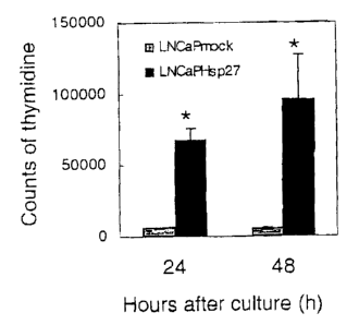
Figs. 1 A and B show increased proliferation in LNCaPHsp27 cells, as compared
to
LNCaPMock cells as observed by [3H]-thymidine incorporation and cell counting,
respectively.
Figs. 2, 3A and B show % of cells in sub G1 after treatment with CH-1 1, Scr
antisense, siHsp27 and CHX.
Fig. 4 shows differential cell counts for cells lines with and without PTEN
after
treatment with siHsp27.
Figs. 5A and B shows effects of siHsp27 in a cell line with inducible PTEN.
Detailed Description of the Invention
The present invention provides a method for evaluating a cancer to assess the
suitability of the cancer to treatment with an hsp27 inhibitor. The cancer may
be a human
cancer, although the method can also be used in connection with veterinary
applications,
for example to evaluate cancers found in dogs, cats and other pets.
The occurrence of elevated levels of hsp27 in various types of cancer and the
-2-
CA 02656577 2009-02-27
demonstrated efficacy of hsp27 inhibitors in multiple types of cancers is
indicative of the
general applicability of the present invention to cancers of many types. In
general, the
method will be employed with cancer types which are considered to be targets
for hsp27
therapy, including in particular those where there has been a previous
determination of
hsp27 overexpression for the patient's cancer. Specific non-limiting examples
of cancer
types that may be treated using the method of the invention include breast,
prostate,
ovarian, uterine, non-small cell lung, bladder, gastric, liver, endometrial,
laryngeal and
colorectal cancers; squamous cell carcinomas such as esophageal squamous cell
carcinoma, glioma, glioblastoma, melanoma, multiple myelmoma and lymphoma.
The first step of the present method is obtaining a sample of cancerous tissue
from
the patient for evaluation. Such samples can be obtained using normal biopsy
and
sampling techniques consistent with the type of cancer. The size of the sample
needed is
based upon the evaluation procedure to be employed.
Once a sample of cancerous tissue is obtained it is evaluated to determine an
expression of level of functional PTEN. As used herein, the term "functional
PTEN"
refers to PTEN that retains its wild-type ability to inhibit the
phosphatidylinositol 3'-
kinase/Akt pathway and hence to act as a tumor suppressor. Reduced levels of
functional
PTEN may result from decreases in the total amount of PTEN expressed, from
modifications to expressed PTEN (for example methylation of PTEN as reported
by
Mirmohammadsadegh et al, Cancer Res. 2006: 66(1) 6546-52), or from mutations
in the
PTEN gene that result in the expressed protein being defective.
There are numerous methods by which the level of functional PTEN may be
determined including immunohistochemical methods, polymerase chain reaction
(PCR
analysis), and PTEN specific immunoassays such as PTEN ELISA. Examples of
specific
known assays include without limitation those in the citations listed in Table
1, all of
which are incorporated herein by reference.
Table 1
Cell Type Assay Type Citation
Breast Cell Oncol. 2007;29(1):25-35.
Breast immunohistochemical Histopathology. 2006 Sep;49(3):248-55
Breast methylation-specific Genes Chromosomes Cancer. 2004 Oct;41(2):117-
PCR assay 24
-3-
CA 02656577 2009-02-27
Breast Breast Cancer Res. 2001;3(6):356-60.
Breast tissue microarray Arch Pathol Lab Med. 2007; 131:767-772
Endometrial immunocytochemical Int J Gynecol Cancer. 2007 May-Jun;17(3):697-
704
Endometrial DNA sequencing Clin Cancer Res. 2006 Oct 15;12(20 Pt 1):5932-5.
Esophageal immunohistochemical Dis Esophagus. 2007;20(6):491-6.
Squamous Cell
Carcinoma
Gastric tissue microarray Appl Immunohistochem Mol Morphol. 2007
Dec;15(4):432-40.
Gastric Int J Cancer. 2008 Jan 15;122(2):433-43
Gastric immunohistochemical World J Gastroenterol. 2006 Feb 21;12(7):1013-7
Glioma RT-PCR Scand J Clin Lab Invest. 2006;66(6):469-75.
Glioma SSCP and sequencing Cancer Res. 1997 October 1; 57: 4187-4190.
Glioblastoma Cancer Res. 2007 May 1;67(9):4467-73
liver Liver Int. 2007 Mar;27(2):155-62
Non-Small Cell immunohistochemical Oncol Rep. 2007 Apr;17(4):853-7
Lung Cancer
Melanoma RT-PCR Cancer Res. 2006 Jul 1;66(13):6546-52
breast, prostate, immunohistochemical Proc Natl Acad Sci U S A. 2007 May
and bladder 1;104(18):7564-9.
carcinoma
renal cell tissue microarray Pathology. 2007 Oct;39(5):482-5
carcinoma and
oncocytoma
astrocytoma yeast-based assay for Oncogene. 2000 Sep 7;19(38):4346-53
the detection of PTEN
nonsense mutation
colorectal Br J Cancer. 2007 Oct 22;97(8):1139-45
ELISA test kits for PTEN are commercially available: PTEN ELISA Assay Kit from
Echelon Biosciences Inc., Salt Lake City, UT and Human/Mouse/Rat PTEN ELISA
development Kit, DuoSet, IC (Intracellular), Minneapolis, MN (Catalog Number
DYC847-2). Materials for immunohistochemical assays for PTEN are also
available
-4-
CA 02656577 2009-02-27
commercially: Pathway Diagnostics, a cell staining assay Phosphatase and
Tensin
Homolog (PTEN); PTEN (clone 17A, NeoMarkers; ready-to-use) and SP kit from
Fujian
Maxin Ltd (China); and monoclonal PTEN antibody, 6H2.1, from Cascade
BioScience,
Inc, Winchester, Mass. Other PTEN monoclonal antibodies are available from
Neomarkers and Zymed. See Modern Pathology 2005; 18: 719-727 which is
incorporated
herein by reference. An RT-PCR kit for PTEN detection is available from
Superarray
Bioscience Corporation, Frederick MD.
An in vitro test for PTEN missense mutations based on a phosphoinositide
phosphatase assay is described in Cancer Res. 2000, June 15; 60:, 3147-3151,
which is
incorporated herein by reference.
The test result of the performed assay are compared to a relevant threshold
level.
The relevant threshold level is determined for the tissue type tested and for
the assay
performed and reflects an average or lower value of PTEN expression. It will
be
appreciated that this threshold value is a balance between the likelihood of
missing the
opportunity to give appropriate therapy to a patient with a higher, but still
reduced level of
PTEN against the risk of treating a patient with a therapeutic that will not
be effective
resulting in a delay in administering alternative therapy. Thus, the specific
threshold
selected for any given cancer will depend on the variability of PTEN
expression levels in
non-cancerous "normal" tissues, the precision and accuracy of the assay
employed, and the
availability of viable alternative treatment modalities.
When the assay reveals an expression level of functional PTEN that is below
the
threshold level, a therapeutic composition comprising as an active agent a
composition
effective to inhibit the expression of hsp27 is administered to the patient.
As noted above,
inhibitors of hsp27 expression of various different types are known in the
art. The specific
route of administration, the dosage level and the treatment frequency will
depend on the
nature of the active agent employed. In general, the therapeutic agent may be
administered
by intravenous, intraperitoneal, subcutaneous, topical or oral routes, or
direct local tumor
injection. For example, antisense targeting hsp27 (such as gggacgcggc
gctcggtcat, OGX-
427, SEQ ID No. 1) may be administered at levels of injection at 200mg, 400mg,
600mg,
800mg or 1000mg once a week as tolerated by the patient.
As discussed above, other inhibitors of hsp27 expression can also be employed,
including evodiamine, which has the formula:
-5-
CA 02656577 2009-02-27
O
N
\
H
berberine derivative, magnolol-containing synthetic suppressors of protein
belonging to
hsp27 family, shikonin-containing synthetic suppressors of protein belonging
to hsp27
family and aconitine-containing synthesis inhibitors of protein belonging to
hsp27
family.
Having described the invention above, the following non-limiting examples are
provided to further illustrate and demonstrate the invention. These
experiments show that
Hsp27 blockade selectively inhibits growth of PTEN deficient cancer cells and
that Hsp27
chaperone is required for Akt stability and activity that, in turn, regulates
phosphorylation
and function of PEA-15. Hsp27 induces dual coordinated effects resulting in
protection
from Fas-induced apoptosis and promotion of cell proliferation through
regulation of PEA-
15 phosphorylation and function in an Akt dependent manner. Hsp27
overexpression
resulted in activation of Akt and increased phosphorylation of its downstream
target PEA-
15 promoting enhanced ERK translocation to nucleus and increased Elk-1
activity which
correlated with increased cyclin D 1 and CDK2 expression with a concomitant
decrease in
p27Kipl expression and increased cell proliferation. Furthermore, Hsp27
overexpression
also led to increased association of PEA-15 with FADD and decreased
sensitivity of cells
to Fas-induced apoptosis . Conversely, Hsp27 knockdown led to reduced Akt
activity and
decreased phosphorylation of PEA-15 leading to reduced association of PEA-15
with
FADD and increased sensitivity of cells to Fas induced apoptosis.
Significantly, siRNA
mediated Hsp27 knockdown in a panel of cell lines and in PTEN Tet-ON LNCaP
cells that
express PTEN in a doxycycline inducible manner demonstrated selective
inhibition of
growth of PTEN deficient cancer cells. These data identify a dual role of
Hsp27 in
regulating cell proliferation and Fas-induced apoptosis through regulation of
PEA-15 and
Akt and indicate that improved clinical responsiveness to Hsp27 targeted
therapy can be
achieved by stratification of patient populations based on expression of PTEN
by cancer
-6-
CA 02656577 2009-02-27
cells in accordance with the present invention.
EXAMPLES
In the following examples, the materials and methods used were as follows:
Cell lines and materials.
LNCaP, PC-3, DU145, 293T, Ku7, RT4, UMUC3, and MDA468 cells were
purchased from American Type Culture Collection (ATCC, Rochville, MD, USA).
PNT1b, LAPC4 and BPH-1 cells were a gift from Prof. N. Maitland (York, UK).
LNCaP
(used up to passage 50 in the present study), DU145, LAPC4, BPH-1, and PNT1b
cells
were routinely maintained in RPMI1640 (Life Technologies, Burlington,
Ontario). RT4
cells were maintained in Macoy's media (Life Technologies, Burlington,
Ontario). Other
cells were maintained in DMEM (Life Technologies, Burlington, Ontario). Media
were
supplemented with 10% fetal bovine serum (FBS) and cultures were grown at 31C
and 5
% CO2. GSK690693C kindly given by Dr. Rakesh Kumar was used as an AKT
inhibitor
in the present study. CH- 11, anti-Fas antibody was purchased from Upstate.
Cyclohexamide (CHX), doxycycline (Dox) and LY-294002 were purchased from
Sigma.
Hsp27 antibody, phospho-Hsp27 (Ser-82) antibody (StressGen), PEA-15 antibody
(Santa
Cruz Biotechnology), phospho- PEA15 (ser-116) antibody (Biospurce), Akt
antibody,
phospho-Akt (Ser-473) antibody, phospho-Foxol (Ser-256) antibody (Cell
Signallig),
FADD antibody (Upstate), p27, cyclin D1, CDK2 (Santa Cruz Biotechnology),
Vinculin
antibody (Sigma Chemical, MO) was purchased from each companies.
Lentiviral infection of Hsp2 7 into LNCaP cells
Two vectors, pHR'-CMV-Hsp27 and pHR'-CMV were used as an empty vector in
the present study as previously described [Araujo, H., et al., J Biol Chem,
1993. 268(8): p.
5911-20.]: pHR'-CMV-Hsp27 including the full-length cDNA for human Hsp27 was
subcloned into the lentiviral vector pHR'-cytomegalovirus (CMV)-enhanced green
fluorescent protein (EGFP) at the BamHI and XhoI sites. Infected LNCaP cells
(LNCaPHsp27) were harvested for UV microscopy to verify green fluorescent
protein
-7-
CA 02656577 2009-02-27
expression, and Western blotting was used to verify Hsp27 expression.
Knockdown by siRNA transfection
Twenty-four hours after culturing in 10 cm dishes at 7 x 10⁵ cells per dish,
Hsp27
siRNA (siHsp27) or scrambled siRNA (Scr) duplexes were transfected into cells.
Briefly,
the RNA duplex was diluted in Opti-MEN serum-free medium and Oligofectamin
(Invitrogen-Life Technologies). After 20 min, cells were transfected at 37?
for 4-6 h and
then were placed in standard medium. Forty-eight hours after transfection,
cells were
harvested and were analyzed in each experiment. To knockdown Akt, Aktl siRNA
(siAkt)
duplexes (Cell signaling) were used. Twenty-four hours after transfection for
24 h, cells
were harvested and then were analyzed as well. An siRNA duplex corresponding
to the
human Hsp27 site (sequence of one strand 5'-AAGUCUCAUCGGAUUUUGCAGC-3',
SEQ ID NO: 2; Dharmacon, Lafayette, CO) was used. A scrambled siRNA duplex (5'-
CAGCGCUGACAACAGUUUCAU-3' SEQ ID NO: 3) was used as a control.
Western blot analysis
Proteins (20-40 g/lane) extracted in RIPA buffer from cells, were
electrophoresed
in SDS-polyacrylamide gels (SDS/PAGE) and transferred to PDVF membranes
(Milipore,
Bedford, MA) by a wet transfer method. After blocking in TBST containing 5%
nonfat
milk powder at room temperature for an hour, membranes were incubated with the
indicated antibodies at 40C overnight. After washing, membranes were then
incubated for
30 min with 1:5000-diluted horseradish peroxidase-conjugate secondary
antibodies (Santa
Cruz Biotechnology, CA) at room temperature. Bands were detected by using an
enhanced
chemiluminescence Western blotting analysis system (Amersham Life Science,
Arlington
Heights, IL).
Clonogenic Proliferation Assay
Cells were cultured at 0.5 x 10⁵ cells per well in 6 well-plates. Twenty-four
hours
after culture, cell growth was compared by a cell count method at 2 days
intervals up to 7
days under standard medium. Each experiment was performed in 3 experiments.
-8-
CA 02656577 2009-02-27
[3HIThymidine Proliferation Assay
Cells were cultured at a concentration of 4 x 10⁴ cells per ml in 12 well-
plates with
standard medium for 24 h. On each day of the study at 24 h or 48 h after
culture, 10 l of
100 Ci/ml [3H]thymidine were added per well and cells were incubated for 3 h.
The cells
were detached from the plate with a trypsin-EDTA solution (0.05% trypsin and
0.53 mM
EDTA; Life Technologies, Inc.). After centrifuging, cells were re-suspended in
IOO I
ddH2O and were transferred to 96-well plates. The collected cells were counted
on a
Packard Top Count counter. Each experiment was performed in 6 experiments.
Immunofluorescence
Cells were grown on glass coverslips in standard media for 24 h. Cells were
fixed
with cold 3% acetone in methanol for 10 min at -20 C and permeabilized in 0.2%
Triton
in phosphate-buffered saline (PBS). Slides were incubated in blocking
solution, 5% BSA
in PBS for 1 h, and simultaneously treated overnight with primary antibodies,
mouse
monoclonal Hsp27 and rabbit polyclonal ERK antibodies. Secondary fluorescent
antibodies, anti-mouse FITC and anti-rabbit Texas red conjugated were added
for 1 h at
room temperature with three 5-min washes (0.1 % Triton in PBS). Cells examined
for
localization of red and green protein were mounted with fluorescent 4',6-
diamidino-2-
phenylindole vectashield mounting medium (Vector Laboratories). Images were
captured
using a Zeiss Axioplan II fluorescence microscope (Zeiss) at x 100
magnification followed
by analysis with imaging software (Northern Eclipse, Empix Imaging, Inc.,
Mississauga,
Ontario, Canada). Analysis of focal co-localization was also done with
Northern Eclipse
and Adobe Photoshop CS software.
Immunoprecipitation
Cell lysates were incubated with 5 g Akt antibody or anti-IgG antibodies.
After 12
h of incubation, 50 L of protein A/G beads (Amersham Pharmacia Biosciences)
were
added into the reaction tubes and incubated for 2 h. The beads were washed
three times
using lx PBS and resolved in 5x loading buffer (MBI, Fermentas Inc.,
Burlington,
Canada). Hsp27 antibody was used and bands were detected as described above.
In vitro Akt kinase assay
-9-
CA 02656577 2009-02-27
Cells were harvested after culture and cell lysates were collected. Assessment
of
Akt activity was performed by the Akt kinase assay kit (Cell signaling),
according to the
manufacture's instructions. Briefly, 500 g of proteins were incubated
overnight with
protein G-agarose beads bearing anti-Akt antibody on rotate at 4AC_ to
immunoprecipitate
Akt. After washing, Akt-antibody-protein G-agarose complexes were added 1 g
of
recombinant GSK-3A/B and 1 l of magnesium/ATP mixture and were incubated for
30
min at 31 After adding SDS sample buffer, samples were boiled for 5 min and
were
electrophoresed on 10% SDS-PAGE. The membranes were incubated with anti GSK-
3A/B(ser21/9) and Akt antibodies. Vinculin expression of input was used as a
control.
Protein stability
LNCaP cells treated with Scr or siHsp27 or LNCaPmock or LNCaPHsp27 cells
were used. Media were changed 18-24 h later to RPMI + 5% serum containing 10
g/mL
of cyclohexamide (CHX) incubated for the indicated time. Western blot was done
by using
Akt, Hsp27, and CAPDH antibodies.
Elk-1 transcription reporter assay
Cells were seeded onto 12-well plates at 105 cells per well. Cells grown in 6-
well
plates were transiently cotransfected in Opti-MEM medium with lipofectin
(Invitrogen),
with 0.5 g GAL-Elb-Luciferase reporter gene and a varying doses of pCMV-GAL4-
Elk-1
kindly provided by Prof. Richard A. Maurer (Oregon, US). Sixteen hours after,
media was
replaced with RPMI1640 plus 10% FBS. Fluorescence was measured in a
luminometer
(MicroLumat Plus, EG & G Berhold) 48 h after transfection. Samples were
normalized by
cotransfection either pCMV-Renila or to protein concentration when Elk-1
effect was
tested. Reporter assays were expressed in arbitrary light units and performed
in 3
experiments.
CH-11-induced apoptosis assay
Cells were treated by 2.5 g/ml CHX alone, 1000ng/ml CH-11 alone or combined
treatment with both drugs in 5% charcoal-stripped serum (CSS) media. Twenty
hours later,
cells were harvested and the percentage of subGl populations were analyzed
using
flowcytometry. On the other hand, cells transfected with Scr 20 nM or siHsp27
20 nM
-10-
CA 02656577 2009-02-27
were treated with 1000 ng/ml CH-l 1 in low-serum (0.5%FBS) media 48 h after
each
treatment. Apoptotic analysis was performed by using the percentage of subGl
populations
after CH-11 administration by flowcytometry. Each assay was performed in 3
experiments.
Statistical analysis
All of the results were expressed as the mean SD. Statistical analysis was
done
with a one-way ANOVA followed by Fisher's protected least significant
difference test
(StatView 512, Brain Power, Inc., Calabasas, CA). *, P <0.05 was considered
significant.
Example 1: Hsp27 regulates Akt pathway in LNCaP cells.
LNCaPHsp27 and LNCaP with empty vector (LNCaPmock) cells were assessed 24
h after culture. Cell lysates were collected 24 hours after culture and bands
were detected
with the indicated antibodies by Western blot assay. Hsp27 antibody, phospho-
Hsp27 (Ser-
82) antibody (StressGen), PEA15 antibody (Santa Cruz Biotechnology), phospho-
PEA15
(ser-116) antibody (Biospurce), Akt antibody, phospho-Akt (Ser-473) antibody,
phospho-
Foxo 1(Ser-256) antibody (Cell Signaling) as a well-known downstream of AKT,
were
assessed both in LNCaP-M and Hsp27-LNCaP cells. Vinculin antibody (Sigma
Chemical,
MO) was used as a control of loading protein. Elevated Hsp27 and phospho-Hsp27
(Ser-
82) protein levels in LNCaP cells stably expressing human Hsp27 cDNA
(LNCaPHsp27)
were observed. Akt and phospho-Akt (Ser-473), PEA15, phospho-PEA15 (ser116)
and
phospho-Foxol (Ser-256) known to be phosphorylated by Akt were up-regulated in
LNCaPHsp27 cells. Hsp27 knockdown by siHsp27 led to down-regulation of Hsp27
in a
dose-dependent manner. Next, effects of treatment by siAkt or GSK690693C, an
Akt
inhibitor, were assessed by Western blot assay in LNCaP cells. siAkt treatment
down-
regulated Akt, phospho-Akt PEA15, and phospho-PEA15, whereas GSK690693C
treatment had a dose-dependent decrease of phospho-GSK-3B and phospho-PEA15
accompanied with up-regulation of phospho-Akt. These data indicates that Hsp27
regulates PEA15 phosphorylation via regulating Akt phosphorylation.
Example 2: Hsp27 directly interacts with Akt and stabilizes Akt in LNCaP
cells.
Hsp27 association with Akt was assessed by immunoprecipitation in LNCaP cells.
Complete knockdown of Hsp27 abolished this interaction with Akt, whereas over-
expression of Hsp27 increased the amount of Hsp27 immunoprecipitation with
Akt. To
-11-
CA 02656577 2009-02-27
estimate whether this interaction is concomitant with functional outcome, in
vitro Akt
kinase assay was examined. Knockdown of Hsp27 decreased Akt activity, while,
increased
Akt activity was seen in LNCaPHsp27 cells. The effect of Hsp27 knockdown on
Akt
stability was next evaluated by using CHX. , which inhibits protein synthesis.
Akt protein
levels rapidly decrease after Hsp27 knockdown. In contrast, Hsp27 over-
expression
prolonged Akt half-life compared to LNCaP cells with empty vector. These data
indicates
that Hsp27 directly interacts with Akt and stabilizes Akt. This results
provides a rational
explanation for the importance of PTEN status to activity of Hsp27 inhibitors
since PTEN
inhibits formation of Akt, such that there is little or no Akt for the Hsp27
to interact with
and stabilize.
Example 3: Hsp27 levels positively correlate with ERK translocation and cell
proliferation rates.
Phospho-PEA 15 is reported to stimulate translocation of ERK to the nucleus.
Effect of Hsp271evels on translocation of ERK in individual cells was
estimated by
immunofluorescent assay. Cell lysates from LNCaP-M cells and HSP27-LNCaP cells
were collected 24 hours after culturing in standard media. LNCaP cells were
treated with
scramble (Scr) 20 nM or 20 nM hsp27 siRNA duplex (siHsp27) 20 nM as described
above. Forty-eight hours after transfection, cells were harvested and cell
lysates were
collected.
Phospho-ERK antibody and ERK antibody (Santa Cruz Biotechnology) were used
to detect bands. Eighteen hours after serum starvation, ERK staining in
LNCaPmock cells
was mainly localized to the cytoplasm as reflected in higher relative levels
of ERP to
phopho-ERK, but increased Hsp27 enhanced the translocation to the nucleus as
reflected
in increased relative amounts of phospo-ERK. Western blot assays also showed
that
Hsp27 over-expression increased ERK phosphorylation. Activity of Elk-1 as a
representative transcription factor downstream of ERK was assessed by
luciferase reporter
assay. These assays demonstrated a dose-dependent and significant increase of
Elk-1
activity in LNCaPHsp27 cells. In addition, shift of cell cycle-dependent
molecules was
investigated by western blot assay. Expression of Cyclin D 1 and CDK2
increased and p27
expression decreased in LNCaPHsp27 cells, accounting for acceleration of cell
cycle by
Hsp27 over-expression.
-12-
CA 02656577 2009-02-27
To identify whether this proliferation is accompanied with up-regulated DNA
synthesis, [3H]Thymidine incorporation assays were used. [3H]Thymidine up-take
in
LNCaPHsp27 cells showed significant increase and reached to 20-fold counts up
to 48
hours (Fig. 1 A), accounting for accelerated DNA synthesis by Hsp27 over-
expression.
Moreover, clonogenic growth assay was performed by using a cell count method.
LNCaPHsp27 cells showed significantly increased proliferation in time-
dependent manner
(Fig. 1B). These data demonstrates that increased Hsp27 functionally activates
phosphorylation and nuclear translocation of ERK via PEA 15 and medicates
increased
cell proliferation in LNCaP cells.
Example 4: Hsp27 regulates induction of Fas-induced apoptosis in LNCaP cells.
In addition to its effect on ERK translocation, Phospho-PEA15 displays anti-
apoptotic effect through loss of FADD function by directly binding to FADD, a
stimulator
of Fas-mediated apoptosis. LNCaP cells are resistant to Fas stimuli. To study
the
involvement of Hsp27, apoptotic assays were performed using CHX, a protein
synthesis
inhibitor that also sensitizes cells to Fas-mediated apoptosis by degrading
expression of c-
FLIPL (cellular FLICE inhibitory protein long forrn).
Cytoprotective effect of Hsp27 to anti-Fas treatment was studied both in
LNCaPHsp27 and LNCaPMock cells. LNCaPHsp27 and LNCaPMock cells were treated
by 2.5 mg/ml CHX alone, 1000ng/ml CH-11 alone and combined treatment with both
drug
in 5% CSS media. Apoptosis (%subGl population) was assessed by flowcytometry
24
hours after treatment. B, Assessment of the effect of Hsp27 knockdown and CH-
11 was
performed 48 hours after transfection with scramble (Scr) 20 nM or Hsp27 siRNA
duplex
(siHsp27) 20 nM as described above. The effect of CH-11, 1000ng/mL was
compared with
the presence or the absence of CH- 11. Cell lysates from LNCaP-M and HSP27-
LNCaP
were collected 24 hours after culturing. Cell lysates from LNCaP cells treated
with Scr 20
nM or siHsp27 20 nM were collected 48 hours after transfection. Proteins were
incubated
with PEA15 antibody at f and bands were detected with FADD antibody (Upstate)
by
Western blot assay.
In LNCaPmock cells treated with CHX, apoptosis (% subGl population) up to
approximately 50% was induced by administration of CH-11. (Figs. 2, 3A and B)
In
contrast, LNCaPHsp27 cells showed significantly less induced after the CH-11
treatment.
-13-
CA 02656577 2009-02-27
Hsp27 knockdown by siHsp27 enhanced CH-11-induced apoptosis 48 hours after
exposure
in low-serum media in LNCaP cells. To estimate whether this protective effect
to CH-11
treatment is involved in the association with FADD, immunoprecipitation with
PEA15
was used. Hsp27 knockdown diminished the interaction with FADD, whereas
increased
Hsp27 strengthened this association. These data demonstrates that Hsp27
regulates the
effect of Fas-induced apoptosis by modifying the interaction with FADD in
LNCaP cells.
Example 5: Effects by Hsp27 knockdown in PC-3 cells.
PC-3 prostate cancer cells were treated by Scr 20 nM or siHsp27 20 nM. Twenty-
four hours after transfection, cells were fixed and were assessed by
fluorescence
microscopy after Hsp27 staining, ERK staining and DAPI nuclear staining. Hsp27
knockdown by siRNA decreased Akt and phospho- Akt (Ser-473), phospho-PEA15
(ser116) and phospho-Foxol (Ser-256) expression levels in dose-dependent
manner in PC-
3 cells. Next, changes of ERK localization after Hsp27 knockdown was assessed
in PC-3
cells by immunofluorescence assay. Media was changed to a low serum (0.5%)
condition
after transfection. After 18 h, ERK nuclear accumulation clearly decreased
after Hsp27
siRNA treatment, accounting for decreased phospho-PEA15 expression level.
Hsp27
siRNA treatment sensitized PC-3 cells to Fas-mediated apoptosis.
Example 6: Correlation between Hsp2 7 Knockdown effect on Growth Rate and PTEN
status
Growth effects by Hsp27 knockdown were compared among 12 cell lines after
transfection with scramble (Scr) 20 nM or Hsp27 siRNA (siHsp27) duplex 20 nM
as
described above. The cell growths were observed with 2 days intervals up to
day7 after
transfection. The date of transfection was considered as dayl. Protein
extracts from cells
24 h after culture in standard media were assessed by Western blot assay using
the
indicated antibodies as described above. Cell number of control treated by Scr
was
considered as 100%.
The relative growth rates for each cell type, represented as a percentage of
cell
count, 100 X (siHsp27/Scr), are shown in Fig. 4. As shown, the cells fell into
two groups,
those there the presence of siHsp27 had not effect, and those where it had a
substantial
-14-
CA 02656577 2009-02-27
inhibition on growth rate. The cell types in the first group (ineffective
cells) are those with
active PTEN. The cell types in the second group (effective cells) have
inactive PTEN.
To further assess the relationship between PTEN status and growth inhibitory
effect by siHsp27 treatment, LNCaP PTEN Tet-on inducible cells were used.
Twenty-four
hours after pre-culturing under the presence or absence of 1 g/mL doxycycline
(Dox),
cells were treated with Scr 20 nM or siHsp27 20 nM and the cell growths were
observed
with 2 days intervals up to day 7. Fig. 5A shows the number of cells when
doxycycline is
absent and shows the ability of siHsp27 to reduce growth rate. In contrast, as
shown in
Fig. 5B, when doxycycline is present resulting in the induction of PTEN, the
growth rates
for the siHsp27 treated cells and the Scr treated cells are the same.
Constitutive PTEN
expression by Dox treatment was confirmed by Western blotting.
Example 7:
LY-294002 (2-(4-morpholinyl)-8-phenyl-4H-l-benzopyran-4-one) is a selective
phosphatidylinositol 3-kinase (PI3K) inhibitor. Combination treatment with
siHsp27 and
LY-294002 treatment was assessed in PC-3 cells. LY-294002 treatment reduced
PTEN
expression and reduced the growth inhibitory effect by Hsp27 knockdown in dose-
dependent manner. These data further demonstrate that growth inhibitory effect
by Hsp27
knockdown is dependent on PTEN/Akt pathway via Hsp27 direct interaction with
Akt.
All of the patents and publications referenced herein are incorporated herein
by
reference as though fully set forth herein.
-15-
CA 02656577 2009-02-27
DEMANDES OU BREVETS VOLUMINEUX
LA PRESENTE PARTIE DE CETTE DENiANDE OU CE BREVETS
COMPREND PLUS D'UN TOME.
CECI EST LE TOME DE _2
NOTE: Pour les tomes additionets, veillez contacter le Bureau Canadien des
Brevets.
JUMBO APPLICATIONS / PATENTS
THIS SECTION OF THE APPLICATION / PATENT CONTAINS MORE
THAN ONE VOLUME.
THIS IS VOLUME `1 OF _2
NOTE: For additional volumes please contact the Canadian Patent Office.