Note: Descriptions are shown in the official language in which they were submitted.
CA 02657511 2013-06-07
72486-35
-1-
SELF-CONTAINED TEST UNIT FOR TESTING BODY FLUIDS
I. Technical Field of Invention
[001] The present invention relates to testing devices used for obtaining a
sample of body fluids,
and then testing that body fluid, normally in conjunction with a testing
device such as a meter.
10021 II. Background of the Invention
[003] In the maintenance of health, it is often desirable to test body fluids
for the presence or
absence of particular substances. To this end, many testing devices have been
invented previously by
the Applicants and others. Testing devices invented by the applicant are shown
in Kloepfer et al.,
U.S. Patent Nos. 6,696,240; 6,840912; and Published Application Nos. EP
013090733; EP
02794170.7; US 10/916,292; and EP 05107323.7
[004] Currently, a need exists for a test unit that is self-contained insofar
as it comprises a unitary
unit that contains all of the primary "disposable" components required for
typical body fluid testing.
These disposable components include a lancet for piercing the skin, and a
testing area, wherein the
fluid (usually blood) that is sought to be obtained, and that flows from the
lanced skin after the skin
has been lanced, can be separated and is separated into a plasma component and
a fraction containing
other components. On such a testing device, the plasma reacts with the
reagents on the test member to
form reagent-bound (or reagent-reacted) reactant compounds that can be used to
quantitatively or
semi-quantitatively determine the presence or absence of a substance within
the blood such as
glucose or cholesterol.
[005] To this end, the readers attention is directed particularly to Kloepfer
et al., U.S. Patent
No.6,840,912 (the "Test Wand" Patent), that discloses a self-contained test
wand. The self-contained
test wand shown in the Kloepfer patent includes a unitary device that includes
the following four
components:(1) a spring-loaded lancet capable of piercing the skin; (2) a
pressure cuff.that contains
an annular lip for exerting pressure around the lancing site, that helps
foster the flow of blood out of
the lanced site; (3) a swab that is provided for cleaning the lanced site
before and after the lancing of
the site; and (4) a test member that includes means for separating the
cellular components of blood
from the plasma components.
[006] The test member also includes one or more reagents that can react with
the components of
interest in the plasma of interest, to thereby convert these components into
reagent reactive
CA 02657511 2009-01-13
WO 2008/019028
PCT/US2007/017211
-2-
components that can then be employed to determine the quantity of the
components of interest. The
test wand is designed to be used in connection with a meter, such as the one
glucose meter disclosed
in Kloepfer et al., U.S. Published Patent Application No. 2006-0034728 (16
February 2006) (the
"Meter Patent"). The meter disclosed in the above-referenced Kloepfer patent
application employs
either reflectance or transmittance photometry techniques to determine the
quantity of the component
of interest.
[007] The reader's attention is directed to the above-referenced Kloepfer
patents and patent
applications, both for their disclosure of various devices, and for their
discussion of the need for =
testing such body components, the chemical aspects of testing for such
components, and the disease
and social aspects of the reasons for the testing for such components.
[008] Although the test wand(s) disclosed in the various Kloepfer patents
perform their intended
function in a most admirable manner, room for improvement still exists. In
particular, room for
improvement exists in producing alternative test wand units that may be
smaller, and thereby take up
less room; or that may be less expensive to produce, or, that may be better
adapted to use in
connection with other types of meters, such as the meter disclosed in
Applicants' co-pending mobile
transmission device meter patent application, U.S. Published Patent
Application No. 2006-0222,567
(05 October 2006) (the "Cell Phone" Patent). Another desire is to provide a
device that has improved
performance, when compared to devices shown in the earlier Kloepfer
references.
- [009] III. Summary of the Invention
[0010] In accordance with the present invention, a self contained disposable
test unit for testing
body fluid comprises a body member and a support member. The support member is
moveable with
respect to the body member between a first position and a second position. The
support member
includes a body part receiving surface for receiving a patient's body part. A
lancet is carried by the
body member and includes a lancet tip capable of piercing the skin of a
patient to produce fluid flow.
A test member is capable of interacting with body fluid to aid in the
determination of information
about body fluid components. A capillary member is capable of directing fluid
flow to the test
member. A pressure cup is capable of exerting pressure on a body part to
foster fluid flow out of a
lanced site and into the capillary, and a calibration member is provided for
containing information for
facilitating calibration of the test unit.
[0011] Preferably, the lancet is moveable between a storage position, a
piercing position and a
retracted position. In the storage position, the lancet tip is disposed below
the body part receiving
surface of the support member. In the piercing position, the tip is disposed
above the body part
receiving surface of the support member. In the retracted position, the tip is
disposed below the body
part receiving surface of the support member. The lancet is moved into the
retracted position through
CA 02657511 2013-06-07
72486-35
-3-
the engagement of the support member with the lancet as the support member
moves between the
first and second position.
[0012] Preferably, the piercing p9sition of the lancet is adjustable to permit
the user to vary a
distance into which the lancet can penetrate the skin when in the piercing
position. Additionally,
when the lancet is in the storage position, the lancet is preferably carried
by the body portion in a
fixed position. The support member preferably includes at least a first and a
second surface that are
selectively engageable with the lancet for moving the lancet into the
retracted position. The first and
second selectively engageable surfaces are axially offset, such that when the
first surface engages a
lancet, the depth to which the tip will penetrate the skin is different than
the depths to which the tip
will penetrate the skin when the second surface engages the lancet.
[0013] One feature of the present invention is that the support member and the
body member are
moveable with respect to each other. This feature has the advantage of
enabling the device to
function with fewer parts than many prior known devices.
[0014] The movement of the body member and support member relative to each
other permits the
lancet to move from a storage position, where it cannot stick the user, to a
piercing position, wherein
the lancet can pierce the user to cause a fluid flow. Continued movement of
the support member
relative to the body member causes the lancet to then move into a retracted
position, where it no
longer is capable of piercing the user. In most cases, the user's skin is
being pierced, and the fluid
that is caused to flow from the lanced site is blood.
[0015] The combination of these features enables the device to provide a
mechanism for sticking
the user with lancet in a quick and relatively painless manner that pierces
the skin, while quickly
removing the lancet, so that it does not remain imbedded within the user.
[0016] Another feature of the present invention is that it includes a lancet
position adjustor for
permitting the user to vary the distance into which the lancet can penetrate
the skin when in a
piercing position.
[0017] This feature has the advantage of enabling the device to be better
suited to different users, by
enabling the user to vary the piercing depth of the lancet. This enables the
user to better select a
minimum piercing depth that will both pierce the skin sufficiently so as to
cause a sufficient flow of
blood, without being inserted any deeper than necessary, and thereby cause any
more pain, or greater
flow of blood than is necessary.
CA 02657511 2013-06-07
72486-35
-3a-
[0017a] In accordance with an aspect of the invention, there is
provided a self
contained disposable testing unit for testing body fluid comprising: a body
member, a support
member moveable with respect to the body member between a first position and a
second
position, the support member including a body part receiving surface for
receiving a patient's
body part, a lancet movable with respect to the support member, the lancet
including a first
end portion pivotably coupled to the body member for pivotal movement with
respect to the
body member, and a second end portion having a second end and a base end, the
base end
being disposed closer to the first end portion than the second end and the
second end
including a lancet tip capable of piercing a patient's skin to produce fluid
flow, wherein
during pivotal movement of the lancet, the base end and lancet tip of the
second end portion
move in an arcuate path and extend along a tangent of an arc scribed by the
pivotal
movement, wherein the base end follows the lancet tip; a test member capable
of interacting
with body fluid to aid in determining information about body fluid components;
a capillary
member capable of directing fluid flow to the test member, a pressure cup for
exerting
pressure on a body part to foster fluid flow out of a lanced site and into the
capillary; and a
calibration member capable of containing information for facilitating
calibration of the
testing unit.
[0017b] In accordance with another aspect of the invention, there is
provided a self
contained disposable testing unit for testing body fluid comprising: a body
member a support
member moveable with respect to the body member between a first position and a
second
position, the support member including a body part receiving surface for
receiving a patient's
body part, a lancet including a first end portion pivotably coupled to the
body member for
pivoting movement with respect to the body member, and a second end portion
having a
second end and a base end, the base end being disposed closer to the first end
portion than the
second end and a the second end including a lancet tip capable of piercing a
patient's skin to
produce fluid flow, wherein during pivotal movement of the lancet, the base
end and lancet
tip of the second end portion move in an arcuate path and extend along a
tangent of an arc
scribed by the pivotal movement, wherein the base end follows the lancet tip;
the lancet being
pivotably moveable between (1) a storage position wherein the tip is disposed
below the body
part receiving surface of the support member, (2) a piercing position wherein
the tip is
CA 02657511 2013-06-07
72486-35
-3b-
disposed above the body part receiving surface of the support member, and (3)
a retracted
position wherein the tip is disposed below the body part receiving surface of
the support
member, wherein the lancet is moved into the retracted position through an
engagement of
the support member with the lancet as the support member moves between the
first and the
second position a test member capable of interacting with body fluid to aid in
determining
information about body fluid components; a capillary member capable of
directing fluid flow
to the test member, a pressure cup for exerting pressure on a body part to
foster fluid flow out
of a lanced site and into the capillary; a calibration member capable of
containing
information for facilitating calibration of the testing unit; and a lancet
position adjustor for
permitting a user to vary a distance into which the lancet can penetrate a
patient's skin when
in the piercing position, and wherein the lancet strikes a skin surface
generally
perpendicularly to the skin surface.
[0017c] In accordance with another aspect of the invention, there is
provided a testing
system for testing body fluid comprising a digital camera containing device
having a case,
and a self contained disposable testing unit for testing body fluid, the self-
contained testing
unit comprising: a body member, a support member moveable with respect to the
body
member between a first position and a second position, the support member
including a body
part receiving surface for receiving a patient's body part, a lancet carried
by the body member
and including a lancet tip capable of piercing a patient's skin to produce
fluid flow; a test
member capable of interacting with body fluid to aid in determining
information about body
fluid components; a capillary member capable of directing fluid flow to the
test member, and
a calibration member capable of containing information for facilitating
calibration of the
testing unit further comprising a coupler for coupling the testing unit to the
case of the digital
camera containing device capable of reading a test result on the reagent
containing test
member.
[0017d] In accordance with another aspect of the invention, there is
provided a self
contained disposable testing unit for testing body fluid comprising: a body
member a support
member moveable with respect to the body member between a first position and a
second
position, the support member including a body part receiving surface for
receiving a patient's
CA 02657511 2013-06-07
72486-35
-3c-
body part, a lancet including a first end portion pivotably coupled to the
body member for
pivoting movement with respect to the body member, and a second end portion
having a
second end and a base end, the base end being disposed closer to the first end
portion than the
second end and the second end including a lancet tip capable of piercing a
patient's skin to
produce fluid flow, wherein during pivotal movement of the lancet, the base
end and lancet
tip of the second end portion move in an arcuate path and extend along a
tangent of an arc
scribed by the pivotal movement, wherein the base end follows the lancet tip;
a test member
capable of interacting with body fluid to aid in determining information about
body fluid
components; a capillary member capable of directing fluid flow to the test
member, pressure
cup for exerting pressure on a body part to foster fluid flow out of a lanced
site and into the
capillary; and a calibration member capable of containing information for
facilitating
calibration of the testing unit wherein the support member is movable with
respect to the
lancet and engagable with the lancet to cause pivotal movement of the lancet
tip in an arcuate
path.
[0017e] In accordance with another aspect of the invention, there is
provided a self
contained disposable testing unit for testing body fluid comprising: a body
member, a support
member moveable with respect to the body member between a first position and a
second
position, the support member including a body part receiving surface for
receiving a patient's
body part, a lancet including a first end portion pivotably coupled to the
body member for
pivoting movement with respect to the body member, and a second end portion
having a
second end and a base end, the base end being disposed closer to the first end
portion than the
second end and the second end including a lancet tip capable of piercing a
patient's skin to
produce fluid flow, wherein during pivotal movement of the lancet, the base
end and lancet
tip of the second end portion move in an arcuate path and extend along a
tangent of an arc
scribed by the pivotal movement, wherein the base end follows the lancet tip;
a test member
capable of interacting with body fluid to aid in determining information about
body fluid
components; a capillary member capable of directing fluid flow to the test
member, a
pressure cup for exerting pressure on a body part to foster fluid flow out of
a lanced site and
into the capillary; and a calibration member capable of containing information
for facilitating
calibration of the testing unit further comprising a skin distance member
disposed interiorly
72486-35 CA 02657511 2015-03-30
-3d-
of the pressure cup for maintaining a patient's body part at an appropriate
position wherein a
patient's body part will not engage an inlet to the capillary member.
[0017f] In accordance with another aspect of the invention, there is
provided a self
contained disposable testing unit for testing body fluid comprising: a body
member a support
member moveable with respect to the body member between a first position and a
second
position, the support member including a body part receiving surface for
receiving a patient's
body part, a lancet including a first end portion pivotably coupled to the
body member for
pivoting movement with respect to the body member, and a second end portion
having a
second end and a base end, the base end being disposed closer to the first end
portion than the
second end and the second end including a lancet tip capable of piercing a
patient's skin to
produce fluid flow, wherein during pivotal movement of the lancet, the base
end and lancet
tip of the second end portion move in an arcuate path and extend along a
tangent of an arc
scribed by the pivotal movement, wherein the base end follows the lancet tip;
and wherein
the lancet includes a radially extending portion extending between the first
and the second
end portions, wherein the support member is movable with respect to the lancet
and
engagable with the lancet to cause pivotal movement of the lancet a test
member capable of
interacting with body fluid to aid in determining information about body fluid
components; a
capillary member capable of directing fluid flow to the test member, a
pressure cup for
exerting pressure on a body part to foster fluid flow out of a lanced site and
into the capillary;
and a calibration member capable of containing information for facilitating
calibration of the
testing unit.
[0017g] In accordance with another aspect of the invention, there is
provided a self
contained disposable testing unit for testing body fluid comprising: a body
member a support
member moveable with respect to the body member between a first position and a
second
position, the support member including a body part receiving surface for
receiving a patient's
body part, a lancet movable with respect to the support member, the lancet
including a first
end portion pivotably coupled to the body member for pivoting movement with
respect to the
body member, and a second end portion having a second end and a base end, the
base end
being disposed closer to the first end portion than the second end and the
second end
CA 02657511 2015-03-30
72486-35
-3e-
including a lancet tip capable of piercing a patient's skin to produce fluid
flow, wherein
during pivotal movement of the lancet, the base end and lancet tip of the
second end portion
move in an arcuate path and extend along a tangent of an arc scribed by the
pivotal
movement, wherein the base end follows the lancet tip; a test member capable
of interacting
with body fluid to aid in determining information about body fluid components;
a capillary
member capable of directing fluid flow to the test member.
[0018] These and other features of the present invention will become
apparent to
those skilled in the art upon a review of the drawings and detailed
description presented
below, that represent the best mode perceived presently of practicing the
invention by the
Applicants.
CA 02657511 2009-01-13
WO 2008/019028
PCT/US2007/017211
-4-
[0019] IV. Brief Description of Drawings
[0020] Fig. 1 is a perspective view of the present invention showing the cap
member in its open
position;
[0021] Fig. 2 is a perspective view, similar to Fig. 1, except rotated 90
degrees, showing the cap in
its closed position;
[0022] Fig. 3 is a rear elevational view of the present invention;
[0023] Fig. 4 is a front elevational view of the present invention;
[0024] Fig. 5 is a bottom plan view of the present invention;
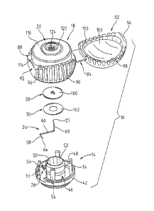
[0025] Fig. 6 is an exploded view of the present invention;
[0026] Fig. 7 is a sectional view taken along lines 7-7 of Fig. 1;
[0027] Fig. 8 is an enlarged sectional view of a portion of the support
member;
[0028] Fig. 9A is a sectional view, similar to Fig. 8, except showing the
lancet in the piercing
position;
[0029] Fig. 9B is a partial, perspective view of the body part engaging
surface showing the lancet
extending there through in the piecing position;
[0030] Fig. 9C is an exploded view of the upper and lower members of the body
part engaging
surface of the present invention;
[0031] Fig. 9D is a partial view of the capillary member of the present
invention;
[0032] Fig. 10 is a perspective and partly broken away view of the capillary
and test member of the
present invention;
[0033] Fig. 11 is a bottom view of the present invention showing the test
member without any
reactant product thereon;
[0034] Fig. 12 is a bottom plan view similar to Fig. 11, except showing
reactant product thereon;
[0035] Fig. 13 is a sectional view of the support member and body member,
showing a lancet in a
piercing position;
[0036] Fig. 14 is a sectional view of the body member and support member
showing the lancet in the
partially retracted position;
. [0037] Fig. 15 is a sectional view of the body member and support
member, showing the lancet in
the fully retracted position;
[0038] Fig. 16 is a perspective view of the body member showing a lancet in
the storage and piercing
position;
[00391 Fig. 17 is a perspective view of the body member, highlighting the
lancet resisting surface;
[0040] Fig. 18 is a front view of a cell phone-type meter that can be used
with the testing device of
the present invention;
[0041] Fig. 19 is a rear perspective view of the cell phone-type meter of the
present invention
CA 02657511 2009-01-13
WO 2008/019028
PCT/US2007/017211
=
-5-
showing the mounting member of the cell phone to which the testing device
mounts;
[0042] Fig. 20 is an enlarged view of the mounting member of the meter;
[0043] Fig. 21 is a rear perspective view showing the testing device mounted
upon the cell phone-
type meter of the present invention;
[0044] Fig. 22 is a sectional view of the device showing the body and support
member in their
respective first or "expanded" positions with respect to each other, and the
lancet in its storage
position;
[0045] Fig. 23 is a sectional view similar to Fig. 22, except showing a
testing device being coupled
to a meter useable with the present invention;
[0046] Fig. 24 is a sectional view similar to Fig. 22, except showing the cap
in the open position;
[0047] Fig. 25 is a view, similar to Fig. 24, except showing the base, rotated
approximately 60
degrees from the view shown in Fig. 24;
[0048] Fig. 26 is a view, similar to Fig. 25;
[0049] Fig. 27 is a view showing the lancet in its piercing position, with the
body member and
support member moved between the first (fully expanded) and second (fully
compressed) positions,
to reside in an intermediate position;
[0050] Fig. 28 is a sectional view, showing the lancet in the partially
retracted position;
[0051] Fig. 29 is a sectional view showing the lancet in the fully retracted
position and the body
member and support member in their second, or fully compressed positions;
[0052] Fig. 30 is a sectional view showing the path of the blood flow, prior
to the blood engaging the
capillary mechanism of the present invention;
[0053] Fig. 31 is a view similar to Fig. 30, showing the path of blood flow
through the capillary
member of the present invention;
[0054] Fig. 32 is a sectional view, similar to Fig. 31;
[0055] Fig. 33 is a sectional view showing the device mounted on to the meter
of the present
invention;
[0056] Fig. 34 is a perspective view of an alternate embodiment showing an
alternate cleansing
member;
[0057] Fig. 35 is a perspective view of the embodiment of Fig. 34, showing the
cover peel strip of
the cleansing member partially removed; and
[0058] Fig. 36 is a sectional view of a second alternate embodiment, showing
an alternate lance
actuator.
[00591 V. Detailed Description of the Preferred Embodiment
CA 02657511 2009-01-13
WO 2008/019028
PCT/US2007/017211
-6-
[0060] The testing device 10 of the present invention is best shown and
described initially with
reference to Figs. 1-8.
[0061] The primary components of the testing device 10 include a body member
14 and a support
member 18. The support member 18 is moveable with respect to the body member
14 between a first
position (Fig. 22) and a second position (Fig. 29). As will be discussed in
more detail below, when
the body member 14 and support member 18 are assembled, they share generally a
common axis,
such that when in the first or expanded position, the body member 14 and
support member 18 are
moved relatively away from each other, so that the height of the testing
device 10 is relatively
maximized. When in the second position, the body member 14 and support member
18 are moved
axially toward,each other so as to compress the testing device 10, so that the
height of the testing
device is at its relative shortest. The support member 18 includes a body part
receiving surface 20,
for receiving a patient's body part.
[0062] The testing device 10 also includes a lancet 24 that is carried by the
body member 14, and is
pivotably coupled to the body member 14 by the lancet 24 being coupled to a
lancet support 26. The
lancet includes a tip 27 that terminates in a point. A reagent containing test
member 28 and
calibration component 30 are also provided.
[0063] As best shown in Figs. 22-29, prior to the testing device being used,
the lancet is normally
positioned in a storage position (Fig. 22) wherein the lancet tip 27 is
disposed below the body part
receiving surface 20 of the support member 18. The lancet 24 is moveable into
a piercing position
(Fig. 27) wherein the tip 27 is disposed above the body part receiving surface
20 of the support. The
lancet 24 is also moveable into a retracted position (Fig. 29) wherein the tip
is disposed below the
body part receiving surface 20 of the support member 18. As will be described
in more detail below,
the lancet 24 is moved into the retracted position through the engagement of
the support member 18
with the lancet 24 as the support member 18 moves between its first (or
expanded) position and its
second (or compressed) position. The "movement" of the lancet 24 from its
storage to its piercing
position actually occurs through the movement of the support member 18
relative to the generally
stationary body member 14 and lancet 24.
[0064] The body member 14 can best be understood with reference to Fig 2, 6
and 7. The body
member 14 includes a base portion 34 that also serves as a coupler, for
coupling the testing device 10
to a meter such as the cell phone type meter 35 shown in Fig. 18. The base 34
should have a
generally planar lip, so that the base can be supported on a surface, such as
a counter top.
[0065] An aperture 34 is formed on the underside of the base 34, and is
defined by an annular ring
like member 38, that also comprises a bayonet-type coupling, to permit the
testing device to be
=
CA 02657511 2009-01-13
WO 2008/019028
PCT/US2007/017211
-7-
coupled to the cell phone like meter 35. A bayonet-type mounting provides a
quick coupling and
release mechanism for coupling and uncoupling the testing device 10 to the
cell phone meter 35.
[0066] The body member 14 also includes a cylindrical perimetral base wall 42
that extends above,
and has a slightly larger diameter than the base 34. The body member also
includes a cylindrical,
axially extending tube 46 that is disposed centrally on the body member 14.
The cylindrical tube 46
has an axially extending, radially outwardly facing exterior wall 48, and an
axially extending, radially
inwardly facing interior wall 50. Interior wall 50 defines a hollow interior
52 that extends generally
between aperture 38, and the upper edge 53 of the cylindrical tube 46.
[0067] The body member 14 also includes four, equi-distantly spaced support
guide members 54 that
are separated from each other at approximately 90 degrees. The upstanding
support guide members
54, extend axially, generally parallel to the axis of the body member 14 and
are provided for
receiving an interior surface of the support member, to appropriately position
the support member 18
on the body member 14, so that the support member 18 can move between its
first or expanded
position arid its compressed position. The body member also includes a lancet
support member 55.
Lancet support 55 includes an angled upper surface that includes a groove 56.
Groove 56 is sized and
positioned for receiving the proximal leg 58 of the lancet 24. Preferably,
groove 56 is sized and
positioned so that the proximal leg 58 can be snap-fit into groove 56, so that
leg 58 will be pivotably
moveable within groove 56, but still will be retained within groove 56.
[0068] The lancet 24 (Fig. 6) includes a first end 64 that is disposed
adjacent to the proximal leg 58,
and an intermediate, radially extending portion 66. The radially extending
portion 66 is so named
because, when the lancet 24 is in its storage position, the portion 66 will
extend in a general radial
direction. However, it will be appreciated that the name given this component,
as with the name
given to the distal or axial portion 68 of the lancet, should not be confined
to specific directions, and
that the claims should always be construed broadly enough to include devices
wherein the various
legs, such as legs 58, 66 and 68 are disposed in other directions.
[0069] As alluded to above, the distal leg 68 is also referred to herein as an
axially extending leg or
portion, because when the lancet 24 is in its rest or storage position, the
portion 68 will generally
extend axially, so that it can fit through the aperture 130 within the body
part supporting surface. It
should also be noted that when in the retracted position (of Fig. 15), the
radially extending portion 66
of the lancet will actually extend in an axial direction, and the axially
extending leg 68 will actually
extend in a radial direction.
[0070] Turning now to Figs. 16 and 17, it will be noted that the cylindrical
tube 46 doe not
comprise a totally endless cylinder. Rather, the cylinder 46 includes an
axially extending slot 72.
The slot includes an angled shelf 73. The angled shelf 73, along with axially
extending wall 75
CA 02657511 2009-01-13
WO 2008/019028
PCT/US2007/017211
-8-
defines slot 72, which together cooperate to form a lancet movement resistant
surface for resisting
pivotal movement of the lancet 24. =
[0071] As best shown in Fig. 16, the lancet 66, when in the storage position
is positioned so that the
radially extending leg 66 extends generally radially, and rests upon angled
shelf 74. Pivotal
movement of the lancet 24 in a direction indicated generally by arrow L in
Fig. 16, causes the radially
extending leg 66 to move through slot 72. The use of the angled shelf 73, and
the spacing between
the side walls 25 of the slot 72 permits the lancet to move through the slot
72, only by overcoming a
predetermined amount of resistance, to thereby prevent the lancet 24 from free
falling unimpededly
through the slot 72.
[0072] This resistence in the movement of the lancet 24 that is induced by the
slot 72 helps to ensure
that the lancet 24 will penetrate the skin of the user, and that the position
of the lancet 24 with the tip
27 pointed upwardly will not be so weakly held so as to be unable to penetrate
the skin.
[0073] The support member 18 has a cap 82 attached to it by a strip of plastic
that comprises a living
hinge 84. The cap 82 is able to move about the living hinge 84 from an open
position, such as shown
in Fig. 1 where the body part receiving surface 20 is exposed, and is
exteriorly disposed, and a closed
position, such as shown in Figs. 2-4. In the closed position, the cap 82 is
disposed in a co-axial
relationship with the support member 18, so that the body part receiving
surface 20 is captured
interiorly within the interior of the cap 82.
[0074] The support member 18 includes an axially extending, radially outwardly
facing exterior wall
88, that includes a knurled or ribbed surface 90, for facilitating the user's
ability to grasp the testing
device 10. The outer surface 88 also includes a small concave portion 92,
that, when the cap 82 is in
its closed position, the concave portion 92 is disposed adjacent to the
overhanging lip 94 of the cap.
The overhanging lip 94 extends radially outwardly past the concave surface 92,
so that the user can
place his finger under the lip 94, to open the cap 82.
[0075] The upper edge of the knurled surface 90 terminates in an axially
facing, radially extending
circumferential mating surface 96, that is sized and positioned for mating
with the axially facing,
radially extending circumferential lip 98 of the cap 82.
[0076] The cap 82 also includes a frusto-spherical exterior surface 104 that
terminates at its lower
end, at the axially facing circumferential lip 98. As discussed above, the
circumferential lip 98 also
includes an overhanging lip portion 94, that is placeable in an opposed, and
adjacent relation to the
small concave surface 92 to form the opening handle. The cap 82 includes a
frusto-spherical interior
wall 106. Preferably, a cleansing pad 108, such as an alcohol soaked cleansing
pad 108 is placed
within the hollow interior defined by the interior wall 106. When the user is
using the testing device
10, the cleansing pad 108 comes in handy, because the user should use the
cleansing pad 108 to
CA 02657511 2009-01-13
WO 2008/019028
PCT/US2007/017211
-9-
cleanse the skin adjacent of the body part that is to be lanced in order to
draw blood from the user.
Preferably, the lance site is cleansed before and after the site is lanced.
[0077] The support member 18, as best shown in Figs. 1, 6, 7 and 9B includes a
testing assembly
118, that includes the body part receiving surface 120, and a radially
outwardly first facing, axially
extending side surface 114. Axially extending side surface 114 and body part
support surface 20 are
normally designed to be disposed interiorly, within the interior of cap 82,
when the device is in its
closed position as is shown in Fig. 4. However, when the cap 82 is opened, the
body part receiving
surface 20 and axially extending surface 114 become exteriorly disposed. The
body part receiving
surface 120, includes several segments or parts, including a beveled,
perimetral edge 117, and a
radially outwardly disposed ring-like portion 118, that is disposed radially
inwardly of the beveled
perimetral edge.
[0078] An elevated, mound-like annular lip 120 is disposed radially interiorly
of the radially
outwardly disposed portion 118, and comprises an endless ring. As will be
described in more detail
below, the elevated annular lip 120 serves as a pressure cup, that is capable
of exerting pressure
against the skin of the user, when the user presses his finger against the
elevated annular lip 120 so
= that the user's skin engages the surface of the elevated annular lip 120.
When the annular lip 120
serves as a pressure cup, the pressure placed upon the user helps to foster
the flow of body fluid, and
in particular, blood out of a lanced site.
[0079] By using a pressure cup, such as that provided by the annular lip 120,
several advantages are
obtained. A first obtained advantage is that a smaller lanced "hole" in the
user's skin can be used,
because the pressure induced by the pressure cup can overcome the smallness of
the hole, to still
permit a sufficient amount of blood to flow out of the lanced hole, to enable
the test to be performed
properly. Additionally, the use of the pressure cup enables the user to use a
non-traditional lancing
site. In this regard, the finger tips are the most typical place for a user to
lance his skin to obtain
blood for a blood test. Finger tips are chosen because of the high rate of
blood flow through the
finger tips.
[0080] Other areas do not give up blood as easily, such as forearms and the
like. However, the use
of a forearm or other body part area has an advantage over the fingers,
because it is not as densely
populated with nerves; and as such, lancing in a site such as the forearm will
generally not hurt as
much. Additionally, the forearm is not used for grabbing and holding objects,
as are the finger tips.
This lack of use by the forearm makes it less likely that the lanced site will
be irritated or injured due
to the activities performed by the body site.
[0081] The elevated annular lip 120 defines a recessed area that is disposed
radially inwardly of the
annular lip.
CA 02657511 2009-01-13
WO 2008/019028
PCT/US2007/017211
-10-
[0082] Within this recessed area is a skin distancing member that includes a
recessed dish 125
surrounded by a lip and platform 127 on which the body part can be placed. The
skin distancing ring
member 124 is disposed concentrically with the pressure cup annular lip 120.
The skin distancing
ring member 124 is sized and positioned so as to maintain the body part, and
preferably the skin of
the body part at an appropriate position relative to the capillary portion of
the device 10. More
particularly, the recessed annular skin distancing lip 127 and recess 125 help
to keep the skin above
the centrally disposed central aperture 130, so that the user's skin does not
plug (close) the aperture,
which is the inlet to the capillary portion of the device 10.
[0083] A central aperture 130 is centrally disposed within the body part
engaging surface 20, and is
= surrounded by a raised central dome 126. The central aperture is sized
and positioned for not only
receiving blood flowing there through into the capillary portion of the
device, but also to receive the
tip 27 of the lancet 24, so that the lancet tip 27 may penetrate the skin of
the user, to cause blood to
flow out of this punctured skin site.
[0084] Turning now to Figs. 8, 9C and 9D, the body part supporting surface 20
is preferably
comprised of separately formed components, including an upper member 134, and
a lower member
136. The primary purpose served by the upper member 134 is to provide a body
support surface upon
which the user can place the body part such as a finger, or forearm that is to
be lanced, so the blood
can be drawn therefrom for testing. To that end, as discussed above, the upper
member 134 includes
the pressure cup 120 and the skin distancing member 124.
[0085] The lower member 136 serves the function primarily of serving as a test
member support, and
it contains the capillary mechanism and test member mechanism thereon.
[0086] The upper member 134 includes a radially outwardly facing cylindrical
side wall 138 that is
sized and positioned to be placed in an opposed relation, so that it is
interiorly received by the
radially inwardly facing side wall 140 of the lower member 136. The lower
member 136 includes a
centrally disposed axially extending capillary tower 144. The central aperture
130, that extends
through the upper member 134, actually opens downwardly in the tower 144 as a
centrally disposed
passageway, that includes a central portion 146, and a radially outwardly
disposed portion 148. The
centrally disposed portion 140 comprises the channel through which the lancet
passes through the
upper 134 and lower 136 members, so that the tip 27 of the lancet can extend
above the central dome
126 (see Fig. 9B) so that it can pierce the skin of the user so that blood may
flow from the lanced site.
[0087] Additionally, blood flows through the central portion 146 in the
radially outwardly disposed
portion. During the flow of the blood through the central and radially
outwardly disposed portions,
the plasma component of the blood starts to become separated from the cellular
components of the
blood. This separation of the plasma from the cellular components is a
separation required for many
CA 02657511 2009-01-13
WO 2008/019028
PCT/US2007/017211
-11-
blood assay tests,.and that is described in more detail in the Kloepfer et
al., patents, and published
applications discussed above, and that are incorporated herein by reference.
[0088] The tower 144 is disposed within a centrally disposed well 152 that
surrounds the tower 144.
Within the well 152 are placed test member components 154. The test member
components 154
include a radially extending capillary space 156, that also serves as a
suction chamber. The capillary
space 156 represents a space into which blood can flow so that the appropriate
components of the
blood (usually the plasma components) will be able to interact with the
reagents contained on the
reagent containing disk shaped test member disk 160, that defines the lower
wall that defines the
capillary space 156. A test member support 162, that can also serve as a
calibration component (See
Fig. 6) is disposed below the reagent containing test member 160.
[0089] The test member 160 can include one or a variety of reagents. Several
well known test
member reagents exist, that can be employed to determine the presence, or
either semi-quantitatively
or quantitatively measure the amount of a particular component, or sets of
components in a body fluid
sample. Examples of reagents that can be placed on the test member to perform
these tests can be
found in patents held by the companies who manufacture such test member
products, including
Bayer, AG, and Roche Diagnostics.
[0090] The path through which the blood flows will be discussed in more detail
below, but before
leaving this area, it should be noted that a foot member 166 is placed at the
base of the radially
outwardly disposed portion 148 of the central channel 130, (Fig. 9D) to
provide a transition and guide
to the blood flowing from the channel 148, and into the capillary space 156.
[0091] The foot member 166 should be in contact with the upper surface of the
reagent containing
test member 160 to facilitate this type of blood flow. A radially extending
air vent channel 168
extends between the base of tower 144, and the radially outer edge 114 of the
lower member 136.
The air vent channel 168 provides an air vent to permit the flow of fluid
radially outwardly in the
capillary space 156 to proceed, without being hindered by air pressure
considerations that would exist
if no vent were present.
[0092] As best shown in Figs. 7 and 11, the underside surface of the support
member 18 includes a
downwardly opening cup member 172, that is generally cylindrical in
configuration, and includes a
radially outwardly facing, axially extending outer wall surface 174, and a
radially inwardly facing
axially extending inner wall surface 176. The purpose of the wall surfaces of
the cup member 172
are to fit between the upstanding support guide member 54 of the body member,
and the outer wall 48
of the cylinder 46 of the body member.
[0093] The outer wall 174 of the inner cup 172 is placed in an opposed
relationship to the radially
inwardly facing wall of the support member 54, and the radially inwardly
facing wall 176 of the
CA 02657511 2009-01-13
WO 2008/019028
PCT/US2007/017211
-12-
support cup 172 is placed in an opposed adjacent relationship with the outer
surface 48 of the
cylindrical support tube 46.
[0094] The support cup 172, support/guide member 54 and cylindrical tube 46
are sized and
positioned, so that the support cup 172 is slideably received by the body
member, and is positioned so
that the support member 18 and body member 14 are disposed generally coaxially
with each other,
and are positioned to be slideable with respect to each other, so that the
support member 18 and body
member 14 can move between an expanded and compressed position.
[0095] The support cup 172 also includes an axially outwardly facing, radially
extending end surface
180, that includes an adjuster member, that permits the user to adjust the
distance that a tip 27 of the
lancet 24 (Fig. 9A), is allowed to extend above the body surface 20. The
adjuster member 182
comprises a series of five axially offset "step" surfaces, that are placed at
a level different than the
general surface 194 of the end surface 180. As best shown in Figs. 11, 12 and
22, the five axially
offset surfaces 184, 186, 188, 190 and 192 are arranged in stair-step fashion,
from the first axially
offset surface 184, which is the "highest" surface, to the lowest surface 194
which actually does not
constitute a step, but rather, constitutes just a continuation of the
remainder of the end surface. It will
be appreciated that the height of the five axially offset surfaces 184-192
differs from the normal end
surface 194.
[0096] Turning now to Fig. 24, the axial movement of the support member 18
relative to the body
member 14, in a direction indicated generally by arrow C, causes the axially
offset steps 184-194 to
move downwardly, toward the radially extending arm 66 of the lancet 24, when
the lancet 24 is in its
storage position. The lancet 24 is in its storage position normally before the
device is used to
perform a test. As the support member 18 continues to move axially downwardly,
it will reach a
point where one of the axially offset steps 184-194 eventually engages the
laterally extending arm 66.
Just prior to this engagement of one of the offset surfaces 184-194, the
lancet is in a position similar
to that shown in Fig. 27 where the tip 27 of the lancet 24 is disposed above
the upper body part
receiving surface 20 of the test member 10. When the lancet tip 27 is in this
position, it is capable of
piercing the skin of the user.
[0097] The user can determine which of the various offset surfaces 1 84-1 94
is chosen to engage the
radially extending leg 66 of the lancet 24. This adjustment is affected by
rotating the support
member 18 relative to the body member 14 about the shared axis A of the
support member 18 and
body member 14. By rotating the support member 18, one can position the
desired offset surface
184-194, above the lancet's 24 radially extending leg 66, so that the desired
surface 184-194 strikes
the lancet's 24 radially extending leg 66. If the user chooses to strike the
lancet leg 66 with the first
or highest step 184, the tip 27 of the lancet 24 will extend a relatively
greater distance above the body
CA 02657511 2009-01-13
WO 2008/019028
PCT/US2007/017211
-13-
surface, and hence pierce the skin of the user to a greater distance or depth,
than will occur if the user
positions the support member so that the lancet leg 66 is engaged by the sixth
or lowest offset surface
194.
[0098] By making this adjustment, the user can determine the depth to which
the lancet 24 tip 27
pierces the skin. Preferably, the lancet 24 pierces the skin to a sufficient
depth to enable a sufficient
amount of blood to flow out of the lanced site, so that enough blood is
available for completing a test.
On the other hand, the lancet should penetrate the skin to the minimal depth
necessary to achieve this
blood flow, because by minimizing the depth, the user also tends to minimize
the amount of pain that
is associated with a lancet "stick".
[0099] By rotating the support member 18 so that it is positioned so that one
of the intermediate
surfaces 186-192 strikes the radially extending leg 66 of the lancet, the
lancet tip 27 would be
allowed to penetrate an intermediate distance somewhere between the relatively
greater distance it
would penetrate if the first step 184 were selected, and the relatively
smaller and shorter depth that it
would penetrate if the lowest offset surface 194 is chosen.
[00100] As will be described in more detail later, after the offset surface
184-194 strikes the
radially extending leg 66, the lancet is pivoted in a direction indicated
generally by arrow R of Fig.
24, on its pivotal connection with the lancet support 55, to move downwardly
and into the retracted
position, such as is shown in Figs. 28 and 29.
[00101] It will also be noted that the radially inwardly facing
surface 200 (Fig. 24) of the
exterior wall 88 of the support member 18 engages and is placed in an opposed
relationship with the
cylindrical perimetral base wall 42 of the body member 14, to further aid in
properly positioning the
support member 18 on the body member 14, so that the body member can move
axially relative to the
body member 14 between the expanded and compressed positions.
[00102] The readers attention is now directed to Figs. 11 and 12.
Figs. 11 and 12 are views
through the bottom of the body member. As discussed above, the interior of the
device is generally
hollow, as is the base 34. This hollowness enables one to look up the hollow
interior, to see the
reactant product that forms on the reactor area 205 of the test member 160
from the reaction between
the reagents contained on the test member 160 and the body fluid that is
placed thereon. Preferably,
the reaction between the reagents and the compound(s) of interest in the blood
will form a
colorometric reaction, wherein the reaction product produced is a colored
reaction product, wherein
the color bears some relationship either to the particular chemical of
interest found on the test
member, or otherwise, to the quantity of the particular chemical (e.g.
glucose, cholesterol) of interest
on the test member. Illustrated dots 202 shown in Fig. 12, can be
"calibration" dots that are placed
on the calibration member 162.
CA 02657511 2009-01-13
WO 2008/019028
PCT/US2007/017211
-14-
[00103] The calibration dots 202 can be pre-printed to replicate
various colors, corresponding
either to various compounds, or else, various quantities of compounds. These
calibration dots 202
can also comprise a type of "bar code" that contains identifying information
about the test device 10.
The colors formed by the reactant product colors from the reaction of the
reagent and the test fluid
are placed adjacent to the calibration color dots 202, so that their color can
be better compared, both
by the meter, and by a visual check. By comparing the colors, one would likely
get a more accurate
and reproducible reading of the quantity of the test compound of interest
formed by the interaction of
the compound with the reagent on the test member 160.
[00104] The manner in which the device moves the lancet 24 between
its storage position and
its retracted position is well illustrated by reference to Figs. 13-17.
[00105] Turning first to Fig. 16, it will be noted that the lancet
24 when in the storage
position, has its proximal end 68 pivotably coupled to lancet support member
55, and has its radially
extending leg 62 positioned to rest on the resistant shelf 73 of the cylinder
46.
[00106] The relative dimensions of the diameter of the lancet 24,
and the width of slot 72 will
cause the radially extending leg 66 of the lancet 24 to rest upon angled shelf
73, and not move axially
through slot 72, unless some force is exerted on the lancet 24 to push it
downwardly.
[00107] The dimensions of slot 72 are chosen so that the amount of
force required to push the
radially extending leg 66 of the lancet through the slot 72 is a greater
amount of force than is
normally required to enable the tip 27 of the lancet 24 to penetrate the skin
of the user. As such, the
shelf 73, wall 75 and slot 72 cooperate to provide enough resistence in the
movement of leg 66, so
that the lancet tip 27 will pierce the skin before moving into its retracted
position.
[00108] Turning now to Fig. 13, the lancet 24 is shown at a
position, just prior to one of the
axially offset surfaces 184, engaging the radially extending leg 66 of the
lancet.
[00109] At this point, the support member 18 still can move an
additional distance
downwardly, in a direction indicated by arrow A to "compress" the support
member 18 and body
member 14. It will also be appreciated that the tip 27 of the lancet 24 lies
just below the body part
receiving surface 20 (and just below the inlet of capillary portion 130), and
that further movement of
the support member 18 in a direction indicated by arrow A, will cause the tip
27 of the lancet to
extend above the surface 20, so that it will be positioned above the body part
engaging surface,
similar to the position shown in Fig. 9B.
[00110] Turning now to Fig. 14, it will be noted that the support
member 18 has moved
axially downwardly on the body portion 14, when compared to the position shown
in Fig. 13. In this
position, one of the axially offset surfaces 184 has already engaged the
radially extending leg 66 of
CA 02657511 2009-01-13
WO 2008/019028
PCT/US2007/017211
-15-
the lancet 24, and has caused the lancet 24 to pivot downwardly, to a position
where the tip 27 is
removed from aperture 130.
[00111] In Fig. 15, a support member 18 is shown in its second or
fully compressed position
vis-a-vis body member 14, so that the lancet 24 is in its fully retracted
position, wherein the distal leg
68 of the lancet is generally disposed radially, and the tip 27 lies generally
near the bottom of the
body member 14. In this position, the lancet 24 is safely tucked interiorly of
the body member 14, in
a position where it is highly unlikely to travel outside the testing device
10, and therefore, is highly
unlikely to be a in a position where it can accidentally stick the user, or
another person.
[00112] A meter 35 with which the testing device is designed to be
used, is shown best in
Figs. 18-20. The meter 35 includes a case 206 that houses all of the internal
components (not shown)
of the meter 35. The meter 35 is shown as being a cell phone-type meter, that
has dual functionality
insofar as it can be used as a cell phone, and also as a test meter. One
benefit of this, as explained in
the Kloepfer cell phone patent application above, is that most cell phones
contain a' camera system
already, that can be used to "read" colorometric" reactions that occur on the
reagent test member 160
of the device, and processing capabilities that can be exploited.
[00113] The front of the cell phone/meter 35 includes a screen 210
upon which information
can be displayed, that preferably comprises a touch-type screen that also
enables commands to be
given through touching appropriate places on the screen 210. A button-laiden
control panel 212 also
appears on the front surface for permitting the user to enter commands to the
cell phone/meter 35.
[00114] Turning now to Fig. 19, the rear of the cell phone/meter 35 is
shown as including a
case member 216, and a testing device receiver/coupler 218. A series of depth
indicia, here shown as
0, 1, 2, 3, 4 and 5 (222) are formed on the rear case member 216, to indicate
the depth at which the
testing device has set the lancet 24. Contained within the receiver/coupler
are a variety of meter
components.
[00115] As shown in Fig. 20, the meter components contained within the test
receiver include
a bayonet mounting surface 228, for receiving the bayonet mount formed in the
base 34 of the testing
device 10. One or more LEDs 232 are provided for serving for as a light
source, to light up in a
controlled manner, the interior of the testing device 10, adjacent to the test
member 160, so that
enough light will be present to enable the meter or camera to perform its
function. A switch 234 is
also operatively coupled in this area to detect the presence or absence of a
testing device 10 on the
receiver/coupler 218.
[00116] Turning now to Fig. 21, a testing device 10 is shown as
being mounted, through the
respective bayonet mounts, to the receiver/coupler 218.
CA 02657511 2009-01-13
WO 2008/019028
PCT/US2007/017211
-16-
[00117] It will be noted that the concave surface 92 of the support
member is placed opposite
the "zero" indicia 222. This placement of the concave surface 92 adjacent to
the zero indicia, can
indicate to the user that the lowest axially offset step 194 will be used so
that the lancet 24 tip 27 will
penetrate the skin, the smallest distance available by the unit.
[00118] If the concave member 92 were pointing to indicia 5, which would
occur if the
support member 18 were rotated about its axis, so that the concave surface 92
faced indicia 5, it
would indicate that the highest axially offset surface 184 of the adjuster was
being employed, so that
the lancet tip 27 would penetrate the greatest possible distance into the skin
of the user. It will also
be appreciated that if the user desired to set the lancet tip 27 depth at an
intermediate level, he would
cause the center of the concave surface 92 to point to one of the intermediate
indicia, such as 1, 2, 3
or 4.
[00119] In this position, the user can place his finger over the
body part receiving surface 20
to begin the testing procedure.
[00120] The reader's attention is now directed to Figs. 22-28, that
depict the sequence that
the lancet goes through, in moving from its storage to its retracted position.
[00121] Turning first to Fig. 22, a sectional view of the device is
shown. It will be noted that
the lancet 24 has its radially extending leg 66 disposed in a radially
extending direction, and that the
tip 27 of the lancet is disposed below the body part receiving surface 20, and
below the tip of the inlet
130 of the capillary portion. It will also be noted that the axially offset
surfaces 184, 186, 188 are
above, and have not yet engaged the lancet 24.
[00122] In this position, the support member 18 and body member 14
are in their first, or
storage position, where they are "expanded" relative to each other and not
compressed. Fig. 23
shows a view similar to Fig. 22, with the exception that the device 10 is
shown as being coupled to
the receiver/coupler 218 of cell phone/meter 35.
[00123] Figs. 24 and 25 also show the device in the storage position,
similar to Figs. 22,
except that Fig. 24 shows the cap 82 in an open position, and Fig. 25 shows
the device rotated about
45 degrees about its axis, from the position shown in Figs. 22, 23 and 24.
[00124] Fig. 26 shows a view generally similar to Fig. 25, and Fig.
27 shows a view, wherein
the support member 18 has moved in an axially compressed direction, which
direction is indicated
generally by arrow A. Fig. 27 shows the device 10 wherein the lancet 24 is in
the piercing position,
as it will be noted that the tip 27 of the lancet 24 is extending above the
body part receiving surface
20, and in fact, the tip 27 of the lancet 24 is about at the same level as the
lip of the pressure receiving
cup 120, and is above the level of the lip 127 of the skin distancing member
124.
CA 02657511 2009-01-13
WO 2008/019028
PCT/US2007/017211
-17-
[00125] It should also be noted that the lancet engaging axially
offset step 184 is just about to
engage the radially extending leg 66 of the lancet. This contrasts from the
view of Fig. 26 where it
will be noticed that the lancet engaging surface 184 is positioned above the
radially extending leg 66
=
of the lancet.
[00126] Fig. 28 shows the lancet 24 after it has begun moving toward its
retracted position. It
will be noted that tip 27 of the lancet 24 is completely removed from the
capillary channel 130, and
the lancet engaging surface 184 is positioned below the pivot point (leg) 58
of the lancet 24. Fig. 29
shows the lancet 24 in the fully retracted position wherein the radially
extending leg 66 of the lancet
extends in a generally axially direction.
[00127] The reader's attention is now directed to Figs. 31 and 32, that
illustrate the sequence
of events that occurs relating to the capillary channel during the use of the
testing device 10. A body
part BP is shown as being placed in Fig. 30 on the body part receiving surface
20. The skin of the
fingers straddles the lip 127 of the body distancing member 124, with the
recess 125 of the body
distancing member providing enough space so that the finger does not plug the
inlet opening 238 of
the capillary channel 130.
[00128] In Fig. 1, the arrow Z shows pressure being exerted
downwardly on the body part
supporting surface 20. This pressure is the pressure that will cause the
support member 18 to
compress the testing device 10, and cause the lancet tip 27 to travel to its
piercing position. Fig. 31
assumes that the piercing has already occurred, and that a body fluid I3F such
as blood has begun to
run out of the body part finger BP, and has begun flowing axially into the
capillary channel 130, and
radially outwardly, in the capillary space 156 that is above the test member
160.
[00129] It is important to note that the suction area 156 is
pinched off at pinch point P against
the testing disk 160. Pinch point P pinches off the capillary channel/suction
area 156, due to the
force that is exerted by the finger, that causes the peripheral area 242 of
the body part supporting
surface to press downwardly on the test disk 160 to cause the test disk 160 to
bend, to thereby form
the pinch point P.
[00130] This pinching prevents the radial movement of the body
fluid past the pinch point P.
Turning now to Fig. 32, arrow Z shows that the pressure of the body part has
been lifted off the body
part receiving surface 20. This removal of force from the body part receiving
member 20, enables the
test disk 160 to straighten out, which enables the capillary channel 156 to
leave its pinch point. This
removal if the pinch point, when coupled with the venting achieved by the
vent channel 168 (Fig.
10), fosters capillarity, and fosters the radially outward movement of the
body fluid BF.
[00131] Left behind (upstream from) the main bolus of body fluid
BF, is the radially inward
area 161, which is the reaction capillary compartment of the test disk 160,
The reaction capillary
CA 02657511 2009-01-13
WO 2008/019028
PCT/US2007/017211
-18-
compartment 161 contains the body fluid that has reacted with the reagent, to
form the reactant
product. This reaction capillary compartment 161 is the area in which the
meter focuses its attention
(e.g. the camera takes its picture) to obtain a reading of the test disk. It
will be noticed that he main
bolus of excess blood BF has traveled downstream of the reaction capillary
compartment to the
excess blood capillary compartment 165
[00132] Capillarity is fostered because air is drawn into the
capillary chamber, that brings
oxygen into the chamber 156, which is necessary for some of the reagent/plasma
reactions to occur,
along with fostering the flow of blood radially outwardly. Additionally, there
exists a capillary force
differential between the reaction capillary compartment 161 and the excess
blood capillary
compartment 165 which also fosters separation and capillarity. Capillarity is
further enhanced due to
the positioning and height differential between the inlet to the capillary
channel 130 at the top of
tower 144, and the relatively lower position of the test disk 160 that
contains both the reaction
capillary compartment 161 and the excess blood capillary compartment 165.
[00133] Fig. 33 presents another sectional view of the testing
device 10, after the test is
finished. After the testing is finished, the cap 82 is placed in its closed
position, and the lens 230, can
view the test results contained on the test member 160.
[00134] Figs. 34 and 35 disclose an alternate embodiment testing
device 300. Testing device
300 is generally identical to testing device 10 shown in Figs. 1-33, with the
exception of the fact that
testing device 306 includes a cleansing member 302 that is placed upon the
body part supporting
surface 20. The cleansing member 302 is generally ring-shaped, and includes a
ring-shaped cleansing
swab 304 that is disposed concentrically with and radially outwardly of the
pressure cup 310. A
cover member 306 covers the cleansing swab and includes a pull tab 308 to
facilitate ;the user
removing the cover member 306_
[00135] Cleansing swab 304 should preferably be fixedly coupled,
such as by glue, to the
testing device, or else, snugly fitted within a channel. Normally, it will be
expected that cleansing
swab will contain some sort of disinfectant, such as alcohol for which the
user can cleanse his skin
both prior to and after his body part is lanced.
[00136] Fig. 36 shows an alternate embodiment test device 400
wherein the lancet movement
mechanism shown in Figs. 1-35 is replaced by a spring-loaded lancet movement
system. The lancet
movement system includes a lancet moving spring 402 that is provided for
engaging a platform 403
that supports lancet 405. This spring 402 expands to move platform 403
upwardly, to thereby move
lancet 408 upwardly through the capillary channel 411, and into engagement
with the user's body
part. A lancet retraction spring 404 is provided for acting against the force
exerted by the lancet
moving spring 402 to cause the lancet 405 to retract downwardly, and back
beneath the body part
CA 02657511 2013-06-07
=
72486-35
-19-
engaging surface 415, after the lancet pierces the skin of the user. A release
member 406 is
positioned to be engaged with one of several axially offset surfaces, e.g.
408, 410 that are designed
and configured similarly to the axial offset surfaces discussed above in
connection with Figs. 1-33.
The axially offset surfaces enable the user to adjust the depth to which the
lancet 405 will penetrate
their skin.
[00137] When the axially offset surface of choice (e.g. 408)
contacts the release member 406,
the spring 402 is then released to urge the lancet 405 upwardly and into
engagement with the body
part.
[00138] Although the invention has been described with reference to
certain preferred
embodiments, it will be appreciated that the scope of the above¨referenced
invention is not
limited to the embodiments disclosed above, but is 'limited only by the
broadest interpretation
allowable of the claims as set forth below.
=
=