Note: Descriptions are shown in the official language in which they were submitted.
CA 02657839 2010-12-10
STENT-VALVES FOR VALVE REPLACEMENT AND
ASSOCIATED METHODS AND SYSTEMS FOR SURGERY
Field of the Invention
[0002] Embodiments of the present invention relate to stent-valves and
associated methods
and systems for their delivery via minimally-invasive surgery.
to Background of the Invention
[0003] Conventional approaches for cardiac valve replacement require the
cutting of a
relatively large opening in the patient's sternum ("sternotomy") or thoracic
cavity
("thoracotomy") in order to allow the surgeon to access the patient's heart.
Additionally,
these approaches require arrest of the patient's heart and a cardiopulmonary
bypass (i.e., use
15 of a heart-lung bypass machine to oxygenate and circulate the patient's
blood). Despite their
invasiveness, these surgical approaches may be reasonably safe. for a first
intervention.
However, tissue adherences resulting from the first surgery may increase the
risks (e.g.,
death) associated with subsequent valve replacement surgeries. See Akins et
at., "Risk of
Reoperative Valve Replacement for Failed Mitral and Aortic Bioprostheses", Ann
Thorac
20 Surg 1998;65:1545-52; and Weerasinghe et Al., "First Redo Heart Valve
Replacement - A 10-
Year Analysis", Circulation 1999;99:655-658.-
CA 02657839 2009-03-05
WO 2008/028569 PCT/EP2007/007413
[0004] Synthetic valves and biological valves have been used for cardiac valve
replacement
with varying results. Synthetic valves rarely fail but require life-long anti-
coagulant
treatment to prevent blood from clotting (thrombosis) in and around the
replacement valve.
Such anti-coagulant treatment significantly limits patients' activities and
can cause various
other complications. Biological valves do not require such anti-coagulation
treatment but
typically fail within 10-15 years. Thus, to limit the need for and risks
associated with re-
operation on failed biological valves, traditionally only patients with less
than about 10-15
years to live have received biological valve replacements. Patients with
longer life
expectancies have received synthetic valves and anti-coagulant treatment.
[0005] Attempts have been made to develop less-invasive surgical methods for
cardiac
valve replacement. These surgical methods, referred to as percutaneous heart
valve
replacement therapies (PHVT), use a catheter to deliver a replacement valve to
an
implantation site using the patient's vascular system. These PHVT attempts
have various
shortcomings, including their inability to ensure proper positioning and
stability of the
replacement valve within the patient's body.
[0006] In view of the foregoing, it would be desirable to provide improved
methods,
systems, and devices for cardiac valve replacement.
Summary of the Invention
[0007] Some embodiments of the present invention are directed to systems,
methods, and
devices for cardiac valve replacement. For example, these methods, systems,
and devices
may be applicable to the full range of cardiac-valve therapies including the
replacement of
failed aortic, mitral, tricuspid, and pulmonary valves. In some embodiments,
the present
invention may facilitate a surgical approach whereby surgery is performed on a
beating heart
without the need for an open-chest cavity and heart-lung bypass. This
minimally-invasive
surgical approach may reduce the risks associated with replacing a failed
native valve in the
first instance, as well as the risks associated with secondary or subsequent
surgeries to
replace failed artificial (e.g., biological or synthetic) valves.
[0008] Stent-valves according to some embodiments of the present invention may
include a
valve component and at least one stent component. The valve component may
include a
3o biological or synthetic (e.g., mechanical) valve and/or any other suitable
material(s). The
stent component may include a first section (e.g., proximal section), a second
section
-2-
CA 02657839 2009-03-05
WO 2008/028569 PCT/EP2007/007413
configured to house the valve component, and a third section (e.g., distal
section). The stent
and valve components may be capable of at least two configurations: a
collapsed
configuration (e.g., during delivery) and an expanded configuration (e.g.,
after implantation).
[0009] In some embodiments, the first section of the stent valve may include a
fixation
element. Such a fixation element may include, for example, an annular groove
for securing
the stent-valve in place at an implantation site. When the stent-valve
includes a single stent
("single-stent-valve"), the annular groove may be configured to receive the
annulus of the
valve in need of replacement. When the stent-valve includes two stents
("double-stent-
valve"), the annular groove of the first stent component may be configured for
matable
attachment to a complimentary annular projection of a second stent component
(i.e., a
positioning stent). In turn, the second stent component may be anchored at the
implantation
site, for example, to the valve in need of replacement and/or adjoining
structures.
[0010] Alternatively or additionally, in some embodiments the third section of
the stent
component may include at least one attachment element. Each attachment element
of the
stent-valve may include, for example, a geometrical opening (e.g., circular or
ovular), hook,
or strap configured for removable attachment to a complimentary structure of a
delivery
device. In addition, each attachment element may correspond to all or a
portion of a
commissural post, to which a commissure between two valve leaflets may be
attached. The
attachment element(s) may allow the stent-valve to be partially expanded
within a patient's
body while the stent-valve remains attached to the delivery device. This may
allow the stent-
valve to be returned to a collapsed configuration and repositioned within the
patient's body
when it is determined that fully expanding the stent-valve would cause the
stent-valve to be
installed incorrectly. Alternatively or additionally, this may allow the stent-
valve to be
returned to the collapsed configuration and removed from the patient's body
when it is
determined that the stent-valve is not functioning properly (e.g., not
permitting sufficient
flow). In some embodiments, the stent-valve may include one attachment
element. In other
embodiments, the stent-valve may include at least two, three, six, or any
other suitable
number of attachment elements. In some embodiments, the fully-expanded stent
diameter in
the region of the attachment element(s) may be smaller than the diameter of
the region that
3o houses an associated valve. This may reduce the risk of injury to the
patient's body (e.g.,
perforation of the aorta) from the attachment elements and/or make it easier
to affix the
attachment elements to the complimentary structure of the delivery device.
-3-
CA 02657839 2009-03-05
WO 2008/028569 PCT/EP2007/007413
[0011] In some embodiments, the stent component of the stent-valve may include
a lattice
structure with a plurality of cells. The lattice structure may be formed from,
for example, a
shape-memory alloy such as nitinol or any other suitable material(s). The
cells in the lattice
structure may be most densely populated in the section of the stent component
that includes
the fixation element. This may provide added support to the fixation element
and increase
the stability of the stent-valve. In some embodiments, the lattice structure
may form at least
one elongate stem (e.g., commissural post) that extends distally along the
stent component
towards the at least one attachment element. The at least one stem may connect
directly to
the at least one attachment element. Alternatively, the lattice structure may
form at least one
supporting element for connecting the at least one stem to the at least one
attachment
element. In some embodiments, all of the cells in the lattice structure may be
closed cells,
which may facilitate recapture of the stent-valve from the partially-expanded
configuration to
the collapsed configuration.
[0012] Still other embodiments of the present invention are directed to a
method for
replacing a valve. A stent-valve is provided that includes a stent component
with an annular
groove, and the stent-valve is secured axially to an annulus of the valve in
need of
replacement. In some embodiments, providing a stent-valve may include suturing
a valve
component to the stent component. Alternatively or additionally, providing a
stent-valve may
include expanding a valve component within the stent component in order to
form a friction
fitting. In some embodiments, providing a stent-valve may include securing a
valve
component to the stent component with a hook-and-loop (e.g., VELCRO )
fastening system.
[0013] In other embodiments of the present invention, a method for replacing a
valve is
provided whereby a first stent component that includes an annular element is
implanted such
that at least a portion of the first stent component is housed within a valve
in need of
replacement. A stent-valve that includes a second stent component is
positioned within the
first stent component by matably attaching a complimentary annular element of
the second
stent component to the annular element of the first stent component.
[0014] In still other embodiments of the present invention, a stent-valve
delivery system is
provided. A first assembly is provided that includes an outer sheath and a
guide wire tubing.
3o The delivery system also includes a second assembly including a stent
holder configured for
removable attachment to at least one attachment element of a stent-valve. The
stent-valve
may be positioned over the guide wire of the first assembly. The first
assembly and the
-4-
CA 02657839 2009-03-05
WO 2008/028569 PCT/EP2007/007413
second assembly may be configured for relative movement with respect to one
another in
order to transition from a closed position to an open position. In the closed
position, the outer
sheath may encompass the stent-valve still attached to the stent holder and
thus constrain
expansion of the stent-valve. In the open position, the outer sheath may not
constrain
expansion of the stent-valve and thus the stent-valve may detach from the
stent holder and
expand to a fully expanded configuration.
[0015] In some embodiments, the first assembly and the second assembly may be
configured to transition from the closed position, to a partially-open
position, to the open
position. In the partially-open position, the stent-valve may expand partially
but not detach
1o from the stent holder because the outer sheath may still encompass the at
least one attachment
element of the stent-valve and the stent holder. When the stent-valve is in
the partially-
expanded configuration, it may be determined whether the stent-valve will be
positioned
correctly if the stent-valve is expanded to the fully expanded configuration.
Alternatively or
additionally, the functionality of the stent-valve may be tested (e.g., to
determine whether the
stent-valve will permit sufficient blood-flow) when the stent-valve is in the
partially-
expanded configuration.
[0016] In some embodiments, the stent-valve delivery system may include at
least one
balloon (e.g., proximal to the stent-valve or other stent to be delivered)
configured to cause
expansion of the stent-valve or positioning stent upon inflation of the at
least one balloon.
[0017] In some embodiments, the stent-valve delivery system may include a push
handle
that causes the relative movement of the first assembly and the second
assembly.
Alternatively, the stent-valve delivery system may include a screw mechanism
for translating
rotational movement of a handle into the relative movement of the first
assembly and the
second assembly.
[0018] In some embodiments, the stent-valve delivery system may include an
integrated
introducer within which the first assembly and the second assembly are
positioned during
delivery of the stent-valve to an implantation site. The integrated introducer
may be
configured to remain within a patient's body even after the first assembly and
the second
assembly are removed, for example, to allow for the introduction of an
occluder.
[0019] In some embodiments, after expansion of the stent-valve to the fully
expanded
configuration, the delivery system may be configured to return to the closed
position by
passing the second assembly through the stent-valve towards a distal end of
the first
-5-
CA 02657839 2009-03-05
WO 2008/028569 PCT/EP2007/007413
assembly.
[0020] Still other embodiments of the present invention are directed to a
method for
delivering a stent-valve to an implantation site whereby the stent-valve is
removably attached
to a delivery device and the stent-valve is delivered to the implantation site
in a collapsed
configuration. The stent-valve may be partially expanded while maintaining the
stent-valve
attached to the delivery device. A determination with respect to the stent-
valve may be made
when the stent-valve is in the partially-expanded configuration. When the
determination
yields a positive response, the stent-valve may be expanded to its fully
expanded
configuration by causing the stent-valve to detach from the delivery device.
[0021] In one particular embodiment, it may be determined whether the stent-
valve is
positioned correctly at the implantation site. The stent-valve may be returned
to the collapsed
configuration and repositioned when the stent-valve is not positioned
correctly at the
implantation site.
[0022] Alternatively or additionally, it may be determined whether a valve
component of
the stent-valve is functioning properly, for example, by testing whether the
valve component
will permit sufficient blood-flow. The stent-valve may be returned to the
collapsed
configuration and removed from a patient's body when the stent-valve is not
functioning
properly.
[00231 In some embodiments, delivering the stent-valve to the implantation
site may
include delivering the stent-valve to the heart for replacement of a cardiac
valve. The
delivery may include accessing a patient's body through an intercostal space
(e.g., fifth
intercostal space) and penetrating the left ventricle at the apex of the
heart.
Brief Description of the Drawings
[0024] For a better understanding of the present invention, reference is made
to the
following description, taken in conjunction with the accompanying drawings, in
which like
reference characters refer to like parts throughout, and in which:
[0025] FIG. 1A shows a valve component in an expanded configuration according
to some
embodiments of the present invention;
[0026] FIG. lB shows a valve component in a collapsed configuration according
to some
embodiments of the present invention;
-6-
CA 02657839 2009-03-05
WO 2008/028569 PCT/EP2007/007413
[0027] FIG. 2A shows a stent component in an expanded configuration according
to some
embodiments of the present invention;
[0028] FIG. 2B shows a single-stent-valve, that includes a stent component and
a valve
component, in an expanded configuration according to some embodiments of the
present
invention;
[0029] FIG. 2C shows a single-stent-valve a collapsed configuration according
to some
embodiments of the present invention;
[0030] FIG. 3A shows a stent component in an expanded configuration according
to some
embodiments of the present invention;
[0031] FIG. 3B shows a stent component in a collapsed configuration according
to some
embodiments of the present invention;
[0032] FIG. 4 shows a double-stent-valve, that includes two stent components
and a valve
component, in an expanded configuration according to some embodiments of the
present
invention;
[0033] FIGS. 5A-7B illustrate the use of a single-stent-valve to replace a
failed biological
(artificial) valve according to some embodiments of the present invention;
[0034] FIGS. 8A and 8B show a stent component that includes attachment
elements for
securing the stent to a delivery device and fixation elements for securing the
stent at the
implantation site according to some embodiments of the present invention;
[0035] FIG. 8C shows a stent component having a diameter in the region of the
attachment
element(s) that is smaller than the diameter of a stent region that houses an
associated valve,
according to some embodiments of the present invention;
[0036] FIG. 8D shows a stent component that includes independently bendable
element(s)
for use in positioning/securing the stent to the geometry/topology at an
implantation site
according to some embodiments of the present invention;
[0037] FIG. 8E shows a stent component that includes locking elements in a
crown
configuration and a fixation element for securing the stent at an implantation
site according to
some embodiments of the present invention;
[0038] FIG. 8F shows a stent component that includes multiple struts for
carrying a valve
component more closely to a region of the stent component that includes
attachment
element(s) for attaching the stent component to a delivery device;
-7-
CA 02657839 2009-03-05
WO 2008/028569 PCT/EP2007/007413
[0039] FIGS. 9A-16 show additional embodiments of stent components that
include
attachment elements for securing the stent to a delivery device and/or
fixation elements for
securing the stent at the implantation site according to the present
invention;
[0040] FIGS. 17/18, 19, and 20 show additional examples of double-stent-valves
according
to some embodiments of the present invention;
[0041] FIG. 21A shows a stent-valve in the shape of an opposed double crown
according to
some embodiments of the present invention;
[0042] FIGS. 21B-E show views of a double-conical stent in accordance with
some
embodiments of the present invention;
[0043] FIGS. 22A-22D show a delivery system for delivering a self-expanding
stent-valve
to an implantation site according to some embodiments of the present
invention;
[0044] FIGS. 23A-23D show a delivery system with inflatable balloon(s)
according to
some embodiments of the present invention;
[0045] FIGS. 24A-24D show a delivery system having a proximal outer shaft with
an
increased diameter according to some embodiments of the present invention;
[0046] FIGS. 25A-25C show a delivery system with inflatable balloon(s)
according to some
embodiments of the present invention;
[0047] FIGS. 26A-26C show a delivery system with an integrated introducer
according to
some embodiments of the present invention;
[0048] FIG. 27 is a flowchart of illustrative stages involved in replacing a
failed native or
artificial valve according to some embodiments of the present invention; and
[0049] FIGS. 28A-C illustrate the replacement of a failed valve through the
use of a
delivery system according to some embodiments of the present invention.
Detailed Description of the Invention
[0050] FIGS. IA-3B show components 100, 200, and 300 for use in replacing, for
example,
a failed (e.g., degenerated) aortic valve, mitral valve, or pulmonary cardiac
valve (e.g., in a
pediatric patient) in accordance with some embodiments of the present
invention. More
particularly, FIGS. IA and lB show a valve component 100. FIGS. 2A-2C show a
stent
component 200 for housing valve component 100. FIGS. 3A and 3B show a stent
component
300 for housing stent component 200 and valve component 100. A device that
includes
-8-
CA 02657839 2009-03-05
WO 2008/028569 PCT/EP2007/007413
components 100 and 200 may be referred to as a single-stent-valve. A device
that
additionally includes component 300 may be referred to as a double-stent-
valve.
[0051] FIG. 4 shows a double-stent-valve 400 that includes valve component
100, stent
component 200, and stent component 300 in accordance with some embodiments of
the
present invention. Double-stent-valve 400 may replace a failed native or
artificial valve. As
used herein, a "native valve" refers to a valve naturally present within a
patient's body. A
failed native valve may be, for example, a stenotic valve. An "artificial
valve" refers to a
biological or synthetic (e.g., mechanical) valve introduced into the patient's
body through
surgery. The implantation site for a device 400 (or other replacement valve)
typically
1o includes at least a part of the area within the failed valve and/or along
at least a portion of
adjacent structure(s). For example, to replace a failed aortic valve, device
400 may be
implanted within the patient's body such that portion 402 of the device is
positioned
substantially entirely within the failed aortic valve. Portion 404 of device
400 may extend
along at least a portion of the aorta. Portion 406 of device 400 may extend
into at least a
portion of the left ventricle of the patient's heart.
[0052] Double-stent-valve 400 may be delivered to the implantation site using
any suitable
delivery approach. In some embodiments of the present invention, device 400
may be
substantially entirely assembled from components 100, 200, and 300 outside the
patient's
body before device 400 is delivered to the implantation site. In other
embodiments of the
present invention, components 100, 200 and 300 of device 400 may be delivered
to the
implantation site separately in multiple steps. For example, stent component
300 may be
delivered and installed at the implantation site, followed by the delivery and
installation of
stent component 200 and valve component 100 in one or more separate steps. In
one
embodiment, components 100 and 200 may be assembled outside the patient's body
and then
delivered and installed within component 300 at the same time. In another
embodiment, stent
component 200 may be delivered and installed within stent component 300,
followed by the
delivery and installation of valve component 100 in a separate step.
Additional
embodiments of double-stent-valves are described in connection with FIGS. 17-
20.
[0053] In some embodiments of the present invention, a single-stent-valve
(FIG. 2B) that
includes valve component 100 and stent component 200 (but not stent component
300) may
be used to replace a failed native or artificial valve. For example, in one
particular
embodiment, the single-stent-valve may replace a failed biological valve
introduced to a
-9-
CA 02657839 2009-03-05
WO 2008/028569 PCT/EP2007/007413
patient's body during a prior valve replacement surgery. Thus, the surgery
involving the
single-stent-valve shown in FIG. 2B may be a secondary or subsequent valve
replacement
surgery. Although in this embodiment no new stent component 300 may be
introduced to the
patient's body, the single-stent-valve including components 100 and 200 may be
housed by a
stent and/or valve remaining at the implantation site from the prior valve
replacement
surgery. In some embodiments, at least a portion of the stent and/or valve
from the prior
surgery may be removed before the single-stent-valve is installed at the
implantation site.
Additional details regarding the replacement of a failed biological valve with
a single-stent-
valve are described in connection with FIGS. 5A-7B.
[0054] In some embodiments of the present invention, valve component 100 may
be
flexible and collapsible such that it can be collapsed, for example, during
delivery via a
catheter to the implantation site. Various embodiments of delivery systems and
surgical
approaches for minimally-invasive surgery are described below in connection
with FIGS.
22A-26C. Upon delivery, the valve component may be at least partially
expanded. FIG. IA
is a perspective view of valve component 100 in an expanded configuration.
FIG. IB is a
perspective view of valve component 100 in a collapsed configuration. As used
herein,
"collapsed configuration" and "expanded configuration" refer to a relative
difference in, for
example, the diameter and/or any other physical characteristic(s) of a
component (e.g.,
length, width). For example, the collapsed valve component shown in FIG. 1B
has an
reduced diameter and may or may not have a longer length than the expanded
valve
component shown in FIG. IA.
[0055] Valve component 100 may include a biological material (e.g., tanned,
untanned,
heterologous or autologous), non-biological material, a synthetic material
(e.g., polymer(s)
such as polyurethane and/or silicon(es)), or a combination thereof. In some
embodiments,
valve component 100 may include preserved biological tissue such as, for
example, human
tissue (e.g., homografts, autografts of valve tissue) or animal tissue
(heterograft or xenograft
valve tissue). In some embodiments, valve component 100 may be a mechanical
valve. For
example, when valve component 100 is a biological valve, expansion of valve
component
100 from a collapsed configuration to an expanded may require self-expansion
of an affixed
stent component 200. In contrast, a synthetic valve component 100 may be
capable of self-
expansion. Valve component 100 may have a shape/form (e.g., length, width,
diameter, etc.)
corresponding to that of the intended valve application (e.g., tricuspid,
pulmonary, mitral or
-10-
CA 02657839 2009-03-05
WO 2008/028569 PCT/EP2007/007413
aortic). In FIGS. 1A and 1B, valve component 100 is a tricuspid valve with
three flaps. This
particular configuration may be particularly suitable, for example, for
replacing a failed aortic
valve. In other embodiments, valve component 100 may have any other suitable
number of
flaps and/or other physical characteristics (e.g., diameter, length, width,
etc.).
[0056] FIG. 2A is a perspective view of stent component 200 in accordance with
an
embodiment of the present invention. As shown in FIG. 2B, stent component 200
houses
valve component 100. In some embodiments, at least a portion of stent
component 200 may
be substantially cylindrical in shape. Alternatively or additionally, stent
component 200 may
have an indentation (e.g., annular groove) or other fixation element 202, for
example, for
1o fixing the stent in place at the implantation site. For example, when stent
component 200 is
part of double-stent-valve 400 (FIG. 4), fixation element 202 may matably
attach to a
complimentary fixation element 302 (e.g., inward annular projection, FIG. 3A)
of stent
component 300. When stent component 200 is part of a single-stent valve (FIG.
2B), fixation
element 202 may affix to at least a portion of the failed valve. Additional
embodiments of
stent components that may include fixation elements are described in
connection with FIGS.
6A and 8A-16.
[0057] In some embodiments of the present invention, stent component 200, like
valve
component 100, may be capable of at least two configurations: a first,
collapsed configuration
(e.g., during delivery) and a second, expanded configuration (e.g., after
installation). FIG. 2A
shows stent component 200 in an illustrative expanded configuration. FIG. 2C
shows stent
component 200 in an illustrative collapsed configuration, with the collapsed
valve component
100 housed therein, for example, for delivery of both components to the
implantation site at
the same time. In some embodiments, stent component 200 may be made from wire
or may
be laser cut from a tube, sheath, or the like. Stent component 200 may include
a shape-
memory alloy material such as, for example, nitinol. The shape-memory alloy
may allow for
compression of stent component 200 (and/or valve component 100) into the first
configuration for, for example, delivery through a small opening in the
patient's body and
expansion of stent component 200 to the second configuration during
installation.
Components 100 and/or 200 may be held in the collapsed configuration, for
example, with a
sheath or wrap. The sheath/wrapping may be removed in order to allow
components 100
and/or 200 to reconfigure into the second configuration.
-11-
CA 02657839 2009-03-05
WO 2008/028569 PCT/EP2007/007413
[0058] Valve component 100 may be secured to stent component 200 via any
suitable
securing mechanism or combination of securing mechanisms. For example, in one
embodiment, valve component 100 may be sutured with one or more stitches to
stent
component 200. In another embodiment, valve component 100 may be secured to
stent
component 200 by way of a friction fitting. For example, valve component 100
may have a
fully-expanded diameter that is slightly larger than the expanded diameter of
stent component
200 such that components 100 and 200 fit securely together upon expansion of
component
100 within component 200. In yet another embodiment, a hook-and-loop type
(e.g.,
VELCRO ) fastening system may be used to secure valve component 100 to stent
component 200. For example, stent component 200 may include microscopic hooks
and
valve component 100 may include corresponding microscopic loops (or vice-
versa). This
hook-and-loop fastening system may include a micro-velour material, which has
been used
previously for surgical applications to improve tissue in-growth. Such a hook-
and-loop
fastening system may allow the position of valve component 100 to be fine-
tuned relative to
the position of stent component 200, for example, after components 100 and 200
have been
implanted within a patient's body. The hooks/loops may also facilitate blood
clotting and the
formation of a seal at the interface between valve component 100 and stent
component 200.
To avoid premature clot formation (e.g., excessive clot formation before
installation is
complete), anti-coagulation monitoring and/or treatment may be provided to the
patient.
Reliable hook-and-loop connections may still be achieved in the presence of
premature clot
formation, although higher activation pressure (described below) may be
required. A
preliminary evaluation shows that reliable hook-and-loop connections can be
formed in the
presence of water, jelly, liquid soap, and/or coagulating proteins. In some
embodiments, such
a hook-and-loop fastening system may be used, alternatively or additionally,
to secure stent
component 200 to stent component 300 (e.g., with the microscopic hooks
attached to an
exterior surface of stent component 200 and the corresponding microscopic
loops attached to
an interior surface of stent component 300, or vice versa).
[0059] Any suitable mechanism or combination of mechanisms (e.g., direct or
indirect
exertion of mechanical compression) can be used to supply the activation
pressure required to
cause the micro-hooks to attach to the micro-loops. For example, in some
embodiments, one
or more balloons may be positioned adjacent to valve component 100 and/or
stent component
200 (e.g., within valve component 100) and inflated temporarily to bring the
micro-hooks
-12-
CA 02657839 2009-03-05
WO 2008/028569 PCT/EP2007/007413
into contact with the micro-loops. Such balloon(s) may placed within the valve
component
100 and/or stent component 200 subsequent to delivery of the stent and/or
valve to the
implantation site. Alternatively, in some embodiments the balloon(s) can be
mounted (e.g.,
removably mounted) within the valve component 100 and/or stent component 200
prior to
delivery of the stent and/or valve to an implantation site (e.g., prior to
loading the stent and/or
valve into a delivery device). The use of such balloon(s) is not limited to
embodiments in
which the valve and stent are affixed to one another by way of hooks/loops.
Rather, such
balloon(s) may be used whenever it is necessary or desirable to use the
balloon(s) to aid in
the expansion and/or engagement at the implantation site of the stent and/or
valve (e.g., when
the valve is sutured to the stent). In some embodiments, a self-expanding
valve component
100 may be provided that self-expands within stent component 200 in order to
cause the
micro-hooks to contact the micro-loops.
[0060] FIG. 3A is a perspective view of stent component 300 in accordance with
an
embodiment of the present invention. As described above, stent component 300
may have a
fixation element 302 (e.g., inward annular projection) that matably attaches
to a
complimentary fixation element 202 of stent component 200 (FIG. 2A). FIG. 4
shows an
embodiment of such matable attachment, in which component 300 houses both
components
100 and 200 to form double-stent-valve 400. The geometry (e.g., length,
width(s),
diameter(s), etc.) of stent component 300 may be particularly suited, for
example, for aortic
valve replacement. In other embodiments, other geometries and configurations
of stent
component 300 may be provided.
[0061] Stent component 300 may be secured in place at the implantation site
using any
suitable securing mechanism or combination of securing mechanisms. For
example, in some
embodiments, fixation element 302 may form a recess (e.g., exterior annular
groove) for
receiving at least a portion of the failed valve. In some embodiments, stent
component 300
may have a diameter slightly larger than a diameter of the implantation site
such that delivery
and expansion of stent component 300 at the implantation site secures stent
component 300 in
place by way of a friction fitting. In some embodiments, stent component 300
may include
one or more projections (e.g., spikes) or clasps for anchoring stent component
300 to the
failed valve and/or adjacent structure(s) at the implantation site.
[0062] FIGS. 5A-7B illustrate embodiments of the present invention for
replacing a failed
artificial (e.g., biological) valve (e.g., stent-valve) introduced to a
patient's body during a
-13-
CA 02657839 2009-03-05
WO 2008/028569 PCT/EP2007/007413
prior surgery. FIG. 5A is a perspective view of a failed biological valve 500
where leaflets
502 of the valve fail to close. FIG. 5B is a perspective view of the failed
biological valve 500
after implantation of the stent-valve shown in FIG. 2B. As shown, failed
biological valve
500 (e.g., and/or its accompanying stent) secure the new stent-valve in place
at the
implantation site. More particularly, fixation element 202 of the stent-valve
(FIGS. 2A and
2B), which may be an annular groove forming the narrowest portion of the stent-
valve, may
receive the annulus of failed biological valve 500 thereby securing the stent-
valve in place.
In other embodiments of the present invention, at least a portion of failed
biological valve
500 may be removed from the patient's body (e.g., the failed valve itself),
whereas other
portion(s) of the failed valve may be left behind at the implantation site
(e.g., a supporting
stent). In still other embodiments, the failed biological valve 500 including
all of its
associated component(s) may be substantially entirely removed from the
implantation site
prior to installation of the new stent-valve.
[0063] FIG. 6A is a perspective view of another example of a stent-valve 600
in accordance
with an embodiment of the present invention. FIG. 6B is a perspective view
showing a use of
stent-valve 600 to replace a failed artificial (e.g., biological) valve. Stent-
valve 600 includes
one or more (e.g., three) locking or retaining elements 602 along an outer
surface of the stent
component. Each locking element 602 may include directionality such that it
collapses (e.g.,
becomes flush with an outer surface of the stent component) upon engagement of
the locking
element with another surface (e.g., the interior of a catheter). When a
locking element 602
protrudes from the outer surface of the stent component, a first end 604 of
the locking
element may be adjacent to the outer surface of the stent component, while a
second end 606
of the locking component may be spaced apart from the outer surface of the
stent component.
When multiple locking elements 602 are provided, first ends 604 of all the
locking elements
may be positioned at substantially the same vertical height/position along the
central axis of
the stent component (e.g., albeit dispersed evenly around the perimeter of the
stent
component), and second ends 606 may be positioned at different vertical
height(s)/position(s)
than first ends 604. First end 604 may be flexible (e.g., allowing hinge-like
movement in two
dimensions) such that movement of the second end relative to the outer surface
of the stent
component does not impair the locking mechanism.
[0064] In some embodiments of the present invention, stent-valve 600 may be
inserted into
the interior of the failed valve in the direction of arrow 608 in FIG. 6B.
When first end 604
-14-
CA 02657839 2009-03-05
WO 2008/028569 PCT/EP2007/007413
of each locking element 602 encounters the interior diameter/annulus of the
failed valve,
second end 606 of the locking element may collapse toward the outer surface of
the stent
component. Upon second end 606 of the locking element reaching an open area of
the failed
valve, the second end may jut outwardly, locking stent-valve 600 in place.
Thus, locking
elements 602 may provide a mechanism for securing the new stent-valve in
place, as an
alternative to or in addition to fixation element 610 (e.g., annular groove)
of the stent
component for affixing stent-valve 600 to (for example) the annulus the failed
valve.
[0065] FIGS. 7A and 7B show another embodiment of a stent component 700 with
locking
elements in accordance with the present invention. FIG. 7A shows that such a
stent
io component can be made from, for example, a sheet of suitable material
(e.g., nitinol).
Referring to FIG. 7B, stent component 700 includes one or more locking
elements 702 that
extend radially from an outer surface of the stent component such that, for
each locking
element, first end 704 and second end 706 of that locking element have
substantially the same
vertical position/height along the central axis of the stent component. In
other embodiments,
such locking elements may be slightly angled, such that ends 704 and 706 of
the same
locking element have different relative vertical positions/heights along the
central axis of the
stent component. In some embodiments, a stent component may be provided that
includes
multiple locking elements, with each locking element having ends 704 and 706
with different
angular orientations. Different locking elements 702 may have the same or
different vertical
positions/heights along the central axis of the stent component.
[0066] FIGS. 8A-16 show additional examples of suitable stent components for
use in
valve replacement in accordance with some embodiments of the present
invention. These
stent components may be used, for example, as part of single-stent-valves and
double-stent-
valves. Each of these stent components includes one or more attachment
elements for
removably attaching the stent component (e.g., together with an integrated
valve component)
to a delivery device (FIGS. 22-26). In some embodiments, these stent
components may also
include a fixation element (e.g., similar to fixation element 202 (FIG. 2A))
for fixing the stent
component in place at the implantation site.
[0067] FIG. 8A shows a perspective view of a stent component 800 in a
collapsed
configuration, as well as an as-cut view of stent component 800 that
illustrates details
regarding its structure. FIG. 8B shows stent component 800 in an expanded
configuration.
Stent component 800 includes first (e.g., proximal) section 802 that includes
a fixation
-15-
CA 02657839 2009-03-05
WO 2008/028569 PCT/EP2007/007413
element (e.g., annular groove), second section 804 that may follow the contour
of a valve
component to be housed therein, and third (e.g., distal) section 806 that
includes one or more
(e.g., three) attachment elements 808. In some embodiments, stent component
800 may
include (for example) a lattice structure (e.g., formed from nitinol wire),
for example, with
section 802 having a denser population of lattice cells than section 804
and/or section 806.
This may provide added support to the fixation element in section 802 and
therefore increase
the stability of device 800 at the implantation site. In some embodiments,
stent component
800 may include only closed lattice cells in order to facilitate the recapture
of stent
component 800 by a delivery device when stent component 800 is in a partially-
expanded
configuration (described below).
[00681 In some embodiments, each of attachment elements 808 may include an
opening
(e.g., circular or ovular) for removably attaching stent component 800 to a
complimentary
element (e.g., wire, strap or hook) of a delivery device. Attachment elements
808 may allow
for partial expansion of the stent component (e.g., together with an
integrated valve
component and/or another stent component) within a patient's body while
causing the stent
component to remain attached to the delivery system. For example, sections 802
and 804
(e.g., and part of section 806) of stent component 800 may expand when stent
component 800
is partially released from a shaft during delivery, whereas no change may be
observed to the
relative positions of attachment elements 808 still constrained by the shaft
(e.g., see FIG. 28
"partial release"). This may allow a surgeon to reposition and/or test the
functionality of the
stent-valve (or double-stent-valve) within the patient's body before
finalizing deployment of
the stent-valve at the implantation site. Such testing of the valve
functionality may include
peripheral pulse monitoring, whereby a pulse wave is measurable if the valve
is functioning
properly. A more reliable assessment of the stent valve function can be made
with
transesophageal echocardiography (TEE), intravascular ultrasound (IVUS) and/or
intracardiac echocardiography (ICE). If the stent-valve malfunctions during
the test (e.g., if
the valve does not permit sufficient blood-flow), the stent-valve may be fully
recaptured by
the delivery device and retrieved from the patient's body. In other
embodiments, stent
component 800 may have a different lattice structure, attachment elements 808
may be
3o reduced or enlarged in length and/or other dimension(s), and/or attachment
elements 808 may
be included in other location(s) relative to stent component 800 (e.g., within
section 804).
-16-
CA 02657839 2009-03-05
WO 2008/028569 PCT/EP2007/007413
[0069] FIG. 8C shows another embodiment of a stent component with integrated
attachment elements 814 that are configured such that the fully expanded
diameter in the
region of the attachment element(s) is smaller than the diameter of the region
that houses an
associated valve. As shown in this example, the attachment elements project
partially
inwardly toward the center axis of the stent component. This may reduce the
risk of injury to
the patient's body (e.g., perforation of the aorta) from the attachment
elements. Alternatively
or additionally, this may make it easier to affix the attachment elements to a
complimentary
structure of the delivery device. For example, when the device is collapsed
for attachment to
the delivery device, the reduced diameter within the region of the attachment
elements may
cause the attachment elements to engage the stent holder earlier.
[0070] FIG. 8D shows yet another embodiment of a stent component in accordance
with the
present invention. In this embodiment, the first (proximal) section of the
stent includes 27
independent, bendable elements 816, each of which may include connected and/or
disconnected cell(s) which can be open and/or closed. In this embodiment, each
bendable
element includes a single, closed cell. In other embodiments, other number(s)
and/or
configuration(s) of the bendable elements may be provided. Bendable elements
816 allow for
accurate positioning/securing of the proximal stent section to the
geometry/topology of (for
example) a calcified annulus/failed biological valve. Each element 816 can
bend/adapt
independently to the topology of the immediately adjacent portion of the
calcified
annulus/failed biological valve. Bendable elements 816 collectively form an
annular groove
in which the location of the bending deformation (grooved portion) for each
bendable
element is controlled by reducing or elongating the lengths of an attached
pair of stent struts
(818, 820) which act as a joint. The length of a single stent strut is shown
by numeral 822.
Primarily, the radial force/resistance of each bendable element 816 is
influenced by the
selection of angle 824 during stent manufacturing. Other design parameters
such as strut
thickness/width also influence the radial force. An advantage of this design
is that the stent
proximal section can more adequately anchor the stent in place at the
implantation site
independently of the stent mid section. Thus, the stent mid section can be
designed to
accommodate (for example) the aortic valve without any over sizing, therefore
reducing the
3o risk of valve failure due to long term mechanical stress. The stent of FIG.
8D also includes
compensation element 826 (e.g., including a triangular wave portion and two
elongate arms)
for accommodating elongation mismatch (if any) within the stent during
manufacturing
-17-
CA 02657839 2009-03-05
WO 2008/028569 PCT/EP2007/007413
and/or crimping. Contrast FIG. 8D with the embodiment shown in FIG. 8C, in
which the
absence of dedicated pairs of struts prevents the stent proximal section from
having elements
that bend independently (e.g., during implantation).
[0071] FIG. 8E shows another embodiment of a stent component in accordance
with the
present invention. In FIG. 8E, only about 1/3 of an as-cut view of the stent
component is
shown in order to more clearly show its features. Similar to the
locking/retaining elements
602 shown in FIGS. 6A and 6B, the stent component shown in FIG. 8E includes a
plurality of
independently bendable locking elements 828 generally located within the
region of the stent
component referenced as region 804 in FIG. 8B. Locking elements 828 form a
crown that
1o may engage, for example, a failed biological valve or calcified native
annulus from the
outflow side. The stent component in FIG. 8E also includes fixation element
830 (e.g.,
annular groove). In FIG. 8E, locking elements 828 are shown as being
positioned at
substantially the same position/height along the central axis of the stent
component. In other
embodiments, different locking elements 828 may have the same or different
vertical
positions/heights along the central axis of the stent component similar to,
for example, the
stent shown in FIG. 7B. Having different positions/heights for at least some
of locking
elements 828 may facilitate engagement with, for example, native valves of
different sizes
(e.g., a thin native valve which can be engaged by locking elements separated
by a small
distance or a thick native valve which can only be engaged by more distantly
spaced locking
elements).
[0072] FIG. 8F shows another embodiment of a stent component in accordance
with the
present invention. In FIG. 8F, only about 1/3 of an as-cut view of the stent
component is
shown in order to more clearly show its features. FIG. 8F includes a Dacron
pocket 832 for
housing a valve component, where Dacron pocket 832 is sutured along the valve
free edge
834. As shown, the valve component within pocket 832 is housed more closely to
attachment element(s) 836, which are similar to attachment elements 808 in
FIG. 8B, in the
embodiment of FIG. 8F than in the embodiment shown in FIG. 9C. A middle
inverted U-
shaped strut 838 is slid into Dacron pocket 832. The valve/pocket is sutured
to an outer
inverted U-shaped strut 840. Inner U-shaped strut 842 is positioned outside
Dacron pocket
832 and serves as a skid during loading/releasing/recapturing of the implant
with a delivery
device by reducing the friction forces between Dacron pocket 832 and the outer
sheath. Inner
U-shaped strut 842 may also be sutured to Dacron pocket 832. In some
embodiments,
-18-
CA 02657839 2009-03-05
WO 2008/028569 PCT/EP2007/007413
Dacron pocket 832 may be closed with further stitching 844. Although the
bottom portion of
the stent is not shown in FIG. 8F, in some embodiments it may include, for
example, a
fixation element (e.g., annular groove) similar to fixation element 802 in
FIG. 8B.
[0073] FIGS. 9A-9C show another example of a stent component 900 with
integrated
attachment element(s) 902 in accordance with an embodiment of the present
invention. FIG.
9A shows a perspective view of stent component 900 in a collapsed
configuration, as well as
an as-cut view of stent component 900 that illustrates details regarding its
structure. FIG. 9B
is a perspective view of stent component 900 in an expanded configuration.
FIG. 9C shows
stent component 900 (with an integrated valve component) positioned beside a
ruler to show
its size (e.g., about 4 centimeters). As shown, each of attachment elements
902 includes a
circular or ovular opening attached to stent component 900 by two supporting
elements 904
(e.g., wires). In turn, each pair of supporting elements 904 attaches to a
stem 906 (e.g.,
commissural post) within the lattice structure. In contrast, each of the
attachment elements
808 in FIG. 8B attaches to stent component 800 by a single supporting element
810, and each
supporting element 810 is attached to a stem 812. All of the stent components
shown in
FIGS. 8A-16 include three stems, although it will be understood that other
suitable numbers
of stems or no stems at all (e.g., FIG. 2A) may be provided in accordance with
some
embodiments of the present invention. Stent component 900 also includes a
fixation element
908, which may be substantially similar to fixation element 202 (FIG. 2A). In
the
embodiment of FIG. 9C, the valve component is sutured around the circumference
of its
annulus. Each of the three leaflets of the valve component is also spot-
sutured to the stent to
permit valve functionality. The locations of the sutures may be selected in
order to permit
elongation of the stent during crimping without damaging the valve or suture.
For example,
the inflow of stent (e.g., within region 802 shown in FIG. 8B) may be covered
on its inner
side with a cloth (e.g., mesh). The cloth and valve component may be sutured
to the stent
(e.g., using a running and/or interrupted technique) in the region adjacent to
the annular
groove (e.g., along the border of stent sections 802 and 804 in FIG. 8B). Some
excess cloth
on the inflow side may be folded over onto the exterior side of the stent and
sutured together
with the valve component in the vicinity of (e.g., further towards section
804) the previous
suturing location. The commissures of the valve component may also be attached
to the
corresponding stent posts, which may have previously been covered with cloth
(e.g., Dacron).
Alternatively, pericardium or other suitable material can be used to cover the
stent
-19-
CA 02657839 2009-03-05
WO 2008/028569 PCT/EP2007/007413
component. In some embodiments, the valve component may be a porcine valve
component
which may be harvested as such or assembled from various donors in order to
have an
optimal match between three cusps. Bovine and equine valves may also be used
that are
made from pericardium. Other suitable sources of valve components can also be
used.
[0074] FIGS. 10A-10B show yet another example of a stent component 1000 with
integrated attachment element(s) 1002 in accordance with an embodiment of the
present
invention. FIG. IOA shows a perspective view of stent component 1000 in a
collapsed
configuration, as well as an as-cut view of stent component 1000 that
illustrates details
regarding its structure. FIG. IOB is a perspective view of stent component
1000 in an
1o expanded configuration. As shown, at least one pair (e.g., all pairs) of
attachment elements
1002 are attached to one another with a bracing element 1004. Each bracing
element 1004
may attach on one end to a first attachment element 1002 and on the other end
to a second
attachment element 1002. In some embodiments, the bracing element(s) 1004 may
include a
wire shaped like a triangular wave. When all attachment elements 1002 include
a bracing
element 1004, collectively the bracing elements 1004 may form a circle around
the perimeter
of stent component 1000. Stent component 1000 may be substantially the same as
stent
component 800 (FIG. 8B) in all other respects.
[0075] FIGS. 11-16 show additional examples of stent components with
integrated
attachment element(s) in accordance with some embodiments of the present
invention. Each
of FIGS. 11-16 includes a perspective view of a stent component in a collapsed
configuration,
as well as an as-cut view of the stent component that illustrates details
regarding its structure.
The following description summarizes various features of the stent components
shown in
FIGS. 11-16. Additional structural features of the embodiments shown in FIGS.
8A-16 will
be apparent to one of ordinary skill in the art from the drawings.
[0076] FIG. 11 shows a stent component that includes shorter supporting
element(s) for
attaching to a corresponding number of ovular/circular attachment element(s)
(i.e., shorter in
comparison to supporting elements 810 of FIG. 8B). The stem(s) in FIG. 11 for
attaching to
the supporting elements may be substantially the same as stems 906 in FIG. 9B.
[0077] FIG. 12 shows a stent component that includes two supporting elements
for
attaching to each ovular/circular attachment element. Each pair of supporting
elements
attaches to a stem such that collectively the supporting elements and stem
form a second
ovular/circular opening, for example, for added support and/or for use as an
additional or
-20-
CA 02657839 2009-03-05
WO 2008/028569 PCT/EP2007/007413
alternative attachment element. The stem(s) in FIG. 12 may be substantially
the same as
stems 906 in FIG. 9B.
[0078] FIG. 13 shows a stent component that includes non-circular/ovular
attachment
components such as, for example, wires, hooks, straps, or a combination
thereof for matably
attaching to a complimentary element of a delivery device (e.g., a circular or
ovular opening).
The stent component in FIG. 13 also includes an increased number of attachment
elements
(e.g., six) when compared to the number of attachment elements (e.g., three)
of stent
component 900 (FIGS. 9A and 9B). In FIG. 13, the attachment elements attach
directly to
the stems of the stent component, two attachment elements per stem. The
stem(s) in FIG. 13
may be substantially the same as stems 906 in FIG. 9B.
[0079] FIG. 14 shows a stent component that replaces the wire/hook attachment
elements in
FIG. 13 with long, narrow openings (e.g., long and narrow in comparison to
attachment
elements 902 of FIG. 9A). The stem(s) in FIG. 14 may be substantially the same
as stems
906 in FIG. 9B.
[0080] FIG. 15 shows a stent component with a modified lattice structure,
including a
modified stem structure. The stent component in FIG. 15 also includes
circular/ovular
attachment elements, where each attachment element is attached to a stem by
two supporting
elements. Each pair of supporting elements and corresponding stem may form a
second
circular/ovular opening, in a manner similar to the supporting element/stem
configuration
shown in FIG. 12.
[0081] FIG. 16 shows a stent component with attachment elements modified
relative to the
attachment elements shown in FIG. 15. Each attachment element in FIG. 16
includes a wire
(e.g., a "U"-shaped wire), with both ends of the wire attaching directly to
the same stem such
that the attachment element/stem configuration forms a substantially
ovular/circular opening.
The stem(s) in FIG. 16 may be substantially the same as the stems shown in
FIG. 15.
[0082] FIGS. 17/18, 19 and 20 show additional examples of double-stent-valves
in
accordance with some embodiments of the present invention. Single-stent valve
1700 of
FIG. 17 includes stent 1702 and valve component 1704. FIG. 18 shows a double-
stent valve
that includes stent-valve 1700 and positioning stent 1802, which may be
attached together by
way of (for example) an annular groove and corresponding annular recess. Stent
component
1802 may be covered with, for example, pericardium in order to prevent
paravalvular leaking.
-21-
CA 02657839 2009-03-05
WO 2008/028569 PCT/EP2007/007413
The double-stent-valve of FIG. 18 may have a generally cylindrical shape that
is suitable for,
for example, pulmonary and/or aortic applications.
[0083] Now referring to FIGS. 19 and 20, FIG. 19 shows a double-stent-valve
with first
stent 1902, second stent 1904, and valve component 1906. FIG. 20 shows a
double-stent-
valve with first stent 2002, second stent 2004, and valve component 2006.
Again, the
positioning stents in FIGS. 19 and 20 may be covered (e.g., with pericardium)
in order to
prevent paravalvular leaking. The stents of FIGS. 19 and 20 may be suitable
for, for
example, pulmonary valve replacement (e.g., in the presence of an aneurysm
that creates a
deformation and where there is no suitable rim for placement of a grooved
stent-valve).
1o More particularly, with respect to pulmonary valve applications, many
candidates for
pulmonary valve replacement have an aneurysm there or a funnel-type
configuration at the
inflow or at the outflow. Thus, the first stent 1902 or 2002 can adapt to this
funnel-type
pulmonary artery configuration and provide the round orifice for securing the
stent-valve
(1904, 1906) or (2004, 2006). In some embodiments, a double-stent-valve
similar to the
double-stent-valve of FIG. 20 may be provided that is suitable for mitral
and/or tricuspid
valve applications, where the positioning stent has a reduced height and an
oval configuration
that provides a round rim for attachment to a groove of a stent-valve
(alternatively, a hook-
loop fastening system can be used). Alternatively or additionally, the
positioning stent may
have independently bendable elements that provide a secure fit at the
implantation site.
Additional structural features of the embodiments shown in FIGS. 17-20 and
details
regarding their use for valve replacement will be apparent to one of ordinary
skill in the art
from the drawings.
[0084] FIG. 21A shows another example of a stent-valve 2100 in accordance with
some
embodiments of the present invention. The embodiment shown in FIG. 21A may be
suitable
for, for example, mitral valve replacement. Stent-valve 2100 may be assembled
from a stent
component and a valve component outside the patient's body prior to delivery
of stent-valve
2100 to an implantation site. Stent-valve 2100 may be a self-expanding stent-
valve adapted
for replacement of the mitral valve. As shown, stent-valve 2100 may have a
shape similar to
an opposed double crown. Stent-valve 2100 may include a porcine pulmonary
valve 2102
sutured into a Dacron conduit (prosthetic tube), with two self-expanding
nitinol Z-stents 2104
and 2106 sutured on the external surface of the prosthesis in such a way to
create two self-
expanding crowns. The self-expanding stent-valve may be loaded for delivery
into a Teflon
-22-
CA 02657839 2010-12-10
sheath, or other suitable delivery system. In this embodiment, Dacron is used
to cover the
stent, although in other embodiments other materials such as Teflon, silicon,
pericardium, etc.
may be used. In one surgical approach, an incision of 1 centimeter may be made
on the left
atrium, controlled by purse string sutures. The Teflon sheath with loaded
stent may be
s pushed along a guide wire (the atrium having been punctured with a needle
and the guide
wire inserted) until the middle of stent-valve reaches the mitral annulus.
Then, the sheath
may be pulled back to deploy the ventricular side first, followed by total
removal of the
sheath to expose the atrial side. Additional details regarding stent-valve
2100 and a surgical
approach for delivering it to an implantation site are described in Liang Ma
et al., "Double-
lo crowned valved stents for off-pump mitral valve replacement", European
Journal of Cardio-
Thoracic Surgery 28:194-199, June 13, 2005.
[0085] FIGS. 21B-E show views of a double-conical stent in accordance with
some
embodiments of the present invention. Referring to FIGS. 21B and 21C, the
double-conical
is stent may include a substantially cylindrical stent 2108 carrying a valve
2110 as well as two
substantially conical stents (2112, 2114) affixed/attached to stent 2108
(e.g., with
VELCRO , suture(s), friction fitting(s), other suitable affixing mechanism(s),
or a
combination thereof). FIG. 21D shows a cross-section of the double-conical
stent shown in
FIGS. 21B and 21C. In other embodiments, at least one of stents 2112 and 2114
may have a
20 crown-shape with protruding spikes formed from open or closed cells or Z-
stents. The first
and second additional stents (2112, 2114) may collectively form a fixation
element 2116
(FIG. 21C; e.g., annular groove) similar to fixation element 202 shown in FIG.
2A. Fixation
element 2116 may allow for fixation, for example, in an orifice of a failed
valve which is of
similar size as the stent 2108 carrying valve component 2110 or to an
anchoring stent with a
25 complimentary annular projection. In some embodiments, stents 2112 and 2114
(and
optionally stent 2108) may be replaced with a single stent in a double-conical
configuration
(e.g., the two cones connected by a continuous region in the area of fixation
element 2116).
An advantage to using separate stent(s) for the cones/fixation element is'that
the mechanical
stresses of the cones/fixation element (e.g., first and second stent 2112 and
2114) can be at
30 least partially separated from stent 2108 containing the valve. In some
embodiments, at least
the additional stent or portion thereof positioned closer to the tip of the
delivery system (e.g.,
stent 2112) may be recapturable by the delivery system. To facilitate such
recapturing, the
-23-
CA 02657839 2009-03-05
WO 2008/028569 PCT/EP2007/007413
additional stent may be formed in a pyramid or wing cross-sectional
configuration 2118 (FIG.
21E). In some embodiments, the wing(s) or spikes of stent 2112 (and/or 2114)
may be
formed at various positions/heights along a central axis of stent 2108 similar
to, for example,
the stent shown in FIG. 7B. Having different positions/heights for at least
some of the wings
or spikes may facilitate engagement with, for example, native valves of
different sizes. In
some embodiments, the stents shown in FIGS. 21B-21E (e.g., stent 2108) may
include at
least one attachment element for removably attaching to a delivery device,
similar to
attachment elements 808 shown in FIG. 8B.
[0086] FIGS. 22A-26C show examples of delivery systems for delivering stent-
valves (e.g.,
1o single-stent-valves or double-stent-valves) to an implantation site in
accordance with some
embodiments of the present invention. In some embodiments, the present
invention provides
a minimally-invasive surgical approach whereby the surgery is performed on a
beating heart
without the need for an open-chest cavity and heart-lung bypass. The heart may
be
penetrated, for example, trans-apically through a relatively small opening in
the patient's
body. For example, to replace a failed aortic valve, the patient's body may be
penetrated
through an intercostal space (e.g., fifth intercostal space), which is a
region between two ribs.
From this access point, the left ventricle may be penetrated at the apex of
the heart. In one
approach, a suitable stent-valve delivery system may initially penetrate the
body/heart (e.g.,
delivery system 2600 (FIGS. 26A-26C) which includes an integrated introducer).
In another
approach, a separate introducer sheath may be used. A guide wire (hollow
needle, catheter,
stiff guide wire, etc.) may be inserted through the introducer to guide
delivery of, for
example, stent component(s), a valve component, and/or other devices (e.g., an
occluder
device). In some embodiments, transluminal, transatrial, or transventricular
access
approaches may be used for, for example, tricuspid and/or mitral valve
replacement. The
right ventricle of the heart may also be accessed for pulmonary valve
replacement. This is in
contrast to other surgical approaches that deliver replacement valves via open-
chest cavities.
Moreover, as described in greater detail below in connection with FIGS. 22A-
28C, delivery
systems according to some embodiments of the present invention release the
proximal portion
of the stent-valve first, which may allow for testing of the valve when the
body is accessed,
for example, trans-parietally. Upon a successful test, the distal portion of
the stent-valve may
be released. This contrasts with stent delivery systems that initially release
the distal portions
of their associated stents.
-24-
CA 02657839 2009-03-05
WO 2008/028569 PCT/EP2007/007413
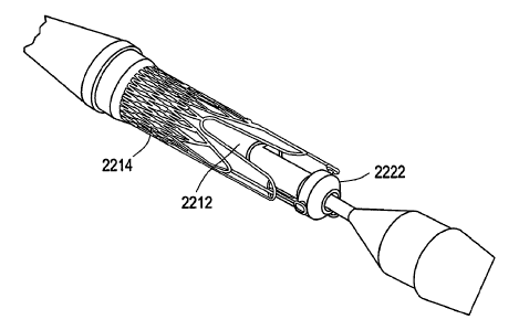
[0087] FIGS. 22A-22D show a delivery system 2200 that includes two
concentrically-
arranged parts, a first assembly (including elements 2202-22 10) and a second
assembly
(including elements 2216-2230). More particularly, the first assembly may
include tip 2202
at the distal end of the delivery system (with a guide wire passing through
the length of the
delivery system and out the tip), inner shaft 2204, outer sheath 2206, metal
shaft 2208, and
push handle 2210. The second assembly may include outer shaft (distal) 2216,
tapered outer
shaft connector 2218, outer shaft (proximal) 2220, stent holder 2222, kink
protector 2224,
hold handle connector 2226, hold handle cup 2228, and 0-ring 2230. As shown,
push handle
2210 is located at the proximal end of the delivery system. In FIGS. 22A and
22B, outer
1o shaft 2220 has been split along its length to allow the components of
delivery system 2200 to
be shown in greater detail. Valve 2212 and stent(s) 2214 form a third assembly
that can be,
for example, loaded and crimped between the first and second assemblies.
[0088] With respect to the first assembly, inner shaft 2204 functions as a
lumen for a guide
wire. Tip 2202 is bonded at its distal end. As used herein, bonding refers to
any suitable
securing/fastening mechanism such as, for example, adhesive bonding using
cyanoacrylate or
UV-curing adhesives or thermal bonding/welding using heat energy to melt the
components
to be assembled. Outer sheath 2206 may be bonded to the proximal section of
tip 2202 and
may constrain the stent-valve (2212, 2214). Outer sheath 2206 may be
perforated to allow
device flushing via hold handle 2210. The proximal part of the first assembly
may be
reinforced with metal shaft 2208 and may end into the push handle with a luer
connector for
guide wire lumen flushing.
[0089] With respect to the second assembly, stent holder 2222 may be bonded
distally on
distal outer shaft 2216. FIG. 22D shows a perspective view better illustrating
the
arrangement between the stent-valve (2212, 2214) and stent holder 2222. Distal
outer shaft
2216 may be bonded proximally to proximal outer shaft 2220 via tapered
connector 2218.
Proximal outer shaft 2220 may be bonded via kink protector 2224 to the hold
handle
assembly, which may include hold handle connector 2226 and hold handle cup
2228. The
hold handle assembly may compress O-ring 2230 for sealing delivery system
2200. A luer
connector may allow for device flushing. The flush mechanism may be used to
remove
trapped air from the delivery system prior to its insertion into the body.
Alternatively or
additionally, the flush mechanism may be used to cool down a stent (e.g.,
nitinol stent) prior
to its release and/or recapture by flushing the stent with a cold saline
solution. Cooling down
-25-
CA 02657839 2009-03-05
WO 2008/028569 PCT/EP2007/007413
the stent may cause a reversible modification of its structure, thus reducing
its Young-
modulus and therefore the stent radial force and the forces necessary for its
delivery and
recapture.
[0090] Delivery system 2200 is said to be in an open position (FIG. 22C) when
(for
example) push handle 2210 contacts the hold handle cup 2228. In the open
position, the
stent-valve (2212, 2214) may detach from stent holder 2222 and fully expand at
an
implantation site. Prior to delivery system 2200 reaching the open position,
the stent-valve
may be crimped onto delivery system 2200 by means of a crimping machine (for
example)
and held in place by stent holder 2222. Stent holder 2222 may affix to the
attachment
elements of the stents shown in Figures 8A-16. The crimped stent-valve may be
maintained
in a collapsed configuration by pulling back the first assembly thus covering
the attachment
components/stent holder 2222 with outer sheath 2206. Once the outer sheath
2206 is
removed such that it no longer constrains the attachment components, the stent-
valve may
automatically detach from stent holder 2222 due to the self-expanding property
of the stent-
valve. Delivery system 2200 is said to be in a closed position (FIGS. 22A and
22B) when
outer sheath 2206 fully encompasses the stent-valve (2212, 2214) such that no
expansion of
the stent-valve occurs.
[0091] Delivery system 2200 is said to be in a partially open position when
(for example)
push handle 2210 is partially pushed towards hold handle cup 2228. In this
partially open
position, the stent-valve (2212, 2214) is deployed proximally and still
attached distally to
stent holder 2222 via the attachment elements. This allows for an accurate
implantation/positioning of the stent-valve. For example, the stent-valve may
be partially
released proximal to the intended implantation site and slightly pushed
distally until
resistance is felt. Final release of the stent-valve (2212, 2214) may occur by
completely
pushing the push handle towards hold handle cup 2228 so that delivery system
2200 reaches
the open position. Such a partially-open position is illustrated in FIG. 28B.
In some
embodiments, an imaging mechanism may be used to determine whether the stent-
valve is
positioned correctly at the implantation site. For example, roadmapping under
fluoroscopy
can be realized with angiography, intra-vascular ultrasound (IVUS), intra-
cardiac
3o echocardiography (ICE), trans-esophageal echocardiography (TEE) or other
mechanism(s) or
combination thereof, which imaging mechanism may be at least partially
integral to or
separate from the delivery system.
-26-
CA 02657839 2009-03-05
WO 2008/028569 PCT/EP2007/007413
[0092] Upon implantation of the stent-valve (2212, 2214), delivery system 2200
may revert
to the closed position prior to retrieval from the patient's body, for
example, by holding the
first assembly and pushing the second assembly distally towards tip 2202/outer
sheath 2206.
In other embodiments, the handle for releasing the stent-valve may comprise a
screw
mechanism for transferring a rotational movement of the handle into a
translational
movement of the outer sheath. This type of release system may allow for
stepwise, more
accurate stent release and recapturing as well as a reduction of the release
force felt by the
surgeon.
[0093] FIGS. 23A-23D show another example of a delivery system 2300 in
accordance
1o with an embodiment of the present invention. Delivery system 2300 may be
substantially
similar to delivery system 2200 (FIG. 22) (e.g., closed position, FIGS. 23A
and 23B; opened
position, FIG. 23C), except delivery system 2300 may additionally include one
or more
folded balloons 2302 (e.g., proximal to the stent-valve). Unless otherwise
indicated, like
features in FIGS. 23A-23D correspond to the same reference numerals in FIGS.
22A-22D,
although the reference numerals have not been reproduced in FIGS. 23A-23D to
avoid
overcomplicating the drawings. The same applies to the stent delivery systems
shown in
FIGS. 24A-D, FIGS. 25A-C, and FIGS. 26A-C. Balloon 2302 may be
inflated/deflated via an
additional lumen in proximal outer shaft 2304, for example, to anchor the
stent-valve (e.g., a
non-self-expanding stent-valve) in place at an implantation site. FIG. 23D
shows a cross
section "A-A" of the lumen structure shown in FIG. 23C. The lumen structure
includes 5-
lumen tubing 2306 and inner shaft 2308. In other embodiments, other structures
for lumen
tubing 2306 may be used (e.g., bi-lumen tubing where the second lumen is used
for balloon
inflation and deflation). Delivery system 2300 may also include access
mechanism 2310 for
balloon inflation/deflation, which may allow connection of a syringe or
inflation device to
inflate/deflate a balloon. Alternatively or additionally, tubing with an
attached stop-cock
may be connected to access mechanism 2310.
[0094] FIGS. 24A-24D show another example of a delivery system 2400 in
accordance
with an embodiment of the present invention. In delivery system 2400, proximal
outer shaft
2402 may have an increased diameter in comparison to the diameter of proximal
outer shaft
2220 (FIG. 22). The increased diameter may reduce bleeding when the delivery
system is
used without an introducer. Alternatively, when an introducer is used, the
increased diameter
may match the internal diameter of the introducer which, in turn, may depend
on the outer
-27-
CA 02657839 2009-03-05
WO 2008/028569 PCT/EP2007/007413
diameter of the outer sheath. Having no gap between the introducer and
delivery system may
reduce the risk of a potential retrieval issue of the delivery system through
the introducer due
to entrapped blood. Accordingly, delivery system 2400 may include a floating
tube 2404 that
fills the gap between the inner and outer assemblies, thus reducing the risk
of the inner
assembly kinking under compression which would result in higher friction
forces within the
delivery system during stent recapturing. Delivery system 2400 may be
substantially similar
to delivery system 2200 in all other respects (e.g., closed position, FIGS.
24A and 24B;
opened position, FIG. 24C).
[0095] FIGS. 25A-C show another example of a delivery system 2500 in
accordance with
1o an embodiment of the present invention. Delivery system 2500 may include
one or more
balloons 2536 distal to the stent-valve. Having the balloon(s) distal to the
stent-valve avoids
having to introduce the delivery system deeper into the body (e.g., into the
ascending aorta)
in order to perform dilation, thereby reducing risk of injury to the body and
improving device
handling (e.g., no bending of rigid device over the aortic arch). Balloon(s)
2536 can be used
for, for example, valvuloplasty prior to stent-valve implantation and/or post-
dilation of the
implanted stent-valve to improve the anchoring of the stent. FIGS. 25B and 25C
show the
balloon(s) 2536 in closed and open positions, respectively.
[0096] The first assembly of delivery system 2500 may include tip 2502, inner
balloon
shaft 2504, outer sheath 2506, and floating tube 2508. The second assembly may
include
inner shaft (distal) 2510, stent holder transition 2512, stent holder 2514,
sleeve 2516, tapered
transition shaft connector 2518, and outer shaft (proximal) 2520. The handle
assembly may
include hold handle connector 2522, hold handle cup 2524, O-ring 2526, metal
shaft 2528,
and push handle 2530. The balloon assembly may include outer shaft 2532, inner
shaft 2534,
balloon 2536, and Y connector 2538.
[0097] FIGS. 26A-C show another example of a delivery system 2600 in
accordance with
an embodiment of the present invention. Delivery system 2600 may include an
integrated
introducer 2602, which may be an additional assembly that houses the second
assembly. The
outer sheath of the delivery system is shown as 2604. Introducer 2602 may
include a
connecting line 2606, a stopcock 2608 and a housing 2610 for the sealing
membrane 2612.
Stopcock 2608 may serve as an access point for, for example, a syringe
containing fluid (e.g.,
saline). Connecting line 2606 may serve to transport the fluid from the
syringe to the inner
lumen of the introducer, and sealing membrane 2612 may seal the introducer
from the outside
-28-
CA 02657839 2009-03-05
WO 2008/028569 PCT/EP2007/007413
environment. Upon stent-valve implantation, the components of delivery system
2600 (e.g.,
first assembly and second assembly) other than introducer 2602 may be
retrieved through the
introducer. Then, another medical device such as, for example, a closure
device may be
introduced through introducer 2602. As another example, intravascular
ultrasound (IVUS)
equipment (e.g., mini-probe) may be introduced through introducer 2602.
Delivery system
2600 may be substantially similar to delivery system 2200 in all other
respects.
[0098] FIG. 27 is a flowchart 2700 of illustrative stages involved in
replacing a failed (e.g.,
native or artificial) valve in accordance with some embodiments of the present
invention.
FIGS. 28A-28C illustrate (without limitation) various stages referenced in the
flowchart of
FIG. 27. At stage 2702, a stent-valve (e.g., single-stent-valve or double-
stent-valve) may be
removably attached to a delivery system. For example, one or more attachment
elements of a
stent component (e.g., attachment elements 808, FIG. 8B) may be affixed to a
stent holder of
the delivery device (e.g., stent holder 2222, FIG. 22). A collapsing element
(e.g., outer
sheath 2206, FIG. 22) may be placed over the attachment elements/stent holder
to maintain
the stent-valve in a collapsed configuration and attached to the delivery
system.
[0099] At stage 2704, the stent-valve may be delivered to an implantation site
in a
collapsed configuration. For example, FIG. 28A ("introduction" and
"positioning") shows
that stent-valve 2802, while still attached to the delivery system via stent
holder 2804 and
fully contained within outer sheath 2806, may be introduced to a patient's
body along guide
wire 2808 so that tip 2810 of the delivery system passes through failed valve
2812. The
delivery system may be manipulated forwards and/or backwards, for example,
until the stent-
valve is believed to be positioned correctly.
[0100] At stage 2706, the stent-valve may be partially expanded, for example,
to determine
(stage 2708) whether the stent-valve is in fact positioned correctly and/or to
test (stage 2710)
whether the stent-valve is functioning properly. For example, FIG. 28A
("partial release")
shows that outer sheath 2806 may be partially removed from proximal section
2814 of the
stent-valve, while attachment elements 2816 of the stent-valve are still
constrained by outer
sheath 2806 onto stent holder 2804.
[0101] At stage 2712, when the stent-valve is positioned correctly at the
implantation site
and/or the stent-valve is functioning properly, the stent-valve may be
detached from the
delivery system in order to cause the stent-valve to expand to its fully-
expanded
configuration. For example, FIG. 28C ("final release") shows that, upon
removal of
-29-
CA 02657839 2009-03-05
WO 2008/028569 PCT/EP2007/007413
attachment elements 2816 and stent holder 2804 from within outer sheath 2806,
attachment
elements 2816 of stent-valve 2802 may detach from stent holder 2804
automatically (or in
response to balloon inflation in other embodiments) thereby causing the stent-
valve to expand
to its fully-expanded configuration. The second assembly of the delivery
device may then be
reunited with the first assembly/outer sheath and removed from the patient's
body. For
example, FIG. 28C ("delivery device retrieval") shows that the second assembly
2818 may be
passed through replacement stent-valve 2802 towards the distal end of the
stent-valve. Then,
the reunited second assembly 2818 and first assembly/outer sheath 2806 may be
passed
through stent-valve 2802 again in the proximal direction before exiting the
patient's body.
io [0102] When the stent-valve is not positioned correctly (stage 2708), at
stage 2714 the
stent-valve may be reverted to the collapsed configuration and repositioned
within the
patient's body. An illustration of this scenario is illustrated in FIG. 28B
("stent
recapturing/repositioning"), in which outer sheath 2806 is slid in the
proximal direction over
proximal section 2814 of the stent-valve in order to recapture the stent-
valve. The stent-valve
is then repositioned and released such that fixation element 2820 of the stent-
valve receives
an annulus 2822 of the failed valve. Similarly, when the stent-valve
malfunctions in response
to a test (stage 2710), at stage 2716 the stent-valve may be reverted to the
collapsed
configuration and removed from the patient's body.
[0103] Thus it is seen that stent-valves (e.g., single-stent-valves and double-
stent-valves)
and associated methods and systems for surgery are provided. Although
particular
embodiments have been disclosed herein in detail, this has been done by way of
example for
purposes of illustration only, and is not intended to be limiting with respect
to the scope of
the appended claims, which follow. In particular, it is contemplated by the
applicant that
various substitutions, alterations, and modifications may be made without
departing from the
spirit and scope of the invention as defined by the claims. Other aspects,
advantages, and
modifications are considered to be within the scope of the following claims.
The claims
presented are representative of the inventions disclosed herein. Other,
unclaimed inventions
are also contemplated. The applicant reserves the right to pursue such
inventions in later
claims.
-30-